Oral Cancer1
Magdalen Foo — Discipline Lead, Oral Maxillofacial Surgery
Learning Outcomes2
- Describe the clinical aspect, diagnosis and management of oral cancer
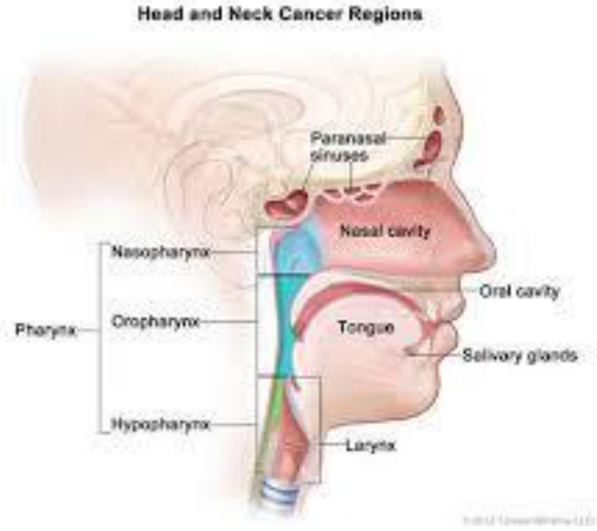
Anatomical sites:
- Paranasal sinuses
- Nasal cavity
- Nasopharynx
- Oral cavity
- Pharynx
- Oropharynx
- Tongue
- Salivary glands
- Hypopharynx
- Larynx
Epidemiology Of Head And Neck Cancer In Australia
Head and Neck Cancer Statistics in Australia3
Incidence
-
5000 new cases of head and neck cancer diagnosed in 2020 (¾ = males, ¼ = females)
- 3.6% of all new cancer cases diagnosed in 2020
- Head and neck cancers (including lip) — 7th most commonly diagnosed cancers in Australia
- 65.1 is the average age of diagnosis
Mortality
-
1000 deaths from head and neck cancer (including lip) in 2020
- 2.4% of all deaths from cancer in 2020
- 70% surviving at least 5 years (2012–2016)
Overview Of Oral Cancer
Sites and Origins of Oral Cancer4
Oral cancer may arise at the following sites:
- Lips
- Anterior 2/3 of the tongue
- Floor of the mouth and ventral tongue
- Hard palate and soft palate
- Gingiva
- Buccal mucosa
It may also arise from:
-
Metastasis from another location in the body
-
Direct spread from surrounding areas, e.g. nasal cavity
-
The retromolar region
-
The lateral and ventral surfaces of the anterior two-thirds of the tongue
Lecturer — Oral Cancer Boundaries
The posterior third of the tongue belongs to the oropharynx rather than the oral cavity.
- It has different symptoms and referral pathways.
- The dorsum of the tongue is not the usual site for oral carcinoma; a lump there is more likely to represent something benign or another condition.
- A large tumour in the sinus cavity may spread rapidly into the upper jaw or nose because the sinus is largely air-filled.
Types of Oral Cancer
- The majority of oral cancers are Squamous Cell Carcinomas
- Rarer types of oral cancer start in the salivary glands
- Lymphomas and Melanomas might also be diagnosed around the oral cavity — approached differently in terms of treatment
Squamous Cell Carcinoma567
- Squamous cell carcinoma represents 90% of all oral cancers
Lecturer — Squamous Cell Presentation
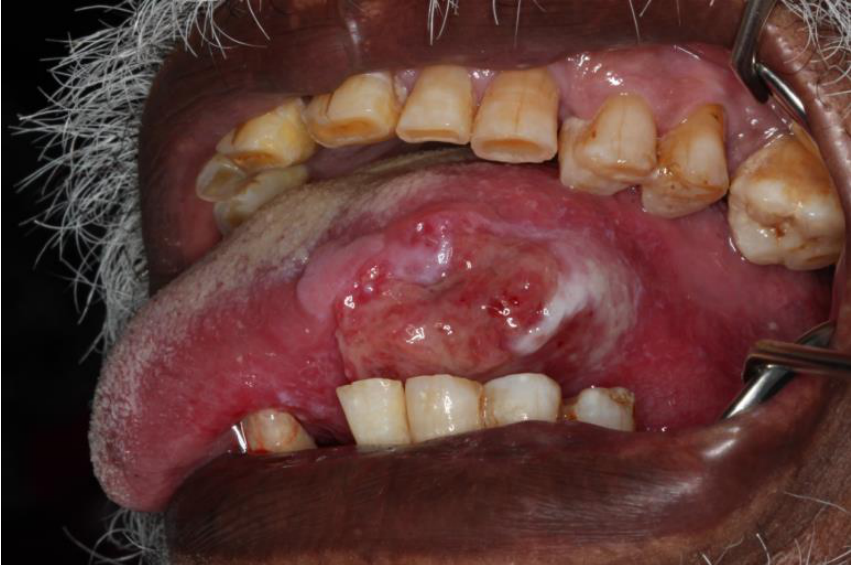
Approximately 90–95% of tumours in the mouth are squamous cell carcinomas, which may initially present in several different ways.
- A non-healing ulcer, red or white patch, indurated lesion, or mass
- Localised bone loss or unusually mobile teeth
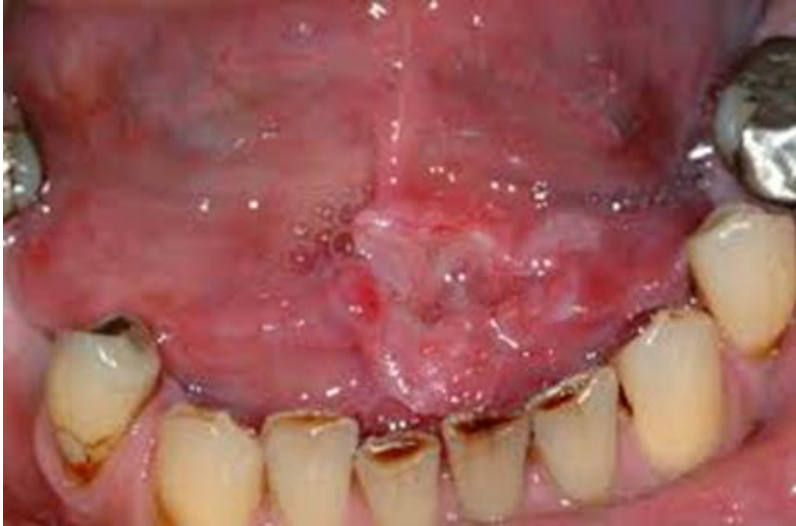
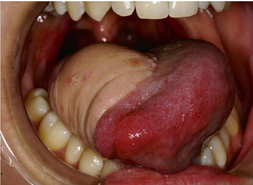
- Oral cancer is not always painful, particularly when it affects the ventral tongue or floor of the mouth.
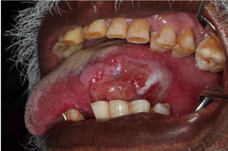
SCC lateral tongue
SCC floor of mouth
Lecturer — Salivary Gland Tumours
The likelihood that a salivary gland tumour is malignant varies according to the gland involved.
- Parotid gland: approximately 20% malignant and 80% likely to be benign
- Submandibular gland: approximately a 50:50 likelihood of being benign or malignant
- Minor salivary glands: more likely to be malignant
- A lower-lip lump is commonly a mucous retention phenomenon caused by lip biting and blockage of a gland.
- An upper-lip tumour is more likely to relate to a minor salivary gland and therefore requires consideration of malignancy.
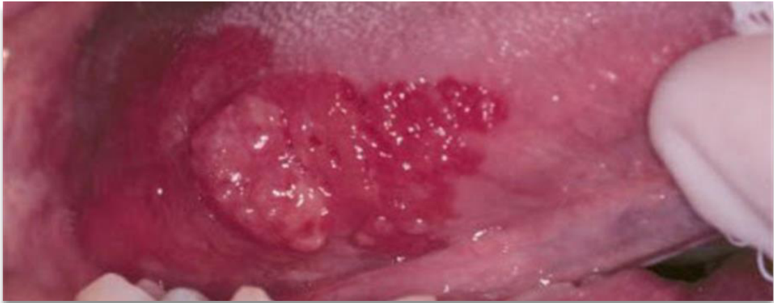
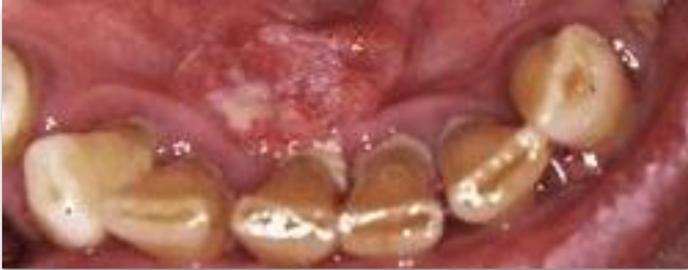
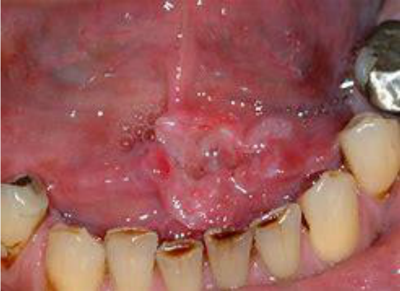
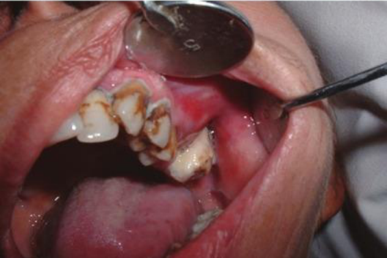
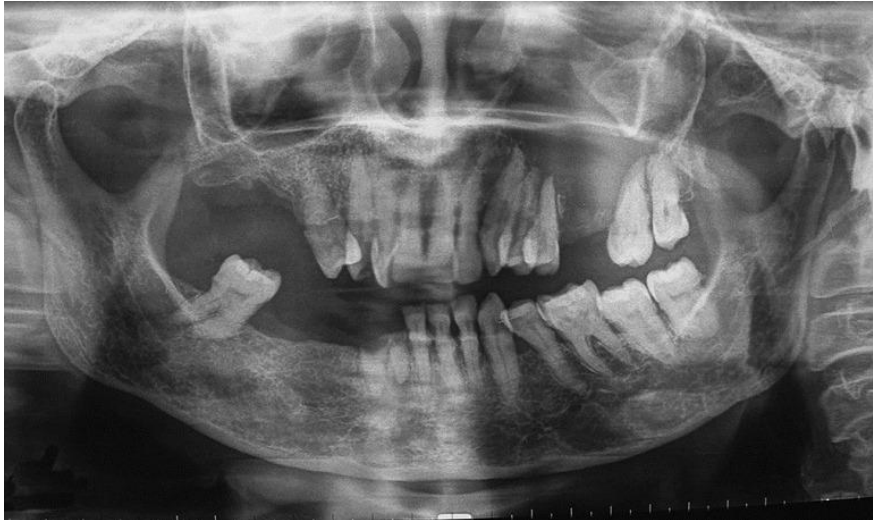
Salivary Gland Carcinoma8
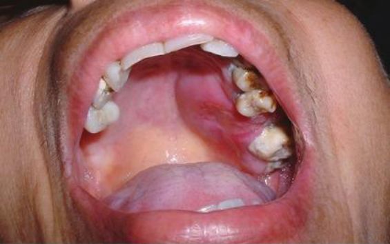 |  |
 |
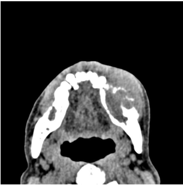
Metastasis To The Jaws9
 |  |
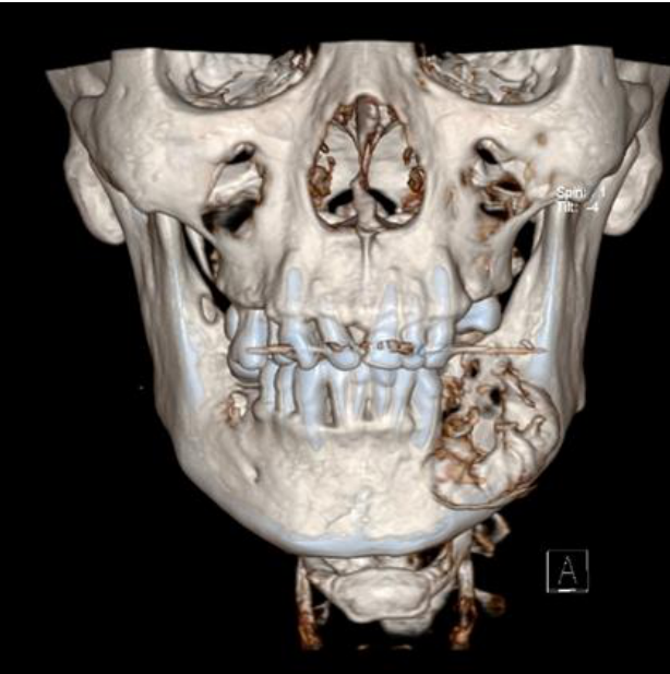 |
Clinical Features10
- Uncommon (<1% of cancer in the oral cavity)
- Metastasize to the mandible, less so to the maxilla
- Majority of tumours are adenocarcinomas
Primary Tumour Origins
- Breast (30%)
- Lungs (20%)
- Kidney (15%)
- Thyroid (5%)
- Prostate (5%)
- Colon (5%)
- Stomach (5%)
- Cutaneous melanoma (5%)
Lecturer — Jaw Metastasis Assessment
Metastatic lesions may present with localised bone loss, unusually mobile teeth, bleeding around teeth, a non-healing ulcer, or numbness caused by nerve involvement.
- A numb lower lip may indicate involvement of the inferior alveolar nerve.
- In the maxilla, infraorbital nerve involvement may cause numbness of the upper lip or side of the nose.
- A history of previous cancer is important when assessing a new oral or jaw lesion.
Radiographic and Symptomatic Presentation
- Radiographic appearance at site of metastatic tumour deposit — ill defined radiolucency / radiopacity or mixed radiolucent and radiopaque lesions
- Can be asymptomatic, or patient may experience pain, loosening of teeth, or unilateral paraesthesia of the lower lip/chin
Lecturer — Metastatic Bone Loss
Metastatic jaw lesions may resemble periodontal disease or an odontogenic problem but show an unusual pattern of destruction.
- Imaging may show generalised bone loss, more severe localised bone loss around particular teeth, a large area of bone destruction, or an unusual area of mandibular bone loss.
- An OPG, periapical radiograph, CT, or other imaging may demonstrate the extent of bone involvement.
- Further consideration is warranted when bone loss does not correspond with the general periodontal condition or when a swelling does not behave like an infection.
Lecturer — Other Oral Cancers
Lymphoma may involve the oral cavity and jaws and can initially resemble an odontogenic or periodontal problem when swelling and marked localised bone loss are present.
- Rare cancers around the oral cavity also include mucosal melanoma.
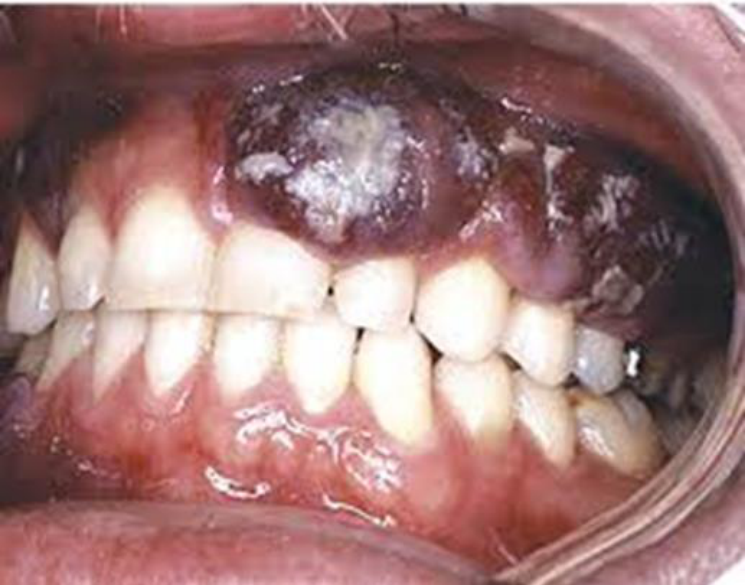
- Oral mucosal melanoma may invade soft tissues, be extensively pigmented, cause bleeding and mobility of teeth, and cause bone loss if it invades bone.
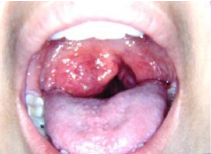
- A tumour involving the uvula or soft palate may interfere with speech and soft-palate function, so the patient may be unable to say “ah” properly and may have a nasal quality to their speech.
Other Cancers Around The Oral Cavity11
Lecturer — Oropharyngeal HPV
HPV was discussed primarily as a risk factor for oropharyngeal cancer involving the posterior third of the tongue, which is separate from the oral cavity region and managed through an ENT pathway.
- Patients may present with a lump in the neck, difficulty swallowing, unintentional weight loss, changes in speech, or pain referred to the ear.
- HPV types 16 and 18 were identified, and HPV-associated disease may have better outcomes than disease in patients without the usual risk factors.
- Vaccination of boys and girls was discussed in relation to reducing HPV-associated oropharyngeal cancer.
 |  |
Risk Factors For Oral Squamous Cell Carcinoma
Oral Squamous Cell Carcinoma12
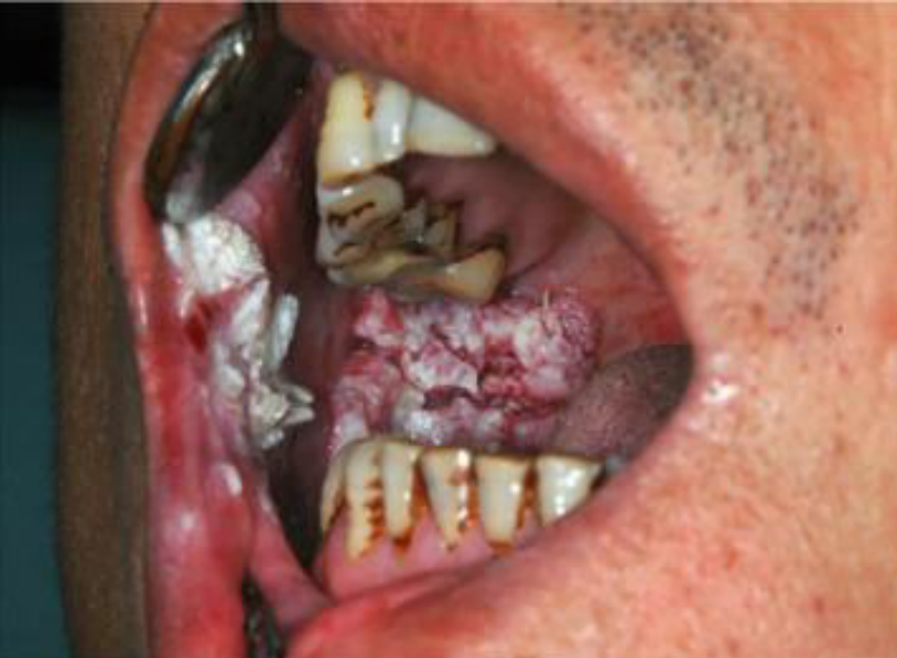
- The main risk factors are tobacco and alcohol consumption (note synergistic).
Lecturer — Oral Cancer Progression
The clinician should consider both the lesion and the patient’s lifestyle when deciding whether a presentation is suspicious.
- Oral cancer does not usually develop overnight; it may progress from normal tissue through changes or premalignant conditions before cancer develops.

Tobacco And Alcohol Consumption1314
“Synergistic consumption of alcohol and tobacco (both smoked and smokeless) significantly increased the odds for the occurrence of Oral Squamous Cell Carcinoma”
Lecturer — Suspicious Lesions
A patient who smokes and drinks heavily and presents with a non-healing, indurated lesion should prompt a high index of suspicion. Clinicians are not expected to diagnose oral cancer in the dental clinic; they are expected to recognise suspicious findings and arrange referral or biopsy.
Clin Oral Investig. 2019 Jul;23(7):2849-2859
The synergistic effect of tobacco and alcohol consumption on oral squamous cell carcinoma: a systematic review and meta-analysis
Fernanda Weber Mello, Gilberto Melo, Júlia Jacoby Pasetto, Carolina Amália Barcellos Silva, Saman Warnakulasuriya, Elena Riet Correa Rivero
 |  |
 |  |
Other Risk Factors15
- Human papilloma virus (HPV)
- Chewing pan (areca or betel nut)
- Epstein-Barr virus (EBV) — for nasopharyngeal carcinoma
- Exposure to the sun
- Family history of mouth cancers / inherited conditions, e.g. Fanconi anaemia
- Post transplant / immunosuppressed
- Oral premalignant conditions
Lecturer — Additional Risk Details
Betel-nut chewing is particularly associated with cancer of the buccal mucosa because the material is stored in the buccal sulcus.
- Long-term sun exposure may lead to changes in the lips that eventually become lip cancer.
- Immunosuppressed patients are at increased risk of cancer because their tissues may not repair abnormal changes effectively.
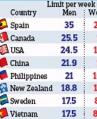
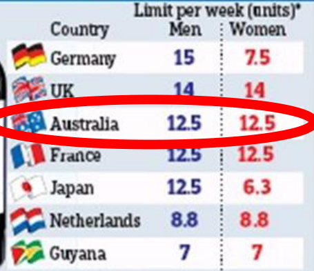
Drinking Guidelines Around the World16
Limit per week (units)*
| Country | Men | Women |
|---|---|---|
| Spain | 35 | 21.3 |
| Canada | 25.5 | 17 |
| USA | 24.5 | 12.3 |
| China | 21.9 | 13 |
| Philippines | 21 | 10.5 |
| New Zealand | 18.8 | 12.5 |
| Sweden | 17.5 | 8.8 |
| Vietnam | 17.5 | 8.75 |
| Country | Men | Women |
|---|---|---|
| Germany | 15 | 7.5 |
| UK | 14 | 14 |
| Australia | 12.5 | 12.5 |
| France | 12.5 | 12.5 |
| Japan | 12.5 | 6.3 |
| Netherlands | 8.8 | 8.8 |
| Guyana | 7 | 7 |
*Note, measurements and guidelines vary globally; comparisons are based on one unit containing 8g of alcohol.
RACGP — Supporting smoking cessation. Royal Australian College of General Practitioners. A guide for health professionals, second edition. racgp.org.au
 |  |
 |
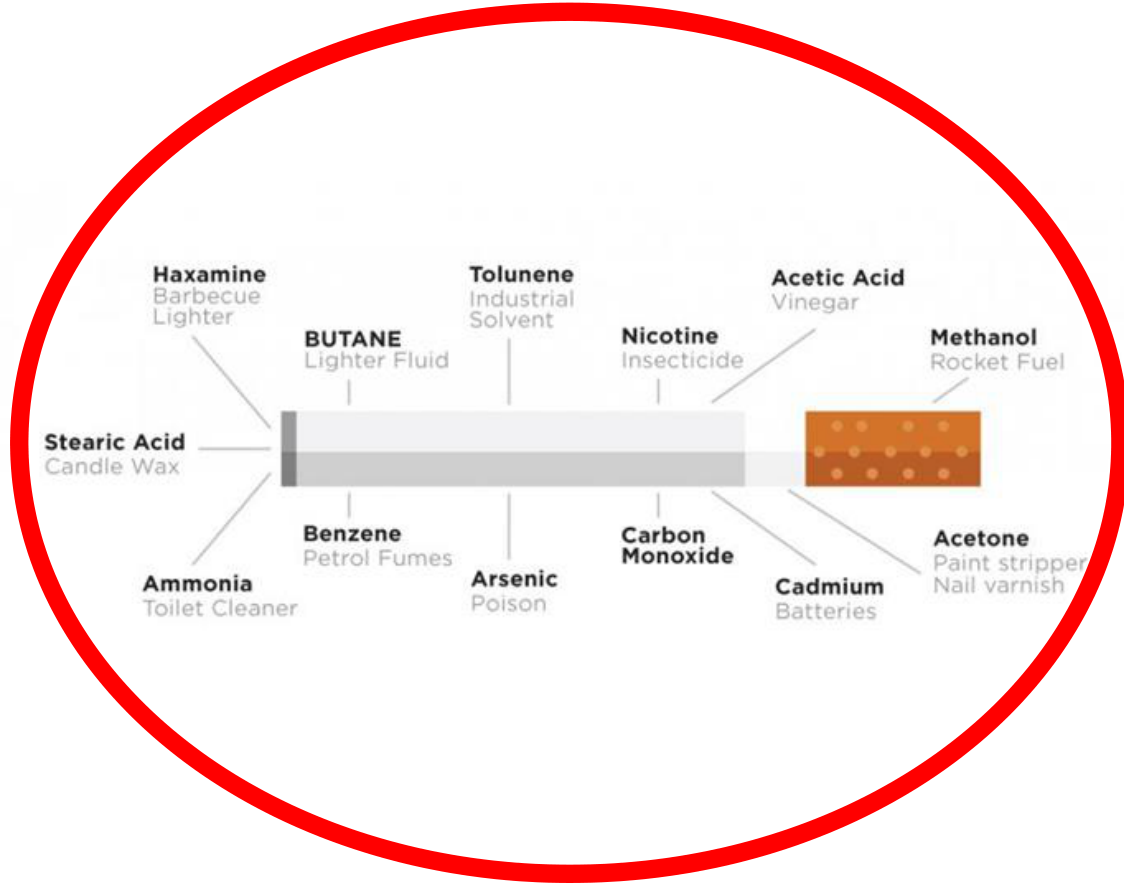
Chemicals in a Cigarette17
- Stearic Acid — candle wax
- Hexamine — barbecue lighter
- Ammonia — toilet cleaner
- Butane — lighter fluid
- Benzene — petrol fumes
- Toluene — industrial solvent
- Arsenic — poison
- Nicotine — insecticide
- Carbon Monoxide
- Acetic Acid — vinegar
- Cadmium — batteries
- Methanol — rocket fuel
- Acetone — paint stripper, nail varnish
HPV And Oropharyngeal SCC18
Oropharyngeal SCC
Oral HPV 16 is a strong risk factor.
“This study corroborates and extends Australian data demonstrating that patients with HPV-positive OPSCC represent a distinct subgroup associated with a significantly better survival”
Australian Journal of Otolaryngology, Vol 2 (2019)
Human papillomavirus and oropharyngeal squamous cell carcinoma: a 12-year retrospective review in a New South Wales tertiary referral centre
Alon Taylor, Thomas Eade, David Veivers, Anthony J. Gill, Leo Pang
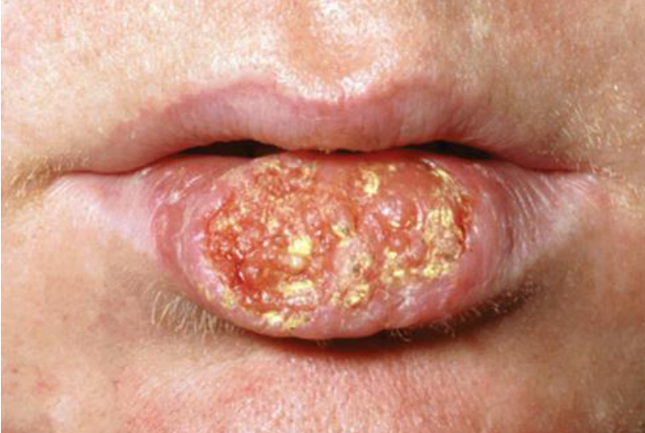
- An ulcer that does not heal within two or three weeks
- An indurated ulcer
- Localised, unexplained tooth mobility
- Localised bone loss
- Bleeding around teeth in an unusual localised area
- Unexplained numbness of the lip, face, or nose
Lecturer — Early Oral Cancer
Most early oral cancers are painless, so a lesion under the tongue may go unnoticed because it does not cause pain.
- A red patch was described as more concerning than a white patch.
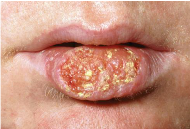
- Lip cancer may present as an ulcerated, indurated, non-healing area, particularly in a patient with substantial sun exposure.
[!note] Lecturer — Neck Lump Assessment
A neck lump may initially be caused by infection, which is the most common cause of lymphadenopathy.
- If the lump persists after review, suspicion should increase.
[!note] Lecturer — Submandibular Obstruction
A tumour near the submandibular duct may present as a blocked submandibular gland rather than as obvious pain from the oral tumour.
- A blocked gland should prompt consideration of infection, a salivary stone, or a tumour obstructing the duct.
- A gland blocked by a stone may intermittently improve and may be associated with sludge or fluid discharge.
- Tumour-related obstruction is less likely to resolve.
[!example] Non-Healing Ulcer With Localised Bone Loss
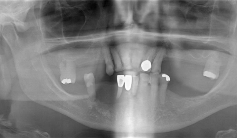
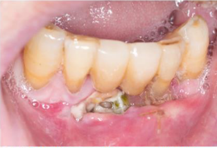
A patient presented with an ulcer in the lower anterior region, generalised bone loss that was more pronounced around the lower anterior teeth, a loose bridge, food impaction, and an ulcer that would not heal.
- The presentation could initially have been attributed to periodontal disease.
- Because the bone loss and ulcer were localised and did not fit the overall periodontal presentation, further investigation was required.
- The lesion was ultimately found to be cancer involving the bone.
[!example] Blocked Submandibular Gland
A tumour near the opening of the submandibular duct prevented the gland from emptying.
- The patient could therefore present with a blocked submandibular gland rather than obvious pain from the oral tumour.
- The differential included infection, a salivary stone, and a tumour obstructing the duct.
[!example] Swelling With Extensive Localised Bone Loss
A patient presented with a swelling that initially suggested infection.
- The teeth were assessed for vitality, cracks, extensive restorations, and non-vitality.
- An OPG revealed a large amount of localised bone loss and a very mobile tooth.
- The lesion was ultimately diagnosed as lymphoma.
- A history of previous cancer was relevant to the assessment.
[!example] Mucosal Melanoma
Mucosal melanoma was described as an extensive pigmented lesion involving the soft tissues.
- The associated teeth may bleed easily and become mobile.
- Bone loss may occur if the melanoma invades the bone.
- A lesion involving the uvula may affect speech and soft-palate function, producing difficulty saying “ah” and a nasal quality to the voice.
Symptoms Of Oral Cancer19
Symptoms can include:
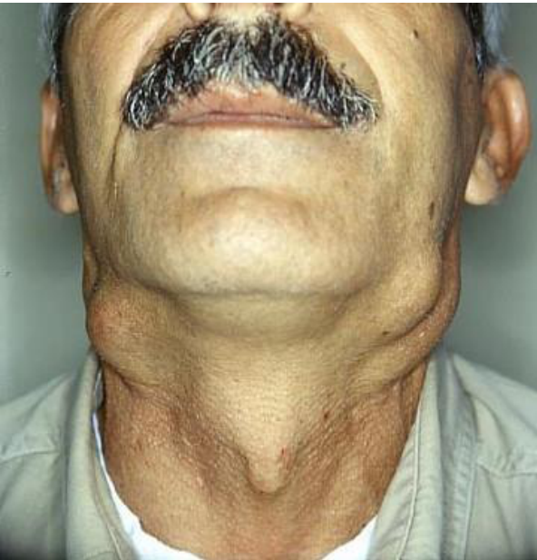
- a lump in the neck
- mobile teeth
- swelling or a sore on the lip that won’t heal
- difficult or painful swallowing
- changes in speech
- bleeding or numbness in the mouth
- white or red patches on the mouth, tongue or gums
- unexplained weight loss
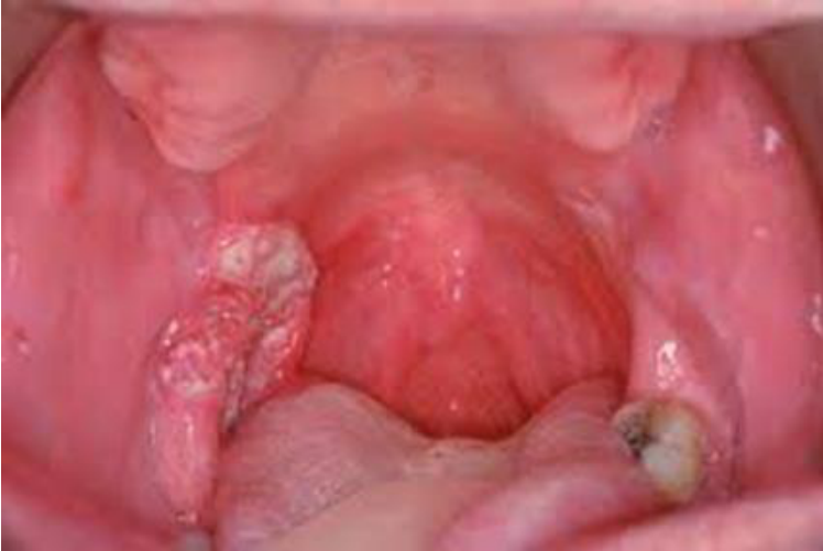
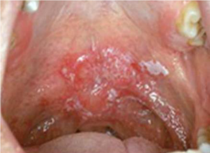
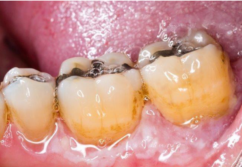
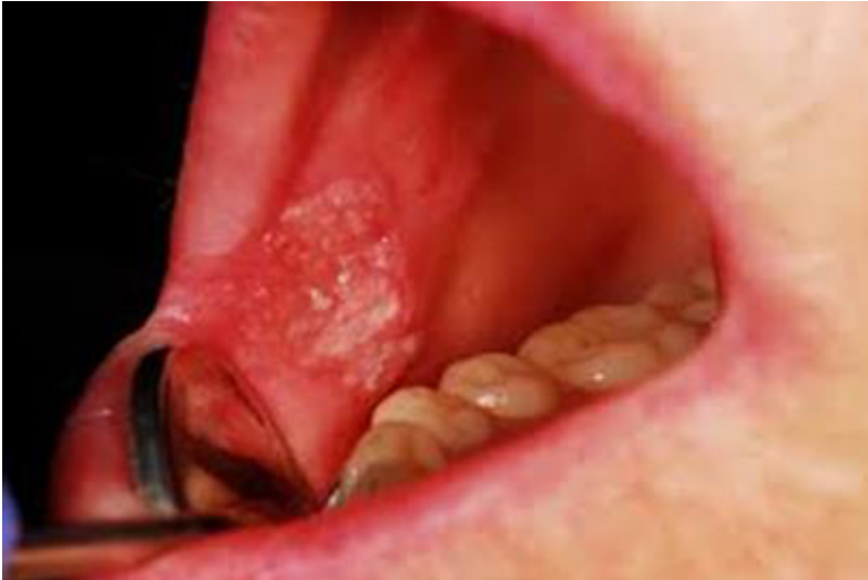
Sites And Incidence Of Oral Carcinoma20212223
Incidence of SCC differs between anatomical sites:
-
Lower lip is most susceptible – 35%
-
Lateral or ventral tongue – 25%
-
Floor of mouth – 20%
-
Soft palate – 15%
-
Other sites – gingiva/alveolar ridge and buccal mucosa (1–4%)
-
Other areas requiring particular attention include the lingual sulcus, retromolar region, and tonsillar region. Intraoral areas prone to development of SCC
Other intraoral sites
Lecturer — Oral Cancer Sites
The lateral and ventral tongue were emphasised as key areas where oral cancer may hide.
- The buccal mucosa is less commonly affected unless the patient chews and stores betel nut there.
- Approximately 30% of lip cancers present as a non-healing ulcer on the lip.
- Lip cancer is associated with sun exposure, including long-term outdoor work or activities without adequate sunscreen.
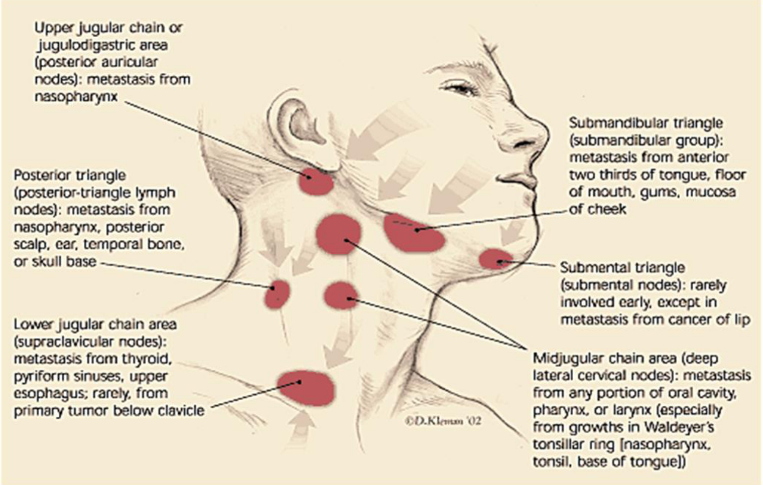
Metastatic Spread2425
-
Lymphatic spread
-
Tumour cells in lymph nodes extend into surrounding tissue (extra-capsular spread) or invade overlying skin
-
Enlarged, hard, fixed lymph nodes – ominous clinical signs
-
Oral cavity SCC metastasize to submandibular, superficial and deep cervical nodes
-
Beyond the regional lymph nodes, metastasize to the lungs
-
Oral cavity tumours commonly spread to the submandibular, sublingual, and jugular-chain lymph nodes.
-
A submandibular duct may become blocked when disease involves the submandibular or sublingual region.
-
A persistent neck lump lasting approximately two weeks or not resolving after review requires investigation.
-
Upper jugular chain or jugulodigastric area (posterior auricular nodes): metastasis from nasopharynx
-
Posterior triangle (posterior-triangle lymph nodes): metastasis from nasopharynx, posterior scalp, ear, temporal bone, or skull base
-
Lower jugular chain area (supraclavicular nodes): metastasis from thyroid, pyriform sinuses, upper esophagus; rarely, from primary tumor below clavicle
-
Submandibular triangle (submandibular group): metastasis from anterior two thirds of tongue, floor of mouth, gums, mucosa of cheek
-
Submental triangle (submental nodes): rarely involved early, except in metastasis from cancer of lip
-
Midjugular chain area (deep lateral cervical nodes): metastasis from any portion of oral cavity, pharynx, or larynx (especially from growths in Waldeyer’s tonsillar ring [nasopharynx, tonsil, base of tongue])
-
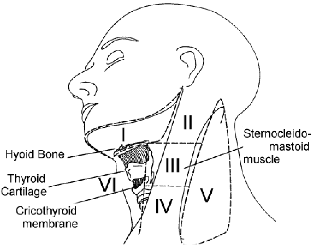
Level 1 comprises the submental and submandibular regions, while levels 2, 3, and 4 lie along the jugular chain.
-
Level 5 was described as more commonly associated with ENT conditions.
Lecturer — Neck Examination
The clinician should examine the neck from behind the patient rather than from the front.
- Check level 1 first because it is an early site of spread.
- Then examine levels 2, 3, and 4.
©D.Klemm ‘02 — © 2002 David Klemm
Investigation26
-
Biopsy - removal of a small amount of tissue which is sent to a pathologist to determine if cancer cells are present
-
Radiographs - OPG and chest x-ray
-
Imaging - PET, CT and MRI scans to determine the location, stage and spread of cancer
-
A flexible nasal endoscope is used to examine the nose, sinuses, larynx and pharynx
-
Suspicious lesions require a definitive diagnosis through biopsy, with referral for biopsy or an incisional biopsy where appropriate.
-
An excisional biopsy removes the entire lesion, but suspicious oral cancers generally require a sample rather than removal of the whole lesion.
-
The biopsy should include abnormal tissue from the centre of the tumour; normal tissue is not essential if adequate abnormal tissue is obtained.
-
A punch biopsy can provide adequate depth for pathological assessment, with 2 mm and 6 mm punch biopsies described as producing the same result.
-
The pathologist is particularly concerned with the depth of invasion, including whether the basement membrane has been invaded.
-
The specimen bottle must be correctly labelled.
-
Relevant clinical information includes smoking and alcohol history, the presence of a non-healing ulcer, and the duration of the lesion.
-
Other radiographs may include periapical radiographs.
-
Imaging is used to determine the size and extent of the tumour and whether adjacent areas, such as the nose or sinuses, are involved.
Lecturer — Biopsy Follow-Up
An inadequate biopsy may require further investigation.
- Repeat biopsy may be required if the first biopsy does not provide enough tissue.
- A lesion that does not heal within two or three weeks, or that appears suspicious, should be referred promptly.
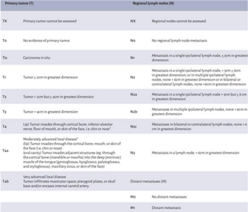
Clinical Staging TNM27
- Clinical staging is used to designate the extent of disease and to determine the most appropriate treatment for the patient
- Uniform staging system provides comparisons of results of types of treatment
- Clinical staging helps predict treatment response and outcomes (prognosis)
Lecturer — Prognosis and Diagnosis
Staging is not completely foolproof but provides a better estimate of prognosis.
- Earlier diagnosis generally means a smaller tumour.
- Earlier diagnosis is associated with better prognosis and better survival.
-
T - primary tumour
-
N - regional lymph node
-
M - distant metastasis
-
T refers to maximum radial dimension of the tumour
-
N refers to the lymph node
-
M refers to metastasis
Primary Tumor (T) and Regional Lymph Nodes (N)28
| Primary tumor (T) | Regional lymph nodes (N) | ||
|---|---|---|---|
| TX | Primary tumor cannot be assessed | NX | Regional nodes cannot be assessed |
| T0 | No evidence of primary tumor | N0 | No regional lymph node metastasis |
| Tis | Carcinoma in situ | N1 | Metastasis in a single ipsilateral lymph node, ≤ 3cm in greatest dimension |
| T1 | Tumor ≤ 2cm in greatest dimension | N2 | Metastasis in a single ipsilateral lymph node, > 3cm ≤ 6cm in greatest dimension; or in multiple ipsilateral lymph nodes, none > 6cm in greatest dimension or in bilateral or contralateral lymph nodes, none > 6cm in greatest dimension |
| T2 | Tumor > 2cm but ≤ 4cm in greatest dimension | N2a | Metastasis in a single ipsilateral lymph node > 3cm but ≤ 6cm in greatest dimension |
| T3 | Tumor > 4cm in greatest dimension | N2b | Metastases in multiple ipsilateral lymph nodes, none > 6cm in greatest dimension |
| T4 | Lip: Tumor invades through cortical bone, inferior alveolar nerve, floor of mouth, or skin of the face, i.e., chin or nose* | N2c | Metastases in bilateral or contralateral lymph nodes, none > 6cm in greatest dimension |
| T4a | Moderately advanced local disease* (lip) Tumor invades through the cortical bone, mouth, or skin of the face (i.e., chin or nose) (oral cavity) Tumor invades adjacent structures (e.g., through the cortical bone [mandible or maxilla] into the deep [extrinsic] muscle of the tongue [genioglossus, hyoglossus, palatoglossus, and styloglossus], maxillary sinus, or skin of the face) | N3 | Metastasis in a lymph node, > 6cm in greatest dimension |
| T4b | Very advanced local disease Tumor infiltrates masticator space, pterygoid plates, or skull base and/or encases internal carotid artery | Distant metastases (M) | |
| M0 | No distant metastases | ||
| M1 | Distant metastasis |
* Superficial erosion alone of bone/tooth socket by gingival primary tumor is not sufficient to classify a tumor as T4.
Lecturer — Tumor and Node Assessment
The tumour must be considered in three dimensions rather than two.
- For a lesion in the lingual sulcus, assessment must determine whether it has invaded the soft tissues, tongue, mandible, or nearby teeth.
- Lymph-node assessment is important because lymph nodes are among the first sites to which oral cancer spreads.
- The biopsy result may indicate cancer, insufficient or inadequate tissue, or no cancer.
- If the sample is inadequate, another biopsy may be required; if no cancer is identified, the patient may be monitored.
Management And The Multidisciplinary Team
Every patient with oral cancer should be presented at a Multidisciplinary Team Meeting (MDT).
This is an integrated team approach to healthcare in which medical and allied health professionals consider all relevant treatment options and collaboratively develop an individual treatment and care plan for each patient.
Lecturer — Treatment Planning
Once cancer is confirmed, the patient is discussed by the multidisciplinary team. The team may include surgeons, dentists, pathologists, radiologists, general practitioners, and other relevant health professionals.
- The team combines the expertise of different professionals to plan the most appropriate treatment.
- Oral cancer is commonly treated with surgery, potentially followed by radiotherapy when there are unfavourable features such as a poorly differentiated tumour, very close margins, or spread to lymph nodes.
- Chemotherapy or radiotherapy may also be used when a large tumour is not operable.
Benefits of a Multidisciplinary Approach29
-
Improving patient care through developing an agreed treatment plan
-
Providing best practice through adopting evidence-based guidelines
-
Improving patient satisfaction with treatment
-
Improving the mental wellbeing of patients
-
Improving access to possible clinical trials of new therapies
-
Increasing the timeliness of appropriate consultations and surgery, and a shorter timeframe from diagnosis to treatment
-
Increasing the access to timely supportive and palliative care
-
Streamlining pathways
-
Reducing duplication of services
-
A clinical trial may provide additional time for a patient with an inoperable tumour even when it is not expected to cure the patient
-
Social-service support may be needed when treatment and recovery prevent the patient from working
-
The general practitioner may help coordinate care, including pain control
Treatment Team30
- GP (General Practitioner) — looks after the patient’s general health and works with specialists to coordinate treatment
- Head and neck surgeon — diagnoses and treats cancers of the head and neck
- Dentist — evaluates and treats the mouth and teeth (before, during and after treatment)
- Radiation oncologist — prescribes and coordinates radiation therapy treatment
- Medical oncologist — prescribes and coordinates the course of chemotherapy
- Cancer nurses — assist with treatment and provide information and support throughout treatment
- Dietitian — recommends an eating plan to follow during treatment and recovery
- Speech pathologist — helps with swallowing and communication difficulties during and after treatment
- Other allied health professionals — such as social workers, pharmacists and counsellors
Lecturer — Team Considerations
The treatment team considers the tumour site, tumour size, lymph-node involvement, margins, possible spread, treatment goals, and quality of life. Treatment goals may differ between patients: some may prioritise minimal intervention, while others may prefer more extensive treatment to preserve function and maintain oral health.
Dental Care Around Cancer Treatment31323334
 |  |
Referral to Oral Health Centre of WA35
Department of Oral Medicine and Oral Pathology — Pre-Chemotherapy / Pre-Radiation Therapy Dental Evaluation
- Diagnosis: Hypopharyngeal SCC, has had surgery
- Proposed treatment: Post-operative radiotherapy (66Gy/33#)
Please advise of extent of radiation therapy field (please also mark teeth to be included on the chart below).
Lecturer — Pre-Radiotherapy Extractions
Before radiotherapy, teeth in the radiation field with a poor prognosis may need to be extracted. A 7 mm periodontal pocket, extensive restoration, or significant periodontal involvement may suggest that a tooth will not do well.
- The lecturer identified approximately 55 Gy as an important level: at lower doses teeth may sometimes be monitored, whereas at 55 Gy or more extraction may be considered if there is doubt about prognosis.
- The aim is to avoid extractions after radiotherapy whenever possible because radiotherapy reduces the healing capacity of the mandible and maxilla.
Dose of radiation to be administered (Gy): 66Gy/33#
Lecturer — Radiation Field
The radiation field should be identified before treatment. If the cancer is on one side of the tongue, the teeth on that side, particularly the molars, may receive a higher radiation dose.

Dental Chart
Upper Teeth
| Central Incisor / Lateral Incisor / Canine (Cuspid) / First Premolar (first bicuspid) / Second Premolar (second bicuspid) | 40 | Central Incisor / Lateral Incisor / Canine (Cuspid) / First Premolar (first bicuspid) / Second Premolar (second bicuspid) | 40 |
| First Molar / Second Molar / Third Molar (wisdom tooth) | 66 | First Molar / Second Molar / Third Molar (wisdom tooth) | 66 |
Lower Teeth
| Third Molar (wisdom tooth) / Second Molar / First Molar | 66 | Third Molar (wisdom tooth) / Second Molar / First Molar | 66 |
| Second Premolar (second bicuspid) / First Premolar (first bicuspid) / Canine (Cuspid) / Lateral Incisor / Central Incisor | 40 | Second Premolar (second bicuspid) / First Premolar (first bicuspid) / Canine (Cuspid) / Lateral Incisor / Central Incisor | 40 |
Right — Left
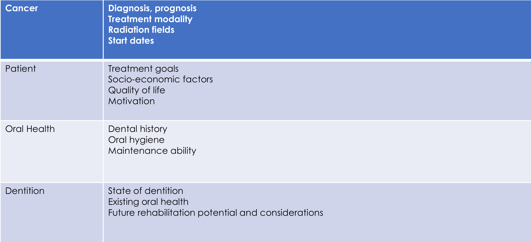
| Cancer | Diagnosis, prognosis — Treatment modality — Radiation fields — Start dates |
| Patient | Treatment goals — Socio-economic factors — Quality of life — Motivation |
| Oral Health | Dental history — Oral hygiene — Maintenance ability |
| Dentition | State of dentition — Existing oral health — Future rehabilitation potential and considerations |
- Changes in taste
- Oral hygiene maintenance — mucositis
- Systemic considerations — pancytopenia, opportunistic infections
Lecturer — Treatment Complications
Chemotherapy and radiotherapy may cause pancytopenia, with reductions in platelets, red blood cells, and white blood cells, leaving patients more prone to infections such as oral thrush. Pseudomembranous thrush may appear as white plaques that can be wiped away, leaving an erythematous surface.
- Treatment may include antifungal medication and instruction on denture cleaning.
- After surgery, the bite may be altered and the patient may have difficulty swallowing or increased drooling.
- Dental care may include caries treatment, root canal treatment, staining and polishing, oral-hygiene instruction, fluoride use, dry-mouth management, and denture hygiene.
Regular review and maintenance.
- Initially 3-monthly review for caries and periodontal management
- Once oral condition stabilizes, 6-monthly recalls — look out for mucosal changes, xerostomia, candidiasis, changes in occlusion
- Fluoride at each recall — Neutrafluor 5000 toothpaste and fluoride varnish
Lecturer — Ongoing Support
The clinician should continue supporting the patient after cancer treatment rather than discontinuing care. The patient’s life and oral cavity may remain permanently changed, with possible altered bite, dry mouth, increased caries risk, recurrent infections, difficulty cleaning dentures, and changes in oral function.
- Saliva replacement and frequent drinking may be required.
- Patients may need longer appointments and more frequent dental review.
Lecturer — Free Tissue Transfer
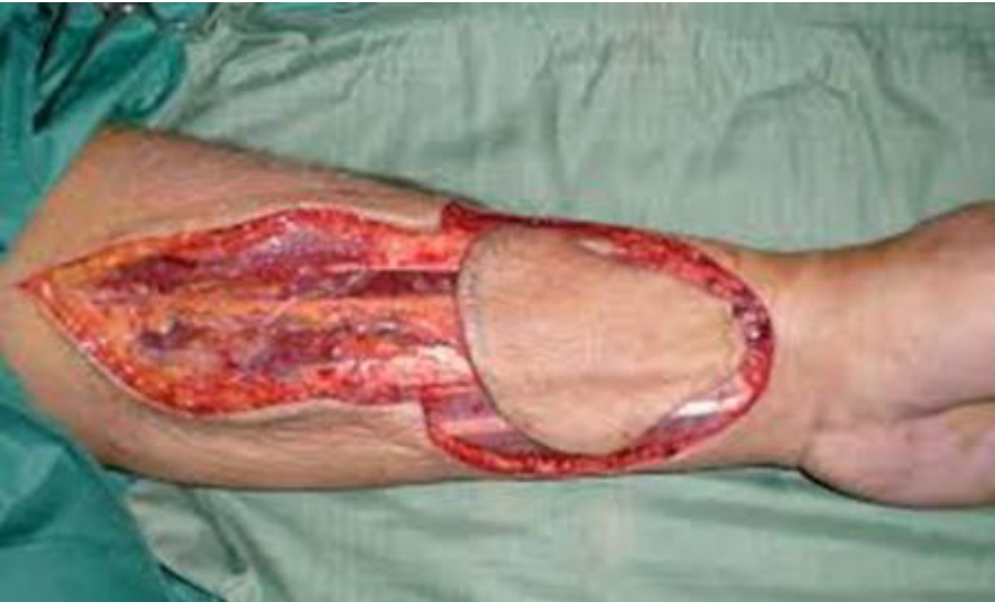
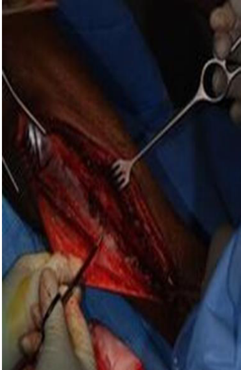
Reconstruction should replace like with like, with soft-tissue defects replaced by soft tissue and bone defects requiring bone and soft-tissue reconstruction.
- A soft-tissue defect involving the tongue or ventral tongue may be reconstructed with tissue from the thigh, leg or arm.
- The transferred tissue includes an artery and vein that must be connected to vessels in the neck.
- A skin graft may not survive in a large defect because it may not receive enough blood supply.
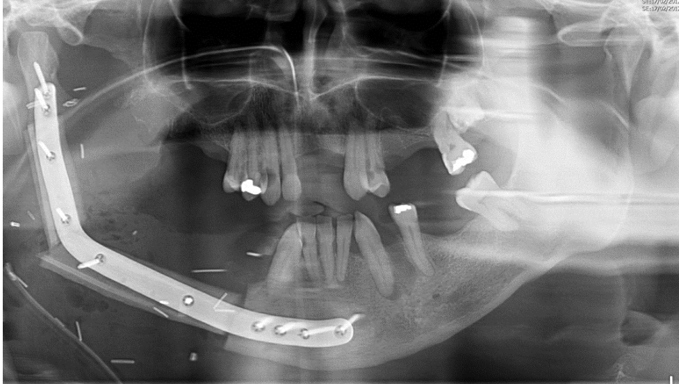
- If the tumour has eroded the mandible, the affected bone is resected and replaced with bone from another site, such as the leg.
- A reconstruction plate may hold the transferred bone in position.
- Preserving occlusion and the patient’s ability to function are important considerations.
- The dentist’s role includes maintaining the remaining teeth and motivating the patient during and after treatment.
Treatment Of Oral Cancer36
-
Intent of treatment — curative versus palliative
-
Surgery — primary modality of treatment for oral cancer; removes the tumour (ablation) and lymph nodes (neck dissection). Adjuvant radiotherapy after surgery depends on the pathological staging of the tumour, surgical margins and nodal status (for patients at high risk of locoregional recurrence)
-
Radiotherapy — used on its own to treat for organ preservation, e.g. oropharyngeal cancers. Usually concurrently with chemotherapy
-
Chemotherapy — concurrent or adjuvant chemoradiation has now become the standard of care in locally advanced head and neck squamous cell carcinoma
-
Treatment depends on the size and extent of the tumour and the presence of unfavourable features.
-
Chemotherapy may be palliative, providing additional time rather than curing the cancer.
-
The aim is also to assess whether microscopic spread has occurred in the lymph nodes and whether radiotherapy is required.
Surgery For Oral Cancer3738
- Surgery to remove the tumour (ablation) and lymph nodes (neck dissection)
- Adjuvant radiotherapy after surgery depends on the pathological staging of the tumour, surgical margins and nodal status
The goal of the surgery is:
- To remove the tumour with a 5 mm microscopic margin of normal tissue around the tumour
- Plus management of the cervical nodes (neck dissection)
- Plus reconstructive surgery to restore speech, swallow and mastication
- Surgery must account for the three-dimensional extent of the tumour, including involvement of the tongue, floor of the mouth, mandible, teeth and other adjacent structures.
- Lymph nodes at levels 1, 2, 3 and 4 may be removed or sampled because they are possible sites of spread.
Lecturer — Defect Closure
Simply closing a large defect by pulling surrounding tissues together may not restore adequate function.
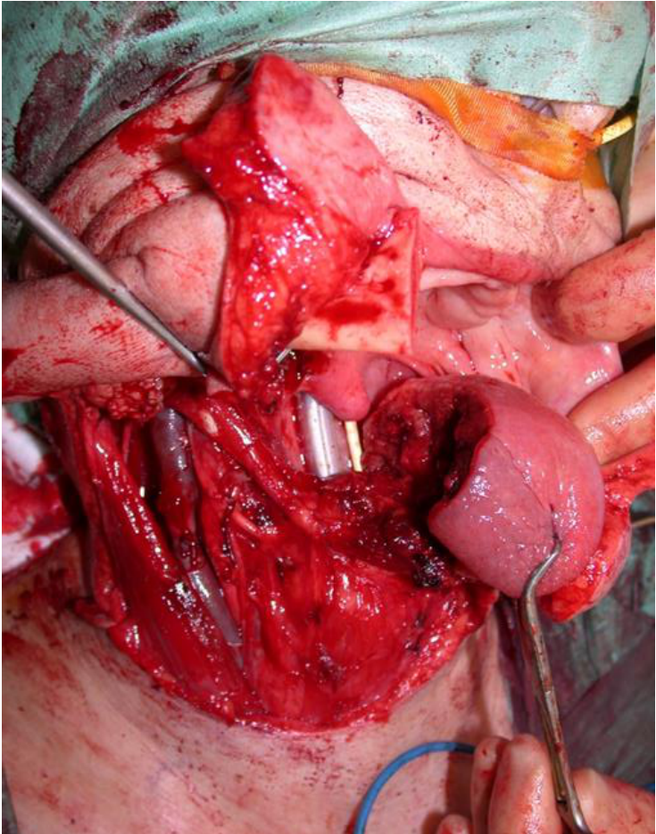
Neck Dissection39
Neck dissection refers to removal of cervical lymph nodes at different levels.
Cervical lymph nodes levels I–VI. Anatomical landmarks shown: hyoid bone, thyroid cartilage, cricothyroid membrane, sternocleidomastoid muscle; node levels labelled I, II, III, IV, V, VI.
Partial resection of tongue and neck dissection.
- Regional lymph nodes are removed or sampled to determine whether microscopic metastasis has occurred.
- For oral cavity tumours, the relevant nodal levels are 1, 2, 3 and 4, and the findings help determine whether additional radiotherapy is required.
Reconstruction With Free Tissue Transfer404142
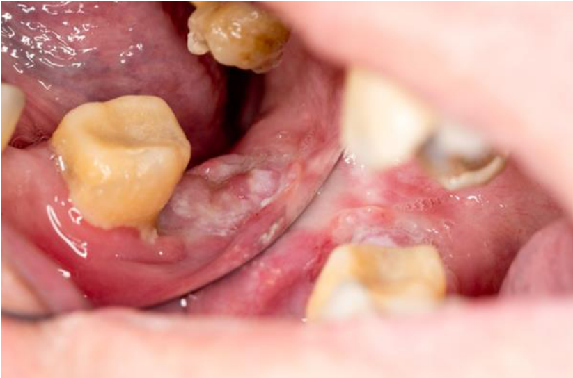
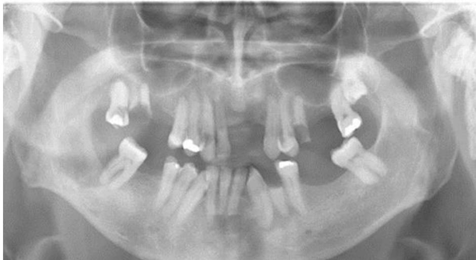
SCC right mandible
Resection and reconstruction with free tissue transfer
Screening And Early Detection43
There is currently no national screening program for mouth cancer available in Australia.
- Dentists routinely use visual examination to check for signs of mouth cancer and early detection
- Dentists play a role in prevention and counselling patients
“Early identification of oral cancers leads to improved treatment options for patients”
Lecturer — Clinical Recognition
The dentist may be the first clinician to identify oral cancer, but is not expected to make the definitive diagnosis.
- The clinician should recognise unusual symptoms, maintain an index of suspicion, and refer for biopsy or perform an appropriate incisional biopsy.
- Early identification improves prognosis and survival.
Symptoms of Oral Cancer44
-
White or red or speckled lesions
-
Lesions or ulcers on the gingiva, palate, tongue, oral mucosa or lip that do not heal in 14 days
-
Cervical lymphadenopathy
-
Difficulty swallowing
-
Unusual bleeding or pain or numbness in the mouth
-
Mobile teeth or swelling of the jaw
-
Changes in speech
-
Unexplained weight loss
-
An indurated lesion
-
Localised bone loss
-
Localised bone loss with a non-healing ulcer
-
An unusual lesion in a smoker and drinker
-
A persistent neck lump with no dental cause
-
Unexplained lip or facial numbness
-
A lesion that does not heal within two or three weeks should be investigated or referred
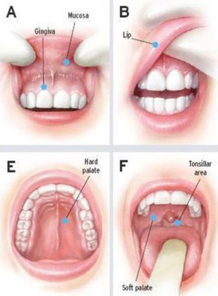
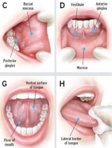
The 8-Step Oral Cancer Screening — Visual Examination45
Areas examined:
- Lips
- Anterior 2/3 of the tongue
- Gingiva
- Buccal mucosa
- Floor of the mouth and ventral tongue
- Hard palate and soft palate
Screening steps:
- A — Mucosa, Gingiva
- B — Lip
- C — Buccal mucosa, Posterior gingiva
- D — Vestibule, Anterior gingiva, Mucosa
- E — Hard palate
- F — Tonsillar area, Soft palate
- G — Ventral surface of tongue, Floor of mouth
- H — Lateral border of tongue
Lecturer — Examination Method
The examination should be carried out systematically at every appointment so that important sites are not missed.
- The sequence may begin with the lips or place the lips later in the examination, provided all areas are assessed.
- Additional areas include the sulci, maxilla, mandible, posterior lateral tongue, lingual sulcus, ventral surface of the tongue, floor of the mouth, palate, gingiva, and hard and soft palate.
- The lingual sulcus must be examined carefully because oral cancer commonly hides there, and the underside of the tongue is particularly important.
- The clinician should decide whether a lesion is confined to soft tissue or has extended into the mandible.
 |  |
Prevention46
- Around 2/3 of mouth cancers in Australia are caused by smoking
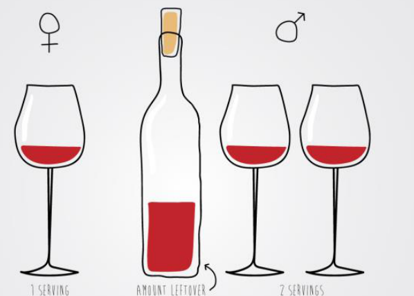
- Around 1/3 are caused by excess alcohol consumption
- Synergistic effect of smoking and alcohol consumption
- Quitting smoking and moderating alcohol consumption will significantly reduce risk of developing mouth cancer
- Cancers of the lip are commonly associated with UV exposure, so sun protection is important
- Prevention and early detection depend on recognising major lifestyle risk factors, identifying suspicious lesions, examining high-risk sites systematically, and referring promptly for biopsy or further investigation.
- Patients who chew betel nut should be considered at increased risk, particularly for buccal mucosal cancer.
- Ongoing oral care is important for patients before, during, and after cancer treatment.
 |  |
 |
Clinical Cases
Case: Lower anterior non-healing ulcer and localized bone loss
Question
Scenario: A patient presents with a non-healing ulcer in the lower anterior region, a loose bridge, and food impaction.
What’s shown: A panoramic radiograph (OPG) and an intraoral clinical photograph of the lower anterior region.
Consider: What should be considered when bone loss and a non-healing ulcer are localized to one specific area rather than being generalized?
Answer
Observations:
- Generalized bone loss, but with more severe, localized bone loss around the lower anteriors (specifically noted around teeth 4 and 2).
- A non-healing ulcer in the lower anterior region.
- A loose bridge and food impaction in the affected area.
Reasoning: While bone loss is most commonly due to periodontal disease, localized bone loss combined with a non-healing ulcer in a specific area should prompt lateral thinking. It is important to consider other causes, such as a malignancy or local infection, rather than assuming it is solely periodontal disease.
Outcome: The area was investigated and the findings turned out to be a cancer in the bone.
Takeaway: When encountering a non-healing ulcer with localized bone loss and a loose prosthesis, maintain a high index of suspicion for malignancy and think laterally beyond generalized periodontal disease.
Case: Floor of mouth ulcer and blocked submandibular gland
Question
Scenario: A patient presents with a blocked submandibular gland or an asymptomatic lesion on the floor of the mouth and ventral tongue.
What’s shown: An intraoral clinical photograph showing the floor of the mouth.
Consider: What is the likely cause of the blocked submandibular gland and the appearance of the ulcer?
Answer
Observations:
- Non-healing ulcer
- Indurated lesion
Reasoning: Tumors on the ventral tongue or floor of the mouth can be painless and go unnoticed by the patient. They may present as a blocked submandibular gland because the tumor surrounds the duct opening, preventing it from emptying. Unlike a salivary stone, which causes intermittent blockage that resolves, a tumor-induced blockage will not improve and presents with a different clinical appearance.
Outcome: The lesion is a tumor causing duct obstruction.
Takeaway: When a patient presents with a blocked submandibular gland, think laterally about tumors on the floor of the mouth or ventral tongue, especially if the blockage does not resolve like a salivary stone would.
Case: Maxillary swelling with localized bone loss
Question
Scenario: A patient presents with a swelling in the minor salivary gland area of the maxilla.
What’s shown: Intraoral clinical photographs of the maxilla and a panoramic radiograph (OPG).
Consider: What is the diagnosis for a swelling accompanied by localized bone loss and a loose tooth?
Answer
Observations:
- Swelling in the minor salivary gland area
- Huge amount of localized bone loss on the OPG
- Very loose tooth
Reasoning: The initial thought for a swelling is usually an infection, prompting a check of tooth vitality, cracks, and restorations. However, the OPG reveals sudden, massive localized bone loss and a loose tooth, which points away from a simple dental infection and suggests a malignancy.
Outcome: Diagnosed as a lymphoma.
Takeaway: While infection is the first consideration for a swelling, localized severe bone loss and tooth mobility on a radiograph should raise suspicion for a malignancy such as lymphoma.
Case: Mandibular metastasis on CT scan
Question
Scenario: A patient with a metastatic lesion in the mandible.
What’s shown: Axial and 3D reconstructed CT scans of the mandible.
Consider: Identify the radiographic findings on the CT scans.
Answer
Observations:
- Bone loss
- An area of lump in the mandible
Reasoning: The CT scan reveals bone loss and a lump in the mandible, which in this case represents metastasis.
Outcome: None stated.
Takeaway: Metastasis to the mandible can present radiographically as bone loss and a lump on CT imaging.
Case: Mucosal melanoma of the oral cavity and uvula
Question
Scenario: A patient presenting with a pigmented oral lesion.
What’s shown: Clinical photographs of the oral cavity showing a pigmented lesion invading the soft tissue, and an intraoral view showing a tumor invading the uvula.
Consider: Identify the clinical features and the functional impact of the lesion.
Answer
Observations:
- Pigmented lesion invading the soft tissue
- Mobile teeth that bleed easily
- Bone loss from bone invasion
- Tumor invading the uvula
Reasoning: The extensive pigmented lesion with soft tissue invasion, tooth mobility, and bone loss is characteristic of mucosal melanoma rather than a simple dark spot. Invasion of the uvula and soft palate impairs their function in sealing off the nasal cavity, leading to a nasal voice.
Outcome: None stated.
Takeaway: Mucosal melanoma presents as an extensive pigmented lesion with soft tissue invasion, tooth mobility, and bone loss, and can impair palatal function causing a nasal voice.
Case: Pre-radiotherapy dental assessment form
Question
Scenario: A patient with hypopharyngeal squamous cell carcinoma is referred for a pre-radiotherapy dental assessment prior to receiving post-operative radiotherapy.
What’s shown: A dental evaluation form detailing the proposed radiation dose (66Gy/33) and a chart indicating the specific radiation dose planned for individual upper and lower teeth (ranging from 40Gy to 66Gy).
Consider: How to use the radiation field and dose information to decide which teeth should be extracted prior to treatment.
Answer
Observations:
- The chart indicates that some teeth will receive 40Gy of radiation, while others will receive 66Gy.
Reasoning: The lecturer explains that the radiation dose determines the risk of complications, such as poor healing, if extractions are needed later. Teeth receiving 55Gy or more are at high risk; if there is any doubt about their long-term prognosis—such as having a 7mm periodontal pocket, a huge MOD restoration, or being heavily restored—they should be extracted pre-radiotherapy. Teeth receiving a much lower dose (e.g., 40Gy) can be given the benefit of the doubt and monitored.
Outcome: The patient is assessed to render them dentally fit before radiotherapy, aiming to avoid extractions within the high-dose radiation field after treatment has commenced.
Takeaway: The threshold for pre-radiotherapy extraction of questionable teeth is typically a radiation dose of 55 Gy or more to avoid post-treatment complications.
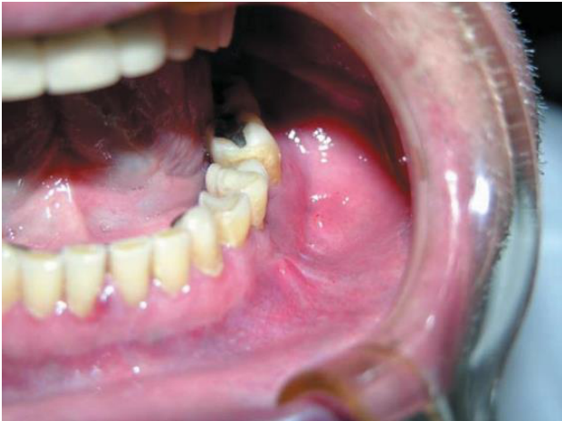
Case: Mobile posterior tooth and non-healing white patch
Question
Scenario: A patient presents with an unusually mobile tooth 48 and a non-healing white patch in the adjacent posterior mandibular region.
What’s shown: An intraoral clinical photograph of the posterior mandible and a panoramic radiograph (OPG).
Consider: What do the clinical and radiographic findings indicate, and what surgical approach is required?
Answer
Observations:
- Tooth 48 is unusually mobile.
- A non-healing white patch is present in the area behind the tooth.
- The panoramic radiograph (OPG) reveals an area of bone erosion in the mandible.
Reasoning:
- Biopsy of the non-healing white patch confirmed it was a carcinoma (transcribed in the lecture as “fcc”).
- The radiographic bone erosion indicates that the lesion has invaded the hard tissue of the mandible.
- Because the tumor has eroded the bone, the surgical resection must include the mandible, not just the soft tissue.
Outcome:
- The patient underwent resection of the tumor and the affected portion of the mandible.
- The defect was reconstructed using a large reconstruction plate and bone harvested from the leg to replace the missing hard tissue.
Takeaway:
- When an oral carcinoma erodes the underlying mandibular bone, surgical management requires resection of the bone and reconstruction with both hard and soft tissue (e.g., a bone graft from the leg and a reconstruction plate) to restore function.
Footnotes
-
Original PDF page 1: W3 Oral Cancer - Foo, p.1 ↩
-
Original PDF page 2: W3 Oral Cancer - Foo, p.2 ↩
-
Original PDF page 3: W3 Oral Cancer - Foo, p.3 ↩
-
Original PDF page 4: W3 Oral Cancer - Foo, p.4 ↩
-
Original PDF page 5: W3 Oral Cancer - Foo, p.5 ↩
-
Original PDF page 6: W3 Oral Cancer - Foo, p.6 ↩
-
Original PDF page 7: W3 Oral Cancer - Foo, p.7 ↩
-
Original PDF page 8: W3 Oral Cancer - Foo, p.8 ↩
-
Original PDF page 10: W3 Oral Cancer - Foo, p.10 ↩
-
Original PDF page 9: W3 Oral Cancer - Foo, p.9 ↩
-
Original PDF page 11: W3 Oral Cancer - Foo, p.11 ↩
-
Original PDF page 12: W3 Oral Cancer - Foo, p.12 ↩
-
Original PDF page 13: W3 Oral Cancer - Foo, p.13 ↩
-
Original PDF page 17: W3 Oral Cancer - Foo, p.17 ↩
-
Original PDF page 14: W3 Oral Cancer - Foo, p.14 ↩
-
Original PDF page 15: W3 Oral Cancer - Foo, p.15 ↩
-
Original PDF page 16: W3 Oral Cancer - Foo, p.16 ↩
-
Original PDF page 19: W3 Oral Cancer - Foo, p.19 ↩
-
Original PDF page 20: W3 Oral Cancer - Foo, p.20 ↩
-
Original PDF page 21: W3 Oral Cancer - Foo, p.21 ↩
-
Original PDF page 22: W3 Oral Cancer - Foo, p.22 ↩
-
Original PDF page 23: W3 Oral Cancer - Foo, p.23 ↩
-
Original PDF page 24: W3 Oral Cancer - Foo, p.24 ↩
-
Original PDF page 25: W3 Oral Cancer - Foo, p.25 ↩
-
Original PDF page 26: W3 Oral Cancer - Foo, p.26 ↩
-
Original PDF page 27: W3 Oral Cancer - Foo, p.27 ↩
-
Original PDF page 28: W3 Oral Cancer - Foo, p.28 ↩
-
Original PDF page 29: W3 Oral Cancer - Foo, p.29 ↩
-
Original PDF page 30: W3 Oral Cancer - Foo, p.30 ↩
-
Original PDF page 31: W3 Oral Cancer - Foo, p.31 ↩
-
Original PDF page 33: W3 Oral Cancer - Foo, p.33 ↩
-
Original PDF page 34: W3 Oral Cancer - Foo, p.34 ↩
-
Original PDF page 35: W3 Oral Cancer - Foo, p.35 ↩
-
Original PDF page 36: W3 Oral Cancer - Foo, p.36 ↩
-
Original PDF page 32: W3 Oral Cancer - Foo, p.32 ↩
-
Original PDF page 37: W3 Oral Cancer - Foo, p.37 ↩
-
Original PDF page 38: W3 Oral Cancer - Foo, p.38 ↩
-
Original PDF page 40: W3 Oral Cancer - Foo, p.40 ↩
-
Original PDF page 39: W3 Oral Cancer - Foo, p.39 ↩
-
Original PDF page 41: W3 Oral Cancer - Foo, p.41 ↩
-
Original PDF page 42: W3 Oral Cancer - Foo, p.42 ↩
-
Original PDF page 43: W3 Oral Cancer - Foo, p.43 ↩
-
Original PDF page 44: W3 Oral Cancer - Foo, p.44 ↩
-
Original PDF page 45: W3 Oral Cancer - Foo, p.45 ↩
-
Original PDF page 46: W3 Oral Cancer - Foo, p.46 ↩
-
Original PDF page 47: W3 Oral Cancer - Foo, p.47 ↩