Principles of Surgery1
Surgical Diagnosis234
- Correlating signs and symptoms
- Targeting investigations and interventions
Lecturer — Diagnostic Reasoning
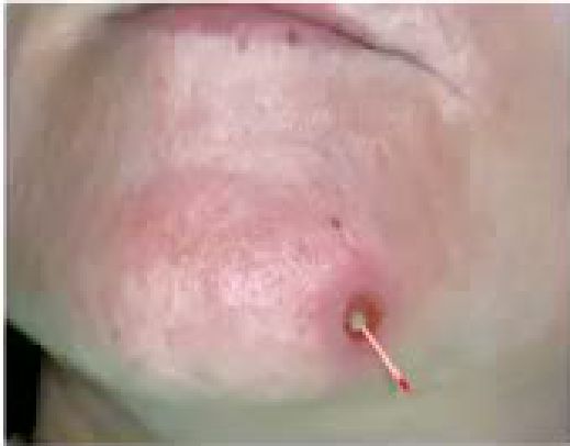
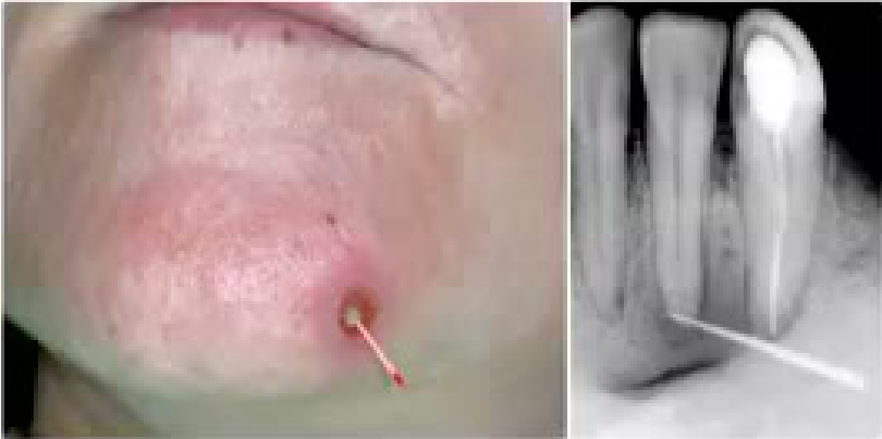
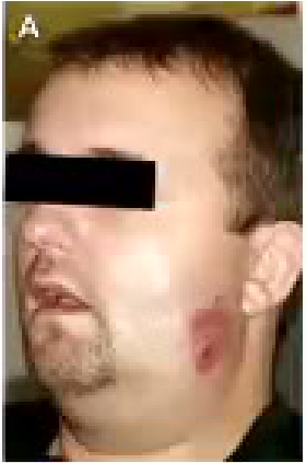
Correct diagnosis is essential before treatment or investigation. A swelling or abscess on the chin may arise from a skin lesion or an infected lower central tooth.
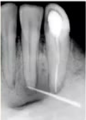
- A non-vital tooth may form a fistula through which pus escapes; a gutta-percha point can be placed in the tract and an X-ray taken to identify the source tooth.
- The thinnest suitable point should be used, but it must not be so thin that it breaks.
- The tooth indicated by the tract may not be the tooth initially suspected.
- A loose tooth may be associated with periodontal disease, a fracture, or a more extensive lesion at the edge of the tooth.
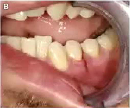
- A tear in the gingiva, localized bruising, or another unusual clinical sign may suggest an underlying fracture.
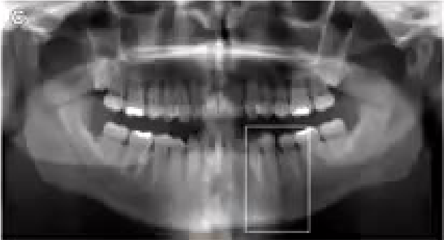
- An X-ray should be taken before extracting an unusually mobile tooth, a tooth with extensive apical radiolucency, or a tooth that may have a lesion beyond the apparent tooth-related problem.
- A surgical sieve can systematically narrow the differential diagnosis to a rational group of three or four possibilities.
Causes of Surgical Disorders5
- Neoplasia
- Autoimmune
- Infection, Inflammatory
- Vascular
- Environment, toxin exposure
- Metabolic
- Drugs
- Degenerative
- Iatrogenic
- Congenital / developmental
- Trauma
Neoplasia6
- Benign neoplasia
- Malignant neoplasia
Lecturer — Neoplastic Features
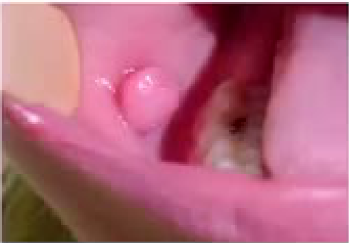
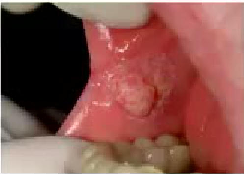
A benign polyp may appear uniform and smooth, whereas a malignant lesion may have poorly defined margins, a speckled white and red appearance, induration, ulceration, or a heaped-up appearance. Bilateral enlargement of a structure is less suggestive of neoplasia because neoplastic growth is usually unilateral.
 |  |
Autoimmune Disease7
- Sjogren’s syndrome
Lecturer — Sjögren Syndrome
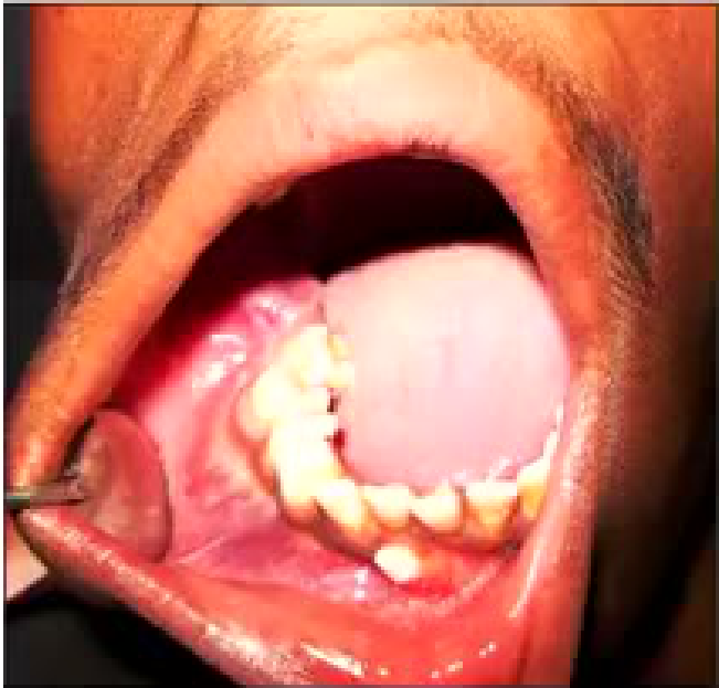
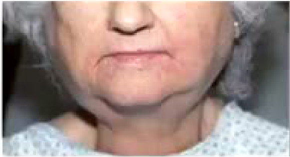
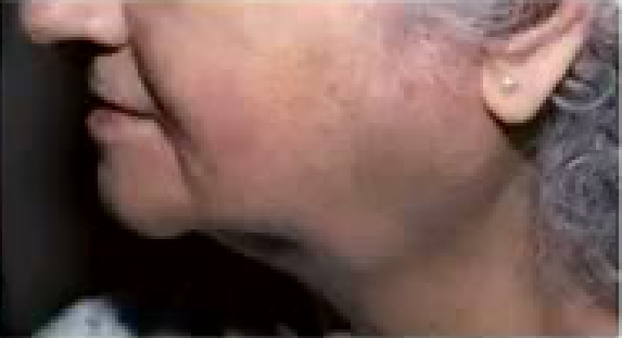
Bilateral swelling of the salivary glands may suggest an autoimmune condition rather than neoplasia. Associated dry eyes and dry mouth may support this possibility.
- Dry mouth may cause cervical caries, create a need for saliva stimulants, and require more frequent reviews.
- Management should be patient-centred and adapted to the patient’s symptoms and risks.
 |  |
Inflammation and Infection89
-
Inflammatory processes often stem from infection
-
Inflammation also result from physical irritation e.g. noxious chemicals
-
Bacterial diffuse through the dentinal tubules toward the pulp and evoke inflammatory changes resulting in pulpitis and pulp necrosis
-
Bacteria from the pulp spreads into periapical tissues and soft tissues and bones of the face and neck
Lecturer — Oral Infection Sources
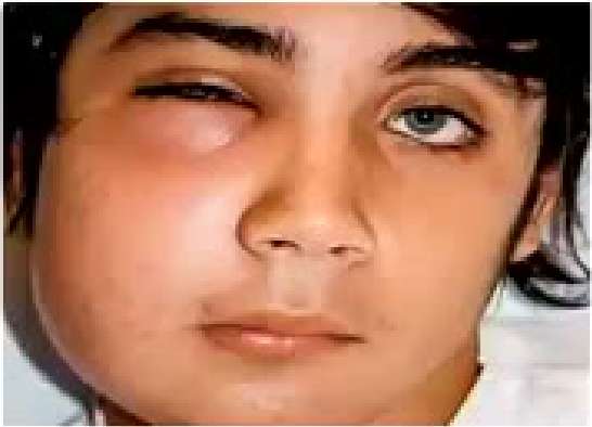
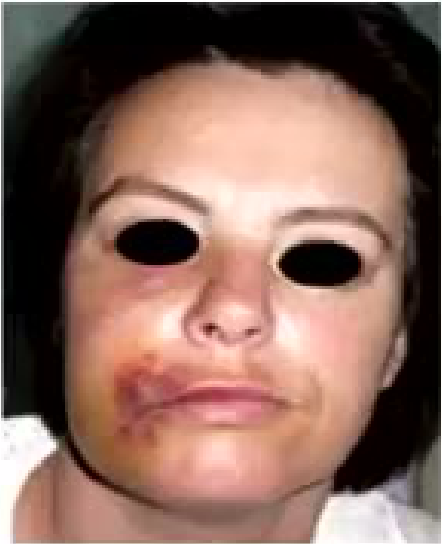
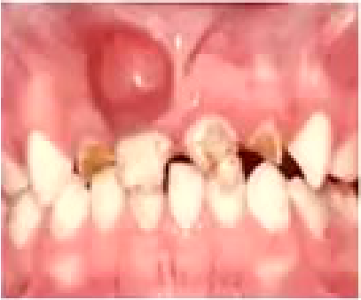
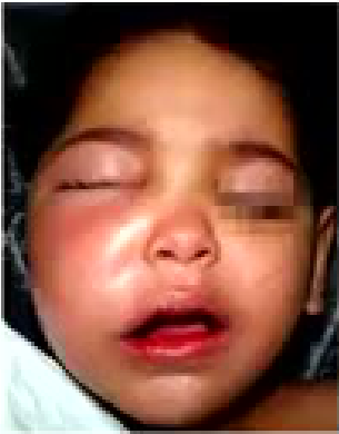
A gum boil represents pus attempting to escape. If the swelling is adjacent to damaged teeth, the infection is likely to originate from one of those teeth and generally requires assessment and treatment of the causative tooth rather than referral to oral medicine.
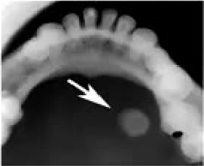
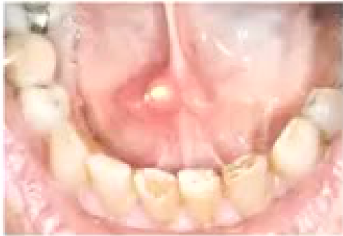
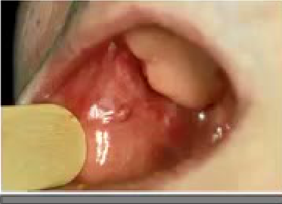
- A blocked duct may result in inflammation and pus emerging from the duct.
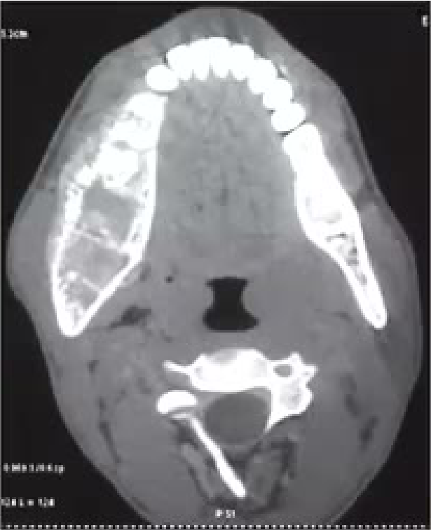
- A duct stone may be visible on an X-ray.
 |  |
 |  |
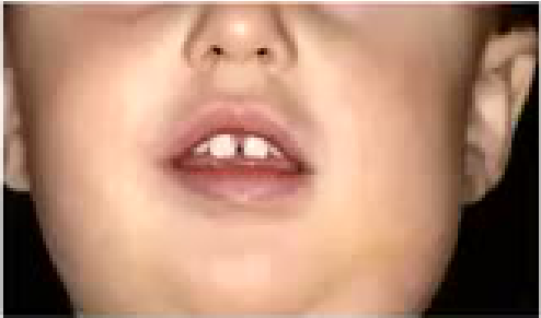
Inflammatory Conditions of Salivary Glands10
- Aetiology – calculus, viral, bacteria infection
 |  |
 |  |
Vascular Malformations11
A-V malformation
Lecturer — Vascular Malformation Risk
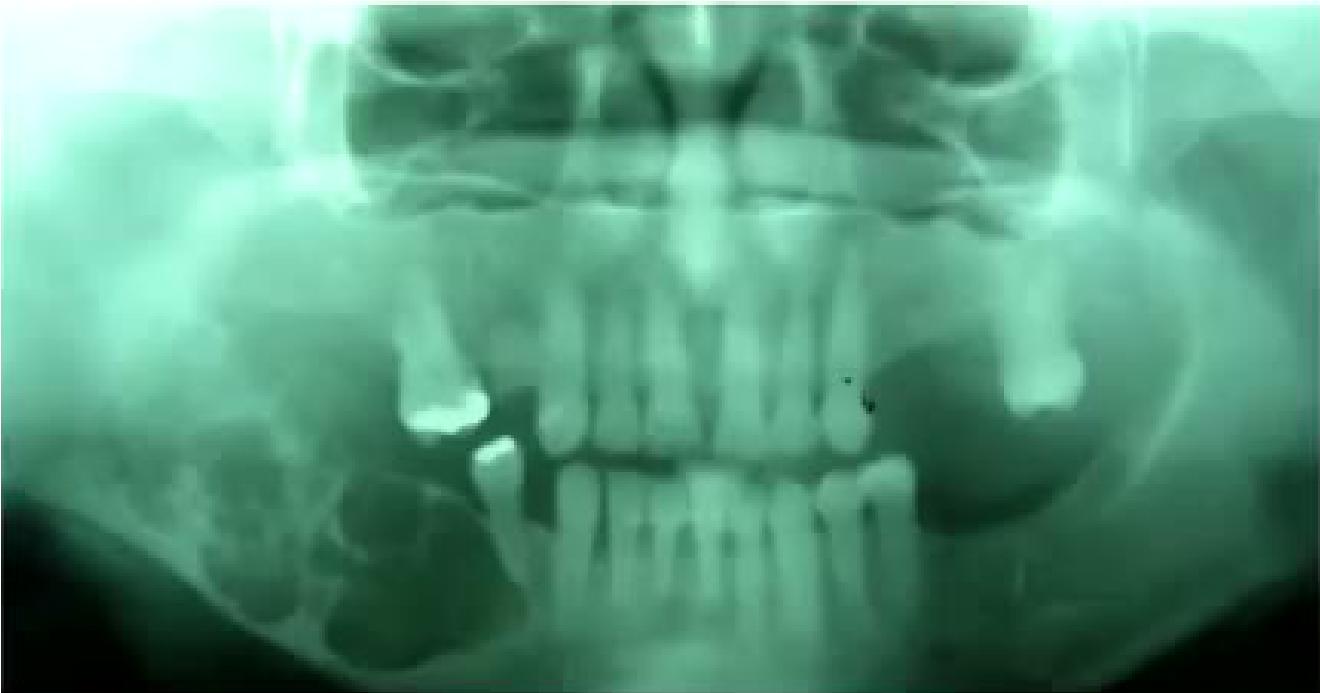
A multilocular radiolucent appearance may represent an arteriovenous malformation, but it may also represent an ameloblastoma. A loose tooth overlying a suspected vascular malformation should not be extracted without further assessment because extraction could result in severe bleeding.
- CT is required when an arteriovenous malformation is suspected; cone-beam CT may not provide sufficient resolution.
- Teeth should not be removed from an area that may contain a significant lesion until appropriate imaging has been performed.
- An apparently loose tooth may be located at the edge of a more serious process.
Oral Cancer and Reconstruction12131415
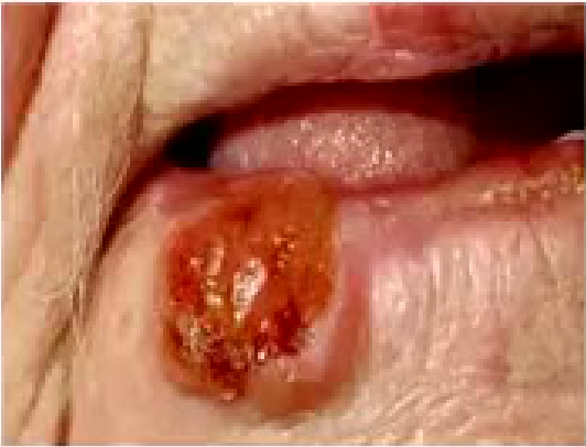
Cutaneous SCC from sun damage
-
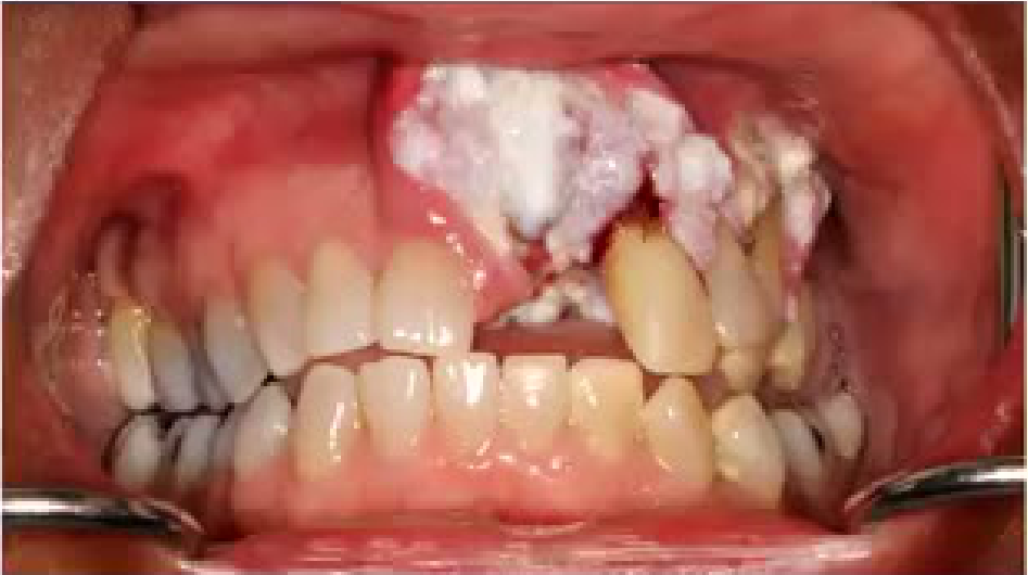
Oral SCC associated with smoking and alcohol excess
-
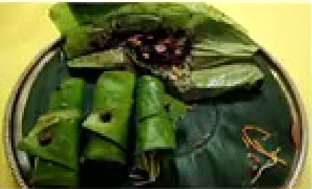
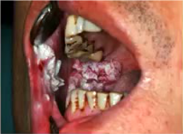
Betel nut habit and oral cancer
Lecturer — Oral Cancer Features
Sun-damaged skin may develop into lip cancer. Features suggestive of cancer include induration, ulceration, a heaped-up growth, speckled white and red areas, a lesion that does not resolve, and poorly defined margins.
- Smoking and drinking were identified as toxin-related contributors in one example.
- Betel nut chewing was associated with buccal mucosal cancer because the betel nut is commonly held there.
- Tongue cancer was described as more frequent in smokers, whereas buccal mucosal cancer was described in relation to betel nut chewing.
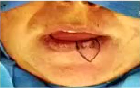
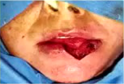
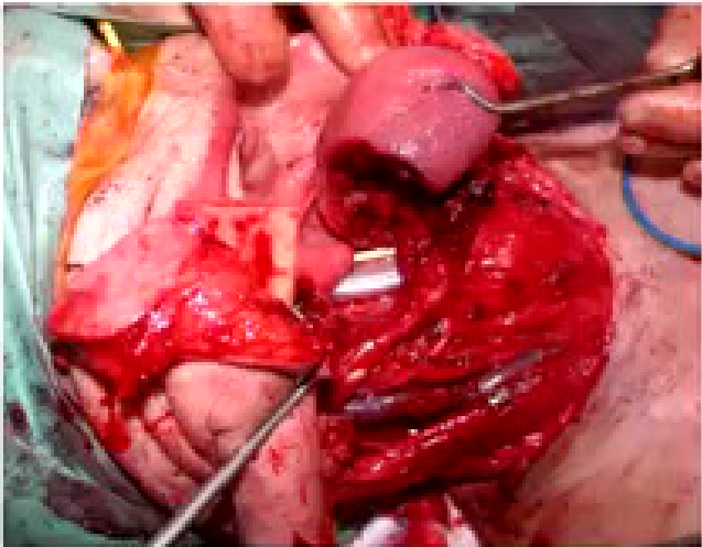
- Surgical treatment may involve excision with a margin and reconstruction, including reconstruction of part of the tongue.
Surgery and reconstruction for oral SCC
Metabolic and Endocrine Disorders1617
- e.g. Diabetes
e.g. Diabetes mellitus
- periodontal disease
- candidosis
- dry mouth and increased caries
- glossitis
- burning mouth syndrome
- orofacial dysaesthesia
- poor healing
Lecturer — Diabetes and Healing
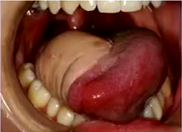
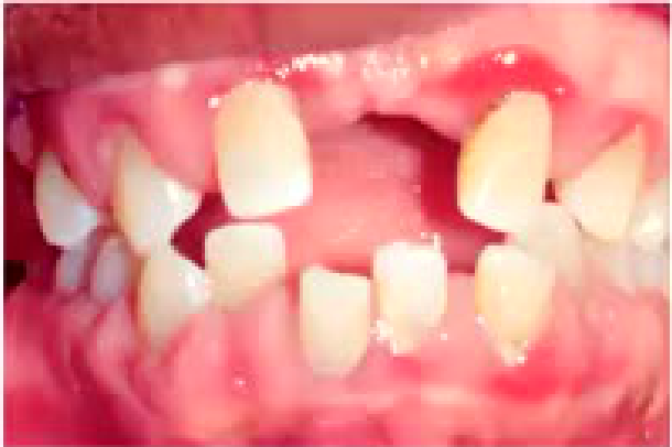
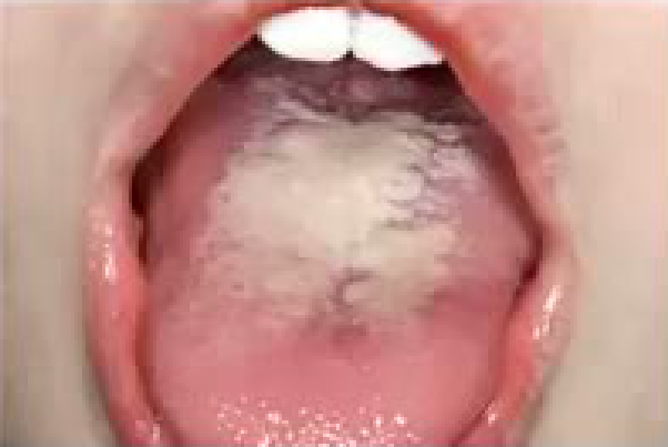
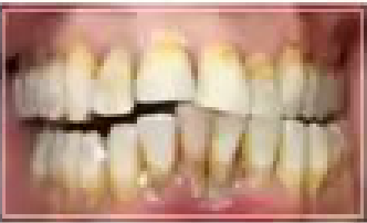
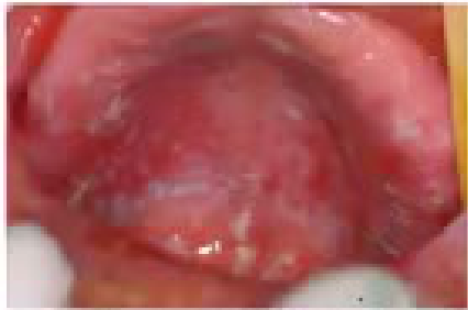
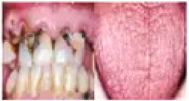
Diabetes may be associated with earlier tooth loss, an increased risk of oral infections, and Candida affecting the tongue and roof of the mouth. Hypocalcaemia and hypercalcaemia were also mentioned as metabolic conditions to consider.
- The patient’s systemic disease should be included when assessing the likely cause of oral symptoms and the expected healing response.
 |  |  |
 |  |
Drug-Induced Disorders1819
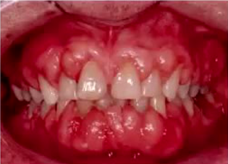
- Phenytoin induced gingival overgrowth
Lecturer — Drug-Related Disorders
Some mental-health medications and antidepressants can cause dry mouth, while antiepileptic drugs may cause gingival hyperplasia. Management may include more frequent cleaning, improved oral hygiene, laser surgery, or gingivectomy.
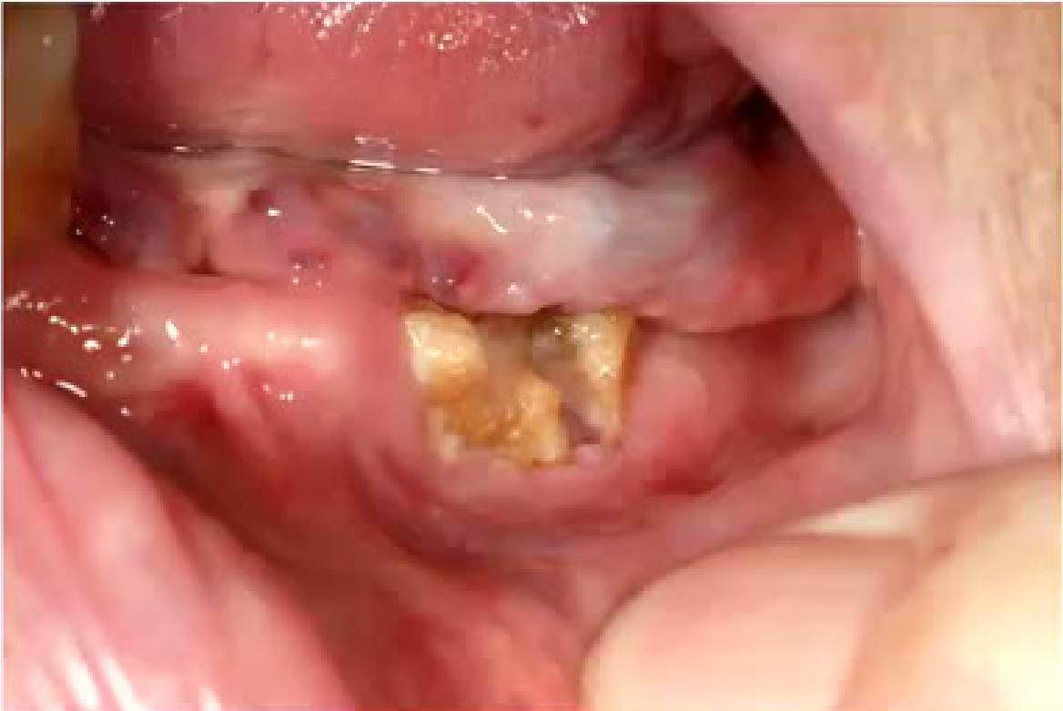
- Prolia was discussed in relation to osteonecrosis and delayed healing after extraction and should not be administered immediately after extraction because the socket has not matured.
- Healing involves an initial haematoma, replacement of the clot, and filling with bone; Prolia may need to be delayed for about a month or longer.
- Aspirin, clopidogrel, and oral anticoagulants affect bleeding and clotting and should not be stopped unless there is a good reason or the prescribing general practitioner agrees.
 |  |
Degenerative Disorders2021
-
Osteoporosis
-
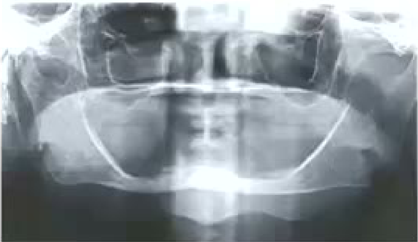
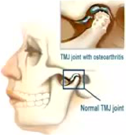
Osteoarthritis
TMJ joint with osteoarthritis Normal TMJ joint
Lecturer — Degenerative Conditions
A degenerative spine and osteoporosis were given as examples of degenerative conditions. A patient may be taking Prolia because of osteoporosis, but osteoporosis or Prolia does not necessarily mean that the medication is the cause of the patient’s pain; infection may be more likely.
 |  |
 |
Iatrogenic Causes2223
- Damage or injury resulting from the action of a health care personnel
Lecturer — Iatrogenic Injury
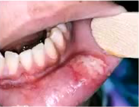
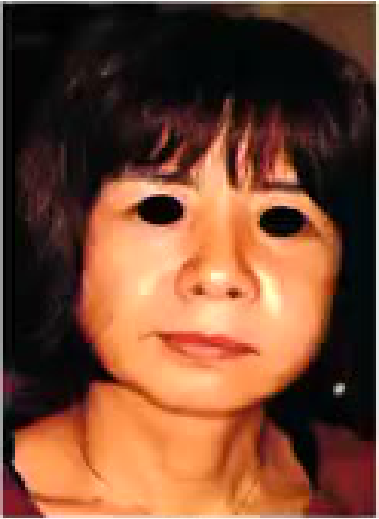
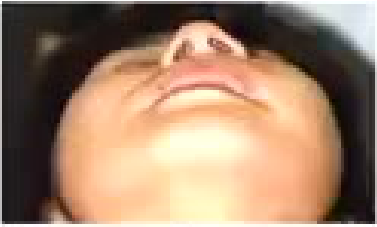
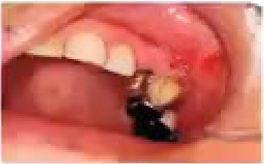
Iatrogenic injury may result from a hot bur, a high-speed instrument used in the mouth, or other treatment-related trauma. A treatment-related complication must be distinguished from the original disease.
Subcutaneous emphysema
 |  |  |
 |  |
Chemical Trauma24
- Sodium hypochlorite solution commonly used in root canal treatments
- Strongly alkaline with pH 11 to 13
- Complications of accidental spillage are damage to clothing, damage to eye, damage to skin and damage to oral mucosa
- Complications from hypochlorite extrusion into peri-radicular soft tissue include chemical burns and tissue necrosis and neurological complications like anaesthesia or paraesthesia
- Using sodium hypochlorite for root canal irrigation without adequate isolation of the tooth can lead to leakage of the solution into the oral cavity and ingestion or inhalation by the patient, resulting in throat irritation and in severe cases, upper airway compromise
Lecturer — Hypochlorite Extrusion
Sodium hypochlorite used during root canal irrigation may cause emphysema if it is forced into the tissues. If swelling begins during irrigation, stop irrigating immediately and flush the area with saline.
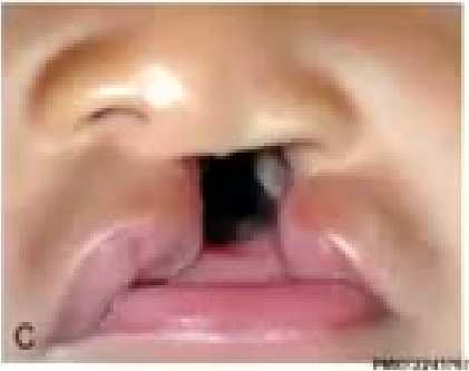
Congenital and Craniofacial Abnormalities25262728293031
- Birth defects of the face or head
- Cleft lip and palate are among the most common of all birth defects. Others are very rare
- Mostly affect how a person’s face or head looks. These conditions may also affect other parts of the body.
- Treatment depends on the type of problem
- Reconstructive surgery may help the person’s appearance
Lecturer — Cleft Management
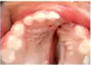
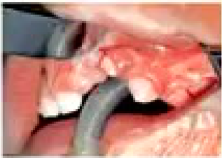
Cleft lip and palate were discussed as becoming less common because mothers are taking folic acid. In a cleft palate, a tooth may lie within the cleft with little or no bone around it.
- A severely deformed tooth may need extraction.
- Alternatively, it may be brought down with orthodontic treatment and supported with bone grafting.
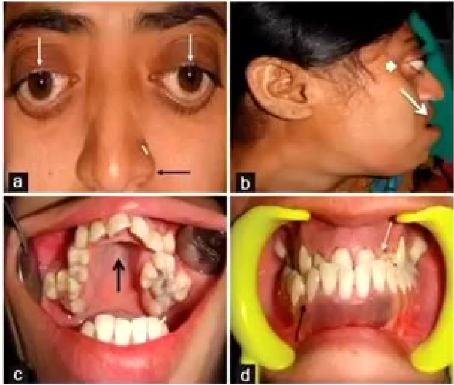
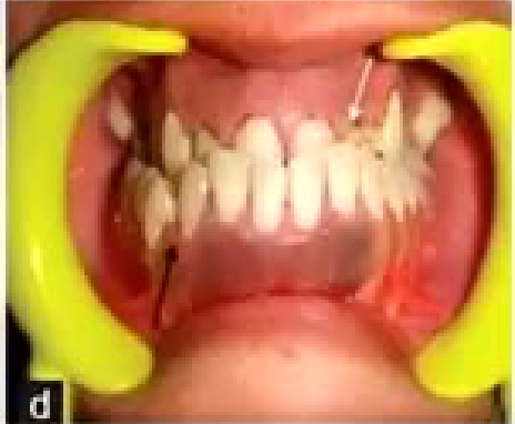
- Other craniofacial abnormalities may produce severe malocclusion, difficult-to-clean teeth, a hypoplastic maxilla, and a Class 3 relationship.
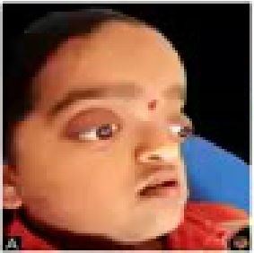
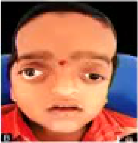
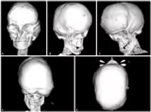
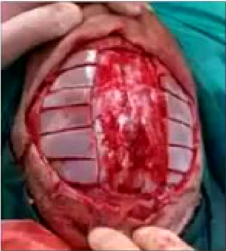
- Some syndromes restrict brain growth; the skull bone may be scored to allow the brain underneath to grow, similar in principle to scoring soft tissue in an escharotomy. This addresses skull growth and does not correct the jaws.
- Cleft lip occurs 1 in 750 live births, cleft palate 1 in every 2000
- Half the cleft lip also have a cleft palate
- 10% patients with cleft have other malformation
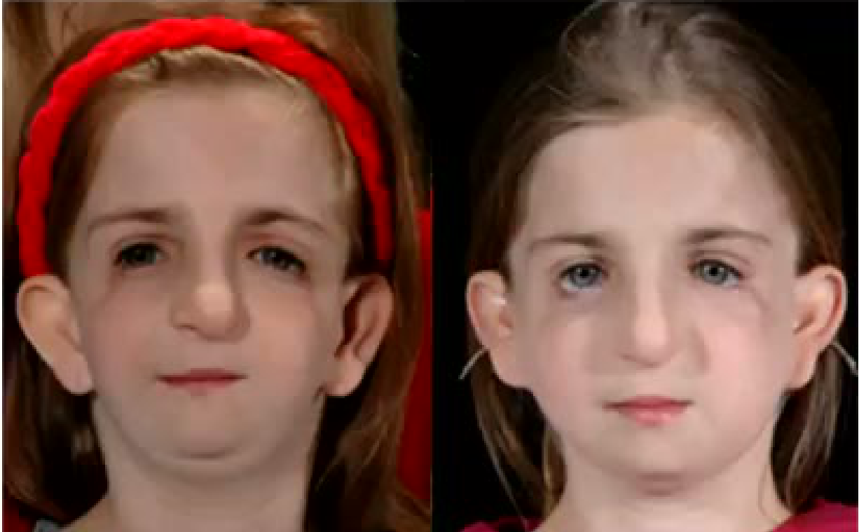
Craniofacial abnormalities
Lecturer — Craniofacial Syndromes
Treacher Collins syndrome was described with downward-slanting eyes and hearing problems; children may use hearing aids. Hemifacial microsomia involves unequal growth of the two sides of the face.
- The smile line may be canted.
- One side of the upper and lower jaws may grow more than the other.
- The upper and lower jaws may need correction.
Orthognathic Surgery32
Lecturer — Orthognathic Planning
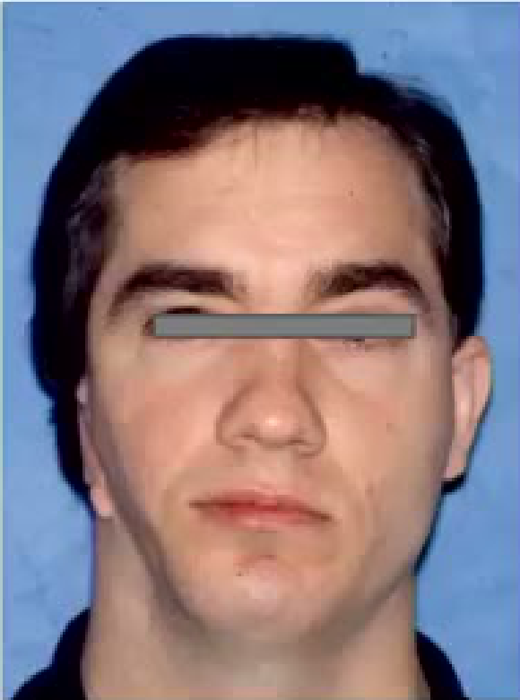
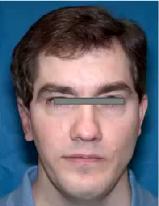
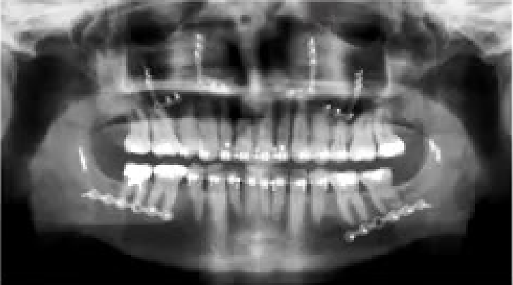
Orthognathic surgery assesses whether the main problem is in the upper jaw, lower jaw, or both, and whether orthodontics alone can correct it. In mild deformity, moving the teeth may be sufficient, but teeth can compensate for a more severe skeletal deformity.
- In a Class 3 patient, a proclined incisor may compensate for the jaw discrepancy.
- Teeth may first need to be moved back to their natural position before the jaw is repositioned; patients are told that the bite may become worse before it becomes better.
- The maxilla is corrected first by assessing its midline, forward and backward position, vertical position, and cant.
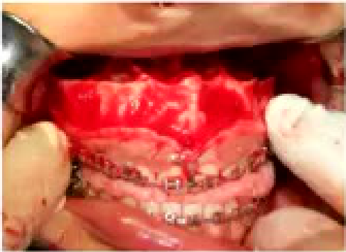
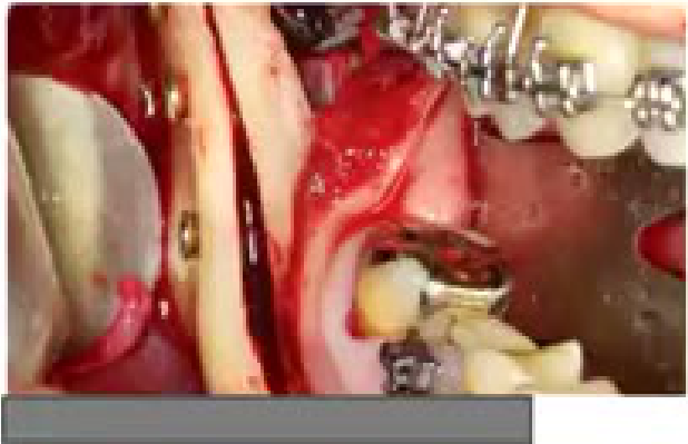
- The upper jaw may be detached while the palatal mucosa remains intact and the greater palatine blood supply is preserved, then held in position with plates and screws.
- The lower jaw is subsequently moved to fit the occlusion.
- Assessment includes lip and soft-tissue support, the nasolabial angle, and the amount of tooth and gum displayed.
- Surgery should reflect whether the patient is unhappy with appearance or function, and measurements of facial angles and projection may vary according to racial group.
 |  |
 |
Developmental Anomalies of Teeth33
- Supernumerary teeth
- Complex and compound odontome
Lecturer — Developmental Teeth
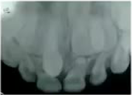
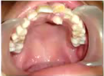
Supernumerary teeth may obstruct the development or eruption of permanent teeth. An upper occlusal X-ray can identify a supernumerary tooth obstructing growth of the upper central incisor, and the supernumerary tooth may need removal.
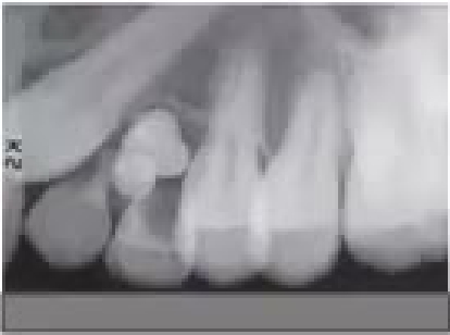
- An odontoma may resemble a tooth but is not a normal tooth; it may contain enamel and dentine or represent a collection of calcified tissues.
 |  |
 |  |
Pre-Prosthetic Surgery3435363738394041
Lecturer — Pre-Prosthetic Reconstruction
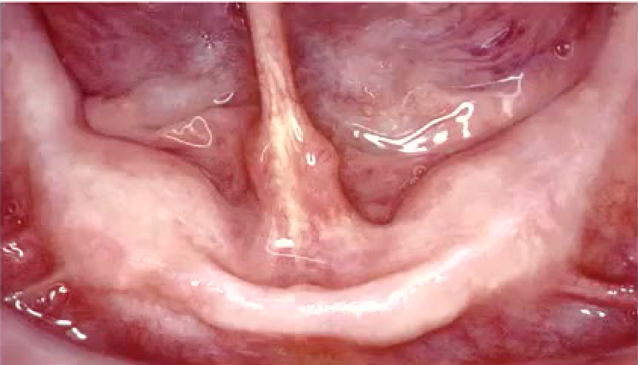
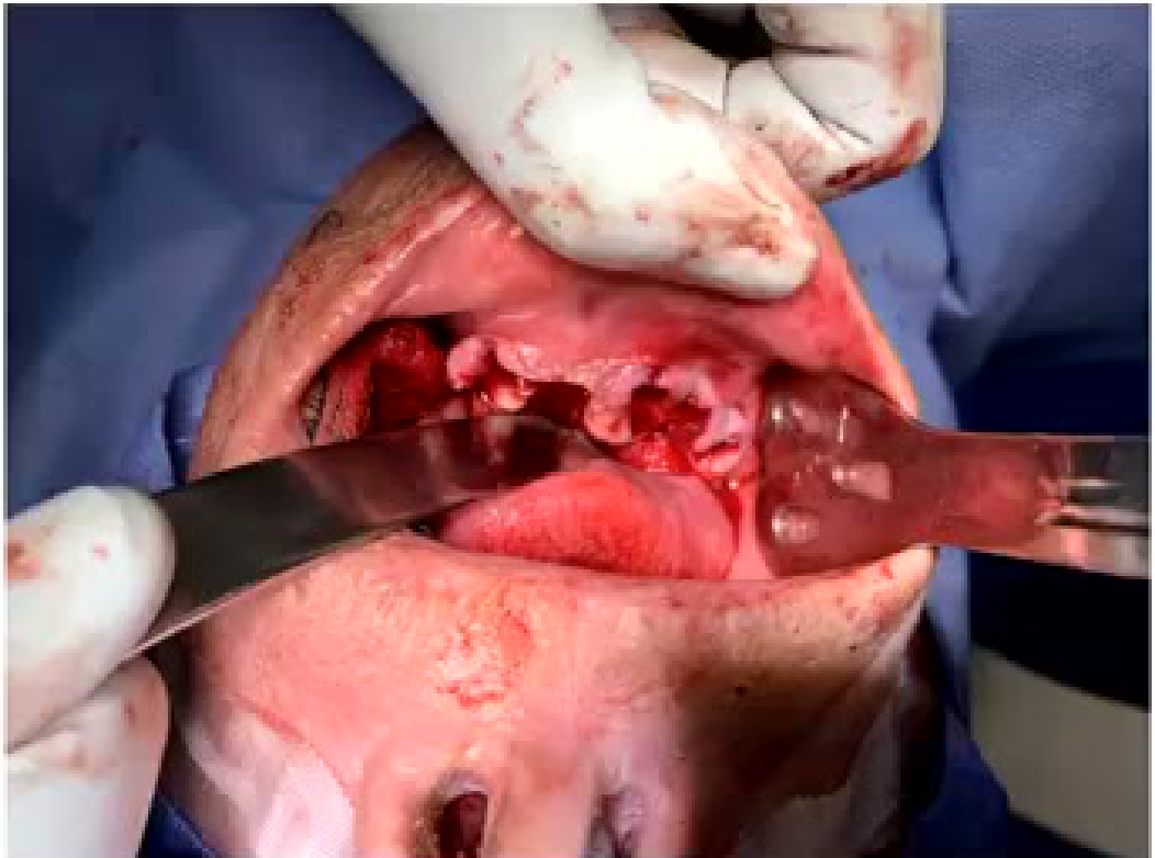
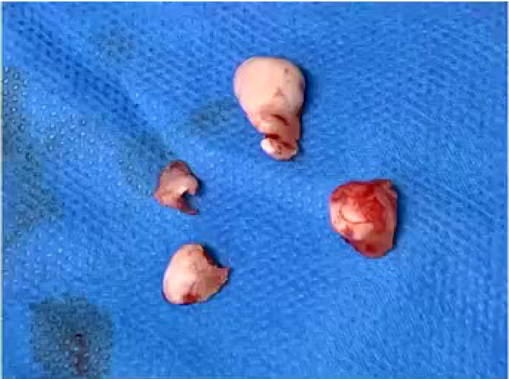
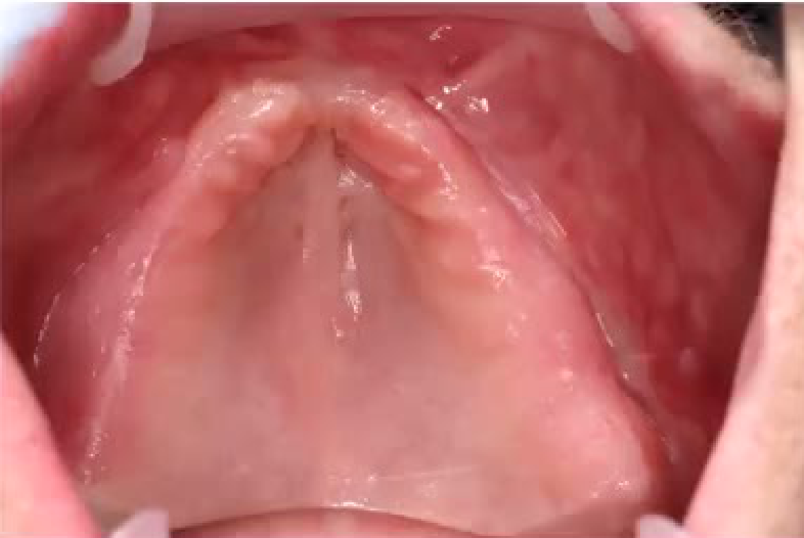
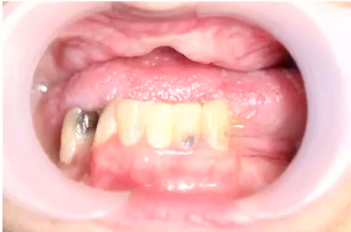
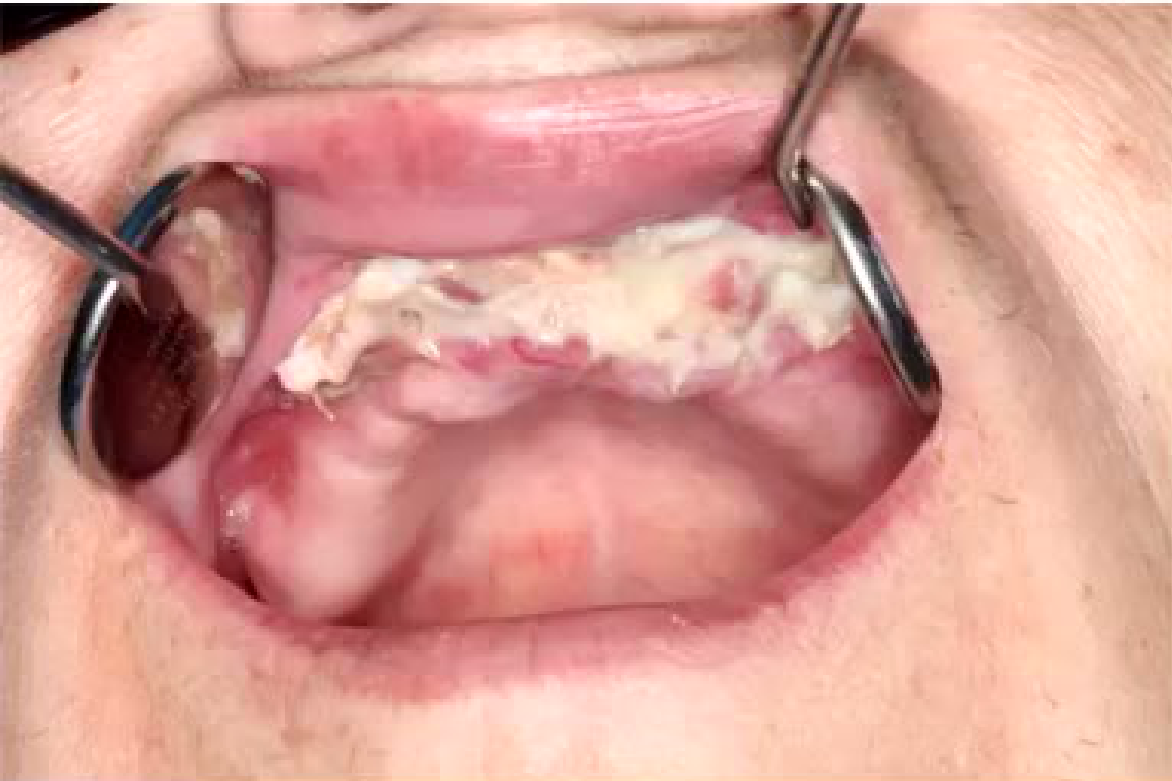
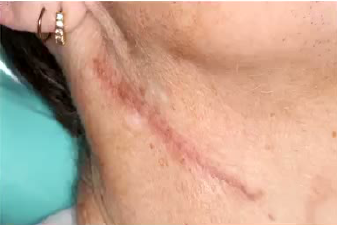
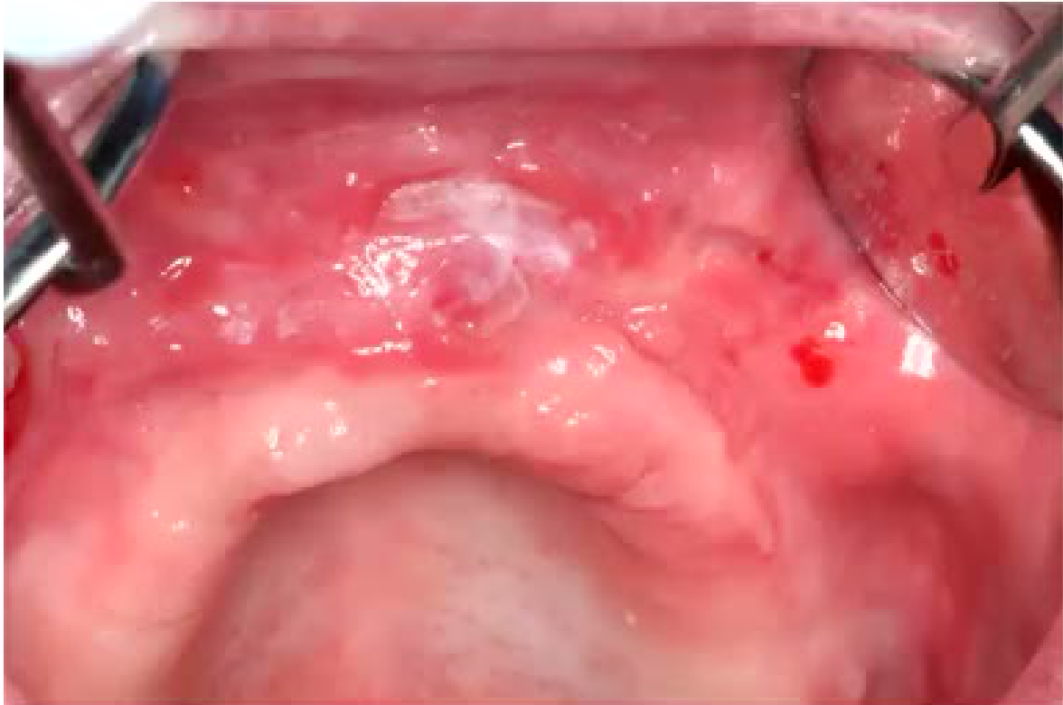
Prominent tori may interfere with denture construction; treatment can involve reflecting the mucosa, smoothing the tori, and removing prominent bony lumps that may resemble osteomas. A defect in the alveolar ridge may also prevent the denture from fitting.
- One example involved a midline defect created when tissue had been removed and the labial sulcus stitched into the gap.
- Treatment reversed the previous arrangement, placed a skin graft, increased the available space, and used a graft to build up the alveolus.
- Skin was taken from the neck because it provided the best match.
- The aim was to create a continuous ridge and provide more room for the denture.
Trauma42
Lecturer — Trauma Assessment
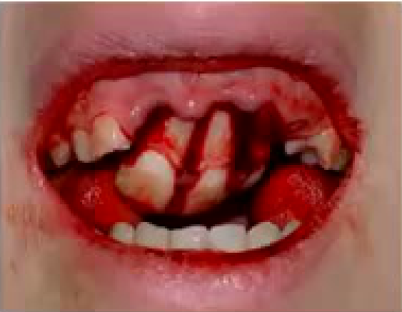
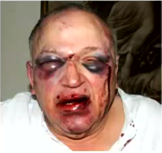
Trauma may affect the teeth, bone, or maxilla. An OPG can help determine whether the maxilla is involved, and general assessment should include the airway, breathing, circulation, history, and special injuries such as burns, electrical injuries, and cold-water immersion.
 |  |
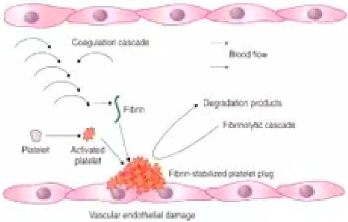
Wound Healing
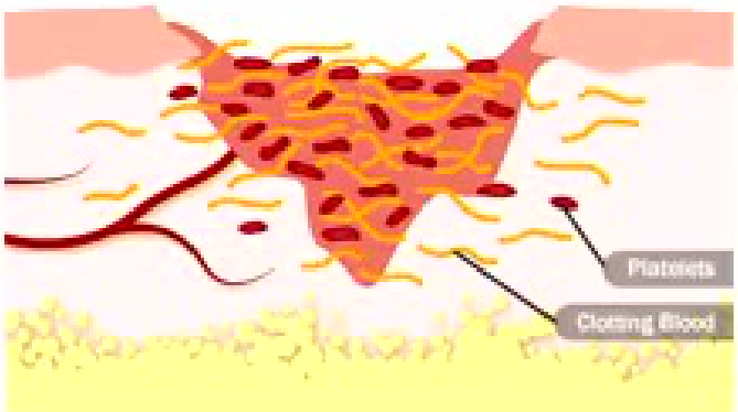
Stage 1: Bleeding Stops43
Stages of Wound Healing
-
Blood vessels contract
-
Platelets migrate to wound
-
Clot forms in seconds to minutes
-
Clotting also helps close and heal the wound
-
The clotting cascade follows the initial platelet plug.
-
Aspirin and clopidogrel affect platelet function.
-
Oral anticoagulants slow the clotting process.
-
Patients with factor V Leiden or antiphospholipid syndrome may be more likely to clot.
-
Saline during extraction helps wash away small pieces of bone, debris, tartar, and plaque.
-
The initial clot should be left undisturbed; patients should chew on the opposite side.
-
Losing the clot early increases the risk of dry socket.
-
Rinsing and cleaning instructions should generally begin the following day.
-
For continued bleeding, consider whether the patient is a bleeder, whether tranexamic acid is required, and whether packing and suturing are sufficient.
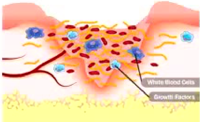
Stage 2: Inflammation44
- Kinins released
- White blood cells migrate to wound to remove bacteria
- Growth factors released to help repair
Lecturer — Wound Infection
Healing depends on the local condition of the wound.
- A dirty or infected wound will not heal well and may need to be cleaned again.
- After haemostasis, macrophages and other inflammatory factors move to the injury, followed later by anti-inflammatory processes.
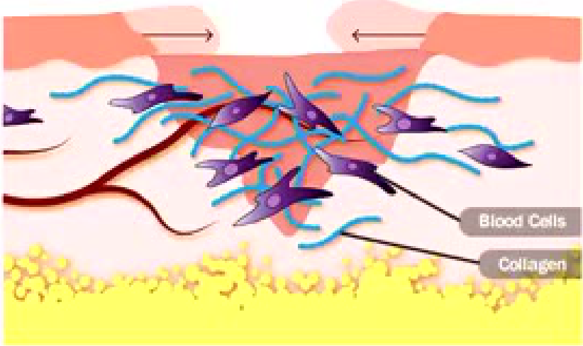
Stage 3: Rebuilding (Growth and Proliferation)45
- Oxygen and nutrients carried to wound site to heal and grow new blood vessels
- Collagen laid down
Lecturer — Oral and Bone Healing
Oral mucosa and bone follow the same broad sequence as other wounds.
- The sequence is haemostasis, inflammation, proliferation, and maturation.
- Bone healing also follows this sequence.
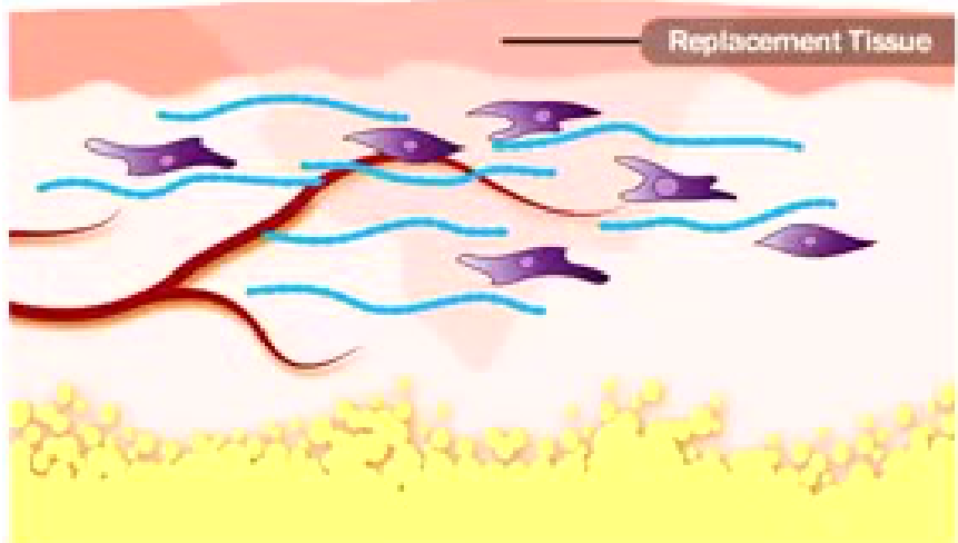
Stage 4: Maturation46
- Replacement tissue takes hold
- Healing can take days, months, even years
Lecturer — Extraction Socket
An extraction socket is not mature shortly after extraction.
- Much of the early healing takes approximately two weeks.
- Further regeneration and maturation continue after this initial period.
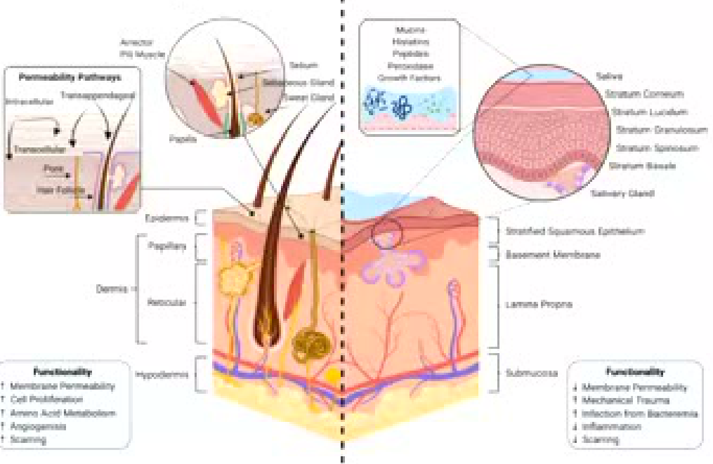
Cutaneous Epithelium vs. Oral Mucosa47
¹Cutaneous Epithelium
- Arrector Pili Muscle
- Sebum
- Sebaceous Gland
- Sweat Gland
- Papilla
- Hair Follicle
- Endodermis
- Papillary Dermis
- Reticular Dermis
- Hypodermis
- Permeability Pathways: Intracellular, Transappendageal, Transcellular, Pores
- Stratum Corneum, Stratum Lucidum, Stratum Granulosum, Stratum Spinosum, Stratum Basale
Functionality:
- ↑ Membrane Permeability
- ↑ Cell Proliferation
- ↑ Amino Acid Metabolism
- ↑ Angiogenesis
- ↑ Scarring
²Oral Mucosa
- Mucin
- Hyaline
- Peptides
- Protease
- Growth Factors
- Saliva
- Salivary Gland
- Stratified Squamous Epithelium
- Basement Membrane
- Lamina Propria
- Submucosa
Functionality:
- ↓ Membrane Permeability
- ↓ Mechanical Trauma
- ↑ Infection from Bacteremia
- ↓ Inflammation
- ↓ Scarring
Oral Wound Healing
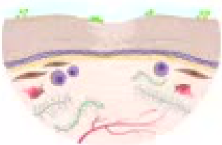
Phases of Oral Wound Healing48
¹Hemostasis — Minutes
- Platelet aggregation
- Neutrophil infiltration
- Inflammatory cell recruitment
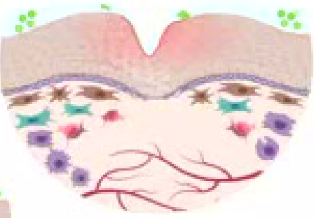
²Inflammation — Days
- Neutrophil apoptosis
- Macrophage migration
- Angiogenesis
- Granulation
- Fibroblast proliferation
³Proliferation
⁴Maturation — Weeks
-
Re-epithelialization
-
Decreased blood vessels
-
Cellular apoptosis
-
Aligned ECM fibrillar network
-
Dense Type I collagen formation
-
An extraction socket initially contains a haematoma, which is replaced before bone formation occurs.
-
The process continues after the first two weeks and requires further maturation.
 |  |
 |  |
Legend
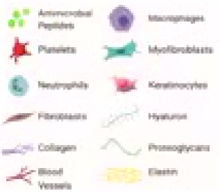
Antimicrobial Peptides, Platelets, Neutrophils, Fibroblasts, Collagen, Blood Vessels, Macrophages, Myofibroblasts, Keratinocytes, Hyaluron, Proteoglycans, Elastin
Oral Mucosa Healing49
- Mucosa re-epithelialized within 2 weeks, but the regeneration of the lamina propria is incomplete
Lecturer — Oral Wound Environment
Oral mucosa heals in layers that differ from those of skin, and a small wound may heal successfully when the local environment is clean and healthy.
- Loose bone fragments can interfere with healing.
- If a loose sequestrum is present, it may be removed with a curette, mouth rinses should continue, and the patient should gently agitate the edges to encourage the gum to grow back.
Metabolic Response to Injury
Injury → Shock → Catabolic phase (1 week) → high metabolic rate, breakdown of proteins and fats, negative nitrogen balance and weight loss.
Overview and Mediating Factors5051
Anabolic phase (2–4 weeks) → protein and fat stores are restored and weight gain occurs (positive nitrogen balance).
- Acute inflammatory response
- Endothelium and blood vessels
- Neuroendocrine response
- Bacterial infection
Lecturer — Injury Response Range
Surgery is a form of trauma, so the physiological response to surgery is also a response to injury. The response can follow injuries ranging from a small extraction or biopsy to major trauma or major surgery, and the magnitude varies with the size of the injury.
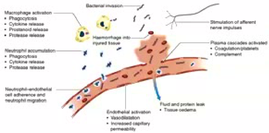
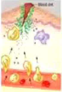
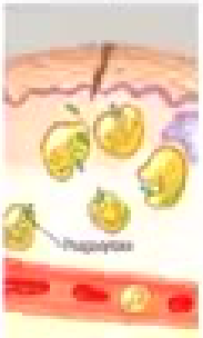
Acute Inflammatory Response5253
-
Injury results in local activation of e.g. macrophages which release cytokines, recruiting macrophages and neutrophils to the site to clear dead tissue and kill bacteria
-
Release of cytokines into circulation initiates systemic features e.g. fever (IL-1) and acute phase protein (IL-6)
-
Other pro-inflammatory e.g. prostaglandins, kinins, complement, proteases and free radicals
-
Anti-inflammatory e.g. antioxidants, protease inhibitors and IL-10, also released
-
Clinical condition of patient depends on extent to which inflammation remains localized and balance between pro- and anti-inflammatory processes
Lecturer — Wound Healing Conditions
The condition of the wound affects the quality of healing. Persistent dirt, debris, or infection can delay healing.
 |  |  |
 |  |
Endothelium and Blood Vessels54
-
Expression of adhesion molecules upon endothelium leads to leucocyte adhesion
-
Release of kinins, prostaglandins and nitric oxide, leads to vasodilatation and increased local blood flow, and increased capillary permeability leads to delivery of inflammatory cells, oxygen and nutrient substrates which are important for healing
-
Oedema results from colloid particles (albumin) leaked into injured tissues
-
Exposure of tissue factor promotes coagulation and together with platelet activation, decreases haemorrhage
-
The initial vascular response includes vessel contraction and platelet activity, followed later by clotting and tissue repair.
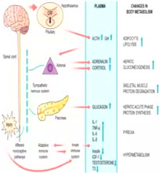
Neuroendocrine Response5556
-
Tissue injury and inflammation leads to impulses in afferent pain fibres that reach the thalamus and mediate metabolic response
-
Activation of sympathetic nervous system leads to release of noradrenaline from sympathetic nerve fibre endings and adrenaline from adrenal medulla
-
This results in tachycardia, increased cardiac output, and carbohydrate, fat and protein metabolism
-
Changes in the circulating concentration of hormones following injury, either by direct gland stimulation or feedback mechanisms
-
Increased secretion of stress hormones
-
Decreased secretion of anabolic hormones
Lecturer — Protective Neuroendocrine Response
The nerve supply and sympathetic response are protective. A major injury may produce increased respiration, tachycardia, and reduced blood pressure; the overall response attempts to restore normality by increasing blood pressure, conserving energy, and providing substrates for repair.
Consequences of Metabolic Response57
- Hypovolaemia
- Increased energy metabolism and substrate cycling
- Catabolism and starvation
- Changes in red blood cell synthesis and coagulation
- Anabolism
Hypovolaemia5859
-
Loss of blood, fluid or water
-
Sequestration of protein-rich fluid into interstitial space, due to increased vascular permeability
-
Decreased circulating volume will reduce oxygen and nutrient delivery and increase healing and recovery time
-
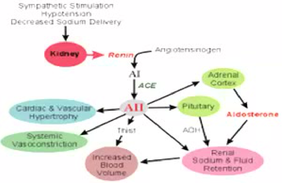
Progressively significant physiological responses were described after losses of approximately 15%, 30%, and 40% of blood volume. Renin-angiotensin-aldosterone system:
-
Triggers: sympathetic stimulation, hypotension, decreased sodium delivery
-
Kidney releases renin → angiotensinogen → AI → (ACE) → AII
-
AII acts on adrenal cortex (via pituitary, TRH, ADH) → aldosterone
-
Effects: systemic vasoconstriction, cardiac and vascular hypertrophy, renal sodium and fluid retention → increased blood volume
Increased Energy Metabolism and Substrate Cycling60
-
Energy for physical work, generate heat, meet basal metabolic requirements
-
Basal metabolic rate (BMR) = energy required for maintenance of membrane polarization, substrate absorption and utilization, mechanical work of heart and respiratory systems
-
Following surgery, overall energy expenditure rise by 50% due to increased generation of heat, fever and BMR
-
The aim of the catabolic and anabolic phases is to replace protein and fat used during the initial response.
Catabolism and Starvation
Catabolism61
-
Breakdown of complex substances to glucose, amino acids and fatty acids
-
Mediated by increase in catecholamines and cytokines and hormones
-
Total energy expenditure increased in proportion to injury severity
-
Reduction in fat and muscle mass until stimulus for catabolism ends
-
The catabolic response occurs after both small and large injuries, although its scale differs.
Starvation
- Occurs when intake less than metabolic demand
- Acute starvation
- Chronic starvation
Lecturer — Catabolism and Equilibrium
Catabolism and starvation were distinguished in the discussion of the body’s response to injury. The body subsequently attempts to restore equilibrium and replace the protein and fat used during healing.
Factors Affecting Magnitude of Response
Patient factors62
- Genetic predisposition
- Coexisting disease
- Drugs
- Nutritional status
Lecturer — Patient Healing Factors
Healing is affected by diabetes, immunosuppression, and the patient’s general systemic condition. Diabetes and immunosuppression may impair healing and increase infection risk.
Trauma factors
- Severity
- Nature
- Ischaemia-reperfusion
- Temperature
- Infection
- Anaesthetic techniques
Lecturer — Socket Healing Conditions
Local wound conditions are important after extraction. A dirty or infected wound may require further cleaning, and loose bone fragments may delay socket healing; the socket should be clean and free of debris, plaque, tartar, and loose bone.
Red Blood Cell Synthesis and Coagulation63
-
Anaemia common after major surgery or trauma
-
Following tissue injury, blood hypercoagulable and increase risk of thromboembolism
-
Aspirin and clopidogrel affect platelets, oral anticoagulants slow coagulation, and Factor V Leiden and antiphospholipid syndrome are associated with increased clotting.
Anabolism64
-
Anabolism involves:
- regaining weight
- restoring skeletal muscle mass
- replenishing fat stores
-
Hormones include insulin, growth hormone, insulin-like growth factors and androgens
-
Hospitalized patients may receive heparin because reduced mobility makes them more prone to clotting.
-
During the anabolic phase, protein and fat are laid down again, so weight and energy stores begin to return.
Effects of Surgery and Nutritional Support6566
-
Stress response to surgery – release of ADH and catecholamines and activation of renin-angiotensin system, resulting in oliguria and water retention
-
Potassium released by damaged tissues. If renal perfusion is poor and urine output poor, potassium not excreted and accumulate. Resultant hyperkalaemia causes arrhythmias
-
Majority of patients require fluid replacement for a brief period postoperatively until normal diet resumed
-
In severely ill patients and those with impaired gastrointestinal function, long-term nutritional support necessary
Lecturer — Postoperative Oral Hygiene
Nutritional support may be needed after surgery, including an NG feed. Patients with a broken jaw or intermaxillary fixation may have difficulty cleaning around the fixation, requiring assistance and increasing their susceptibility to infection.
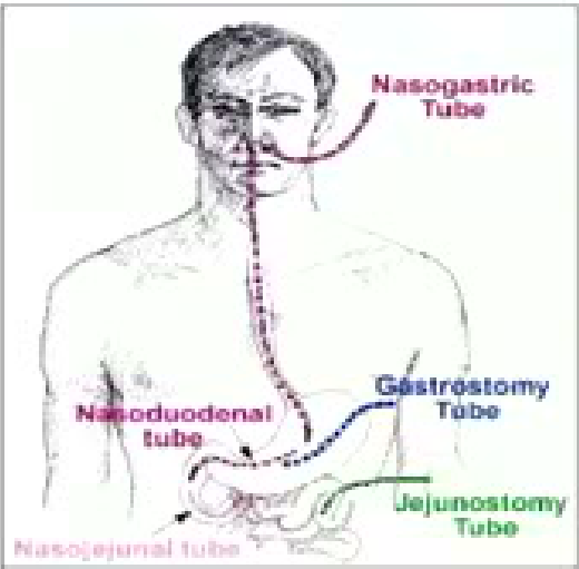
Enteral feeding67
- Nasogastric tube
- Gastrostomy tube
- Jejunostomy tube
- Nasoduodenal tube
- Nasojejunal tube
 |  |
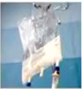 |
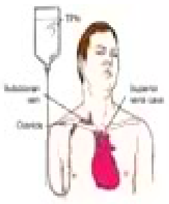
Total parenteral nutrition
Infections and Antibiotics
Development of Infection
-
When normal defences (e.g. skin) are breached, commensals can cause infection
-
Infection = proliferation of micro-organisms in body tissue with adverse physiological consequences
-
A swab may be useful when an infection has recurred or failed to respond to previous treatment.
Factors Important in the Development of Infection68
- Host defence system — physical barriers, antagonism by commensals, antibacterial substances, immune state, acquired immunity, innate immunity
- Bacterial factors — virulence, inoculum, growth rate
- Insults — foreign body, necrosis, ischemia, haematoma
- Local factors include dirt, debris, loose bone, plaque, tartar, and persistent infection.
- Patient factors include diabetes, immunosuppression, and poor healing.
- Surgical factors include extensive bone removal, procedures involving bone drilling, and major surgery.
- Recurrent infection may require consideration of anaerobic organisms.
Preventing Infection69
Minimize risks to patients and staff from infections acquired during or after surgery:
- Aseptic technique
- Hand decontamination
- Personal protective equipment (PPE)
- Skin preparation
- Surgical instruments
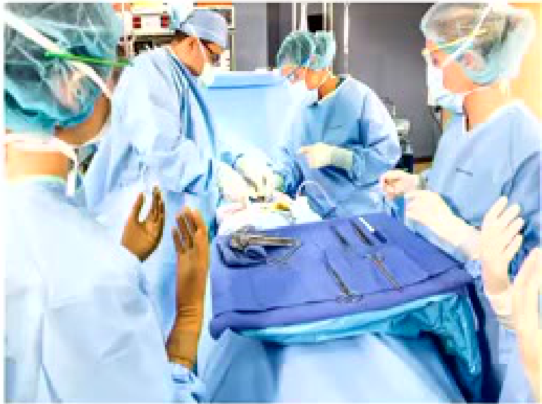
Aseptic Technique70
Specific practices immediately before and during surgery to reduce postoperative infection:
-
Hand washing
-
Surgical scrub
-
Skin preparation of patient
-
Maintaining sterile field
-
Safe operating practices
-
In theatre, only sterile items should be touched.
-
Protective glasses or a face shield should be worn.
-
During extraction, saline can be used to wash away small bone fragments and other debris.
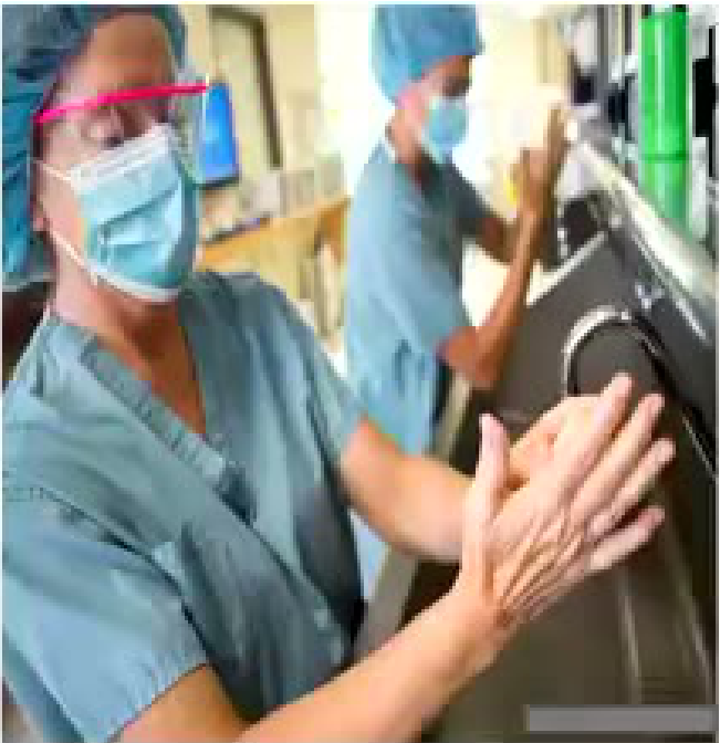
Hand Decontamination71
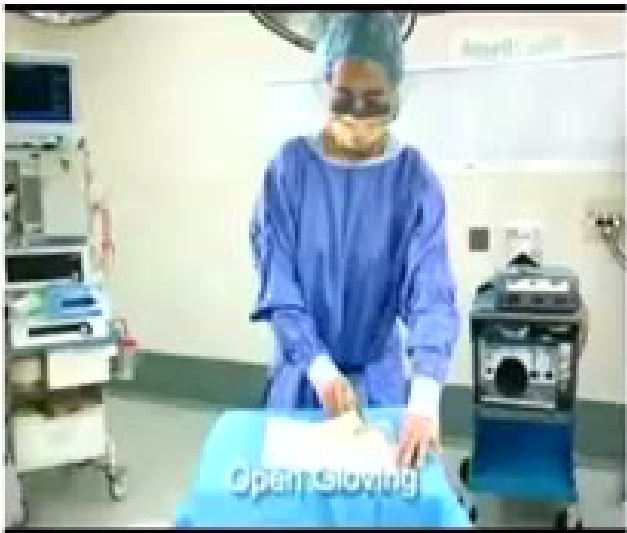
Personal Protective Equipment72
-
Cap
-
Face mask
-
Eye protection
-
Sterile gown and gloves
-
Protective glasses or a face shield should be used.
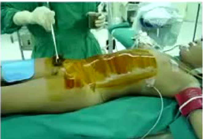
Skin Preparation73
Antiseptics (e.g. chlorhexidine or povidone-iodine) applied to the surgical site prior to incision reduce the number of resident organisms and the risks of wound infection.
Surgical Instruments74
-
Prevent cross-infection
-
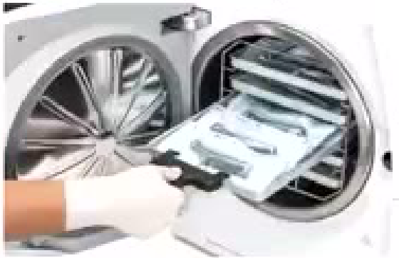
Surgical instruments are washed in washer disinfectors to remove organic matter and kill most micro-organisms except spores
-
Instruments are then packed and autoclaved to destroy any remaining micro-organisms and spores
-
Instruments should be cleaned and handled using an aseptic technique.
Prophylactic Use of Antibiotics7576
-
Timing and dose
-
Choice of antibiotic
-
Carriage of resistant organisms and prophylaxis
-
Prophylaxis for immunosuppressed patients
-
Antibiotic prophylaxis aims to prevent infection by achieving high concentrations of antibiotic during surgery
-
Choice of antibiotic must cover likely pathogens
-
Single dose of antibiotics usually adequate for prophylaxis
-
In patients with multi-resistant bacteria (e.g. carriage of MRSA or ESBL-producing E. coli) or patients who are immunocompromised, the antibiotic choice may need to be modified and expert advice sought
-
Prophylactic antibiotics may be considered when bone will be drilled, extensive bone removal is planned, or the wound is likely to become infected.
-
They may also be considered when the patient is diabetic, immunosuppressed, or unlikely to heal well.
-
Amoxicillin was described as usually sufficient.
-
Cefalexin may be used in patients who are allergic to amoxicillin.
-
Erythromycin may be considered when the patient cannot tolerate cefalexin.
-
Doxycycline was mentioned in relation to an AVS tooth.
-
Metronidazole may be added to cover anaerobes.
Prophylaxis in Dental Surgery77
- Low risk of wound infection balanced against risk of adverse effects and risk of developing resistant organisms
- For most dentoalveolar procedures in fit immunocompetent patients, antibiotic prophylaxis is not required
- Consider antibiotic prophylaxis for:
- Substantial bone removal or drilling
- Maxillofacial surgery
- Surgery in patients with recurrent infections
- Dentoalveolar surgery in immunocompromised patients
There is limited evidence supporting the efficacy of commonly used antibiotics in preventing complications after lower third molar removal.
- Antibiotic prophylaxis should be considered for substantial bone removal or drilling and for patients with diabetes or immunosuppression.
- Antibiotics are not automatically required for every dental procedure; the patient and procedure must be considered.
Principles of Antibiotic Use78
-
Microbiology guides therapy
-
Indications should be evidence-based
-
Narrowest spectrum required
-
Dosage appropriate to site and type of infection
-
Minimise duration of therapy
-
Ensure monotherapy in most situations
-
A recurrent infection that has not responded to amoxicillin may involve a different organism.
-
Antibiotics should be used alongside appropriate cleaning and treatment of the source of infection.
Postoperative Care and Day Surgery
- Clinical assessment and monitoring — assess patient’s colour, pulse, blood pressure, respiratory rate, oxygen saturation, level of consciousness
- Respiratory management — maintenance of airway and adequate ventilation
- Cardiovascular management — monitor for blood loss
- Fluid, electrolyte and renal management
- Control of sepsis
- Nutrition
Postoperative Care79
Lecturer — Extraction Aftercare
After extraction, protect the clot and assess persistent bleeding.
- Leave the clot undisturbed and have the patient chew on the opposite side.
- If an oroantral communication is suspected, do not probe it or ask the patient to perform a Valsalva manoeuvre.
- Place a stitch with the edges as close together as possible, prescribe antibiotics, advise the patient not to blow their nose, and review after approximately one or two weeks.
- A small communication may close spontaneously, whereas a larger opening may require formal repair.
- Advise patients not to use straws, smoke, go underwater, dive, fly, or increase pressure in the area.
- Swimming followed by forceful nose clearing was described as preventing one communication from healing.
[!note] Lecturer — Retained Root Management
Imaging helps determine whether a suspected retained root should be removed and how to approach it.
- Use an X-ray, preferably cone-beam CT, to localize the root and show its length; a long root may track forward from another tooth.
- A small, asymptomatic root tip may be left, but a root associated with pus or ongoing problems may need removal.
- Raise a palatal flap rather than simply making a hole through the palate, with imaging guiding whether bone must be removed.
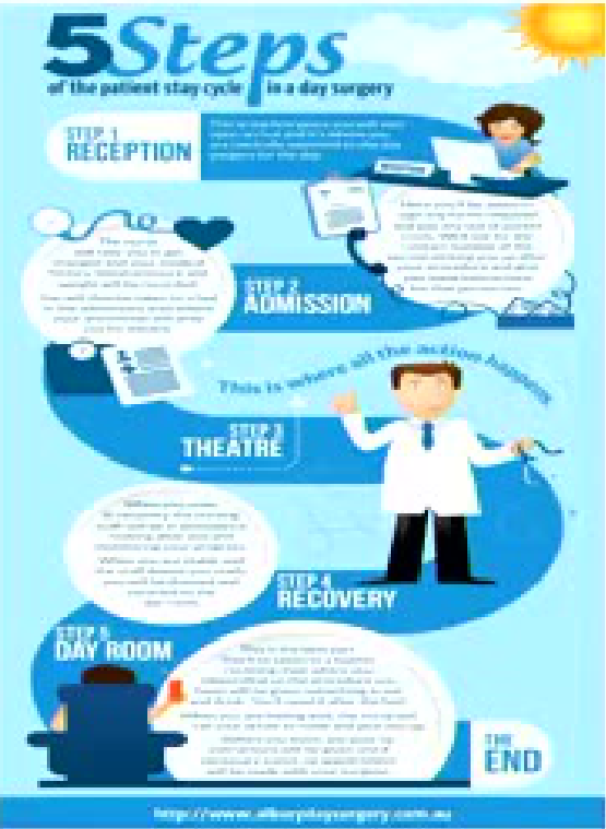
Day Surgery808182
-
Surgical procedure performed on a day-case basis
-
Well-defined and streamlined pathway
-
Minimizes risk of hospital acquired infection
-
Reduces risk of venous thromboembolism
-
Economic benefit
-
Maximize surgical throughput
-
Criteria based pre-assessment reduce unnecessary investigations
-
Protocol-based discharge with comprehensive patient information reduces unplanned hospital readmissions
-
Wisdom tooth removal was given as a common example of day surgery.
 |  |
 |
Patient Selection83
- Efficient and safe
- Only specified patients accepted for operation
Obesity Criteria
- Body Mass Index (BMI) = kg/m²
- Obese if BMI > 30
- Risks of obesity include morbidities of diabetes and obstructive sleep apnoea
Lecturer — Obesity and Anaesthesia
Day surgery services may apply strict body-mass criteria because anaesthetic gas can behave differently in patients with a high amount of fat.
- Gas may remain in the fat and be released later, increasing concern about discharge and readmission.
- Some services also consider whether a patient is very tall or very muscular rather than relying only on body-mass index.
ASA Status
Anaesthesiologists’ Physical Status Classification System (ASA status). Day units accept:
-
ASA I (normal healthy)
-
ASA II (mild systemic disease e.g. well controlled non-insulin dependent diabetes, mild hypertension, epilepsy or asthma)
-
Patients with more serious systemic disease may require treatment in a hospital with intensive-care facilities.
-
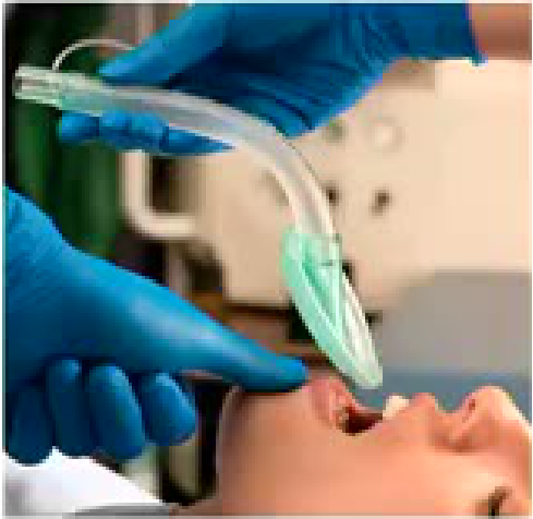
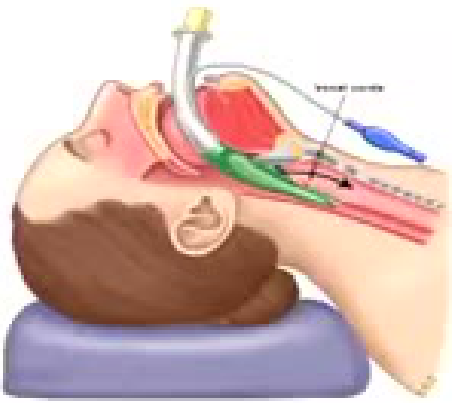
Laryngeal mask airway
Lecturer — Laryngeal Mask Airway
A laryngeal mask sits at the back of the throat rather than entering the trachea.
- The patient does not need to be paralysed, can continue breathing, and may wake relatively quickly.
- Admission for surgery
- Anaesthesia
- Analgesia
- Recovery
- Discharge criteria
Clinical Cases
Case: Chin abscess traced with a gutta percha point
Question
Scenario: A patient presents with an abscess on the chin.
What’s shown: A clinical photograph of the lower face showing an abscess on the chin, alongside a composite image.
Consider: How to determine the source of the chin abscess and differentiate it from a skin boil.
Answer
Observations:
- An abscess is present on the chin.
- A gutta percha (GP) point is inserted into the fistula or track.
- A radiograph is taken with the GP point in place.
Reasoning: The GP point traces the path of the pus escaping from the necrotic tooth. The radiograph reveals the exact tooth involved by following the track. In this specific case, although it might initially be suspected to be a root-filled tooth, the GP point points to the adjacent tooth.
Outcome: The correct source tooth is identified for targeted treatment.
Takeaway: Using a gutta percha point to trace a fistula is an effective way to identify the source of an abscess and ensure an accurate diagnosis.
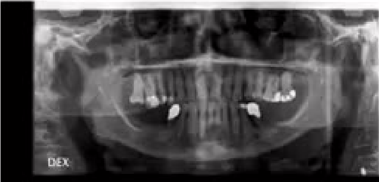
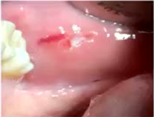
Case: Mandibular fracture suspected from a gingival tear
Question
Scenario: A patient presents with clinical signs that might indicate a facial fracture.
What’s shown: An intraoral clinical photograph showing a tear in the gingiva, and a panoramic radiograph (OPG).
Consider: What subtle clinical clues might indicate a hard-to-spot fracture, and what does the radiograph reveal?
Answer
Observations:
- There is a tear in the gingiva.
- The OPG shows a small crack between the first and second premolars (teeth 4 and 5).
- Localized bruising may also be present.
Reasoning: A tear in the gingiva can be a subtle clue of an underlying fracture. The OPG confirms the presence of a fracture line between the 4 and 5. The lecturer also notes that a lingual hematoma is another mnemonic clue for such injuries.
Outcome: The fracture is identified on the OPG.
Takeaway: Subtle clinical signs like a gingival tear or localized bruising should prompt radiographic investigation to rule out a fracture.
Case: Bilateral salivary gland swelling
Question
Scenario: A lady presents with swollen salivary glands.
What’s shown: A clinical photograph of the lower face of a patient.
Consider: What is the likely cause of bilateral salivary gland swelling, and how should the condition be managed?
Answer
Observations:
- Bilateral swelling of the salivary glands.
- The patient also reports symptoms of dry eyes.
Reasoning: Bilateral swelling is unlikely to be neoplasia, which typically presents as a unilateral growth. The bilateral nature, along with dry eyes, points towards an autoimmune condition such as Sjogren’s syndrome. The resulting dry mouth can lead to an increased caries rate.
Outcome: Management involves patient-centered care, such as using saliva stimulants, monitoring for and treating cervical caries, and scheduling more frequent reviews.
Takeaway: Bilateral salivary gland swelling with dry eyes suggests an autoimmune condition like Sjogren’s syndrome, requiring management of the secondary effects of dry mouth, such as increased caries risk.
Case: Child with a gum boil and decayed primary incisors
Question
Scenario: A child presents with a swelling on the gum.
What’s shown: Clinical intraoral photograph of the maxillary anterior teeth and a close-up of the child’s face.
Consider: Identify the source of the infection and the appropriate management.
Answer
Observations:
- Swelling on the gum indicating pus trying to escape.
- Severely decayed primary incisors (A’s and B’s).
Reasoning: The infection is likely originating from one of the severely decayed teeth, which needs to drain.
Outcome: No referral to oral medicine is needed; the source tooth must be treated to allow drainage.
Takeaway: A gum boil indicates pus trying to escape from an infected tooth, which should be treated directly rather than referred.
Case: Salivary duct stone and blocked duct
Question
Scenario: A patient presents with an inflammatory salivary gland issue.
What’s shown: Dental radiograph (likely an occlusal view) and clinical photographs of the lower jaw and intraoral views.
Consider: Identify the radiographic and clinical findings and the diagnosis.
Answer
Observations:
- Radiograph shows a stone in the salivary duct.
- Clinical photos show a blocked duct with pus coming out.
Reasoning: The presence of a stone on the X-ray and pus discharge from the blocked duct indicates an inflammatory condition of the salivary duct.
Outcome: Diagnosis of a salivary duct stone and blockage.
Takeaway: Salivary duct stones can often be visualized on an X-ray, and a blocked duct may present with pus discharge.
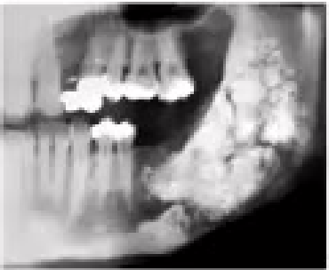
Case: Patient with a loose 44 and multilocular radiolucency
Question
Scenario: A patient requests the extraction of a very loose lower right first premolar (44).
What’s shown: Panoramic radiograph (OPG) showing the maxilla and mandible.
Consider: Determine the diagnosis based on the radiograph and the appropriate management for the loose tooth.
Answer
Observations:
- OPG shows a multilocular radiolucency around the loose tooth.
Reasoning: The multilocular appearance suggests an arteriovenous (AV) malformation rather than just periodontal disease. Extracting the tooth would cause severe, life-threatening bleeding.
Outcome: Do not extract the tooth. Refer the patient for a CT scan (as CBCT lacks sufficient resolution) to confirm the AV malformation.
Takeaway: Always take an X-ray before extracting a loose tooth to rule out serious underlying pathology like an AV malformation, which would contraindicate extraction and require a CT scan.
Case: Lip cancer from sun-damaged skin
Question
Scenario: A patient presents with a lesion on the lip.
What’s shown: Clinical photos of the lower lip showing a large, ulcerated lesion, and an intraoral surgical field.
Consider: Identify the clinical features of the lesion and the diagnosis.
Answer
Observations:
- The lip lesion has indurated edges.
- Surgical photos show the lesion excised with a margin, allowing the lip to be brought together.
Reasoning: The indurated edges and history of sun-damaged skin indicate a lip cancer.
Outcome: The cancer was surgically excised with a margin, and the lip was reconstructed.
Takeaway: Lip cancer often arises from sun-damaged skin and presents with indurated edges, requiring surgical excision with clear margins.
Case: Oral cancer in a smoker and drinker
Question
Scenario: A patient with a history of smoking and drinking presents with an oral lesion.
What’s shown: Intraoral clinical photograph of the anterior maxilla.
Consider: Identify the clinical features of the lesion and the likely diagnosis.
Answer
Observations:
- The patient is missing teeth 21 and 22.
- The area has a heaped, indurated, and florid growth that looks abnormal.
Reasoning: The heaped, indurated growth in a patient with a history of smoking and drinking is characteristic of oral cancer.
Outcome: Diagnosis of oral cancer.
Takeaway: Oral cancer can present as a heaped, indurated, and florid growth, particularly in patients with a history of smoking and alcohol use.
Case: Buccal mucosa cancer from betel nut chewing
Question
Scenario: A patient who chews betel nut presents with a buccal mucosal lesion.
What’s shown: Intraoral clinical photograph of the buccal mucosa.
Consider: Identify the clinical features of the lesion and the diagnosis.
Answer
Observations:
- The lesion on the buccal mucosa is indurated and ulcerated with central ulceration.
- It exhibits speckled leukoplakia (bits of white and red).
Reasoning: Betel nut chewing is frequently associated with buccal mucosa cancer. The indurated, non-healing ulcer with speckled leukoplakia is characteristic of malignancy.
Outcome: Diagnosis of buccal mucosa cancer.
Takeaway: Betel nut chewers are at high risk for buccal mucosa cancer, which presents as an indurated, non-healing ulcer with speckled leukoplakia.
Case: Diabetic patient with a dry, caked tongue
Question
Scenario: A diabetic patient presents for a dental examination.
What’s shown: Intraoral and clinical photographs of the oral cavity, tongue, and a composite image.
Consider: Identify the oral manifestations visible in the images.
Answer
Observations:
- The tongue appears caked and dry.
Reasoning: Diabetic patients are more prone to oral infections like candida, periodontal disease, and dry mouth, which manifests as a caked and dry tongue.
Outcome: The patient exhibits oral manifestations of diabetes, including a dry, caked tongue and increased risk of candida and caries.
Takeaway: Diabetic patients are prone to oral infections like candida, periodontal disease, and dry mouth, which can present as a caked and dry tongue.
Case: Medication-related osteonecrosis of the jaw
Question
Scenario: A patient on Prolia presents with exposed bone in the jaw.
What’s shown: Intraoral clinical photograph showing exposed bone.
Consider: Identify the condition and the appropriate management for the exposed bone.
Answer
Observations:
- The mucosa has broken down, exposing necrotic, dead bone.
Reasoning: The patient is on Prolia, which predisposes them to medication-related osteonecrosis. The exposed bone is necrotic and needs to be managed to allow the gum to heal.
Outcome: Management involves feeling for loose bits of bone (sequestrum) and removing them with a curette, followed by encouraging the patient to use mouth rinses and gently agitate the edges to promote gum healing.
Takeaway: Medication-related osteonecrosis presents as exposed necrotic bone; management involves removing loose sequestra with a curette and using mouth rinses to encourage soft tissue healing.
Case: Osteoarthritis of the temporomandibular joint
Question
Scenario: A patient presents with temporomandibular joint issues.
What’s shown: Panoramic radiograph (OPG) of the maxillofacial region.
Consider: Identify the radiographic findings in the temporomandibular joints.
Answer
Observations:
- The OPG shows that both temporomandibular joints are severely worn down.
Reasoning: The worn-down appearance of the joints on the radiograph is indicative of osteoarthritis.
Outcome: Diagnosis of osteoarthritis in the temporomandibular joints.
Takeaway: Osteoarthritis of the temporomandibular joint can be identified on an OPG by observing severely worn-down joint spaces.
Case: Subcutaneous emphysema from hypochlorite irrigation
Question
Scenario: A patient suddenly develops swelling during a root canal treatment.
What’s shown: Frontal portrait, low-angle view, and intraoral photograph of a patient.
Consider: Identify the cause of the swelling and the immediate management.
Answer
Observations:
- The clinical photos show a patient with significant facial swelling.
Reasoning: The swelling is caused by subcutaneous emphysema resulting from the accidental extrusion of sodium hypochlorite into the peri-radicular soft tissues during canal irrigation.
Outcome: Immediate management is to stop irrigating with hypochlorite immediately to prevent forcing more chemical into the circulation, and then flush the area with saline.
Takeaway: Accidental extrusion of sodium hypochlorite during root canal irrigation can cause subcutaneous emphysema and severe swelling; immediate management is to stop irrigation and flush with saline.
Case: Cleft lip and palate patient
Question
Scenario: A patient with a cleft lip and palate is presented to discuss the dental and bony findings in the cleft area and the management options.
What’s shown: Clinical photographs of a patient with a cleft lip and palate, including views of the maxillary dental arch and intraoral structures.
Consider: What findings are present in the cleft area regarding the teeth and surrounding bone, and what are the management options?
Answer
Observations:
- A tooth is located within the cleft gap.
- There is very little or no bone surrounding the tooth.
Reasoning: Due to the lack of supporting bone and potential tooth deformity, a decision must be made regarding the tooth’s management.
Outcome: Management options include extracting the deformed tooth or bringing it down into the arch with bone grafting, as is done at the children’s hospital.
Takeaway: Teeth located within a cleft gap often lack surrounding bone, requiring a decision between extraction or orthodontic alignment combined with bone grafting.
Case: Syndromic craniofacial abnormality patient (Class III)
Question
Scenario: A female patient with syndromic craniofacial abnormalities is presented, highlighting her occlusal and skeletal features.
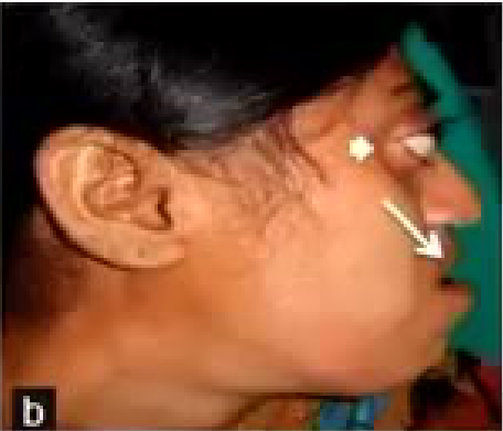
What’s shown: Extraoral and intraoral clinical photographs showing a lateral profile view and the upper and lower dental arches.
Consider: What are the key skeletal and dental findings in this patient?
Answer
Observations:
- Malocclusion with jumbled teeth, making oral hygiene difficult.
- Class III skeletal relationship with the lower jaw positioned further forward than the upper jaw.
- A very hypoplastic maxilla.
Reasoning: The combination of a hypoplastic maxilla and a relatively forward-positioned mandible results in a Class III malocclusion and significant dental crowding.
Outcome: The patient is diagnosed with a Class III malocclusion secondary to a hypoplastic maxilla and syndromic craniofacial abnormalities.
Takeaway: Syndromic craniofacial abnormalities can present with severe skeletal discrepancies, such as a hypoplastic maxilla leading to a Class III malocclusion and significant dental crowding.
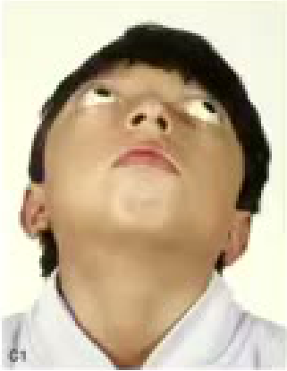
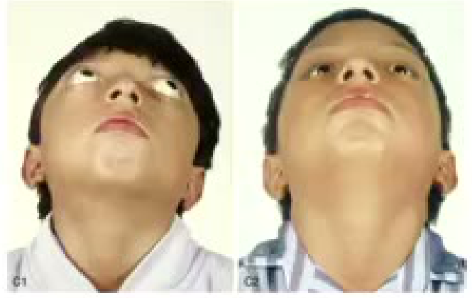
Case: Child with syndromic craniosynostosis undergoing skull scoring
Question
Scenario: A young child with a syndromic syndrome that affects skull growth and retards brain growth is presented.
What’s shown: Clinical photographs of a young child with craniofacial abnormalities and an intraoperative photograph showing a surgical procedure on the skull.
Consider: What surgical intervention is being performed, and what is its physiological purpose?
Answer
Observations:
- The intraoperative image shows the surgical scoring of the cranial bone.
Reasoning: Scoring the bone (similar to an escharotomy for soft tissue) releases the restriction, allowing the skull to expand and the brain underneath to grow.
Outcome: The skull was scored to allow for brain growth.
Takeaway: In syndromic conditions where skull growth is restricted and retards brain development, scoring the cranial bone can be performed to allow for brain expansion.
Case: Hemifacial microsomia, facial asymmetry
Question
Scenario: A patient presenting with facial asymmetry.
What’s shown: Clinical photographs showing facial asymmetry.
Consider: Identify the clinical findings and the required treatment approach.
Answer
Observations:
- A downward slant and cant of the smile line.
- Asymmetric growth where one side of the upper and lower jaw has grown more than the other.
Reasoning: The facial asymmetry and cant are caused by unilateral overgrowth of the jaws. To correct the functional and aesthetic discrepancies, both the upper and lower jaws must be addressed.
Outcome: Correction of the upper and lower jaw.
Takeaway: Hemifacial microsomia causes asymmetric jaw growth resulting in a cant and slanted smile line, requiring orthognathic correction of both jaws.
Case: Supernumerary tooth blocking eruption
Question
Scenario: A patient with concerns about tooth development.
What’s shown: An upper occlusal radiograph.
Consider: Identify the radiographic finding causing the developmental issue and the appropriate management.
Answer
Observations:
- A supernumerary tooth is visible on the radiograph.
- The supernumerary tooth is blocking the growth and eruption of the central incisors.
Reasoning: The supernumerary tooth is physically obstructing the normal eruption path of the adjacent permanent teeth.
Outcome: Referral for the removal of the supernumerary tooth.
Takeaway: A supernumerary tooth can block the eruption of adjacent teeth and should be identified on radiographs and removed.
Case: Prominent mandibular tori requiring pre-prosthetic surgery
Question
Scenario: An edentulous patient requiring dentures.
What’s shown: Clinical photographs of the mandibular ridge and excised bony specimens.
Consider: Identify the anatomical variation interfering with prosthetics and the surgical management required.
Answer
Observations:
- Prominent bony lumps (tori) on the mandibular ridge.
- Excised bony specimens following surgery.
Reasoning: The prominent tori interfere with the fitting, stability, and comfort of dentures. Removing them creates a smooth, continuous ridge suitable for prosthetic rehabilitation.
Outcome: The mucosa was stripped back and the tori were smoothed off to allow for denture fabrication.
Takeaway: Prominent tori can interfere with denture placement and may require pre-prosthetic surgical removal to ensure a proper fit.
Case: Maxillary ridge defect requiring skin graft
Question
Scenario: A patient presenting with a severe maxillary ridge defect.
What’s shown: Clinical photographs showing a missing midline ridge and the post-operative result.
Consider: Identify the cause of the defect and the surgical reconstruction method used.
Answer
Observations:
- Absence of the alveolar ridge in the midline.
- The labial sulcus was previously stitched directly to the defect gap.
Reasoning: The previous surgical approach created a defect and reduced vestibular depth. To restore a continuous ridge and provide adequate room for a denture, the defect needs to be augmented and the sulcus deepened.
Outcome: The previous suturing was reversed, and a skin graft taken from the neck was used to plumb up the alveolus, resulting in a continuous ridge and adequate room for a denture.
Takeaway: Severe ridge defects with loss of vestibular depth can be reconstructed using a skin graft to restore alveolar volume and denture space.
Footnotes
-
Original PDF page 1: Wx Surgery for dentists, p.1 ↩
-
Original PDF page 2: Wx Surgery for dentists, p.2 ↩
-
Original PDF page 3: Wx Surgery for dentists, p.3 ↩
-
Original PDF page 4: Wx Surgery for dentists, p.4 ↩
-
Original PDF page 5: Wx Surgery for dentists, p.5 ↩
-
Original PDF page 6: Wx Surgery for dentists, p.6 ↩
-
Original PDF page 7: Wx Surgery for dentists, p.7 ↩
-
Original PDF page 8: Wx Surgery for dentists, p.8 ↩
-
Original PDF page 9: Wx Surgery for dentists, p.9 ↩
-
Original PDF page 10: Wx Surgery for dentists, p.10 ↩
-
Original PDF page 11: Wx Surgery for dentists, p.11 ↩
-
Original PDF page 12: Wx Surgery for dentists, p.12 ↩
-
Original PDF page 13: Wx Surgery for dentists, p.13 ↩
-
Original PDF page 14: Wx Surgery for dentists, p.14 ↩
-
Original PDF page 15: Wx Surgery for dentists, p.15 ↩
-
Original PDF page 16: Wx Surgery for dentists, p.16 ↩
-
Original PDF page 17: Wx Surgery for dentists, p.17 ↩
-
Original PDF page 18: Wx Surgery for dentists, p.18 ↩
-
Original PDF page 19: Wx Surgery for dentists, p.19 ↩
-
Original PDF page 20: Wx Surgery for dentists, p.20 ↩
-
Original PDF page 21: Wx Surgery for dentists, p.21 ↩
-
Original PDF page 22: Wx Surgery for dentists, p.22 ↩
-
Original PDF page 23: Wx Surgery for dentists, p.23 ↩
-
Original PDF page 24: Wx Surgery for dentists, p.24 ↩
-
Original PDF page 25: Wx Surgery for dentists, p.25 ↩
-
Original PDF page 26: Wx Surgery for dentists, p.26 ↩
-
Original PDF page 27: Wx Surgery for dentists, p.27 ↩
-
Original PDF page 28: Wx Surgery for dentists, p.28 ↩
-
Original PDF page 29: Wx Surgery for dentists, p.29 ↩
-
Original PDF page 30: Wx Surgery for dentists, p.30 ↩
-
Original PDF page 31: Wx Surgery for dentists, p.31 ↩
-
Original PDF page 32: Wx Surgery for dentists, p.32 ↩
-
Original PDF page 33: Wx Surgery for dentists, p.33 ↩
-
Original PDF page 34: Wx Surgery for dentists, p.34 ↩
-
Original PDF page 35: Wx Surgery for dentists, p.35 ↩
-
Original PDF page 36: Wx Surgery for dentists, p.36 ↩
-
Original PDF page 38: Wx Surgery for dentists, p.38 ↩
-
Original PDF page 39: Wx Surgery for dentists, p.39 ↩
-
Original PDF page 40: Wx Surgery for dentists, p.40 ↩
-
Original PDF page 41: Wx Surgery for dentists, p.41 ↩
-
Original PDF page 42: Wx Surgery for dentists, p.42 ↩
-
Original PDF page 37: Wx Surgery for dentists, p.37 ↩
-
Original PDF page 43: Wx Surgery for dentists, p.43 ↩
-
Original PDF page 44: Wx Surgery for dentists, p.44 ↩
-
Original PDF page 45: Wx Surgery for dentists, p.45 ↩
-
Original PDF page 46: Wx Surgery for dentists, p.46 ↩
-
Original PDF page 47: Wx Surgery for dentists, p.47 ↩
-
Original PDF page 48: Wx Surgery for dentists, p.48 ↩
-
Original PDF page 49: Wx Surgery for dentists, p.49 ↩
-
Original PDF page 50: Wx Surgery for dentists, p.50 ↩
-
Original PDF page 51: Wx Surgery for dentists, p.51 ↩
-
Original PDF page 52: Wx Surgery for dentists, p.52 ↩
-
Original PDF page 53: Wx Surgery for dentists, p.53 ↩
-
Original PDF page 54: Wx Surgery for dentists, p.54 ↩
-
Original PDF page 55: Wx Surgery for dentists, p.55 ↩
-
Original PDF page 56: Wx Surgery for dentists, p.56 ↩
-
Original PDF page 57: Wx Surgery for dentists, p.57 ↩
-
Original PDF page 58: Wx Surgery for dentists, p.58 ↩
-
Original PDF page 59: Wx Surgery for dentists, p.59 ↩
-
Original PDF page 60: Wx Surgery for dentists, p.60 ↩
-
Original PDF page 61: Wx Surgery for dentists, p.61 ↩
-
Original PDF page 62: Wx Surgery for dentists, p.62 ↩
-
Original PDF page 63: Wx Surgery for dentists, p.63 ↩
-
Original PDF page 64: Wx Surgery for dentists, p.64 ↩
-
Original PDF page 65: Wx Surgery for dentists, p.65 ↩
-
Original PDF page 66: Wx Surgery for dentists, p.66 ↩
-
Original PDF page 67: Wx Surgery for dentists, p.67 ↩
-
Original PDF page 68: Wx Surgery for dentists, p.68 ↩
-
Original PDF page 69: Wx Surgery for dentists, p.69 ↩
-
Original PDF page 70: Wx Surgery for dentists, p.70 ↩
-
Original PDF page 71: Wx Surgery for dentists, p.71 ↩
-
Original PDF page 72: Wx Surgery for dentists, p.72 ↩
-
Original PDF page 73: Wx Surgery for dentists, p.73 ↩
-
Original PDF page 74: Wx Surgery for dentists, p.74 ↩
-
Original PDF page 75: Wx Surgery for dentists, p.75 ↩
-
Original PDF page 76: Wx Surgery for dentists, p.76 ↩
-
Original PDF page 77: Wx Surgery for dentists, p.77 ↩
-
Original PDF page 78: Wx Surgery for dentists, p.78 ↩
-
Original PDF page 79: Wx Surgery for dentists, p.79 ↩
-
Original PDF page 80: Wx Surgery for dentists, p.80 ↩
-
Original PDF page 82: Wx Surgery for dentists, p.82 ↩
-
Original PDF page 83: Wx Surgery for dentists, p.83 ↩
-
Original PDF page 81: Wx Surgery for dentists, p.81 ↩