Maxillofacial Infections1
The University of Western Australia — Oral Health Centre of Western Australia
Maxillofacial Infections
Magdalen Foo Discipline Lead, Oral Maxillofacial Surgery
Course Overview And Objectives
Course Topics2
- Immunology and pathology of infectious disease
- Bacterial and non-bacterial microbiology
- Laboratory diagnostics – identification and antimicrobial susceptibility
- Anatomy of fascial spaces
- Diagnostic imaging (plain film, CT, MRI, Ultrasound)
- Principles of antimicrobial and surgical management
Learning Objectives34
-
Recognise clinical features of odontogenic infection – anatomical spread, time scale, signs of inflammation and treatment
-
Distinguish clinical pattern of infection of dental origin from infection of salivary glands, sinuses, skin, eyes and ears
-
Predict the likely course of behaviour of an infection and distinguish unusual presentations and elect to investigate them further
-
Select cases requiring surgical treatment
-
Select cases requiring antimicrobial therapy and suggest a regime
-
Appropriately refer cases requiring inpatient treatment and suggest what that treatment would be
Immunology And Pathology Fundamentals
Acute Infections And Pathogens5
- Acute infections of the oral maxillofacial regions due to pathogens, e.g. bacteria, viruses and fungi
- Surgical infections mainly due to bacterial infection
- Organisms vary in their virulence and are divided into:
- Commensals
- Potential pathogens
- Pathogens
Commensal organisms6
May become pathogenic by change of site or host resistance, e.g. age, concurrent disease, immunosuppressants, chemotherapy, radiotherapy.
Spread Of Infection7
- Once established, infection may spread, depending on host factors and pathogen factors
- Local anatomy is an important host factor in the direction of spread
- Three serious sequelae of spread of infection in the oral maxillofacial region:
- Airway obstruction, intracranial spread, septicaemia
- Infection spreads by:
- Tissue planes and spaces, lymphatics, blood
Immunology Of Infections8
Three factors to consider:
- Host — humoral and cellular immunity
- Environment
- Organism — virulence, quantity
Disease results from:
- Host defence
- Organism and host interaction
- Environment
Host-Microorganism Interactions910
-
Initial contact between host and microorganism will usually result in a transient relationship where the organism is eliminated by host defences
-
If the organism finds the host suitable for growth, they may cause an infection
-
Host may eliminate the organism or host may die
-
Infections can be caused by commensal organisms or invasion by exogenous organisms
-
If an organism moves from its usual site to an unaccustomed site, it can behave as a pathogen, e.g. ‘normal’ inhabitants of the oral cavity moving into deeper soft tissues or bone, causing infection
-
Infections result from disruption of the normal flora:
- Use of antibiotics
- Immunosuppression
- Chemotherapeutic agents
-
Individuals susceptible to infections that are opportunistic or minimally pathogenic, e.g. viridans streptococci causing sepsis in a patient with neutropenia
-
Infections may occur when natural defences are compromised, e.g. burns patients
-
Oral microorganisms (commensals) enter the circulation and lodge on prosthetic heart valves
Effects Of Infection On The Host11
- Non-specific effects
- Inflammation
- Specific effects
- Release of toxins or inducing altered host inflammatory response
SIRS And Sepsis12
- Systemic inflammatory response syndrome (SIRS) in response to severe clinical insults, manifested by:
- Temperature >38 or <36 degrees Celsius
- Heart rate >90 beats/min
- Respiratory rate >20 breaths/min
- WBC >12,000 <4,000 per mm³
- Sepsis is systemic inflammatory response to a documented infection
- Severe sepsis occurs with associated hypotension, hypoperfusion or organ dysfunction
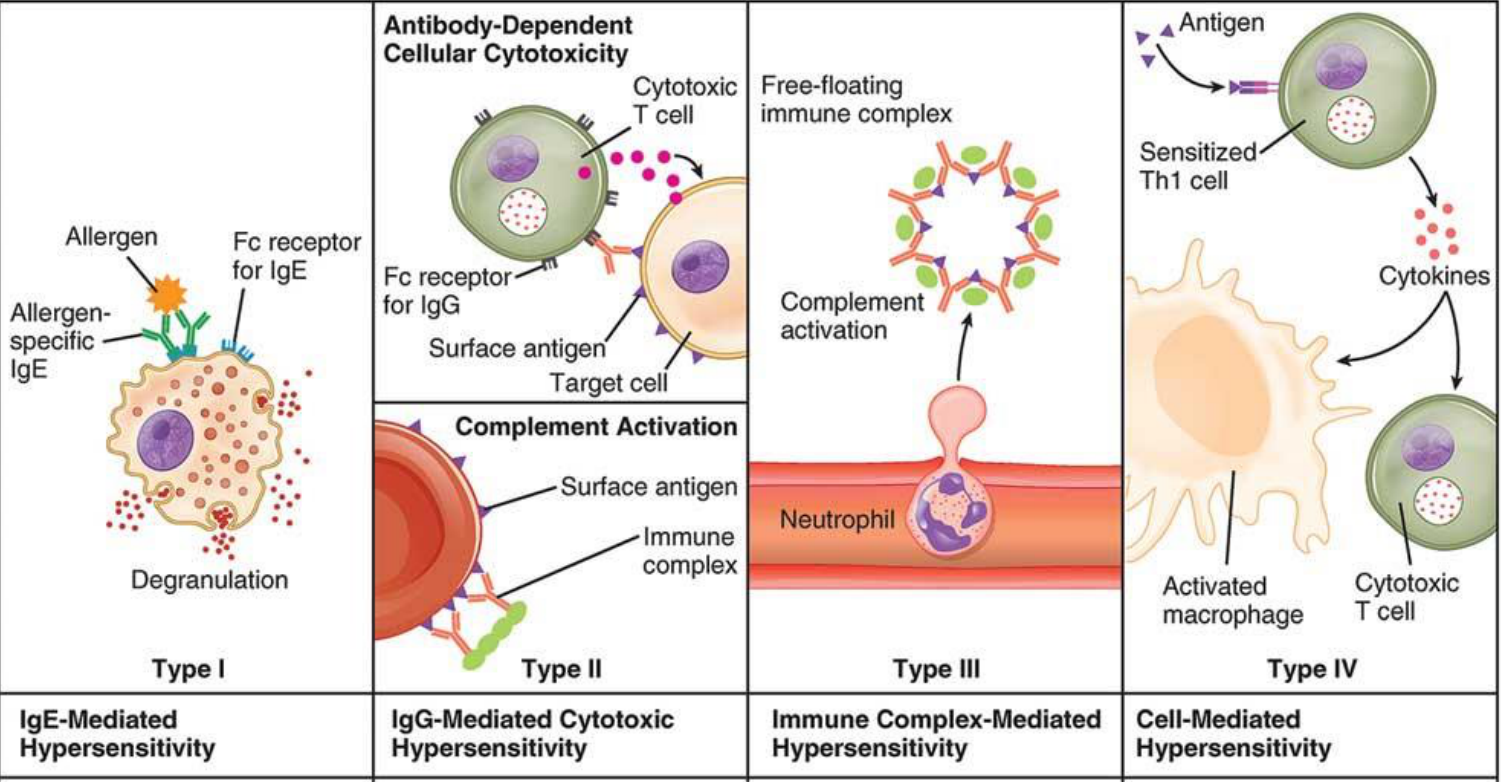
Mechanisms Of Immunological Injury1314
The immune response exists to protect the host. However, its effects are not always beneficial to the host.
Immunological injury can be immediate or delayed.
- Type I reaction (anaphylactic)
- Type II reaction (cytotoxic antibody)
- Type III reaction (immune complex)
- Type IV reaction (delayed type hypersensitivity)
| Type I | Type II | Type III | Type IV |
|---|---|---|---|
| IgE-Mediated Hypersensitivity | IgG-Mediated Cytotoxic Hypersensitivity | Immune Complex-Mediated Hypersensitivity | Cell-Mediated Hypersensitivity |
| Allergen; Fc receptor for IgE; Allergen-specific IgE; Degranulation | Cytotoxic T cell; Fc receptor for IgG; Surface antigen; Target cell — Complement Activation: Surface antigen; Immune complex | Free-floating immune complex; Complement activation; Neutrophil | Antigen; Sensitized Th1 cell; Cytokines; Activated macrophage; Cytotoxic T cell |
Overview Of Maxillofacial Infection Types15
- Dental pulp
- Periodontium
- Odontogenic infections of fascial spaces
- Osteomyelitis of the jaws
- Salivary gland infections
- Infections of the facial skin and scalp
- Orbital infections
- Infections of the ear and mastoid
- Nasal and Para-Nasal Sinus infections
- Oropharyngeal and Tonsillar infections
Dental And Fascial Space Infections
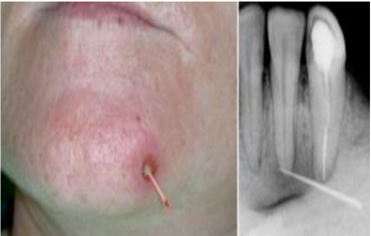
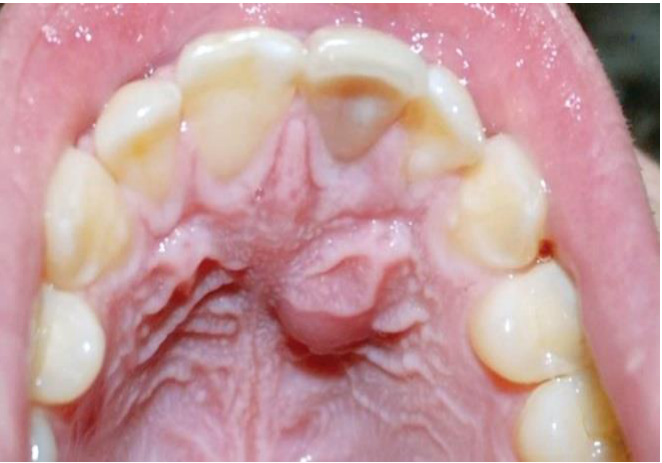
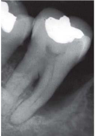
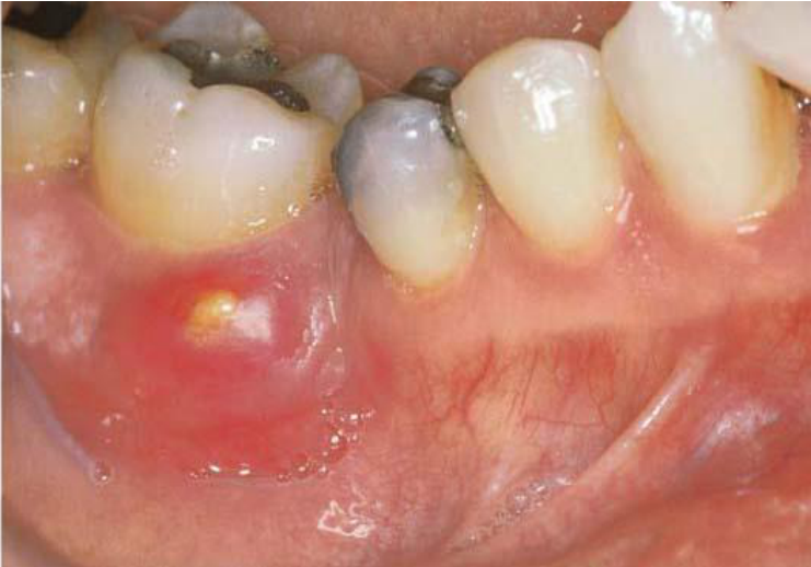
Infection Of Dental Pulp And Periodontium16
 |  |
 |  |
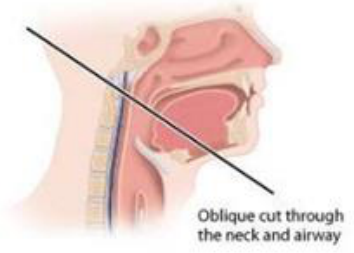
Fascial Spaces And Spread Of Odontogenic Infection17
 | 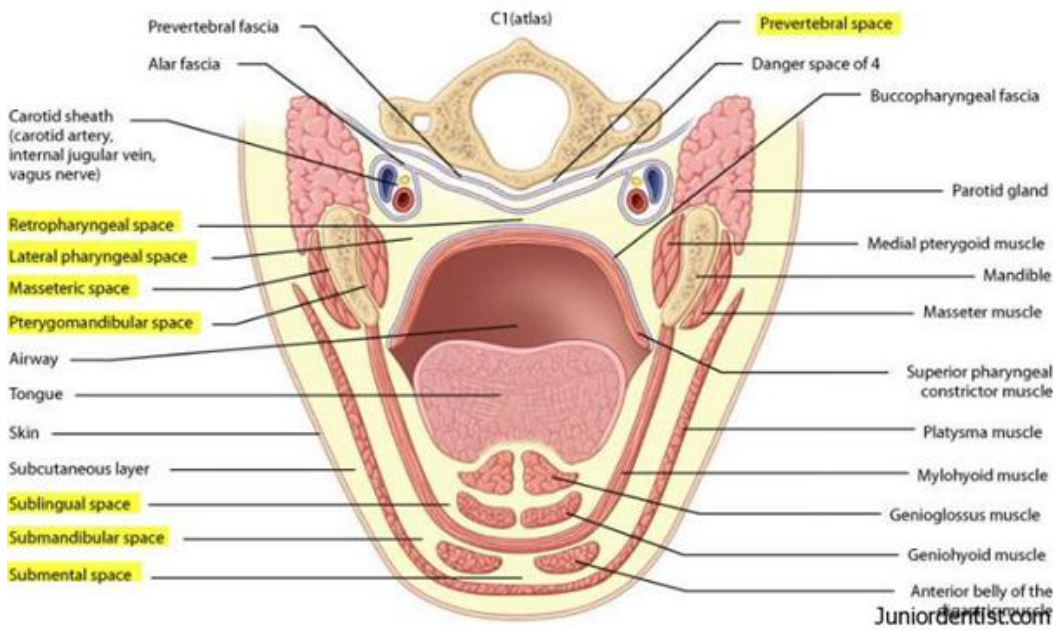 |
Fascial Spaces18
- Potential spaces between layers of fascia that do not exist in a healthy individual but become filled during infections
- Spaces have boundaries formed by muscles, fascia, periosteum, bone, mucous membranes
Infection in these spaces may progress from superficial infection to cellulitis to the formation of an abscess requiring immediate drainage. Spread of infection between spaces depends on anatomic location.
Oblique cut through the neck and airway, showing: prevertebral fascia; C1 (atlas); prevertebral space; alar fascia; danger space of 4; buccopharyngeal fascia; carotid sheath (carotid artery, internal jugular vein, vagus nerve); parotid gland; medial pterygoid muscle; mandible; masseter muscle; superior pharyngeal constrictor muscle; platysma muscle; mylohyoid muscle; genioglossus muscle; geniohyoid muscle; anterior belly of the digastric muscle; retropharyngeal space; lateral pharyngeal space; masseteric space; pterygomandibular space; airway; tongue; skin; subcutaneous layer; sublingual space; submandibular space; submental space.
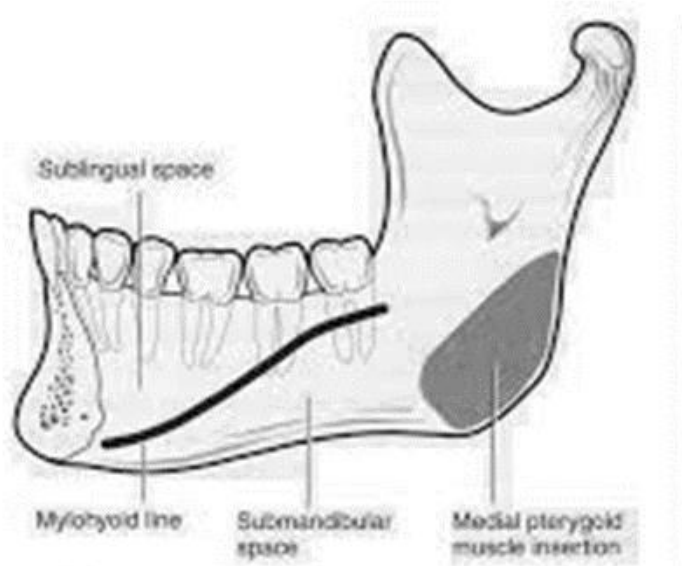
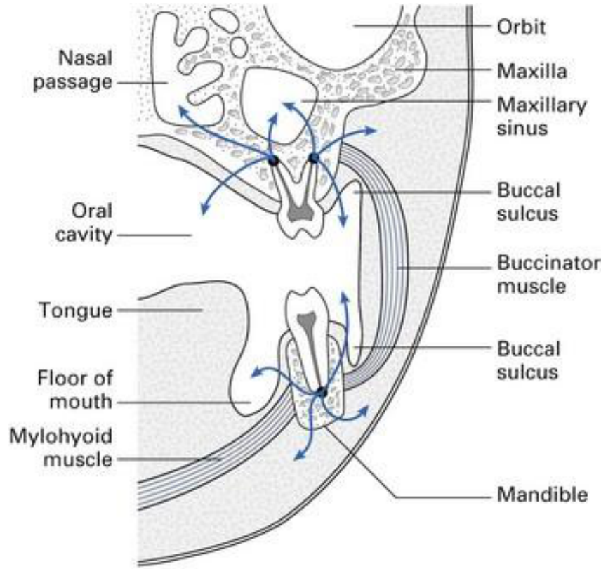
Spread of Odontogenic Infection19
The floor of the mouth is divided by the mylohyoid muscle into the Sublingual Space above and the Submandibular Space below (note the relationship of the Mylohyoid Line to the apices of the mandibular teeth).
Figure labels: sublingual space; mylohyoid line; submandibular space; medial pterygoid muscle insertion; nasal passage; orbit; maxilla; maxillary sinus; buccal sulcus; buccinator muscle; mandible; oral cavity; tongue; floor of mouth; mylohyoid muscle.
 |  |
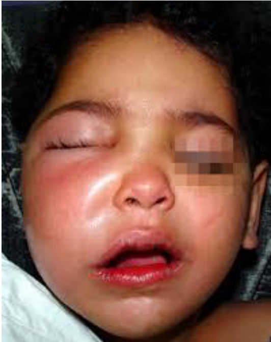
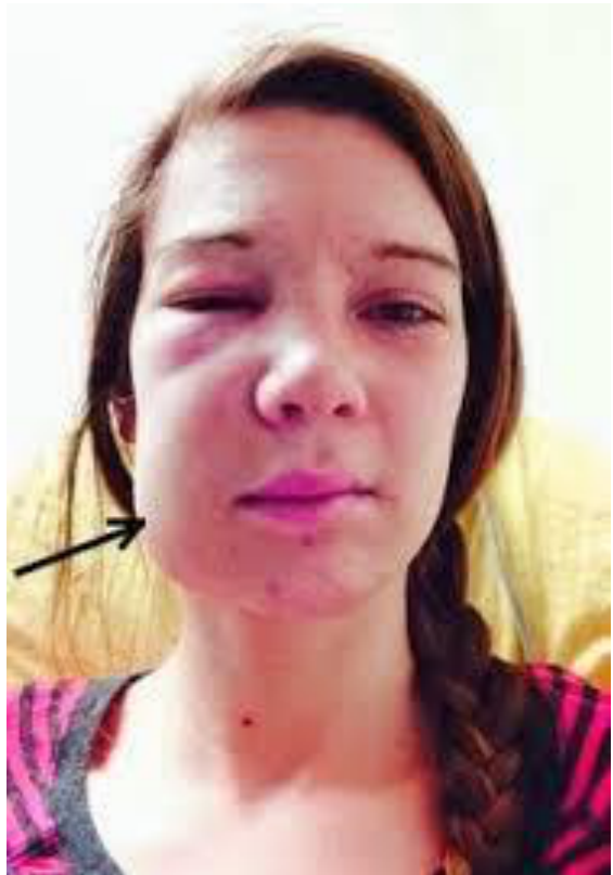
Specific Space Infections2021
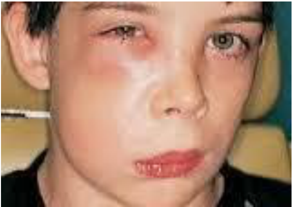
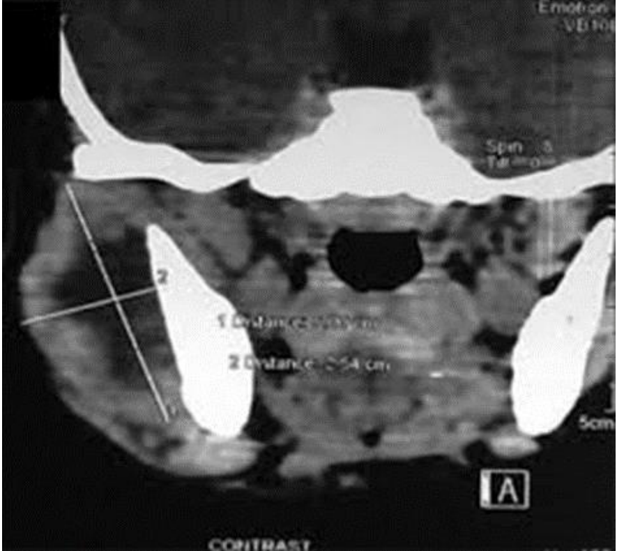
Maxillary periapical abscess
 |  |  |
 | 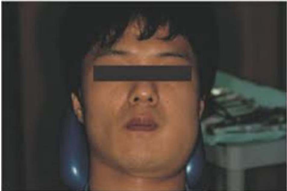 |
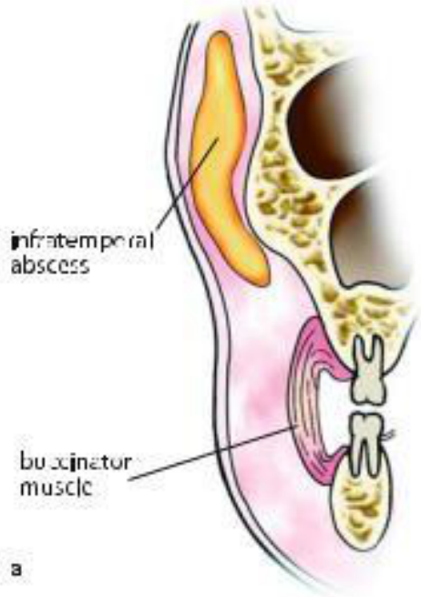
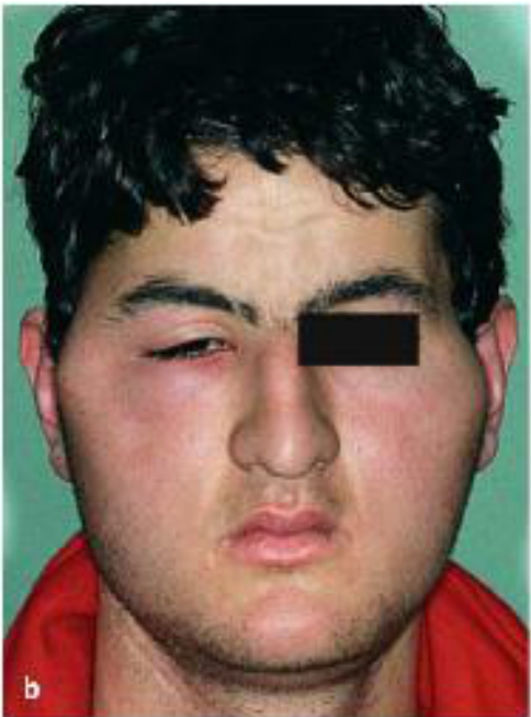
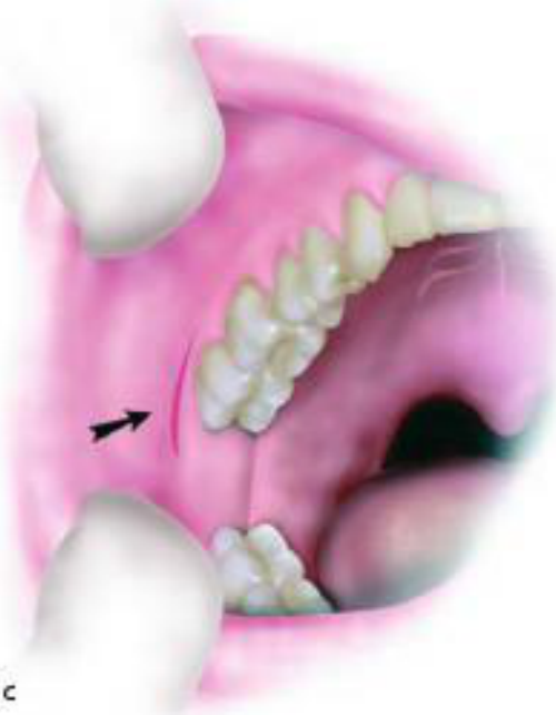
Infratemporal Space Infection22
Figure labels: infratemporal abscess; buccinator muscle.
 |  |
 |
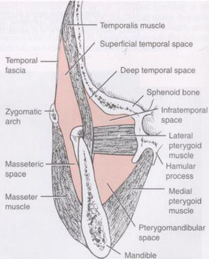
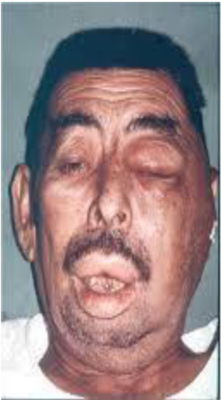
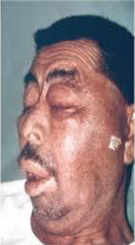
Temporal Space Infection23
Figure labels: temporal fascia; zygomatic arch; masseteric space; masseter muscle; temporalis muscle; superficial temporal space; deep temporal space; sphenoid bone; infratemporal space; lateral pterygoid muscle; hamular process; medial pterygoid muscle; pterygomandibular space; mandible.
 |  |
 |
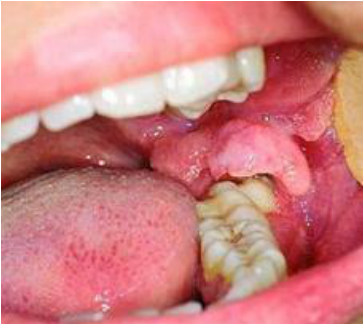
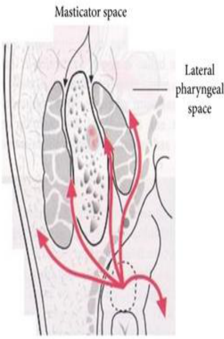
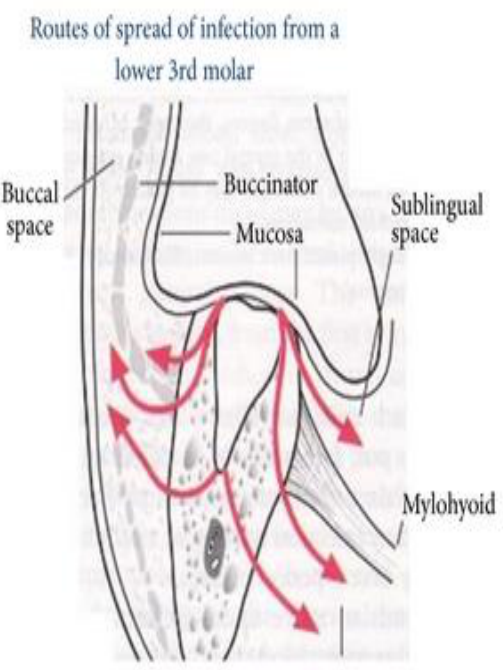
Pericoronitis24
Figure labels: masticator space; lateral pharyngeal space; buccal space; buccinator; mucosa; sublingual space; mylohyoid.
Submasseteric space infection
 |  |
 |
Deep Neck And Pharyngeal Space Infections25
- Commonly from septic focus of mandibular teeth, tonsils, parotid gland, deep cervical lymph nodes, middle ear, or sinuses
- Involves the submandibular space, lateral pharyngeal space, retropharyngeal space
- Relatively uncommon in the post-antibiotic era
- Can progress to life-threatening complications due to proximity to airway and potential spread into carotid sheath, cavernous sinus, cranium and mediastinum
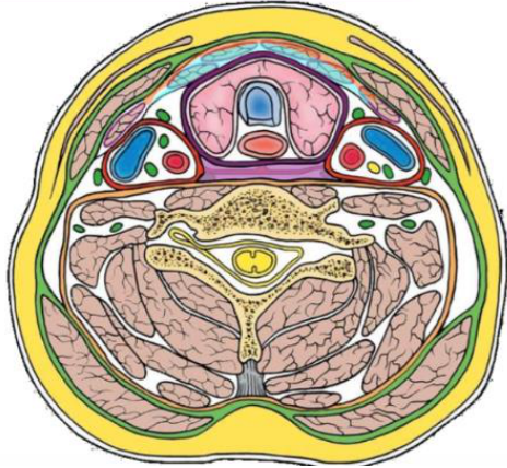
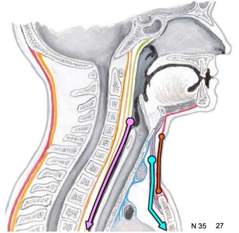
Cervical Fascia: Neck Fascia and Clinical Concerns26
Infections:
- Between investing & muscular pretracheal fascia
- typically stops at manubrium
- Between investing & visceral pretracheal fascia
- will likely spread to anterior thorax
- Within retropharyngeal space
- swelling affecting swallowing, speech; may spread to thorax
 |  |
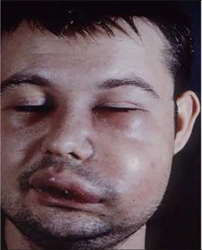
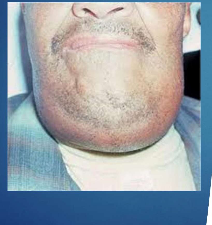
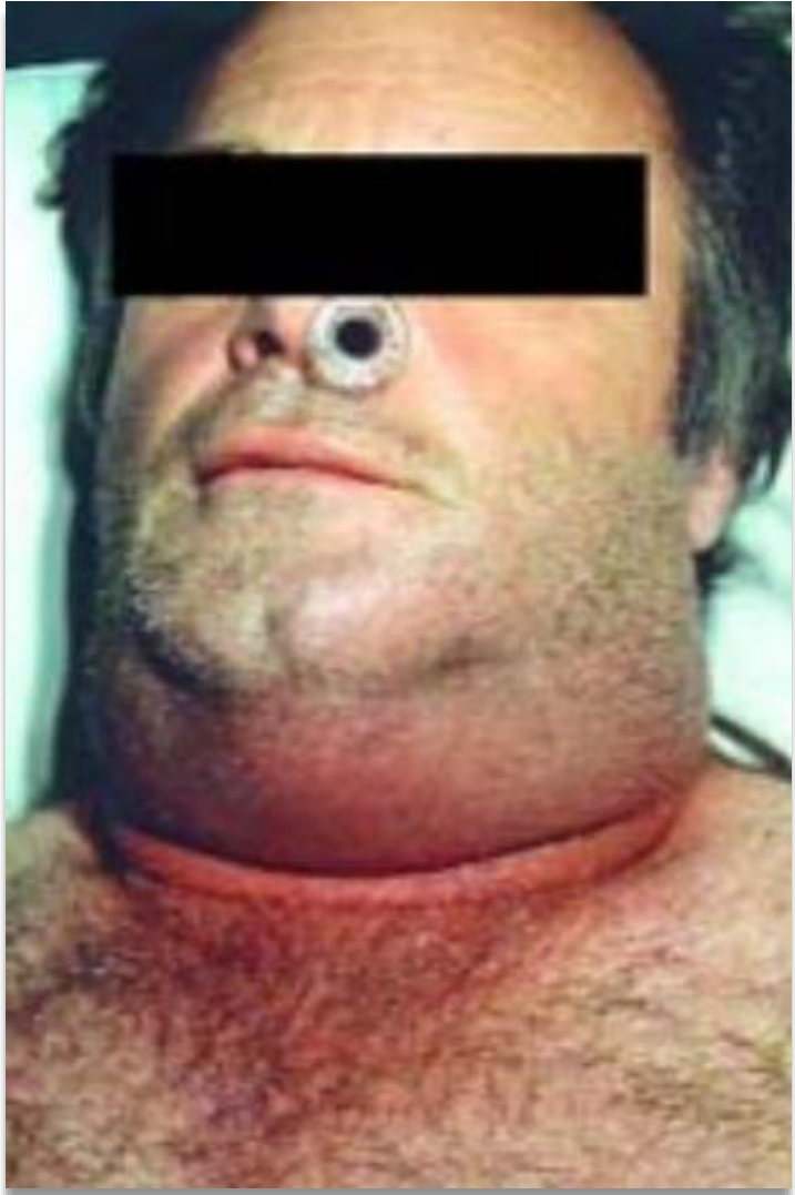
Ludwig’s Angina27
- Cellulitis of sublingual and submandibular space
- Life-threatening airway compromise
- Organisms: Strep. viridans and Staph. aureus
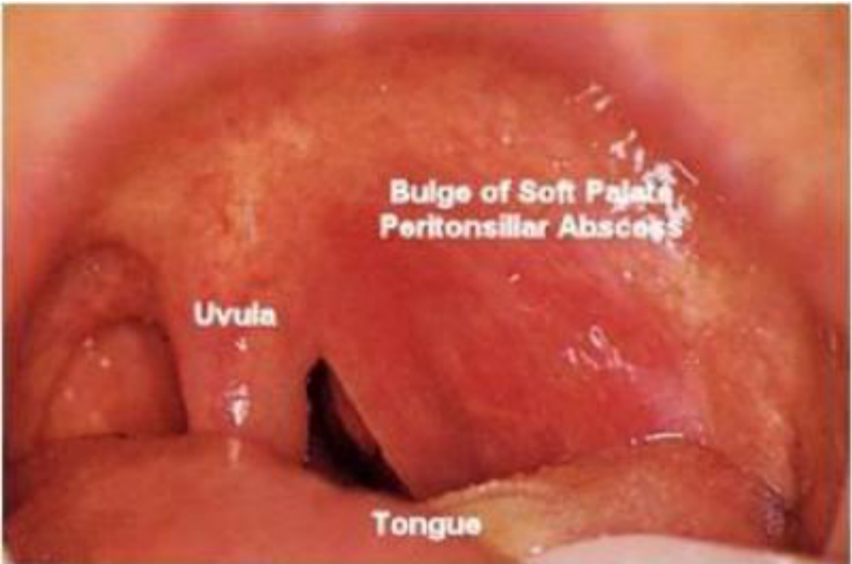
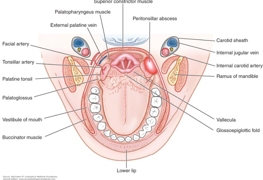
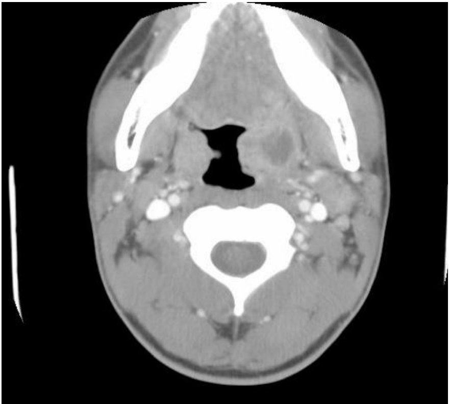
Peritonsillar Abscess2829
Figure labels: bulge of soft palate; peritonsillar abscess; uvula; tongue.
Figure labels: superior constrictor muscle; palatopharyngeus muscle; peritonsillar abscess; external palatine vein; facial artery; tonsillar artery; palatine tonsil; palatoglossus; vestibule of mouth; buccinator muscle; carotid sheath; internal jugular vein; internal carotid artery; ramus of mandible; vallecula; glossoepiglottic fold; lower lip.
Reichman EF. Emergency Medicine Procedures, Second Edition. www.accessemergencymedicine.com. Copyright © The McGraw-Hill Companies, Inc. All rights reserved.
 |  |
 |
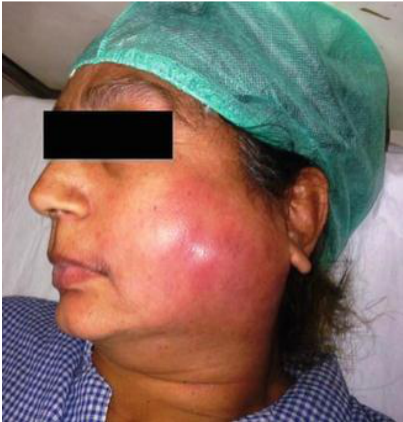
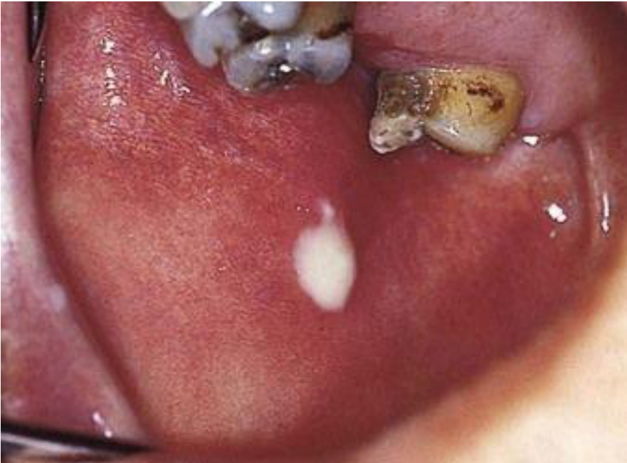
Salivary Gland And Airway Infections30
Infection of the salivary glands
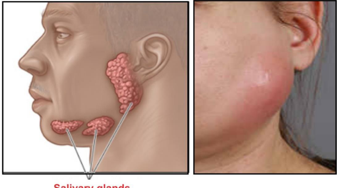
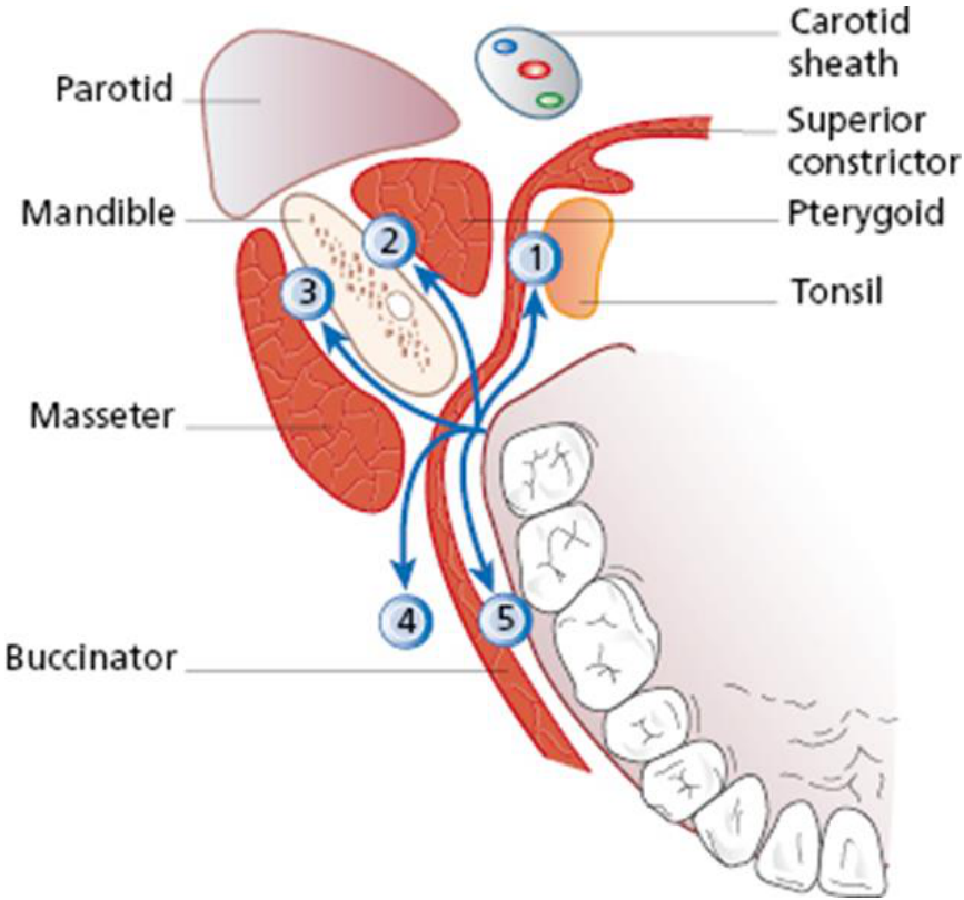
Parotid Gland Infection3132
- Proximity of parotid and tonsil to lateral pharyngeal space
- Potential to cause airway compromise
 |  |
 |
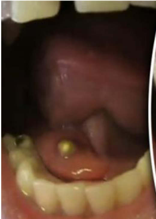
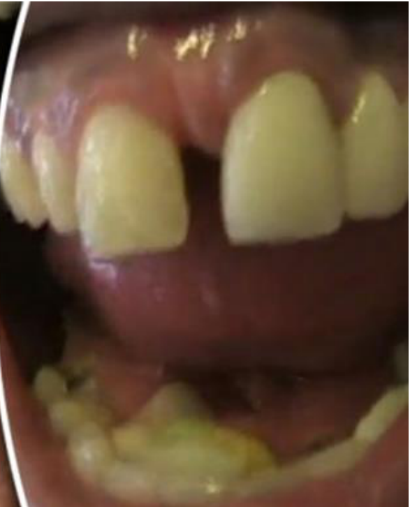
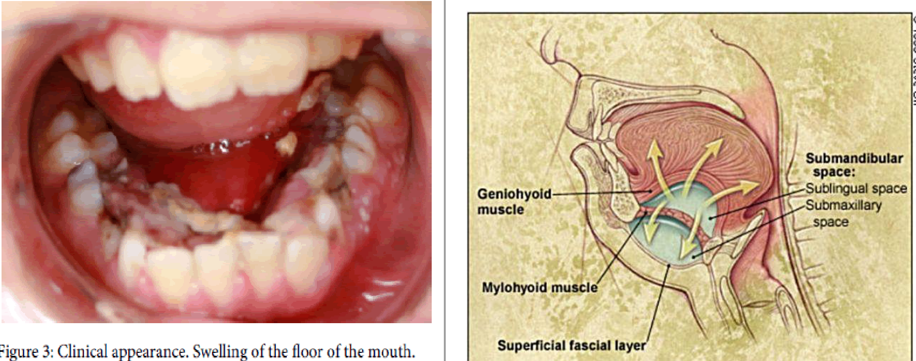
Floor Of Mouth Swelling3334
- Floor of mouth swelling, causing potential airway compromise
© 1999 Steve Oh
- Geniohyoid muscle
- Mylohyoid muscle
- Superficial fascial layer
- Submandibular space:
- Sublingual space
- Submaxillary space
 |  |
 |
Ear And Sinus Infections
Middle Ear Infections35
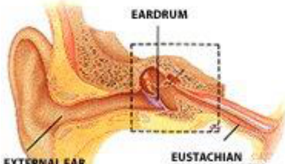
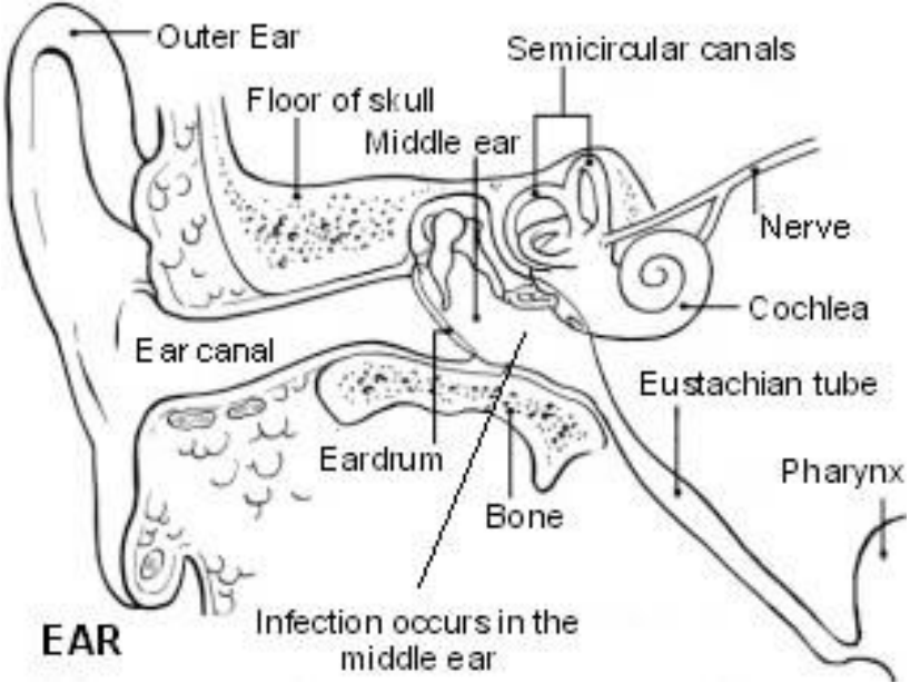
Anatomy of the Middle Ear36
- External ear canal
- Eardrum
- Eustachian tube (connects ear to nose)
Labeled structures: outer ear, floor of skull, middle ear, semicircular canals, nerve, cochlea, ear canal, eustachian tube, eardrum, bone, pharynx. Infection occurs in the middle ear.
 |  |
 |
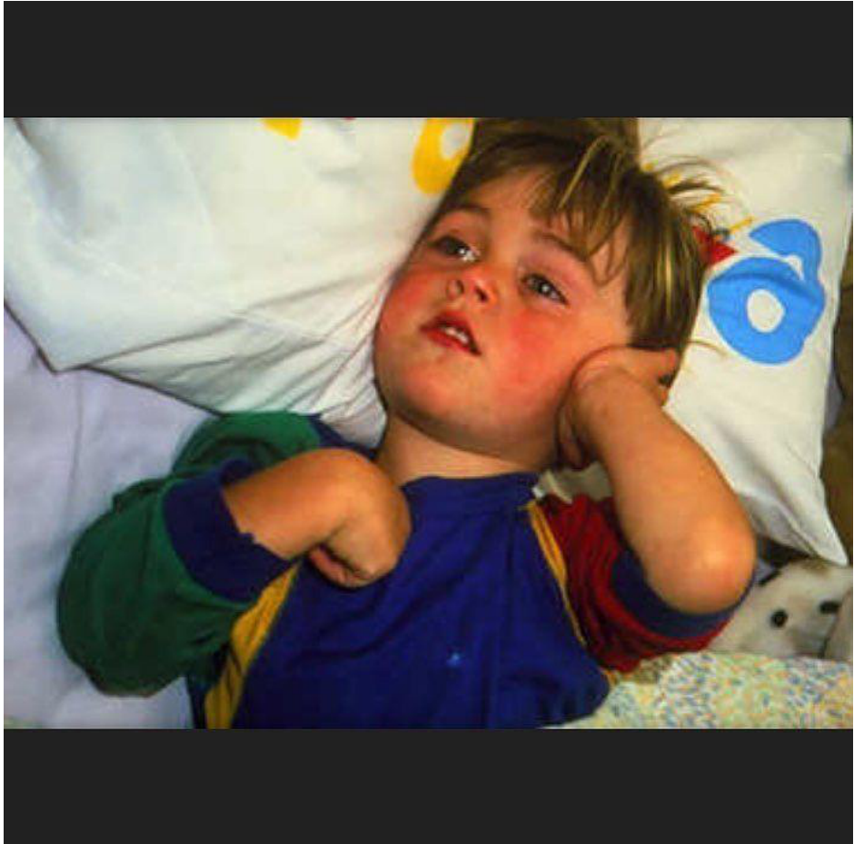
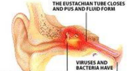
Pathogenesis
- Viruses and bacteria have entered the tube from the throat
- The eustachian tube closes and pus and fluid form
- Fluid puts pressure on the eardrum causing redness and pain
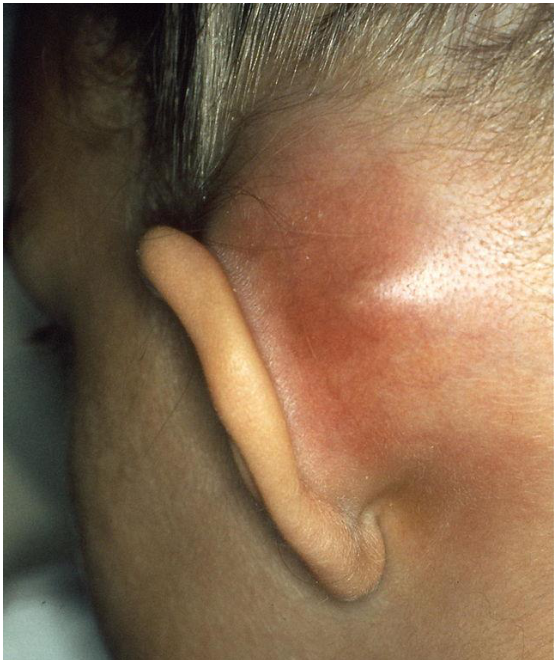
Mastoiditis37
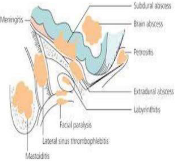
Complications associated with mastoiditis:
- Subdural abscess
- Meningitis
- Brain abscess
- Petrositis
- Extradural abscess
- Labyrinthitis
- Facial paralysis
- Lateral sinus thrombophlebitis
- Mastoiditis
 |  |
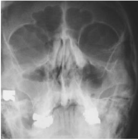
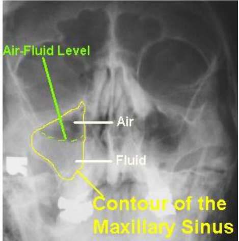
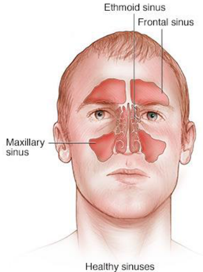
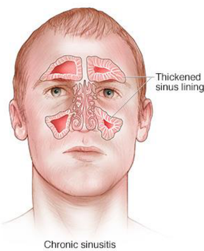
Sinus Infection38
Figure labels: air-fluid level (air above, fluid below); ethmoid sinus, frontal sinus, maxillary sinus, thickened sinus lining.
© Mayo Foundation for Medical Education and Research. All rights reserved.
 |  |
 |  |
Orbital And Intracranial Complications
Orbital Complications Of Sinusitis39
- Orbital cellulitis
- Intracranial infections
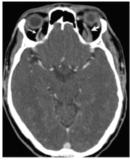
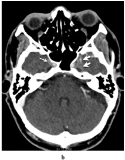
- Cavernous sinus thrombosis
- Septic emboli of optic nerve
- Optic nerve ischemia may result in visual loss
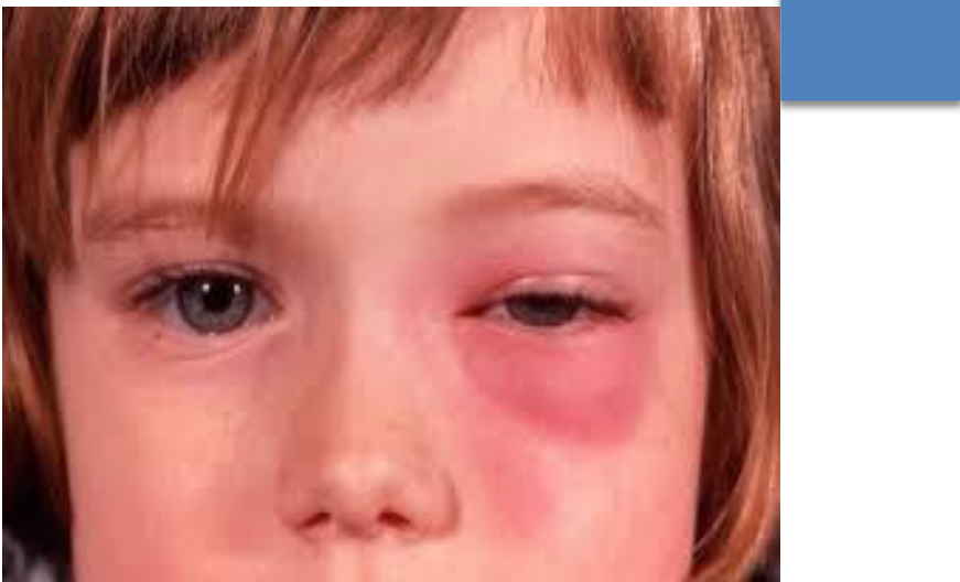 | 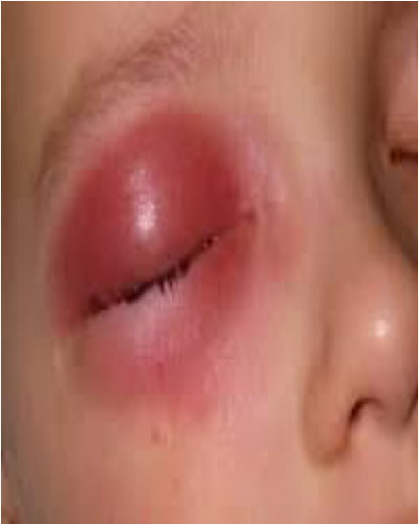 |
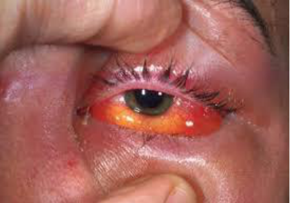
Orbital Cellulitis40
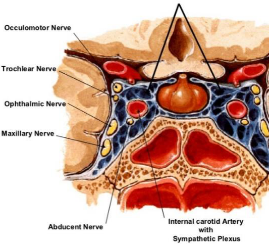
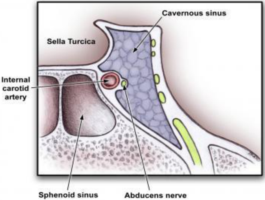
The complications of orbital cellulitis are ominous and include optic neuropathy, retinal vein occlusion, cavernous sinus thrombosis, meningitis and death
Figure labels: occulomotor nerve, trochlear nerve, ophthalmic nerve, maxillary nerve, abducent nerve, internal carotid artery with sympathetic plexus.
 |  |
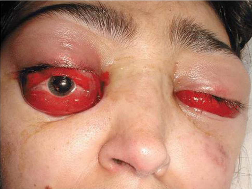
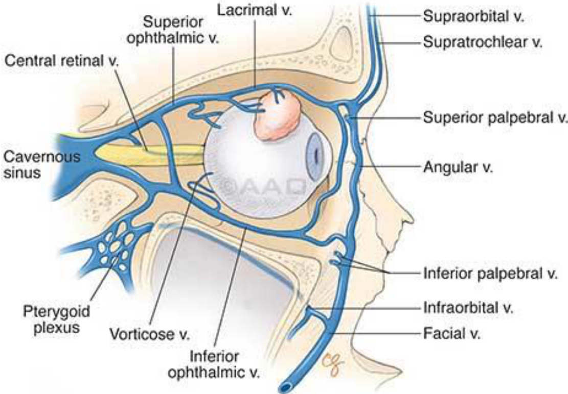
Cavernous Sinus Thrombosis414243
- Fevers, rigors, malaise, severe frontal and periorbital pain
- Exophthalmos
- Oedema of eyelid and chemosis of conjunctiva
- Ophthalmoplegia
- Ptosis
- Dilated pupil with loss of accommodation reflex
Figure labels: lacrimal v., superior ophthalmic v., supraorbital v., supratrochlear v., central retinal v., superior palpebral v., cavernous sinus, angular v., inferior palpebral v., pterygoid plexus, vorticose v., infraorbital v., facial v., inferior ophthalmic v.
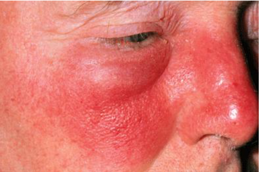
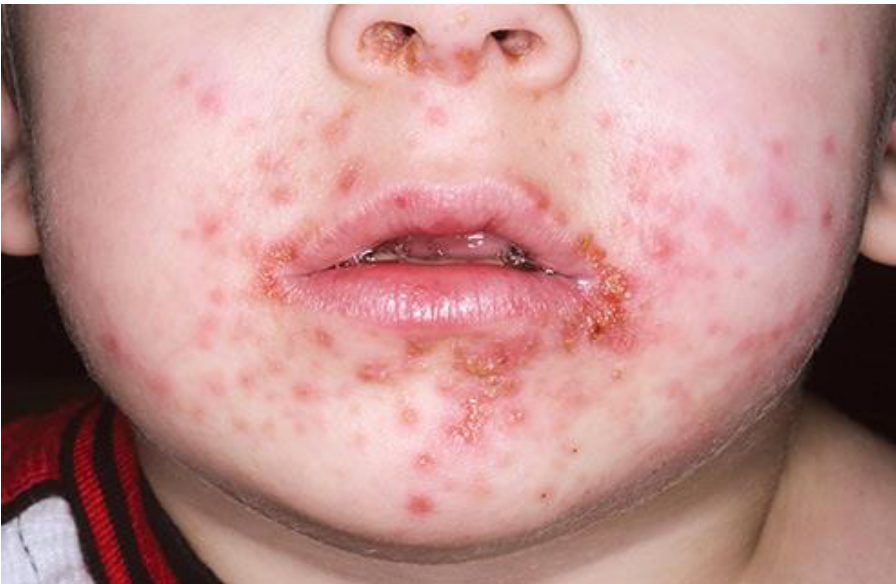
Skin And Soft Tissue Infections44
Skin infections
 |  |
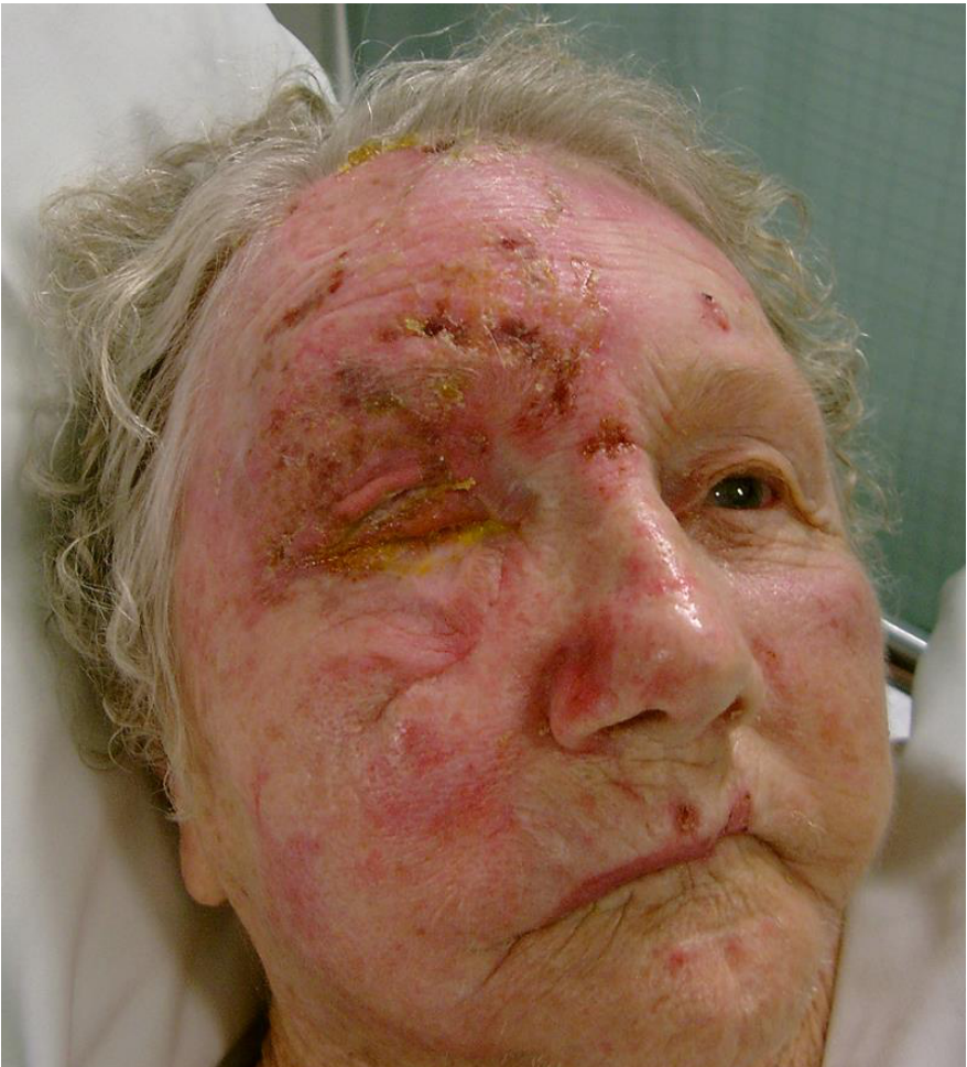
Herpes Zoster Ophthalmicus45
- Can lead to chronic ocular complications, reduced vision, even blindness
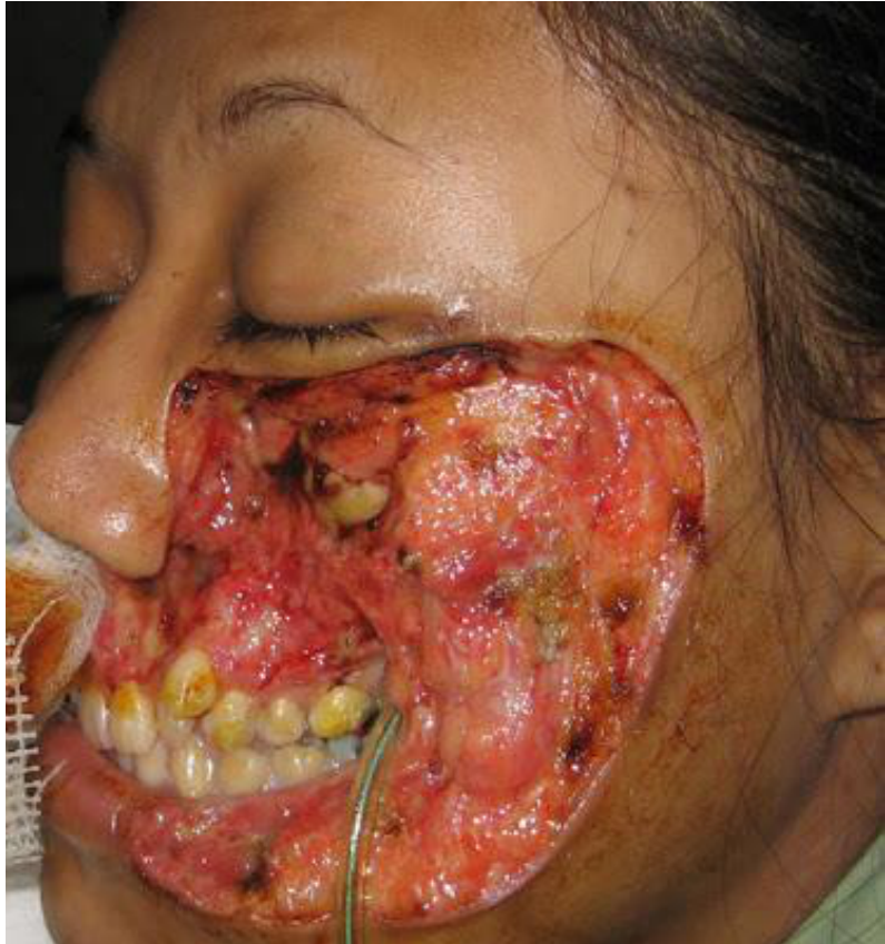
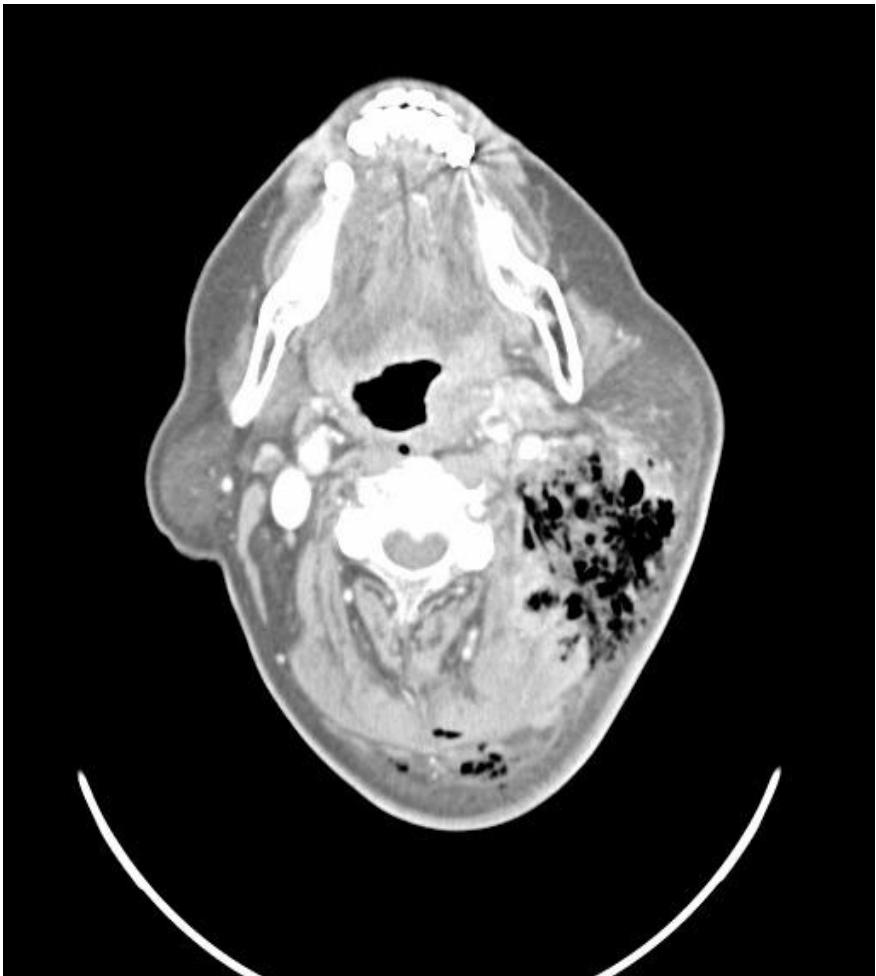
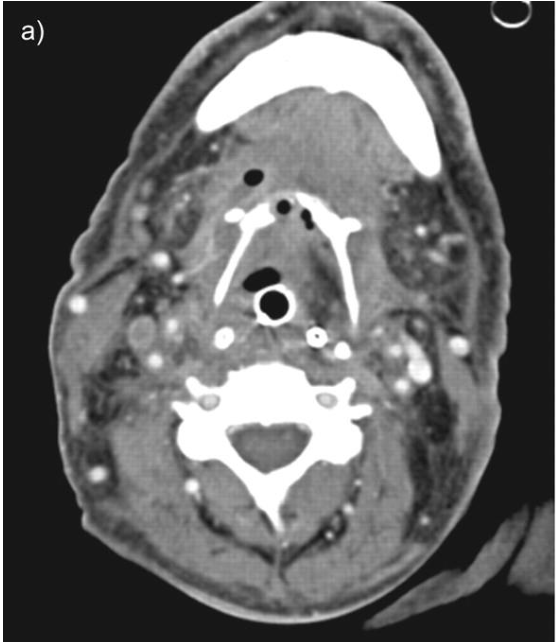
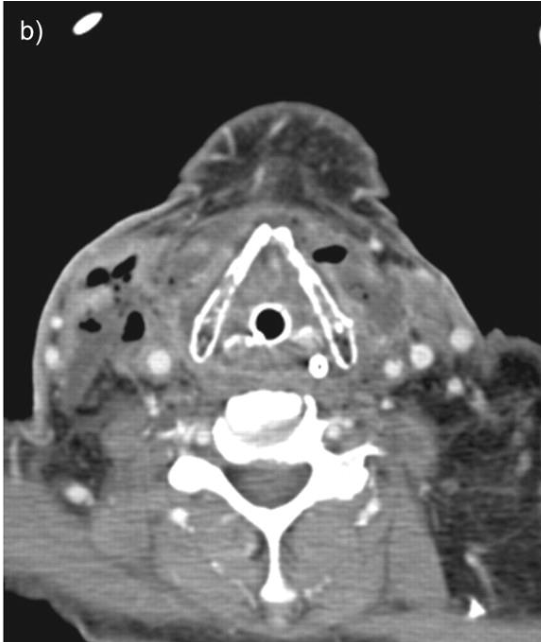
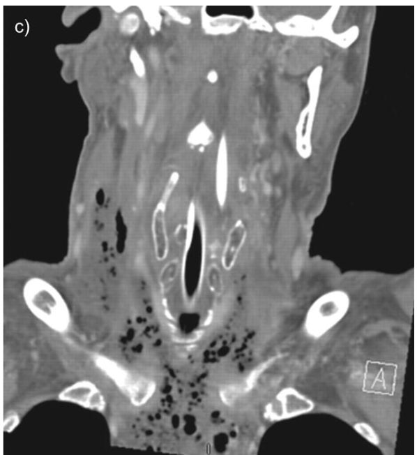
Necrotising Fasciitis4647
-
Rapidly progressing necrosis of the subcutaneous tissue and overlying skin
-
Type I: polymicrobial flora
-
Type II: Group A Beta-Streptococcus bacteria
-
Type III: gas gangrene, or clostridial myonecrosis
-
Presence of gas-forming organisms; subcutaneous air is classically described in necrotizing fasciitis
 |  |
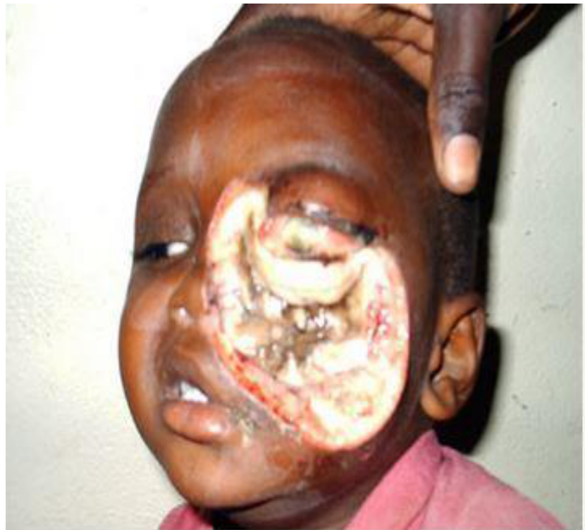
Cancrum Oris48
- Orofacial gangrene which causes mutilating destruction of soft and hard tissues of the oral and perioral structures
- Typically affects children who are malnourished, with poor oral hygiene and concurrent debilitating illnesses
Management Of Odontogenic Infections
Steps In Management4950
-
Severity of infection
-
Host defences
-
Setting of care
-
Surgical treatment
-
Medical support
-
Antibiotic therapy
-
Frequent evaluation
-
The possibility of deep space infection should be considered in any patient who does not respond to the usual treatment of an abscessed tooth or tonsillitis
-
This type of infection also should be considered in a toxic patient who has a fever of unknown origin, with or without blood cultures that show anaerobic organisms.
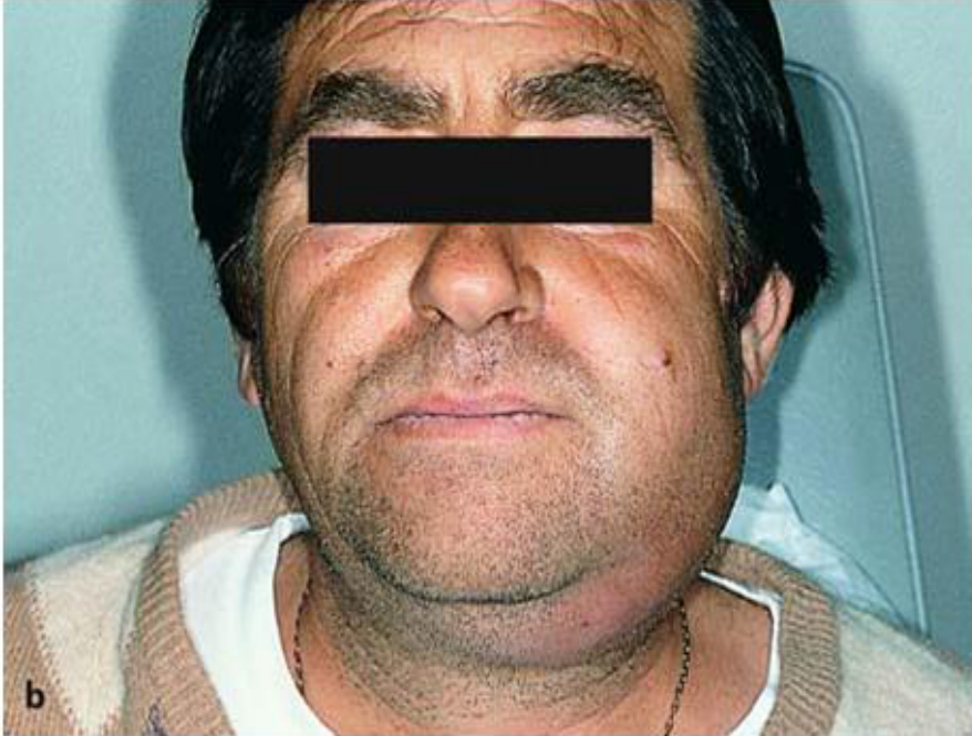
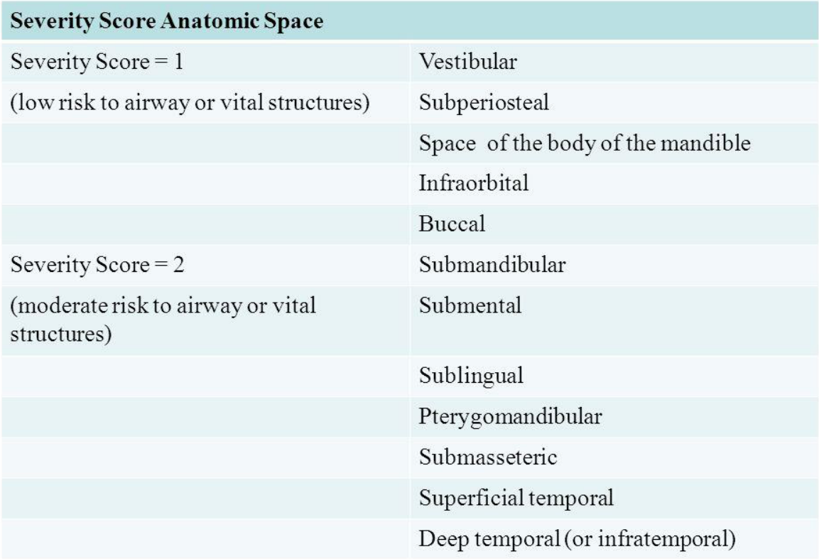
Severity Of Infection5152535455
- Buccal space spreading to submandibular space
- Submandibular space spreading to mediastinum
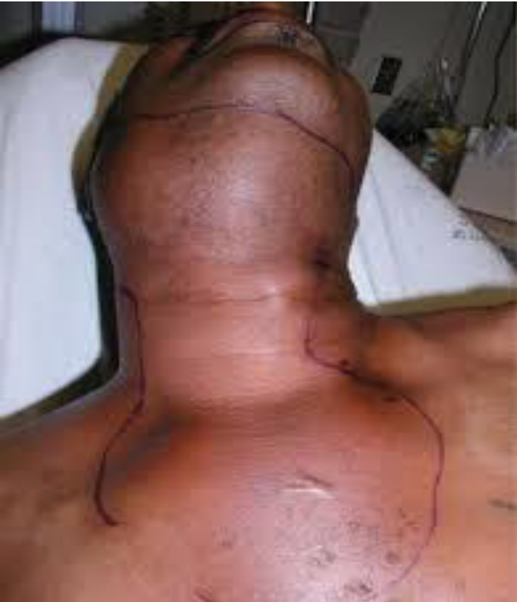
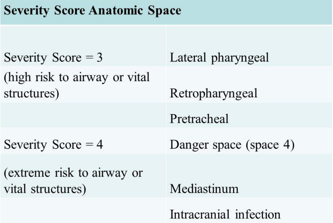
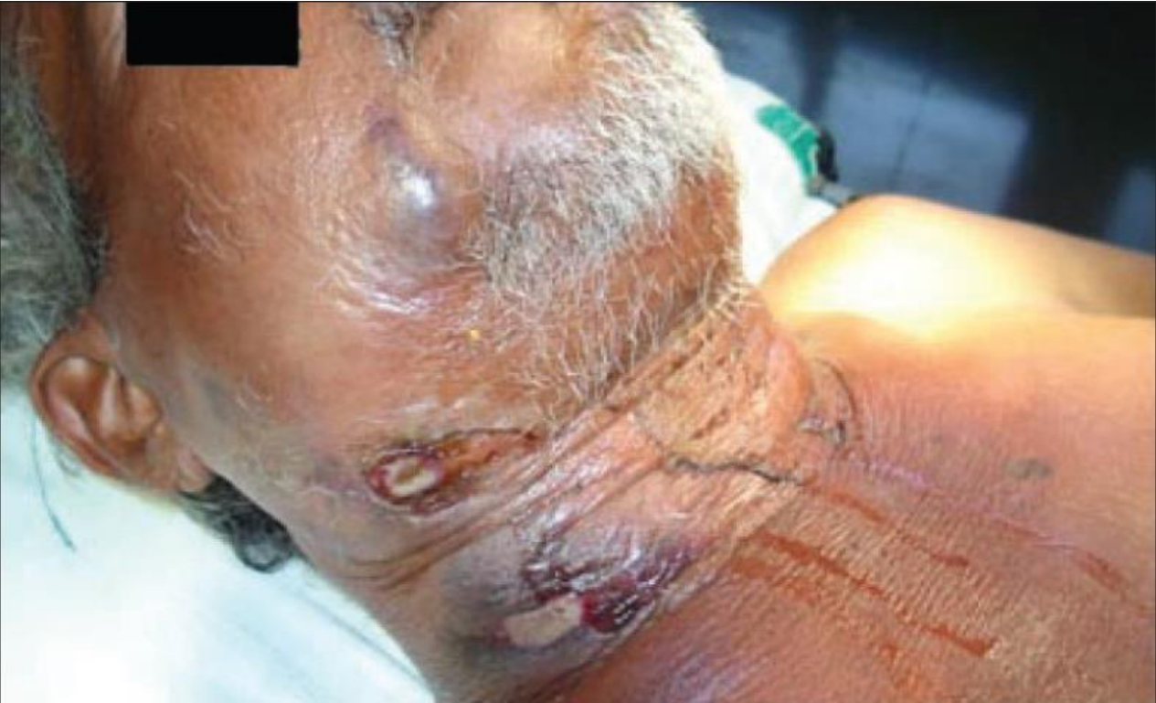
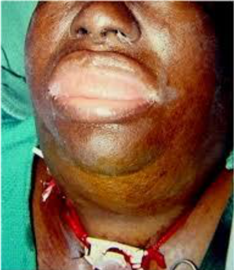
Severity Scores of Facial Space Infections56
| Severity Score | Anatomic Space |
|---|---|
| Severity Score = 1 (low risk to airway or vital structures) | Vestibular, Subperiosteal, Space of the body of the mandible, Infraorbital, Buccal |
| Severity Score = 2 (moderate risk to airway or vital structures) | Submandibular, Submental, Sublingual, Pterygomandibular, Submasseteric, Superficial temporal, Deep temporal (or infratemporal) |
| Severity Score | Anatomic Space |
|---|---|
| Severity Score = 3 (high risk to airway or vital structures) | Lateral pharyngeal, Retropharyngeal, Pretracheal |
| Severity Score = 4 (extreme risk to airway or vital structures) | Danger space (space 4), Mediastinum, Intracranial infection |
Rate of progression
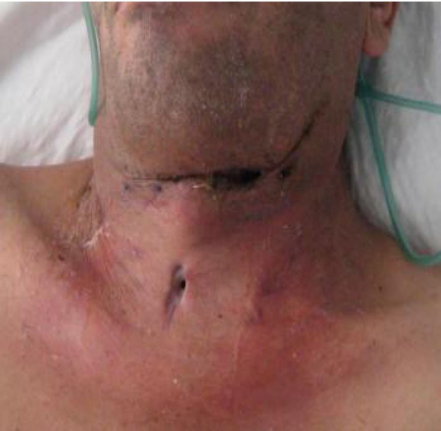
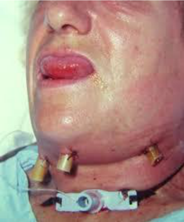
Airway compromise may require a tracheostomy to secure airway
- Ominous sign — pulse oximeter O2 saturation of below 94% in an otherwise healthy patient
- Indication for immediate establishment of a secure airway
Host Defences And Inpatient Care57
- Immune system
- Systemic reserve
Indications for hospital admission58
- Temperature > 38.3 degree Celsius
- Dehydration
- Threat to airway or vital structures
- Infection in moderate or high severity anatomic spaces
- General anaesthesia
- Inpatient control of systemic disease
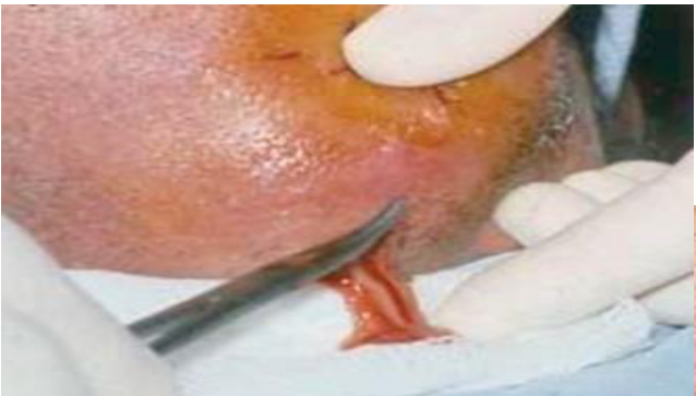
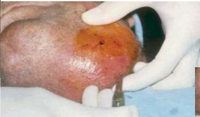
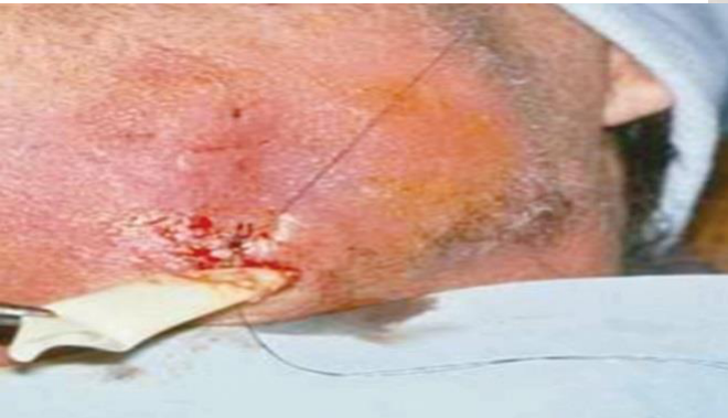
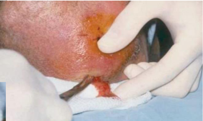
Surgical Treatment596061
- Airway security — immediate establishment of airway by early intubation or tracheostomy
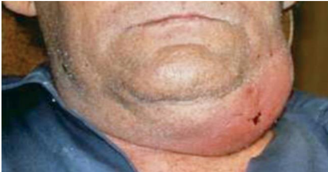
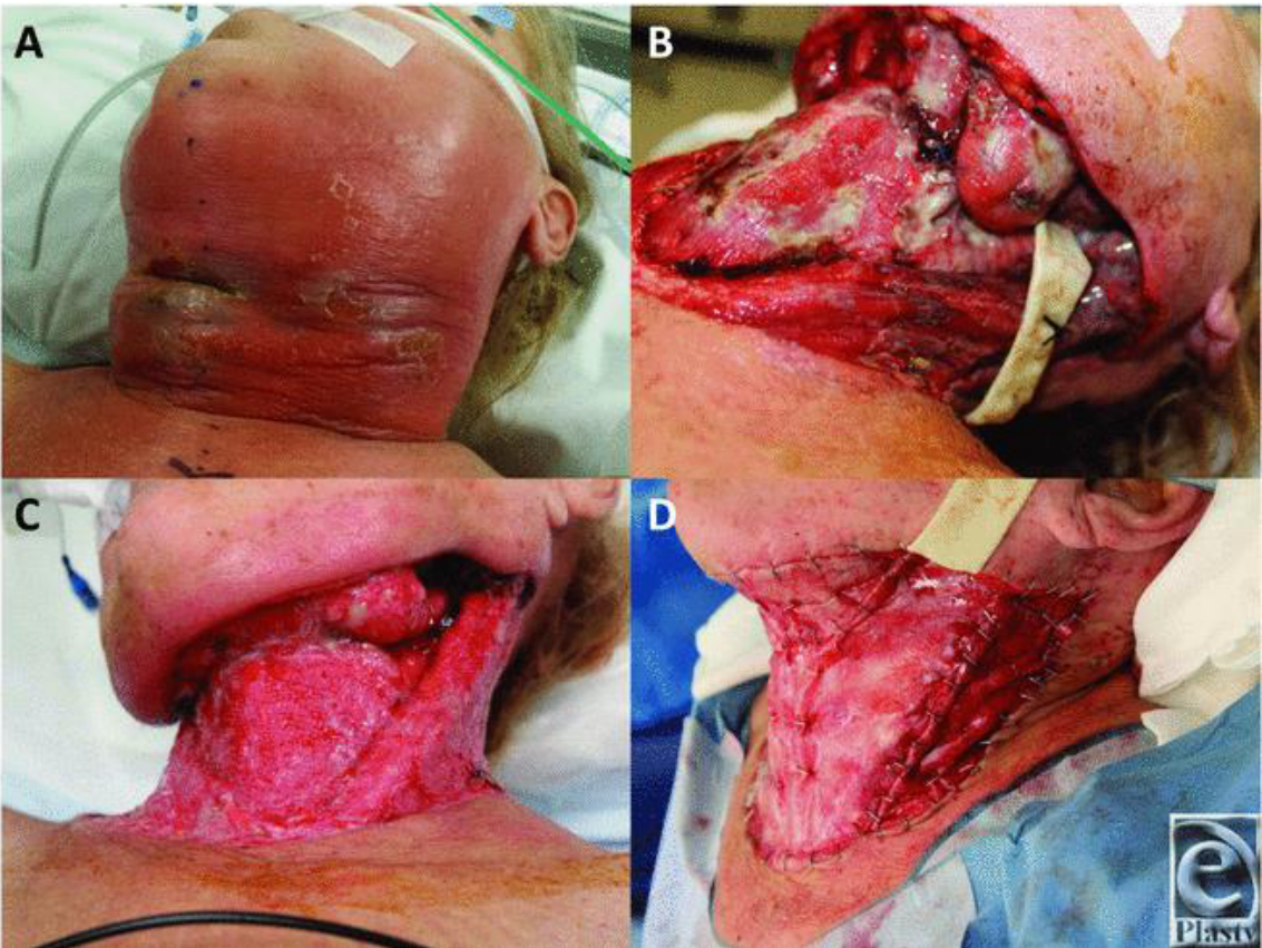
- Surgical drainage — early surgical intervention
Surgical drainage
- Expectant approach to surgical drainage of deep neck infections or undertake surgical drainage undertaken early
- Deep space infection may be occult; a high index of suspicion is required for diagnosis
- Early recognition is necessary to avoid tissue damage, bacteremia or airway compromise
 |  |  |
 |  |  |
Culture and sensitivity testing62636465
- Infection involves anatomic spaces of moderate or greater severity
- Significant medical or immune system compromise
- Chronic infections recalcitrant to therapy
Gas-forming organisms
Aggressive debridement
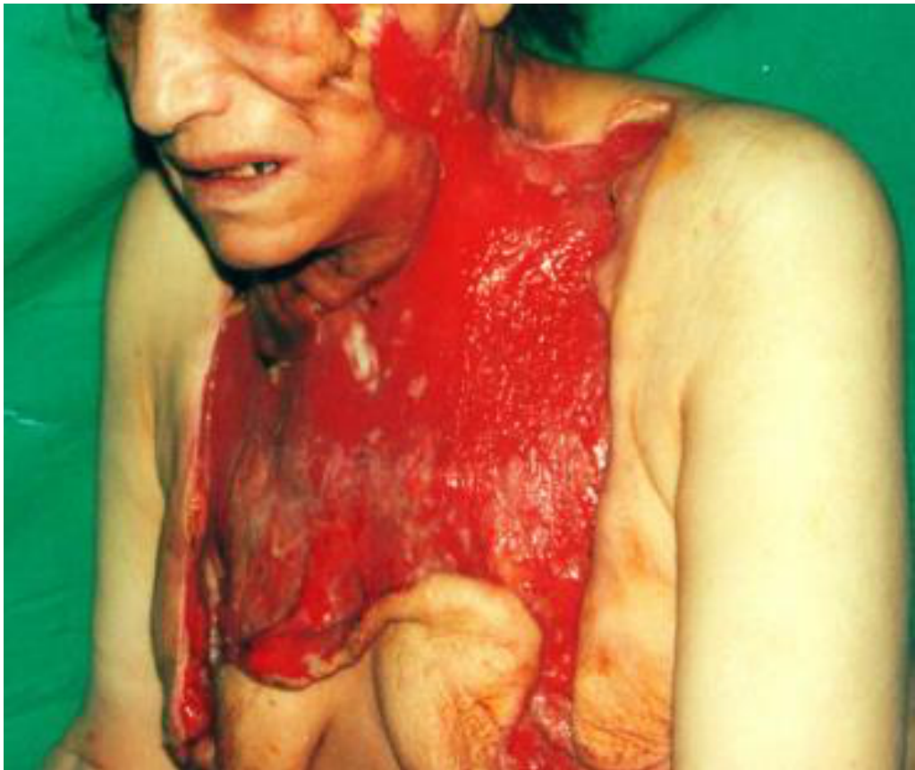
Medical Support And Antibiotic Therapy66
Hydration, nutrition and control of fever
Empiric antibiotics for odontogenic infections requiring only outpatient management67
- Penicillin, Clindamycin, Cephalexin
Empiric antibiotics for odontogenic infections requiring inpatient management
- Clindamycin, Third generation cephalosporin (intravenous route), Metronidazole
Frequent Evaluation68
- Improvement in signs and symptoms — decreasing swelling, cessation of wound drainage, declining white cell count, decreased malaise, decrease airway swelling
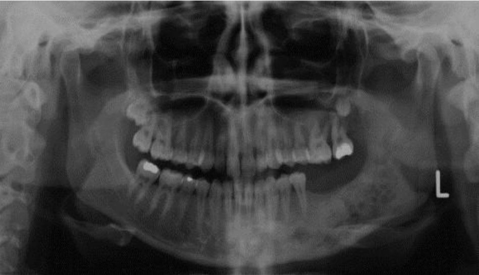
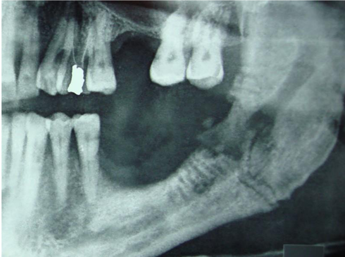
Osteomyelitis6970
-
Bacteria-induced inflammatory process (extractions, RCT, fractures)
-
Inflammation of the bone marrow, extending to involve periosteum
-
Incidence higher in the mandible
-
Predisposing factors: diminished host defences, medications, local pathology, odontogenic infections, trauma
-
Acute or chronic (1 month duration)
-
Clinical features: pain, swelling, lymphadenopathy, fever, paraesthesia, trismus, malaise, fistulas
-
CT evaluation
-
Medical management — antibiotic treatment
-
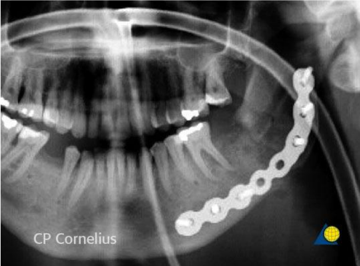
Surgical options:
- sequestrectomy
- reconstruction type plate to prevent pathological fracture
-
Hyperbaric oxygen treatment
References7172
CP Cornelius
 |  |
Footnotes
-
Original PDF page 1: Wy Maxillofacial Infections Mag Foo, p.1 ↩
-
Original PDF page 2: Wy Maxillofacial Infections Mag Foo, p.2 ↩
-
Original PDF page 3: Wy Maxillofacial Infections Mag Foo, p.3 ↩
-
Original PDF page 4: Wy Maxillofacial Infections Mag Foo, p.4 ↩
-
Original PDF page 5: Wy Maxillofacial Infections Mag Foo, p.5 ↩
-
Original PDF page 6: Wy Maxillofacial Infections Mag Foo, p.6 ↩
-
Original PDF page 7: Wy Maxillofacial Infections Mag Foo, p.7 ↩
-
Original PDF page 8: Wy Maxillofacial Infections Mag Foo, p.8 ↩
-
Original PDF page 9: Wy Maxillofacial Infections Mag Foo, p.9 ↩
-
Original PDF page 10: Wy Maxillofacial Infections Mag Foo, p.10 ↩
-
Original PDF page 11: Wy Maxillofacial Infections Mag Foo, p.11 ↩
-
Original PDF page 12: Wy Maxillofacial Infections Mag Foo, p.12 ↩
-
Original PDF page 13: Wy Maxillofacial Infections Mag Foo, p.13 ↩
-
Original PDF page 14: Wy Maxillofacial Infections Mag Foo, p.14 ↩
-
Original PDF page 15: Wy Maxillofacial Infections Mag Foo, p.15 ↩
-
Original PDF page 16: Wy Maxillofacial Infections Mag Foo, p.16 ↩
-
Original PDF page 18: Wy Maxillofacial Infections Mag Foo, p.18 ↩
-
Original PDF page 17: Wy Maxillofacial Infections Mag Foo, p.17 ↩
-
Original PDF page 19: Wy Maxillofacial Infections Mag Foo, p.19 ↩
-
Original PDF page 20: Wy Maxillofacial Infections Mag Foo, p.20 ↩
-
Original PDF page 24: Wy Maxillofacial Infections Mag Foo, p.24 ↩
-
Original PDF page 21: Wy Maxillofacial Infections Mag Foo, p.21 ↩
-
Original PDF page 22: Wy Maxillofacial Infections Mag Foo, p.22 ↩
-
Original PDF page 23: Wy Maxillofacial Infections Mag Foo, p.23 ↩
-
Original PDF page 25: Wy Maxillofacial Infections Mag Foo, p.25 ↩
-
Original PDF page 26: Wy Maxillofacial Infections Mag Foo, p.26 ↩
-
Original PDF page 27: Wy Maxillofacial Infections Mag Foo, p.27 ↩
-
Original PDF page 28: Wy Maxillofacial Infections Mag Foo, p.28 ↩
-
Original PDF page 29: Wy Maxillofacial Infections Mag Foo, p.29 ↩
-
Original PDF page 30: Wy Maxillofacial Infections Mag Foo, p.30 ↩
-
Original PDF page 31: Wy Maxillofacial Infections Mag Foo, p.31 ↩
-
Original PDF page 32: Wy Maxillofacial Infections Mag Foo, p.32 ↩
-
Original PDF page 33: Wy Maxillofacial Infections Mag Foo, p.33 ↩
-
Original PDF page 34: Wy Maxillofacial Infections Mag Foo, p.34 ↩
-
Original PDF page 36: Wy Maxillofacial Infections Mag Foo, p.36 ↩
-
Original PDF page 35: Wy Maxillofacial Infections Mag Foo, p.35 ↩
-
Original PDF page 37: Wy Maxillofacial Infections Mag Foo, p.37 ↩
-
Original PDF page 38: Wy Maxillofacial Infections Mag Foo, p.38 ↩
-
Original PDF page 39: Wy Maxillofacial Infections Mag Foo, p.39 ↩
-
Original PDF page 40: Wy Maxillofacial Infections Mag Foo, p.40 ↩
-
Original PDF page 41: Wy Maxillofacial Infections Mag Foo, p.41 ↩
-
Original PDF page 42: Wy Maxillofacial Infections Mag Foo, p.42 ↩
-
Original PDF page 43: Wy Maxillofacial Infections Mag Foo, p.43 ↩
-
Original PDF page 44: Wy Maxillofacial Infections Mag Foo, p.44 ↩
-
Original PDF page 45: Wy Maxillofacial Infections Mag Foo, p.45 ↩
-
Original PDF page 46: Wy Maxillofacial Infections Mag Foo, p.46 ↩
-
Original PDF page 47: Wy Maxillofacial Infections Mag Foo, p.47 ↩
-
Original PDF page 48: Wy Maxillofacial Infections Mag Foo, p.48 ↩
-
Original PDF page 49: Wy Maxillofacial Infections Mag Foo, p.49 ↩
-
Original PDF page 50: Wy Maxillofacial Infections Mag Foo, p.50 ↩
-
Original PDF page 51: Wy Maxillofacial Infections Mag Foo, p.51 ↩
-
Original PDF page 53: Wy Maxillofacial Infections Mag Foo, p.53 ↩
-
Original PDF page 54: Wy Maxillofacial Infections Mag Foo, p.54 ↩
-
Original PDF page 55: Wy Maxillofacial Infections Mag Foo, p.55 ↩
-
Original PDF page 56: Wy Maxillofacial Infections Mag Foo, p.56 ↩
-
Original PDF page 52: Wy Maxillofacial Infections Mag Foo, p.52 ↩
-
Original PDF page 57: Wy Maxillofacial Infections Mag Foo, p.57 ↩
-
Original PDF page 58: Wy Maxillofacial Infections Mag Foo, p.58 ↩
-
Original PDF page 59: Wy Maxillofacial Infections Mag Foo, p.59 ↩
-
Original PDF page 60: Wy Maxillofacial Infections Mag Foo, p.60 ↩
-
Original PDF page 61: Wy Maxillofacial Infections Mag Foo, p.61 ↩
-
Original PDF page 62: Wy Maxillofacial Infections Mag Foo, p.62 ↩
-
Original PDF page 63: Wy Maxillofacial Infections Mag Foo, p.63 ↩
-
Original PDF page 64: Wy Maxillofacial Infections Mag Foo, p.64 ↩
-
Original PDF page 65: Wy Maxillofacial Infections Mag Foo, p.65 ↩
-
Original PDF page 66: Wy Maxillofacial Infections Mag Foo, p.66 ↩
-
Original PDF page 67: Wy Maxillofacial Infections Mag Foo, p.67 ↩
-
Original PDF page 68: Wy Maxillofacial Infections Mag Foo, p.68 ↩
-
Original PDF page 69: Wy Maxillofacial Infections Mag Foo, p.69 ↩
-
Original PDF page 70: Wy Maxillofacial Infections Mag Foo, p.70 ↩
-
Original PDF page 71: Wy Maxillofacial Infections Mag Foo, p.71 ↩
-
Original PDF page 72: Wy Maxillofacial Infections Mag Foo, p.72 ↩