Imaging of Infection and Inflammatory Lesions of the Jaws12
Dr Dayea Oh
Oral & Maxillofacial Radiologist
Contents
- Inflammatory lesions of the Jaws
- Imaging Modalities & Features of:
- Periapical inflammatory lesions
- Osteomyelitis
- Jaw Infection involving Soft Tissues
Inflammatory Lesions of the Jaws3
- Most common disease
- Disruption of bone metabolism
- Imbalance of osteoblastic (bone production) and osteoclastic (bone resorption) activities
- Altered by the pathogenicity of bacterial microorganisms, the host immune response, tissue vascularity, and time.
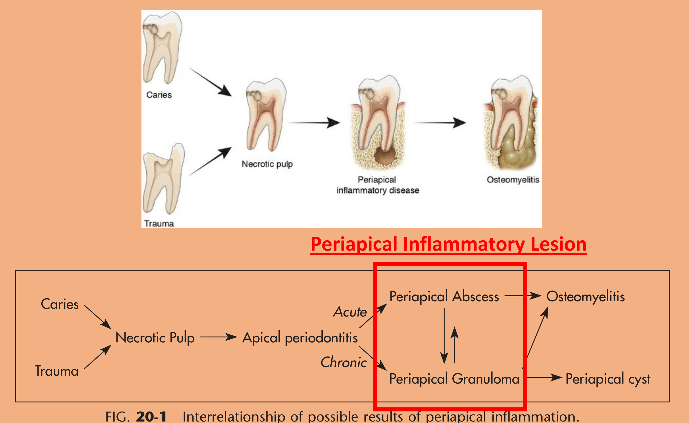
Lecturer — Infection Origin
- Jaw infections are most often odontogenic.
- Caries
- Trauma
- Necrotic pulp
- Periapical inflammatory disease
- Osteomyelitis
Periapical Inflammatory Lesions4
- Caries
- Trauma
- Necrotic pulp
- Apical periodontitis
- Acute
- Chronic
- Periapical abscess
- Periapical granuloma
- Periapical cyst
- Osteomyelitis
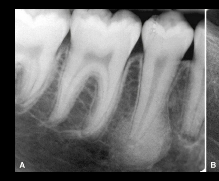
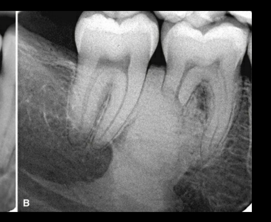
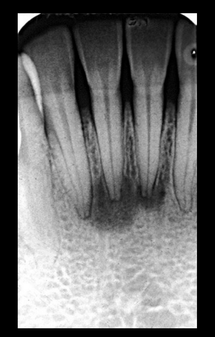
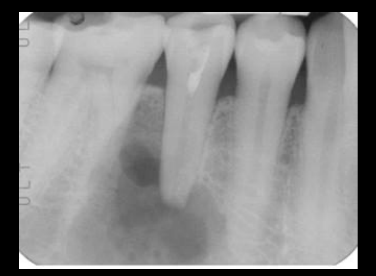
Periapical inflammatory lesions encompass both imaging approach and characteristic radiographic findings.
Lecturer — Inflammatory Progression
Odontogenic infection commonly begins with pulp necrosis after bacterial invasion through caries or trauma.
- Acute disease predominantly involves neutrophils.
- More chronic disease involves monocytes, macrophages and lymphocytes.
- A cyst arising from an infected tooth is more correctly termed a radicular cyst.
Imaging Modalities and Features
Imaging Modalities
- Periapical radiographs
- Panoramic radiographs (OPG)
- CT
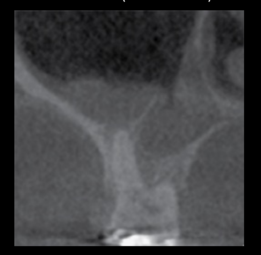
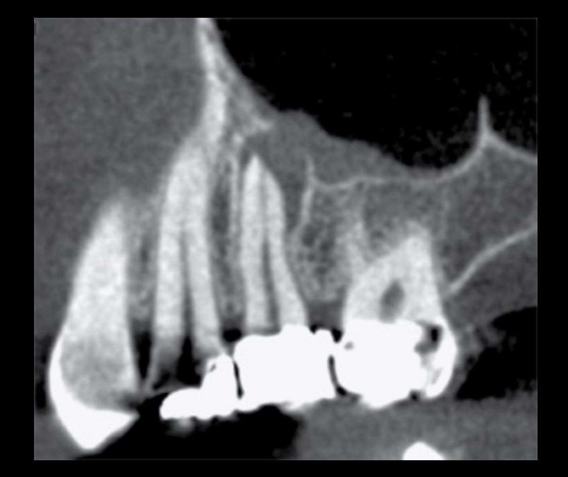
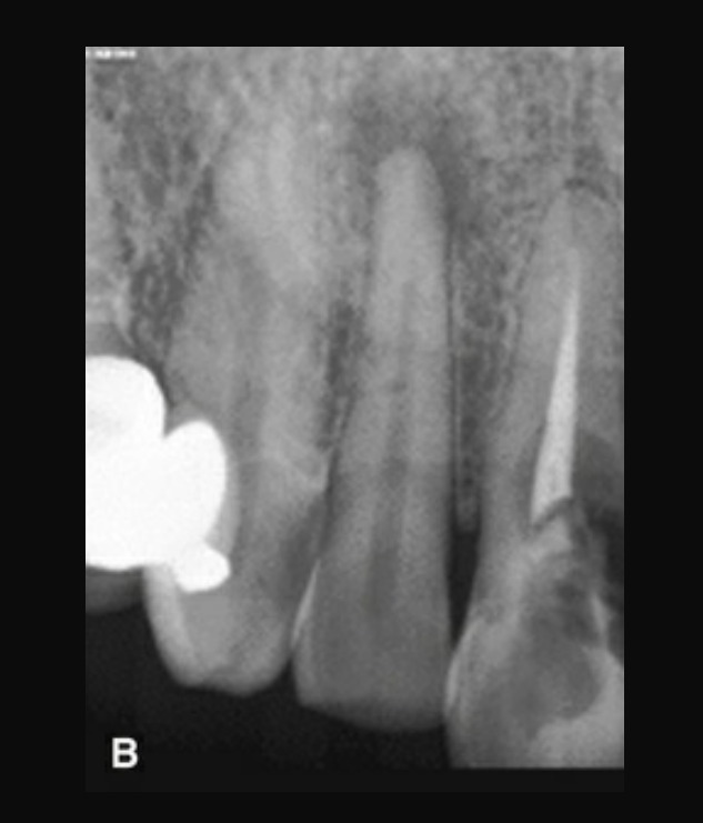
- Both cone beam CT and multislice CT are more sensitive for periapical lesions than intraoral and panoramic radiographs.
Lecturer — Imaging Modality Roles
Orthopantomograms are useful for surveying multiple lesions and assessing larger or heavily compromised areas.
- Cone beam CT is particularly useful when periapical radiographs, OPGs or other two-dimensional imaging are inconclusive.
- Three-dimensional imaging is less affected by beam-projection angle and may show small apical lesions or cortical perforation not visible on plain radiographs.
General Imaging Features
- Radiolucent → can be well-defined or ill-defined
- Centred at the apical foramen
- Effacement of apical lamina dura
- Surrounding reactive sclerosis
- +/- apical root resorption
- +/- cortical effacement
- +/- periostitis of antral floor with or without effacement
Case Examples56
Ref. White and Pharoah’s Oral Radiology: Principles and Interpretation 8th Edition
Case courtesy of Dr Bernard Koong
 |  |
 |
Multiple Inflammatory Lesions78910
Case courtesy of Dr Bernard Koong
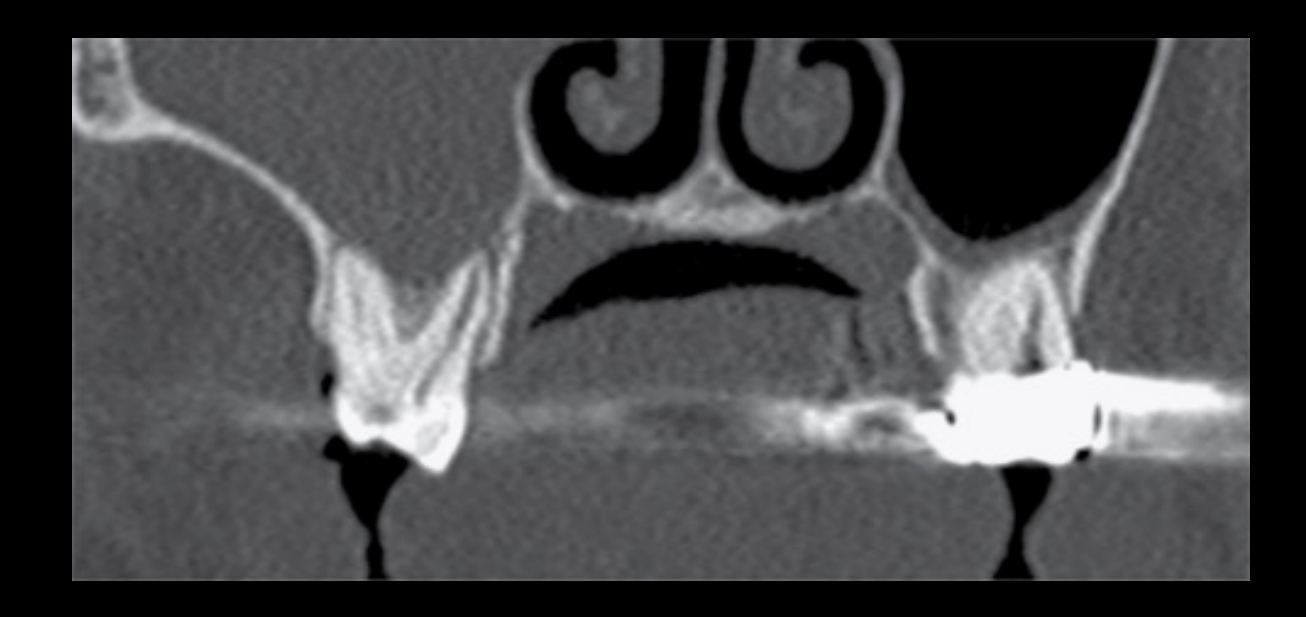
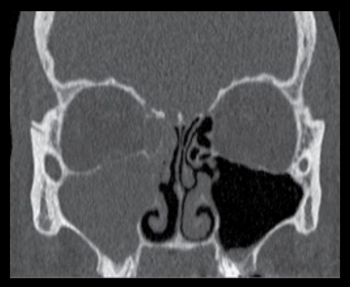
Odontogenic Sinusitis Cases11
Inflammatory change in right paranasal sinuses secondary to odontogenic infection.
Case courtesy of Dr Bernard Koong
 |  |
Non-Periapical Inflammatory Lesion Cases1213141516
Find inflammatory lesions.
Hint: not periapical
Ref. White and Pharoah’s Oral Radiology: Principles and Interpretation 8th Edition
Case courtesy of Dr Bernard Koong
Lecturer — Osteomyelitis Pathogenesis
Osteomyelitis usually begins with infection and progresses through inflammation, vascular compromise, and bone necrosis.
- Infection commonly starts in a necrotic pulp and spreads from the apex through cancellous or medullary bone, marrow spaces, Haversian canals, and Volkmann canals.
- Acute inflammation produces pus and raises intraosseous pressure, compressing blood vessels; the mandible is particularly vulnerable because of its limited collateral circulation.
- Ischaemia produces necrotic bone that separates as a sequestrum, which remains a focus of infection that antibiotics cannot effectively penetrate.
- The body attempts to contain the infection by forming new bone around the sequestrum, while chronic inflammation may produce fibrosis, sclerosis, and sinus tracts.
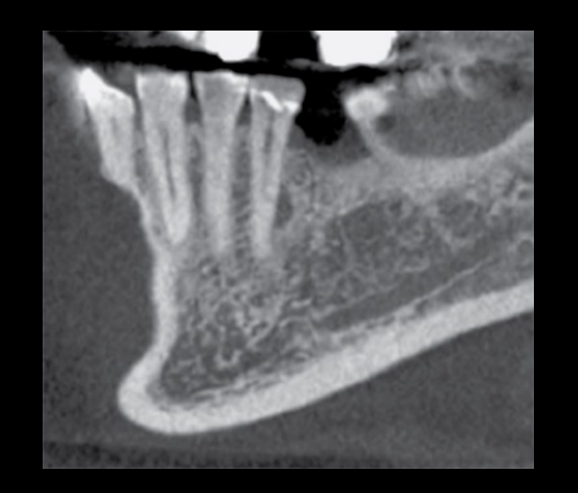
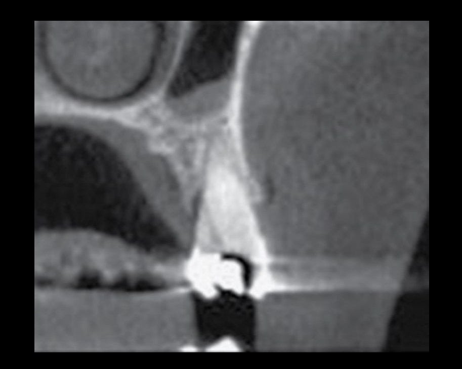
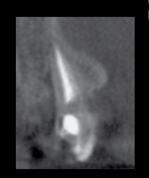
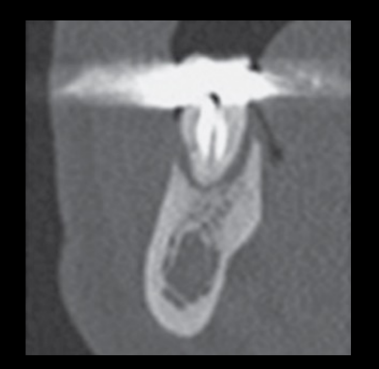
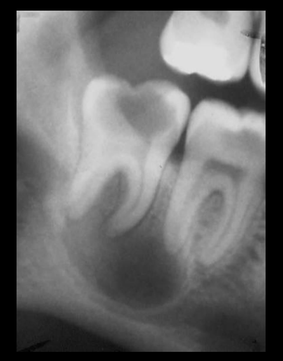
Osteomyelitis
- Widespread inflammation of bone
- Most are associated with odontogenic infections, i.e. periapical inflammatory lesions
- Others: trauma (esp. compound fractures) and radiation therapy
- Most common in the posterior mandible
Classification and Pathogenesis1718
Numerous classifications exist, but there are 2 broad groups:
- ACUTE
- Suppurative and rarefying
- CHRONIC (> 4 weeks)
- Non-suppurative and sclerosing
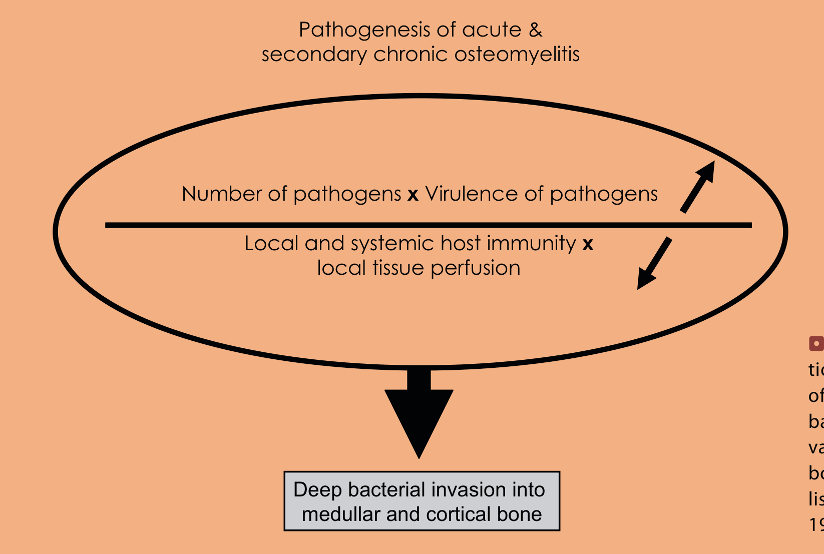
Pathogenesis of acute and secondary chronic osteomyelitis
- Number of pathogens × virulence of pathogens
- Local and systemic host immunity × local tissue perfusion
- Deep bacterial invasion into medullary and cortical bone
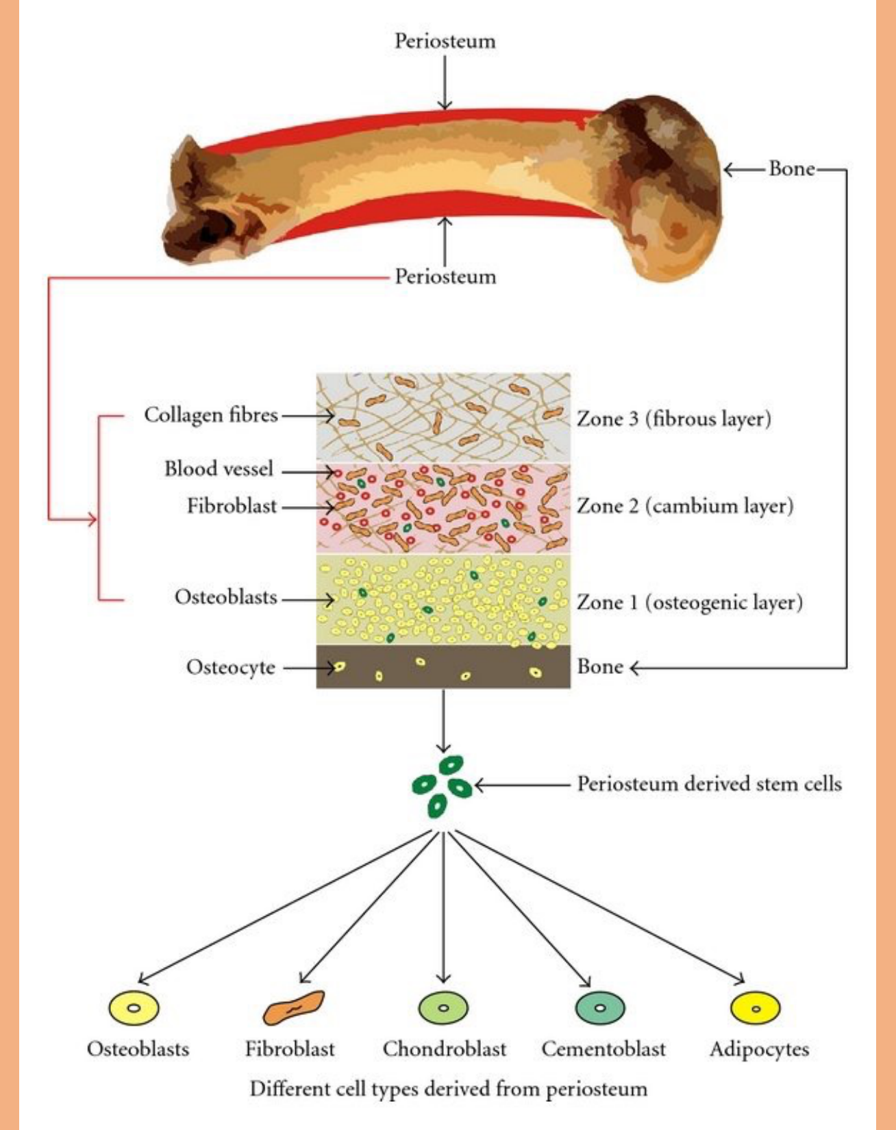
Periosteum structure (labeled diagram): Periosteum overlying bone; collagen fibres (Zone 3, fibrous layer); blood vessel; fibroblast; Zone 2 (cambium layer); osteoblasts; Zone 1 (osteogenic layer); osteocyte; bone. Periosteum-derived stem cells give rise to osteoblasts, fibroblasts, chondroblasts, cementoblasts, and adipocytes.
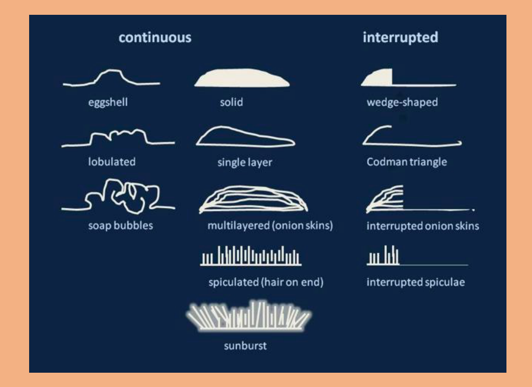
Periosteal Reaction Patterns
Pattern of Periosteal Reaction19
-
Single layer
-
Solid
-
Multilayered (lamellated / onion skin)
-
Spiculated
- Perpendicular “hair-on-end”
- Divergent “sunburst”
- Sloping “velvet”
-
Complex
-
Codman’s triangle
-
Spiculated and sunburst patterns were described as more associated with malignant tumours.
-
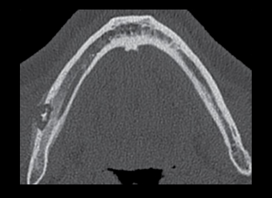
In adult osteomyelitis, the periosteal reaction may be thin and single-layered or thicker and solid.
-
Periosteal reaction may be minimal, and some patients may have a cortical defect without an obvious periosteal reaction. Descriptive terms for periosteal reaction patterns: continuous; interrupted; eggshell; solid; wedge-shaped; lobulated; single layer; Codman triangle; soap bubbles; multilayered (onion skins); interrupted onion skins; spiculated (hair on end); interrupted spiculae; sunburst.
 |  |
Imaging Modalities and Features
Imaging Modalities20
-
2D imaging is insufficient
-
Cone Beam CT
-
Multislice CT
-
MRI
-
Nuclear Medicine Imaging (Gallium Scan)
-
CT was described as the gold-standard modality for osteomyelitis.
-
Multislice CT demonstrates both hard-tissue and soft-tissue changes.
-
MRI is the most sensitive modality for very early osteomyelitis confined to the marrow without substantial bony destruction.
-
If CT does not demonstrate substantial abnormality but early osteomyelitis is suspected, MRI may be appropriate.
 |  |
Imaging Features
-
Ill-defined lucencies (lytic / destruction of bone) in the ACUTE phase, becoming more well-defined in the CHRONIC phase
-
Loss of normal trabecular pattern
-
Cortical destruction is common
-
Extensive adjacent sclerosis, depending on chronicity
-
Periosteal reaction (periosteal new bone formation)
-
Sequestra formation → key feature
-
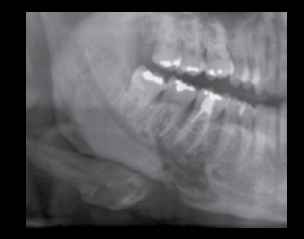
Chronic osteomyelitis often produces mixed lucent and opaque appearances because of sclerosis and new bone formation.
-
Sequestrum represents necrotic bone separated from healthy bone.
-
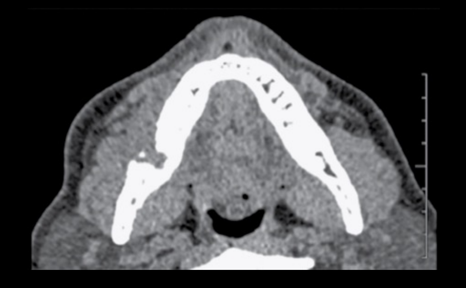
Soft-tissue swelling may appear on CT as loss of the normal distinction between muscle, fat, and other soft tissues.
-
Gas may appear as small black foci within involved tissues and was associated with gas-producing bacteria.
Periosteum Structure and Age Variation212223
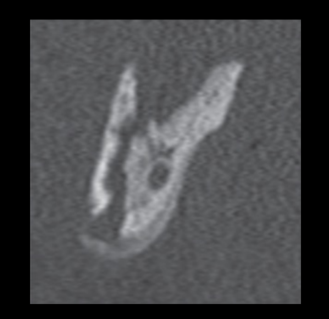
The structure of periosteum varies with age. In children it is thicker, more vascular, active, and loosely attached, as compared to adults where it is thinner, less active, and firmly adherent.
Lecturer — Age and Periosteal Response
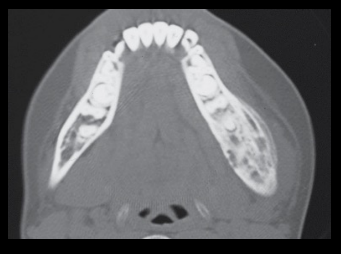
The age-related structure of the periosteum affects the appearance of osteomyelitis.
- In adults, osteomyelitis may produce a relatively thin, single-layered periosteal reaction.
- In children, the active periosteum may produce a much thicker, multilayered or onion-skin appearance and more extensive periosteal change in response to infection.
Jaw Infection Involving Soft Tissues
Infection may fail to drain into the oral cavity and may involve various spaces, including the submandibular, sublingual, masticator, parapharyngeal and parotid spaces.
Lecturer — Deep-Space Assessment
Deep neck spaces are potential sites for spread from extensive tooth infections and can be assessed on CT and MRI.
- MRI provides detailed visualisation of soft tissues.
- When reporting collections, describe their relationship to adjacent anatomical structures.
Lecturer — Soft-Tissue Spread
When osteomyelitis becomes extensive or is not treated early, infection may break through the cortex and produce cellulitis.
- The infection is no longer confined to bone and may initially localise around the source before spreading rapidly through deep spaces.
- Extensive swelling may narrow the airway.
- Cellulitis and abscess collection can be life threatening.
- Other potential complications include airway compromise, cavernous sinus thrombosis, cerebral abscess, orbital infections and mediastinitis.
Overview and Imaging Modalities24
- Multislice CT with intravenous contrast is the first modality of choice.
- MRI is also useful.
Post-Contrast CT Findings25262728
- Thickening of the skin and subcutaneous fat stranding
- Fat planes often demonstrate increased density ‘dirty’ from oedema
- Involved muscles are swollen and enhanced
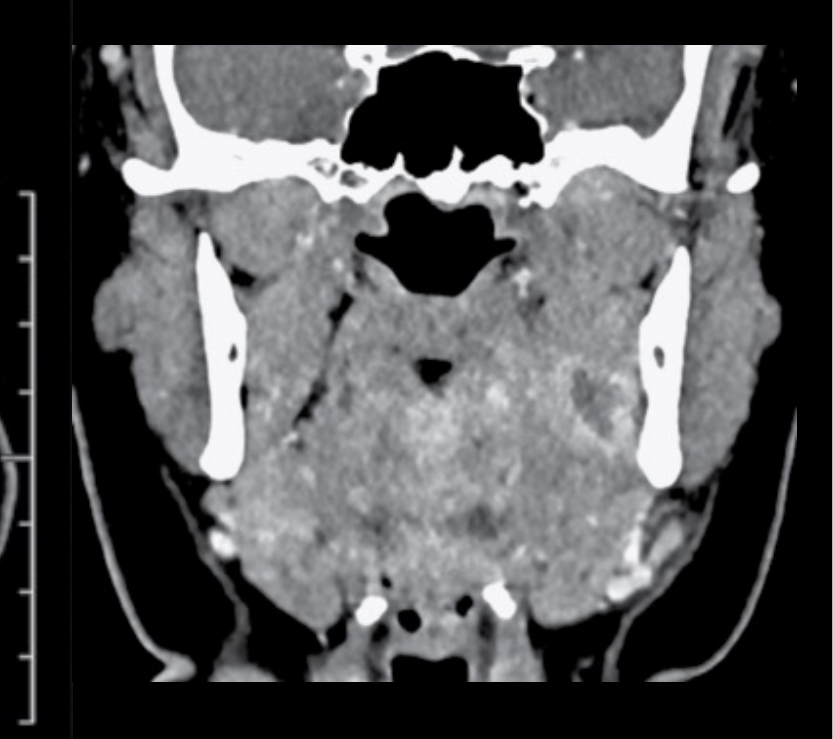
- Abscess collection presents as a spherical or lobulated fluid density focus with rim enhancement.
Lecturer — Abscess Enhancement
Normal fat appears dark and clearly defined, but oedema and inflammation alter its density so that it becomes dirty or strandy.
- Contrast does not enter the abscess itself; it surrounds the collection because of the inflammatory process around it.
 |  |  |
 |  |
Post-Extraction Infection Cases293031
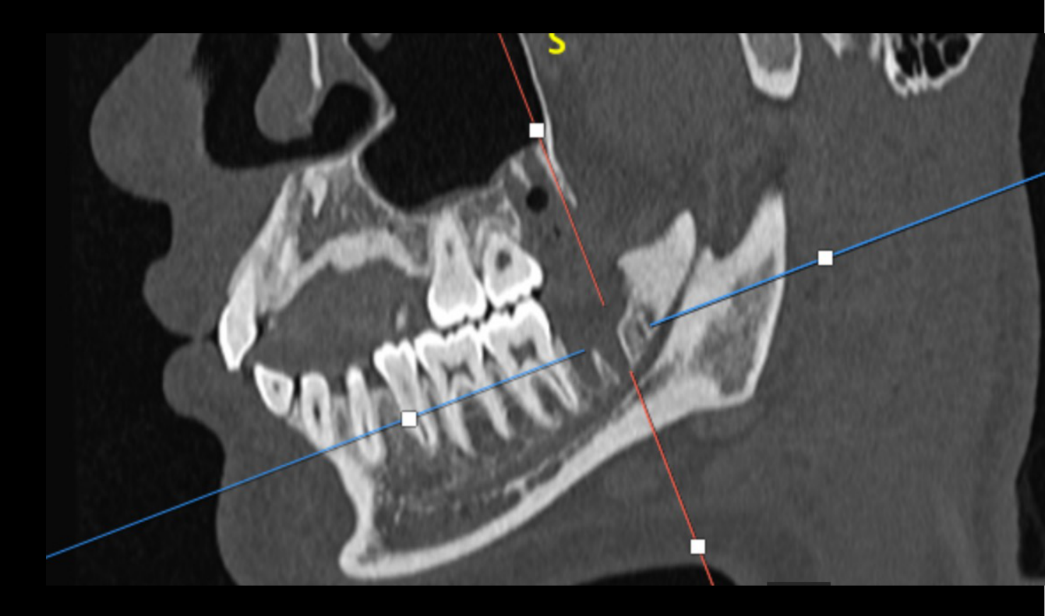
Post 28 & 38 Exo Infection
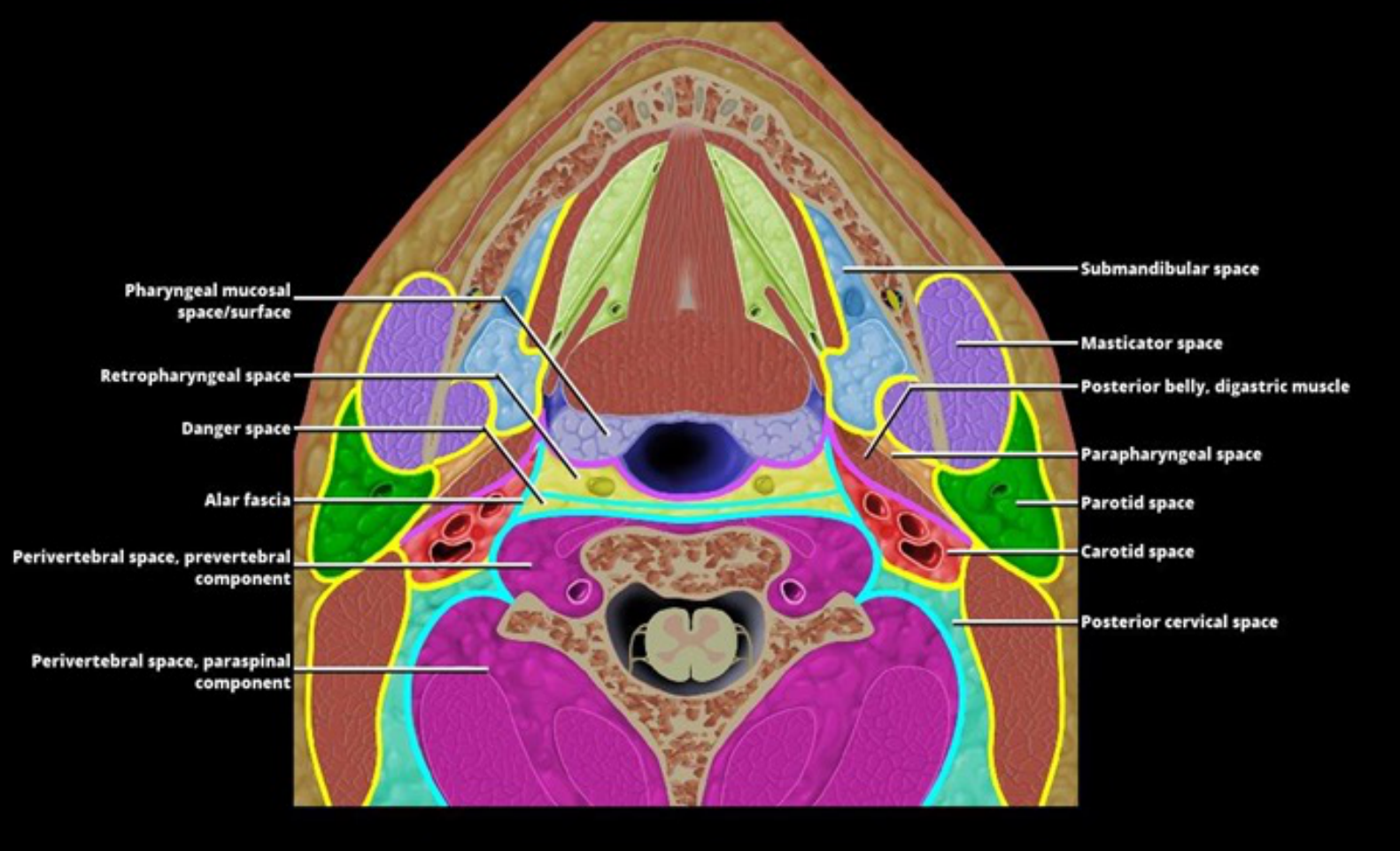
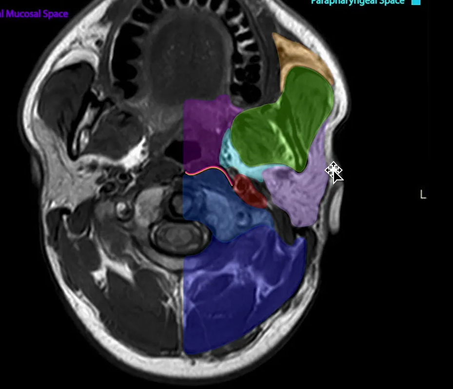
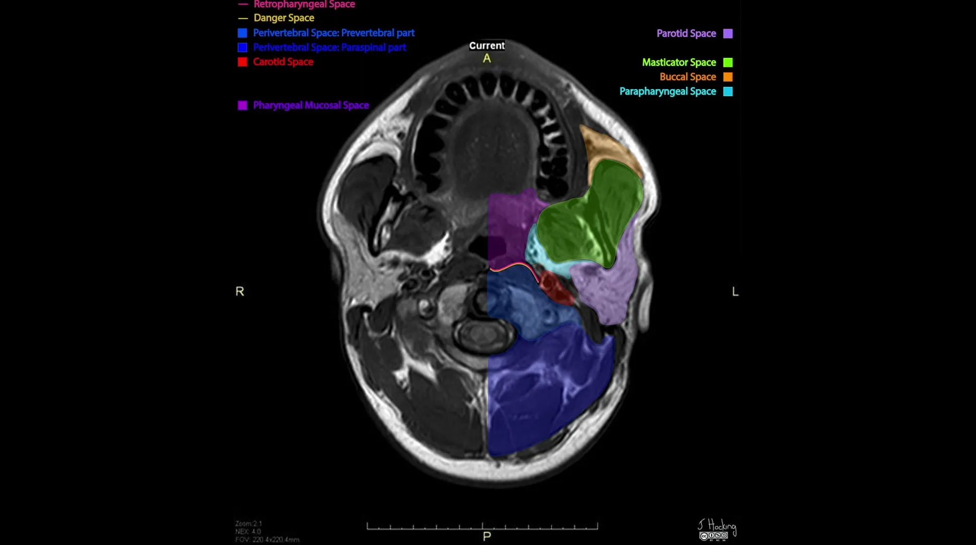
Anatomy of Deep Neck Spaces3233
 |  |
Anatomical Spaces and Fascia34
- Pharyngeal mucosal space/surface
- Retropharyngeal space
- Danger space
- Alar fascia
- Perivertebral space, prevertebral component
- Perivertebral space, paraspinal component
- Submandibular space
- Masticator space
- Posterior belly, digastric muscle
- Parapharyngeal space
- Parotid space
- Carotid space
- Posterior cervical space
 | 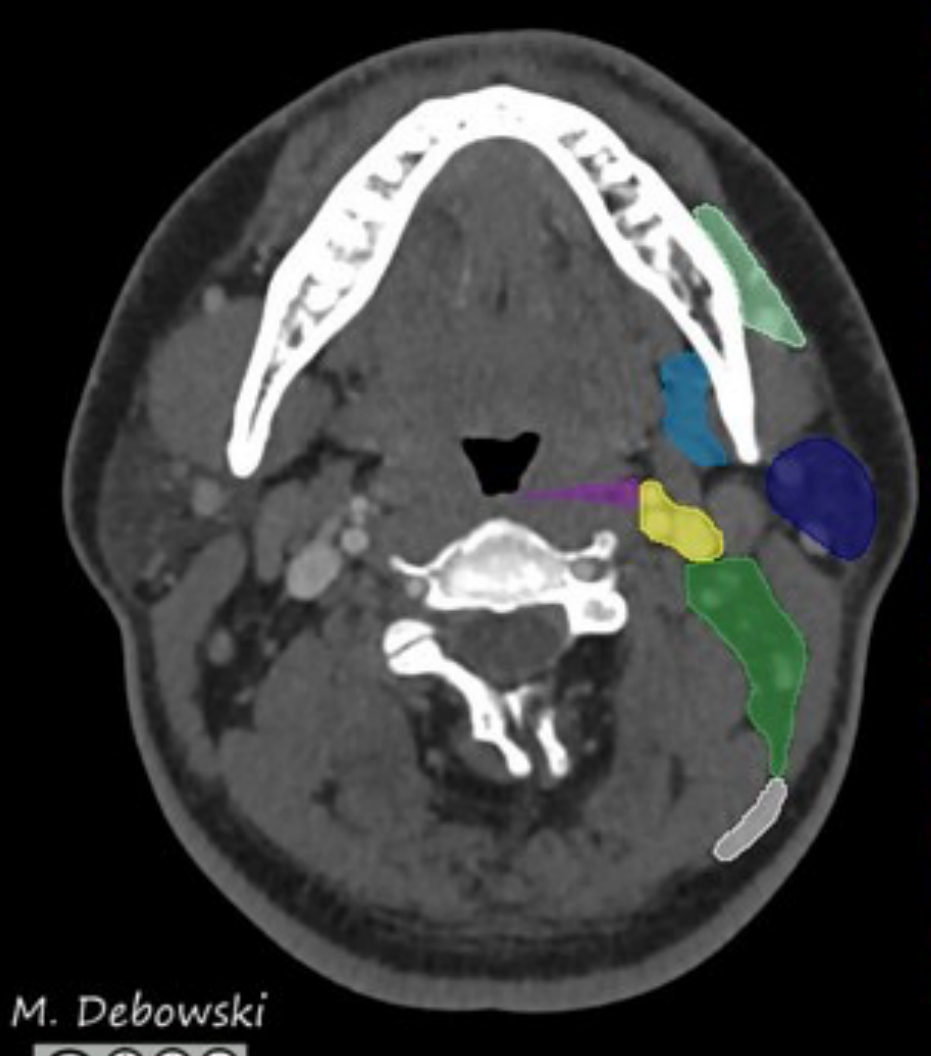 |
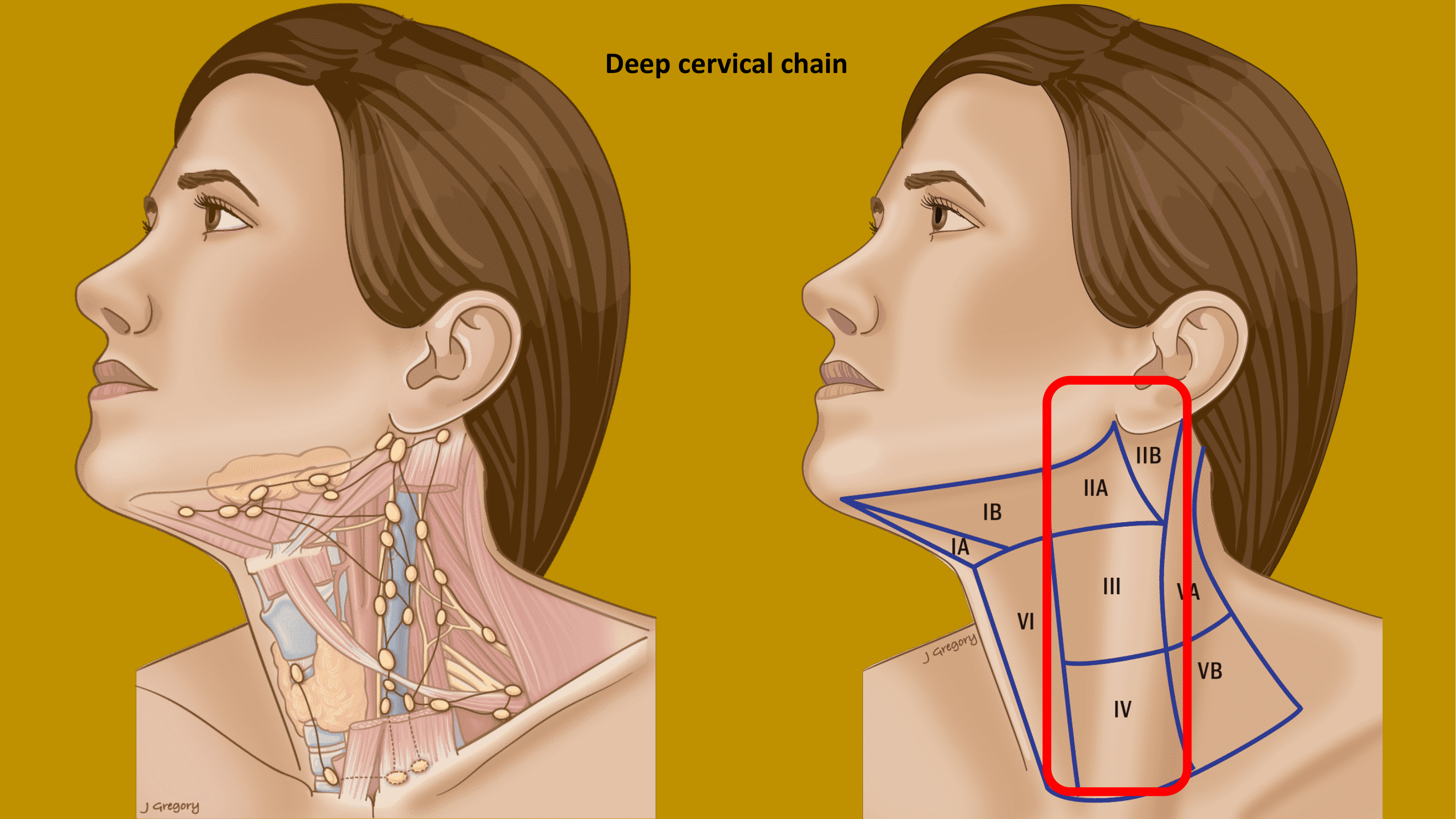
Nodal Levels
- Ia Submental
- Ib Submandibular
- IIa Upper jugular (anterior)
- IIb Upper jugular (posterior)
- III Mid jugular
- IVa Lower jugular
- IVb Medial supraclavicular
- Va Upper posterior triangle
- Vb Lower posterior triangle
- Vc Lateral supraclavicular
- VIa Anterior jugular
- VIb Para-tracheal
- VIIa Retropharyngeal
- VIIb Retro-styloid
- VIII Parotid
- IX Bucco-facial
- Xa Retro-auricular
- Xb Occipital
M. Debowski — STAT
A, R, P, S, A, I, S, I
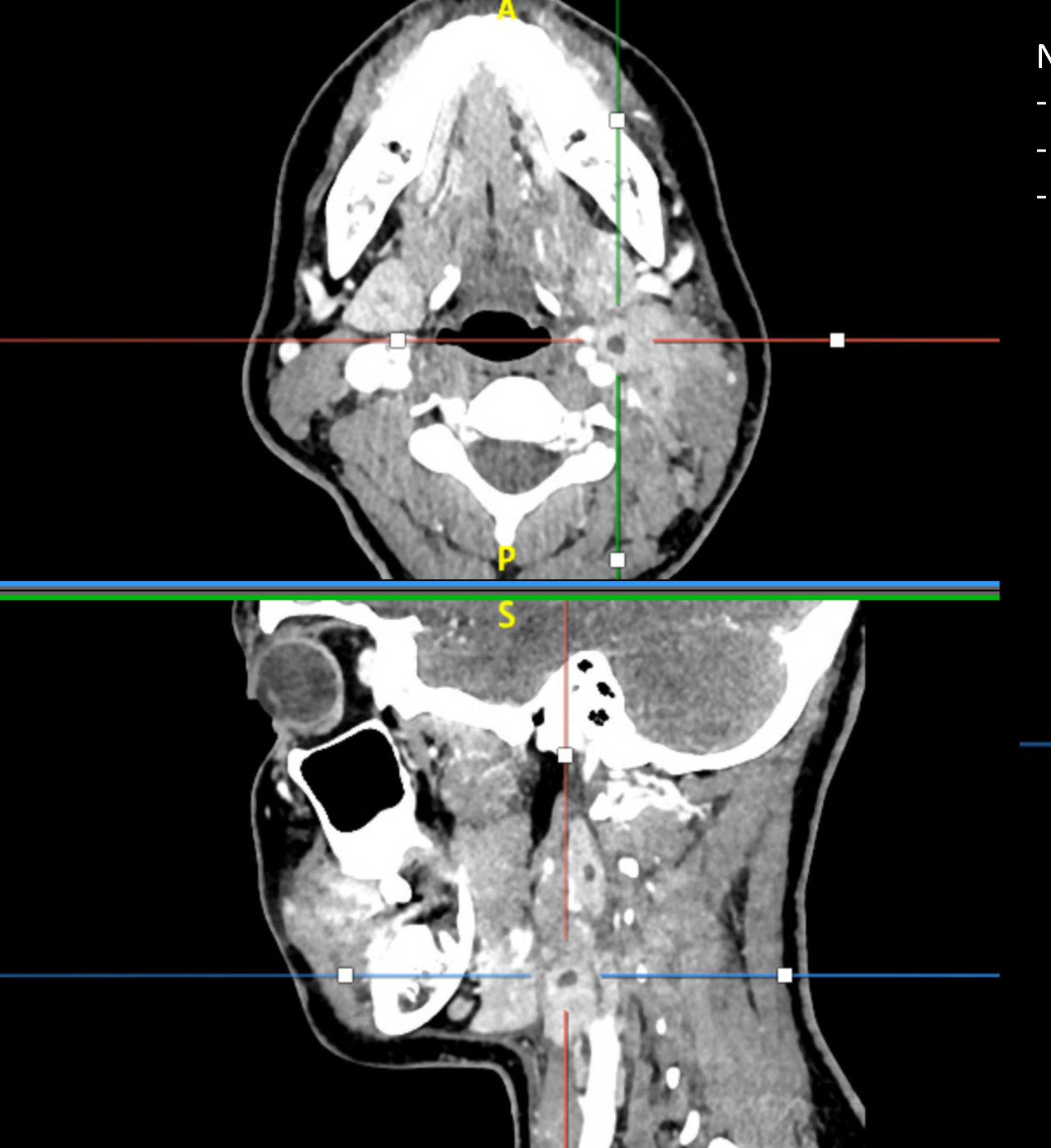
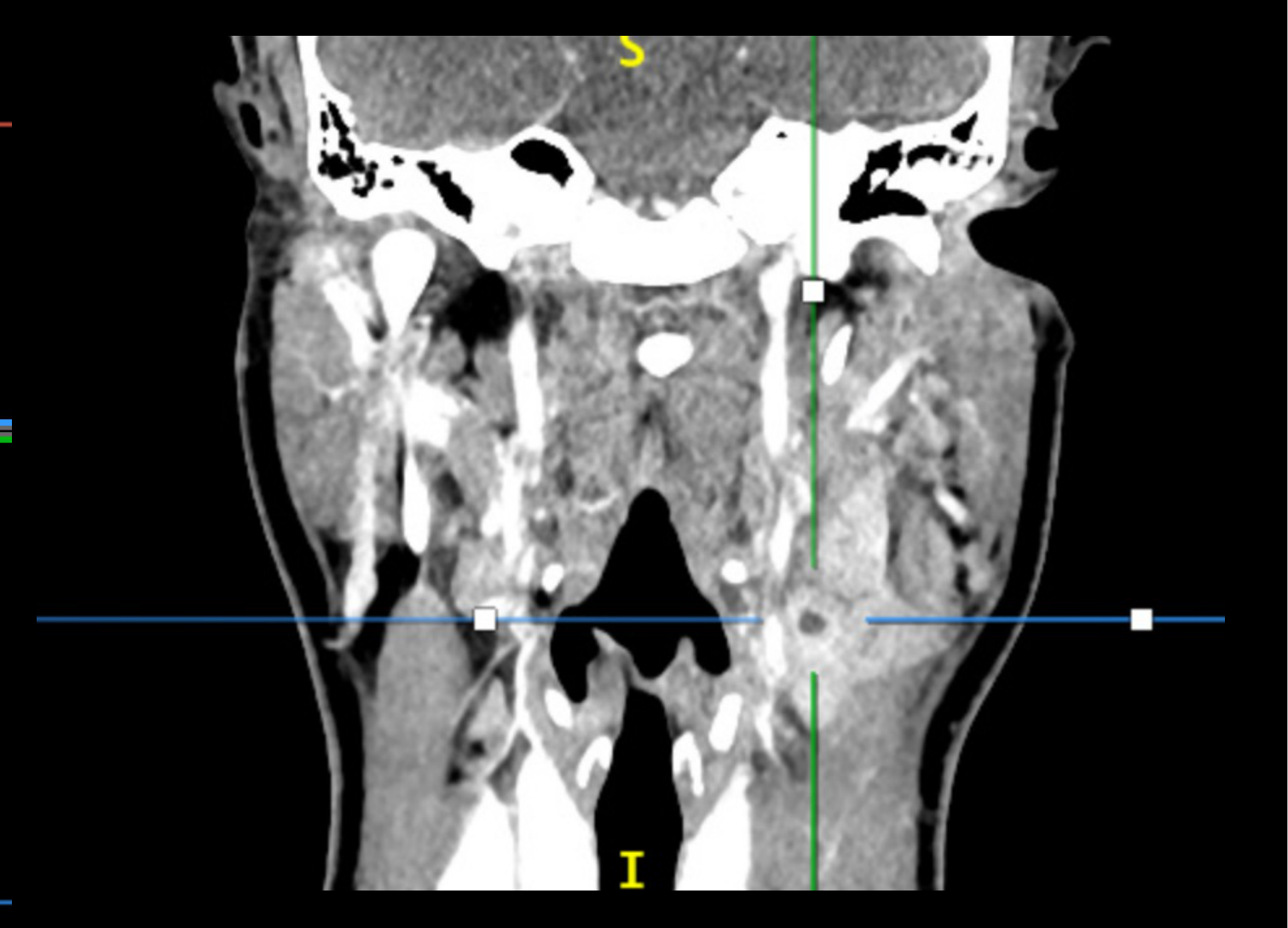
Abscess and Necrotic Collection Cases353637
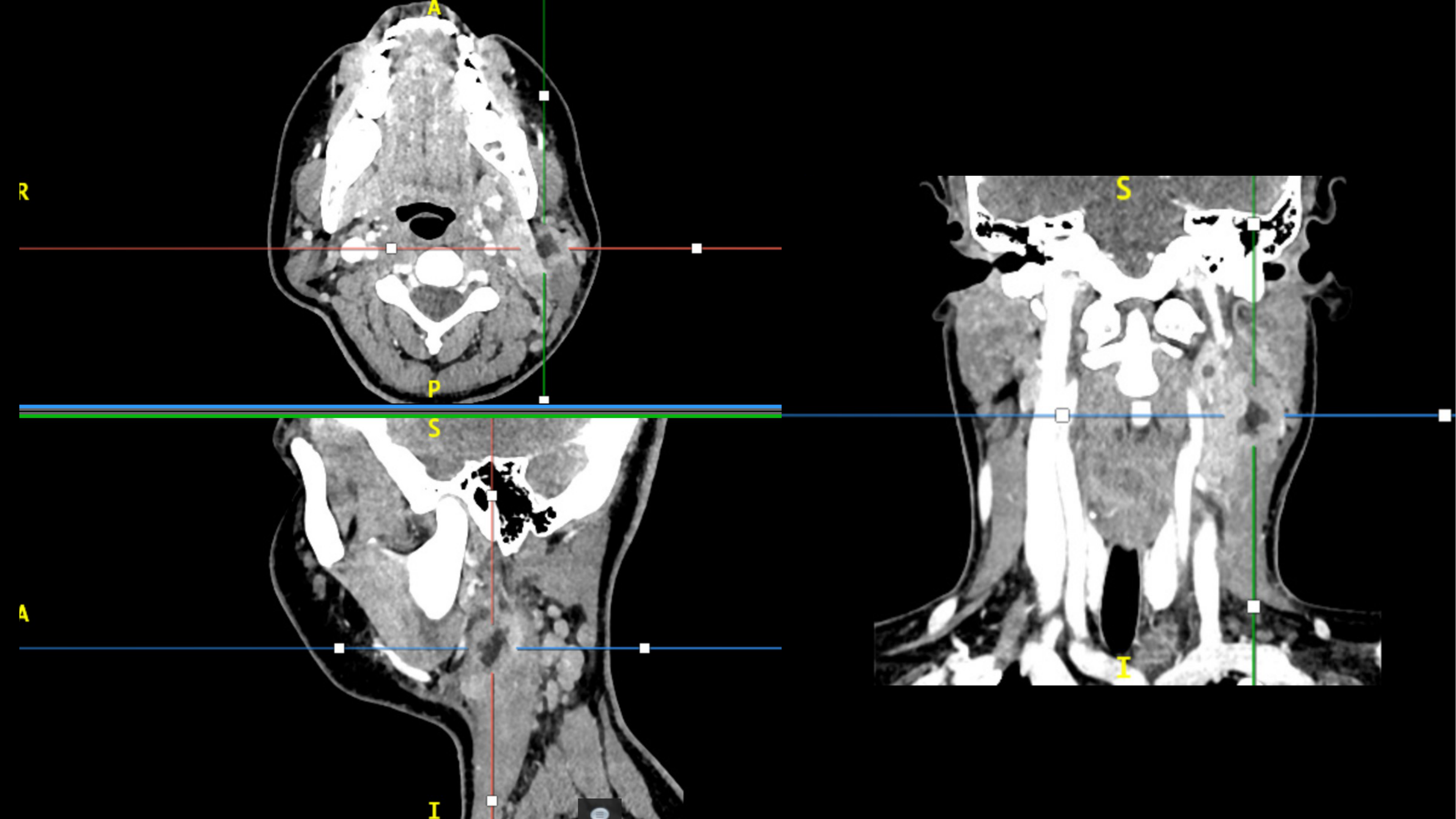
Necrotic mass with multiple foci of fluid collection:
- posterior to the submandibular gland
- superficial to the carotid sheath
- anterior to the sternocleidomastoid muscle
 |  |
 |
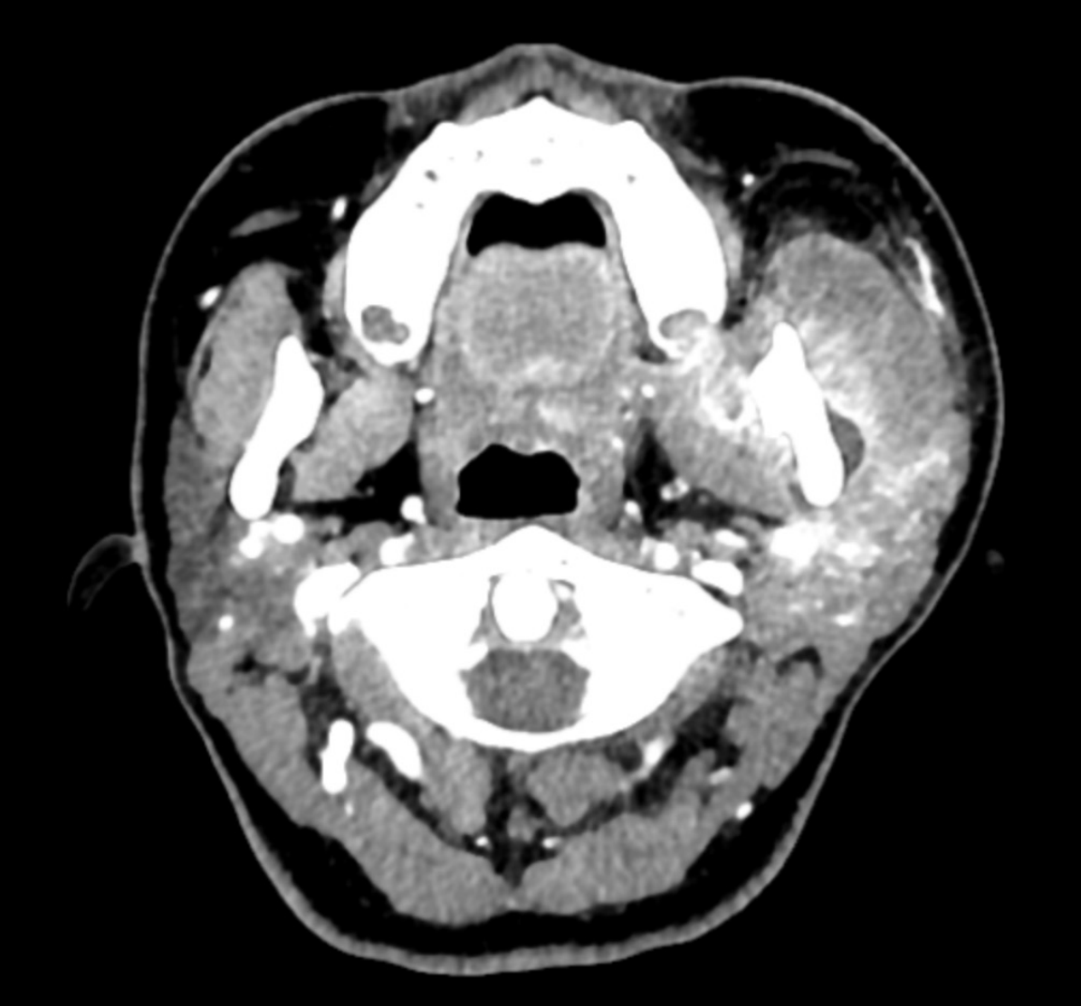
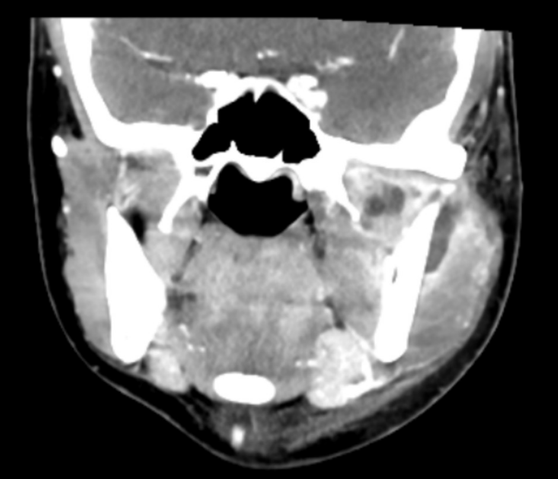
Cellulitis Cases383940
Post 8’s Exo Infection Pt 2.
- Multiple foci of collection in the left masticator space medial pterygoid, lateral pterygoid and deep portion of the masseter and direct extension into the left temporomandibular joint
- Oedema of affected muscles of mastication
- Thickening of platysma muscle
- Subcutaneous fat stranding / oedema
- Fat stranding in left submandibular space. Reactive nodes (1B)
- Parapharyngeal & pharyngeal mucosal spaces are normal
End.
Clinical Cases
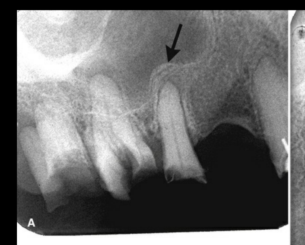
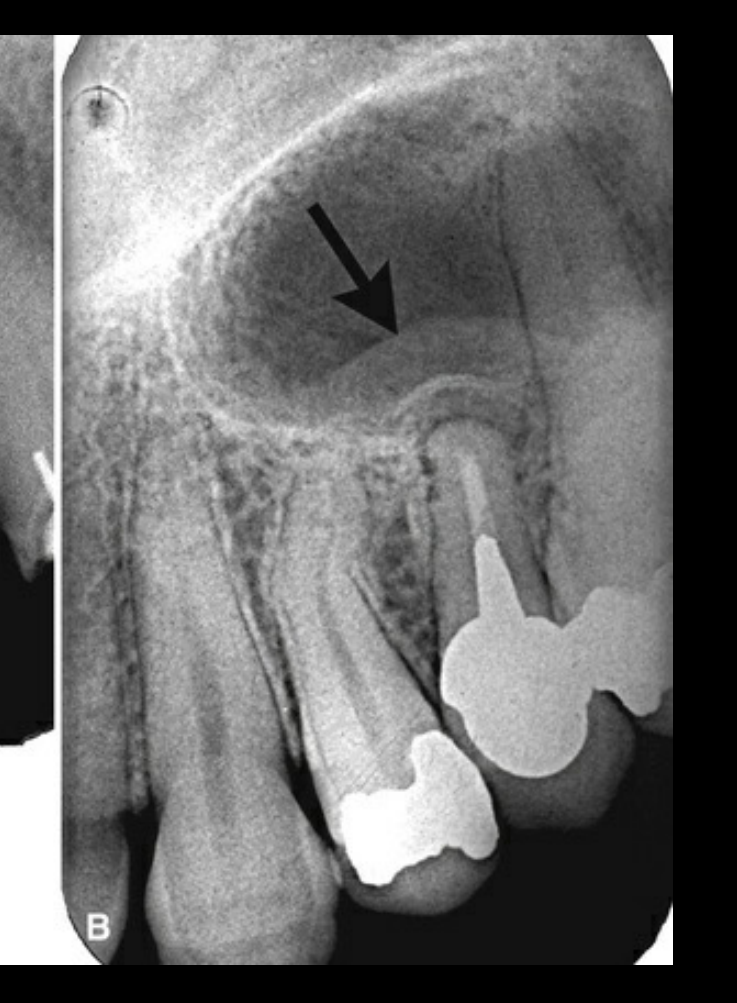
Case: Maxillary sinus floor changes adjacent to root remnants
Question
Scenario: A patient with multiple decoronated teeth or root remnants in the maxilla.
What’s shown: Radiographs showing periapical inflammatory lesions related to the root remnants, with the adjacent maxillary sinus floor appearing lifted and sclerosed, along with reactive changes within the antrum.
Consider: What is the term for the reactive changes seen in the maxillary sinus floor related to these adjacent inflammatory lesions?
Answer
Observations:
- Lifting and mild sclerosis of the maxillary sinus floor.
- Reactive changes within the maxillary antrum.
Reasoning: The inflammatory lesions adjacent to the root remnants cause a reactive periosteal response on the adjacent sinus floor, leading to sclerosis and lifting.
Takeaway: Inflammatory lesions in the maxilla can cause secondary reactive changes in the adjacent maxillary sinus floor, known as periostitis or periosteal reaction.
Case: Furcation lucency in a heavily restored molar
Question
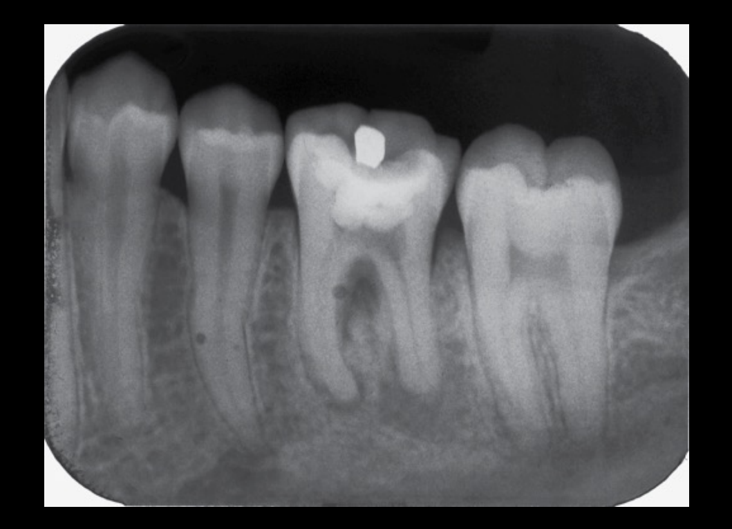
Scenario: A heavily restored mandibular molar with evidence of commenced endodontic therapy.
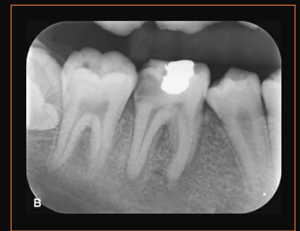
What’s shown: A radiograph demonstrating a well-defined periapical lucency with surrounding sclerosis on the distal root, widening of the apical PDL space on the mesial root, and a separate lucency centered on the furcation region.
Consider: What is the most likely cause of the localized bone loss centered on the furcation region in this infected molar?
Answer
Observations:
- Well-defined periapical lucency with sclerosis on the distal root (chronic change).
- Widened PDL space on the mesial root (apical periodontitis).
- Localized bone loss centered on the furcation, not the apical foramen.
Reasoning: While furcation bone loss can be periodontal, a localized inflammatory lesion centered on the furcation in an endodontically involved molar is highly indicative of a structural tooth defect.
Takeaway: Localized furcation bone loss in an infected molar is often an inflammatory lesion associated with an undisplaced vertical root fracture or root crack.
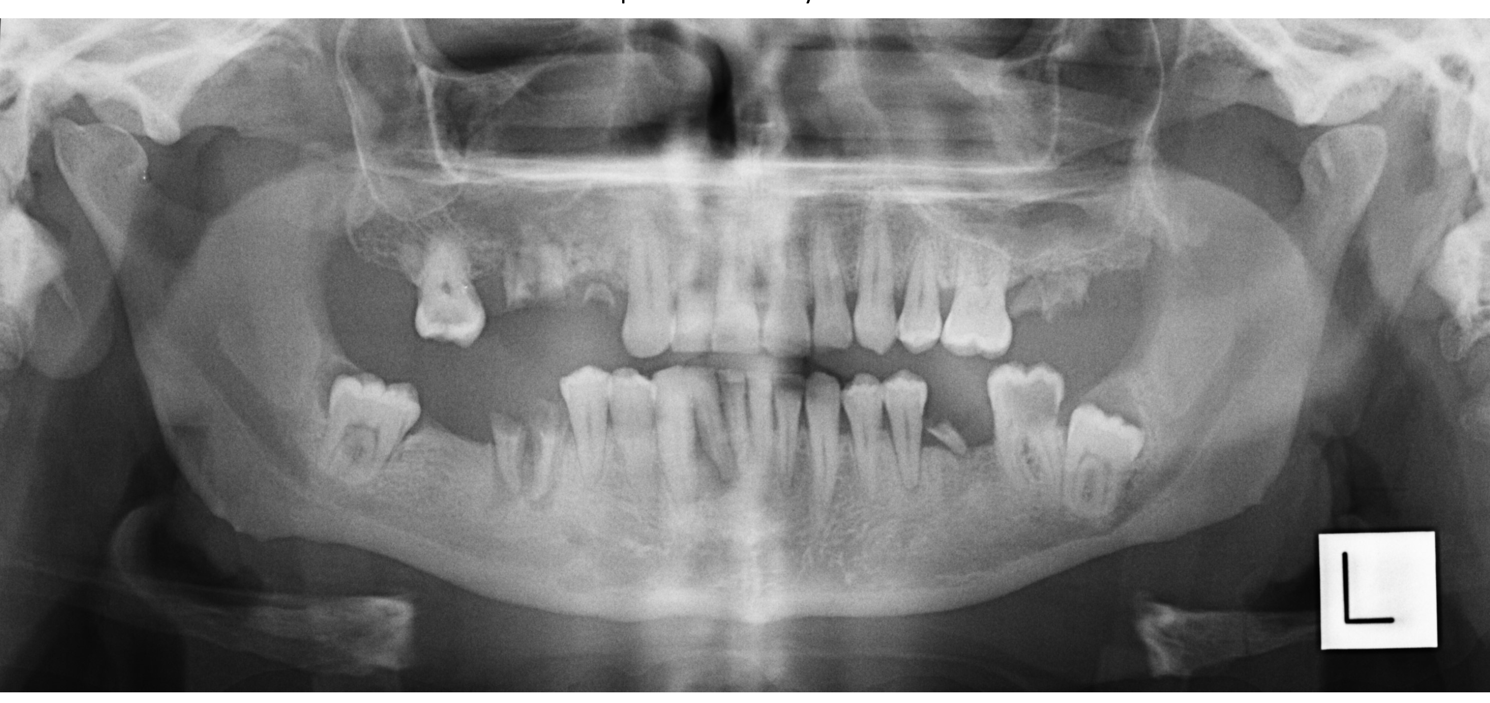
Case: OPG overview of multiple root remnants with sinus changes
Question
Scenario: An overview radiograph of a patient with multiple heavily compromised teeth and root remnants.
What’s shown: An OPG showing multiple root remnants with associated inflammatory lesions and sclerotic changes. On the right, there is disruption of the sinus floor with mucosal changes. On the left, the sinus floor is intact but shows reactive mucosal thickening.
Consider: What secondary sinus changes are visible on this OPG related to the inflammatory lesions of the maxillary teeth?
Answer
Observations:
- Disruption of the right maxillary sinus floor with mucosal changes.
- Reactive mucosal thickening within the left maxillary sinus despite an intact floor.
Reasoning: The chronic inflammatory lesions from the maxillary root remnants trigger reactive mucosal and bony changes in the adjacent sinuses, manifesting as periostitis and mucosal thickening.
Takeaway: OPGs are useful for providing an overview of multiple inflammatory lesions and their secondary reactive effects on adjacent structures like the maxillary sinuses.
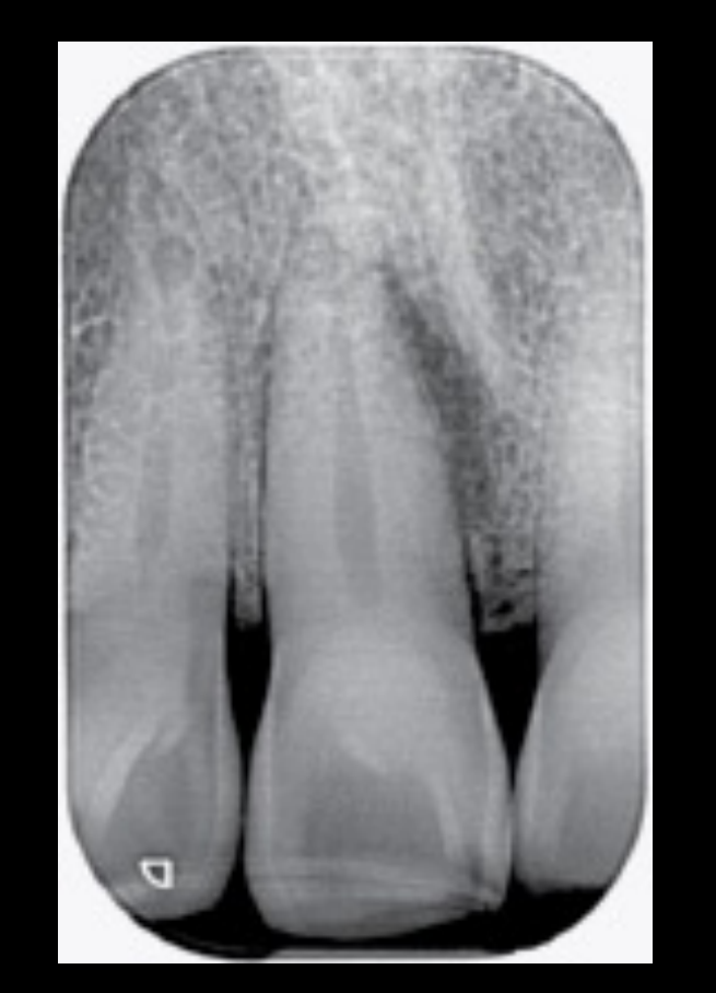
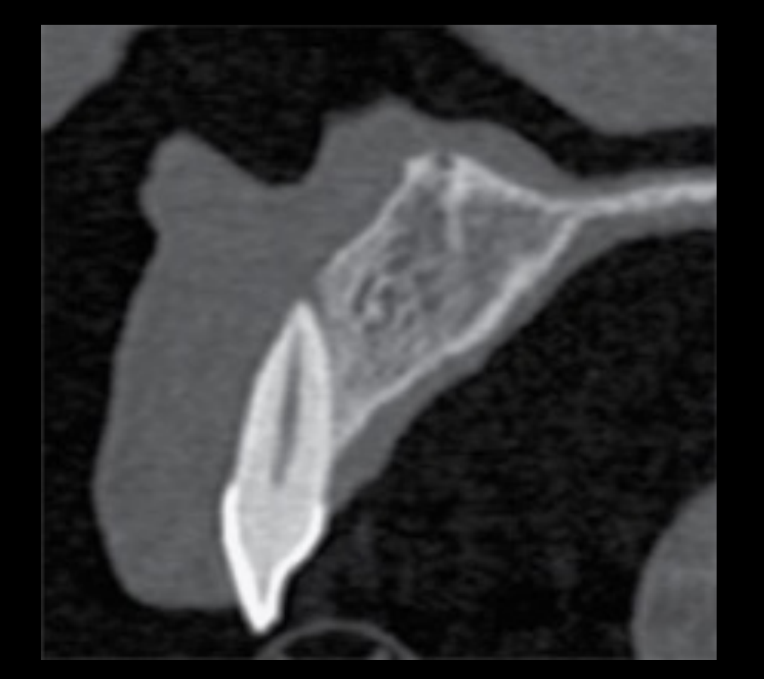
Case: CBCT evaluation of a painful anterior tooth with equivocal PA findings
Question
Scenario: A patient with a painful maxillary anterior tooth.
What’s shown: A periapical radiograph showing apparent widening of the distal PDL space. A subsequent CBCT scan reveals a tiny apical lucency and perforation of the overlying labial cortex.
Consider: Why was CBCT indicated for this tooth, and what did it reveal that the 2D radiograph missed?
Answer
Observations:
- PA radiograph shows apparent distal PDL widening (actually an artifact from the incisive canal projection).
- CBCT shows a tiny apical lucency and labial cortical perforation.
Reasoning: 2D imaging is governed by beam projection angles, which can obscure or mimic pathology. CBCT provides 3D imaging free from superimposition and projection artifacts.
Takeaway: When 2D imaging is inconclusive or shows equivocal findings in a symptomatic tooth, CBCT is indicated to accurately detect small apical lucencies and cortical perforations.
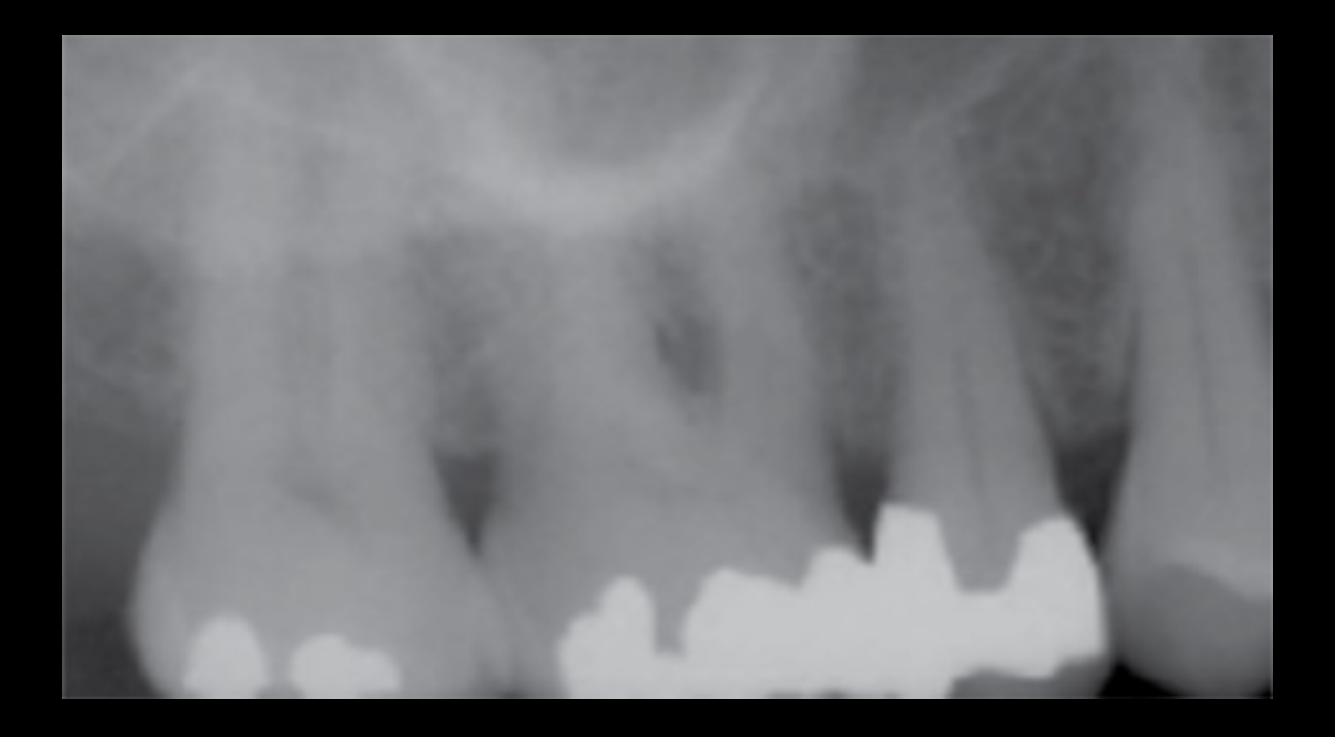
Case: CBCT evaluation of heavily restored posterior teeth with obscured OPG findings
Question
Scenario: A patient with heavily restored maxillary posterior teeth (15, 16, 17).
What’s shown: An OPG showing opaque superimposition with no clear apical lucency. A CBCT coronal view of tooth 16 reveals an apical lucency, effacement/perforation of the sinus floor, reactive mucosal thickening in the antrum, surrounding sclerosis, and root resorption.
Consider: What does the CBCT reveal for tooth 16 that was obscured on the OPG, and what stage of disease does this represent?
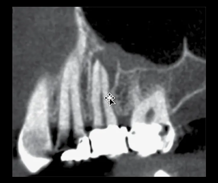
Answer
Observations:
- OPG shows superimposition obscuring details.
- CBCT shows an apical lucency, sinus floor perforation, reactive mucosal thickening, surrounding sclerosis, and root resorption.
Reasoning: The OPG’s 2D superimposition masked the pathology. The CBCT clearly delineates the chronic inflammatory lesion extending into the sinus.
Takeaway: CBCT is essential for evaluating heavily restored teeth where OPG superimposition obscures chronic inflammatory lesions and their extension into the maxillary sinus.
Case: Severe unilateral odontogenic sinusitis
Question
Scenario: A case of maxillary sinus inflammation arising from a dental infection.
What’s shown: An image showing complete opacification of the maxillary sinus due to extensive reactive mucosal thickening, with complete obstruction of the sinus draining pathway.
Consider: What imaging features characterize this severe case of unilateral odontogenic sinusitis?
Answer
Observations:
- Complete opacification of the maxillary sinus.
- Extensive reactive mucosal thickening.
- Complete obstruction of the draining pathway.
Reasoning: The dental infection perforated the sinus floor and disrupted the Schneiderian membrane, leading to severe reactive mucosal changes that completely filled the sinus and blocked drainage.
Takeaway: Odontogenic sinusitis can present with complete sinus opacification and obstruction of the draining pathway due to severe reactive mucosal thickening.
Case: Periradicular lesion from a palatally running tooth fracture
Question
Scenario: A tooth with an inflammatory lesion not centered on the apex.
What’s shown: A CBCT coronal view showing a distally positioned lucency and a visible line extending to the palatal vault, causing localized palatal PDL space thickening.
Consider: What is the cause of the localized palatal PDL space thickening and distally positioned lucency in this case?
Answer
Observations:
- Distally positioned lucency.
- A palatally running fracture line visible on the coronal CBCT view.
- Localized palatal PDL space thickening.
Reasoning: The fracture line extends palatally, causing localized periradicular inflammation rather than a classic apical periodontitis.
Takeaway: Not all odontogenic infections are centered at the apex; lateral canals or tooth fractures can cause localized periradicular periodontitis away from the apical foramen.
Case: Periradicular lesions and vertical bone loss in an endodontically treated tooth
Question
Scenario: An endodontically treated tooth presenting with periradicular radiolucencies.
What’s shown: CBCT images showing a tiny palatal periradicular lucency, as well as buccal and lingual periradicular inflammatory lesions, with isolated vertical bone loss adjacent to the root.
Consider: What is the likely etiology of the isolated vertical bone loss and multiple periradicular lucencies in this endodontically treated tooth?
Answer
Observations:
- Tiny palatal, buccal, and lingual periradicular lucencies.
- Isolated vertical bone loss adjacent to the root.
Reasoning: Multiple periradicular lesions and isolated vertical bone loss in an endodontically treated tooth are classic signs of a structural failure rather than a simple missed canal.
Takeaway: Isolated vertical bone loss and multiple periradicular lucencies adjacent to an endodontically treated tooth are highly indicative of a vertical root fracture.
Case: Apparent periapical lucencies mimicking pathology on OPG
Question
Scenario: A cropped OPG showing apparent periapical lucencies in the posterior mandible.
What’s shown: Radiographs showing lucencies adjacent to dense opacities and near the submandibular fossa. The lamina dura and PDL spaces of the adjacent teeth are completely intact.
Consider: Why do these apparent lucencies appear on the radiograph despite an intact lamina dura and PDL space?
Answer
Observations:
- Apparent periapical lucencies.
- Intact lamina dura and PDL spaces.
- Adjacent dense opacities (bone islands) and proximity to the submandibular fossa.
Reasoning: The lucencies are radiographic artifacts caused by the contrast of adjacent dense bone islands and the anatomical concavity of the submandibular fossa, not true bone destruction.
Takeaway: Always check the lamina dura and PDL space; apparent lucencies with intact periodontal structures adjacent to dense bone islands or anatomical concavities are likely radiographic artifacts.
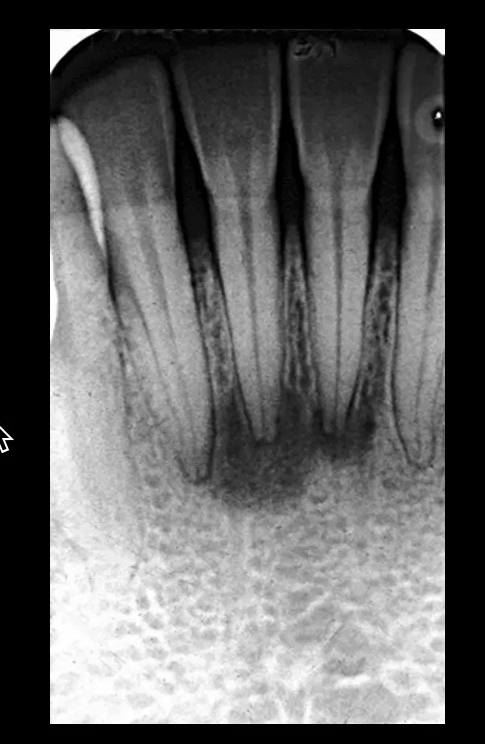
Case: Periapical lucencies with internal bone density and intact PDL
Question
Scenario: A patient with periapical lucencies in the anterior teeth.
What’s shown: Radiographs showing periapical lucencies with intact PDL spaces and partially intact lamina dura. The density within the lucencies shows some remaining bone trabeculation rather than being completely radiolucent.
Consider: What is the diagnosis for these periapical lucencies that mimic inflammatory lesions but have intact PDL spaces and internal bone density?
Answer
Observations:
- Periapical lucencies in anterior teeth.
- Intact PDL spaces and partially intact lamina dura.
- Internal bone density remaining within the lucencies.
Reasoning: The intact periodontal ligament indicates the teeth are not the source of infection. The mixed density represents fibrous and osseous tissue rather than pure inflammatory destruction.
Takeaway: Cemento-osseous dysplasia is a benign fibro-osseous lesion that mimics periapical inflammatory lesions but is distinguished by intact PDL spaces and internal bone density.
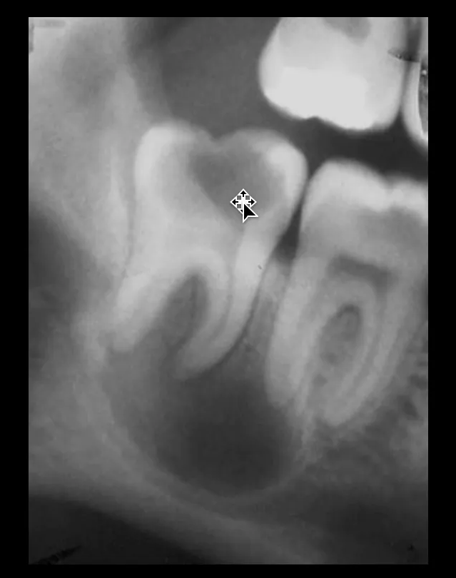
Case: Large mixed-density lucency mimicking a periapical lesion
Question
Scenario: A patient with a large periapical lucency and evidence of previous endodontic therapy, but the tooth responds to vitality tests.
What’s shown: A radiograph showing a large periapical lucency with multiple densities and multilocular features. The associated tooth responds to cold and EPT vitality tests.
Consider: What is the diagnosis for this large, mixed-density lucency that mimics a periapical inflammatory lesion, and how does vitality testing help?
Answer
Observations:
- Large periapical lucency with multiple densities.
- Evidence of previous endodontic therapy.
- Tooth responds to vitality tests (vital).
Reasoning: A vital tooth indicates the pulp is not necrotic, meaning the lesion is not of endodontic/inflammatory origin. The large size and mixed density are characteristic of a neoplasm.
Takeaway: Ameloblastoma can mimic periapical inflammatory lesions; vitality testing is crucial, as a vital tooth indicates the lucency is likely a tumor rather than an inflammatory lesion.
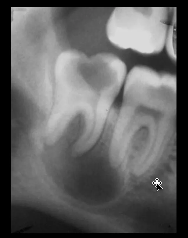
Case: Well-defined periapical lucency displacing the mandibular canal
Question
Scenario: A patient with a heavily carious tooth and a well-defined periapical lucency.
What’s shown: A radiograph showing a well-defined periapical lucency with loss of the lamina dura and widened PDL space. The mandibular canal is heavily displaced and compressed by the lesion.
Consider: What feature distinguishes this lesion from a standard periapical abscess or granuloma, and what is the diagnosis?
Answer
Observations:
- Well-defined periapical lucency.
- Loss of lamina dura and widened PDL.
- Heavy displacement and compression of the mandibular canal.
Reasoning: Inflammatory lesions like abscesses and granulomas do not typically displace or compress adjacent canals. Displacement indicates expansion via hydraulic pressure.
Takeaway: Displacement and compression of the mandibular canal by a periapical lesion is a classic sign of cystic activity (radicular cyst) rather than a simple abscess or granuloma.
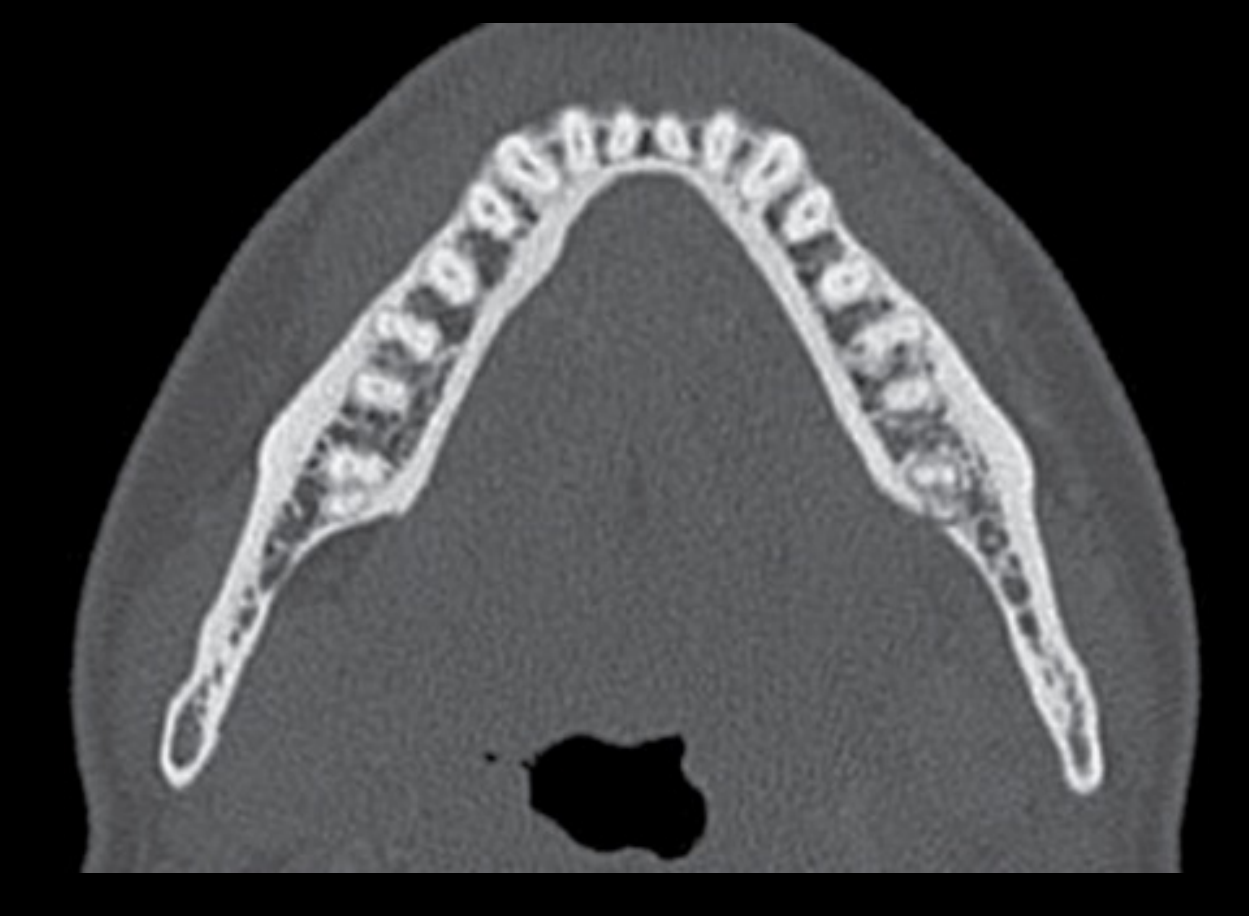
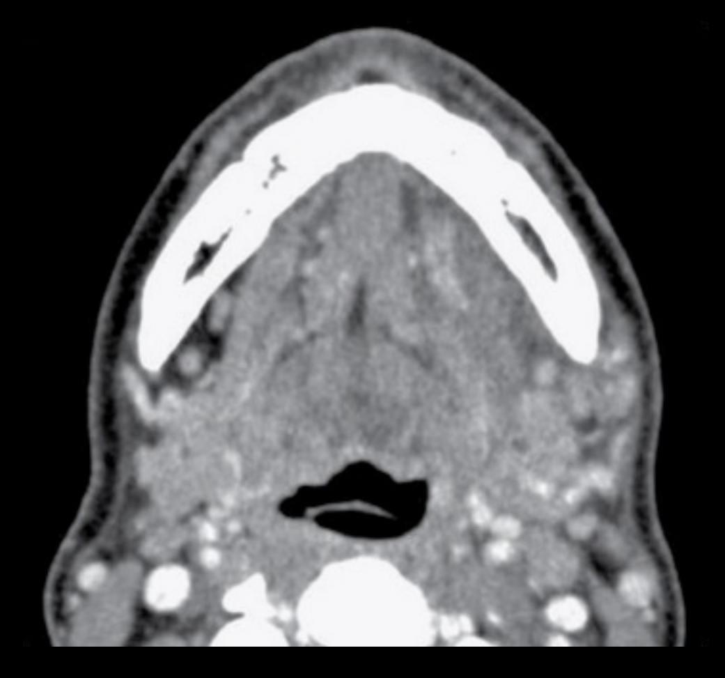
Case: Adult osteomyelitis with soft tissue swelling
Question
Scenario: An adult patient with osteomyelitis.
What’s shown: A CT scan showing single-layer, solid periosteal reactions of varying thickness. The soft tissue window reveals loss of normal fat planes and a lump of swollen soft tissue overlying the bone.
Consider: What periosteal reaction pattern and soft tissue changes are seen in this adult case of osteomyelitis?
Answer
Observations:
- Single-layer, solid periosteal reactions.
- Loss of normal soft tissue detail and fat planes.
- Active swelling forming a lump of soft tissue over the bone.
Reasoning: In adults, the periosteum is firmly attached and less active, resulting in single-layer reactions. The infection extends into the soft tissue, causing edema and loss of fat planes.
Takeaway: Adult osteomyelitis typically presents with single-layer, solid periosteal reactions and extensive overlying soft tissue swelling with loss of normal fat planes.
Case: Osteomyelitis periosteal reaction in a child
Question
Scenario: A pediatric patient with osteomyelitis.
What’s shown: An image demonstrating a thick, multi-layered periosteal reaction resembling an onion skin.
Consider: How does the periosteal reaction in a child with osteomyelitis differ from that of an adult?
Answer
Observations:
- Thick, multi-layered (“onion skin”) periosteal reaction.
Reasoning: A child’s periosteum is thicker, more vascular, loosely attached, and highly active, allowing for a much more robust and multi-layered reactive response to infection.
Takeaway: Children with osteomyelitis exhibit a thick, multi-layered (“onion skin”) periosteal reaction due to their highly active and loosely attached periosteum.
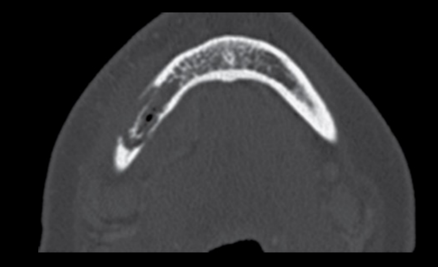
Case: Pathologic fracture and gas-producing bacteria in osteomyelitis
Question
Scenario: A patient with a pathologic fracture secondary to osteomyelitis.
What’s shown: A CT scan showing a pathologic fracture defect with mixed lytic and sclerotic changes. The soft tissue appears “dirty” with thickening, and there are small black dots indicating air within the defect.
Consider: What additional finding on the CT indicates the presence of gas-producing bacteria in this case of osteomyelitis?
Answer
Observations:
- Pathologic fracture defect with mixed lytic/sclerotic changes.
- “Dirty” soft tissue thickening.
- Small black dots (air) within the defect.
Reasoning: The presence of air (gas) in the soft tissue or bone defect, in the absence of a recent surgical opening, is a hallmark of infection by gas-producing bacteria.
Takeaway: The presence of air pockets (gas) within an osteomyelitis defect on CT is a key indicator of infection by gas-producing bacteria.
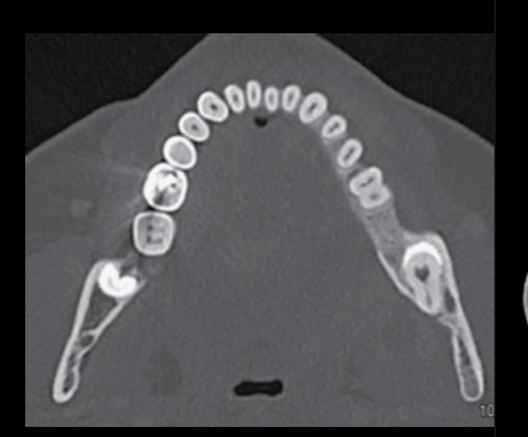
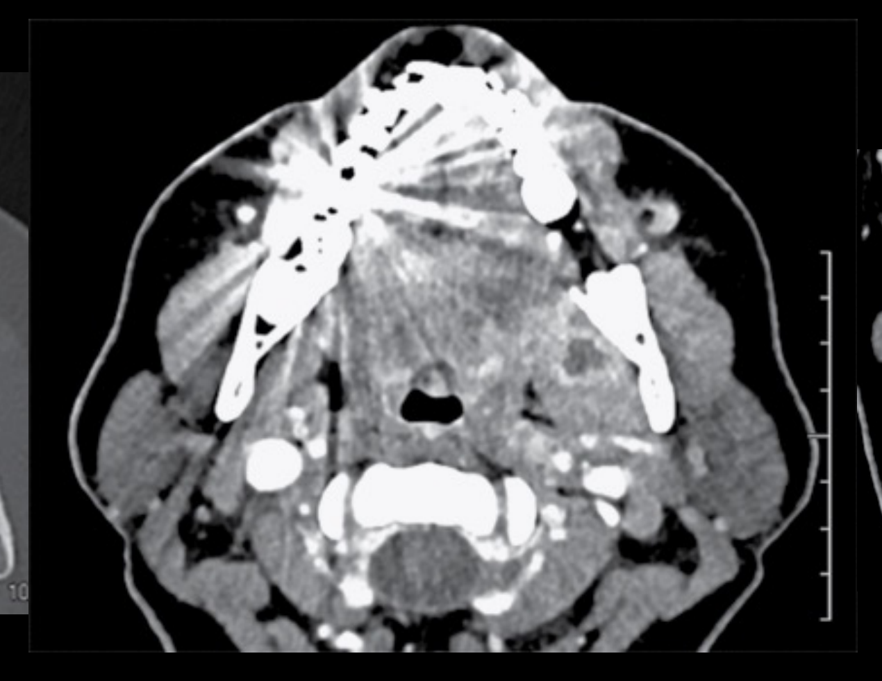
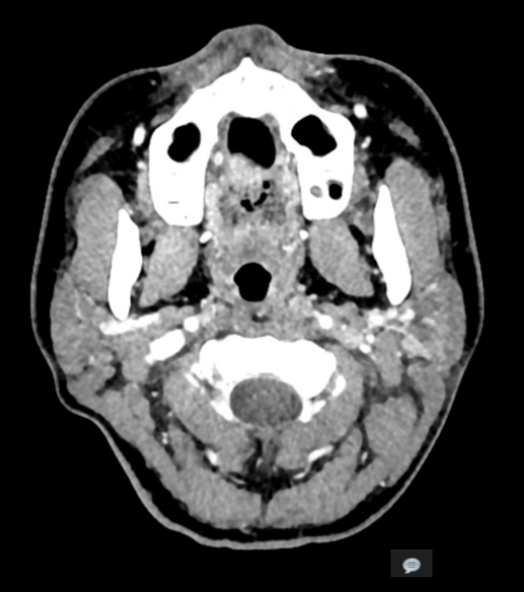
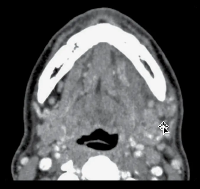
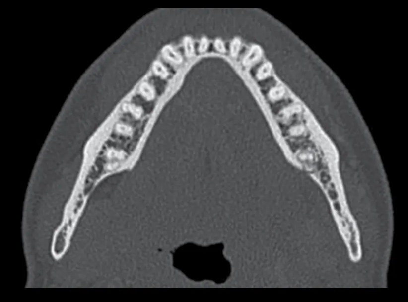
Case: Airway narrowing from tooth 37 infection on soft tissue window
Question
Scenario: A patient with a widened PDL and lingual cortical breach related to tooth 37.
What’s shown: CT bone and soft tissue windows. The bone window shows the cortical breach but minimal soft tissue detail. The soft tissue window reveals extensive swelling, loss of normal fat, and medial displacement of the airway.
Consider: What critical soft tissue complication is revealed on the soft tissue window that was not apparent on the bone window for this tooth 37 infection?
Answer
Observations:
- Bone window shows lingual cortical breach.
- Soft tissue window shows extensive swelling, loss of normal fat planes, and medial displacement/narrowing of the airway.
Reasoning: Bone windows are optimized for cortical and trabecular bone, masking soft tissue edema. Soft tissue windows reveal the true extent of fascial space involvement and airway compromise.
Takeaway: Soft tissue windowing on CT is critical for evaluating the extent of fascial space infection and airway compromise, which are often invisible on bone windows.
Case: Cheek edema from tooth 16 infection visible only on soft tissue window
Question
Scenario: A patient with a periapical lucency related to tooth 16.
What’s shown: An OPG and CT bone window showing the periapical lucency but relatively clean soft tissues and an intact sinus floor. A CT soft tissue window reveals significant thickening of the right cheek and “dirty fat” density.
Consider: Why is a contrast CT with soft tissue windowing preferred over OPG or CBCT for evaluating the extent of this tooth 16 infection?
Answer
Observations:
- OPG and CT bone window show minimal soft tissue changes.
- CT soft tissue window shows right cheek thickening and “dirty fat” (edema).
Reasoning: OPGs and CBCTs (and CT bone windows) do not adequately display soft tissue edema and fascial space involvement. Soft tissue windowing on a medical CT reveals the true extent of the cellulitis.
Takeaway: Medical CT with soft tissue windowing is superior to OPG and CBCT for evaluating soft tissue edema and fascial space involvement in odontogenic infections.
Case: Abscess with rim enhancement and medial pterygoid thickening
Question
Scenario: A patient with an abscess formation related to osteomyelitis.
What’s shown: A contrast-enhanced CT showing a trapped fluid collection surrounded by enhancing tissue. There is also notable thickening of the medial pterygoid muscle.
Consider: What imaging sign on the contrast-enhanced CT confirms the presence of an abscess, and what muscle is notably thickened?
Answer
Observations:
- Trapped fluid collection with rim enhancement.
- Thickening of the medial pterygoid muscle.
Reasoning: Contrast cannot penetrate the necrotic center of an abscess but enhances the surrounding inflamed capsule, creating rim enhancement. Adjacent muscles thicken due to inflammatory edema.
Takeaway: Rim enhancement surrounding a fluid collection on contrast CT confirms an abscess, and adjacent muscles (like the medial pterygoid) will show inflammatory thickening.
Case: Deep neck and buccal space abscesses post third molar extraction
Question
Scenario: A patient presenting with pain and neck swelling following extraction of teeth 28 and 38.
What’s shown: CT soft tissue windows showing an abscess in the left cervical chain involving the sternocleidomastoid muscle (posterior to the submandibular gland, superficial to the carotid sheath) and another abscess in the buccal space.
Consider: Where are the two distinct abscess collections located in this patient following third molar extractions?
Answer
Observations:
- Abscess in the left cervical chain involving the sternocleidomastoid muscle.
- Abscess in the buccal space.
Reasoning: Infections from mandibular third molars can spread to the cervical chain, while maxillary third molars can spread to the buccal space. The CT clearly delineates these deep space collections.
Takeaway: Post-extraction infections can spread to multiple deep fascial spaces, such as the cervical chain and buccal space, requiring careful CT evaluation to map the extent.
Case: Extensive left masticator space abscesses extending to the TMJ
Question
Scenario: A patient with severe left-sided facial swelling following a third molar extraction.
What’s shown: A contrast CT showing multiple abscesses in the left masticator space (medial to the lateral pterygoid, lateral/deep to the masseter, extending into the left TMJ). There is swelling of the muscles of mastication, platysma, subcutaneous fat stranding, and reactive lymph nodes in the submandibular space.
Consider: What are the extent and specific locations of the abscesses and inflammatory changes in this severe post-extraction cellulitis case?
Answer
Observations:
- Multiple abscesses in the left masticator space extending into the left TMJ.
- Swelling of mastication muscles and platysma.
- Subcutaneous fat stranding and reactive submandibular lymph nodes.
Reasoning: The infection has extensively involved the masticator space and surrounding soft tissues, demonstrating the potential for widespread fascial space involvement and reactive lymphadenopathy.
Takeaway: Severe post-extraction infections can cause extensive multi-space abscesses in the masticator space, extending to the TMJ, with widespread soft tissue edema and reactive lymphadenopathy.
Footnotes
-
Original PDF page 1: L1 - Imaging for Infection and Inflammatory Lesions of the Jaws, p.1 ↩
-
Original PDF page 2: L1 - Imaging for Infection and Inflammatory Lesions of the Jaws, p.2 ↩
-
Original PDF page 3: L1 - Imaging for Infection and Inflammatory Lesions of the Jaws, p.3 ↩
-
Original PDF page 4: L1 - Imaging for Infection and Inflammatory Lesions of the Jaws, p.4 ↩
-
Original PDF page 6: L1 - Imaging for Infection and Inflammatory Lesions of the Jaws, p.6 ↩
-
Original PDF page 7: L1 - Imaging for Infection and Inflammatory Lesions of the Jaws, p.7 ↩
-
Original PDF page 8: L1 - Imaging for Infection and Inflammatory Lesions of the Jaws, p.8 ↩
-
Original PDF page 9: L1 - Imaging for Infection and Inflammatory Lesions of the Jaws, p.9 ↩
-
Original PDF page 10: L1 - Imaging for Infection and Inflammatory Lesions of the Jaws, p.10 ↩
-
Original PDF page 11: L1 - Imaging for Infection and Inflammatory Lesions of the Jaws, p.11 ↩
-
Original PDF page 12: L1 - Imaging for Infection and Inflammatory Lesions of the Jaws, p.12 ↩
-
Original PDF page 13: L1 - Imaging for Infection and Inflammatory Lesions of the Jaws, p.13 ↩
-
Original PDF page 14: L1 - Imaging for Infection and Inflammatory Lesions of the Jaws, p.14 ↩
-
Original PDF page 15: L1 - Imaging for Infection and Inflammatory Lesions of the Jaws, p.15 ↩
-
Original PDF page 16: L1 - Imaging for Infection and Inflammatory Lesions of the Jaws, p.16 ↩
-
Original PDF page 17: L1 - Imaging for Infection and Inflammatory Lesions of the Jaws, p.17 ↩
-
Original PDF page 18: L1 - Imaging for Infection and Inflammatory Lesions of the Jaws, p.18 ↩
-
Original PDF page 19: L1 - Imaging for Infection and Inflammatory Lesions of the Jaws, p.19 ↩
-
Original PDF page 20: L1 - Imaging for Infection and Inflammatory Lesions of the Jaws, p.20 ↩
-
Original PDF page 5: L1 - Imaging for Infection and Inflammatory Lesions of the Jaws, p.5 ↩
-
Original PDF page 22: L1 - Imaging for Infection and Inflammatory Lesions of the Jaws, p.22 ↩
-
Original PDF page 23: L1 - Imaging for Infection and Inflammatory Lesions of the Jaws, p.23 ↩
-
Original PDF page 24: L1 - Imaging for Infection and Inflammatory Lesions of the Jaws, p.24 ↩
-
Original PDF page 25: L1 - Imaging for Infection and Inflammatory Lesions of the Jaws, p.25 ↩
-
Original PDF page 26: L1 - Imaging for Infection and Inflammatory Lesions of the Jaws, p.26 ↩
-
Original PDF page 27: L1 - Imaging for Infection and Inflammatory Lesions of the Jaws, p.27 ↩
-
Original PDF page 28: L1 - Imaging for Infection and Inflammatory Lesions of the Jaws, p.28 ↩
-
Original PDF page 29: L1 - Imaging for Infection and Inflammatory Lesions of the Jaws, p.29 ↩
-
Original PDF page 30: L1 - Imaging for Infection and Inflammatory Lesions of the Jaws, p.30 ↩
-
Original PDF page 31: L1 - Imaging for Infection and Inflammatory Lesions of the Jaws, p.31 ↩
-
Original PDF page 32: L1 - Imaging for Infection and Inflammatory Lesions of the Jaws, p.32 ↩
-
Original PDF page 34: L1 - Imaging for Infection and Inflammatory Lesions of the Jaws, p.34 ↩
-
Original PDF page 35: L1 - Imaging for Infection and Inflammatory Lesions of the Jaws, p.35 ↩
-
Original PDF page 33: L1 - Imaging for Infection and Inflammatory Lesions of the Jaws, p.33 ↩
-
Original PDF page 36: L1 - Imaging for Infection and Inflammatory Lesions of the Jaws, p.36 ↩
-
Original PDF page 37: L1 - Imaging for Infection and Inflammatory Lesions of the Jaws, p.37 ↩
-
Original PDF page 38: L1 - Imaging for Infection and Inflammatory Lesions of the Jaws, p.38 ↩
-
Original PDF page 39: L1 - Imaging for Infection and Inflammatory Lesions of the Jaws, p.39 ↩
-
Original PDF page 40: L1 - Imaging for Infection and Inflammatory Lesions of the Jaws, p.40 ↩
-
Original PDF page 41: L1 - Imaging for Infection and Inflammatory Lesions of the Jaws, p.41 ↩