Soft Tissue Calcifications and Ossifications in the Head and Neck1
Dr. May Lam
Oral and Maxillofacial Radiologist
Senior lecturer
BDSc (Hons) FRACDS (GDP) DClinDent (DMFR) MRACDS (DMFR)
Heterotopia2
Definition: The presence of a particular tissue type at a non-physiological site, but usually co-existing with original tissue in its correct anatomical location. In other words, it implies ectopic tissue, in addition to retention of the original tissue type.
Two types:
-
Heterotopic calcification
-
Heterotopic ossification
-
Because these findings remain in their anatomical locations, interpretation on panoramic radiographs requires familiarity with the head and neck anatomy visible on the panoramic image.
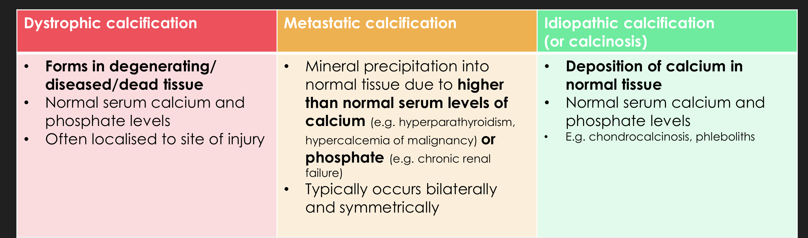
Heterotopic Calcifications3
- When deposition of calcium salts occurs in an unorganised fashion in soft tissue
- Divided into 3 categories:
| Dystrophic calcification | Metastatic calcification | Idiopathic calcification (or calcinosis) |
|---|---|---|
| • Forms in degenerating/diseased/dead tissue • Normal serum calcium and phosphate levels • Often localised to site of injury | • Mineral precipitation into normal tissue due to higher than normal serum levels of calcium (e.g. hyperparathyroidism, hypercalcemia of malignancy) or phosphate (e.g. chronic renal failure) • Typically occurs bilaterally and symmetrically | • Deposition of calcium in normal tissue • Normal serum calcium and phosphate levels • E.g. chondrocalcinosis, phleboliths |
- The cause is unknown
- Bone formation within muscles in myositis ossificans
- Extra bone formation around the spine in ankylosing spondylitis
Heterotopic Ossification4
-
When mineral is deposited in soft tissue as organised, well-formed bone
-
“Heterotopic bone” = bone that has formed in an abnormal (extraskeletal) location
- May be all compact bone, or exhibit some trabeculae and fatty marrow
-
Size ranges from 1mm to several cm in diameter
-
One or more may be present
-
Examples:
- Post-traumatic ossification
- Bone produced by tumours
- Ossification from diseases such as progressive myositis ossificans and ankylosing spondylitis
-
Calcium and phosphate are deposited as organised bone rather than as unorganised clumps of salt crystals.
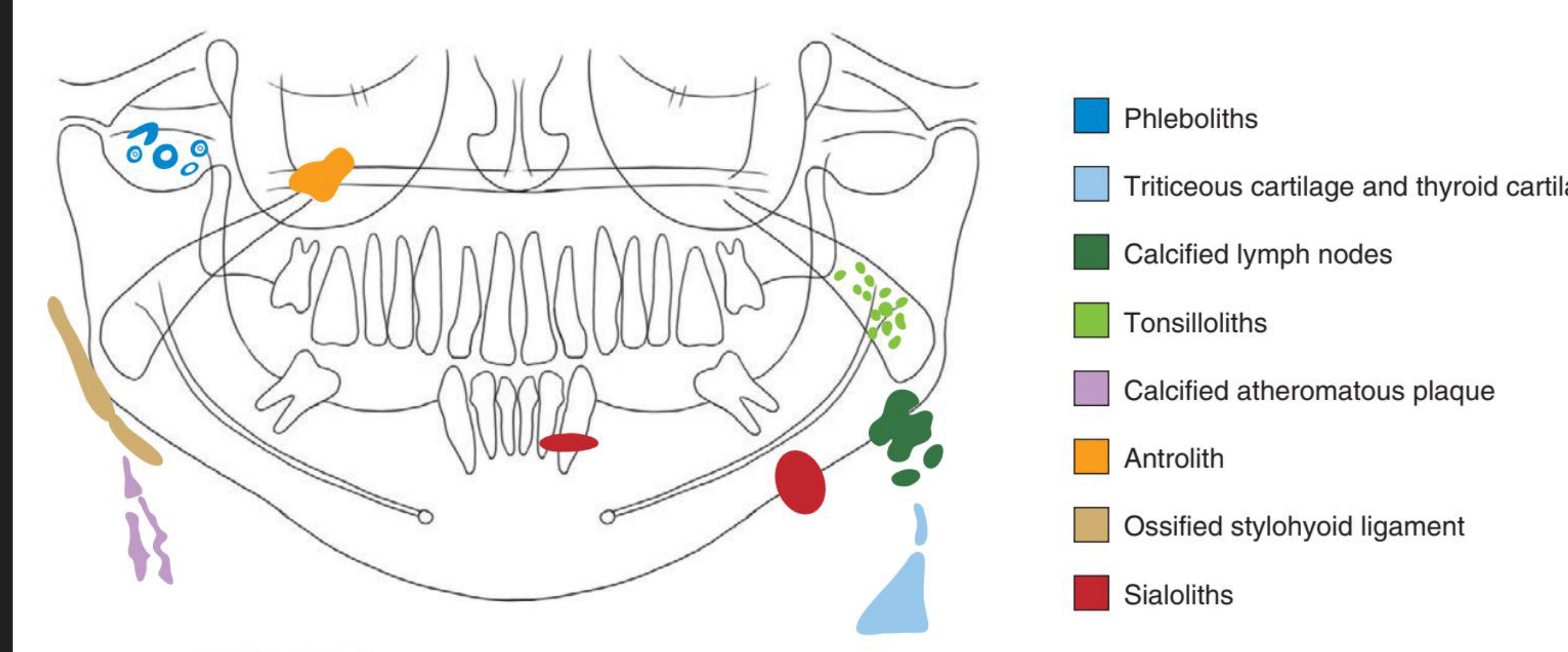
Examples of Soft Tissue Calcifications5
-
Phleboliths
-
Triticeous cartilage and thyroid cartilage
-
Calcified lymph nodes
-
Tonsilloliths
-
Calcified atheromatous plaque
-
Antrolith
-
Ossified stylohyoid ligament
-
Sialoliths
-
Common locations for soft tissue calcifications can be anticipated by reviewing the anatomical structures represented on a panoramic radiograph.
-
Dystrophic calcifications
-
Post-inflammatory tonsillar calcifications
-
Calcified atherosclerotic plaques
-
Mönckeberg’s medial calcific sclerosis
-
Laryngeal cartilage calcifications
-
Rhinoliths
-
Metastatic calcification White and Pharoah, 2014
Dystrophic Calcifications6
- Calcified lymph nodes
- Post-inflammatory tonsillar calcifications
- Calcified atherosclerotic plaque
- Arteriosclerosis
E.g. long standing chronically infected cyst or polyp
Location: gingiva, tongue, lymph nodes, and cheek
Clinical Sign/Symptoms7
-
Typically none
-
Enlargement and ulceration of overlying soft tissue
-
Palpation of solid mass of calcium salts
-
Dystrophic calcifications occur in degenerating, diseased, or dead tissue despite normal serum calcium and phosphate levels.
-
Evidence of previous trauma may include soft-tissue hyperplasia or ulceration.
-
A fibrous hyperplasia following repeated trauma to the alveolar ridge is another example.
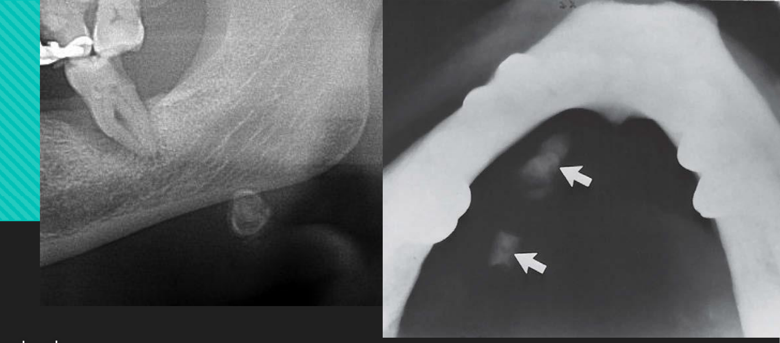
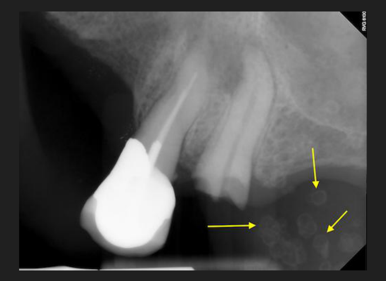
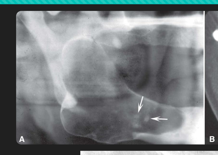 Dystrophic Calcification on chronically infected residual cyst Dystrophic Calcification on chronically infected residual cyst | 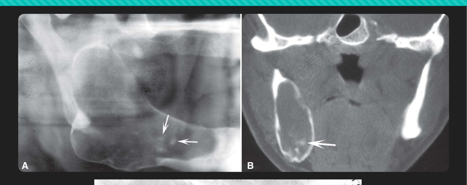 |
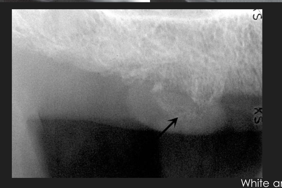 Fibrous hyperplasia in edentulous patient , arrow pointng to opacitiices Fibrous hyperplasia in edentulous patient , arrow pointng to opacitiices |
Imaging Features
- Varies from being barely perceptible as fine grains of radiopacities to larger, irregular radiopaque particles (but rarely exceed 0.5 cm in diameter)
- One or more radiopacities may be seen
- Periphery: irregular or indistinct
- Internal: may be homogeneous or contain punctate areas
White and Pharoah, 201
Calcified Lymph Nodes8
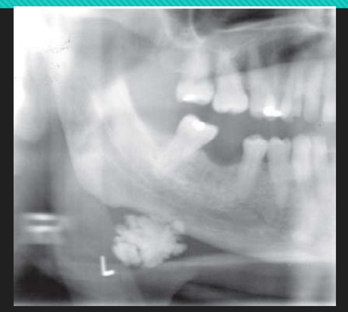
- Deposition of hydroxyapatite-like calcium salts within the lymph node (LN), nearly effacing all nodal architecture
- An isolated calcified LN in the level I nodes is usually related to post-inflammatory calcification
- More extensive dystrophic calcification in LNs that are chronically inflamed due to various diseases (frequently granulomatous disorders, e.g. TB, sarcoidosis, etc.)
- Implies either active or previously treated disease
- Occasionally associated with malignancies
Clinical Features9
- Generally asymptomatic
- Often incidental finding
- When nodes can be palpated, are hard, lumpy, round-oblong masses
- Most common: submandibular and superficial/deep cervical nodes (Level Ib and II nodes)
- Less common: preauricular and submental nodes
Management
- Typically no treatment
- Need to manage underlying cause
Location10
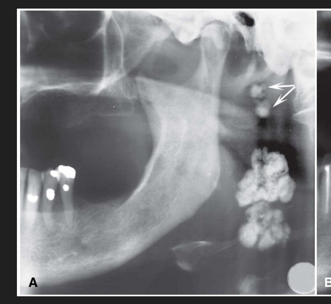
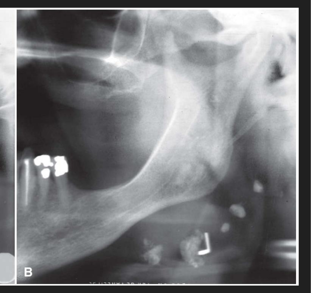
- Submandibular region — at or just below the mandibular angle or between the posterior border of the ramus and the C-spine
- May affect a single node or a linear series of nodes (“LN chaining”)
 |  |
Periphery/Shape
- Well-defined
- Usually irregular
- Often lobulated, like the shape of a cauliflower
Internal Features11
- May vary in degree of opacity
- Occasional laminated appearance
Surrounding Features
N/A
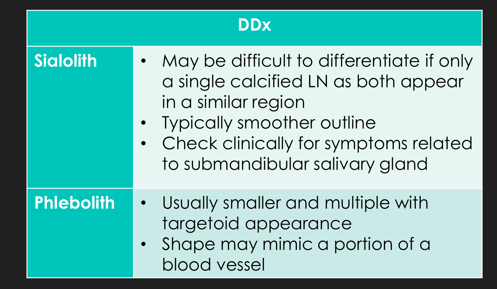
Differential Diagnosis from calcified lymph nodes
| DDx | Features |
|---|---|
| Sialolith | May be difficult to differentiate if only a single calcified LN, as both appear in a similar region; typically smoother outline; check clinically for symptoms related to submandibular salivary gland |
| Phlebolith | Usually smaller and multiple with targetoid appearance; shape may mimic a portion of a blood vessel |
Post-Inflammatory Tonsillar Calcifications
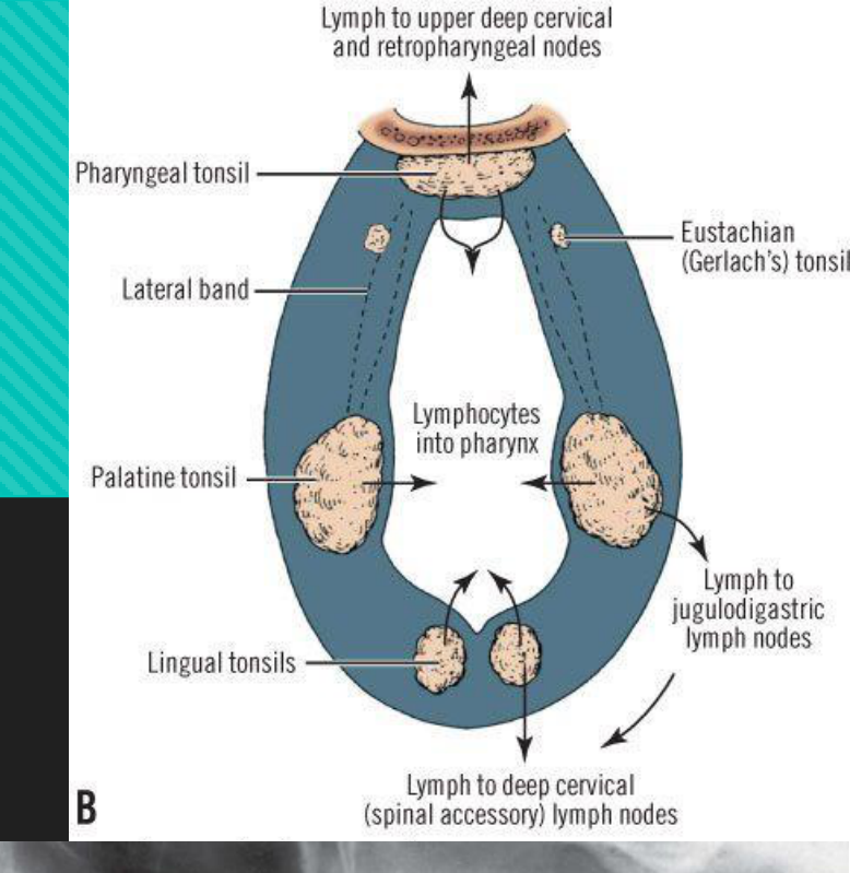
Synonyms: tonsilloliths, dystrophic calcifications in the tonsils, tonsillar calculi, tonsil concretions
Clinical Features12
- Hard, white/yellow objects projecting from the tonsillar crypts
- Usually palatine tonsil
- Typically no clinical signs and symptoms
- Larger calcifications — pain, swelling, fetor oris, dysphagia or foreign body sensation on swallowing
 |  |
Disease Mechanism13
- Formed when repeated bouts of inflammation enlarge the tonsillar crypts
- Incomplete resolution of organic debris can serve as a nidus for dystrophic calcification
Lecturer — Tonsillar Debris
Repeated tonsillitis enlarges the crypts, allowing organic debris to become lodged before calcification.
- The debris may include dead bacteria, pus, epithelial cells, and food.
Management
- Asymptomatic — no treatment
- Unless elderly with manual deglutition disorders or immunocompromised patients, for risk of aspiration pneumonia
- Symptomatic — manual expression or surgical removal if large
Location14
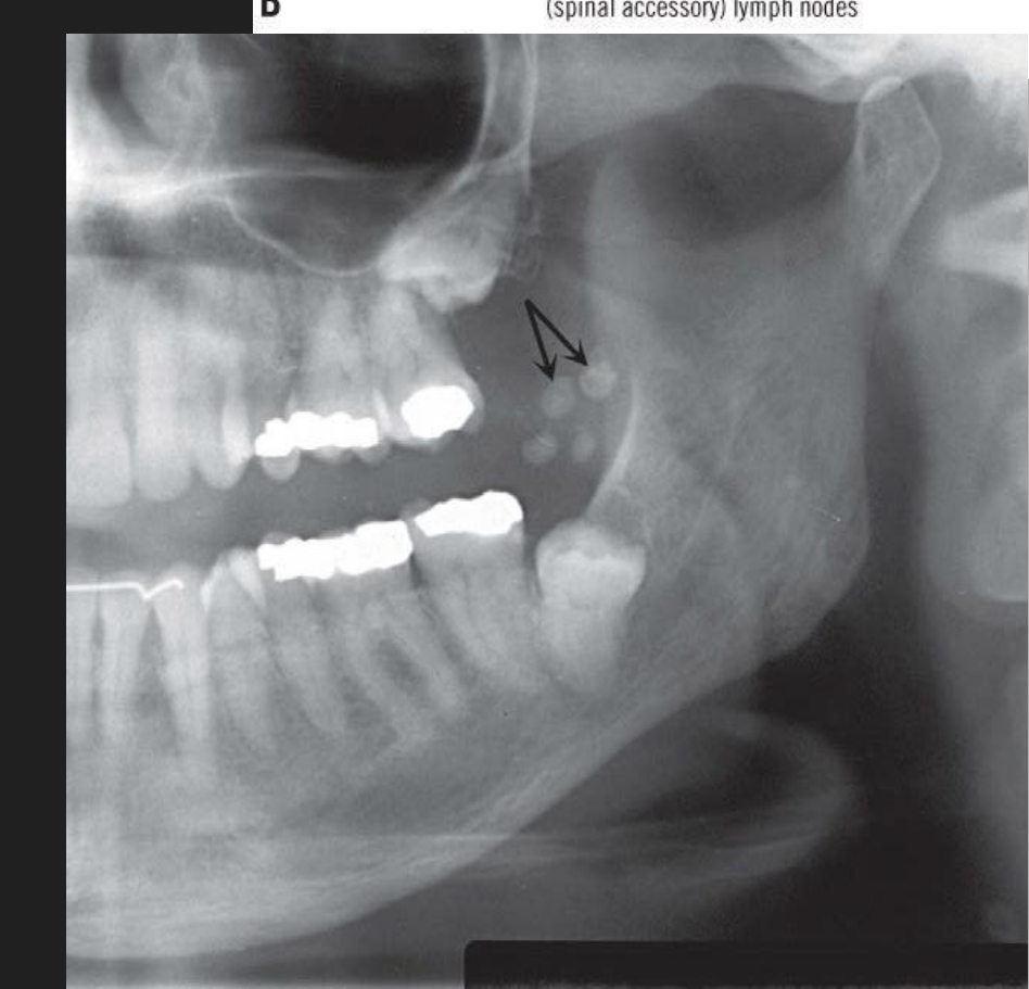
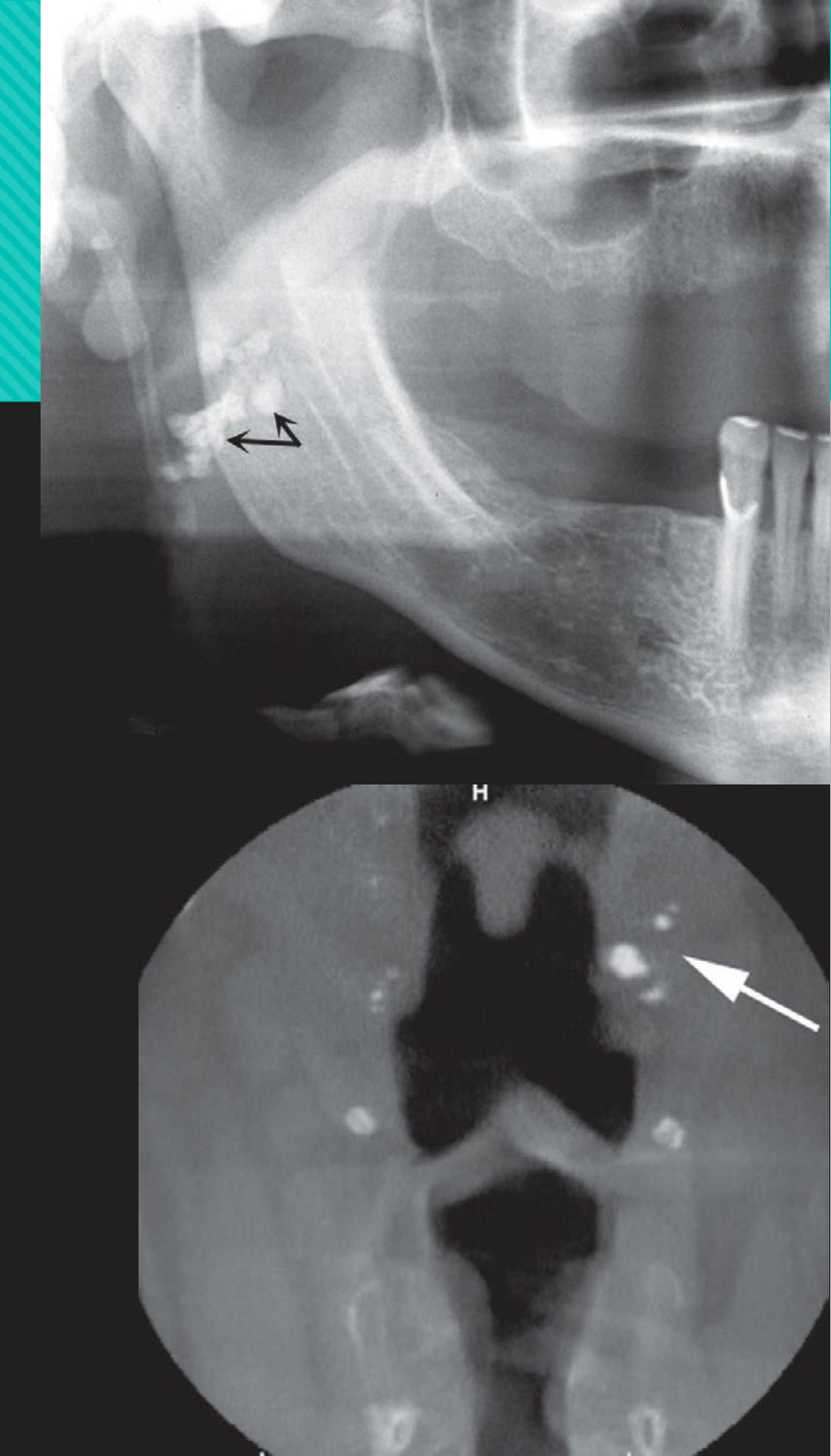
- [PAN] Superimposed over the mid-ramus region where the dorsum of tongue crosses the ramus in the oropharyngeal air spaces, often inferior to the IAC
- [CT] Medial to the ramus, next to the lateral wall of the pharyngeal air space
Periphery/Shape
-
Cluster of multiple small radiopacities
-
Rarely large size
-
Tonsilloliths may also occur in the lingual and pharyngeal tonsils, but are almost never seen in the eustachian tonsil.
Internal Features
- Slightly more radiopaque than cancellous bone
- Roughly the same as cortical bone
Surrounding Features
N/A
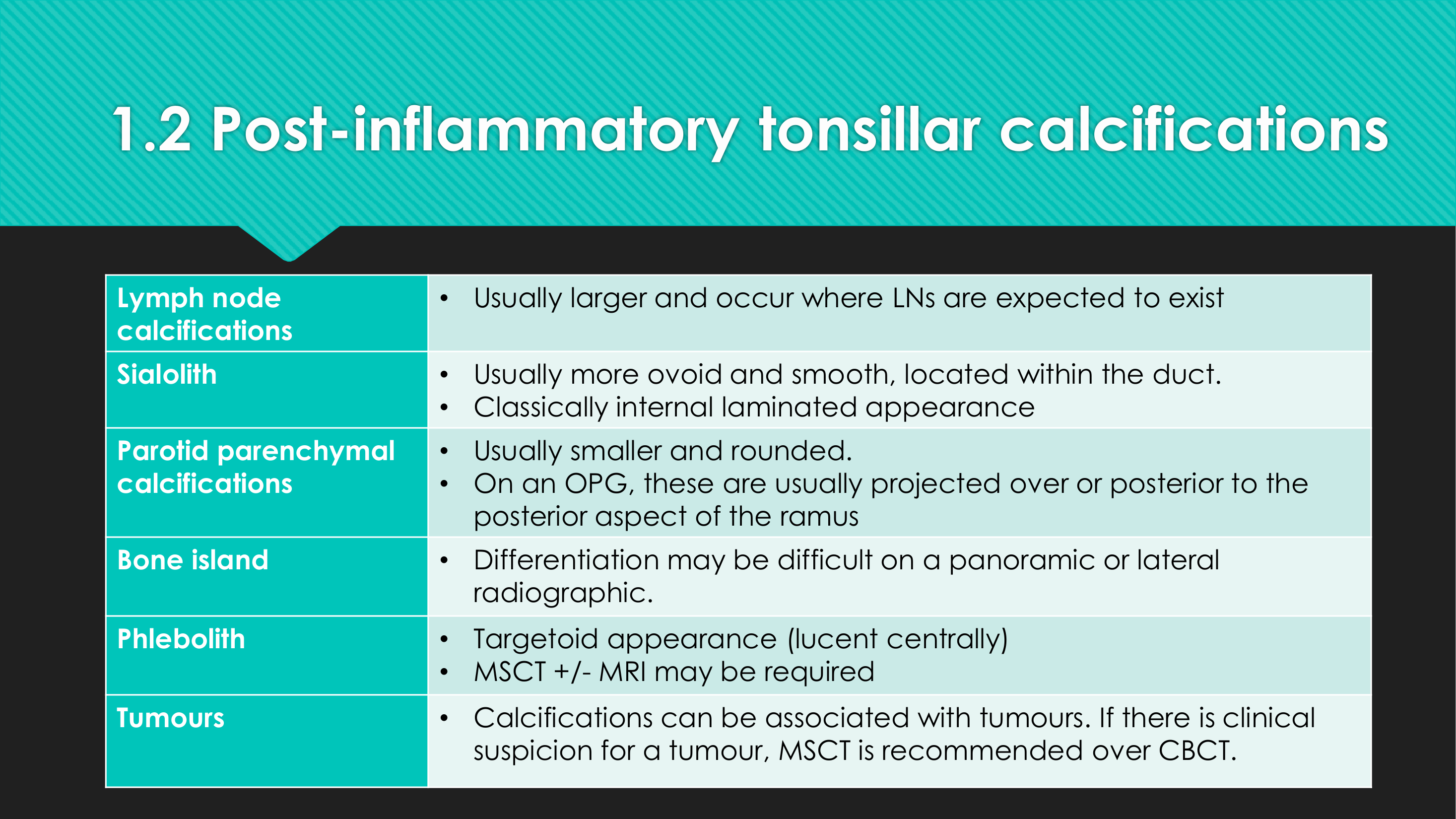
Differential Diagnosis from tonsiloliths15
| DDx | Features |
|---|---|
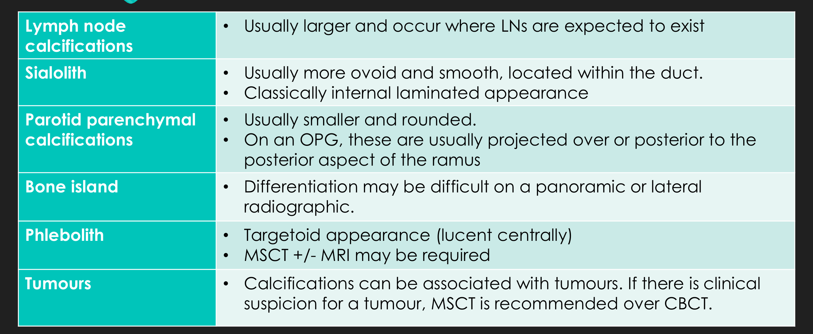
| Lymph node calcifications | Usually larger and occur where LNs are expected to exist |
| Sialolith | Usually more ovoid and smooth, located within the duct; classically internal laminated appearance |
| Parotid parenchymal calcifications | Usually smaller and rounded; on an OPG, these are usually projected over or posterior to the posterior aspect of the ramus |
| Bone island | Differentiation may be difficult on a panoramic or lateral radiograph |
| Phlebolith | Targetoid appearance (lucent centrally); MSCT +/- MRI may be required |
| Tumours | Calcifications can be associated with tumours. If there is clinical suspicion for a tumour, MSCT is recommended over CBCT. |
- Their position beside the pharyngeal airspace and medial to the ramus helps identify tonsilloliths.
Calcified Atherosclerotic Plaque
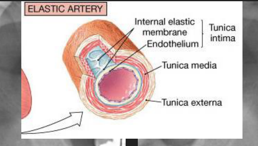
Definition: dystrophic calcification occurring in atheromatous plaques in the intima of large or medium sized vessels
Disease Mechanism16
- Atheromatous plaques begin as fatty streaks composed of lipid-laden macrophages (foam cells)
- Composed of soft necrotic core surrounded by chronic inflammatory cells, smooth muscle cells, and neovascularisation, covered by a fibrous cap
- Often undergo calcification
Aetiology17
- Not completely understood
- Thought to involve chronic endothelial injury leading to inflammatory response, accumulation of lipids, platelet aggregation and activation of smooth muscle cells
Disease Progression
- Stenosis, occlusion, ischaemia, infarction, distal embolism, haemorrhage, aneurysmal dilatation
Management
-
1 in 7 patients with CAP on OPG will have a clinically significant carotid artery stenosis (Constantine et al. 2019)
-
Referral for medical review if:
- Patients with established risk factors for cerebrovascular and cardiovascular disease
- Younger patients
- Substantial carotid calcifications
-
Referral to a general practitioner should be considered when a plaque is seen on an OPG, particularly in a patient who is unusually young for this finding.
Location18
- First develop at arterial bifurcations due to increased endothelial damage from shear forces at these sites
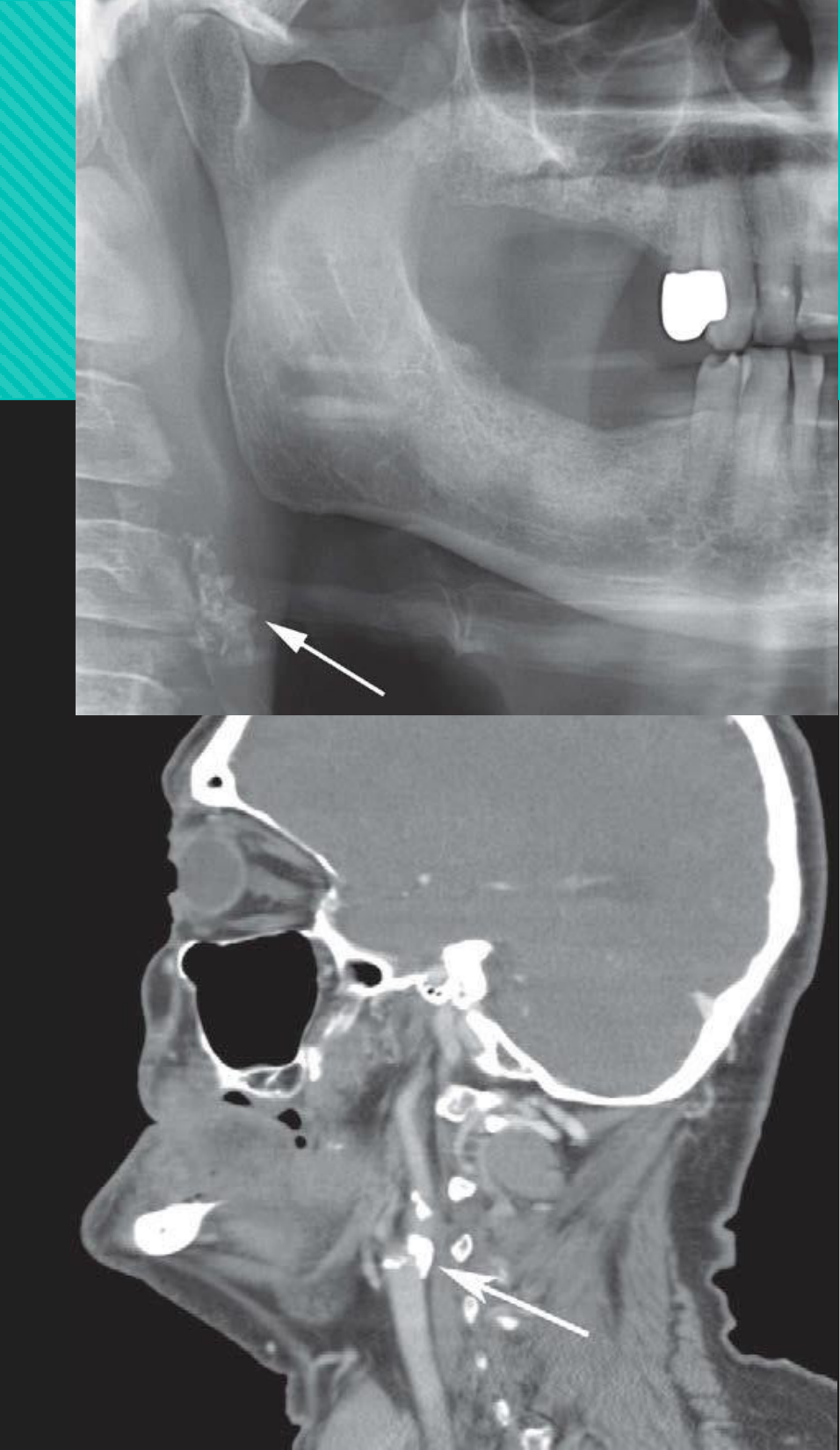
- [PAN] Either superior or inferior to the greater cornu of the hyoid, adjacent to C3-4 or the intervertebral space between them
Lecturer — Carotid Bifurcation
Calcified atherosclerotic plaques commonly develop where the common carotid artery divides into the external and internal carotid arteries.
- On a panoramic radiograph, this bifurcation is approximately at the level of the C3 and C4 vertebrae, around the level of the hyoid bone.
Periphery/Shape
- May be singular or multiple
- Well-defined and irregular
- May be C-shaped or circular in morphology
- Vertical linear distribution
Internal Features
- Heterogeneous radiopacity with radiolucent voids
Surrounding Features
N/A
Arteriosclerosis19
Synonyms: Mönckeberg’s medial calcific sclerosis
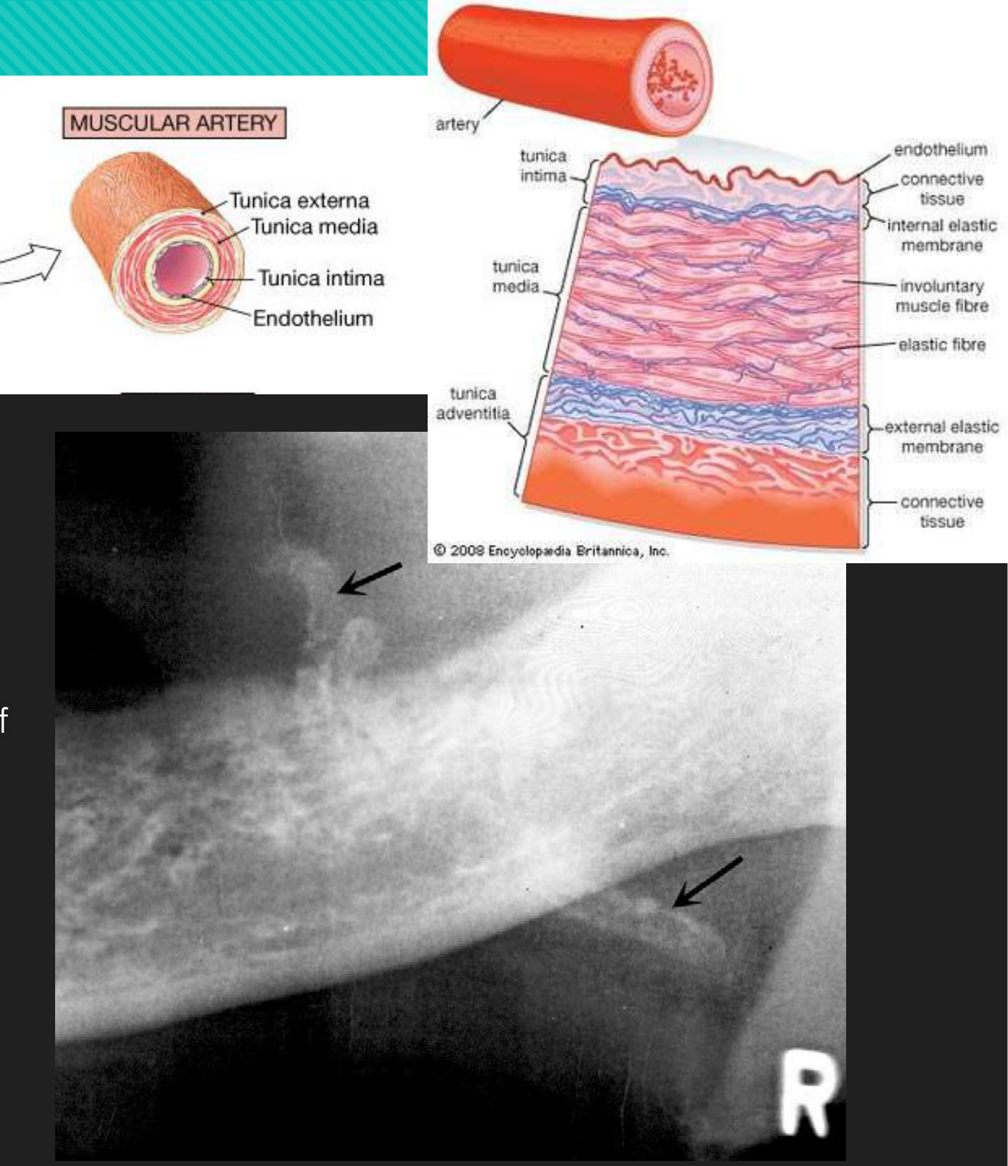
Definition: thickening and loss of elasticity of the walls of muscular arteries due to calcification of the tunica media
Disease Mechanism20
- Deposition of calcium within the medial coat of vessel → fragmentation, degeneration, and eventual loss of elastic fibres
- No luminal narrowing
Aetiology
- Uncertain
- More common with advancing age, diabetes, and chronic renal disease
Clinical Features21
- Initially asymptomatic
- Late in disease: peripheral vascular disease, cutaneous gangrene, myositis as a result of vascular insufficiency
Management
- Evaluation for occlusive arterial disease or peripheral vascular disease
Location22
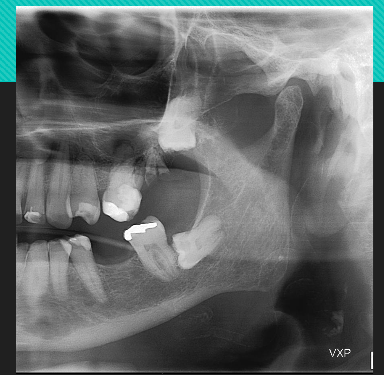
- Facial artery > carotid artery on PAN
Lecturer — Arterial Involvement
In the head and neck, the facial artery is most commonly affected, while the lingual arteries may also be involved.
- Lingual artery involvement is not seen on an OPG and requires three-dimensional imaging.
- When the carotid vessels are outlined, this is almost always due to atheromatous plaque calcification.
Periphery/Shape
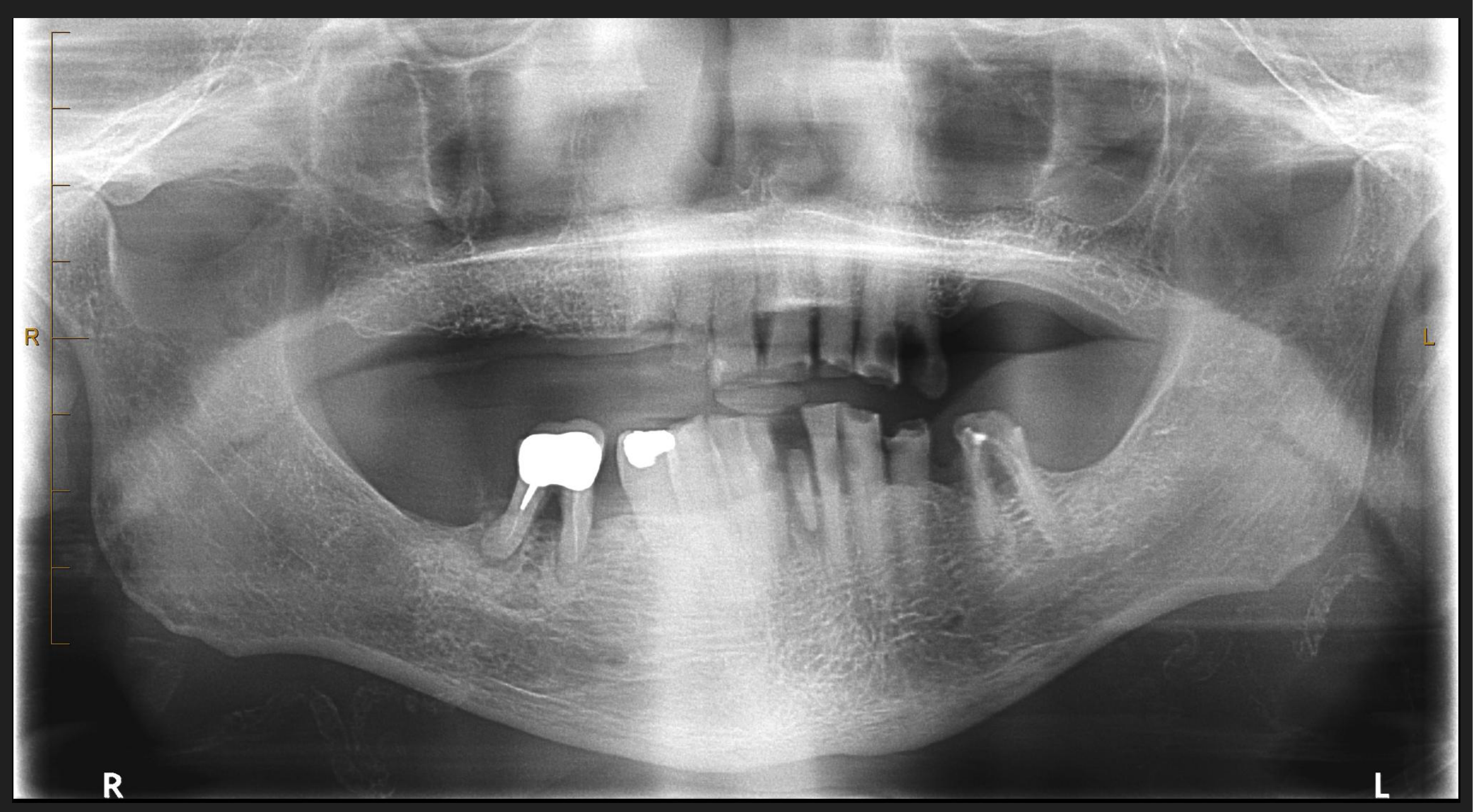
- Outline of an artery (due to calcific deposits in the wall)
- When viewed from the side: a parallel pair of thin, radiopaque lines with a straight course or tortuous path
- Described as ‘tram-track’ or ‘pipe stem’ appearance
- In cross-section, circular, tubular, or ring-like pattern
Internal Features
- No internal structure, as the diffuse, finely divided calcium deposits occur solely in the medial wall of vessels
Lecturer — Arterial Cross Section
In cross-section, the calcification appears as a circular ring with a central lucency representing the non-occluded lumen.
- The finding is often bilateral.
Surrounding Features
N/A
Idiopathic Calcification23
Lecturer — Idiopathic Calcification
Idiopathic calcifications occur in normal tissue despite normal serum calcium and phosphate levels, and their cause is unknown. They are relatively common in the head and neck.
- Sialolith
- Phlebolith
- Laryngeal cartilage calcification
- Antroliths and Rhinoliths
Sialoliths
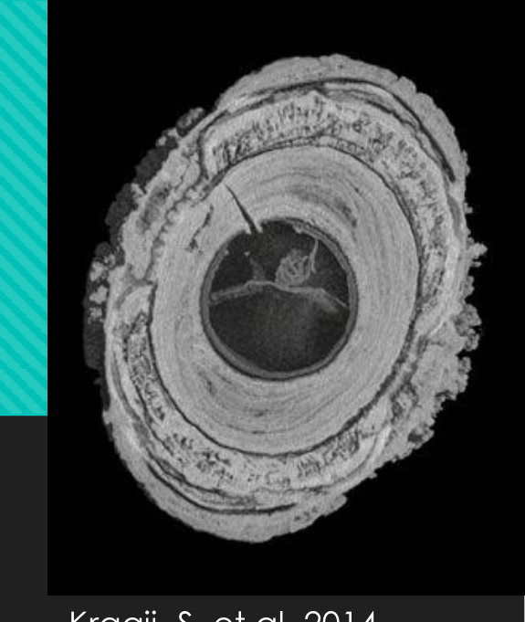
Definition: Salivary gland stones — “ductal sialolith” vs “parenchymal calcifications”
Lecturer — Sialolith Epidemiology
Ductal sialoliths are increasingly seen in younger and teenage patients. The lecture suggested that increased consumption of energy drinks instead of water may contribute, noting an association between kidney stones and salivary gland stones.
- Within the submandibular gland, approximately 50% are in the distal portion of Wharton’s duct, 20% are in the proximal duct, and 30% are within the gland itself.
Disease Mechanism24
Ductal sialolith:
- Arises due to alteration in mechanical conditions (e.g. slow flow rate or physiochemical characteristics of gland secretion)
- Leads to nidus formation and subsequent precipitation of Ca and PO4 salts
Parenchymal calcifications:
- May be related to a previous inflammatory condition or ongoing chronic sialadenitis (e.g. Sjogren’s syndrome)
Epidemiology
Ductal sialoliths:
- Usually occur singly (70-80%)
- Most common in submandibular glands of middle-aged or older men
Parenchymal calcifications:
- Generally multiple in the parotid gland
Clinical Features25
- May be asymptomatic
- History of pain and swelling in floor of mouth/gland/cheek
- Discomfort intensifies during mealtimes, when salivary flow is stimulated — usually abates if the stone does not occlude the duct completely
Prognosis and Recurrence
- 9% recurrent sialolithiasis
- 10% with sialolithiasis also have nephrolithiasis
Management
- Small stones can be ‘milked out’ by bimanual palpation through ductal orifice
- If too large or located in proximal duct, may require lithotripsy or sialendoscopy
- Surgical removal of stone/gland in cases of exceedingly large or intraparenchymal sialoliths
- Parenchymal calcifications require treatment of the underlying sialadenitis
Location26
- 83-94% submandibular gland
- 4-10% parotid gland
- 1-7% sublingual gland
Periphery/Shape
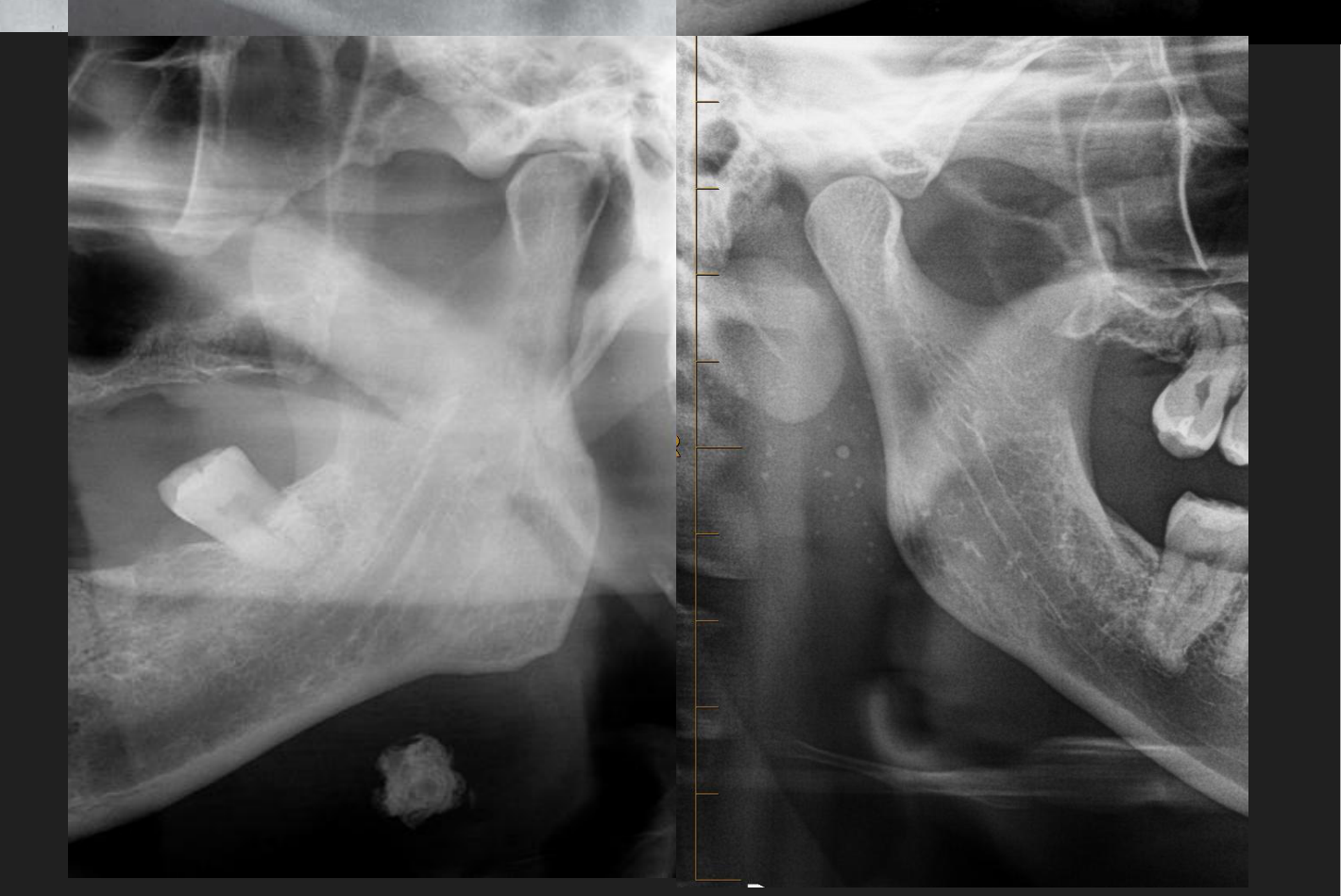
- Ductal: cylindrical, smooth, elongated
- Hilum: larger, more irregularly shaped/ovoid
- Parenchymal: ranges from a few small opacities within a focal region to widespread opacities throughout the gland
Internal Features27
- Either homogeneously radiopaque or laminated
- <20% of submandibular and 40% of parotid sialoliths are radiolucent due to low mineral content of parotid secretions — requires a sialogram for visualisation
- Radiolucent sialoliths very rare in the sublingual gland
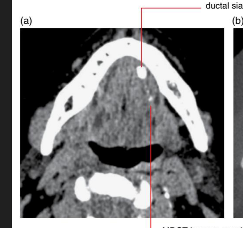
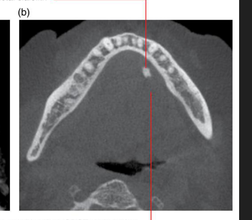
 |  |
Surrounding Features
- Ductal sialoliths may contribute to sialodochitis, sialadenitis, cellulitis, and atrophy of the salivary gland
Lecturer — Sialolith Obstruction
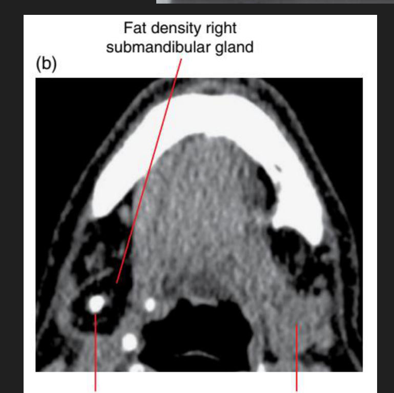
On soft-tissue-window multislice CT, a chronically obstructed and atrophic gland may be replaced by fat. The lecture described a right submandibular gland replaced by fat in association with an opaque sialolith.
(b) Sialolith within the intraglandular proximal duct; normal left submandibular gland. Koong, 2017
Further Imaging Techniques2829303132
- Plain films: Rarely used today as they only detect moderately large, fairly dense calcifications and further imaging is often required.
- CBCT: Not indicated for sialoliths due to poor soft tissue contrast and inability to detect small changes in radiodensities.
- US or MSCT is preferred.
(a) Large distal ductal sialolith. Koong, 2017
- US:
- Very good initial imaging choice, esp. parotid gland
- Advantages: no ionising radiation, real time imaging, accessibility, inexpensive, good soft tissue discrimination
- Disadvantages: limited to superficial structures, technique sensitive, operator dependent, hard to interpret
- MSCT:
- May be performed as sialography
- Advantages: multiplanar imaging, good soft tissue and hard tissue contrast, small changes in radiodensity can be detected, provides 3D information in conventional sialography, can detect ‘mucus plugs’
- Disadvantages: higher radiation dose, cost
- MRI:
- Not typically needed for sialolith evaluation, although may be used as an alternative to conventional MSCT sialography in evaluating ductal pathosis
Lecturer — Sialolith Imaging
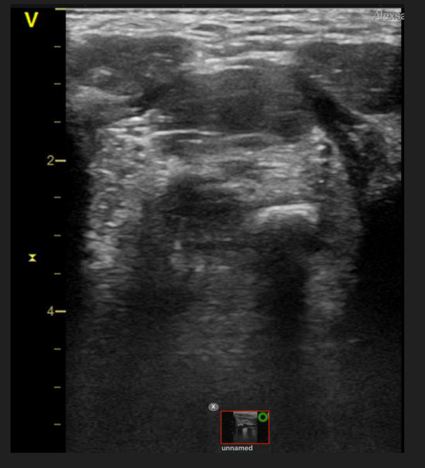
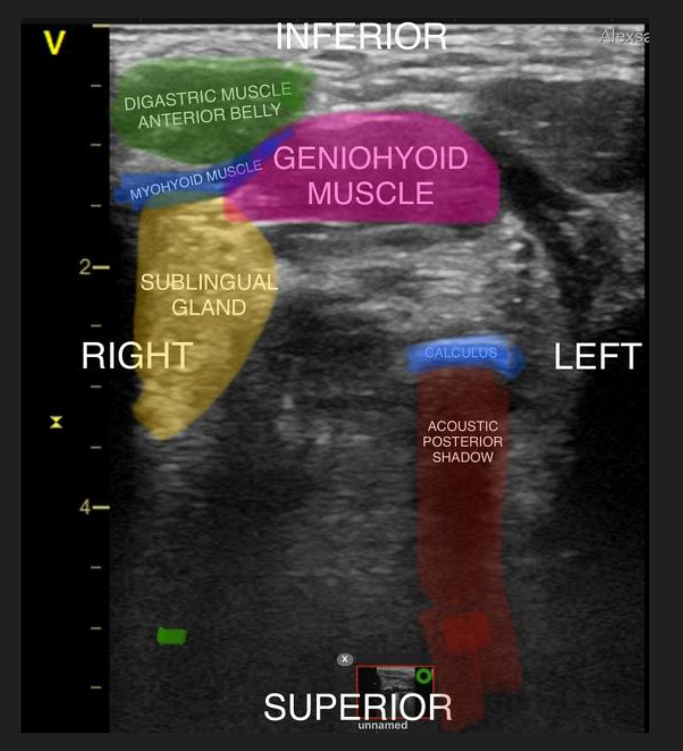
On ultrasound, a sialolith appears as a hyperechoic, well-defined structure with posterior acoustic shadowing. Ultrasound is limited for deep structures, including the deep lobe of the parotid, may not fully visualise the submandibular gland, and is operator dependent.
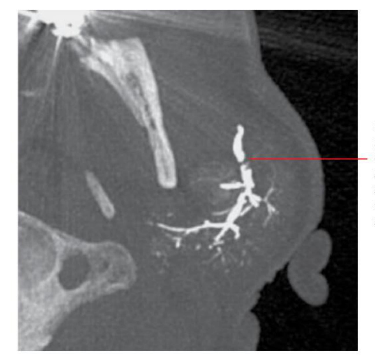
- A multislice CT sialogram can show a flow void caused by a mucus plug or non-calcified sialolith; such an obstruction may not be visible on conventional two-dimensional imaging or non-contrast multislice CT.
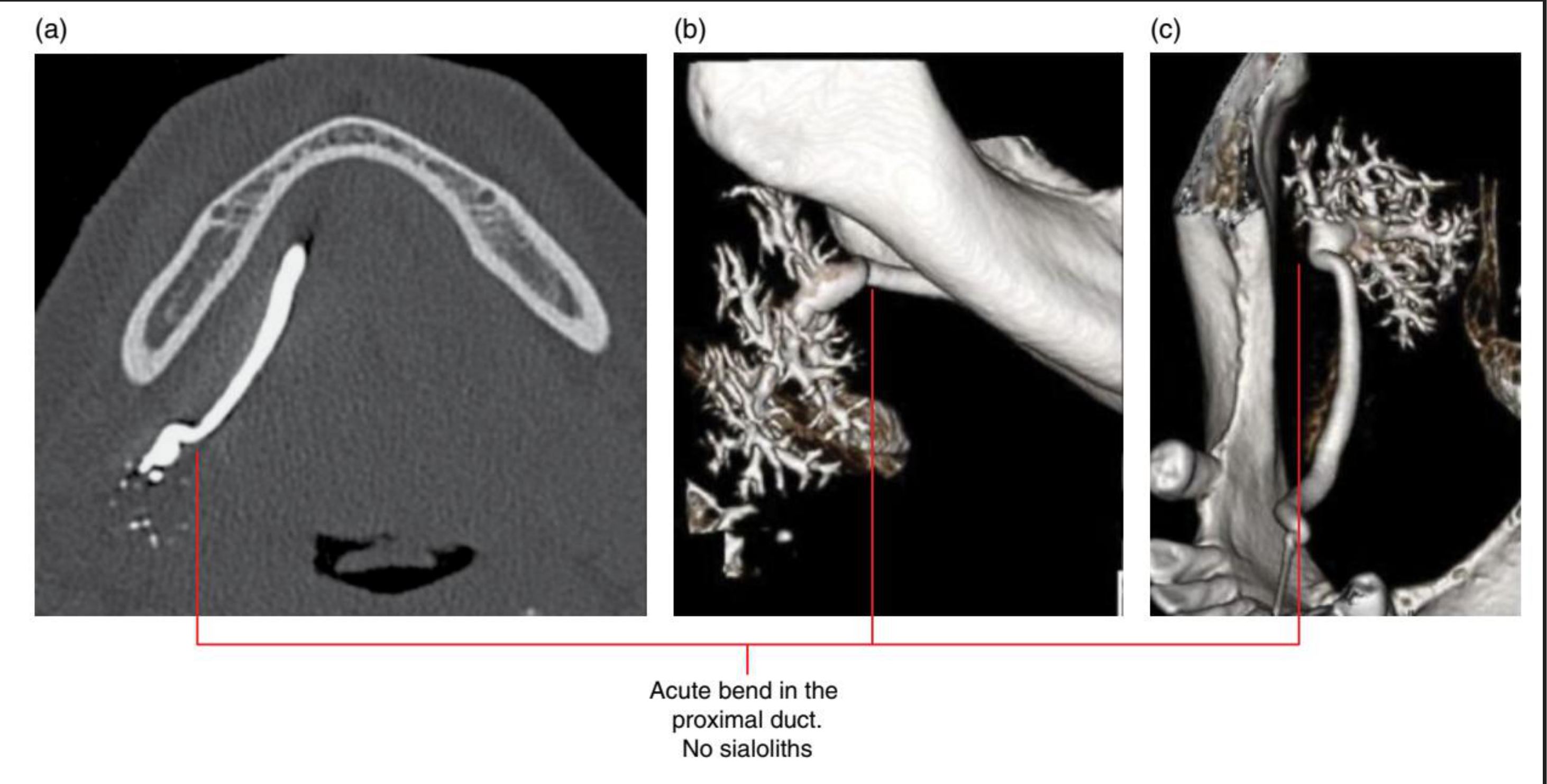
- Sialography may also demonstrate an acute bend in a duct that interferes with salivary flow.
https://radiopaedia.org/cases/sialolithiasis-submandibular-gland-7
Flow void within the proximal duct reflects either a mucus plug or a non-calcified ductal sialolith. This would not be identified without a sialogram.
Koong, 2017
(a) (b) (c) Acute bend in the proximal duct. No sialoliths.
Figure 16.25 Acute bend of the right submandibular gland proximal duct (recurrent swelling related to meals): axial (a) and surface-rendered (b,c) MDCT sialogram images.
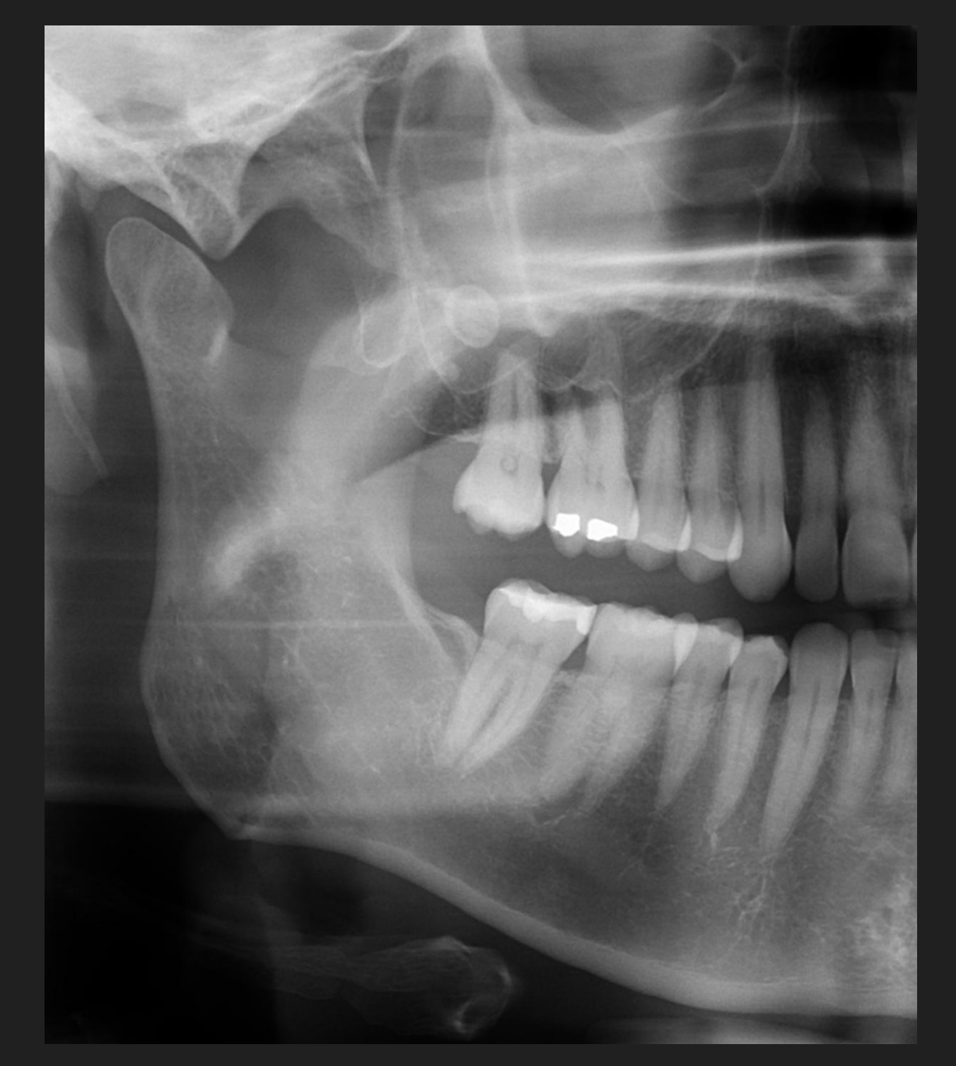
Phleboliths33
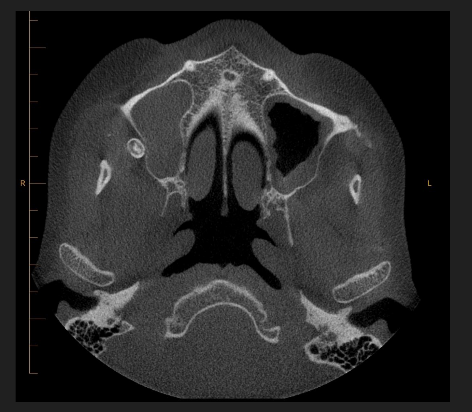
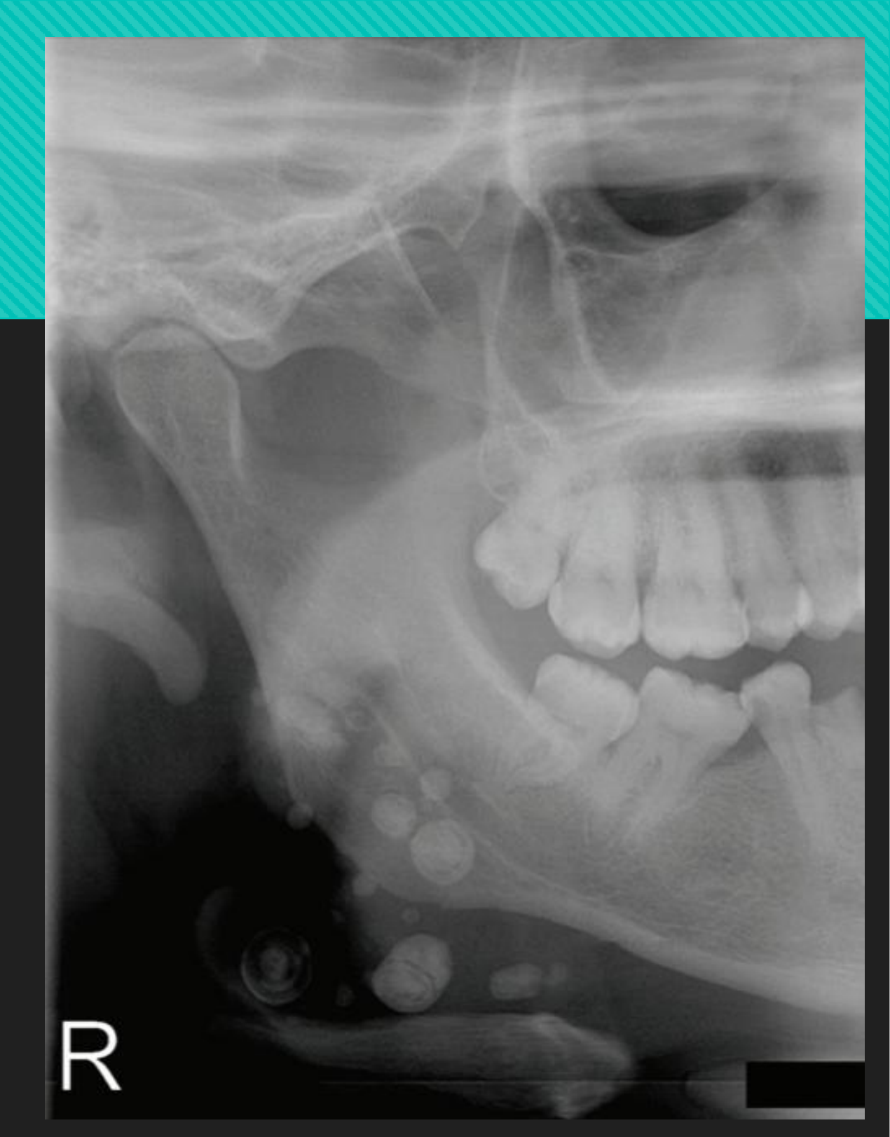
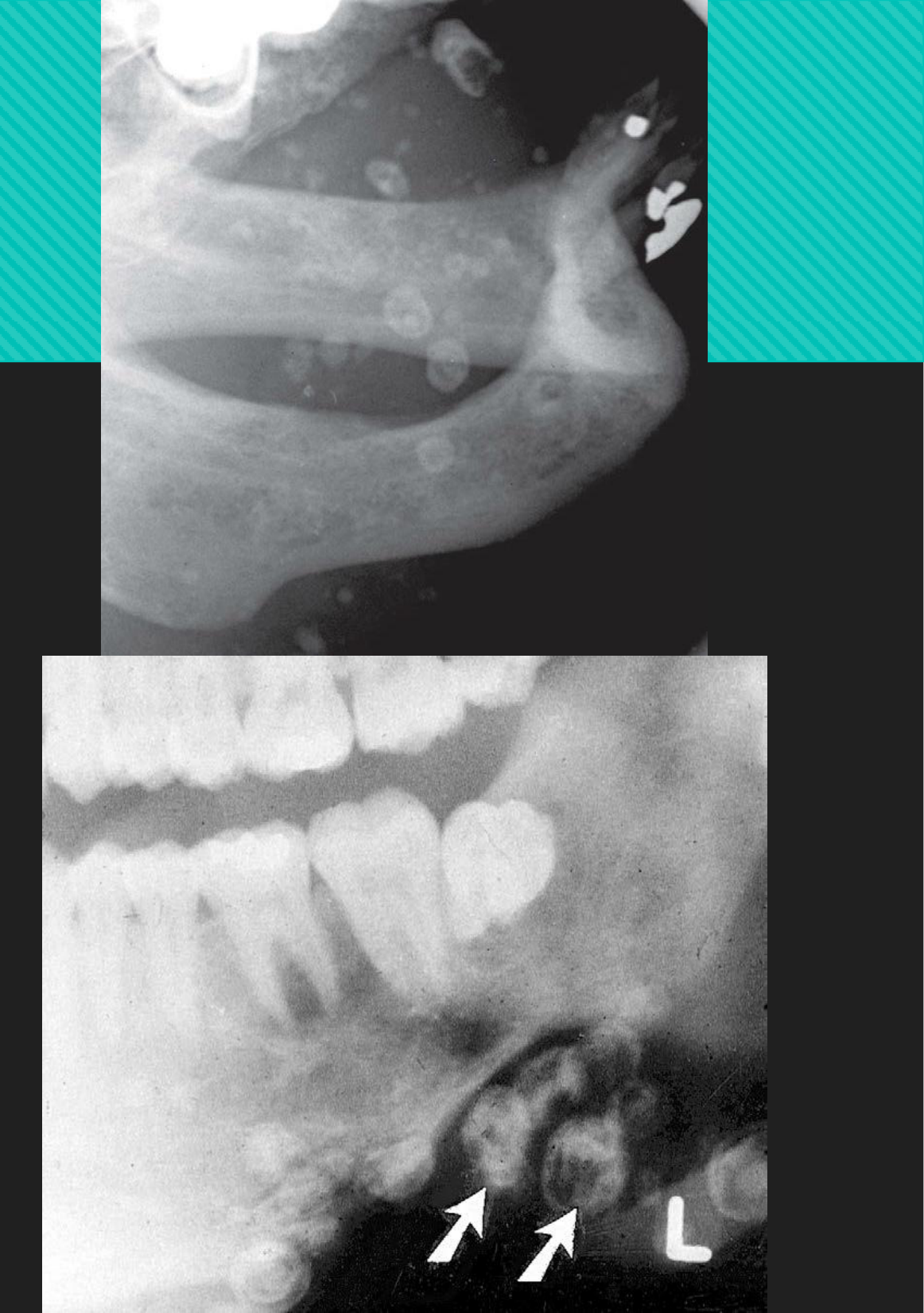
Definition: Calcified thrombi found in veins, venulae, or sinusoidal vessels of haemangiomas (esp. cavernous type)
 |  |
Disease Mechanism34
- Venous stagnation → intravascular thrombi → become organised and mineralised
- Mineralisation begins in core of thrombus
- Consists of calcium carbonate-fluorohydroxyapetite
Clinical Features35
- In the head and neck, almost always signify presence of haemangioma
- In adults, may be the sole residua of a childhood haemangioma that has regressed
- Soft tissues may be swollen, throbbing, or discoloured by presence of veins or haemangioma
- Haemangiomas often fluctuate in size (associated with body positioning or Valsalva manoeuvre); should blanch/change in colour with pressure
- Auscultation may reveal a bruit in cases of cavernous hemangioma but not in the capillary type
Management
- May need further imaging with MSCT or MRI +/- contrast to visualise the vascular malformation
- Central haemangiomas are treated without delay due to risk of lethal exsanguination
- Embolisation, surgery, or sclerosing techniques
Lecturer — Vascular Malformation Precautions
The phlebolith itself does not require treatment, but the associated vascular malformation does. Dental clinicians should establish whether the patient knows about the malformation, has received treatment, and requires precautions before procedures in the region because puncturing a vessel may cause severe or lethal exsanguination.
Location36
- Most commonly found in haemangiomas (Mand > Max; typically mandibular body, ramus, or within IAC)
Periphery/Shape
- In cross-section: round/oval shape with smooth periphery, up to 6mm in diameter
- If viewed from the side, resembles a straight or slightly curved sausage
Internal Features
- Laminated, bulls-eye or targetoid appearance
- May be homogeneously radiopaque
- Radiolucent flow voids may be seen
Surrounding Features
- N/A
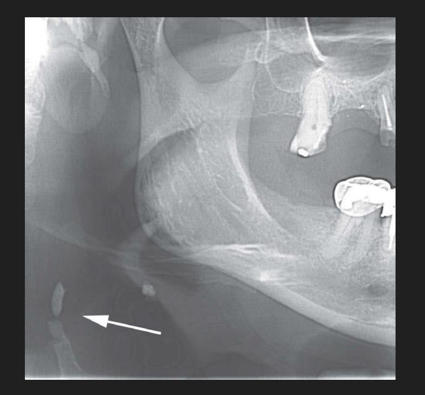
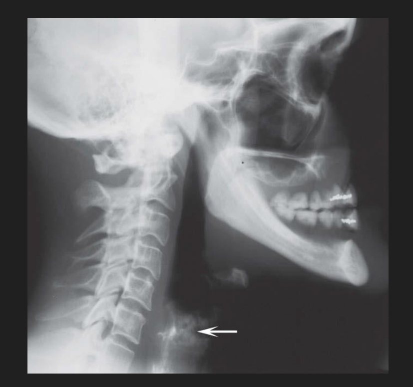
Laryngeal Cartilage Calcifications
Mechanism37
- Progresses as a physiologic process
- Endochondral calcification and ossification of the hyaline laryngeal cartilages begins on attainment of skeletal maturity
Lecturer — Laryngeal Cartilage
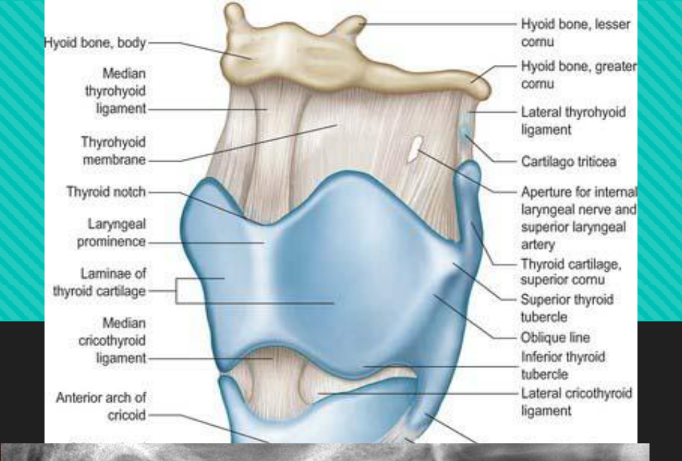
Most laryngeal cartilages are hyaline cartilage and may ossify. The epiglottis and vocal processes of the arytenoid cartilages are fibroelastic and are excluded.
 |  |
Clinical Features
- Asymptomatic
- Typically an incidental finding
Management
- No treatment needed
Location38
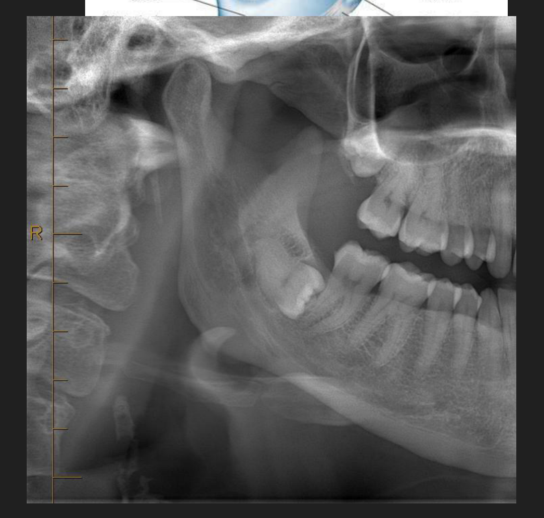
- Triticeous cartilage: small, paired, found within lateral thyrohyoid ligaments
- [PAN] located inferior to greater cornu of hyoid and adjacent to superior border of C4
- Superior cornu of thyroid cartilage: medial to C4
 |  |
Periphery/Shape
- Triticeous: “grain of wheat” measuring 7-9mm (L) and 2-4mm (W) with smooth, well-defined, regular geometry
- Thyroid cartilage: usually only 2-3mm visible at lower edge of radiograph
Internal Features
- Homogeneous radiopaque, but occasionally with an outer cortex
Surrounding Features
- N/A
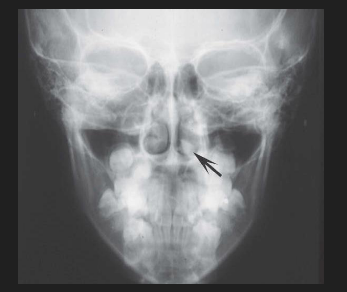
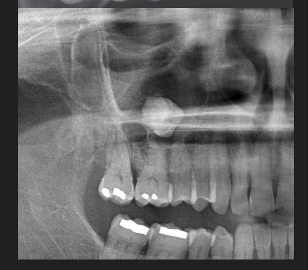
Rhinoliths and Antroliths
Disease Mechanism39
- Arise from deposition of nasal, lacrimal, and inflammatory mineral salts by accretion around a nidus
- Rhinolith nidus = typically an exogenous foreign object
- Usually enters from anterior, but may be from posterior choana
- Antrolith nidus = typically endogenous
- Dystrophic calcification within chronically inflamed mucosa in long-standing sinusitis, or
- A non-invasive aspergillosis mycetoma in patients with chronic sinus disease → necrotic fungus ball or hard mycolith
Lecturer — Rhinolith Nidus
A rhinolith nidus is usually an exogenous object, such as a coin, bead, seed, fruit pit, or other object introduced through the nostril. Drug smuggling was also mentioned as a possible source of a nasal foreign object.
Clinical Features40
- Asymptomatic
- Expanding mass may impinge on mucosa → pain, congestion, and ulceration
- May have nasal obstruction, unilateral purulent or blood-stained rhinorrhoea, sinusitis, headache, epistaxis, anosmia, fetor, and fever
Management
- Referral to ENT for endonasal or sinus endoscopic surgical removal
- Lithotripsy has been used in some cases to debulk large rhinoliths
Location41
- Rhinoliths: develop in nose
- Antroliths: develop in maxillary antrum
- Concretions rarely form in frontal or ethmoid sinus
Periphery/Shape
- Various shapes and sizes, depending on nature of nidus
Internal Features
- Homogeneous or heterogeneous radiopacities, depending on nature of nidus
- May have laminations
- Occasionally, density exceeds surrounding bone
Surrounding Features
- N/A
Metastatic Calcification4243
- Caused by conditions involving elevated serum Ca and PO₄ levels
- E.g. hyperparathyroidism, hypercalcaemia of malignancy
- Occurs in soft tissues of orofacial region
- Extremely rare
Lecturer — Metastatic Calcification
Metastatic calcification may also occur in chronic renal failure. These findings are uncommon in dental practice.
Heterotopic Ossifications44
- Stylohyoid ligament ossification
- Osteoma cutis
- Myositis ossificans
- Heterotopic ossification is the deposition of calcium and phosphate as organised bone within soft tissue.
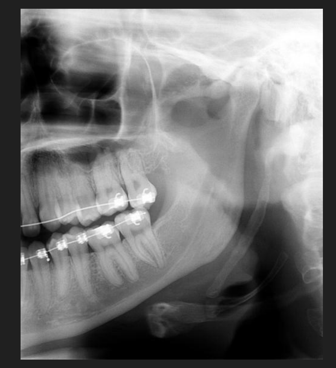
Stylohyoid Ligament Ossification
Disease Mechanism45
- The styloid process arises from second branchial arch (Reichert’s cartilage).
- The stylohyoid ligament is a remnant of the second branchial arch.
- Ossification of the styloid process begins from the base of the skull downwards.
- Length of styloid process is variable, but maximum 3 cm is considered to be normal.
- This ossification process may extend to the stylohyoid ligament
- Commonly bilateral
Epidemiology
- Prevalence: ~18% of population showed ossification of >3 cm of the stylohyoid ligament
- Age: may show some calcification at any age
Clinical Features46
- Usually an incidental feature on panoramic images
- The ossified ligament can be palpated at the palatine tonsil as a hard, pointed structure
- Only a few patients have symptoms
- Little correlation between extent of ossification and intensity of symptoms
- Symptoms related to the ossification of the stylohyoid ligament is known as Eagle Syndrome with two subtypes:
- Classic Eagle syndrome
- Results from cranial nerve impingement
- Carotid artery syndrome
- Results from impingement of carotid vessels
- Classic Eagle syndrome
Classic Eagle Syndrome
Stylohyoid ligament ossification + presence of clinical discomfort + a recent history of neck trauma (typically tonsillectomy)
The ossified stylohyoid complex and local scar tissue are thought to cause symptoms by impinging on CN V, VII, IX, X or XII (pass in close proximity to the styloid process)
Symptoms
- Vague, nagging to intense pain in the pharynx on speaking, chewing, swallowing, turning the head or opening the mouth widely, especially on singing or yawning
- A foreign body sensation in the throat on swallowing
- Tinnitus
- Otalgia
Lecturer — Carotid Artery Syndrome
Carotid artery syndrome, also called vascular Eagle syndrome, consists of stylohyoid ligament ossification with symptoms but no history of neck trauma.
- It is more prevalent than classic Eagle syndrome and tends to affect individuals over 40 years of age.
- Head turning may cause compression of the internal or external carotid artery.
- Reported effects include vertigo, syncope, carotid-distribution pain and carotidynia from stimulation of the sympathetic plexus around the artery.
- Other reported symptoms include eye pain, temporoparietal headaches, migraines, aphasia, visual symptoms and weakness.
DDx for Stylohyoid Ligament Ossification and Eagle Syndrome47
Symptoms are often vague, but symptoms + evidence of ligament ossification is usually distinctive for Eagle syndrome.
NOTE: Presence of stylohyoid ligament ossification WITHOUT symptoms is not considered to be Eagle syndrome.
- TMD: Diagnostic confirmation of stylohyoid ligament ossification can be achieved by topical anaesthesia to suppress gag reflex, palpation of tonsillar fossa to reproduce symptoms, and detection of the hard submucosal mass
Management
- Most are asymptomatic: no treatment
- Vague symptoms: conservative management, reassurance, steroid or lidocaine injections into tonsillar fossa
- Persistent or intense symptoms: stylohyoidectomy (amputation of styloid process)
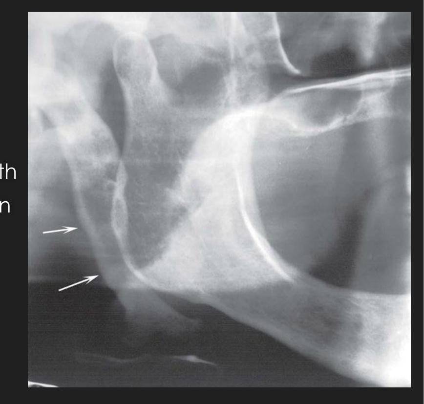
Location48
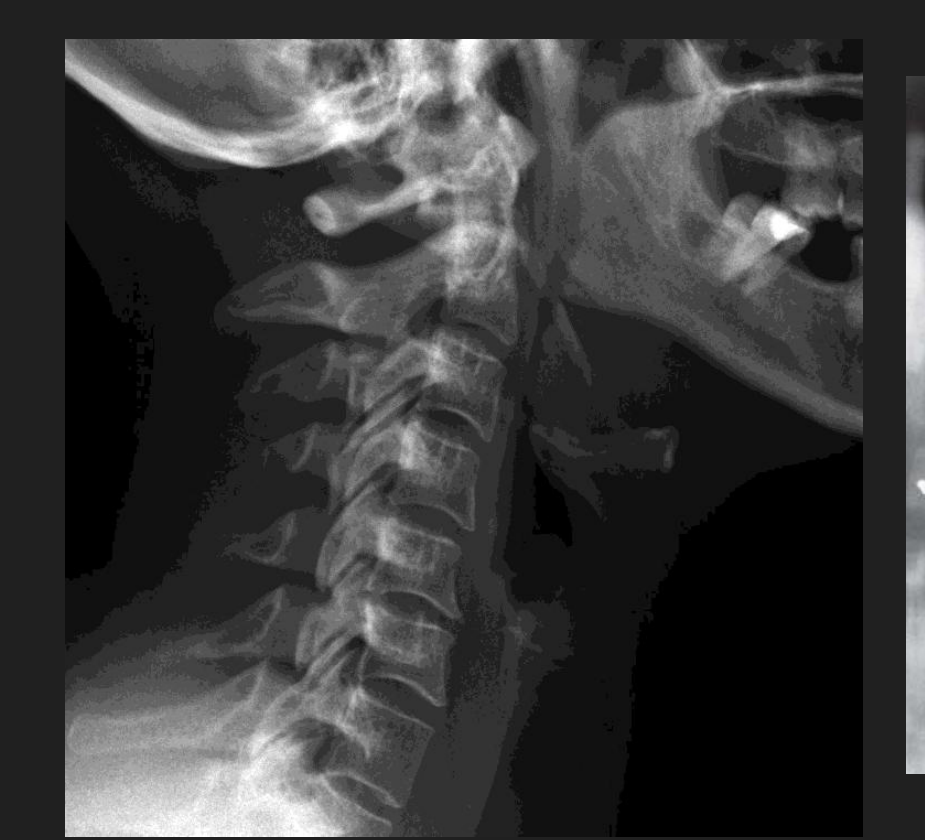
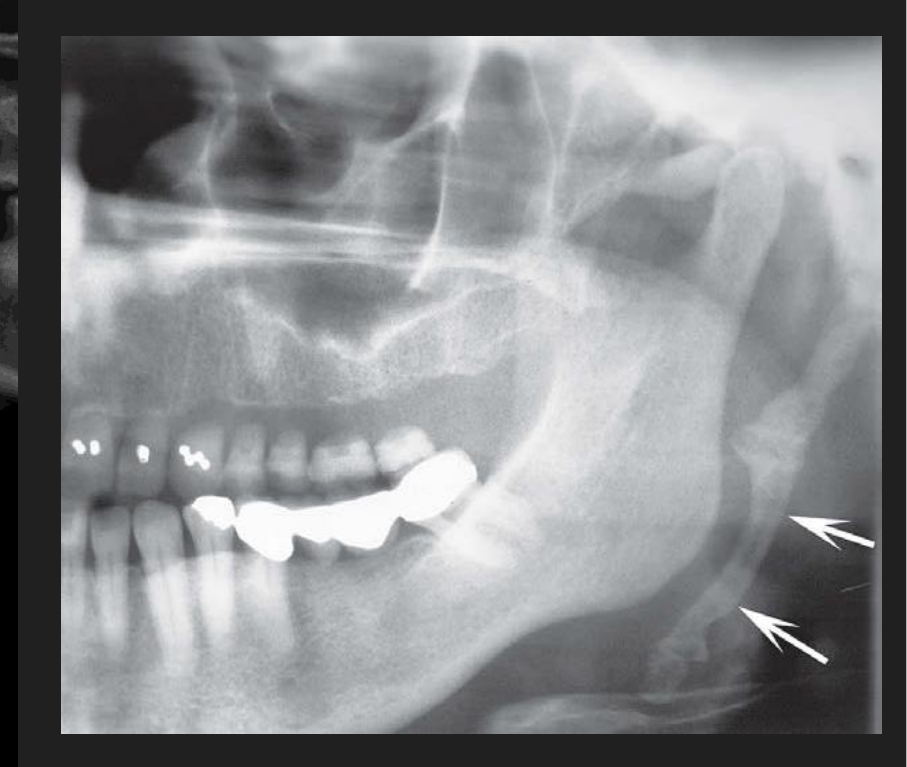
Linear ossification extends from region of mastoid process, crosses the posteroinferior aspect of the ramus towards the hyoid bone
 |  |
 |
Periphery/Shape
- Styloid process appears as long, tapering, thin radiopaque process of 0.5–2.5 cm in length
- Ossified ligament has roughly straight outline, although some irregularity may be seen on the outer surface
- Pseudoarticulations (radiolucent, joint-like junctions) may interrupt the ligament
Internal Features
- Small: homogeneously radiopaque
- As the ossification increases in length and girth, the outer cortex of the bone becomes apparent (radiopaque band at periphery)
Surrounding Features
N/A
Osteoma Cutis
Disease Mechanism49
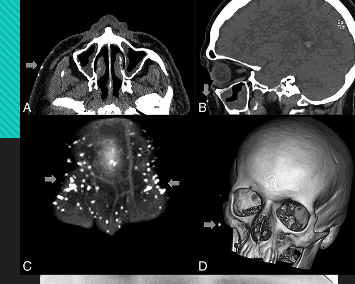
A rare soft tissue calcification in the skin or subcutaneous tissues that manifests as focal development of bone within the dermis physically removed from any original osseous tissue.
Divided into:
- Primary (15%): occurring in normal tissue without any pre-existing condition
- Secondary (85%): developing in damaged or disrupted skin (e.g. acne)
Occasionally found in diffuse scleroderma — replaces the altered collagen in the dermis and subcutaneous septa
Clinical Features50
- Can occur anywhere
- EO: face most common
- IO: tongue most common (osteoma mucosae or osseous choristoma)
- Does not cause any visible change in the overlying skin
- Occasional colour change to yellowish-white
- If large, individual osteoma may be palpated
- If needle inserted, stone-like resistance is met
- Multiple miliary osteoma cutis: numerous (dozens to hundreds) of lesions
- Female: face; Male: scalp or chest
Management
- No treatment required
- Primary osteoma cutis often removed for cosmetic reasons
- Resurfacing of the skin in multiple miliary osteoma cutis with Er:YAG laser + tretinoin cream, curettage and CO₂ continuous wave laser, or needle microincision-extirpation technique
Location51
- Most commonly cheek and lip regions
- May be superimposed over tooth root or alveolar process → appearance of area of dense bone
 | 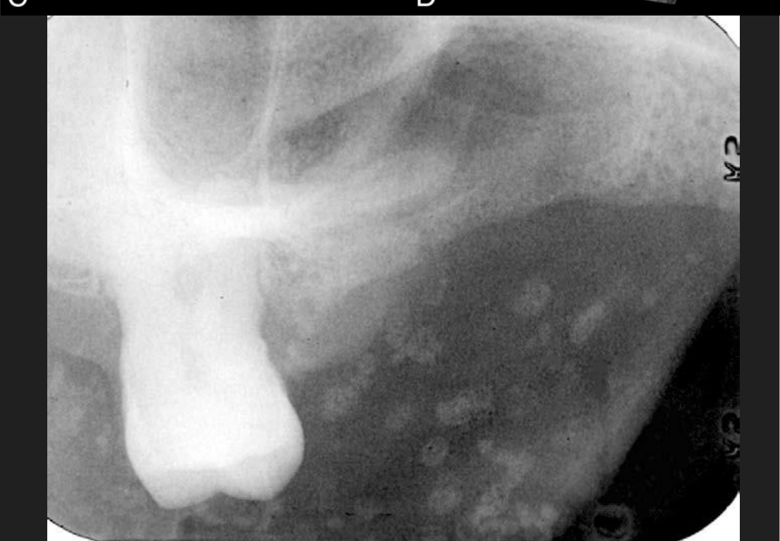 |
Periphery/Shape
- Smoothly outlined, radiopaque, washer-shaped
- Single or multiple
- Usually very small (range: 0.1–5 cm)
Internal Features
- Homogeneously radiopaque with radiolucent centre (represents fatty marrow) → donut appearance
- Trabeculae occasionally develop in marrow cavity of larger osteomas
- Snowflake-like radiopacity corresponding to clinical location of scar tissue in calcified cystic acne
Surrounding Features
N/A
Myositis Ossificans52
- Fibrous tissue and heterotopic bone form within the interstitial tissue of muscle and associated tendons/ligaments.
- Leads to secondary destruction and atrophy of muscle (due to interdigitation of the fibrous tissue and bone which separates the muscle fibres)
- Two principle forms:
- Localised
- Progressive
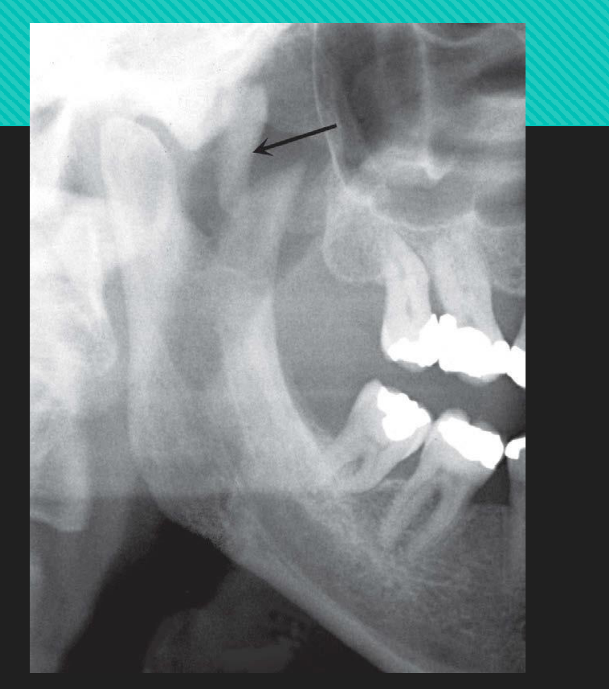
Localised (Traumatic) Myositis Ossificans
- Synonyms: post-traumatic myositis ossificans, solitary myositis
- Epidemiology:
- Can develop at any age and gender (more common in young men who engage in vigorous activity)
- Aetiology: acute/chronic trauma, heavy muscular strain caused by certain occupations and sports, muscle injury from multiple injections (e.g. IANB)
Disease Mechanism
- Skeletal muscle has limited capacity for regeneration following significant physical trauma
- → haemorrhage into muscle, tendons, or fascia
- → exuberant proliferation of vascular granulation tissue subsequently undergoes metaplasia to cartilage and bone during healing
- NB: no inflammation is involved despite the term myositis
- NB: the fibrous tissue and bone form within the interstitial tissue of muscle; no actual ossification of the muscle fibres occurs
Clinical Features
- Site of trauma remains swollen, tender, and painful for longer than expected
- Overlying skin red and inflamed
- Opening jaws may be difficult
- After 2–3 weeks, the area of ossification becomes apparent with palpation of a firm intramuscular mass
- May enlarge slowly but eventually stops growing
- May appear fixed or be freely moveable
Management
- Rest and limitation of use to reduce extent of calcific deposit
- Medical management with bone morphogenetic protein type I receptor inhibition to reduce heterotopic ossification
- Surgical excision for lesions that cause a functional restriction or neurologic impairment
- With intensive physiotherapy to minimise postsurgical scarring
- Incomplete excision or excision at immature stage can result in recurrence
Location
Most common in head and neck: masseter and SCM
Periphery/Shape
- Periphery more radiopaque than internal structure
- Variation in shape: irregular oval to linear streaks (pseudotrabeculae) running in same direction as muscle fibres — characteristic for myositis ossificans
- Heterotopic bone may lie along long axis of muscle
- Generally <6 cm in greatest dimension
Internal Features
Varies with time:
- (3–4 weeks) faintly homogeneous radiopaque
- (2 months) further organisation into a delicate, lacy or feathery radiopaque internal structure (indicative of bone formation but without normal trabecular pattern)
- (6–12 months) fully matured where entity becomes denser, more homogeneous and better defined
- (after this) lesion may shrink
Surrounding Features
N/A
DDx
-
Ossification of stylohyoid ligament: form and location of myositis ossificans often enough to make DDx
-
Other soft tissue ossifications
-
Osteogenic neoplasms: e.g. osteogenic sarcoma — can form a linear bone pattern, however, lesion is contiguous with adjacent bone and signs of bone destruction often present
-
Further imaging and investigations may be needed to exclude bone-forming neoplasms.
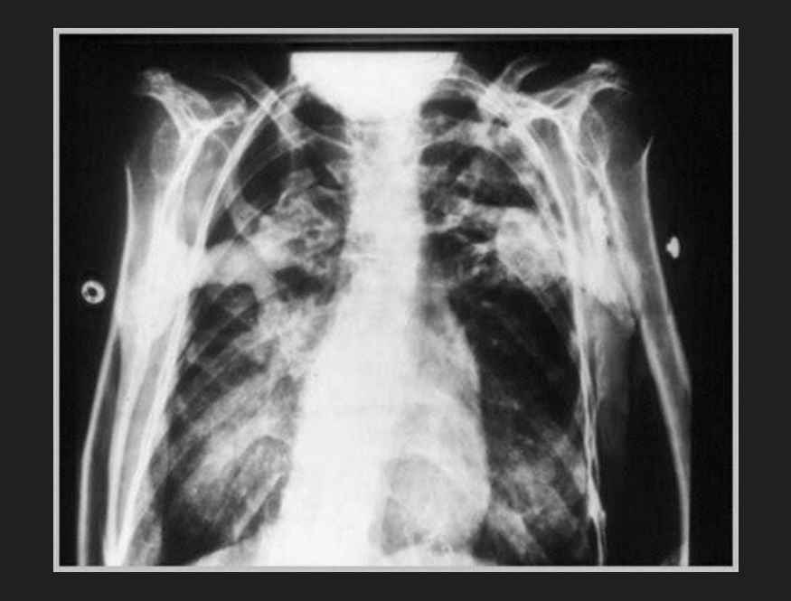
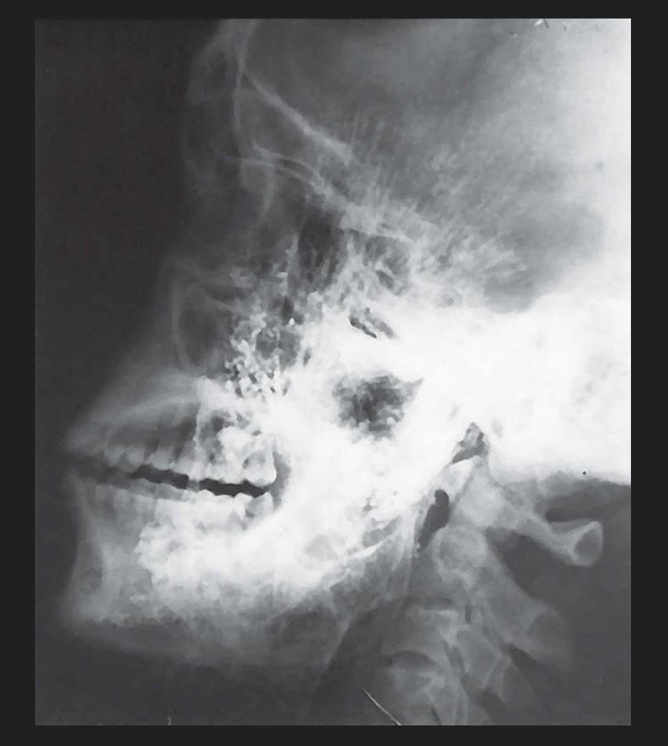
Progressive Myositis Ossificans53
- Synonym: fibrodysplasia ossificans progressiva
Disease Mechanism
- Rare hereditary autosomal dominant disease
- Symptoms from early infancy
Epidemiology
- M > F
Clinical Features
- Begins in striated muscles of neck and upper back, then extend to extremities
- Initially presents as soft tissue swelling with pain, with increasing stiffness and limitation of movement as ossification occurs, eventually resulting in the ‘petrified man’ condition
- Life expectancy: 3rd–4th decade
Lecturer — Progressive Disease Outcome
Premature death generally occurs in the third or fourth decade.
- Causes include ossification of the respiratory muscles.
- Involvement of the muscles of mastication may cause inadequate nutrition.
Management
-
No effective treatment exists
-
Excision of nodules that are traumatised and ulcerate frequently
-
Supportive therapy may be required in later stages of disease when interference with respiration or respiratory infection occurs
-
Imaging features similar to localised form
-
Heterotopic bone more commonly oriented along long axis of involved muscle
-
Osseous malformation of the regions of muscle attachment (e.g. Md condyles) may be seen
 |  |
References54
- Koong, B. (2017). Atlas of Oral and Maxillofacial Radiology. Chichester, UK: John Wiley and Sons.
- White, and Pharoah. (2014). Oral radiology : Principles and interpretation (7th ed.). St. Louis: Elsevier/Mosby.
Audio Appendix
Additional Audio Content
The following sections from the lecture audio did not correspond to any heading in the main document.
Further Reading
- Two resources were presented as useful further reading on soft tissue calcifications and ossifications in the head and neck.
Clinical Cases
Case: Radiopaque opacities within a cystic cavity
Question
Scenario: A patient presents with a chronically infected residual cyst.
What’s shown: A cropped panoramic radiograph and a coronal slice of a multi-slice CT scan of the same patient.
Consider: What do the internal opacities within the cystic cavity represent, and what is the underlying mechanism of their formation?
Answer
Observations:
- Opacities are visible internally within the cystic cavity on both the panoramic radiograph and the CT scan.
Reasoning: These opacities represent dystrophic calcification, which forms in degenerating, diseased, or necrotic tissue (such as a chronically infected cyst) while serum calcium and phosphate levels remain normal.
Takeaway: Dystrophic calcifications can occur within the cavities of chronically infected cysts due to tissue degeneration and necrosis.
Case: Soft tissue overgrowth in an edentulous ridge
Question
Scenario: An edentulous patient has a history of trauma to the alveolar ridge over the years.
What’s shown: A radiograph showing the outline of a soft tissue overgrowth with internal opacities.
Consider: What is the diagnosis for the soft tissue overgrowth, and what do the internal opacities indicate?
Answer
Observations:
- The outline of a soft tissue overgrowth is visible.
- Internal opacities are present within the soft tissue.
Reasoning: The soft tissue overgrowth is fibrous hyperplasia resulting from chronic trauma to the alveolar ridge. The internal opacities represent dystrophic calcification within this hyperplastic tissue.
Takeaway: Fibrous hyperplasia caused by chronic trauma can undergo dystrophic calcification, presenting as radiopacities within the soft tissue outline.
Case: Linear chain of irregular opacities near the mandibular angle
Question
Scenario: A panoramic radiograph is evaluated for soft tissue calcifications in the submandibular and cervical chain region.
What’s shown: A linear chain of opacities located at or just below the angle of the mandible.
Consider: What is the most likely identity of these calcifications, and what are their characteristic morphological features?
Answer
Observations:
- The opacities form a linear chain reflecting the cervical chain of nodes.
- They are well-defined, irregular, and described as cauliflower-like.
- Internally, they may have a laminated appearance resembling an onion cross-section.
Reasoning: The location and morphology are characteristic of calcified lymph nodes, which typically result from chronic inflammation (e.g., granulomatous diseases like tuberculosis) or previous oral cavity inflammation. The irregular, cauliflower-like shape distinguishes them from smoother sialoliths or phleboliths.
Takeaway: Calcified lymph nodes often present as irregular, cauliflower-like, or laminated opacities in the cervical chain and submandibular regions.
Case: Cluster of small opacities superimposed over the mid-ramus
Question
Scenario: A panoramic radiograph and cross-sectional imaging (CBCT/MSCT) are reviewed for a patient.
What’s shown: A cluster of multiple small opacities superimposed over the mid-ramus level on the panoramic view, and located medial to the ramus next to the pharyngeal airspace on CT.
Consider: What is the most likely diagnosis for these calcifications, and what is their typical composition and origin?
Answer
Observations:
- Multiple small opacities with a density similar to cortical bone.
- Located medial to the ramus and adjacent to the lateral wall of the pharyngeal airspace.
Reasoning: This presentation is classic for a tonsillolith (post-inflammatory tonsillar calcification), typically affecting the palatine tonsil. They form when organic debris (dead bacteria, pus, epithelial cells) lodges in enlarged tonsillar crypts due to repeated tonsillitis and serves as a nidus for calcification.
Takeaway: Tonsilloliths present as clusters of small, cortically dense opacities medial to the ramus and are a common incidental finding on panoramic radiographs.
Case: Heterogeneous opacity at the carotid bifurcation
Question
Scenario: A multi-slice CT and panoramic radiograph are evaluated at the level of the C3 and C4 vertebrae.
What’s shown: A well-defined, irregular, C-shaped or circular opacity with radiolucent voids located at the carotid bifurcation.
Consider: What vascular pathology does this calcification represent, and what is its clinical significance?
Answer
Observations:
- Heterogeneous opacity with radiolucent voids.
- C-shaped or circular morphology lining a tubular structure.
- Located at the carotid bifurcation (C3-C4 level).
Reasoning: These findings represent a calcified atherosclerotic plaque. The calcification occurs in the tunica intima of the vessel due to chronic endothelial injury and lipid accumulation. Its presence on a panoramic radiograph can indicate significant carotid artery stenosis, warranting referral for cardiovascular risk evaluation.
Takeaway: Calcified atherosclerotic plaques at the carotid bifurcation appear as heterogeneous, C-shaped opacities and may indicate significant carotid artery stenosis.
Case: Bilateral tortuous radiopaque lines along the jaw
Question
Scenario: A panoramic radiograph is examined for vascular calcifications.
What’s shown: A parallel pair of thin radiopaque lines with a tortuous path, visible bilaterally along the mandible.
Consider: What specific artery is outlined by these calcifications, and what is the underlying pathological process?
Answer
Observations:
- Parallel pair of thin radiopaque lines (tram-track or pipe-stem appearance).
- Tortuous path, present bilaterally.
Reasoning: This represents Mönckeberg’s medial calcific sclerosis, which affects the tunica media of the arteries (most commonly the facial artery in the head and neck). Unlike atherosclerosis, it causes thickening and loss of elasticity without narrowing the lumen, hence the visible central lucency and bilateral presentation.
Takeaway: Mönckeberg’s arteriosclerosis outlines the facial artery as parallel, tortuous radiopaque lines without causing luminal stenosis.
Case: Laminated structure with a central nidus
Question
Scenario: A micro-CT scan of a salivary gland stone is analyzed.
What’s shown: A calcified structure with a central core surrounded by concentric layers.
Consider: What does the central core represent, and how does the surrounding laminated structure form?
Answer
Observations:
- A central nidus of organic material.
- Surrounding lamellated layers of organic and inorganic material.
Reasoning: This is a ductal sialolith. It forms when alterations in salivary flow or composition lead to a nidus of organic material, which then stimulates the precipitation of concentric layers of calcium and phosphate salts, creating a laminated appearance.
Takeaway: Ductal sialoliths form around an organic nidus that triggers the layered precipitation of calcium and phosphate salts.
Case: Elongated opacities conforming to a ductal path
Question
Scenario: Panoramic and other radiographic projections of the floor of the mouth are reviewed.
What’s shown: Smooth, elongated, and cylindrical radiopacities located within the distal and proximal portions of a duct that bends around a muscle.
Consider: What is the diagnosis, and why do the calcifications take on this specific shape?
Answer
Observations:
- Smooth, elongated, cylindrical opacities.
- Located within the distal and proximal portions of Wharton’s duct.
Reasoning: These are sialoliths within Wharton’s duct. The stones conform to the tubular structure of the duct, resulting in their smooth, elongated, and cylindrical shape, especially as the duct bends around the mylohyoid muscle.
Takeaway: Sialoliths within a salivary duct often take on a smooth, elongated, cylindrical shape as they conform to the tubular structure of the duct.
Case: Irregular opacity within a salivary gland hilum
Question
Scenario: A radiograph of the submandibular region is evaluated.
What’s shown: A slightly irregular-shaped radiopacity located within the hilum of the submandibular salivary gland.
Consider: Why does this calcification have a more irregular shape compared to those found within the duct?
Answer
Observations:
- Irregular-shaped radiopacity.
- Located within the hilum of the submandibular salivary gland.
Reasoning: This is a sialolith located in the hilum, where the ducts collect and exit the gland. Because there is more space to calcify in the hilum compared to the narrow tubular duct, the stone can grow into a larger, more irregular shape.
Takeaway: Sialoliths located in the hilum of a salivary gland tend to be larger and more irregularly shaped due to the available space for calcification.
Case: Collection of multiple small opacities in the parotid region
Question
Scenario: A radiograph of the parotid region is reviewed.
What’s shown: A collection of very small, multiple radiopacities spread throughout the gland.
Consider: What is the diagnosis, and what is the typical underlying etiology for this pattern of calcification?
Answer
Observations:
- Multiple, very small opacities.
- Spread throughout the parotid gland parenchyma.
Reasoning: These are parenchymal calcifications of the parotid gland. Unlike ductal stones, parenchymal calcifications are generally multiple and small, often occurring secondary to chronic inflammation (sialadenitis) such as in Sjögren’s syndrome.
Takeaway: Parenchymal calcifications in the parotid gland present as multiple small opacities and are typically secondary to chronic sialadenitis.
Case: Two radiopaque regions separated by a radiolucent gap
Question
Scenario: A radiograph of a suspected salivary gland stone is evaluated.
What’s shown: Two distinct radiopaque regions with a non-calcified, radiolucent middle section.
Consider: What explains the radiolucent gap between the two calcified regions?
Answer
Observations:
- Two radiopaque regions.
- A radiolucent middle section between them.
Reasoning: The middle section represents a mucus plug where the duct is blocked but the material has not yet calcified due to low mineral content. It is a single continuous blockage rather than two separate stones.
Takeaway: A radiolucent gap between radiopaque sialoliths often represents a non-calcified mucus plug blocking the duct.
Case: Asymmetric soft tissue density in the submandibular region
Question
Scenario: A multi-slice CT soft tissue window is evaluated for a patient with a known opaque sialolith.
What’s shown: Normal soft tissue grey density on the left side, but dark black fat density on the right side where the submandibular gland is expected.
Consider: What pathological process has occurred in the right submandibular gland?
Answer
Observations:
- Normal grey soft tissue density on the left.
- Dark black fat density on the right.
Reasoning: The right submandibular gland has undergone fat-replaced atrophy. Chronic blockage of the duct by the sialolith leads to backed-up secretions, a negative feedback loop that stops saliva production, and eventual replacement of the glandular tissue with fat.
Takeaway: Chronic ductal obstruction by a sialolith can lead to fat-replaced atrophy of the affected salivary gland.
Case: Hyperechoic structure with posterior shadowing
Question
Scenario: An ultrasound of the submandibular gland is performed for suspected salivary gland pathology.
What’s shown: A well-defined, white (hyperechoic) structure with a dark area directly behind it.
Consider: What does the hyperechoic structure represent, and what causes the dark area behind it?
Answer
Observations:
- Hyperechoic (white), well-defined structure.
- Acoustic posterior shadowing (dark area behind the structure).
Reasoning: The hyperechoic structure is a sialolith (calculus). Because ultrasound uses sound waves, the hard surface of the stone reflects the waves (appearing white), preventing them from penetrating deeper and creating an acoustic posterior shadow where no information is recorded.
Takeaway: On ultrasound, sialoliths appear as hyperechoic structures with acoustic posterior shadowing due to the reflection of sound waves.
Case: Flow void in the proximal parotid duct
Question
Scenario: A multi-slice CT sialogram with contrast is reviewed for a patient with recurrent left parotid swelling during mealtimes.
What’s shown: A flow void where there is no contrast in the proximal duct of the parotid gland.
Consider: What is the cause of the flow void, and why is it not visible on conventional non-contrast imaging?
Answer
Observations:
- Flow void lacking contrast in the proximal parotid duct.
Reasoning: The flow void represents a mucus plug or a non-calcified sialolith. Because it lacks calcification (low mineral content), it is radiolucent and cannot be detected on conventional 2D imaging or non-contrast CT; contrast injection is required to outline the ductal system and reveal the filling defect.
Takeaway: Non-calcified sialoliths or mucus plugs appear as flow voids on contrast sialograms and are invisible on non-contrast imaging.
Case: Acute ductal bend causing recurrent swelling
Question
Scenario: A multi-slice CT sialogram with surface-rendered images is evaluated for a patient with recurrent right submandibular swelling during mealtimes.
What’s shown: Contrast outlining the ductal system, revealing no stones but a sharp angulation in the proximal duct.
Consider: What is the anatomical cause of the patient’s symptoms?
Answer
Observations:
- No sialoliths blocking the duct.
- An acute bend in the proximal duct.
Reasoning: The acute bend in the proximal duct affects the flow of saliva, causing backup and recurrent swelling during mealtimes when salivary flow is stimulated. This structural anomaly, rather than a stone, is the cause of the symptoms.
Takeaway: Recurrent salivary swelling can be caused by structural ductal anomalies, such as an acute bend, which impairs saliva flow even in the absence of sialoliths.
Case: Targetoid opacity in the retroantral region
Question
Scenario: A CBCT scan of the pterygopalatine/retroantral region is evaluated.
What’s shown: A round or oval radiopacity with a classic targetoid or bull’s-eye appearance.
Consider: What is the diagnosis, and what underlying vascular condition does this finding imply?
Answer
Observations:
- Round/oval radiopacity.
- Targetoid or bull’s-eye internal appearance.
Reasoning: This is a phlebolith, which is a calcified thrombus within a vein or vascular malformation. In the head and neck, the presence of phleboliths almost always indicates an underlying hemangioma or vascular malformation, which requires careful management to avoid lethal exsanguination during surgical procedures.
Takeaway: Phleboliths exhibit a targetoid bull’s-eye appearance and strongly indicate the presence of an underlying vascular malformation or hemangioma.
Case: Radiopacity with a central lucency near the hyoid bone
Question
Scenario: A cropped panoramic radiograph is evaluated inferior to the greater cornu of the hyoid bone and adjacent to C4.
What’s shown: A relatively homogeneous radiopacity that presents with an outer cortex and a central lucent appearance.
Consider: What anatomical structures are calcifying, and is this a pathological process?
Answer
Observations:
- Radiopacity with an outer cortex and central lucency.
- Located inferior to the hyoid bone and adjacent to C4.
Reasoning: This represents physiological endochondral ossification of the laryngeal cartilages, specifically the triticeous cartilage and the superior cornu of the thyroid cartilage. It is a normal, age-related process that requires no management.
Takeaway: Calcification of the laryngeal cartilages (triticeous and thyroid) is a normal physiological process that can present with an outer cortex and central lucency on panoramic radiographs.
Case: Linear ossified structure with joint-like interruptions
Question
Scenario: A panoramic radiograph is evaluated in the region of the styloid process and hyoid bone.
What’s shown: A linear ossified structure extending from the styloid process towards the hyoid bone, featuring irregular interruptions that resemble joints.
Consider: What is the diagnosis, and what do the joint-like interruptions represent?
Answer
Observations:
- Linear ossified structure crossing the posterior inferior border of the ramus.
- Irregular interruptions resembling joints (pseudoarthroses).
Reasoning: This represents ossification of the stylohyoid ligament. The joint-like interruptions are pseudoarthroses, which are fake joint structures that can interrupt the ligament as it ossifies. Unless symptomatic (Eagle syndrome), it is an incidental finding.
Takeaway: Ossification of the stylohyoid ligament can present with pseudoarthroses, appearing as joint-like interruptions along the ossified linear structure.
Case: Water-shaped opacity superimposed over a tooth
Question
Scenario: A PA radiograph of the facial region is reviewed.
What’s shown: A roughly circular radiopacity with a central lucency, sometimes showing a trabecular or snowflake-like pattern, superimposed over a tooth or alveolar process.
Consider: What is the diagnosis, and what is the typical clinical presentation of this condition?
Answer
Observations:
- Water-shaped or donut-like radiopacity (circular with central lucency).
- Trabecular or snowflake-like internal pattern.
- Superimposed over dental structures.
Reasoning: This is osteoma cutis, a focal development of bone in the dermis or subcutaneous tissues. It is often asymptomatic and discovered incidentally on radiographs when superimposed over teeth. Treatment is usually only required for cosmetic reasons.
Takeaway: Osteoma cutis presents as a water-shaped, radiopaque lesion with a central lucency in the soft tissues and is typically asymptomatic.
Case: Linear ossifications along the temporalis muscle
Question
Scenario: Radiographs of a patient with a known hereditary condition causing progressive muscle stiffness are evaluated.
What’s shown: Radiopaque linear lines oriented along the long axis of the temporalis muscle, along with osseous malformation around the mandibular condyle.
Consider: What is the diagnosis, and how does the ossification pattern relate to the underlying anatomy?
Answer
Observations:
- Radiopaque linear lines along the long axis of the temporalis muscle.
- Osseous malformation around the mandibular condyle (muscle attachment region).
Reasoning: This represents progressive myositis ossificans, a rare hereditary condition where heterotopic bone forms in the interstitial tissues of muscles, tendons, and ligaments. The ossification characteristically aligns along the long axis of the involved muscles and can affect tendon attachments near joints.
Takeaway: Progressive myositis ossificans causes heterotopic bone formation that aligns along the long axis of muscles and can involve tendon attachments.
Footnotes
-
Original PDF page 1: L2 - Opacities in Dental Imaging, p.1 ↩
-
Original PDF page 2: L2 - Opacities in Dental Imaging, p.2 ↩
-
Original PDF page 3: L2 - Opacities in Dental Imaging, p.3 ↩
-
Original PDF page 4: L2 - Opacities in Dental Imaging, p.4 ↩
-
Original PDF page 5: L2 - Opacities in Dental Imaging, p.5 ↩
-
Original PDF page 6: L2 - Opacities in Dental Imaging, p.6 ↩
-
Original PDF page 7: L2 - Opacities in Dental Imaging, p.7 ↩
-
Original PDF page 8: L2 - Opacities in Dental Imaging, p.8 ↩
-
Original PDF page 9: L2 - Opacities in Dental Imaging, p.9 ↩
-
Original PDF page 10: L2 - Opacities in Dental Imaging, p.10 ↩
-
Original PDF page 11: L2 - Opacities in Dental Imaging, p.11 ↩
-
Original PDF page 12: L2 - Opacities in Dental Imaging, p.12 ↩
-
Original PDF page 13: L2 - Opacities in Dental Imaging, p.13 ↩
-
Original PDF page 14: L2 - Opacities in Dental Imaging, p.14 ↩
-
Original PDF page 15: L2 - Opacities in Dental Imaging, p.15 ↩
-
Original PDF page 16: L2 - Opacities in Dental Imaging, p.16 ↩
-
Original PDF page 17: L2 - Opacities in Dental Imaging, p.17 ↩
-
Original PDF page 18: L2 - Opacities in Dental Imaging, p.18 ↩
-
Original PDF page 22: L2 - Opacities in Dental Imaging, p.22 ↩
-
Original PDF page 19: L2 - Opacities in Dental Imaging, p.19 ↩
-
Original PDF page 20: L2 - Opacities in Dental Imaging, p.20 ↩
-
Original PDF page 21: L2 - Opacities in Dental Imaging, p.21 ↩
-
Original PDF page 23: L2 - Opacities in Dental Imaging, p.23 ↩
-
Original PDF page 24: L2 - Opacities in Dental Imaging, p.24 ↩
-
Original PDF page 25: L2 - Opacities in Dental Imaging, p.25 ↩
-
Original PDF page 26: L2 - Opacities in Dental Imaging, p.26 ↩
-
Original PDF page 27: L2 - Opacities in Dental Imaging, p.27 ↩
-
Original PDF page 28: L2 - Opacities in Dental Imaging, p.28 ↩
-
Original PDF page 29: L2 - Opacities in Dental Imaging, p.29 ↩
-
Original PDF page 30: L2 - Opacities in Dental Imaging, p.30 ↩
-
Original PDF page 31: L2 - Opacities in Dental Imaging, p.31 ↩
-
Original PDF page 32: L2 - Opacities in Dental Imaging, p.32 ↩
-
Original PDF page 36: L2 - Opacities in Dental Imaging, p.36 ↩
-
Original PDF page 33: L2 - Opacities in Dental Imaging, p.33 ↩
-
Original PDF page 34: L2 - Opacities in Dental Imaging, p.34 ↩
-
Original PDF page 35: L2 - Opacities in Dental Imaging, p.35 ↩
-
Original PDF page 37: L2 - Opacities in Dental Imaging, p.37 ↩
-
Original PDF page 38: L2 - Opacities in Dental Imaging, p.38 ↩
-
Original PDF page 39: L2 - Opacities in Dental Imaging, p.39 ↩
-
Original PDF page 40: L2 - Opacities in Dental Imaging, p.40 ↩
-
Original PDF page 41: L2 - Opacities in Dental Imaging, p.41 ↩
-
Original PDF page 42: L2 - Opacities in Dental Imaging, p.42 ↩
-
Original PDF page 43: L2 - Opacities in Dental Imaging, p.43 ↩
-
Original PDF page 44: L2 - Opacities in Dental Imaging, p.44 ↩
-
Original PDF page 45: L2 - Opacities in Dental Imaging, p.45 ↩
-
Original PDF page 46: L2 - Opacities in Dental Imaging, p.46 ↩
-
Original PDF page 49: L2 - Opacities in Dental Imaging, p.49 ↩
-
Original PDF page 50: L2 - Opacities in Dental Imaging, p.50 ↩
-
Original PDF page 51: L2 - Opacities in Dental Imaging, p.51 ↩
-
Original PDF page 52: L2 - Opacities in Dental Imaging, p.52 ↩
-
Original PDF page 53: L2 - Opacities in Dental Imaging, p.53 ↩
-
Original PDF page 54: L2 - Opacities in Dental Imaging, p.54 ↩
-
Original PDF page 60: L2 - Opacities in Dental Imaging, p.60 ↩
-
Original PDF page 61: L2 - Opacities in Dental Imaging, p.61 ↩