Cysts of the Jaws1
Dr. May Lam Oral and Maxillofacial Radiologist Senior Lecturer BDSc (Hons) FRACDS (GDP) DClinDent (DMFR) MRACDS (DMFR)
Introduction
-
Definition: a pathologic cavity filled with fluid, lined by epithelium, and surrounded by a definite connective tissue wall.
-
Cysts occur more often in the jaws than in any other bone because most cysts originate from the numerous rests of odontogenic epithelium that remain after tooth formation.
-
The cystic fluid either is secreted by the cells lining the cavity or is derived from the surrounding tissue fluid.
-
Clinical Features: asymptomatic swelling (most common)
-
Some cysts produce a hard swelling.
Definition and Clinical Features of Cysts2
General Imaging Features
Location3
-
Within bone anywhere in the maxilla or mandible (rare in condyle and coronoid process)
-
Odontogenic cysts occur in tooth-bearing regions of the jaws (above IAC)
-
In the maxilla, odontogenic cysts are generally below the floor of the maxillary antrum.
 |  |
Periphery/Shape
-
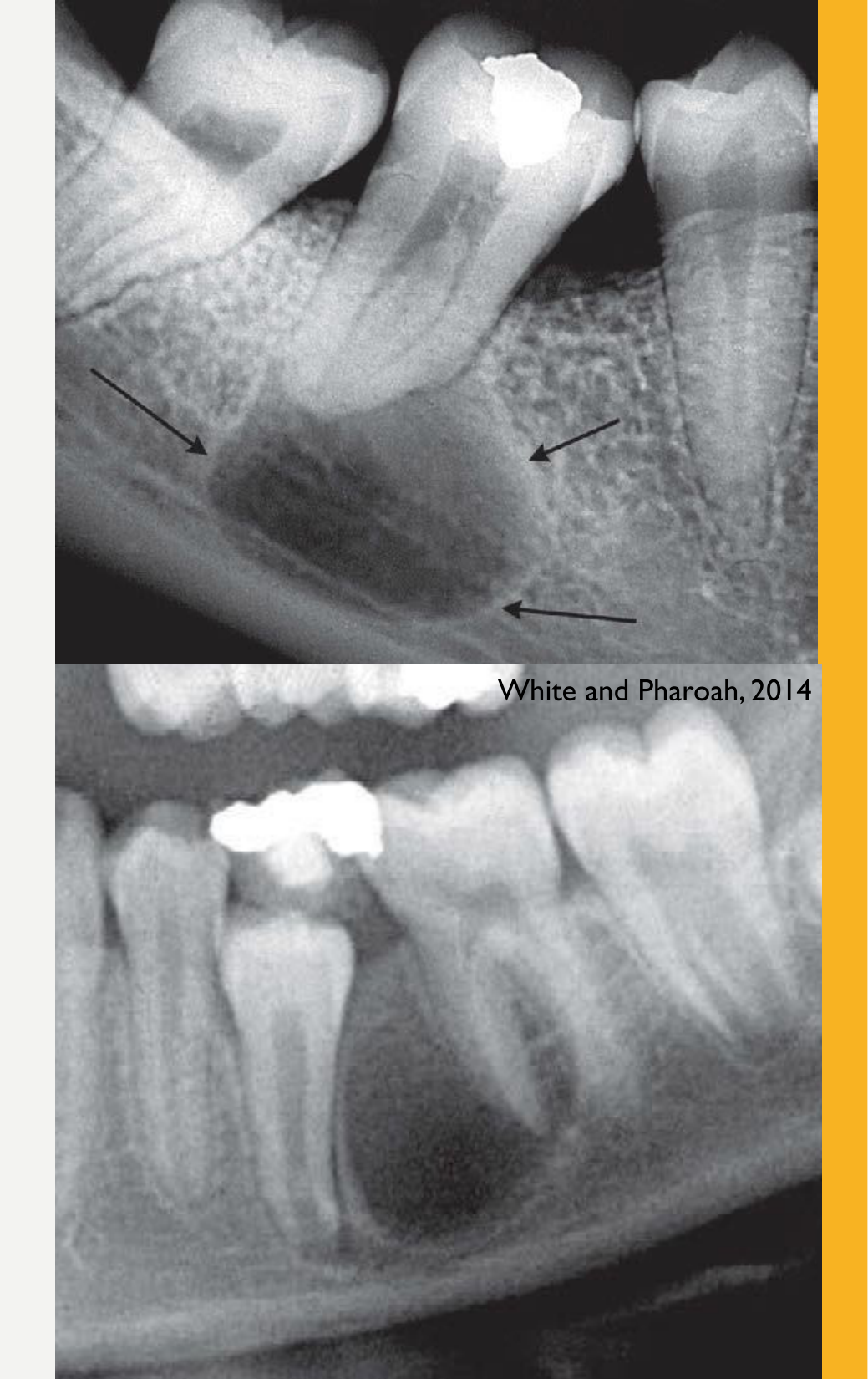
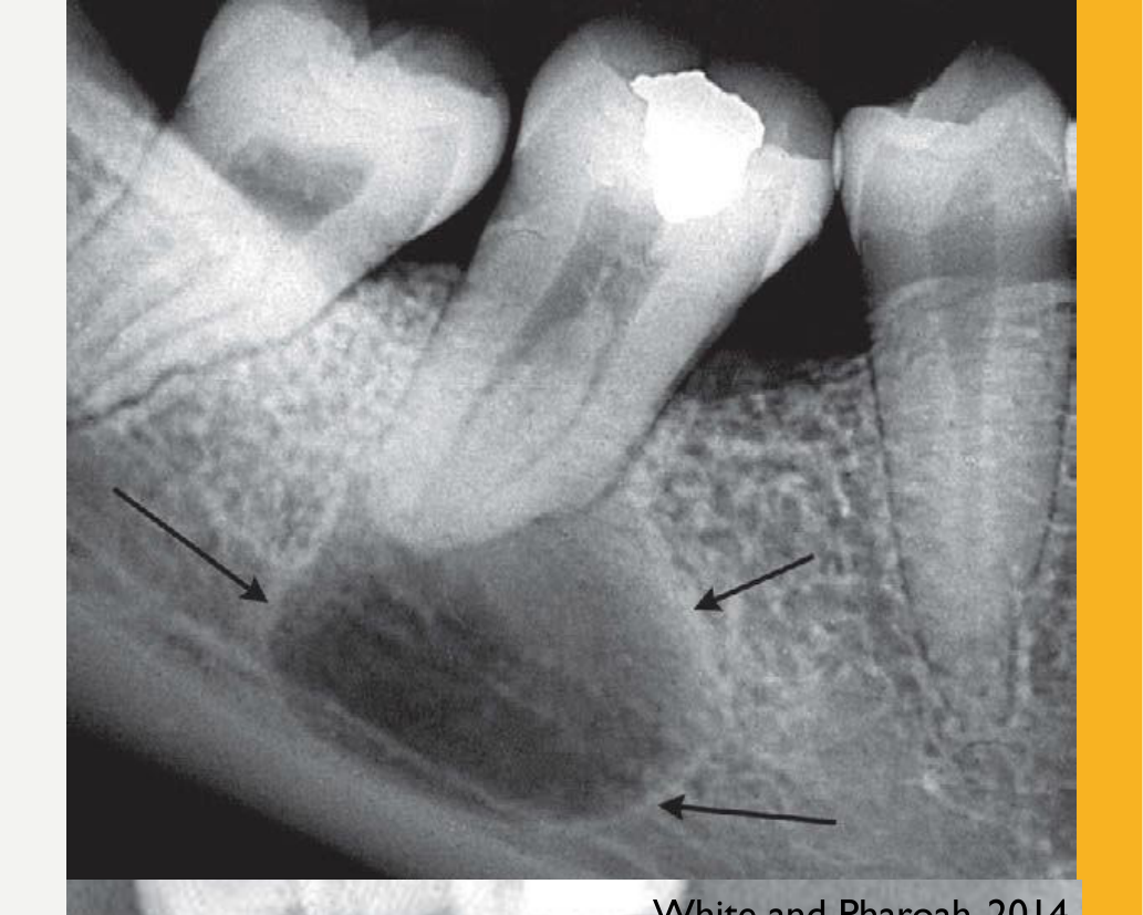
Usually spherical/round, but its shape is influenced by adjacent structures
-
Well-defined, corticated
- If secondarily infected, this may change to a thicker, sclerotic border or make the cortex less apparent
-
A cyst suspended in the maxillary sinus may grow concentrically and remain spherical.
-
A cyst contacting thick cortical bone may develop a flattened edge.
-
The well-defined, corticated border is usually thin and uniform.
Internal Structure4
-
Mostly radiolucent
-
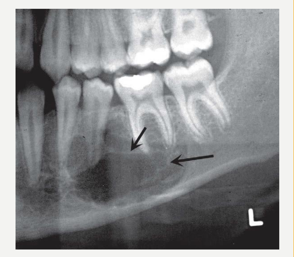
Long standing cysts may have dystrophic calcification (gives a sparse particulate appearance)
-
Some cysts have septa
-
Cysts are usually radiolucent because fluid is lucent on X-rays.
-
Dystrophic calcifications in long-standing cysts appear as sparse, small radiopaque dots.
-
Septa are seen as thin radiopaque lines.
 |  |
Effects on Surrounding Structures
-
Displacement and resorption of teeth
-
Expansion and thinning of the jaw cortices
-
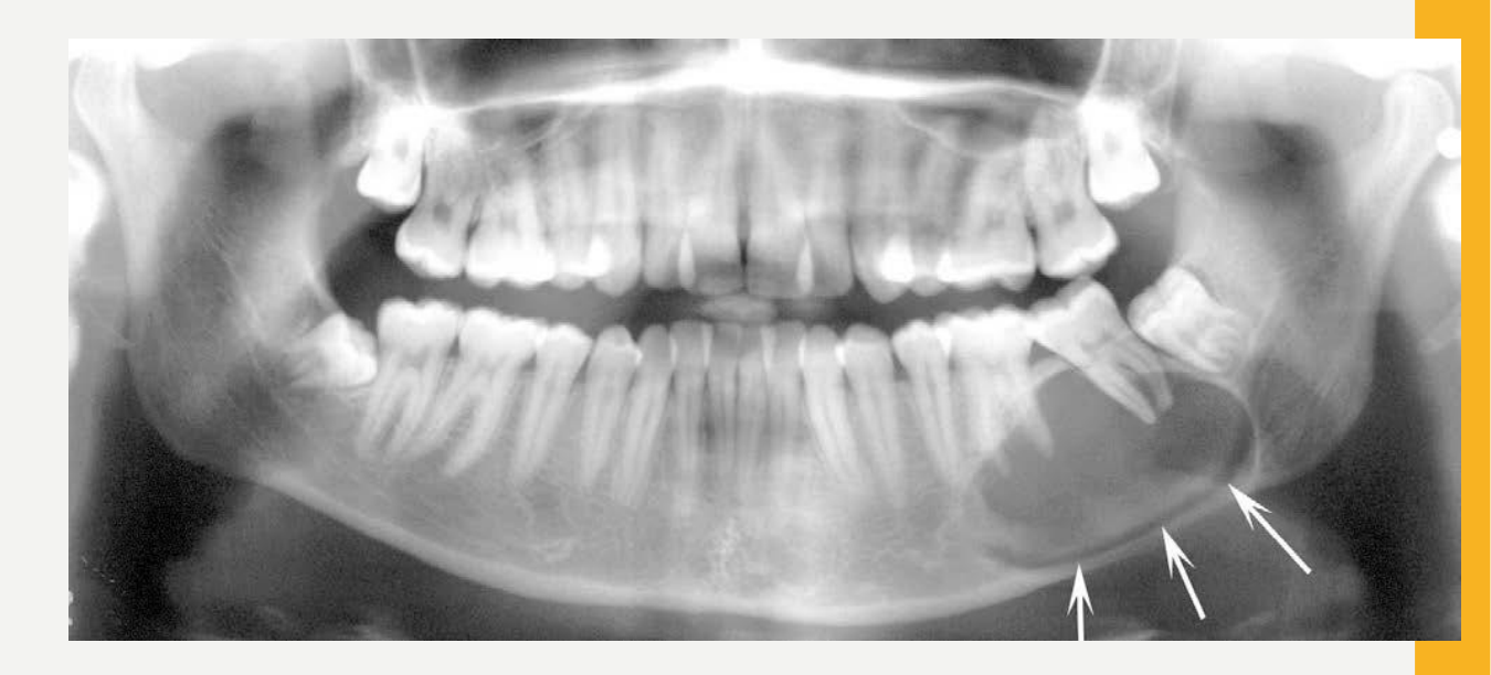
Displacement of the inferior alveolar canal (IAC)
-
Elevation of the antral +/- nasal cortical floors
-
Expansion and thinning of the jaw cortices may be difficult to appreciate on periapical, bitewing, and panoramic radiographs.
-
These cortical changes are more clearly demonstrated on CBCT and multislice CT.
-
The inferior alveolar canal is commonly displaced in an inferior direction. White and Pharoah, 2014
Classification of Jaw Cysts5
- Radicular cyst
- Residual radicular cyst
- Dentigerous cyst
- Odontogenic keratocyst
- Inflammatory collateral cysts
- Mandibular buccal bifurcation cyst
- Paradental cyst
- Lateral periodontal cyst and botryoid odontogenic cyst
- Nasopalatine duct cyst
- Surgical ciliated cyst
- Orthokeratinised odontogenic cyst
- Calcifying odontogenic cyst
- Glandular odontogenic cyst
Radicular Cyst
-
Definition: An inflammatory odontogenic cyst associated with the root of a non-vital tooth
-
Related terminology:
- Acceptable: periapical cyst, apical cyst
- Not recommended: (Apical) periodontal cyst, inflammatory dental cyst, dental cyst
-
Subtype: Residual radicular cyst
-
Epidemiology: Most common cyst in the jaw (60% of all odontogenic cysts)
-
Age: Wide range, peak incidence in 4th–5th decades of life
-
Gender: Slight male predilection
-
A residual radicular cyst remains after extraction when the cystic epithelium is left behind and continues to grow.
Radicular Cyst Definition and Epidemiology6
WHO, 2022
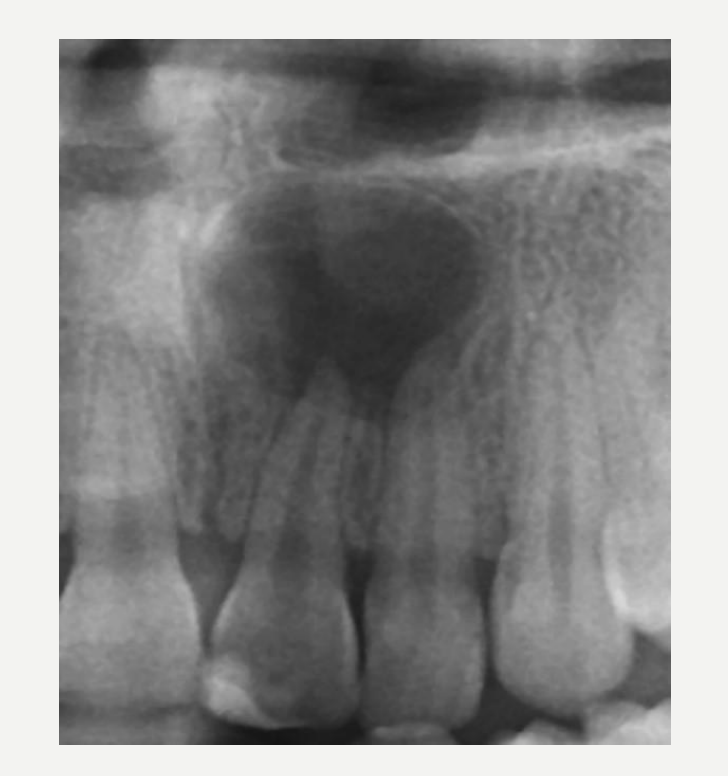 | 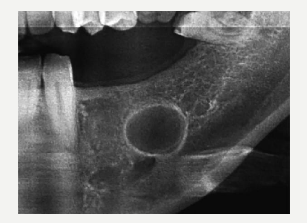 |
Radicular Cyst Aetiology and Clinical Features
Aetiology7
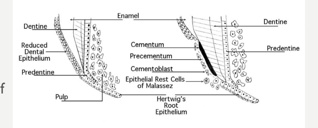
- Chronic inflammation at the apex of a non-vital tooth causes proliferation of the epithelial cell rests of Malassez (remnants of Hertwig’s epithelial root sheath) in the PDL
- This forms the epithelial lining of the radicular cyst
- A cyst cavity is formed and enlarges as a result of osmotic pressure and peripheral bone resorption
 |  |
Clinical Features
- Always associated with a non-vital tooth
- Asymptomatic, unless secondarily infected
- There may be a history of dental pain or abscess
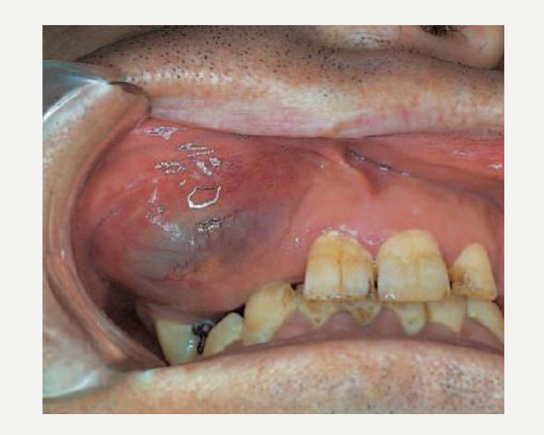
- May cause bluish swelling
- May feel bony and hard (cortex intact), crepitant (bone thinning), rubbery and fluctuant (cortex perforated)
Tooth Anatomy (Figure Labels)
Dentine; Reduced Dental Epithelium; Predentine; Pulp; Enamel; Cementum; Precementum; Cementoblast; Epithelial Rest Cells of Malassez; Hertwig’s Root Epithelium; Dentine; Predentine
Figure courtesy of Dr T Matias — RACDS Oral Histology Orientation Course Booklet
Cawson’s Essentials of Oral Pathology and Oral Medicine, 2008.
Radicular Cyst Imaging Features
Location8
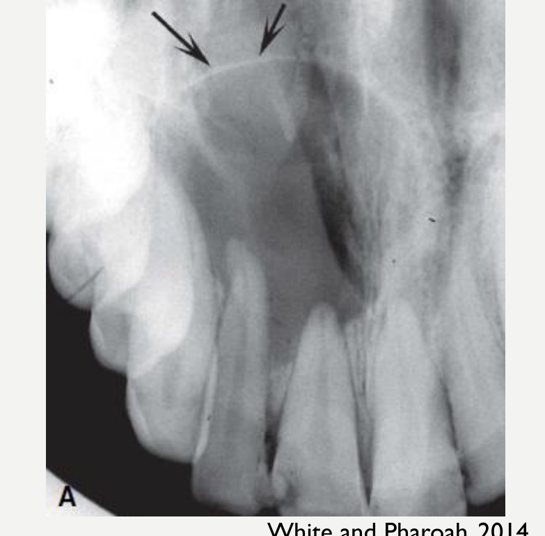
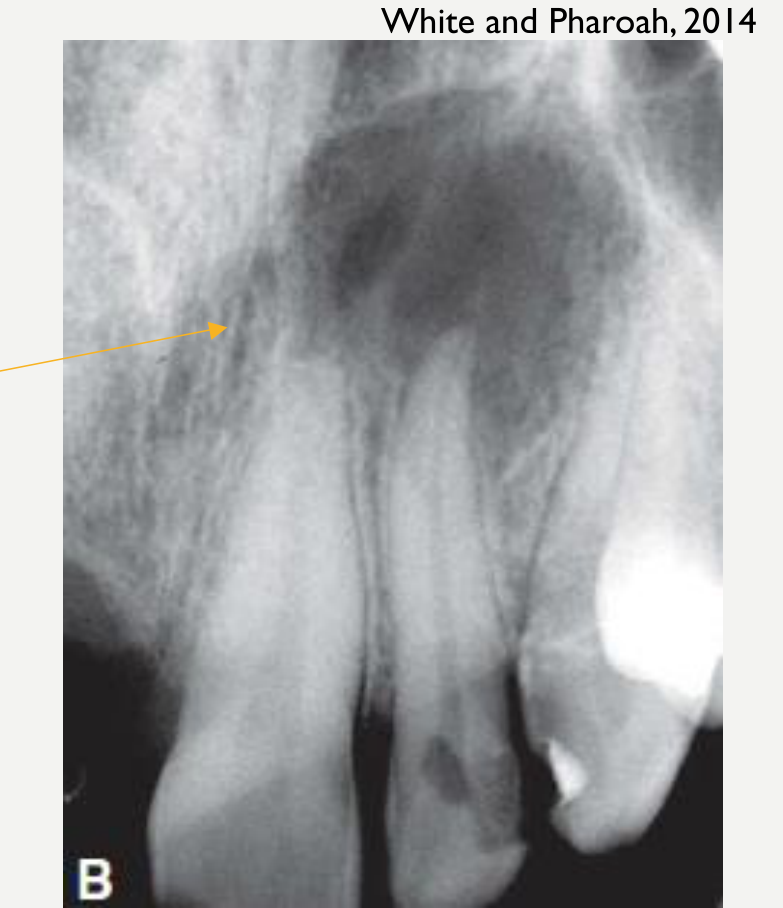
- Maxilla > Mandible
- 40–50% arise in the anterior maxilla, followed by the posterior mandibular region
- Epicentre at tooth apex
- Unless associated with a lateral root canal (Lateral radicular cyst)
 |  |
Periphery/Shape
-
Well-defined, cortical border
- Cortex may be sclerotic or lost if cyst is secondarily infected
-
Curved or circular
- Unless influenced by surrounding structures
-
10 mm
-
A periapical radiolucency smaller than 10 mm is considered apical periodontitis.
-
The border may become thicker and sclerotic with inflammatory change, but less distinct with secondary infection.
-
A periapical radiolucency smaller than 10 mm is considered apical periodontitis. White and Pharoah, 2014.
Internal Structure9
- Radiolucent
- Dystrophic calcifications may develop in long-standing cysts
- Sparsely distributed, small particulate radiopacities
 |  |
Surrounding Structures
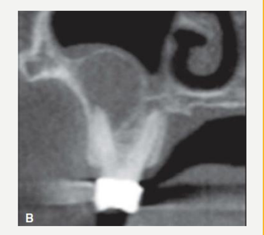
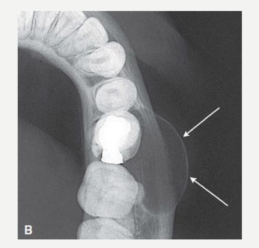
- Displacement and resorption of roots of adjacent teeth
- Resorption pattern has curved outline
- Can resorb the root(s) of the involved non-vital tooth (rare)
- May extend into the maxillary sinus (sinus floor remains intact but is elevated)
- Outer cortical plates may expand in curved or circular shape
- May displace the IAC in an inferior direction
White and Pharoah, 2014.
Radicular Cyst Histopathology
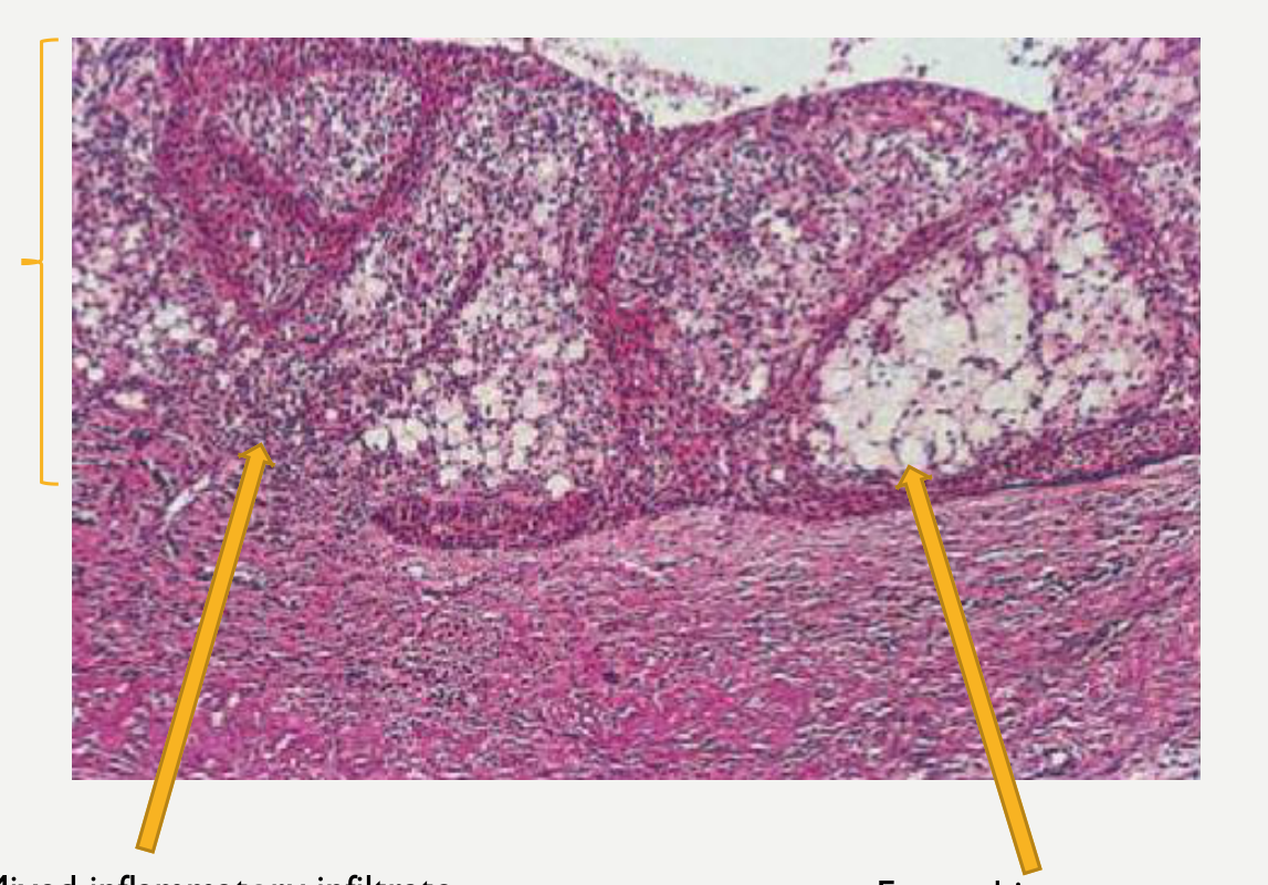
Histopathology10
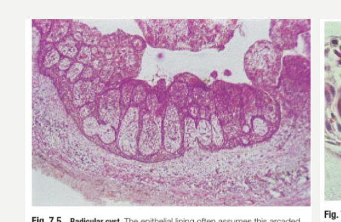
- Epithelium: Non-keratinised stratified squamous epithelium that is proliferative with elongated rete pegs in a characteristic arcading pattern
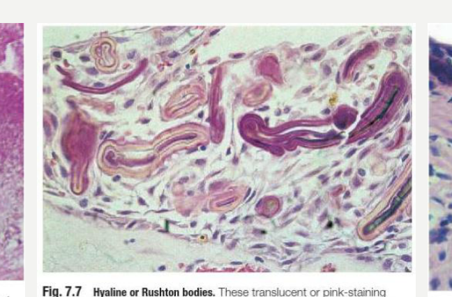
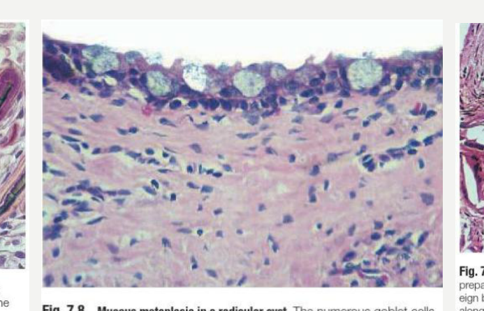
- +/- hyaline (Rushton) bodies, mucous (goblet) cells, or small areas of keratinisation
- Cyst wall: Inflamed fibrous tissue, often with foamy histiocytes
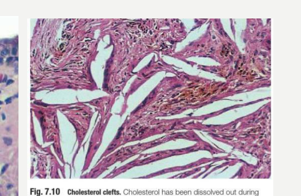
- Deposits of cholesterol crystals (clefts) with foreign body giant cells are often seen and may form luminal nodules
- Residual and long-standing cysts are less inflamed and have more regular thin epithelium
Figure labels: Mixed inflammatory infiltrate; Foamy histocytes
Cawson’s Essentials of Oral Pathology and Oral Medicine, 2008.
 |  |  |
 |  |
Radicular Cyst Treatment and Prognosis
Treatment11
-
Extraction or root canal therapy of tooth and enucleation of cyst +/- apicoectomy
-
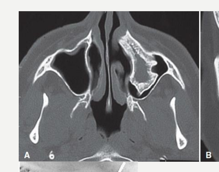
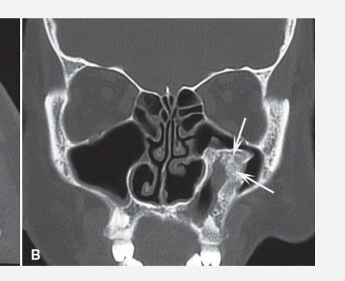
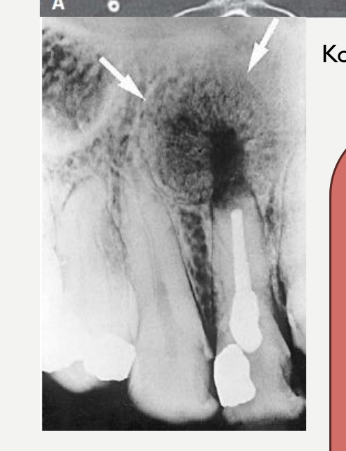
Healing: new bone grows into defect from periphery and may result in a radiating pattern resembling the spokes of a wheel
-
A central lucent area may persist because it contains fibrous scar tissue and is considered normal.
 |  |
 |
Recurrence
- Rare, although may persist as residual cysts (may follow extraction or RCT alone)
Essential Diagnostic Criteria
- Non-vital tooth
- Cyst lined by non-keratinising epithelium in characteristic arcading pattern
Koong, 2017.
Dentigerous Cyst
Dentigerous Cyst Definition and Epidemiology12
-
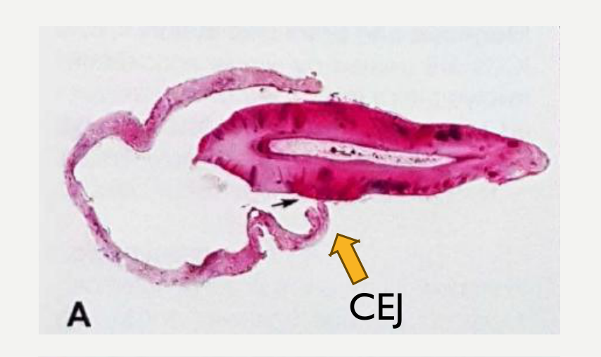
Definition: A developmental odontogenic cyst of the jaws surrounding the crown of an unerupted tooth, the lining attached to the cementoenamel junction (CEJ).
-
Related terminology: follicular cyst
-
Subtype: eruption cyst (a superficial dentigerous cyst over an erupting tooth in a child, usually a deciduous first molar)
-
Epidemiology: 2nd most common cyst in the jaws (~25% of all jaw cysts)
-
Age: wide range (5–83 years), peak incidence in 2nd–3rd decades
-
Gender: 1.7 times more frequent in males
-
An eruption cyst is located entirely within soft tissue above an erupting tooth.
-
In children, dentigerous cysts account for approximately 25% of jaw cysts. Cawson’s Essentials of Oral Pathology and Oral Medicine, 2008.
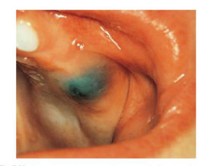
Dentigerous Cyst Aetiology and Clinical Features
Aetiology13
- Develops from accumulation of fluid between the reduced enamel epithelium and crown of the unerupted tooth
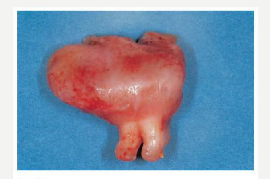
Clinical Features
- Asymptomatic and usually discovered on routine dental radiographs or when investigating the failure of a tooth to erupt
- Larger cysts cause slow expansion of jaw (hard swelling)
- Pain and swelling if infected (usually due to communication with oral cavity)
Cawson’s Essentials of Oral Pathology and Oral Medicine, 2008.
Dentigerous Cyst Imaging Features
Location14
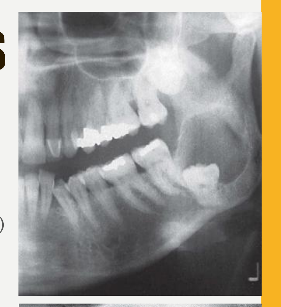
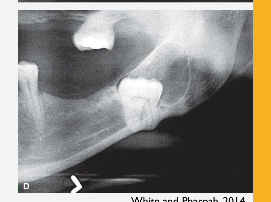
- ~75% associated with unerupted mandibular 3rd molar
- Frequency: lower 8s > upper 3s > upper 8s > lower 5s
- ~5% around supernumerary teeth (esp. mesiodens in anterior maxilla)
- Epicentre above crown
- Cyst attaches at CEJ
 |  |
Periphery and Shape
- Well-defined corticated (may be missing if secondarily infected)
- Curved or circular outline
- More likely cystic if follicular cortex is >5 mm from crown surface
White and Pharoah, 2014.
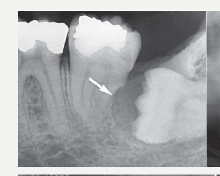
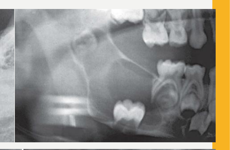
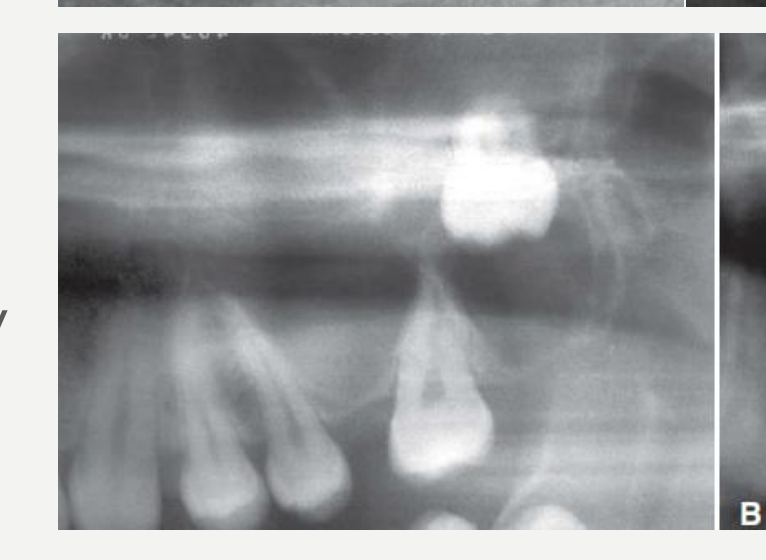
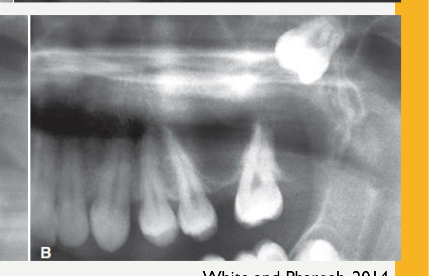
Internal Structure15
- Radiolucent, except crown of tooth
 |  |
 |  |
Surrounding Structures
- Displacement and resorption of adjacent teeth
- Displacement of involved tooth
- Displacement of maxillary antrum/IAC
- Expansion of outer cortex of involved jaw
Lecturer — Imaging Displacement
The cyst may displace developing teeth, including mandibular second molars and premolars.
- In the maxilla, it may elevate the maxillary antral floor.
- In the mandible, it may displace the inferior alveolar canal inferiorly.
- Expansion of the outer cortex may be difficult to appreciate on two-dimensional imaging.
White and Pharoah, 2014.
Dentigerous Cyst Histopathology
Histopathology16
-
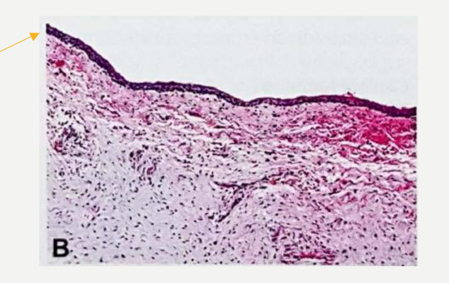
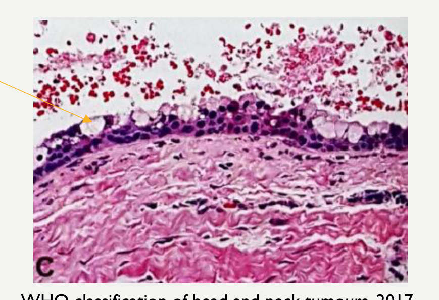
Epithelium: thin, non-keratinized stratified squamous epithelial lining
- +/- cuboidal, columnar and ciliated cells
- +/- mucous or sebaceous metaplasia
- +/- hyaline (Rushton) bodies and focal keratinisation
-
Cyst wall: loose, often myxoid, fibrous connective tissue and may contain occasional odontogenic epithelial rests
-
Attachment of the cyst lining to the CEJ is the defining feature both radiographically and histopathologically. Panels A, B, C; CEJ labelled on figure.
WHO classification of head and neck tumours, 2017.
 |  |
 |
Dentigerous Cyst Treatment and Prognosis
Treatment17
- Surgical enucleation (may include removal of tooth)
- Large cysts may require marsupialisation before removal
- Marsupialisation of eruption cysts to allow affected tooth to erupt normally
- Submit cyst lining for histology as benign/malignant tumours have been reported to arise (e.g. ameloblastomas, SCC, mucoepidermoid carcinoma)
Lecturer — Marsupialisation Purpose
Marsupialisation may be used to decompress a large cyst and reduce its size.
- It may also stimulate peripheral bony healing.
Recurrence
- Do not recur
Essential Diagnostic Criteria
- Well-defined radiolucency associated with the crown of an unerupted tooth
- Epithelium and cyst wall attached to the CEJ of the unerupted tooth
Odontogenic Keratocyst
-
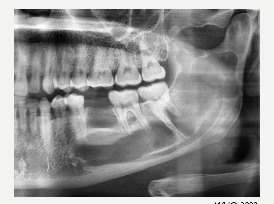
Definition: a developmental odontogenic cyst that is characterized histologically by a thin parakeratinized stratified squamous epithelial lining with palisaded and hyperchromatic basal cells and clinically by a tendency to recur after treatment.
-
Related terminology: keratocystic odontogenic tumour
-
Epidemiology: 3rd most common, ~10–20% of odontogenic cysts
- 5% of all OKCs occur as part of naevoid basal cell carcinoma syndrome (NBCCS) (usually multiple and in younger patients)
-
Age: all ages, peak incidence 2nd–3rd decade and smaller peak at 50–70 years
-
Gender: slight male predilection
-
It is also known as Gorlin-Goltz syndrome when referring to the syndromic condition.
Odontogenic Keratocyst Definition and Epidemiology18
WHO, 2022
Odontogenic Keratocyst Aetiology and Clinical Features
Aetiology19
- Arise from remnants of dental lamina
- Mutations in the PTCH1 gene (tumour suppressor gene)
- In syndromic cases and 80% of sporadic cases
Lecturer — PTCH1 Signalling
PTCH1 mutation or inactivation activates the SHH signalling pathway and produces abnormal epithelial proliferation.
Clinical Features
- Painless, unless secondarily infected
- Lesions are often large at diagnosis due to insidious growth pattern (significant bone destruction, but minimal bone expansion)
- Tooth displacement if lesion is large
- Pathologic fracture risk
- Aspiration reveals yellow, cheesy material (keratin)
Lecturer — Growth Pattern
The cyst tends to tunnel through bone between the cortices with minimal expansion.
- A jaw fracture may occur because the bone has been hollowed out and replaced by fluid.
Odontogenic Keratocyst Imaging Features
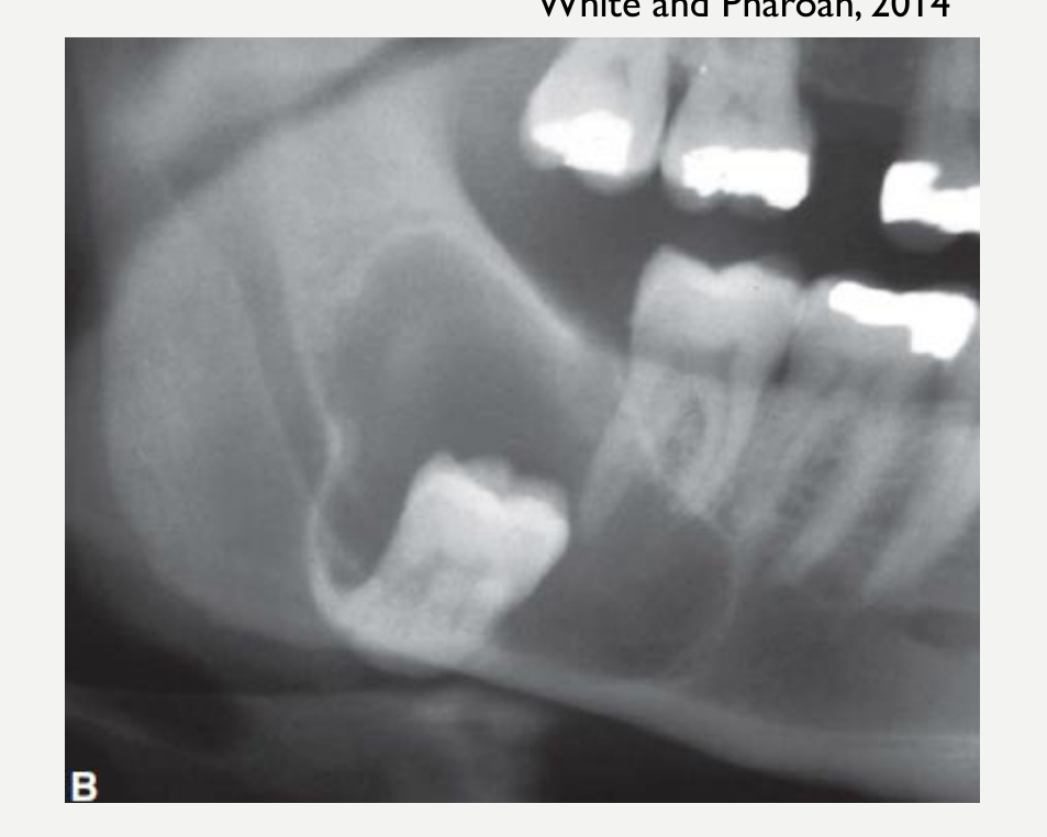
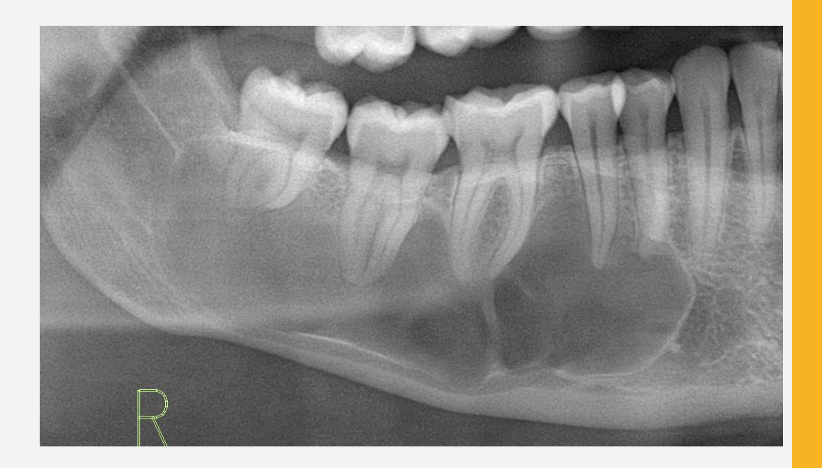
Location20
- ~80% in mandible
- Mostly posterior body and ramus
- Epicentre superior to IAC
- If found in posterior maxilla, often associated with NBCCS
- Sometimes form around an unerupted tooth
- Rarely extraosseous (in the gingiva)
 |  |
Periphery and Shape
-
Well-defined and corticated (unless secondarily infected)
-
Smooth, round/oval shape, or scalloped outline
-
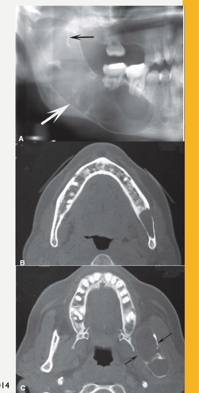
Multislice CT is useful for assessing the lesion’s extent and any cortical perforation. White and Pharoah, 2014
Internal Structure21
- Radiolucent
- Curved internal septa may be present = multilocular appearance
Lecturer — Internal Septa
Large lesions may appear multilocular, but they are not considered true multilocular lesions with separate compartments.
- The septa represent residual dense bone and are often incomplete rather than extending from cortex to cortex.
Surrounding Structures
- Minimal expansion for size
- Except upper ramus and coronoid process
- Cystic wall may contact soft tissue peripheral to outer cortex of mandible
- Occasionally displace and resorb teeth
- IAC displaced inferiorly
- May invaginate and occupy the entire maxillary antrum
Lecturer — Ramus Expansion
In the ramus or coronoid process, the lesion may balloon out and cause marked thinning, expansion, or perforation.
Lecturer — Surgical Planning
Cortical perforation indicates contact with adjacent soft tissues and may affect surgical planning. Tooth displacement and resorption may occur but are less pronounced than with other cysts.
White and Pharoah, 2014
Odontogenic Keratocyst Histopathology
Histopathology22
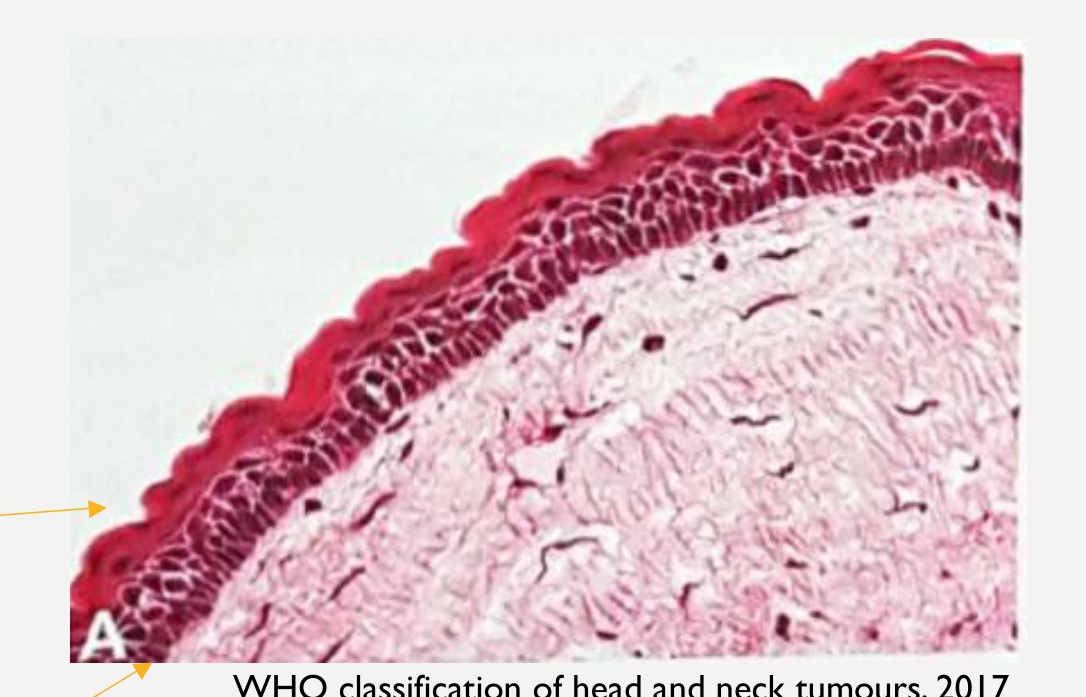
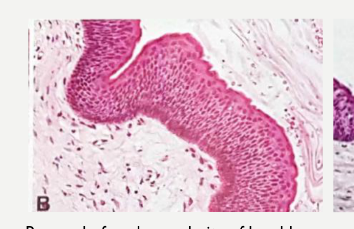
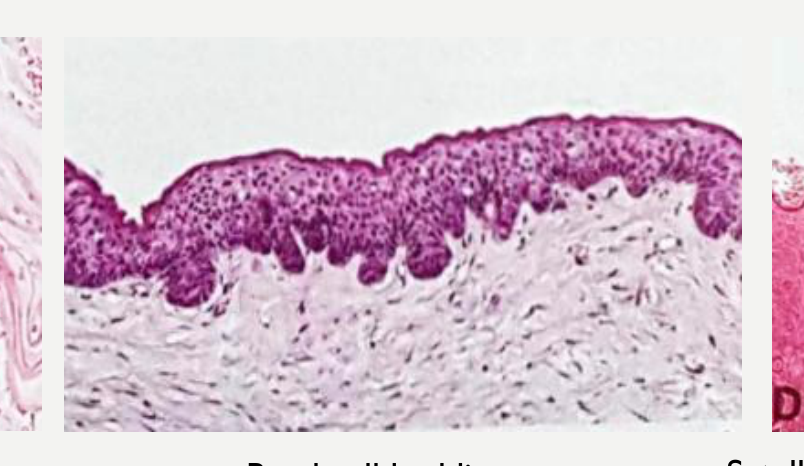
- Epithelium: thin, regular, folded parakeratinised epithelium 4–8 cell layers thick without rete ridges
- Corrugated with parakeratin
- +/- focal orthokeratosis
- Characteristic palisading basal layer with hyperchromatic nuclei
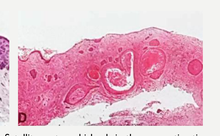
- Cyst wall: uninflamed fibrous wall
- Satellite cysts are more common in NBCCS
 |  |
 |  |
Odontogenic Keratocyst Treatment and Prognosis
Treatment23
-
Need CT imaging to determine extent and location of any cortical perforations with soft tissue extension
-
Enucleation, marsupialisation or resection
-
Periodic post-treatment clinical and radiographic examination
-
Submit for histopathology — odontogenic carcinoma arising from the epithelial lining of OKCs has been reported
-
Marsupialisation may be used for decompression of larger lesions.
-
More aggressive approaches may include ostectomy, cryotherapy or chemical cautery with Carnoy solution.
Recurrence
- High recurrence rate with traditional enucleation (20–30%)
- Even higher in NBCCS (50%)
- Recurrence can be reduced by more aggressive or adjunctive treatment including curettage, resection, peripheral ostectomy, cryotherapy or chemical cautery of the cavity, with excision of overlying mucosa
- Usually within first 5 years, but may occur as late as 10 years
- Likely due to small satellite cysts or fragments of epithelium left behind after surgical removal
Essential Diagnostic Criteria
- Located in jaws
- Stratified squamous epithelial lining with surface parakeratin
- Palisaded hyperchromatic basal cells
Inflammatory Collateral Cysts
Inflammatory Collateral Cysts Definition and Epidemiology24
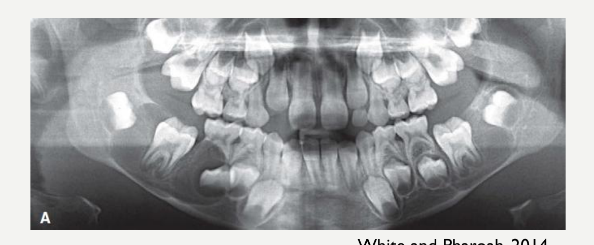
- Definition: inflammatory cysts on the buccal or distobuccal aspect of the roots of partially or recently erupted teeth.
- Two main types:
- Paradental cysts (PC) (60%)
- Mandibular buccal bifurcation cysts (MBBC) (40%)
- Epidemiology: 5% of all odontogenic cysts
- Average age: 30 years (PC); 9 years (1st molar MBBC); 17 years (2nd molar MBBC)
- Gender: more common in males (70%)
Inflammatory Collateral Cysts Aetiology and Clinical Features
Aetiology25
-
Inflammation and dilation of the pericoronal tissues and lined by sulcular/junctional epithelium (derived from reduced enamel epithelium)
-
Cyst formation may be exacerbated by food impaction or enamel cervical projections
-
Enamel cervical projections may extend beyond the CEJ onto the root surface, particularly near the bifurcation.
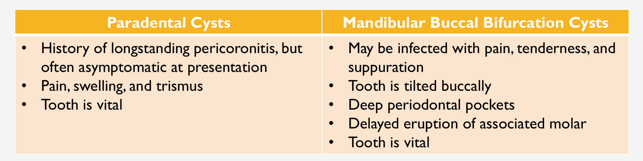
Clinical Features
| Paradental Cysts | Mandibular Buccal Bifurcation Cysts |
|---|---|
| • History of longstanding pericoronitis, but often asymptomatic at presentation • Pain, swelling, and trismus • Tooth is vital | • May be infected with pain, tenderness, and suppuration • Tooth is tilted buccally • Deep periodontal pockets • Delayed eruption of associated molar • Tooth is vital |
Inflammatory Collateral Cysts Imaging Features
Location26
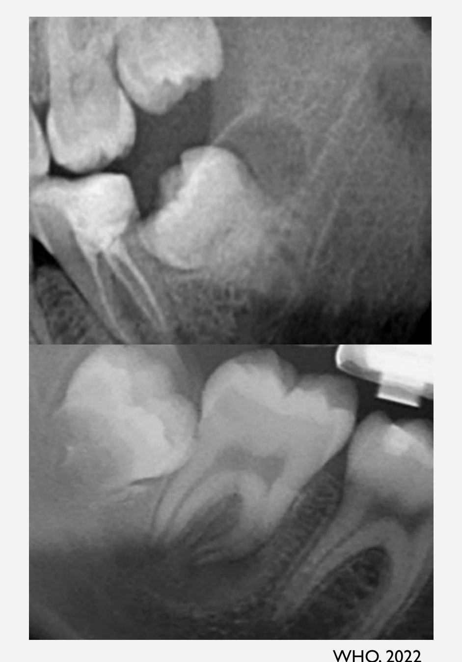
- PC: typically distobuccal aspect of mandibular 3rd molars (depending on angle of impaction)
- MBBC: at the buccal aspect of mandibular 1st or 2nd molars and are often bilateral
- Other sites: rare (4%)
Periphery and Shape
- May not be readily apparent with very subtle radiolucent region superimposed over the roots
- Circular shape with well-defined cortical border
- Can become quite large
WHO, 2022
Internal Structure27
- Radiolucent
Surrounding Structures
-
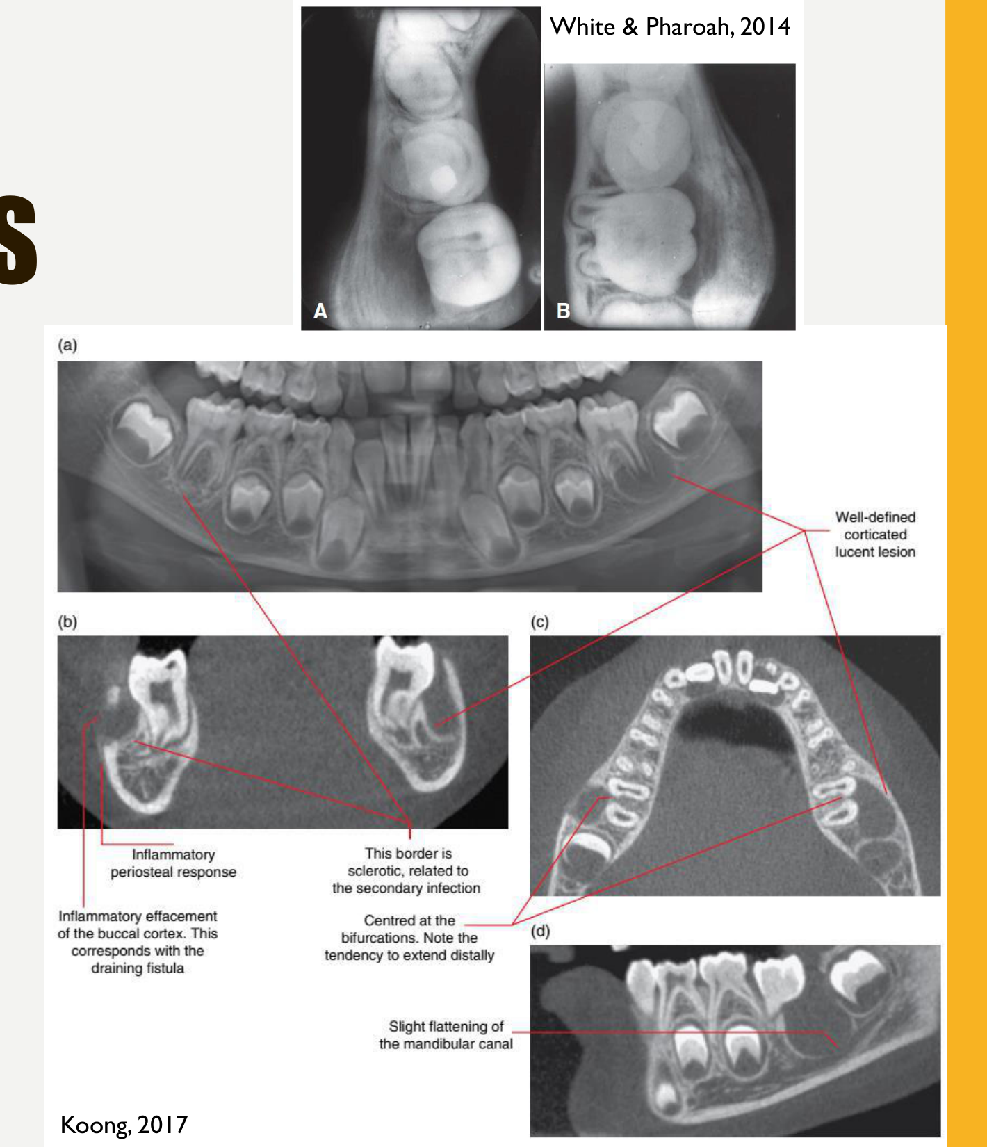
Tipping of involved molar so root tips are pushed into the lingual cortical plate of mandible and occlusal surface tipped toward buccal aspect
-
May displace and resorb adjacent teeth
-
May cause considerable amount of smooth expansion of the buccal cortical plate
-
If secondarily infected, periosteal new bone formation may be seen on the buccal cortex adjacent to involved tooth
-
Infection may produce layers of periosteal new bone, creating an onion-peel appearance.
Figure Annotations (A, B)
- (a) Well-defined corticated lucent lesion — centred at the bifurcations. Note the tendency to extend distally
- (b) This border is sclerotic, related to the secondary infection
- (c) Inflammatory periosteal response; inflammatory effacement of the buccal cortex. This corresponds with the draining fistula
- (d) Slight flattening of the mandibular canal
White & Pharoah, 2014 Koong, 2017
- The tilted molar has more prominent, superiorly positioned lingual cusps, and CBCT demonstrates the tooth angulation and contact between the root apices and lingual cortical plate.
Inflammatory Collateral Cysts Histopathology, Treatment and Prognosis
Histopathology28
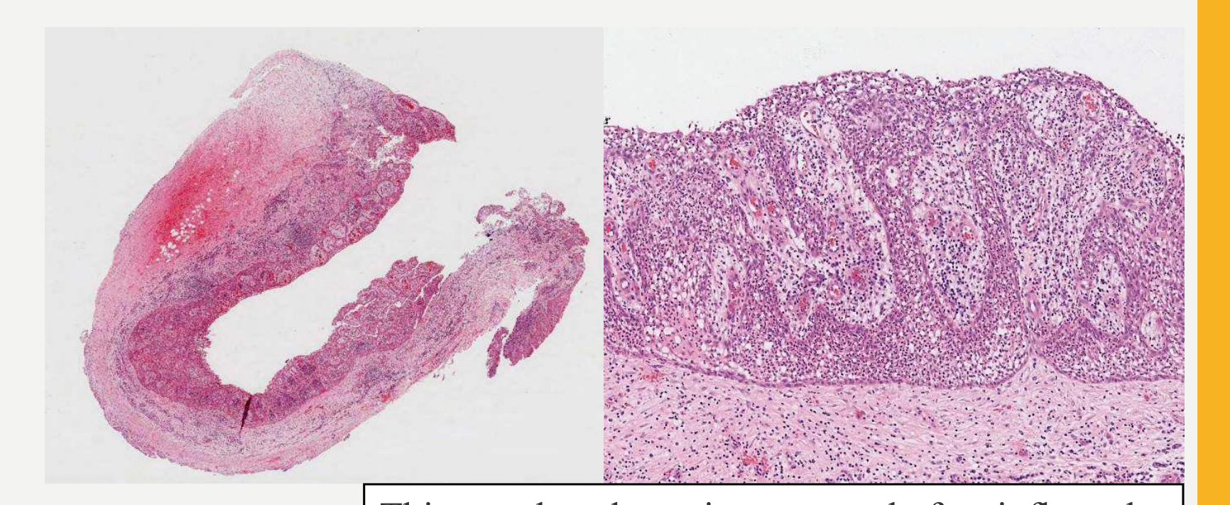
- Resembles radicular cyst
This paradental cyst is composed of an inflamed fibrous wall lined by hyperplastic epithelium. The cyst appears as an open pocket. (WHO, 2022)
Treatment
- Resolution without intervention
- Curettage and enucleation
- 3rd molars associated with PC are often removed
- 1st and 2nd molars with MBBC can be conserved and erupt normally
Recurrence
- Does not recur
Essential Diagnostic Criteria
- Associated with partially or recently erupted vital tooth
- Radiolucency distinct from dental follicle
- Intact lamina dura
- Non keratinised epithelium
Nasopalatine Duct Cyst
Nasopalatine Duct Cyst Definition and Epidemiology
- Definition: a developmental non-odontogenic cyst arising in the incisive canal.
- Related terminology: Incisive canal cyst
Epidemiology29
- 5% of all cysts in the jaws; 80% of all non-odontogenic cysts
- Age: 30-60 years
- Gender: M>F; 3:1
Nasopalatine Duct Cyst Aetiology and Clinical Features
Aetiology30
- Arises from respiratory and squamous epithelial vestigial remnants of an embryonic nasopalatine duct present in some individuals
- In one third of cases, inflammation from non-vital teeth or periodontal ligament may be a factor by stimulating the epithelial remnants to proliferate
Clinical Features
-
Sessile swelling just posterior to maxillary incisors OR asymptomatic
-
May arise deeper and present as swelling on labial alveolus or bulging of nasal floor
-
Often traumatised and may become infected
-
A sessile swelling may be located at the incisive papilla.
Nasopalatine Duct Cyst Imaging Features
Location31
- Found only in the midline of anterior hard palate
- Between roots of incisors
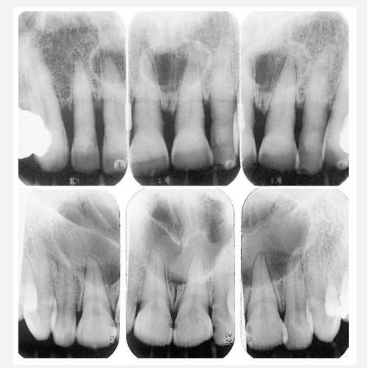
Periphery and Shape
-
Well-defined
-
Often corticated
-
May have characteristic heart shape
-
Size: incisive canal >6mm considered to be cyst (average diameter 18mm)
-
The characteristic heart shape is particularly apparent on two-dimensional imaging.
Internal Structures
- Radiolucency
Surrounding Structures
- Incisor roots may be displaced, but are vital
- Lamina dura is intact
White and Pharoah, 2014
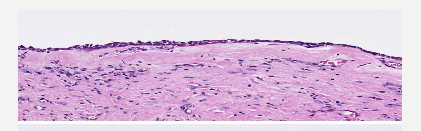
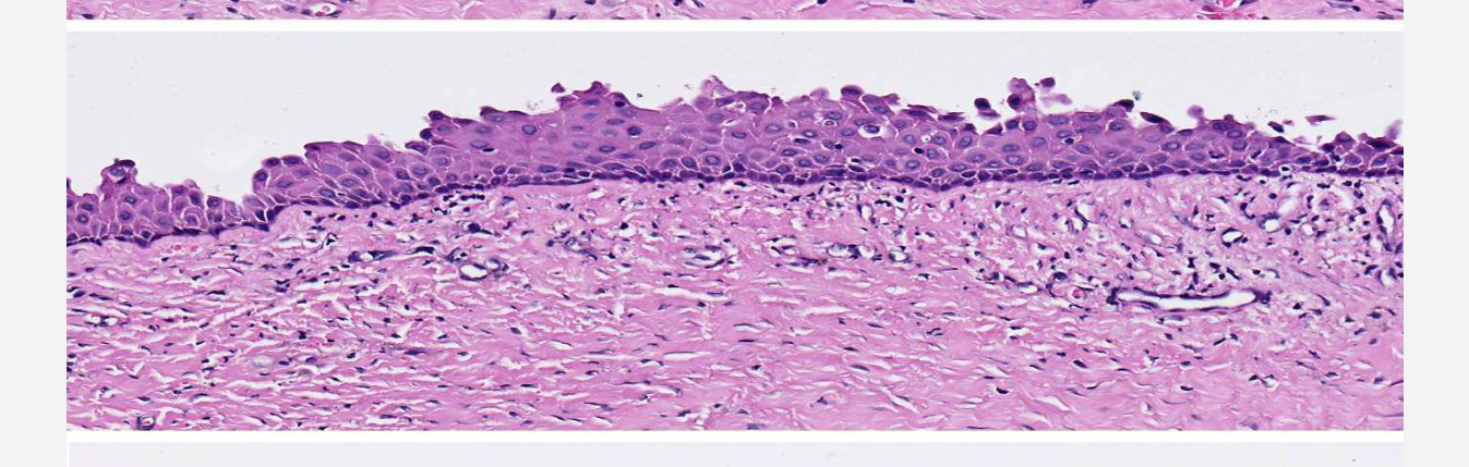
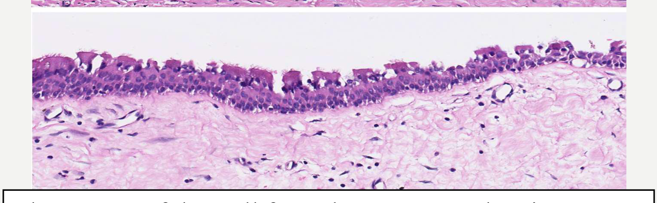
Nasopalatine Duct Cyst Histopathology32
- Epithelium: 90% lined by stratified squamous epithelium with focal areas of cuboidal, columnar, ciliated change
- 50% of cases contain respiratory epithelium (but <10% are lined entirely by respiratory epithelium)
- Cyst wall: Prominent neurovascular bundles with occasional small mucous glands or cartilage
- Traumatised cysts are inflamed
 |  |
 |
Nasopalatine Duct Cyst Treatment and Prognosis
Treatment33
- Enucleation
Recurrence
- Do not normally recur
Essential Diagnostic Criteria
- Epicentre at incisive canal
- Lining of non-keratinised squamous or respiratory epithelium
- +/- Neurovascular bundle in the cyst wall
Lateral Periodontal and Botryoid Odontogenic Cyst
Lateral Periodontal and Botryoid Odontogenic Cyst Definition and Epidemiology
Definition34
-
Lateral periodontal cyst (LPC): a developmental odontogenic cyst lined by non-keratinized epithelium with characteristic thickenings.
-
Botryoid odontogenic cyst (BOC): a less common multilocular subtype of LPC.
-
The lateral periodontal cyst and botryoid odontogenic cyst are considered the same lesion.
Epidemiology
- Rare, <1% of odontogenic cysts
- Age: wide range, peak incidence 5th–7th decades
- Gender: slight male predilection
Lateral Periodontal and Botryoid Odontogenic Cyst Aetiology and Clinical Features
Aetiology35
- Unknown; thought to arise from odontogenic epithelial remnants, however the source is controversial (either the dental lamina, reduced enamel epithelium, or epithelial rests of Malassez)
Clinical Features
-
Asymptomatic in 90% of cases
-
Can mimic a lateral periodontal abscess if secondarily infected
-
Expansion of buccal bone (50% of cases)
-
Usually small (<1 cm), but can attain considerable size
-
Botryoid odontogenic cysts may become much larger.
Lateral Periodontal and Botryoid Odontogenic Cyst Imaging Features
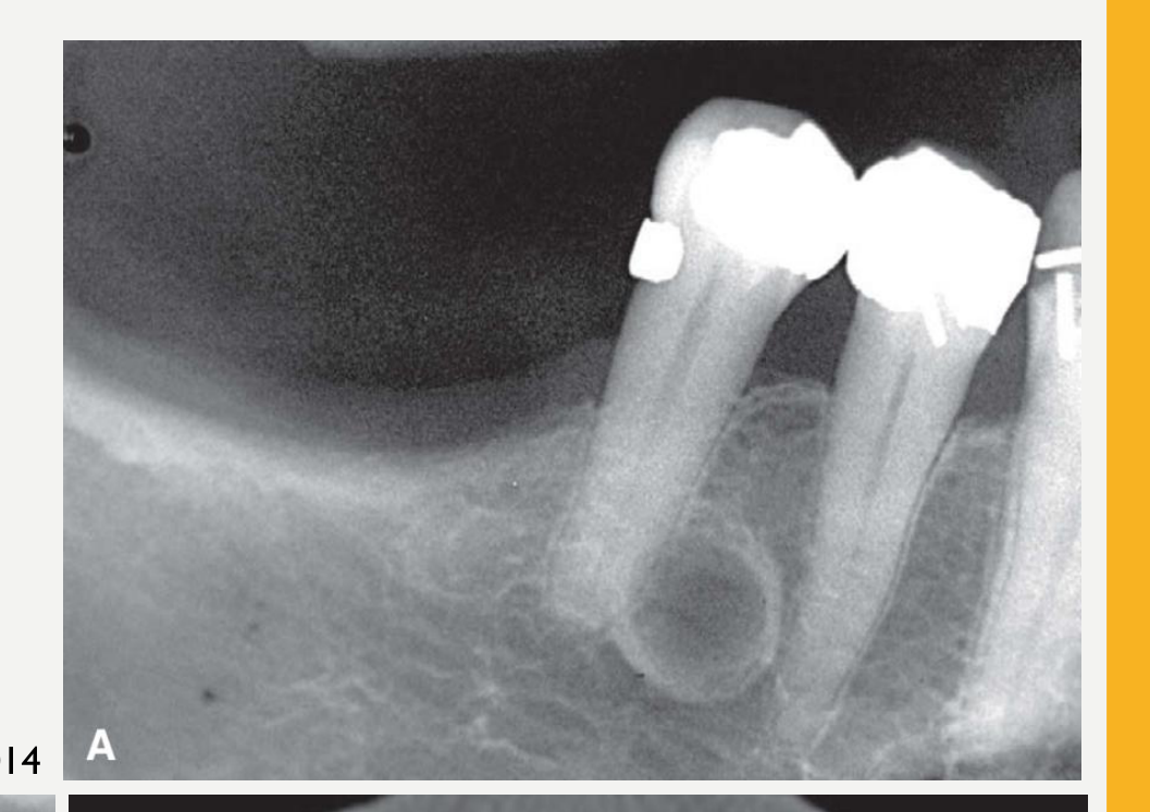
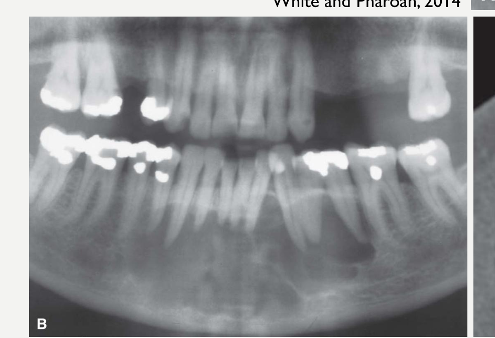
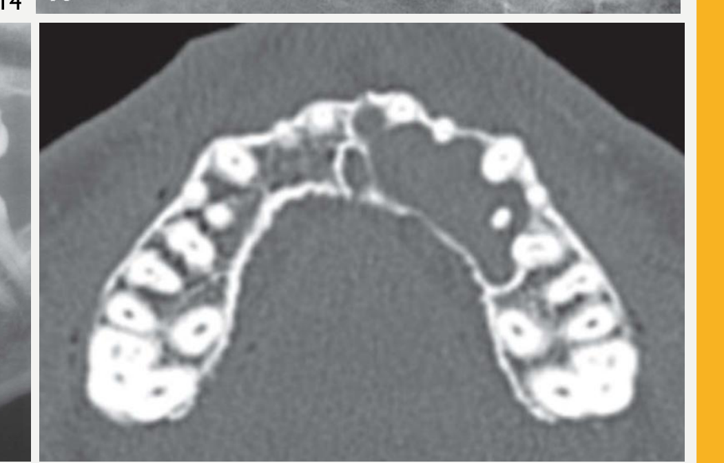
Location36
- Mandible > Maxilla
- Anterior to molars (particularly premolar region)
- Multifocal occurrence has been reported
- Centred upon the lateral surface of the root
 |  |
 |
Periphery and Shape
- Well-defined with prominent cortical boundary
- Round/oval shape
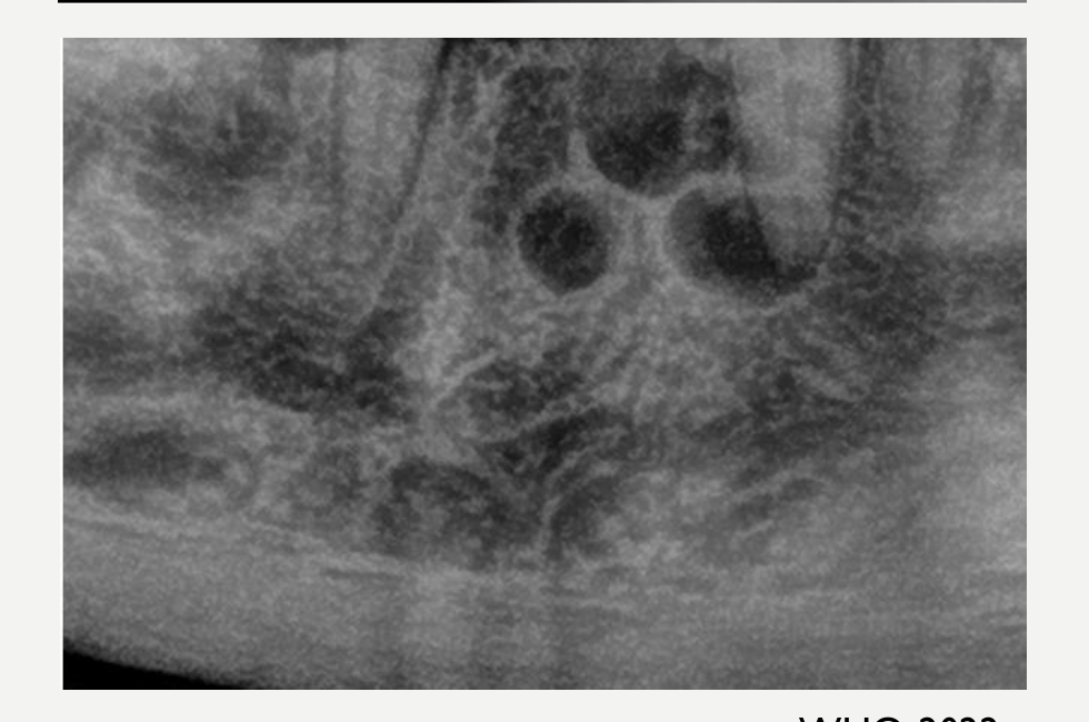
- LPC: unilocular
- BOC: multilocular
White and Pharoah, 2014
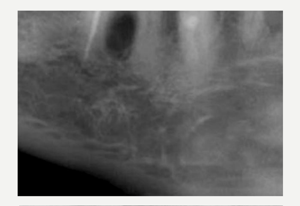
Internal Structure37
-
Radiolucent
-
BOC may have a multilocular appearance (related more to histologic appearance)
-
Botryoid odontogenic cysts contain multiple separate locules and internal septa.
 |  |
Surrounding Structures
- Small cysts may efface the lamina dura of adjacent roots (but tooth roots are rarely resorbed)
- Large cysts may displace adjacent teeth and cause expansion
- May have a similar growth pattern to OKCs with minimum expansion of the involved bone
WHO, 2022
Lateral Periodontal and Botryoid Odontogenic Cyst Histopathology
Histopathology38
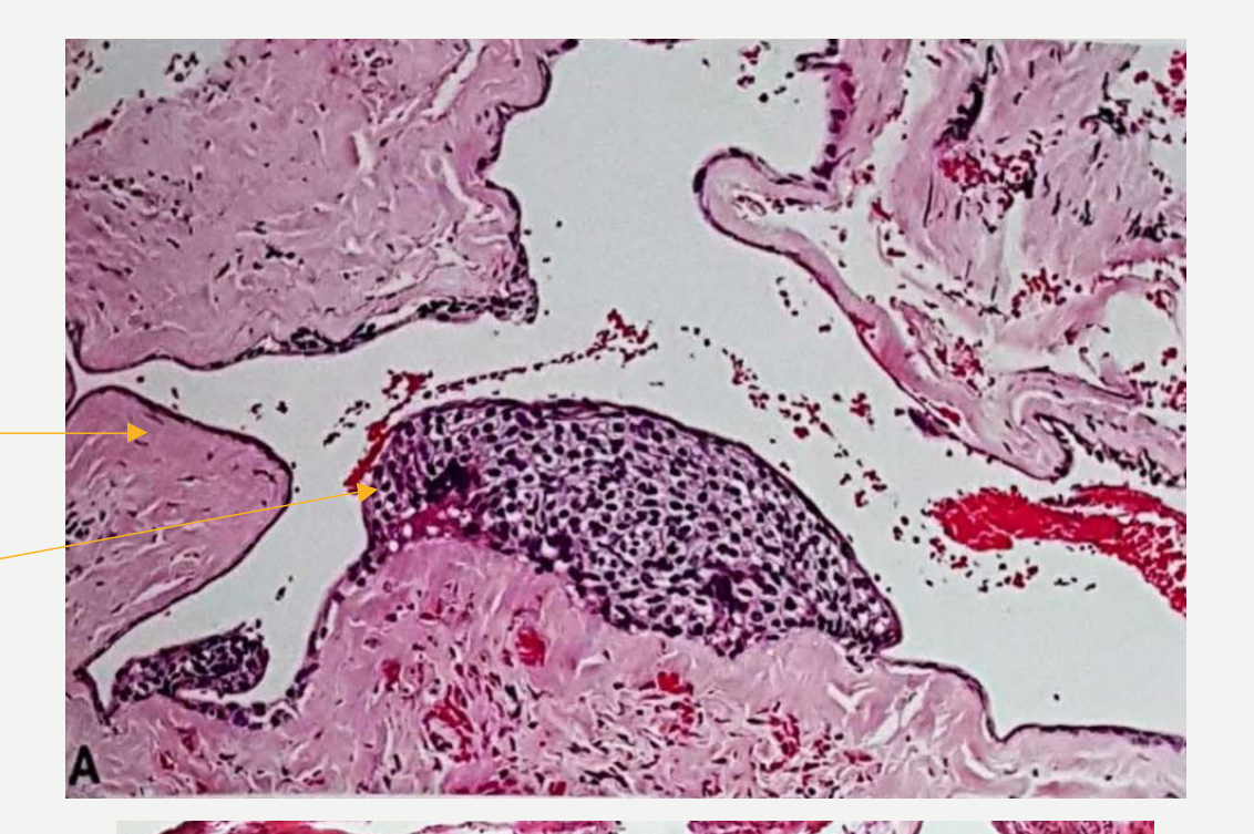
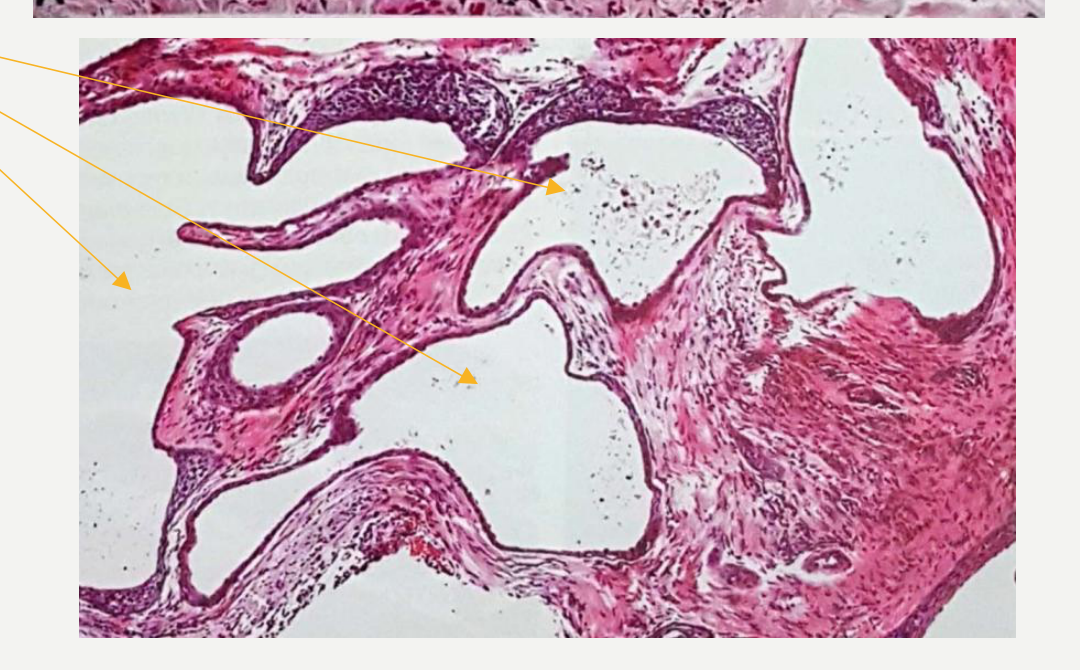
- Epithelium: thin, non-keratinised squamous or cuboidal epithelium 1–3 cells thick with focal, whorled, plaque-like epithelial thickenings
- Cyst wall: uninflamed fibrous wall
- BOC: same appearance, but with multicystic spaces
WHO classification of head and neck tumours, 2017
 |  |
Lateral Periodontal and Botryoid Odontogenic Cyst Treatment and Prognosis
Treatment39
- Enucleation, without removal of the adjacent tooth/teeth
Recurrence
- Rare for simple unilocular cysts
- Approximately 22% for BOCs
- Likely due to the multicystic nature of the lesion
Essential Diagnostic Criteria
-
Located on the lateral aspect or between the roots of vital erupted teeth
-
Characteristic whorled epithelial plaques
-
Multilocular (BOC only)
-
A history of previous surgery or trauma in the region
Surgical Ciliated Cyst
- Definition: a benign cyst caused by the traumatic implantation, usually surgical, of respiratory epithelium in the gnathic bones.
- Related terminology:
- Acceptable: surgical ciliated cyst of the maxilla; postoperative maxillary cyst (if maxillary location); (Respiratory) implantation cyst
- Epidemiology: rare
- Age: 5th to 6th decade
- Gender: no gender predilection
Surgical Ciliated Cyst Definition and Epidemiology40
WHO, 2022
Surgical Ciliated Cyst Aetiology and Clinical Features41
- Aetiology:
- Cysts develop from entrapped sinus or nasal mucosa in the jaw bones following trauma or surgery (e.g. Calwell-Luc or Le Fort I procedures, sinus surgery, maxillary fracture, midface osteotomy, or traumatic tooth extraction)
- Mandibular cases are generally caused by implantation of sinus epithelium by contaminated instruments, or, transfer of epithelium with autologous nasal osteocartilagenous grafts for chin augmentation
- The cyst usually develops after a long latent period, the reported delay being up to 20 years after the causative surgery. It is thought that the cyst cavity forms due to persistent mucous secretion.
- Clinical Features:
- May be asymptomatic or present with swelling, pain, or tenderness
Surgical Ciliated Cyst Imaging Features42
- Location:
- The gnathic bones, most common in posterior maxilla
- Very rare in the mandible
- Radiographic features:
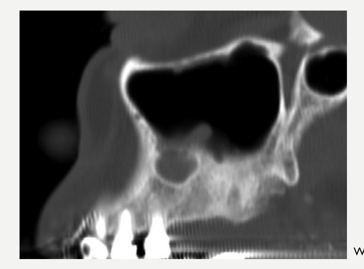
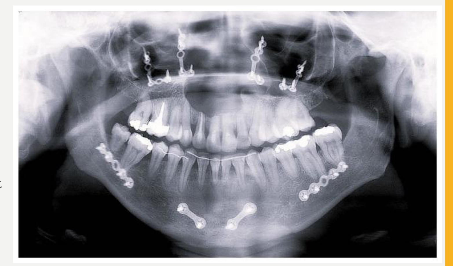
- Radiographs show a well-demarcated unilocular radiolucency of the jaws.
- Approximately 60% of cases are at least partially surrounded by a corticated margin.
- Occasional large lesions in the maxilla may fill the sinus or be multilocular
- Surgical plates may provide evidence of previous surgery.
Surgical Ciliated Cyst Histopathology, Treatment and Prognosis
- Histopathology:
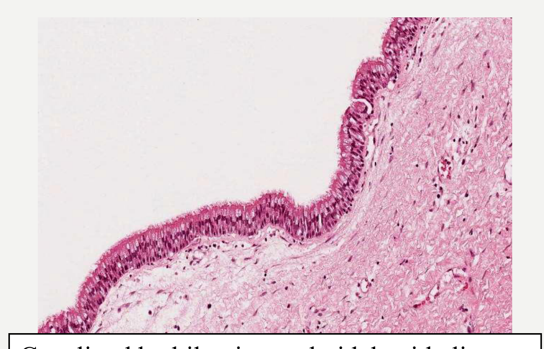
- Epithelium: pseudostratified ciliated columnar respiratory epithelium
- +/- squamous metaplasia or simple cuboidal cells
- +/- mucous (goblet) cells
- Cyst wall: loose fibrous connective tissue, which may be inflamed
- +/- subepithelial fibrosis or hyalinisation
- Epithelium: pseudostratified ciliated columnar respiratory epithelium
- Treatment:
- Simple enucleation
- Removal of the cyst lining
- Simple enucleation
- Recurrence:
- Rare
Essential Diagnostic Criteria43
- A history of previous surgery
- Radiolucent well demarcated cyst
- Respiratory epithelial lining
Orthokeratinised Odontogenic Cyst
Orthokeratinised Odontogenic Cyst Definition and Epidemiology44
- Definition: a developmental cyst lined by orthokeratinised stratified squamous epithelium
- Related terminology:
- Not recommended: Orthokeratinized variant of odontogenic keratocyst
- It is now considered a separate entity.
- Not recommended: Orthokeratinized variant of odontogenic keratocyst
- Epidemiology: rare (<1% of odontogenic cysts)
- Age: wide range, average age 35 years; peak incidence in 3rd – 4th decades
- Gender: 65% occur in males
Orthokeratinised Odontogenic Cyst Aetiology and Clinical Features45
- Aetiology:
- Unknown, but most likely arises from the remnants of dental lamina
- Clinical Features:
- Painless swelling, but many are incidental findings during radiographic examination
- Rare cases of multiple or bilateral OOC have been reported, but no evidence of association with Gorlin Syndrome (naevoid basal cell carcinoma)
Orthokeratinised Odontogenic Cyst Imaging Features46
- Location:
- Mandible (80%)
- 65% in angle/ramus region
- Radiographic features:
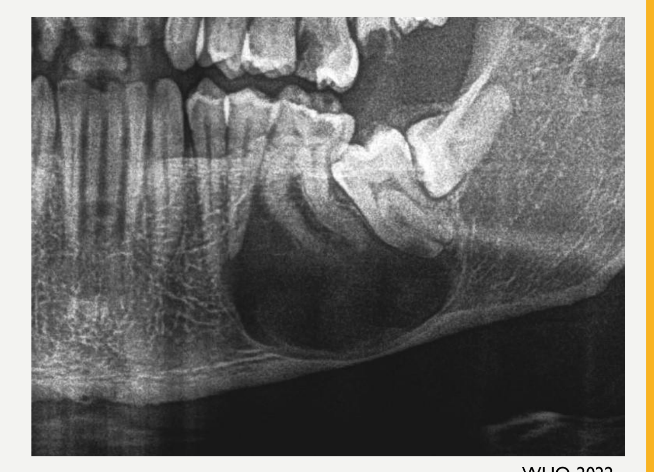
- Well-demarcated, spherical unilocular radiolucent lesions, often with a corticated margin
- Occasional cases (<10%) are multilocular
- Up to 70% of all lesions are associated with an impacted tooth, with a radiological similarity to dentigerous cysts
WHO, 2022
Orthokeratinised Odontogenic Cyst Histopathology47
- Histopathology:
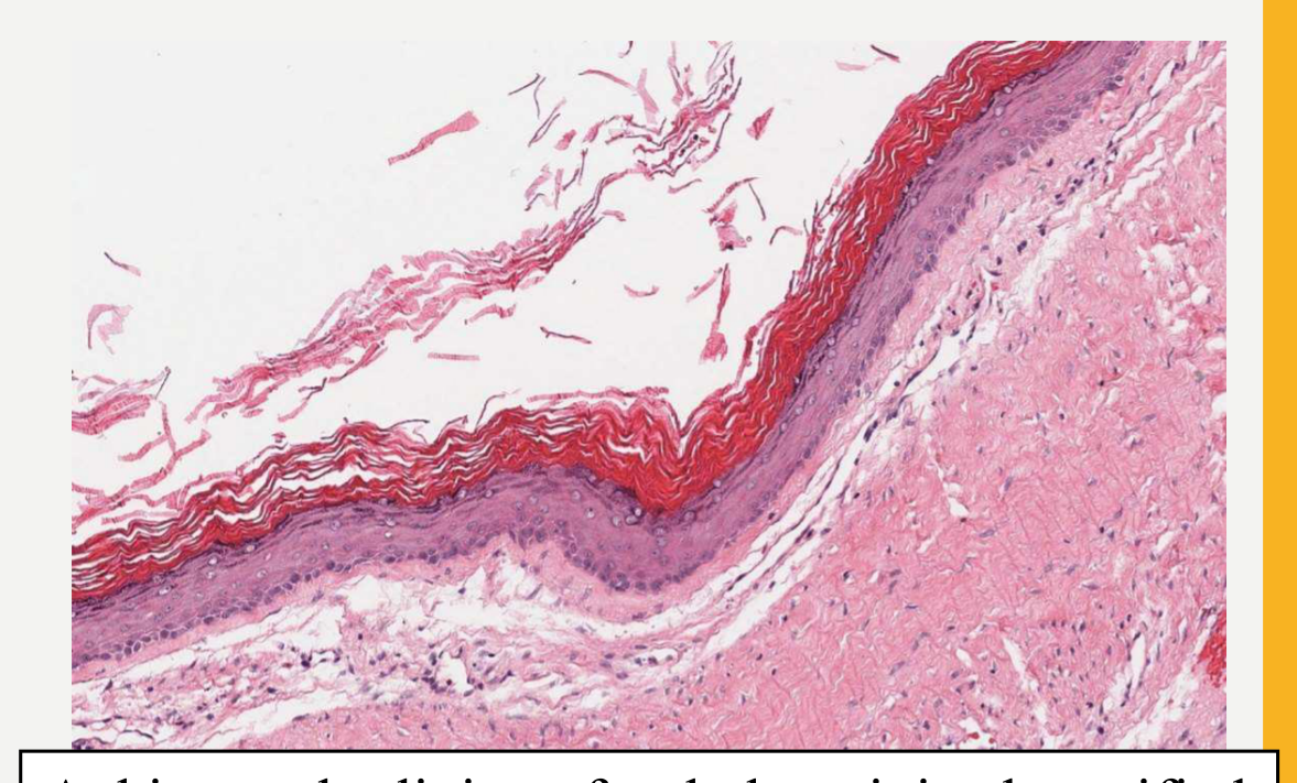
- Epithelium: thin regular orthokeratinized stratified squamous epithelium with a prominent granular cell layer and inconspicuous or low cuboidal basal cells. It is heavily keratinized, often with lamellae of keratin filling the lumen
- The keratinised layer lacks cell nuclei.
- Cyst wall: fibrous and may show areas of inflammation
- +/- parakeratinized or non-keratinized areas, but these form a small part of the lining and are often associated with inflammation
- Rare examples include sebaceous glands in the lining
- Microcysts may be seen in the wall in 5% of cases
- Epithelium: thin regular orthokeratinized stratified squamous epithelium with a prominent granular cell layer and inconspicuous or low cuboidal basal cells. It is heavily keratinized, often with lamellae of keratin filling the lumen
- Treatment:
- Enucleation
- Enucleation includes removal of the cyst lining.
- Enucleation
- Recurrence:
- Rare (less than 5%)
Essential diagnostic criteria:
- Located in tooth bearing areas of jaw
- Thin, regular epithelial lining with orthokeratinization
Calcifying Odontogenic Cyst
Calcifying Odontogenic Cyst Definition and Epidemiology48
-
Definition: A developmental odontogenic cyst characterized histologically by ghost cells, which often calcify.
-
Related terminology: calcifying cystic odontogenic tumour, Gorlin cyst
-
Epidemiology: rare, <1% of odontogenic cysts
-
Age: peak incidence 2nd – 3rd decade
-
Gender: no gender predilection
-
It occupies a spectrum from a cystic lesion to a solid odontogenic neoplasm.
Calcifying Odontogenic Cyst Aetiology and Clinical Features
Aetiology49
- Unknown
- Has mutations in the CTNNB1 gene which implicate the Wnt pathway. This pathway is also involved in odontomas, with which COC is often associated.
Clinical Features
- Asymptomatic in most cases
- Painless swelling of jaws
- Extraosseous lesions present as gingival swellings, sometimes with pain and tenderness
Calcifying Odontogenic Cyst Imaging Features
Location50
- Maxilla = mandible
- Anterior > posterior (especially with cuspids and incisors)
- Association with odontomas (more common in maxilla)
- Central > peripheral
- Up to 10% are extraosseous (anterior regions of jaw)
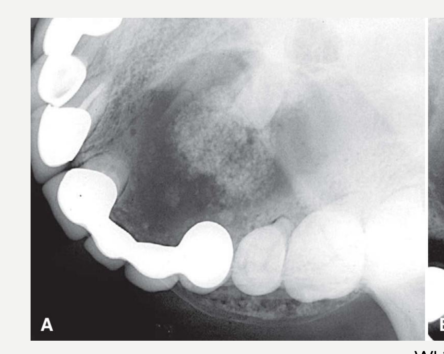 | 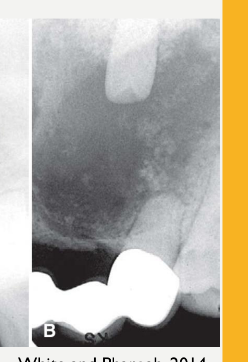 |
Periphery and Shape
- Variable
- Well-defined, corticated with curved, cyst-like shape
- Ill-defined and irregular
White and Pharoah, 2014
Internal Structure51
- Variable:
- Completely radiolucent
- Evidence of small foci of calcified material – white flecks or small smooth pebbles
- Larger, solid, amorphous masses
- Rarely appear multilocular
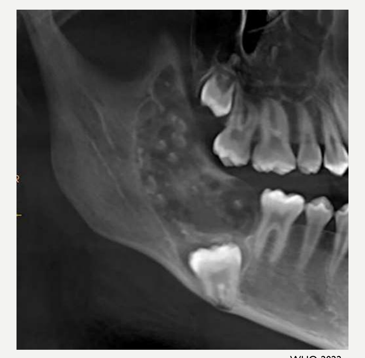
Surrounding Structures
- Impedes eruption of associated tooth
- Tooth displacement and root resorption
- Expansion and perforation of cortical plates
WHO, 2022
Calcifying Odontogenic Cyst Histopathology
Histopathology52
- Unilocular cyst
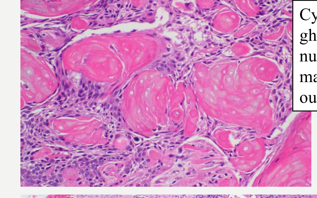
- Epithelium: stratified epithelial lining of varying thickness resembling stellate reticulum and with palisaded and hyperchromatic columnar ameloblast-like basal cells.
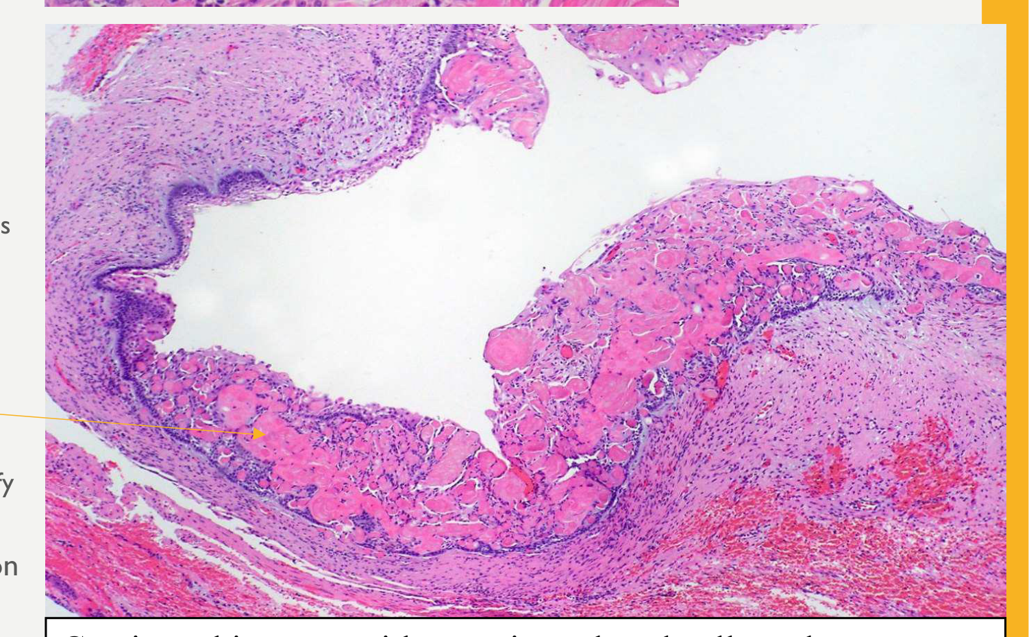
- Ghost cells are characteristic:
- Rounded or stacked flattened pale eosinophilic cells within the epithelium with distinct outlines and karyolysis resulting in a ‘ghost-like appearance’
- Variable in number and often pass into the connective tissue of the cyst wall, eliciting a foreign body reaction and inducing dentinoid
- Although characteristic, ghost cells also occur in other odontogenic tumours and do not alone justify a diagnosis of COC
- Ghost cells lack nuclei but retain the cell outline.
- Ghost cells are characteristic:
- +/- intraluminal and/or mural epithelial proliferation producing ameloblastoma-like areas
 |  |
Calcifying Odontogenic Cyst Treatment and Prognosis
Treatment53
- Conservative surgical removal, enucleation and/or curettage
Recurrence
- Post-treatment follow-up required
- 8% recurrence rate
Essential Diagnostic Criteria
- Cystic architecture
- Numerous ghost cells
Glandular Odontogenic Cyst
Glandular Odontogenic Cyst Definition and Epidemiology54
- Definition: a developmental cyst in which the epithelial lining resembles glandular tissue.
- Related terminology:
- Not recommended: sialo-odontogenic cyst; mucoepidermoid odontogenic cyst
- Epidemiology: rare, <0.5% of odontogenic cysts
- Age: average age = 50 years, peak incidence 5th–7th decades
- Gender: no gender predilection
Lecturer — Terminology Clarification
The previous names are not recommended because they can be confused with other entities.
Glandular Odontogenic Cyst Aetiology and Clinical Features55
- Aetiology:
- Unknown
- Thought to arise from remnants of the dental lamina
- Clinical Features:
- Slowly expanding, painless swelling
- Association with an impacted tooth is extremely rare
- Mandibular lesions may reach a large size and cross the midline
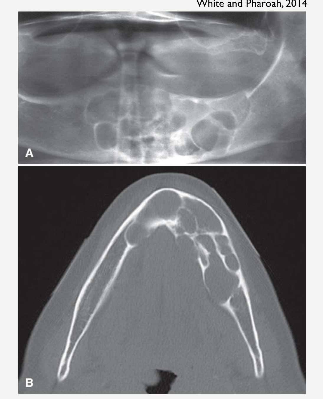
Glandular Odontogenic Cyst Imaging Features56
White and Pharoah, 2014
- Location:
- Exclusively in the jaws
- Mandible > Maxilla
- Anterior > Posterior
- Periphery/Shape:
- Well-defined cortical boundary
- Smooth or scalloped
- Internal Structure:
- Unilocular or multilocular
- Surrounding Structures:
- Expansion of the outer cortical plate of the jaws
- Regions of perforation
- Displacement ± resorption (25%) of teeth
- Cortical thinning may also occur.
- Expansion of the outer cortical plate of the jaws
Glandular Odontogenic Cyst Histopathology57
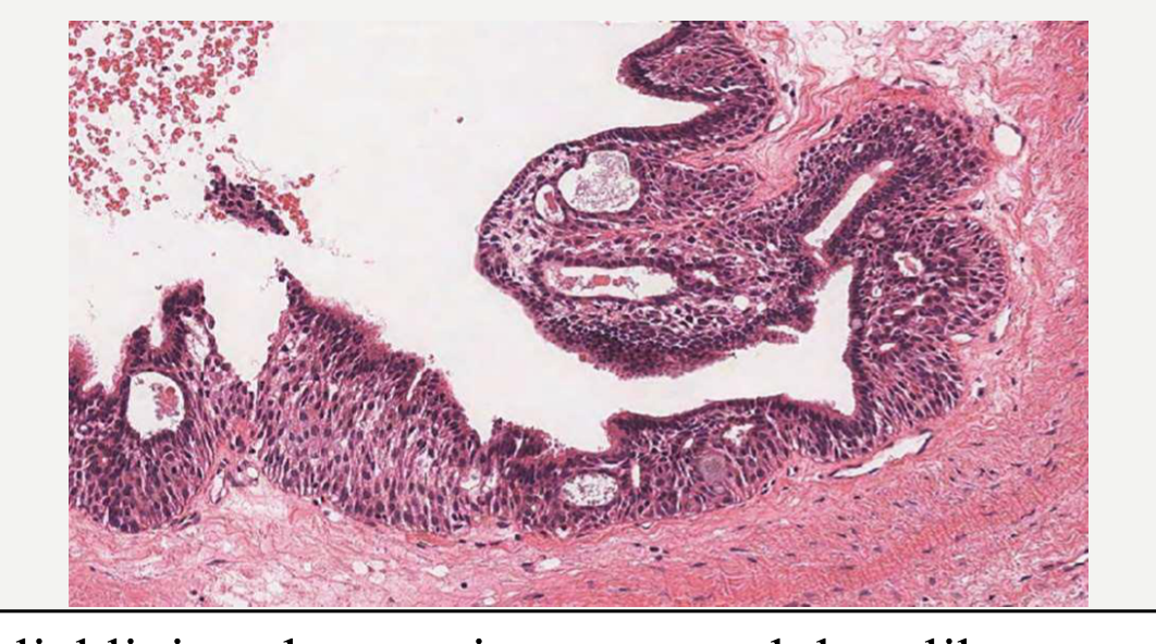
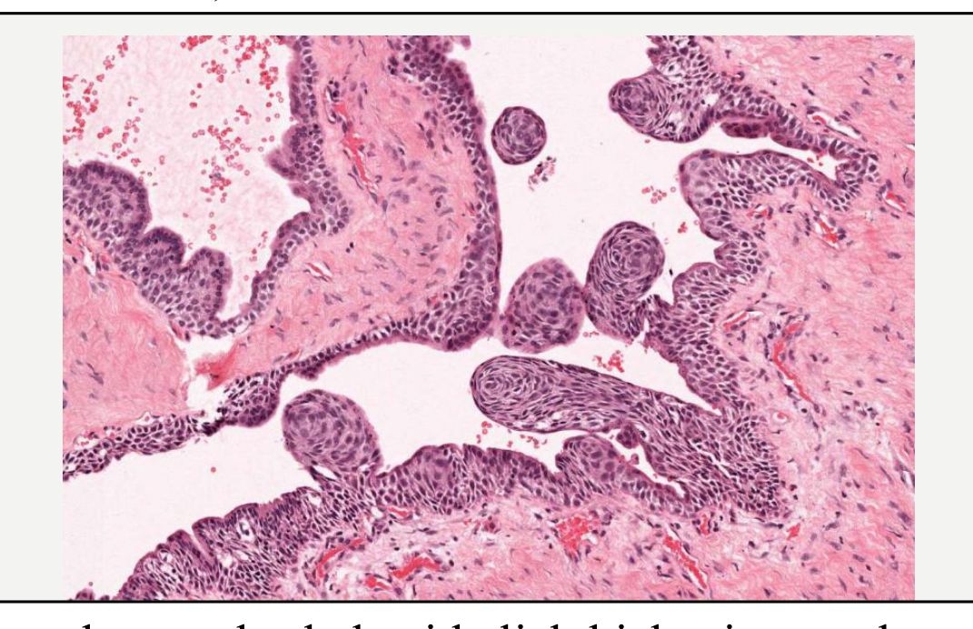
- Histopathology:
- Often multilocular cysts
- Epithelium: of variable thickness, ranging from a thin layer of flattened squamous or cuboidal cells to stratified squamous epithelium
-
- Hobnail cells in all cases (cuboidal or low columnar cells on the luminal surface)
- ± whorled epithelial thickenings, or plaques similar to those in the lateral periodontal cyst
- ± intraepithelial microcysts, apocrine metaplasia, clear cells, papillary projections (tufting), cilia and mucous cells
-
Not all features are present in every case, and a higher number of features allows a more confident diagnosis.
- Previously, diagnosis was based on the presence of seven of ten specified criteria.
 |  |
Glandular Odontogenic Cyst Treatment and Prognosis
- Treatment:
- Enucleation
- Removal of the cyst lining is usually performed with enucleation.
- Resection has been advocated due to high recurrence, particularly for large or multilocular lesions
- Enucleation
- Recurrence:
- Recurs in 22% of cases after enucleation (especially large and multilocular lesions)
- Post-treatment radiological review
- May occur up to 8 years later
- Radiological review is required for at least eight years.
Essential Diagnostic Criteria58
- Radiolucent cystic lesion of the tooth-bearing area of the jaw
- Often multilocular
- Lining of variable thickness with epithelial thickening, plaques or papillary projections + Hobnail cells ± microcysts or duct-like structures ± mucous or clear cells
References59
- Cawson, R.A., and Odell, E.W. (2008). Cawson’s Essentials of Oral Pathology and Oral Medicine E-Book (8th ed.). Churchill Livingstone.
- WHO Classification of Tumours Editorial Board. Head and neck tumours. Lyon (France): International Agency for Research on Cancer; 2022 (WHO classification of tumours series, 5th ed.; vol. 9).
- Koong, B. (2017). Atlas of Oral and Maxillofacial Radiology. Chichester, UK: John Wiley and Sons.
- White, and Pharoah. (2014). Oral radiology: Principles and interpretation (7th ed.). St. Louis: Elsevier/Mosby.
Audio Appendix
Additional Audio Content
The following sections from the lecture audio did not correspond to any heading in the main document.
Further Reading
- The lecture identified four references as useful resources for further reading on cysts of the jaws.
Clinical Cases
Case: Cyst with a corticated border
Question
Scenario: A radiograph of a jaw cyst is presented to illustrate its border characteristics.
What’s shown: A well-defined radiolucent lesion surrounded by a thin, uniform radiopaque line.
Consider: Identify the specific border feature shown and explain what it indicates about the cyst.
Answer
Observations:
- A thin, uniform radiopaque line surrounding the radiolucent cyst.
Reasoning: This thin, uniform radiopaque line represents a corticated border. Most cysts of the jaws are well-defined and exhibit this characteristic corticated margin, indicating a slow-growing, benign process that allows the surrounding bone to form a distinct boundary.
Takeaway: Most cysts of the jaws are well-defined and corticated, presenting with a thin, uniform radiopaque border.
Case: Long-standing cyst with dystrophic calcification and septa
Question
Scenario: A radiograph of a long-standing jaw cyst is presented to demonstrate internal changes over time.
What’s shown: A radiolucent lesion containing sparse, opaque, dotty appearances and thin opaque lines crossing through it.
Consider: Identify the internal features shown and explain what they indicate about the duration and structure of the cyst.
Answer
Observations:
- Sparse, opaque, dotty radiopacities within the radiolucency.
- Thin opaque lines traversing the cyst.
Reasoning: The sparse, opaque, dotty appearances represent dystrophic calcifications, which can occur in cysts that have been present for a long time. The thin opaque lines represent septa within the cyst itself.
Takeaway: Long-standing cysts may demonstrate internal dystrophic calcifications and septa, appearing as sparse radiopacities and thin opaque lines on radiographs.
Case: Dentigerous cyst causing displacement of developing teeth 47 and 4
Question
Scenario: A radiograph is presented showing a dentigerous cyst in the mandible.
What’s shown: A radiolucent lesion associated with an unerupted tooth, causing positional changes to adjacent developing teeth.
Consider: Identify the positional changes of the developing teeth and explain the mechanism.
Answer
Observations:
- Displacement of the developing tooth 47.
- Mesial displacement of the developing tooth 4.
Reasoning: The expanding dentigerous cyst occupies space within the jaw, physically pushing the adjacent developing teeth out of their normal eruption paths, resulting in displacement.
Takeaway: Dentigerous cysts can cause significant displacement of adjacent developing teeth due to their space-occupying mass effect.
Case: Cyst causing displacement of tooth 37
Question
Scenario: A radiograph is presented showing a cyst in the mandible affecting the adjacent teeth.
What’s shown: A radiolucent lesion located between teeth 37 and 36, with tooth 37 positioned at a noticeably different angle compared to the contralateral tooth 47.
Consider: Identify the effect the cyst is having on tooth 37 and explain the mechanism behind this change.
Answer
Observations:
- Tooth 37 is angled differently from the contralateral tooth 47.
- The cyst is occupying space between teeth 37 and 36.
Reasoning: The cyst is acting as a space-occupying lesion. By taking up space between the teeth, it physically pushes tooth 37 posteriorly, altering its angle and position.
Takeaway: Cysts of the jaws can cause mass effect features, including the displacement and resorption of adjacent teeth due to space occupation.
Case: Cyst causing inferior displacement of the inferior alveolar canal
Question
Scenario: A radiograph is presented showing a cyst in the mandible affecting the inferior alveolar canal.
What’s shown: A radiolucent lesion in the mandible with the inferior alveolar canal pushed inferiorly, compared to the contralateral side where it is located much closer to the apices of the teeth.
Consider: Identify the effect of the cyst on the inferior alveolar canal and explain how this is determined.
Answer
Observations:
- The inferior alveolar canal is displaced inferiorly on the affected side.
- The contralateral canal is in its normal, more superior position near the tooth apices.
Reasoning: The cyst exerts a mass effect, occupying space within the mandible and physically pushing the inferior alveolar canal in an inferior direction. Comparing it to the normal contralateral side confirms the displacement.
Takeaway: Displacement of the inferior alveolar canal is a mass effect feature of jaw cysts, often pushed inferiorly by the expanding lesion.
Case: Residual radicular cyst after tooth extraction
Question
Scenario: A radiograph is presented showing a cystic lesion in an edentulous area.
What’s shown: A well-defined radiolucent lesion in the jaw where a tooth has previously been extracted.
Consider: Identify the anatomical context of this radiolucency and explain how it differs from a standard radicular cyst.
Answer
Observations:
- A cystic lesion is present in an area where the associated tooth has been removed.
Reasoning: Normally, a radicular cyst requires a non-vital tooth to be present. In this case, the tooth was extracted, but the cystic epithelium was left behind, allowing the cyst to continue growing as a residual radicular cyst.
Takeaway: A residual radicular cyst occurs when cystic epithelium is left behind after the extraction of a non-vital tooth, allowing the cyst to persist and grow.
Case: Radicular cyst centered at the tooth apex
Question
Scenario: Radiographs of radicular cysts are presented to demonstrate their typical location.
What’s shown: Radiolucent lesions centered directly over the apical foramen of non-vital teeth.
Consider: Identify the epicenter of the radiolucency in relation to the tooth and explain its clinical significance.
Answer
Observations:
- The center of the cystic lesion is located around the apex of the involved tooth.
Reasoning: Radicular cysts arise from chronic inflammation at the apex of a non-vital tooth. Consequently, the epicenter of the radiolucency is generally centered over the apical foramen, unless it arises from a lateral canal.
Takeaway: A radicular cyst is typically centered around the apex of a non-vital tooth, reflecting its inflammatory origin at the apical foramen.
Case: Radicular cyst extending into the maxillary sinus
Question
Scenario: A radiograph is presented showing a radicular cyst in the posterior maxilla.
What’s shown: A radiolucent lesion extending into the maxillary sinus, with the buccal plate expanded into a curved, circular shape and appearing very thin.
Consider: Describe the effect of the lesion on the adjacent anatomical structures and the cortical bone.
Answer
Observations:
- The cyst extends into the maxillary sinus, elevating the sinus floor.
- The buccal cortical plate is expanded, thinned, and curved.
Reasoning: As a space-occupying lesion, the radicular cyst expands into the path of least resistance. It can extend into the maxillary sinus while keeping the sinus floor intact, and it causes significant thinning and expansion of the buccal cortical plate.
Takeaway: Radicular cysts can extend into the maxillary sinus and cause significant expansion and thinning of the buccal cortical plate.
Case: Dentigerous cyst causing curved root resorption of tooth 37
Question
Scenario: A radiograph is presented showing a dentigerous cyst affecting an adjacent tooth.
What’s shown: A radiolucent lesion surrounding the crown of an unerupted tooth, with the root of the adjacent tooth 37 showing resorption.
Consider: Identify the effect of the lesion on the adjacent tooth root and explain the shape of the resorption.
Answer
Observations:
- Curved resorption of the root of tooth 37.
Reasoning: The dentigerous cyst acts as a space-occupying lesion. The resorption pattern on the adjacent tooth root has a curved outline because it is being resorbed by the spherical shape of the cyst itself.
Takeaway: Dentigerous cysts can cause mass effect features, including curved root resorption of adjacent teeth due to the spherical shape of the cyst.
Case: Radicular cyst histology showing an arcading pattern
Question
Scenario: A histological slide of a radicular cyst is presented.
What’s shown: Non-keratinised stratified squamous epithelium with elongated structures extending into the underlying connective tissue.
Consider: Identify the specific epithelial pattern shown and explain its diagnostic significance.
Answer
Observations:
- Elongated rete pegs extending into the connective tissue.
- A characteristic arcading pattern of the epithelium.
Reasoning: The proliferative non-keratinised stratified squamous epithelium of a radicular cyst characteristically demonstrates elongated rete pegs in an arcading pattern, which is a key histological feature of this inflammatory cyst.
Takeaway: Radicular cysts histologically demonstrate non-keratinised stratified squamous epithelium with elongated rete pegs in a characteristic arcading pattern.
Case: Healing residual cystic cavity with a spoke-wheel pattern
Question
Scenario: A radiograph is presented showing a healing bone cavity following cyst treatment.
What’s shown: A central lucent area surrounded by a radiating spoke-wheel pattern of bone trabeculae.
Consider: Identify the radiographic pattern seen in the healing bone and explain its significance.
Answer
Observations:
- A radiating spoke-wheel pattern of bone.
- A central lucent area.
Reasoning: When a residual cystic cavity heals, new bone can form in a radiating spoke-wheel pattern, which is a perfectly normal healing response. The central lucent area is also normal and is simply filled with fibrous scar tissue.
Takeaway: A radiating spoke-wheel pattern with a central lucent area is a normal radiographic sign of a healing residual cystic cavity filled with fibrous scar tissue.
Case: Dentigerous cyst causing apical displacement of tooth 46
Question
Scenario: A radiograph is presented showing a dentigerous cyst originating from a specific tooth.
What’s shown: A radiolucent lesion surrounding the crown of tooth 46, with the tooth itself positioned deeper in the bone than expected.
Consider: Identify the positional change of the involved tooth and explain its cause.
Answer
Observations:
- Tooth 46 is displaced in an apical direction.
Reasoning: The dentigerous cyst originates from the follicle of tooth 46. As the cyst accumulates fluid and expands, it pushes the involved tooth apically, away from the occlusal plane.
Takeaway: Dentigerous cysts can cause apical displacement of the involved unerupted tooth as the cyst expands.
Case: Dentigerous cyst causing apical displacement of tooth 28
Question
Scenario: A radiograph is presented showing a dentigerous cyst in the maxilla.
What’s shown: A radiolucent lesion associated with an unerupted maxillary tooth, with the tooth positioned high in the maxilla.
Consider: Identify the positional change of the involved tooth and explain its cause.
Answer
Observations:
- Tooth 28 is displaced in an apical direction.
Reasoning: Similar to mandibular teeth, the expansion of the dentigerous cyst in the maxilla exerts pressure on the involved tooth, pushing it apically and superiorly into the maxilla.
Takeaway: Dentigerous cysts can cause apical displacement of the involved unerupted tooth in the maxilla as the cyst expands.
Case: Dentigerous cyst causing inferior displacement of the inferior alveolar canal
Question
Scenario: A radiograph is presented showing a large dentigerous cyst in the mandible.
What’s shown: A radiolucent lesion in the mandibular body/ramus area, with the inferior alveolar canal positioned lower than its normal anatomical course.
Consider: Identify the effect of the cyst on the inferior alveolar canal and explain the mechanism.
Answer
Observations:
- The inferior alveolar canal is displaced inferiorly.
Reasoning: The large dentigerous cyst occupies space within the mandible, exerting a mass effect that physically pushes the inferior alveolar canal in an inferior direction.
Takeaway: Large dentigerous cysts in the mandible can cause inferior displacement of the inferior alveolar canal due to their mass effect.
Case: OKC with a scalloped outline
Question
Scenario: A radiograph of an odontogenic keratocyst (OKC) is presented.
What’s shown: A radiolucent lesion in the jaw with a border that forms a series of contiguous arches.
Consider: Describe the shape of the lesion’s border and explain its characteristic appearance.
Answer
Observations:
- A scalloped outline consisting of a series of contiguous arches.
Reasoning: While OKCs can be smooth and round, they frequently present with a scalloped outline. This appearance is formed by a series of contiguous arches along the border of the radiolucency.
Takeaway: OKCs often present with a characteristic scalloped outline, appearing as a series of contiguous arches along the lesion’s border.
Case: OKC with a multilocular appearance and internal septa
Question
Scenario: A radiograph of a large odontogenic keratocyst (OKC) is presented.
What’s shown: A large radiolucent lesion containing curved internal opaque lines.
Consider: Identify the internal features shown and explain their composition and significance.
Answer
Observations:
- Curved internal septa appearing as opaque lines within the radiolucency.
Reasoning: When large enough, an OKC can give the appearance of a multilocular lesion with curved internal septa. However, these septa are actually residual, dense bone and are often incomplete, meaning the OKC is not considered a true multilocular lesion with separate compartments.
Takeaway: Large OKCs can appear multilocular with curved internal septa, but these septa are incomplete residual bone rather than true compartmentalizing walls.
Case: OKC tunneling along the mandible
Question
Scenario: A radiograph of an odontogenic keratocyst (OKC) in the mandible is presented.
What’s shown: A large radiolucent lesion within the mandible that does not appear balloon-like or circular, but rather extends longitudinally through the bone.
Consider: Describe the growth pattern of this lesion and how it affects the surrounding bone compared to typical cysts.
Answer
Observations:
- The lesion is large but has not ballooned out into a circular shape.
- It appears to have grown longitudinally through the bone.
Reasoning: Unlike other cysts that tend to grow concentrically and expand the bone, the OKC has an insidious growth pattern where it tunnels along the mandible between the cortices with minimal jaw expansion.
Takeaway: OKCs characteristically tunnel along the bone with minimal expansion for their size, unlike the concentric, ballooning growth seen in other cysts.
Case: OKC causing inferior displacement of the inferior alveolar canal
Question
Scenario: A radiograph of an odontogenic keratocyst (OKC) in the mandible is presented.
What’s shown: A radiolucent lesion in the mandible with a white arrow indicating the position of the inferior alveolar canal, which is located lower than expected.
Consider: Identify the effect of the OKC on the inferior alveolar canal and explain the mechanism.
Answer
Observations:
- The inferior alveolar canal is displaced inferiorly.
Reasoning: As the OKC tunnels and expands within the mandible, it exerts a mass effect on surrounding structures, frequently pushing the inferior alveolar canal in an inferior direction.
Takeaway: OKCs in the mandible often cause inferior displacement of the inferior alveolar canal due to their mass effect.
Case: OKC histology showing a flat basal layer and corrugated parakeratin
Question
Scenario: Histological slides of an odontogenic keratocyst (OKC) are presented.
What’s shown: Epithelium with a flat basal layer, a corrugated surface, and basal cells with dark nuclei lined up in a specific pattern.
Consider: Identify the specific epithelial and basal cell features shown and explain their diagnostic significance.
Answer
Observations:
- A flat basal layer without rete ridges.
- A corrugated parakeratin surface.
- Palisading basal cells with hypochromatic (dark) nuclei lined up like a picket fence.
Reasoning: These are the hallmark histological features of an OKC. The thin, regular, folded parakeratinised epithelium lacks rete ridges, has a corrugated surface, and features a characteristic palisading basal layer with hypochromatic nuclei.
Takeaway: OKCs are characterized histologically by a thin parakeratinised epithelium with a corrugated surface and a palisading basal layer of hypochromatic nuclei.
Case: Inflammatory collateral cyst causing buccal tipping of teeth 36 and 46
Question
Scenario: A radiograph is presented showing inflammatory collateral cysts affecting mandibular molars.
What’s shown: Teeth 36 and 46 with prominent lingual cusps and root tips pushed lingually, compared to primary molars where cusps are superimposed.
Consider: Identify the positional changes of the teeth and explain the mechanism behind this presentation.
Answer
Observations:
- The lingual cusps of teeth 36 and 46 are more prominent and located more superiorly.
- The teeth are tilted buccally.
Reasoning: Because the inflammatory collateral cyst is located on the buccal aspect of the tooth, it exerts pressure that tilts the tooth buccally. This tipping makes the lingual cusps appear more prominent and pushes the root tips into the lingual cortex.
Takeaway: Inflammatory collateral cysts on the buccal aspect of molars can cause buccal tipping of the tooth, resulting in prominent lingual cusps and root tips pushed into the lingual cortex.
Case: Inflammatory collateral cyst superimposed over roots of teeth 36 and 46
Question
Scenario: A radiograph is presented showing an inflammatory collateral cyst in the mandible.
What’s shown: A circular radiolucent region superimposed over the roots of teeth 36 and 46, with a subtle or poorly defined corticated border.
Consider: Identify the location of the radiolucency and explain why its border might be difficult to detect.
Answer
Observations:
- A circular radiolucent region superimposed over the roots of the molars.
- A subtle or poorly defined corticated border.
Reasoning: Inflammatory collateral cysts, such as the buccal bifurcation cyst, arise on the buccal aspect of the roots. Because of this buccal location and superimposition over the roots on a 2D radiograph, the corticated border can be subtle and harder to detect.
Takeaway: Inflammatory collateral cysts may present as a subtle radiolucency superimposed over the roots of recently erupted molars, with a poorly defined border due to their buccal location.
Case: Inflammatory collateral cyst on CBCT showing root tips in the lingual cortex
Question
Scenario: A coronal CBCT slice is presented showing an inflammatory collateral cyst.
What’s shown: A coronal view of the mandible showing buccally tilted molars with their root tips directed toward and contacting the lingual cortical plate.
Consider: Identify the positional changes of the root tips and explain their relationship to the cyst.
Answer
Observations:
- The long axis of the teeth is directed such that the apices contact the lingual cortical plate.
- The lingual cusp tips are located higher than the buccal cusp tips.
Reasoning: The buccal location of the cyst pushes the crowns buccally, which in turn forces the root tips lingually. On a coronal CBCT slice, this tipping is clearly visualized as the apices contacting the lingual cortex.
Takeaway: CBCT imaging clearly demonstrates the buccal tipping of molars caused by inflammatory collateral cysts, showing the root tips pushed into the lingual cortex.
Case: Inflammatory collateral cyst on occlusal view showing buccal expansion and onion-skin appearance
Question
Scenario: Occlusal radiographs are presented showing an inflammatory collateral cyst.
What’s shown: Significant buccal cortical expansion with layers of opaque lines forming a specific pattern.
Consider: Identify the cortical changes and the specific pattern of the opaque lines, and explain their etiology.
Answer
Observations:
- Significant buccal cortical expansion.
- Layers of opaque lines forming an onion-peel appearance.
Reasoning: The cyst causes expansion of the buccal cortex. The layers of opaque lines represent periosteal new bone formation laid down sequentially as a result of the chronic infection and inflammation associated with the cyst.
Takeaway: Inflammatory collateral cysts can cause significant buccal cortical expansion and an onion-peel appearance due to sequential periosteal new bone formation from chronic infection.
Case: Inflammatory collateral cyst histology showing an open pocket
Question
Scenario: A histological slide of an inflammatory collateral cyst is presented.
What’s shown: Inflamed fibrous tissue with hyperplastic epithelium and a distinct open communication to the surface.
Consider: Identify the specific structural feature shown and explain its clinical correlation.
Answer
Observations:
- An open pocket communicating with the epithelial lining.
Reasoning: The histology of an inflammatory collateral cyst often demonstrates an open pocket, which correlates clinically with the deep periodontal pocket that can be probed on the buccal aspect of the involved tooth.
Takeaway: Inflammatory collateral cysts histologically demonstrate an open pocket, correlating with the deep periodontal pocket found clinically on the buccal aspect.
Case: Lateral periodontal cyst with a heavily corticated border
Question
Scenario: A radiograph of a lateral periodontal cyst is presented.
What’s shown: A small, round or oval radiolucent lesion between the roots of vital teeth with a very thick, radiopaque border.
Consider: Describe the border of the lesion and explain how it differs from typical cysts.
Answer
Observations:
- A very heavily corticated border that is thicker than the usual corticated margin.
Reasoning: While lateral periodontal cysts are well-defined, they characteristically have a very heavily corticated border. It is not quite sclerotic, but it is distinctly thicker than the thin, uniform corticated margin seen in most other cysts.
Takeaway: Lateral periodontal cysts characteristically present with a very heavily corticated border that is thicker than the typical corticated margin of other jaw cysts.
Case: Botryoid odontogenic cyst with internal septa
Question
Scenario: A radiograph of a botryoid odontogenic cyst is presented.
What’s shown: A multilocular radiolucent lesion between the roots of teeth with internal opaque lines separating the spaces.
Consider: Identify the internal structure of the lesion and explain its relationship to the lateral periodontal cyst.
Answer
Observations:
- Internal septa creating multiple separate locules.
Reasoning: The botryoid odontogenic cyst is the multilocular subtype of the lateral periodontal cyst. Unlike the unilocular lateral periodontal cyst, the botryoid variant demonstrates internal septa, giving it a multilocular appearance with multiple separate cystic spaces.
Takeaway: The botryoid odontogenic cyst is the multilocular subtype of the lateral periodontal cyst, characterized radiographically by internal septa and multiple locules.
Case: Lateral periodontal cyst histology showing whorled plaque-like epithelial thickenings
Question
Scenario: A histological slide of a lateral periodontal cyst is presented.
What’s shown: Thin, non-keratinised epithelium with focal, nodular thickenings.
Consider: Identify the specific epithelial features shown and explain their diagnostic significance.
Answer
Observations:
- Focal whorled plaque-like epithelial thickenings.
Reasoning: The epithelium of a lateral periodontal cyst is typically thin (one to three cells thick) but characteristically demonstrates focal whorled plaque-like epithelial thickenings, which is a key diagnostic histological feature.
Takeaway: Lateral periodontal cysts histologically demonstrate thin, non-keratinised epithelium with characteristic focal whorled plaque-like epithelial thickenings.
Case: Surgical ciliated cyst in the anterior maxilla with surgical plates
Question
Scenario: A radiograph of the anterior maxilla is presented from a patient with a history of surgery.
What’s shown: A well-defined corticated radiolucency in the anterior maxilla, with surgical plates visible in the surrounding bone.
Consider: Identify the radiographic findings and correlate them with the patient’s surgical history to determine the likely diagnosis.
Answer
Observations:
- A well-defined, corticated radiolucency in the anterior maxilla.
- Presence of surgical plates indicating previous orthognathic surgery.
Reasoning: The well-defined lucency represents a cyst, and the presence of surgical plates indicates previous trauma or surgery in the region. This combination is characteristic of a surgical ciliated cyst, which develops from entrapped respiratory epithelium following surgery.
Takeaway: A surgical ciliated cyst should be suspected when a well-defined radiolucency is found in a region with a history of previous surgery or trauma, such as orthognathic surgery.
Case: Orthokeratinised odontogenic cyst histology showing surface keratinization
Question
Scenario: A histological slide of an orthokeratinised odontogenic cyst is presented.
What’s shown: A stratified squamous epithelium with a heavily keratinized surface layer lacking cell nuclei.
Consider: Identify the specific keratinization pattern shown and explain its significance.
Answer
Observations:
- Heavy surface keratinization.
- Absence of cell nuclei in the keratinized layer.
Reasoning: The term “orthokeratinised” means that the cell nuclei are not present in the keratinized layer. This is in contrast to parakeratinisation, where flattened, condensed nuclei are retained. This feature distinguishes it from the odontogenic keratocyst.
Takeaway: Orthokeratinised odontogenic cysts histologically demonstrate a thin regular epithelial lining with orthokeratinisation, meaning the keratinized layer lacks cell nuclei.
Case: Calcifying odontogenic cyst histology showing ghost cells
Question
Scenario: A histological slide of a calcifying odontogenic cyst is presented.
What’s shown: Epithelial cells that retain their cell outline but lack visible nuclei.
Consider: Identify the specific cells shown and explain their diagnostic significance.
Answer
Observations:
- Cells with visible outlines but absent nuclei.
Reasoning: These are characteristic “ghost cells.” While their presence is characteristic of the calcifying odontogenic cyst and they often calcify, it is important to note that ghost cells can also occur in other odontogenic tumours, so their presence alone does not justify the diagnosis without cystic architecture.
Takeaway: Calcifying odontogenic cysts are characterized histologically by the presence of ghost cells, which retain their cell outline but lack nuclei.
Case: Glandular odontogenic cyst showing unilocular and multilocular examples
Question
Scenario: Two radiographs of glandular odontogenic cysts are presented.
What’s shown: One radiograph showing a single radiolucent space, and another showing multiple radiolucent spaces separated by opaque lines.
Consider: Identify the structural variations shown and explain their significance in diagnosing this cyst.
Answer
Observations:
- One lesion is unilocular.
- The other lesion is multilocular with internal septa.
Reasoning: The glandular odontogenic cyst can present as either a unilocular or multilocular radiolucency. Both presentations demonstrate well-defined corticated borders with smooth, scalloped outlines, and both will exhibit mass effect features like cortical expansion.
Takeaway: Glandular odontogenic cysts can present as either unilocular or multilocular radiolucent lesions with well-defined, scalloped borders.
Footnotes
-
Original PDF page 1: L3.1 - Cysts of the Jaws, p.1 ↩
-
Original PDF page 2: L3.1 - Cysts of the Jaws, p.2 ↩
-
Original PDF page 3: L3.1 - Cysts of the Jaws, p.3 ↩
-
Original PDF page 4: L3.1 - Cysts of the Jaws, p.4 ↩
-
Original PDF page 5: L3.1 - Cysts of the Jaws, p.5 ↩
-
Original PDF page 6: L3.1 - Cysts of the Jaws, p.6 ↩
-
Original PDF page 7: L3.1 - Cysts of the Jaws, p.7 ↩
-
Original PDF page 8: L3.1 - Cysts of the Jaws, p.8 ↩
-
Original PDF page 9: L3.1 - Cysts of the Jaws, p.9 ↩
-
Original PDF page 10: L3.1 - Cysts of the Jaws, p.10 ↩
-
Original PDF page 11: L3.1 - Cysts of the Jaws, p.11 ↩
-
Original PDF page 12: L3.1 - Cysts of the Jaws, p.12 ↩
-
Original PDF page 13: L3.1 - Cysts of the Jaws, p.13 ↩
-
Original PDF page 14: L3.1 - Cysts of the Jaws, p.14 ↩
-
Original PDF page 15: L3.1 - Cysts of the Jaws, p.15 ↩
-
Original PDF page 16: L3.1 - Cysts of the Jaws, p.16 ↩
-
Original PDF page 17: L3.1 - Cysts of the Jaws, p.17 ↩
-
Original PDF page 18: L3.1 - Cysts of the Jaws, p.18 ↩
-
Original PDF page 19: L3.1 - Cysts of the Jaws, p.19 ↩
-
Original PDF page 20: L3.1 - Cysts of the Jaws, p.20 ↩
-
Original PDF page 21: L3.1 - Cysts of the Jaws, p.21 ↩
-
Original PDF page 22: L3.1 - Cysts of the Jaws, p.22 ↩
-
Original PDF page 23: L3.1 - Cysts of the Jaws, p.23 ↩
-
Original PDF page 24: L3.1 - Cysts of the Jaws, p.24 ↩
-
Original PDF page 25: L3.1 - Cysts of the Jaws, p.25 ↩
-
Original PDF page 26: L3.1 - Cysts of the Jaws, p.26 ↩
-
Original PDF page 27: L3.1 - Cysts of the Jaws, p.27 ↩
-
Original PDF page 28: L3.1 - Cysts of the Jaws, p.28 ↩
-
Original PDF page 29: L3.1 - Cysts of the Jaws, p.29 ↩
-
Original PDF page 30: L3.1 - Cysts of the Jaws, p.30 ↩
-
Original PDF page 31: L3.1 - Cysts of the Jaws, p.31 ↩
-
Original PDF page 32: L3.1 - Cysts of the Jaws, p.32 ↩
-
Original PDF page 33: L3.1 - Cysts of the Jaws, p.33 ↩
-
Original PDF page 34: L3.1 - Cysts of the Jaws, p.34 ↩
-
Original PDF page 35: L3.1 - Cysts of the Jaws, p.35 ↩
-
Original PDF page 36: L3.1 - Cysts of the Jaws, p.36 ↩
-
Original PDF page 37: L3.1 - Cysts of the Jaws, p.37 ↩
-
Original PDF page 38: L3.1 - Cysts of the Jaws, p.38 ↩
-
Original PDF page 39: L3.1 - Cysts of the Jaws, p.39 ↩
-
Original PDF page 40: L3.1 - Cysts of the Jaws, p.40 ↩
-
Original PDF page 41: L3.1 - Cysts of the Jaws, p.41 ↩
-
Original PDF page 42: L3.1 - Cysts of the Jaws, p.42 ↩
-
Original PDF page 43: L3.1 - Cysts of the Jaws, p.43 ↩
-
Original PDF page 44: L3.1 - Cysts of the Jaws, p.44 ↩
-
Original PDF page 45: L3.1 - Cysts of the Jaws, p.45 ↩
-
Original PDF page 46: L3.1 - Cysts of the Jaws, p.46 ↩
-
Original PDF page 47: L3.1 - Cysts of the Jaws, p.47 ↩
-
Original PDF page 48: L3.1 - Cysts of the Jaws, p.48 ↩
-
Original PDF page 49: L3.1 - Cysts of the Jaws, p.49 ↩
-
Original PDF page 50: L3.1 - Cysts of the Jaws, p.50 ↩
-
Original PDF page 51: L3.1 - Cysts of the Jaws, p.51 ↩
-
Original PDF page 52: L3.1 - Cysts of the Jaws, p.52 ↩
-
Original PDF page 53: L3.1 - Cysts of the Jaws, p.53 ↩
-
Original PDF page 54: L3.1 - Cysts of the Jaws, p.54 ↩
-
Original PDF page 55: L3.1 - Cysts of the Jaws, p.55 ↩
-
Original PDF page 56: L3.1 - Cysts of the Jaws, p.56 ↩
-
Original PDF page 57: L3.1 - Cysts of the Jaws, p.57 ↩
-
Original PDF page 58: L3.1 - Cysts of the Jaws, p.58 ↩
-
Original PDF page 59: L3.1 - Cysts of the Jaws, p.59 ↩