Benign Tumours of the Jaws1
Part 1 — Odontogenic Tumours
Dr. May Lam
Oral and Maxillofacial Radiologist, Senior Lecturer
BDSc (Hons) FRACDS (GDP) DClinDent (DMFR) MRACDS (DMFR)
Introduction to Benign Tumours
Definition: an abnormal mass of tissue that exhibits uncontrolled and uncoordinated growth, persisting even after the stimuli that initiated the growth have ceased. It tends to be slow-growing, well-differentiated and well-circumscribed, resemble the tissue of origin, and remain localised to the site of origin.
Clinical Features2
- Insidious onset with slow growth
- Painless
- Does not metastasise
General Imaging Features
Location3
-
Have specific anatomical predilection
-
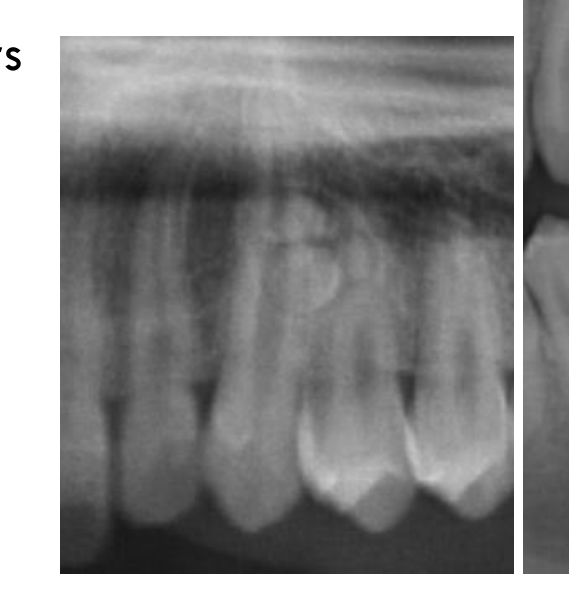
E.g. odontogenic tumours occur in the alveolar processes, above the IAC; cartilaginous tumours occur in jaw locations with residual cartilaginous cells
-
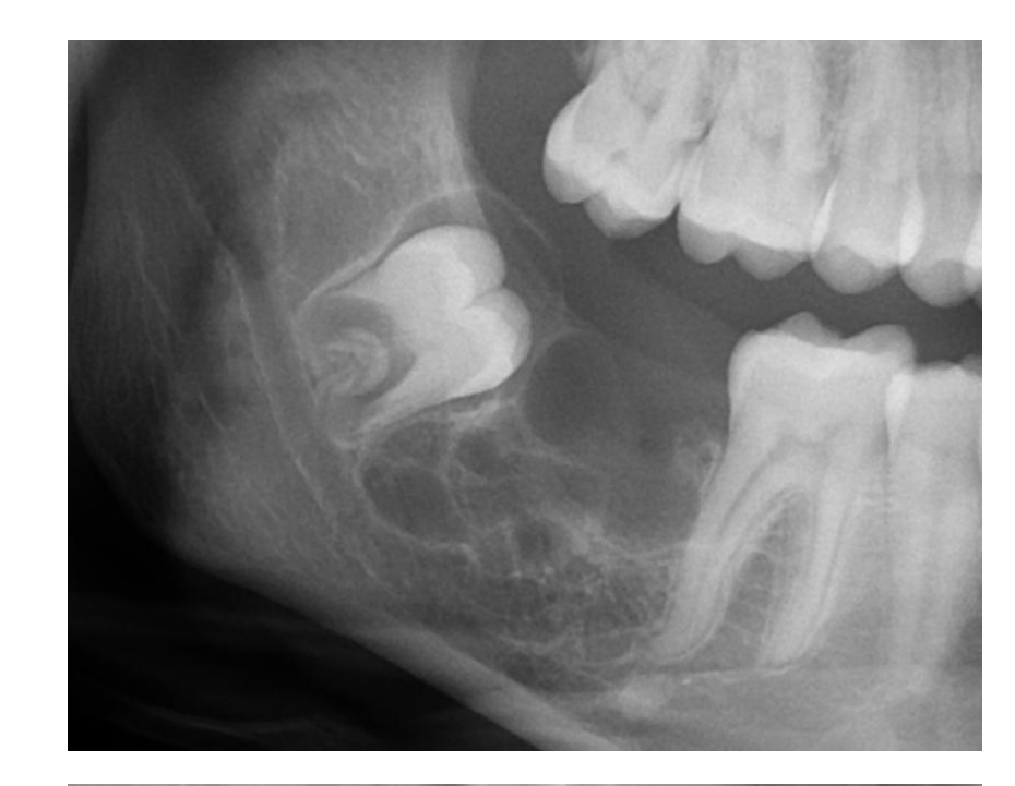
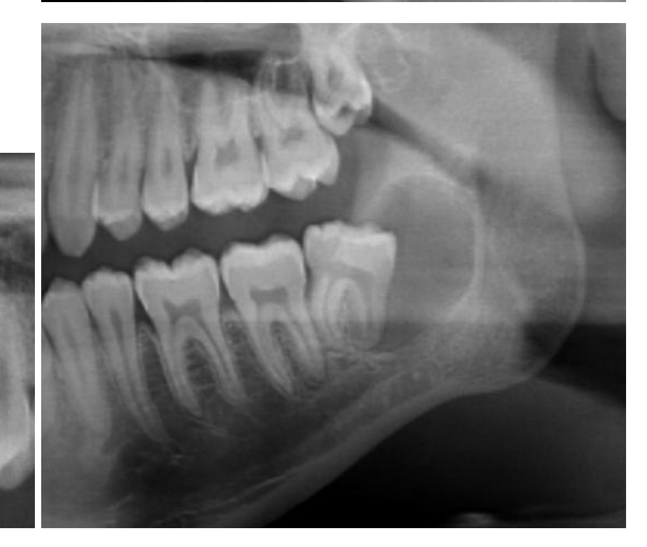
The anatomical location can help narrow the differential diagnosis, including the specific jaw, anterior or posterior position, relationship to teeth, and relationship to the inferior alveolar canal or maxillary sinus.
 |  |
 |
Periphery and Shape
- Usually smooth, well-defined, sometimes corticated borders
- Due to the slow enlargement by formation of additional internal tissue
- A corticated border appears as a thin radiopaque line and reflects slow tumour enlargement with formation of additional internal tissue.
- Due to the slow enlargement by formation of additional internal tissue
- May be unilocular or multilocular
- A radiolucent band may be seen
- This represents a soft tissue capsule
- The radiolucent band may be present around a lesion and may represent a soft-tissue capsule.
- This represents a soft tissue capsule
Internal Structure4
- Variable
- May be totally RL, totally RO, or a mixture of RL/RO tissues
- If the lesion contains RO elements, these usually represent residual bone, reactive bone formation, or a calcified material produced by the tumour
- The internal pattern may be characteristic for specific types of tumours
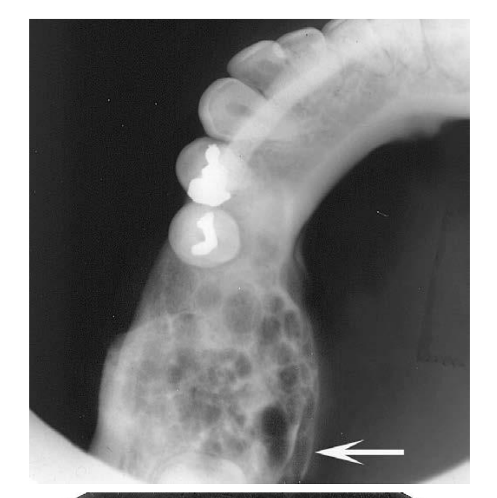
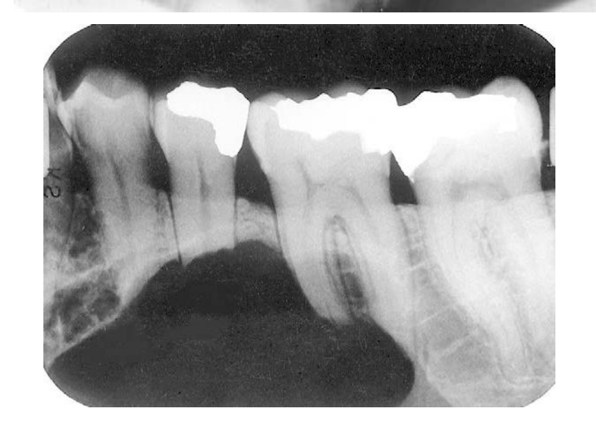
- E.g. curved coarse septa are characteristic for ameloblastomas
Lecturer — Ameloblastoma Septa
Coarse, curved septa are particularly characteristic of ameloblastoma.
- They represent residual bone trapped within the tumour and remodelled into a curved shape by internal cystic structures.
 |  |
Effects on Surrounding Structures
- Displacement and resorption of teeth
- Expansion and thinning of the jaw cortices +/- perforation
- Displacement of the inferior alveolar canal (IAC)
- Elevation of the antral +/- nasal cortical floors
Lecturer — Tumour Mass Effect
Benign tumours produce space-occupying or mass-effect features because they grow by taking up space.
- Root resorption is more common with benign tumours than with cysts and may produce blunting of the roots.
- Pressure against the cortex causes resorption of the inner cortical surface, while the periosteum deposits new bone on the outer surface.
- Repeated remodelling results in expansion and thinning of the cortices, with possible cortical perforation if the lesion grows faster than periosteal bone formation.
White and Pharoah, 2014
WHO Classification Overview
5th ed (2022)
Odontogenic Benign Tumours5
-
Benign epithelial odontogenic tumours
- Ameloblastoma
- Ameloblastoma, conventional
- Ameloblastoma, unicystic type
- Ameloblastoma, extraosseous/peripheral type
- Adenoid ameloblastoma
- Metastasising ameloblastoma
- Adenomatoid odontogenic tumour
- Squamous odontogenic tumour
- Calcifying epithelial odontogenic tumour
- Ameloblastoma
-
Benign mixed epithelial & mesenchymal odontogenic tumours
- Odontoma
- Ameloblastic fibroma
- Primordial odontogenic tumour
- Dentinogenic ghost cell tumour
- Ameloblastic fibro-odontoma
-
Benign mesenchymal odontogenic tumours
- Odontogenic myxoma/myxofibroma
- Odontogenic fibroma
- Cementoblastoma
- Cemento-ossifying fibroma
Non-Odontogenic Benign Tumours
-
Benign maxillofacial bone and cartilage tumours
- Osteoma
- Osteochondroma
- Osteoblastoma
- (Osteoid osteoma)
- Chondroblastoma
- Chondromyxoid fibroma
- Desmoplastic fibroma of bone
-
Soft tissue tumours
- Vascular tumours
- Haemangioma
- Peripheral nerve sheath tumours
- Neurofibroma
- Schwannoma
- Neuroma
- Vascular tumours
Part 1: Benign Epithelial Odontogenic Tumours6
- Ameloblastoma
- Ameloblastoma, conventional
- Ameloblastoma, unicystic type
- Ameloblastoma, extraosseous/peripheral type
- Adenoid ameloblastoma
- Metastasizing ameloblastoma
- Adenomatoid odontogenic tumour
- Squamous odontogenic tumour
- Calcifying epithelial odontogenic tumour
- Apparent septa may represent residual bone rather than true septa.
- Radicular cyst, particularly when the lesion is located periapically.
- Recurrence may result from incomplete excision when the lesion has been mistaken for a cyst.
Ameloblastoma78
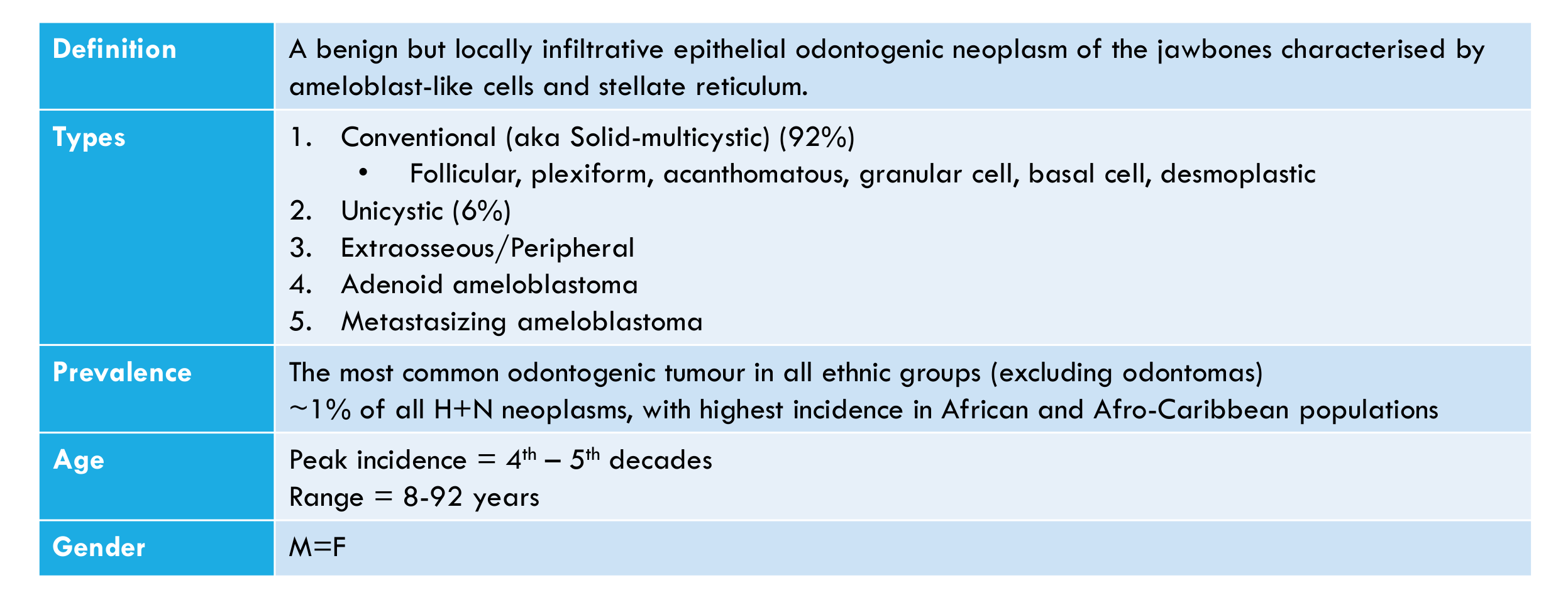
| Definition | A benign but locally infiltrative epithelial odontogenic neoplasm of the jawbones characterised by ameloblast-like cells and stellate reticulum. |
|---|
| Types | 1. Conventional (aka Solid-multicystic) (92%)
- Follicular, plexiform, acanthomatous, granular cell, basal cell, desmoplastic
- Unicystic (6%)
- Extraosseous/Peripheral
- Adenoid ameloblastoma
- Metastasizing ameloblastoma | |---|---|
| Prevalence | The most common odontogenic tumour in all ethnic groups (excluding odontomas) ~1% of all H+N neoplasms, with highest incidence in African and Afro-Caribbean populations | |---|---|
| Age | Peak incidence = 4th – 5th decades Range = 8-92 years | |---|---|
| Gender | M=F |
|---|
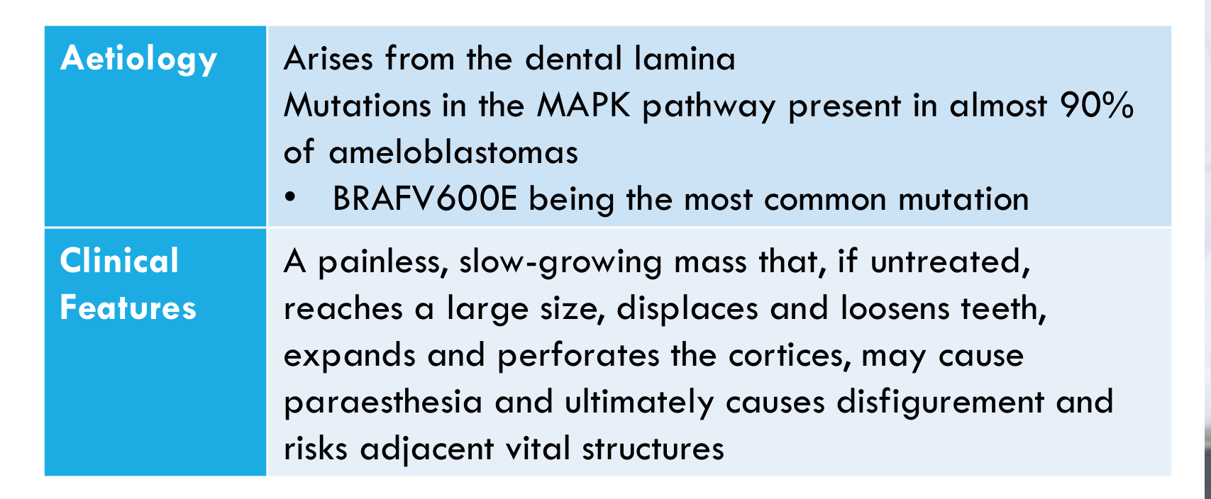
| Aetiology | Arises from the dental lamina Mutations in the MAPK pathway present in almost 90% of ameloblastomas
- BRAF V600E being the most common mutation | |---|---|
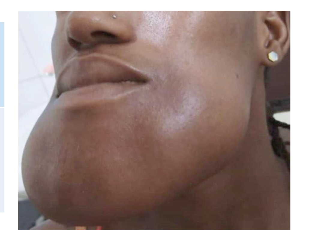
| Clinical Features | A painless, slow-growing mass that, if untreated, reaches a large size, displaces and loosens teeth, expands and perforates the cortices, may cause paraesthesia and ultimately causes disfigurement and risks adjacent vital structures |
|---|
- Ameloblastoma is one of the tumours most likely to be encountered in dental practice.
 |  |
 |
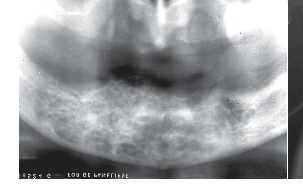
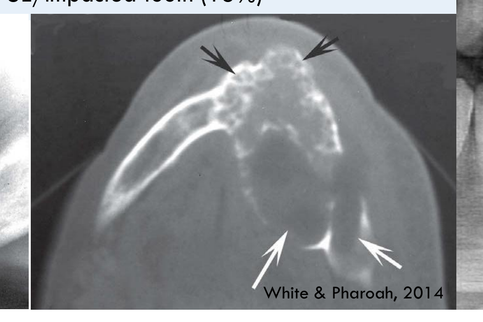
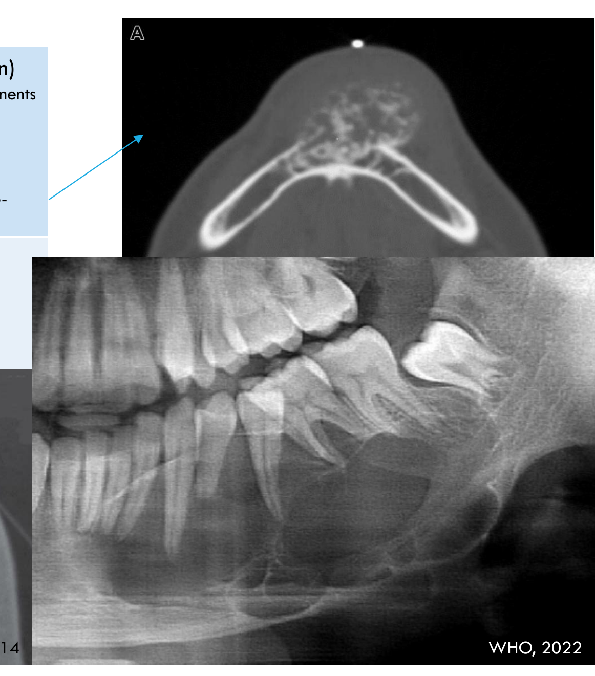
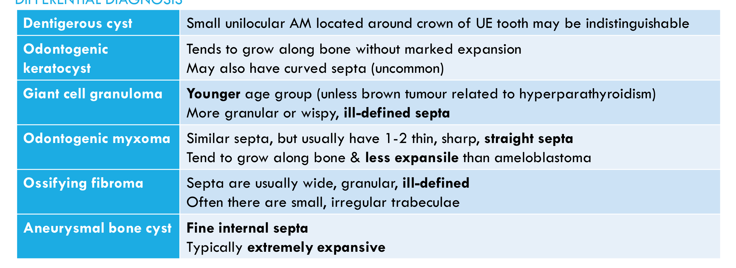
Conventional Ameloblastoma91011
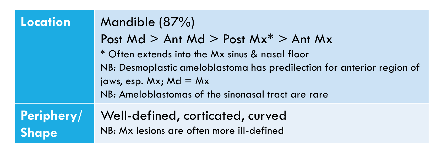
| Location | Mandible (87%) Post Md > Ant Md > Post Mx* > Ant Mx
- Often extends into the Mx sinus & nasal floor NB: Desmoplastic ameloblastoma has predilection for anterior region of jaws, esp. Mx; Md = Mx NB: Ameloblastomas of the sinonasal tract are rare | |---|---|
| Periphery/ Shape | Well-defined, corticated, curved NB: Mx lesions are often more ill-defined | |---|---|
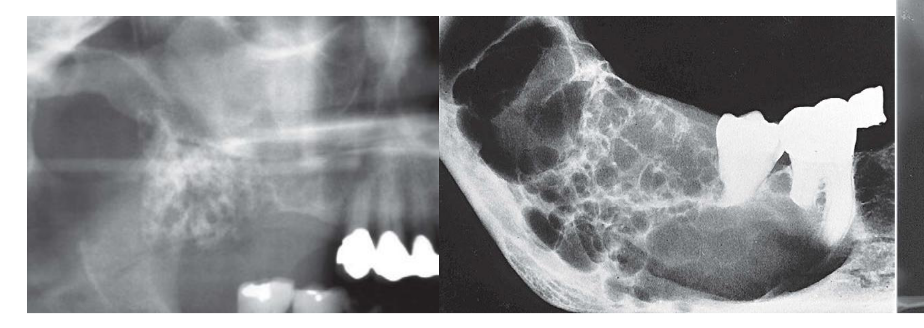
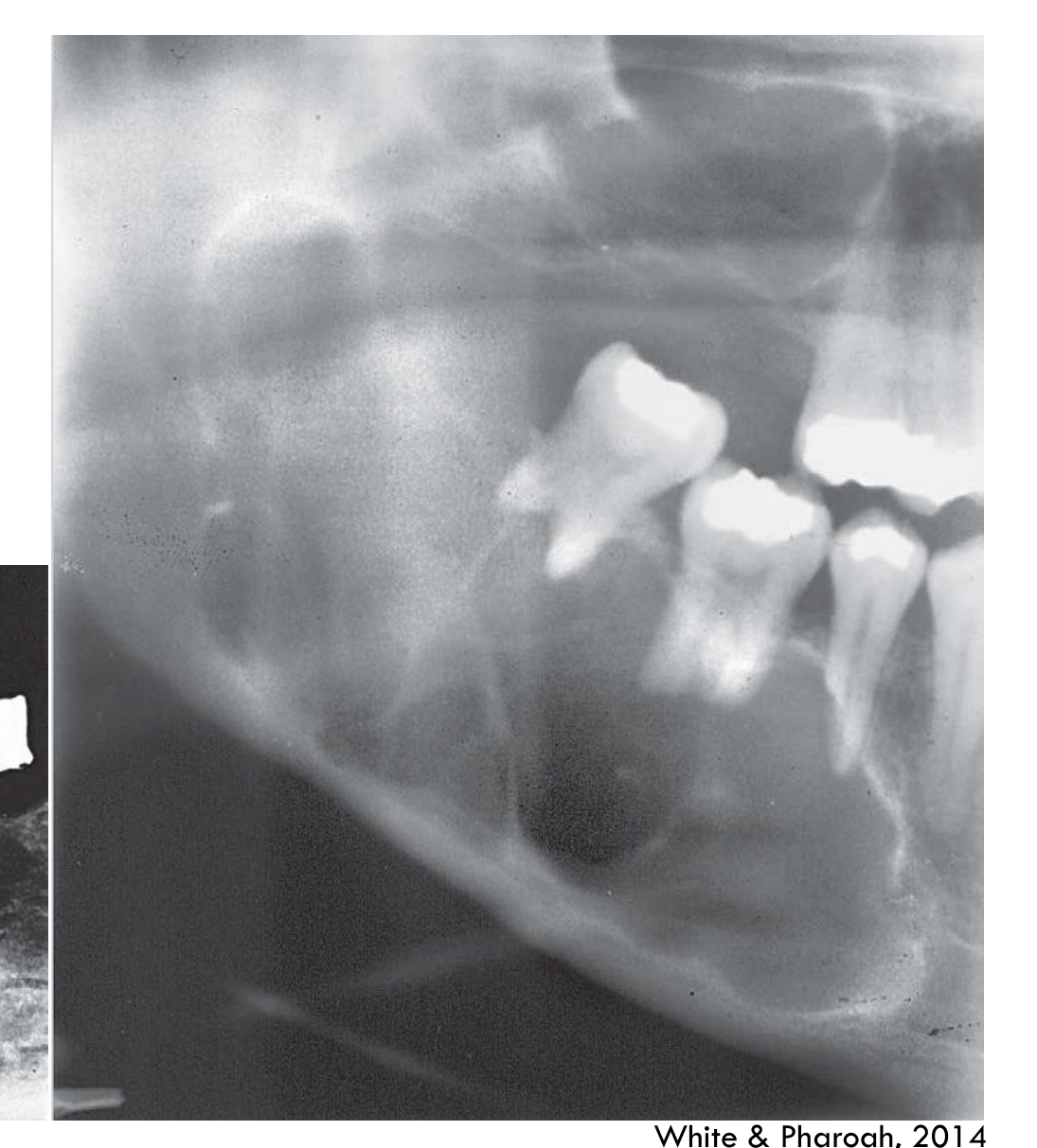
White & Pharoah, 2014
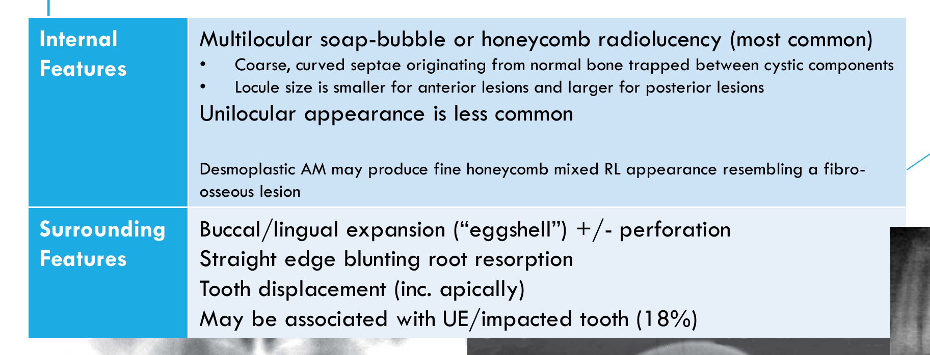
| Internal Features | Multilocular soap-bubble or honeycomb radiolucency (most common)
- Coarse, curved septae originating from normal bone trapped between cystic components
- Locule size is smaller for anterior lesions and larger for posterior lesions Unilocular appearance is less common Desmoplastic AM may produce fine honeycomb mixed RL appearance resembling a fibro-osseous lesion | |---|---|
| Surrounding Features | Buccal/lingual expansion (“eggshell”) +/- perforation Straight edge blunting root resorption Tooth displacement (inc. apically) May be associated with UE/impacted tooth (18%) | |---|---|
White & Pharoah, 2014 WHO, 2022
| Dentigerous cyst | Small unilocular AM located around crown of UE tooth may be indistinguishable |
|---|
| Odontogenic keratocyst | Tends to grow along bone without marked expansion May also have curved septa (uncommon) | |---|---|
| Giant cell granuloma | Younger age group (unless brown tumour related to hyperparathyroidism) More granular or wispy, ill-defined septa | |---|---|
| Odontogenic myxoma | Similar septa, but usually have 1-2 thin, sharp, straight septa Tend to grow along bone & less expansile than ameloblastoma | |---|---|
| Ossifying fibroma | Septa are usually wide, granular, ill-defined Often there are small, irregular trabeculae | |---|---|
| Aneurysmal bone cyst | Fine internal septa Typically extremely expansive | |---|---|
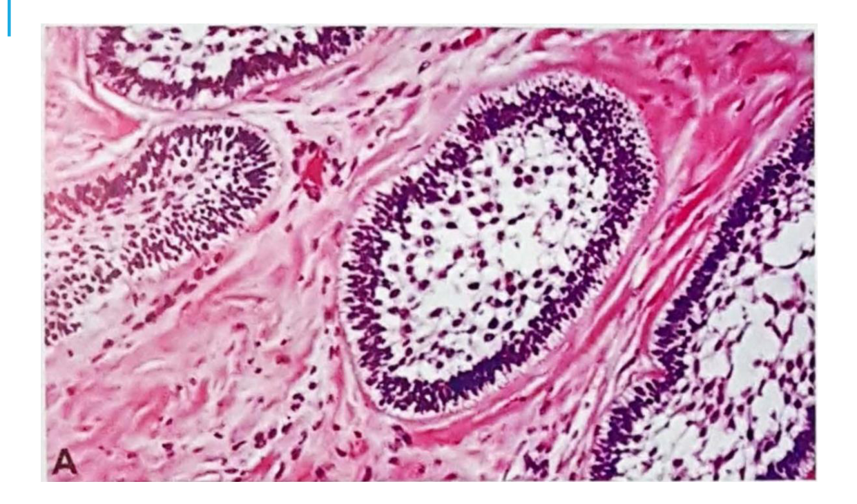
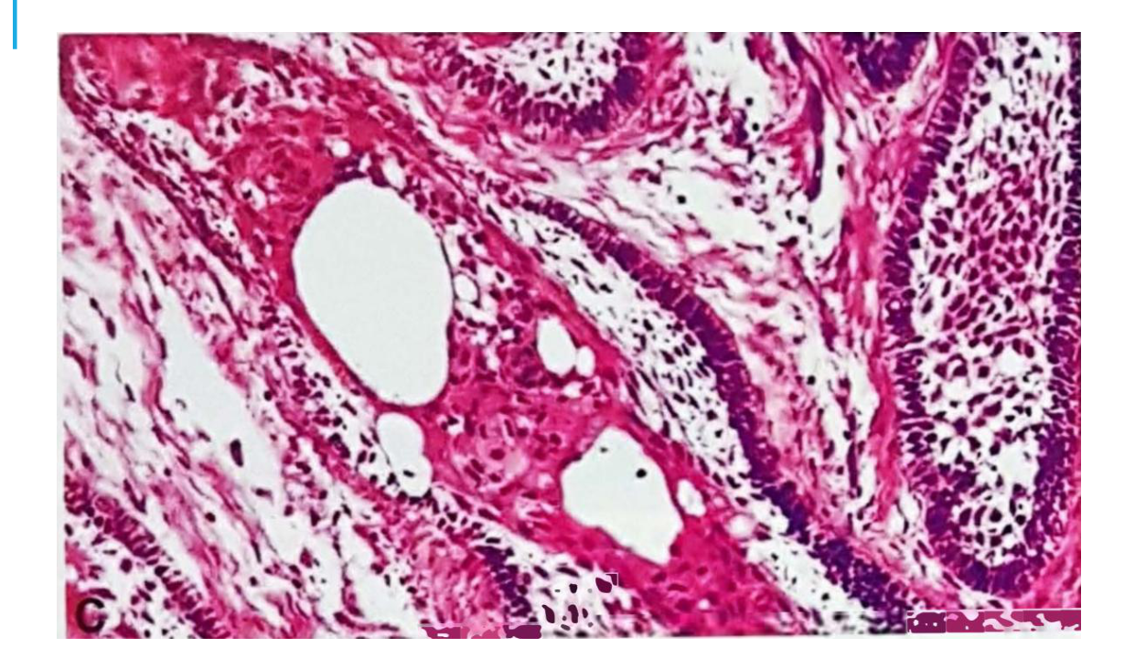
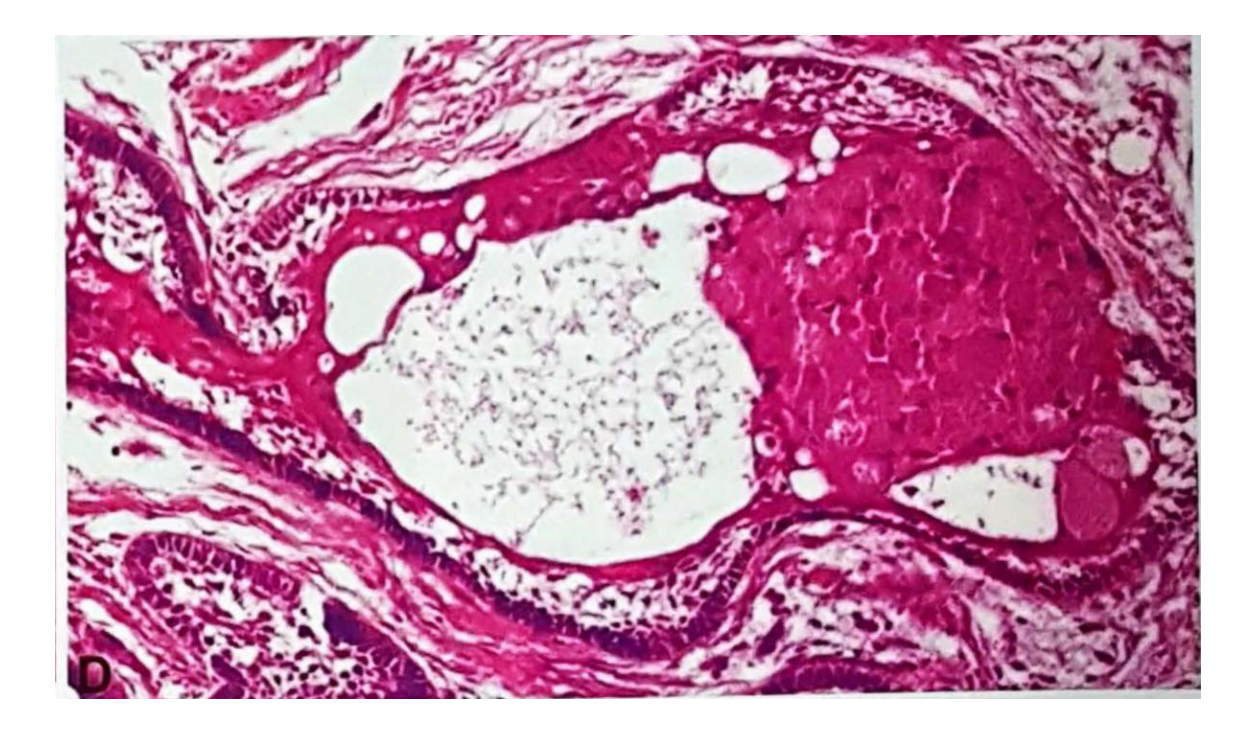
Follicular Type (Most Common)1213
- Islands of odontogenic epithelium with columnar peripheral cells
- Hyperchromatic nuclei with palisading pattern & reverse polarity
- Reminiscent of the enamel organ (ameloblast-like peripheral cells with a central core akin to stellate reticulum which often undergoes cystic degeneration)
- Connective tissue is moderate to highly collagenised
 |  |
 |  |
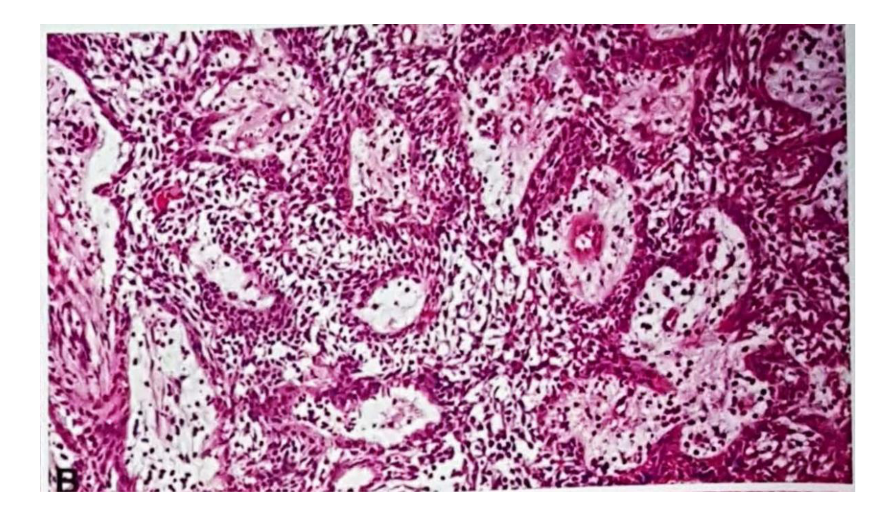
Plexiform Type
-
Strands & cords of ameloblastomatous epithelium that form anastomoses
-
Peripheral cells: less pronounced
-
Inconspicuous stellate reticulum
-
Connective tissue: loose & often undergoes cystic changes
-
Squamous metaplasia in stellate reticulum-like central areas
-
May have keratin pearl formation
-
Granular change in the stellate reticulum-like central areas
Other types: Basaloid, desmoplastic, clear cell
Mixed patterns are seen frequently
Infiltration into bony trabeculae is commonly seen
No clinical significance is associated with these histological patterns
Management1415
- Complete excision with negative margins, irrespective of histopathological subtype
- This may involve a segmental resection, mandibulectomy or maxillectomy, depending on lesion size
- Requires removal of bone margin of at least 10mm beyond radiographic margin
- Maxillary lesions are usually treated more aggressively
- BRAF inhibitor treatment has been proposed, alone or in combination with MAPK/ERK kinase (MEK) inhibitors
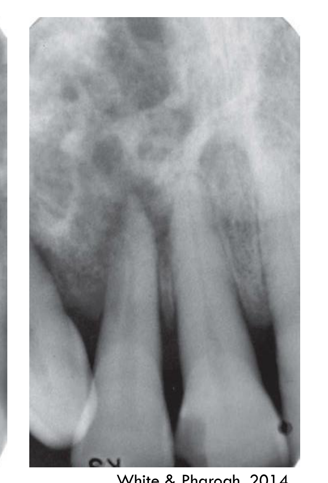
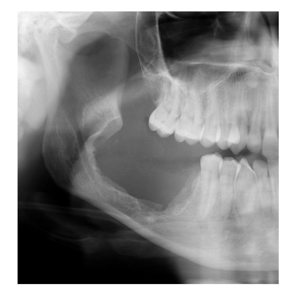 | 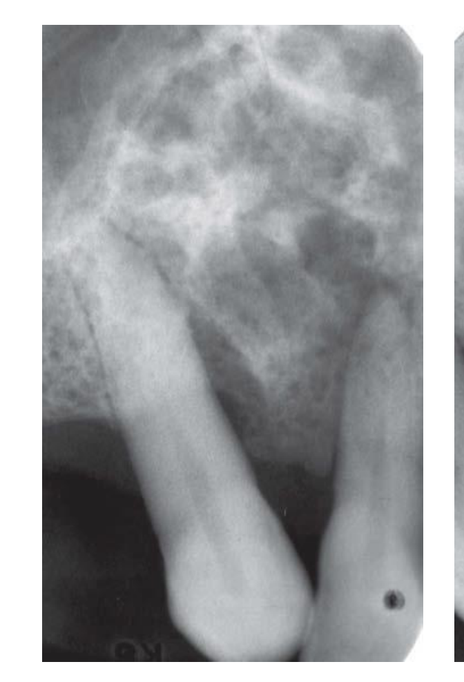 |
 |
Prognosis/Recurrence
- Recurrence rate = approx. 60-80% with conservative surgery
- Long follow up (1-2 decades) is mandatory – consider lifelong
Multiple small cyst-like structures with very coarse sclerotic cortical margins, sometimes separated by normal bone
More aggressive both radiographically & histologically than the original lesion
White & Pharoah, 2014
Unicystic Ameloblastoma161718
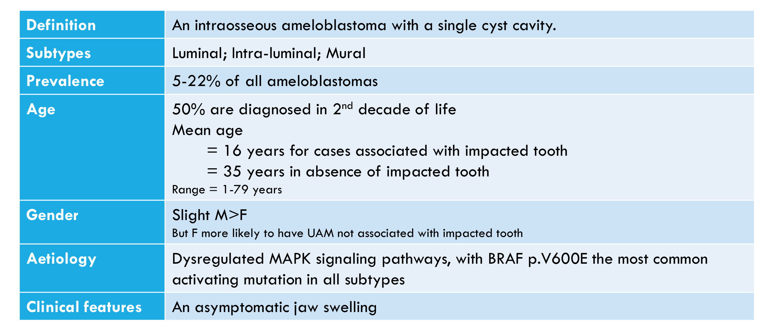
| Definition | An intraosseous ameloblastoma with a single cyst cavity. |
|---|
| Subtypes | Luminal; Intra-luminal; Mural |
|---|
| Prevalence | 5-22% of all ameloblastomas |
|---|
| Age | 50% are diagnosed in 2nd decade of life Mean age
- = 16 years for cases associated with impacted tooth
- = 35 years in absence of impacted tooth Range = 1-79 years | |---|---|
| Gender | Slight M>F But F more likely to have UAM not associated with impacted tooth | |---|---|
| Aetiology | Dysregulated MAPK signaling pathways, with BRAF p.V600E the most common activating mutation in all subtypes |
|---|
| Clinical features | An asymptomatic jaw swelling |
|---|
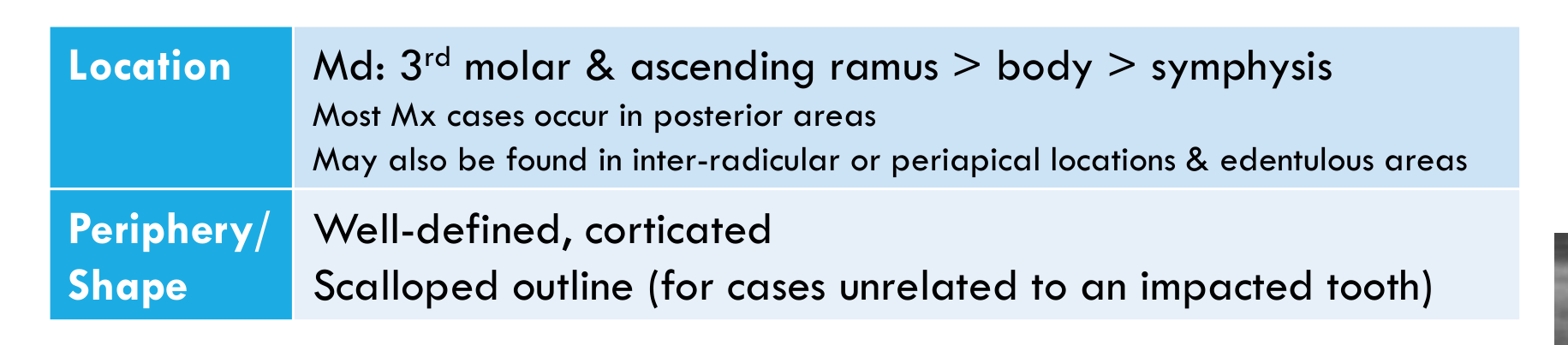
| Location | Md: 3rd molar & ascending ramus > body > symphysis Most Mx cases occur in posterior areas May also be found in inter-radicular or periapical locations & edentulous areas | |---|---|
| Periphery/ Shape | Well-defined, corticated Scalloped outline (for cases unrelated to an impacted tooth) | |---|---|
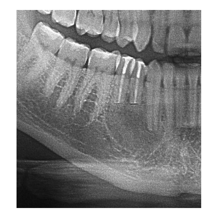
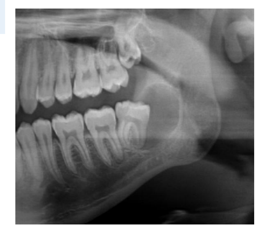
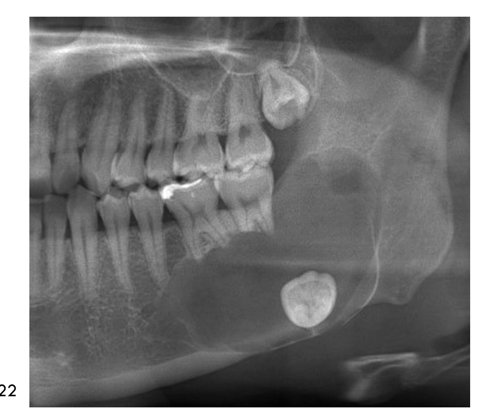
| Internal Features | Unilocular radiolucency Often associated with UE tooth (most often Mand 8s) | |---|---|
| Surrounding Features | Root resorption Cortical perforation in 1/3 of cases | |---|---|
White & Pharoah, 2014 WHO, 2022

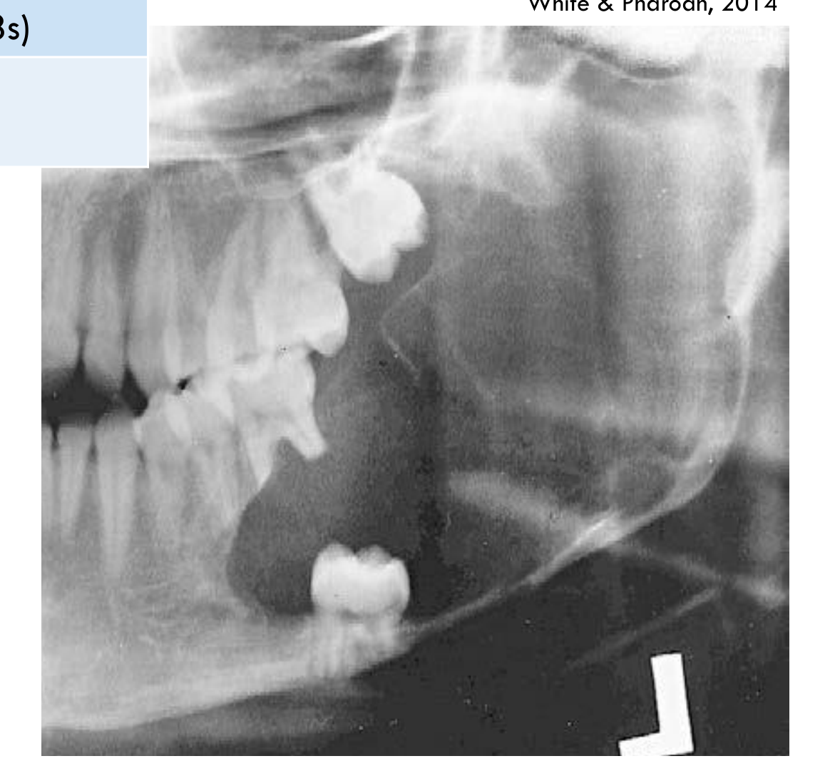
Histopathology19
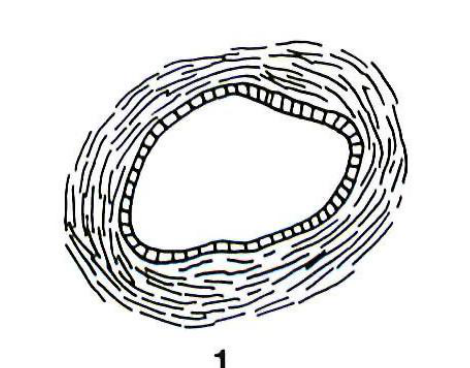
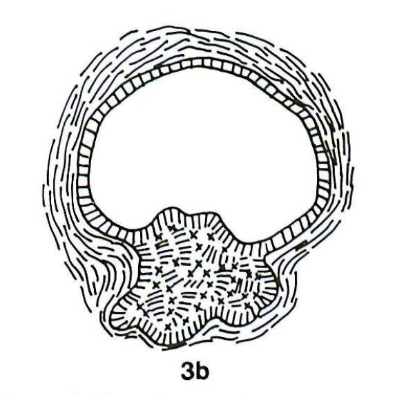
- Single cyst lined by epithelium with palisaded columnar basal layer with reverse polarity and stellate reticulum-like upper layers in most of the lining constitutes the luminal subtype
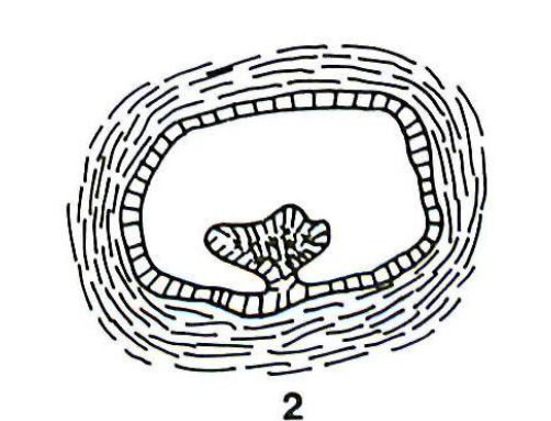
- Additional plexiform epithelial masses may extend into the lumen only, constituting the intra-luminal type
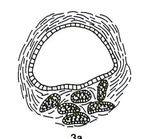
- Additional islands of epithelium extending into the wall constitute the mural subtype
- 50-66% of UAM may have a mural component → behave more aggressively like conventional ameloblastomas
- Extensive sampling is required for accurate definition of subtype
- Similar but focal changes may be seen in dentigerous and radicular cysts, especially in areas of inflammation
Fig. 1. Histologic types 1. Group 1 – cyst lined by variable, often nondescript, epithelium with no infiltration into fibrous cyst wall. 2. Group 2 – cyst showing intraluminal plexiform epithelial proliferation with no infiltration. 3. Group 3 – cyst with invasion of epithelium into the cyst wall in either (a) a follicular or (b) a plexiform pattern.
 |  |
 |  |
Differential Diagnosis20
- May be indistinguishable from a dentigerous cyst or odontogenic keratocyst
- Histopathologic correlation required for definitive diagnosis
Management/Prognosis/Recurrence
- Up to 30% may recur after enucleation, as definitive diagnosis prior to initial treatment is not possible
- Conservative marsupialisation followed by enucleation is proposed for luminal and intraluminal UAM
- Risk of recurrence so long-term follow up required
- Mural UAM appears to be intermediate between UAM and conventional ameloblastoma
- Might require consideration of more extensive surgery as for conventional ameloblastoma, depending on size, extent of intra-mural proliferation and radiological findings
- However, accurate diagnosis often only follows definitive removal, allowing a period of follow up to confirm recurrence before more aggressive treatment.
Adenomatoid Odontogenic Tumour
Definition21
A benign encapsulated epithelial odontogenic tumour that contains rosette or duct-like structures and has an indolent behaviour.
 |  |
Prevalence
<10% of all odontogenic tumours
- It is much less common than ameloblastoma.
Age
Wide age range, but >80% are diagnosed in the 2nd and 3rd decades of life
Gender
F>M; 2:1
Aetiology
- Unknown, although KRAS p.G12V and p.G12R mutations are detected in approximately 70% of sporadic AOTs
- Multiple AOTs can occur in Schimmelpenning syndrome
Clinical Features
- Most are asymptomatic
- All have limited growth potential
- Large AOTs present as bony hard swellings with cortical expansion but not perforation
- Peripheral AOTs appear as small gingival nodules
WHO, 2022
Location22
-
95% intraosseous (Mx>Md, Ant>Post)
-
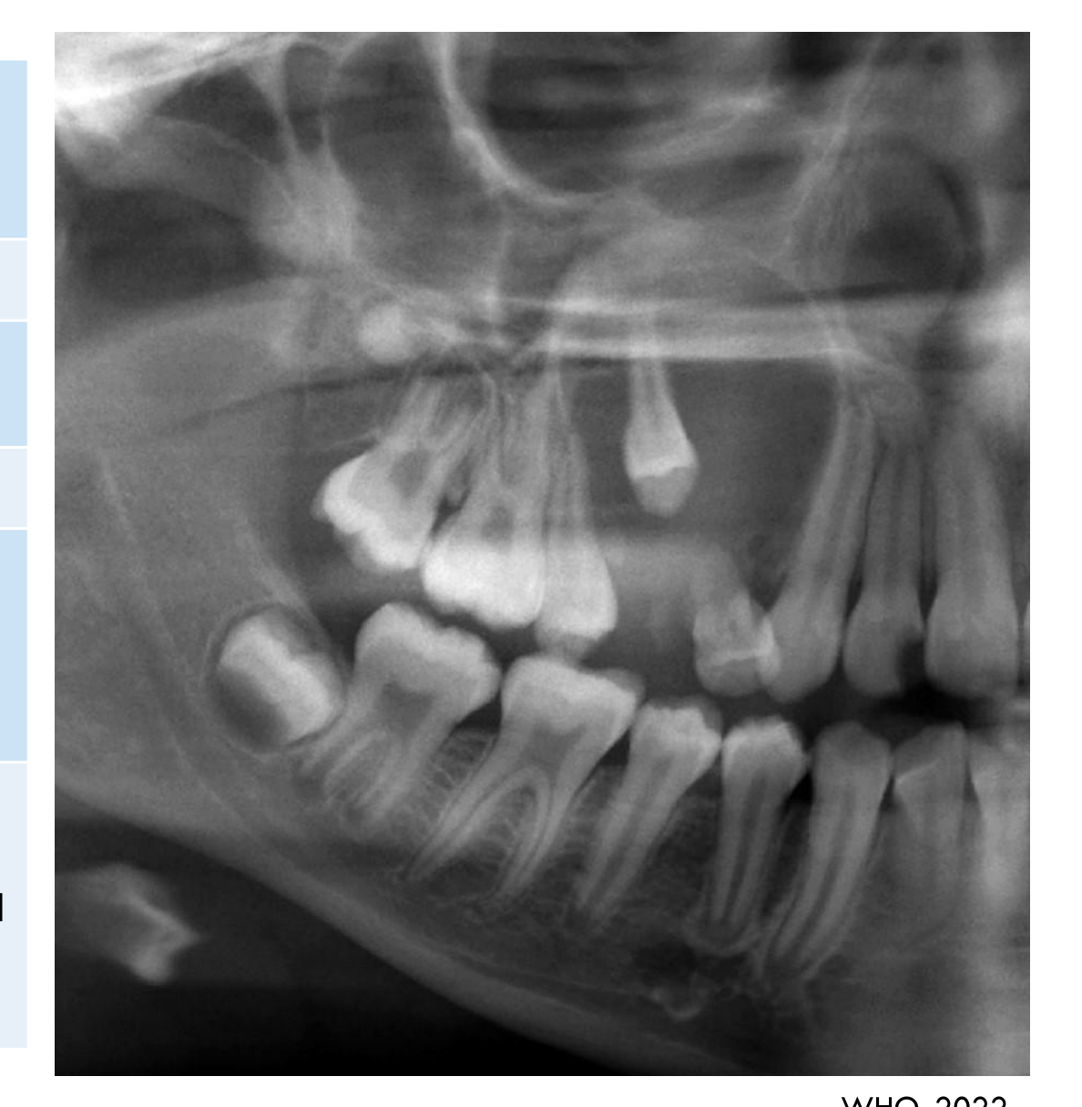
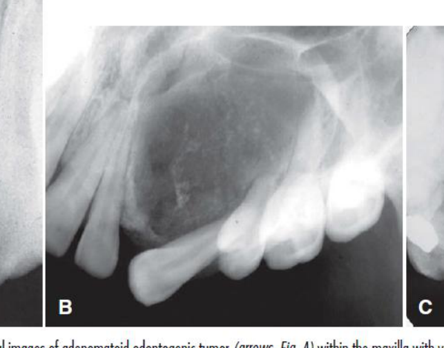
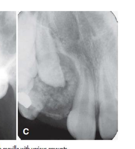
75% cases are located around or alongside the crown of an UE permanent tooth, often extending apically past CEJ
-
5% extraosseous (anterior maxillary gingiva)
-
The tumour may expand the follicle and surround most of the tooth.
-
Unlike a dentigerous cyst, it does not usually attach at the cemento-enamel junction; it tends to surround much of the root and part of the crown.
 |  |
 |  |
Periphery
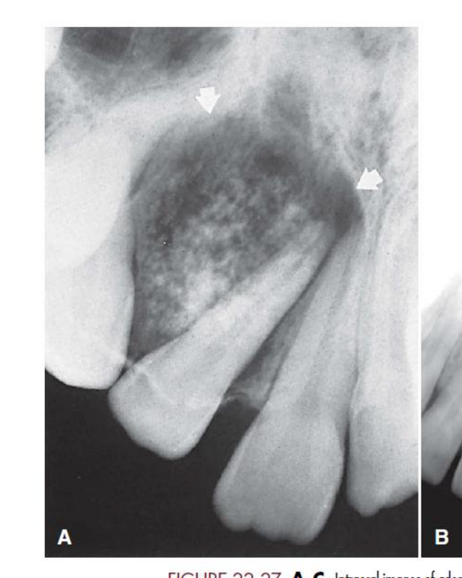
Well defined, unilocular, symmetric with corticated/sclerotic border
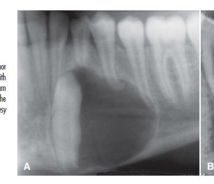
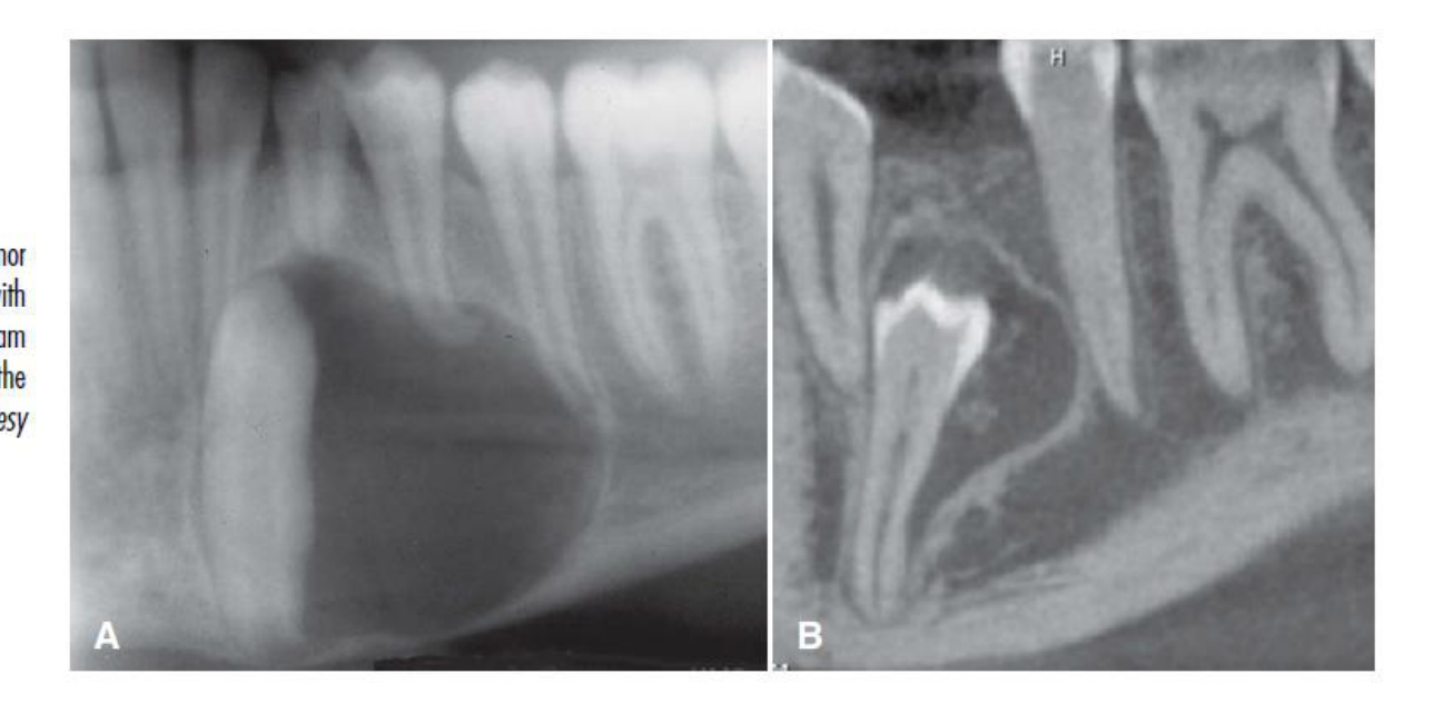
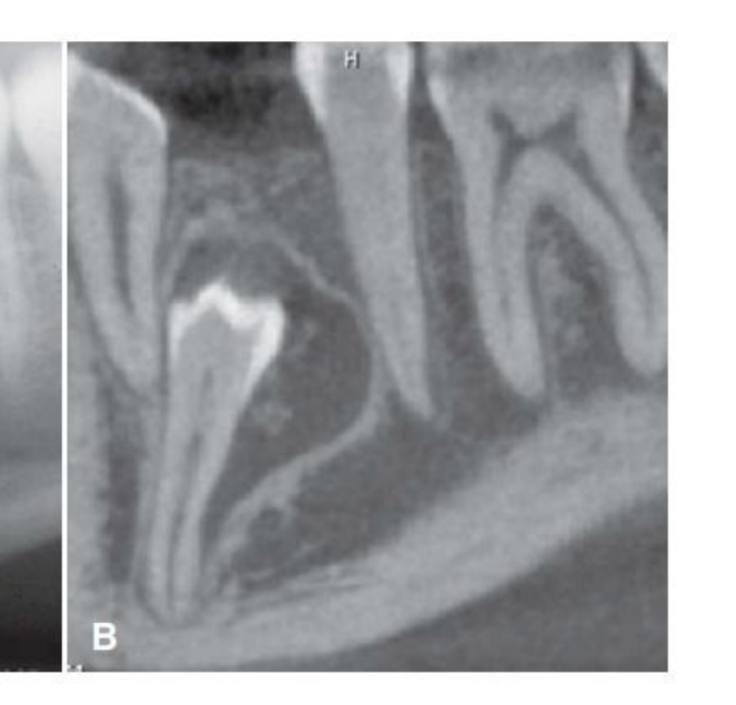
FIGURE 22-27 A-C, Intraoral images of adenomatoid odontogenic tumor (arrows, Fig. A) within the maxilla with various amounts of calcification, some of which have a pebble-like shape. (A, Courtesy R. Howell, DDS, Morgantown, WV.)
White & Pharoah, 2014
Internal Features23
Radiolucent, 2/3rd of cases show small radiopaque foci — “snowstorm”
 |  |
 |  |
Surrounding Features
- Tooth displacement frequent
- Root resorption rare
- Cortical expansion (perforation rare)
White & Pharoah, 2014
Extraosseous Variant
-
Appears as a unilocular radiolucency located between, above, or superimposed upon the roots of erupted teeth
-
May cause superficial erosion of the underlying alveolar bone
-
The peripheral or extraosseous variant is uncommon.
-
A slightly loosened appearance may occur when the lesion is positioned on the buccal or lingual aspect of the jaw.
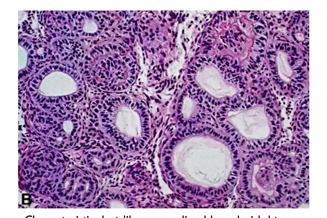
Histopathology24
-
Encapsulated
-
Contain variably sized nodules of spindle, cuboidal and columnar epithelial odontogenic cells with minimal stroma
-
Within the nodules are rosette or duct-like structures, which produce the adenomatoid or gland-like appearance
-
Eosinophilic amorphous secretory material similar to enamel matrix present between the epithelial cells and rosette-like structures
-
+/- Small foci of calcification, dentinoid matrix, & haemorrhage
-
+/- cysts lined by non-keratinising stratified epithelium
-
The cysts may be lined by non-keratinising stratified epithelium resembling a dentigerous cyst.
Differential Diagnosis25
-
AOT-like areas have been recognized within other odontogenic tumours (inc. odontomas)
-
AOT may contain areas resembling calcifying epithelial odontogenic tumour with clear cells
-
These histological overlaps makes radiological and clinical correlation essential for definitive diagnosis.
-
Dentigerous cyst.
-
Odontoma.
-
Calcifying epithelial odontogenic tumour.
-
Other lesions containing calcification.
Management
- Enucleation (including capsule)
Prognosis/Recurrence
- Almost no risk of recurrence following conservative enucleation.
Squamous Odontogenic Tumour
Definition26
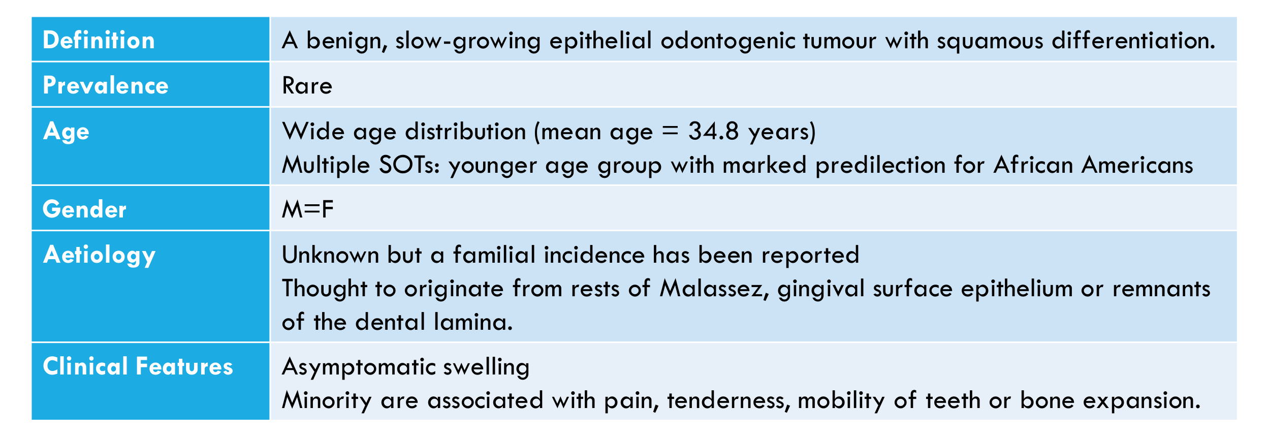
A benign, slow-growing epithelial odontogenic tumour with squamous differentiation.
Prevalence
Rare
Age
- Wide age distribution (mean age = 34.8 years)
- Multiple SOTs: younger age group with marked predilection for African Americans
Gender
M = F
Aetiology
- Unknown but a familial incidence has been reported
- Thought to originate from rests of Malassez, gingival surface epithelium or remnants of the dental lamina.
Clinical Features
-
Asymptomatic swelling
-
Minority are associated with pain, tenderness, mobility of teeth or bone expansion.
-
It may occur in edentulous patients.
Location27
-
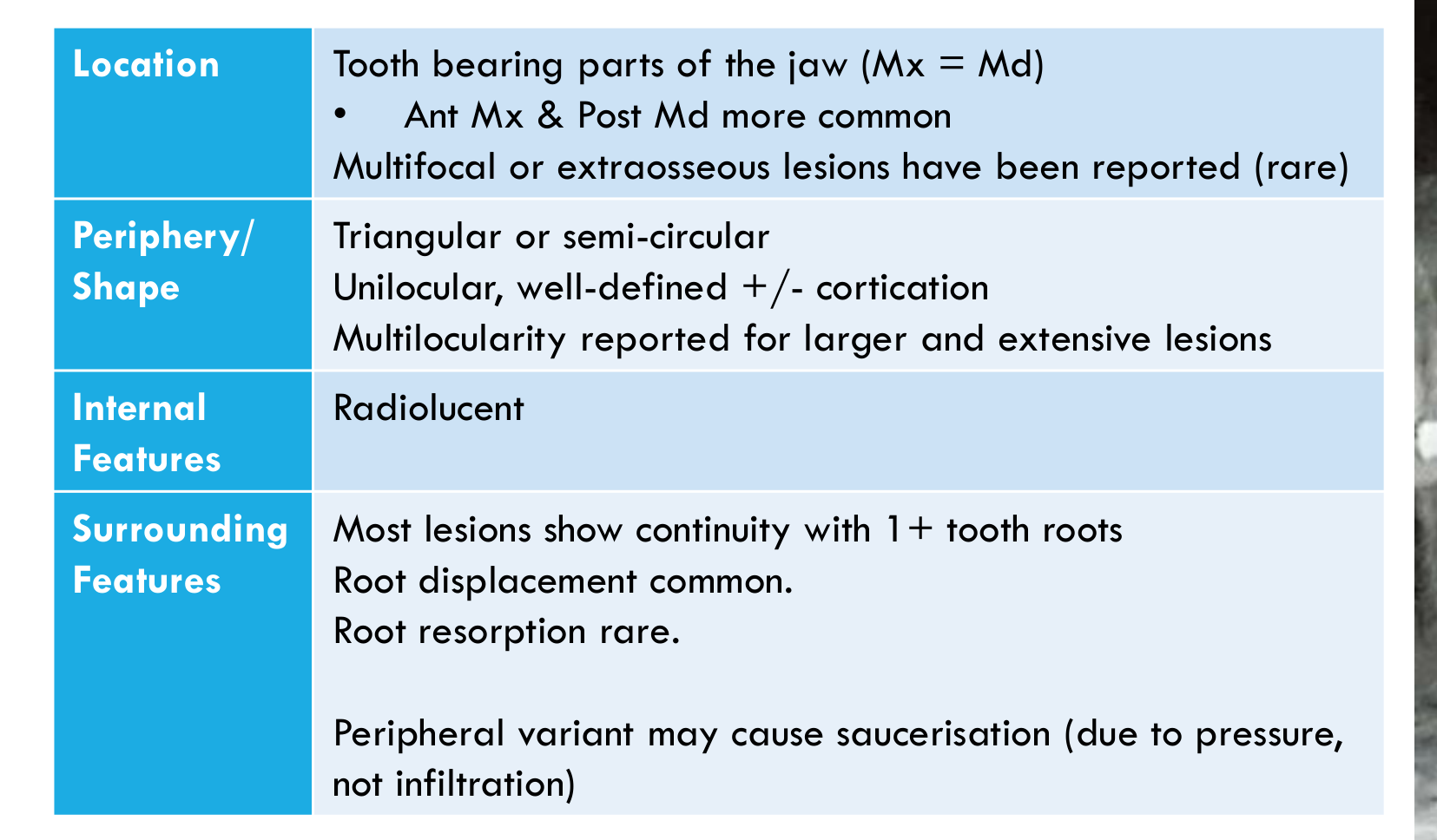
Tooth bearing parts of the jaw (Mx = Md)
- Ant Mx & Post Md more common
-
Multifocal or extraosseous lesions have been reported (rare)
-
Most lesions are solitary.
 |  |
Periphery / Shape
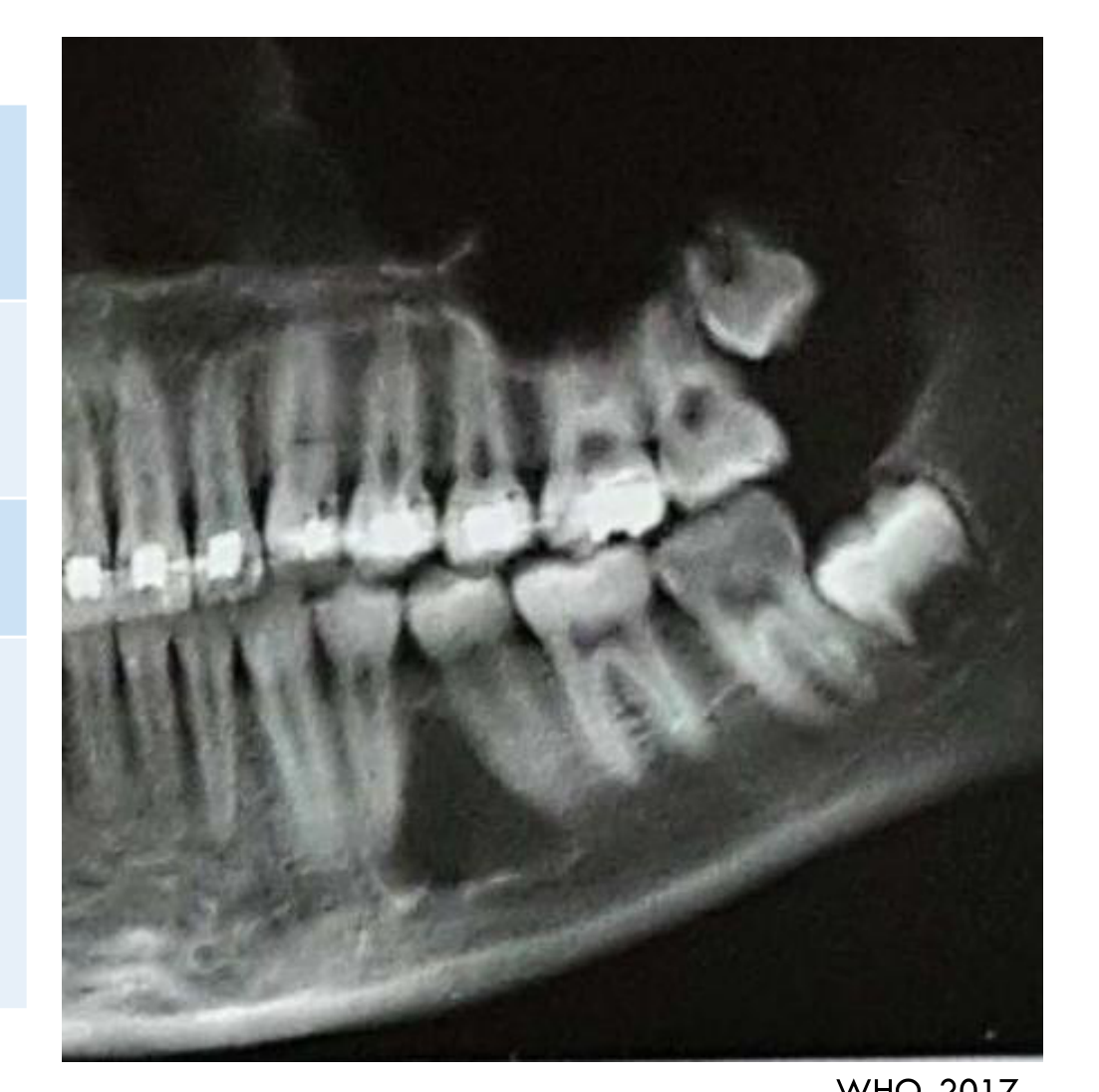
- Triangular or semi-circular
- Unilocular, well-defined +/- cortication
- Multilocularity reported for larger and extensive lesions
Internal Features
Radiolucent
Surrounding Features
-
Most lesions show continuity with 1+ tooth roots
-
Root displacement common.
-
Root resorption rare.
-
Peripheral variant may cause saucerisation (due to pressure, not infiltration)
-
Multilocular or more aggressive lesions may produce cortical expansion or perforation.
Histopathology28
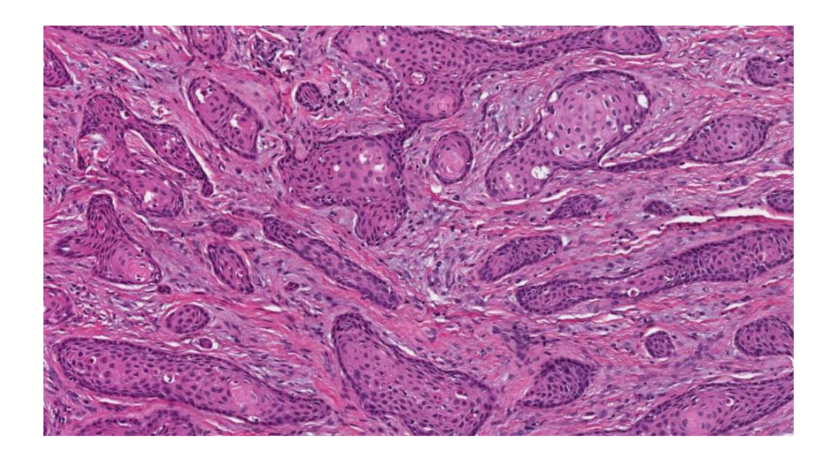
- Islands of bland well-differentiated squamous epithelium in a fibrous or myxoid stroma
- Peripheral cells of the islands are flat to cuboidal with very infrequent mitoses
- Central cells have a tendency for cystic degeneration, individual cell keratinization, and calcification
- +/- Mucous metaplasia, sebaceous differentiation, and ghost cell-like areas
Differential Diagnosis29
-
Ameloblastoma – acanthomatous or desmoplastic variants
- SOT lacks peripheral columnar cells & palisading nuclei
-
Well-differentiated squamous cell carcinoma
- SOT lacks peripheral palisading with reverse nuclear polarity, and the cytological features are bland (i.e. no dysplasia)
-
SOT-like proliferation of epithelium may develop in the lining of odontogenic cysts
-
Well-differentiated squamous cell carcinoma is particularly considered when there is no corticated border.
Management
-
Enucleation
-
A solitary unilocular lesion is generally treated by simple enucleation.
Prognosis / Recurrence
- Unifocal SOT rarely recurs after conservative surgery.
- Multifocal or recurring SOT, especially of the maxilla, may require a more radical approach.
Calcifying Epithelial Odontogenic Tumour303132
| Feature | Details |
|---|---|
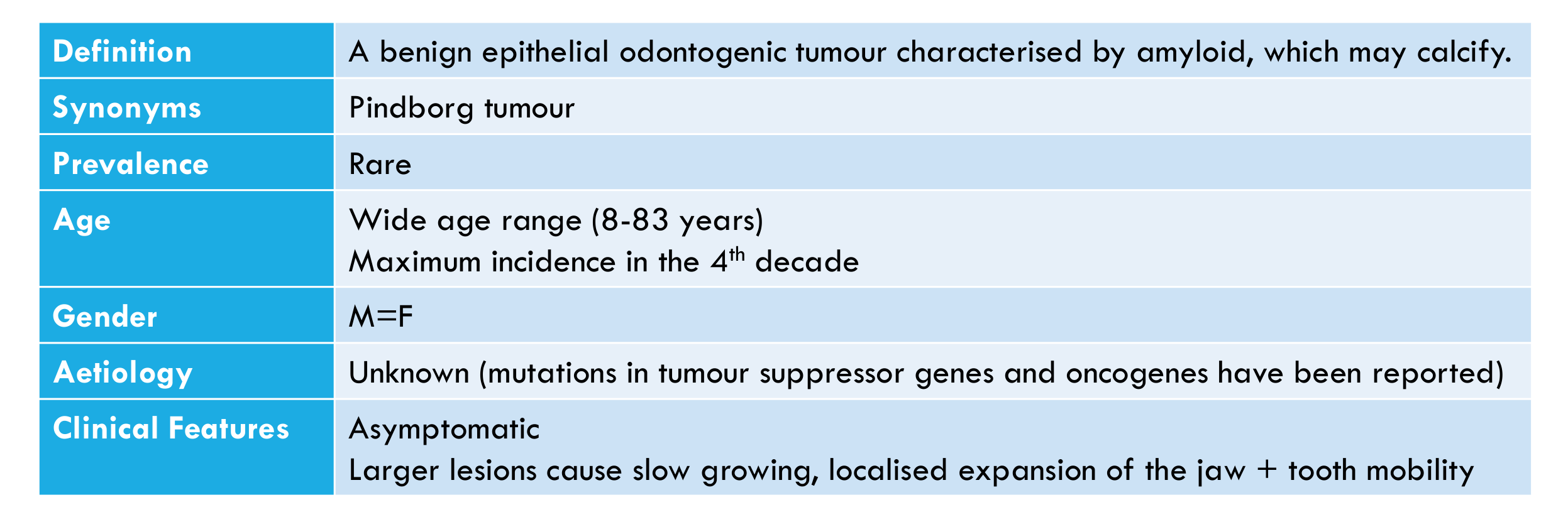
| Definition | A benign epithelial odontogenic tumour characterised by amyloid, which may calcify. |
| Synonyms | Pindborg tumour |
| Prevalence | Rare |
| Age | Wide age range (8–83 years); maximum incidence in the 4th decade |
| Gender | M = F |
| Aetiology | Unknown (mutations in tumour suppressor genes and oncogenes have been reported) |
| Clinical Features | Asymptomatic; larger lesions cause slow growing, localised expansion of the jaw + tooth mobility |
Lecturer — Alternative Name
The tumour was also referred to as a “pinball tumour.”
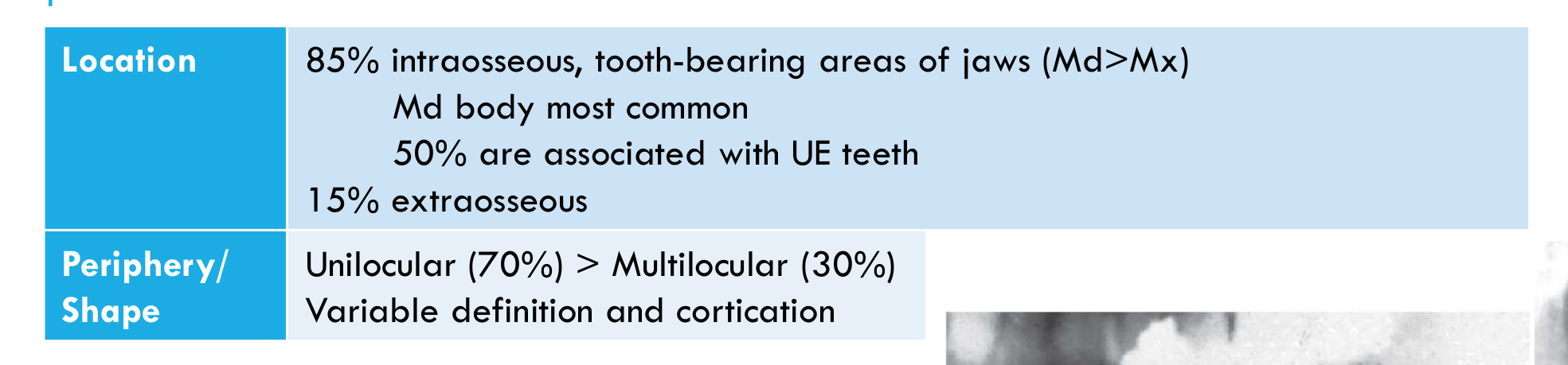
| Location | 85% intraosseous, tooth-bearing areas of jaws (Md > Mx); Md body most common; 50% are associated with UE teeth; 15% extraosseous |
|---|---|
| Periphery/Shape | Unilocular (70%) > Multilocular (30%); variable definition and cortication |
Lecturer — Mandibular Distribution
Approximately 60% of tumours occur in the mandible, with a predilection for the mandibular body.
White & Pharoah, 2014
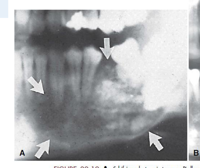
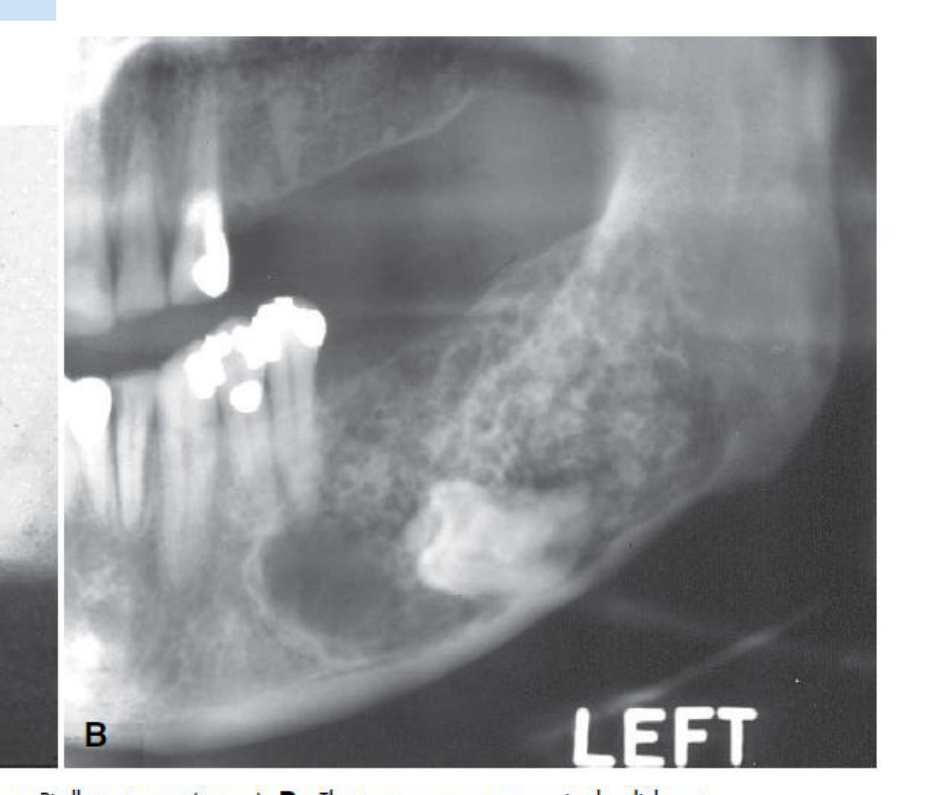
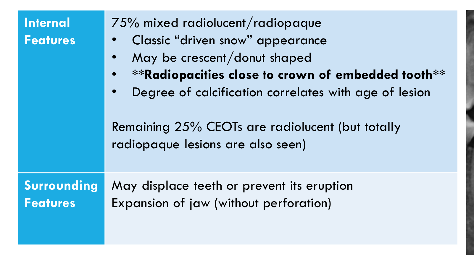
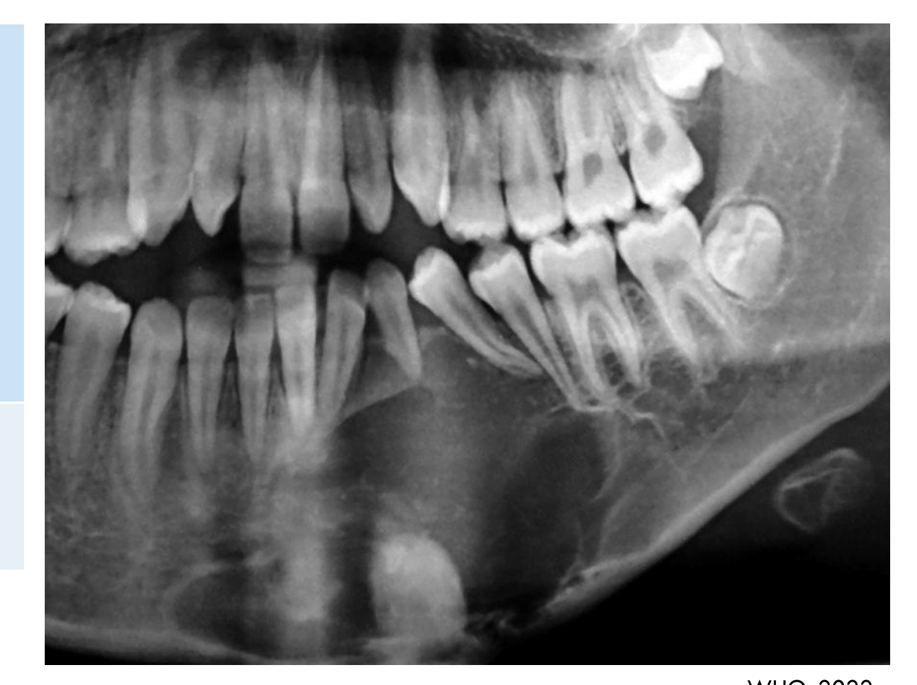
| Internal Features | 75% mixed radiolucent/radiopaque:
-
Classic “driven snow” appearance
-
May be crescent/donut shaped
-
Radiopacities close to crown of embedded tooth
-
Degree of calcification correlates with age of lesion
-
The opacities may be embedded around the crown of an unerupted tooth Remaining 25% CEOTs are radiolucent (but totally radiopaque lesions are also seen) | | :--- | :--- | | Surrounding Features | May displace teeth or prevent its eruption; expansion of jaw (without perforation) |
WHO, 2022
Histopathology33
-
Variable appearance with 4 subtypes:
- Clear cell
- Cystic/microcystic
- Non-calcifying/Langerhans cell rich
- AOT subtype
-
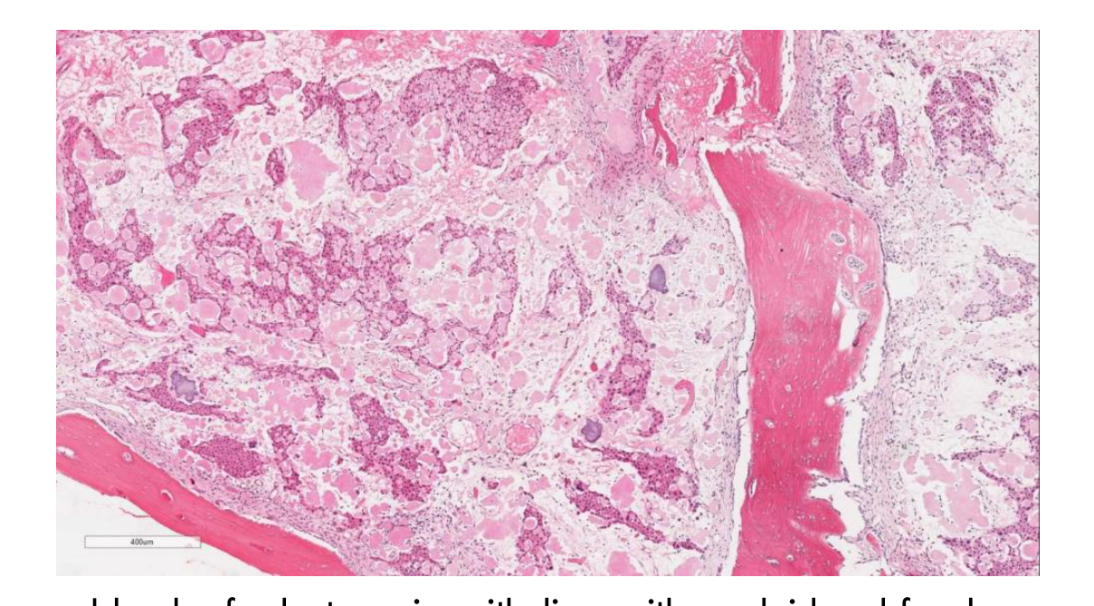
Comprise sheets, cords or nests of polyhedral epithelial cells with distinct cell borders and prominent intercellular bridges
- +/- nuclear pleomorphism (mitoses are rare)
-
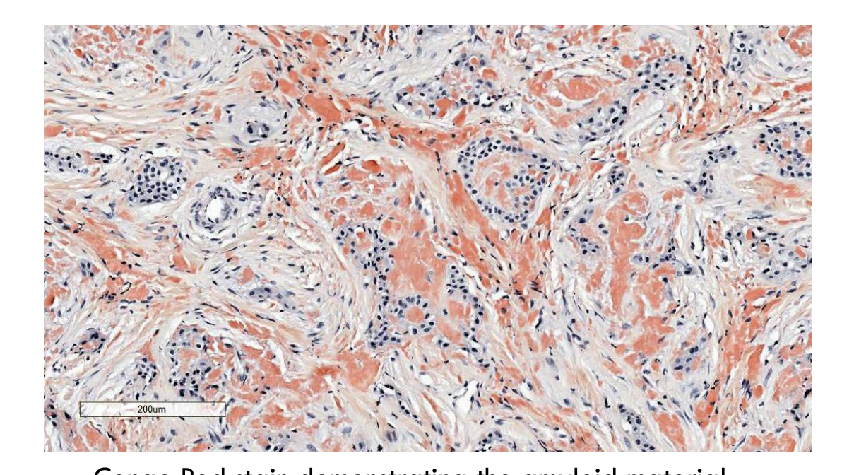
Deposits of amorphous lightly eosinophilic amyloid composed of ameloblast-associated proteins (stains with Congo red)
- These may calcify forming large masses or small round concentric densely basophilic calcifications with Liesegang rings
-
Amyloid deposits occur between the epithelial cells Congo Red stain demonstrating the amyloid material. (WHO, 2022)
 |  |
Differential Diagnosis34
- May extend into adjacent medullary spaces in an infiltrative pattern worrying for malignancy
- Foci resembling CEOT may be found in odontomas and the follicles of unerupted teeth
Lecturer — Radiological Correlation
The relationship between crescent- or donut-shaped opacities and the crown of an unerupted tooth can help distinguish this tumour from other mixed-density lesions.
- Histopathological findings should be correlated with radiological and clinical features.
Management
-
Local surgical removal
-
More aggressive treatment is associated with less recurrence
Prognosis/Recurrence
- Recurrence rate = approx. 13%
- Varies with treatment modality, being much higher in those treated by curettage
- No histological parameters predict recurrence
- Malignant transformation rare
Part 2: Benign Epithelial and Mesenchymal Odontogenic Tumours35
- Odontoma
- Ameloblastic fibroma
- Primordial odontogenic tumour
- Dentinogenic ghost cell tumour
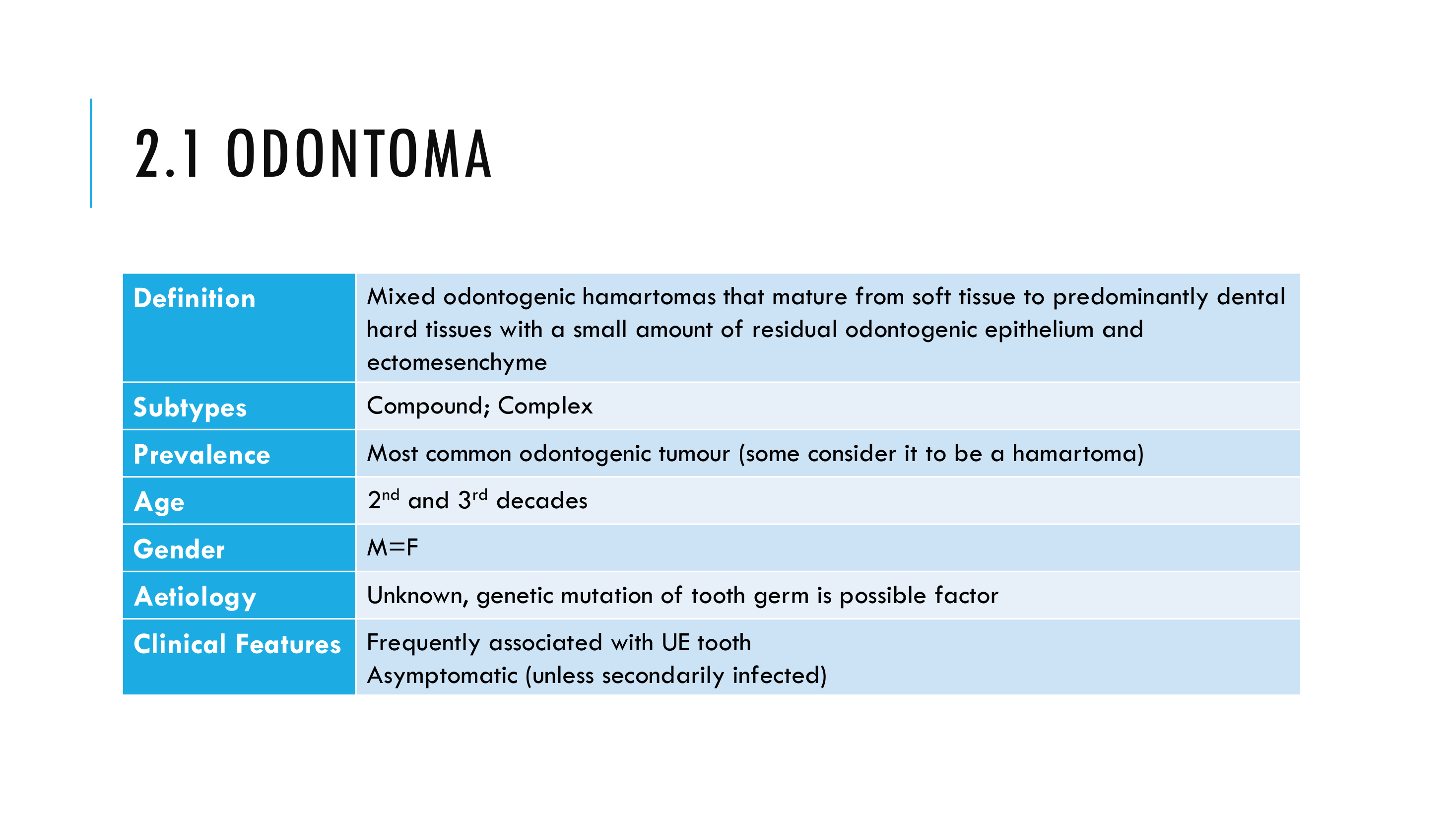
Odontoma
Definition36
Mixed odontogenic hamartomas that mature from soft tissue to predominantly dental hard tissues with a small amount of residual odontogenic epithelium and ectomesenchyme.
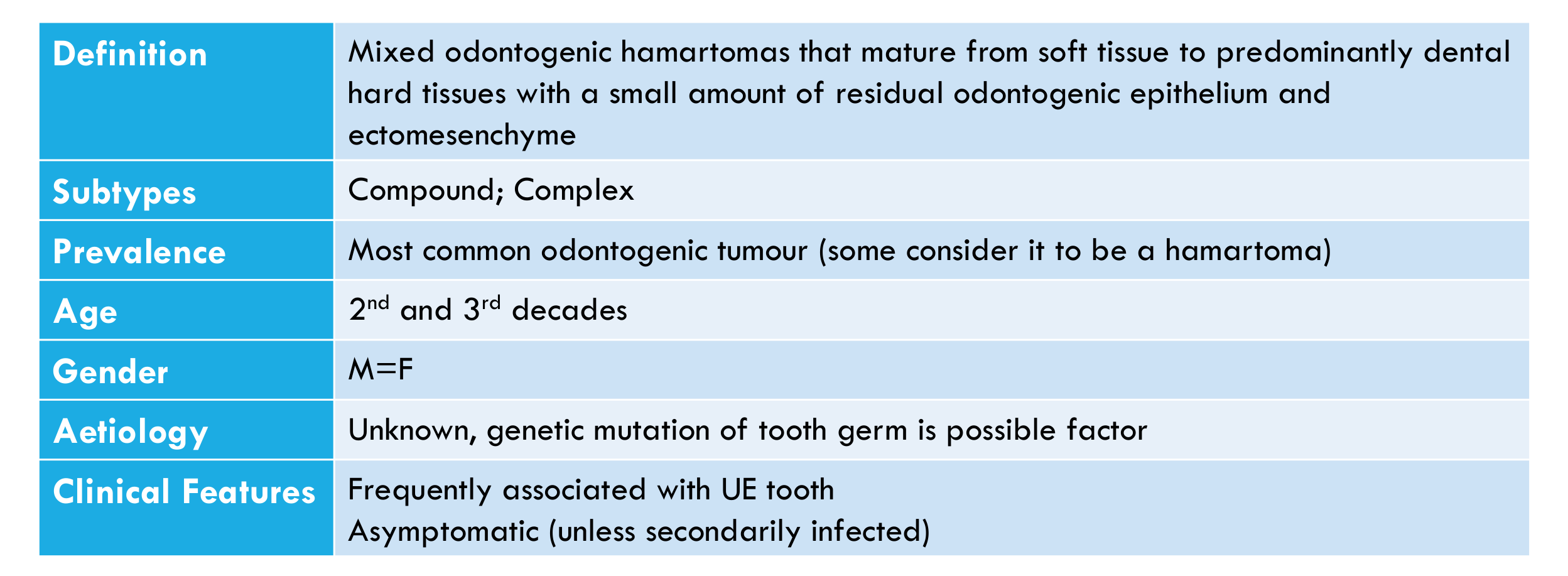
Subtypes
- Compound
- Complex
Prevalence
Most common odontogenic tumour (some consider it to be a hamartoma).
Age
2nd and 3rd decades.
Gender
M = F
Aetiology
Unknown; genetic mutation of tooth germ is a possible factor.
Clinical Features
-
Frequently associated with an unerupted tooth
-
Asymptomatic (unless secondarily infected)
-
Odontomas may also interfere with tooth eruption.
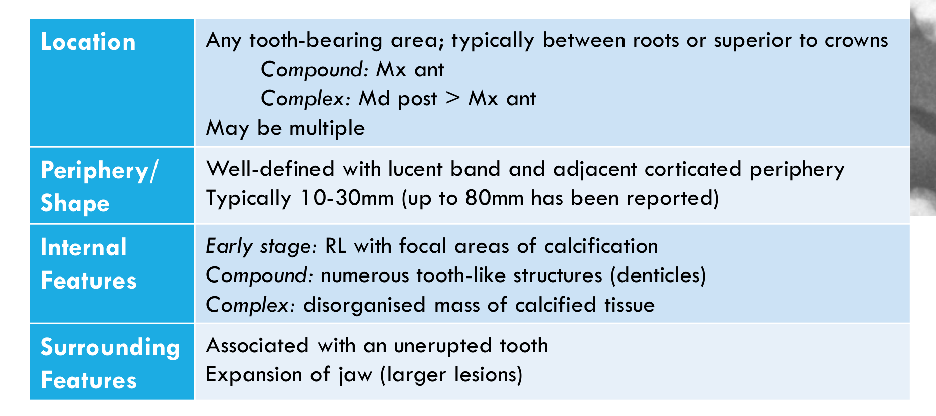
Location37
- Any tooth-bearing area; typically between roots or superior to crowns
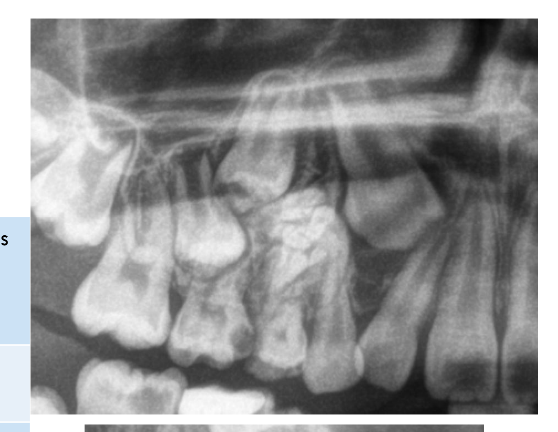
- Compound: maxillary anterior
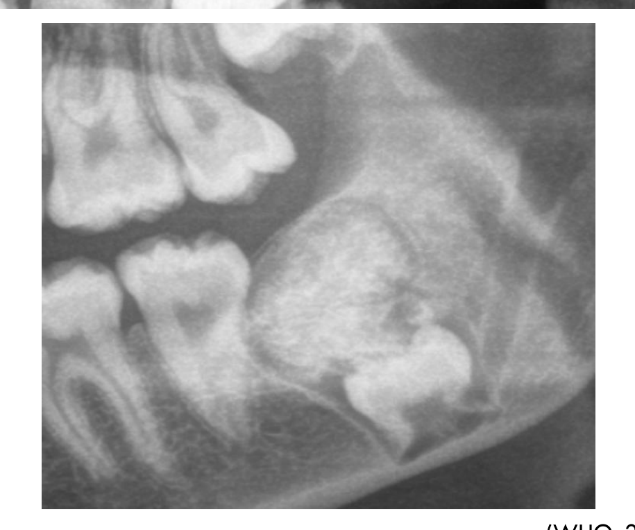
- Complex: mandibular posterior > maxillary anterior
- May be multiple
 |  |
 |
Periphery / Shape
- Well-defined with a lucent band and adjacent corticated periphery
- Typically 10–30 mm (up to 80 mm has been reported)
Internal Features
- Early stage: radiolucency with focal areas of calcification
- Compound: numerous tooth-like structures (denticles)
- Complex: disorganised mass of calcified tissue
Surrounding Features
- Associated with an unerupted tooth
- Expansion of the jaw (larger lesions)
Histopathology38
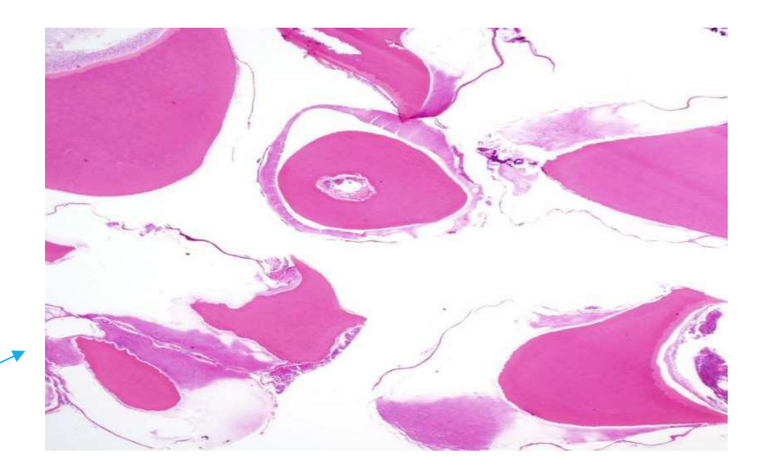
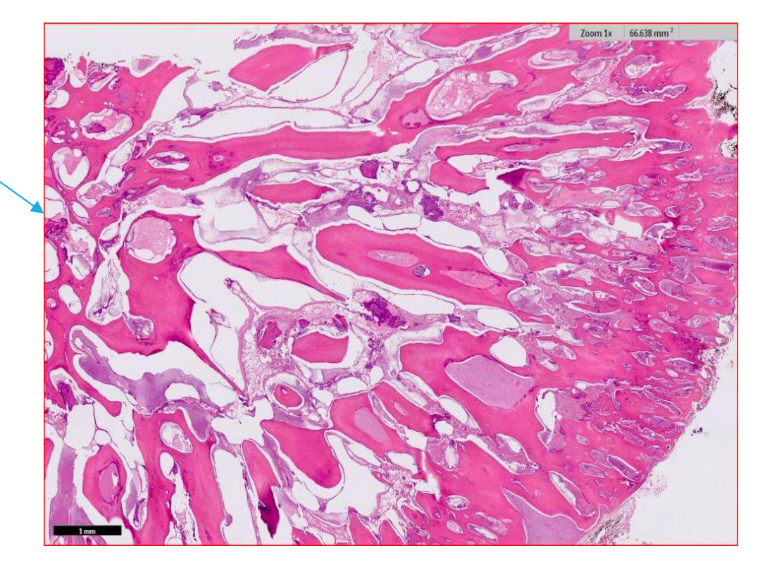
- Compound odontoma: contains multiple rudimentary teeth exhibiting dentin and enamel matrix. Dentine in odontomas usually shows some irregularity of tubule structure.
- Complex odontoma: consists of a disorganized mass of mature tubular dentine intermixed with rounded zones of enamel matrix where enamel has been lost on decalcification, with areas of dental pulp and cementum.
 |  |
Management
Local surgical removal.
Prognosis / Recurrence
Do not recur (unless removal is incomplete).
WHO, 2022
Ameloblastic Fibroma
Definition39
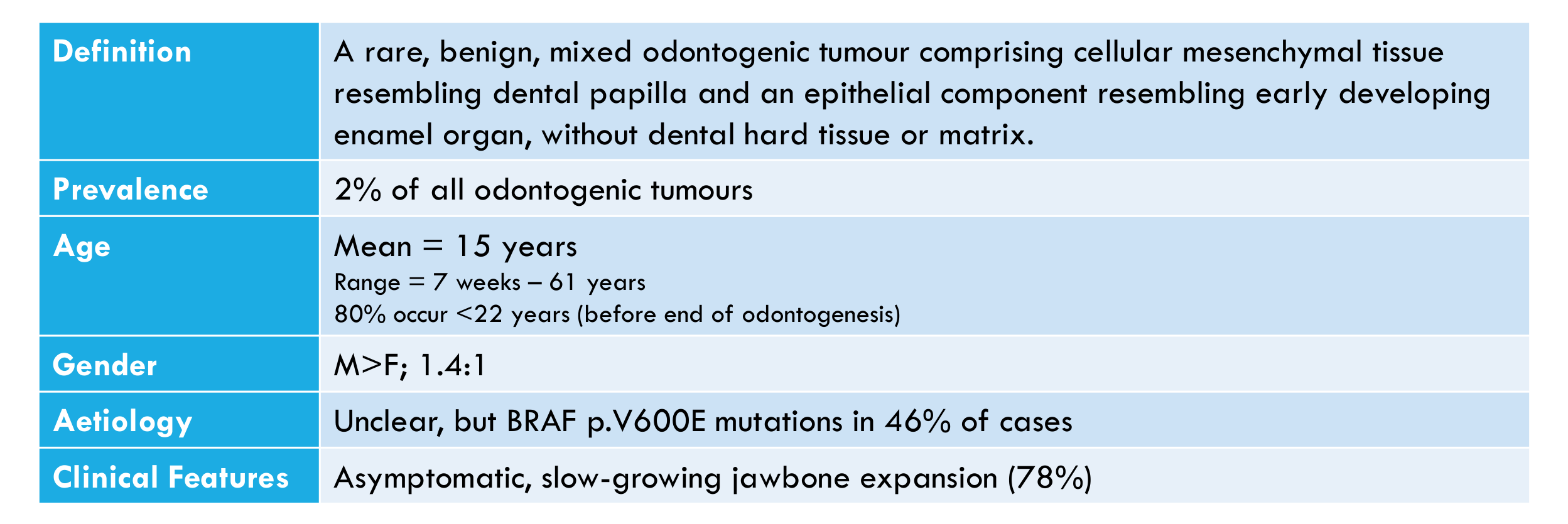
A rare, benign, mixed odontogenic tumour comprising cellular mesenchymal tissue resembling dental papilla and an epithelial component resembling early developing enamel organ, without dental hard tissue or matrix.
Clinical Profile
- Prevalence: 2% of all odontogenic tumours
- Age:
- Mean = 15 years
- Range = 7 weeks – 61 years
- 80% occur <22 years (before end of odontogenesis)
- Gender: M>F; 1.4:1
- Aetiology: Unclear, but BRAF p.V600E mutations in 46% of cases
- Clinical features: Asymptomatic, slow-growing jawbone expansion (78%)
Location40
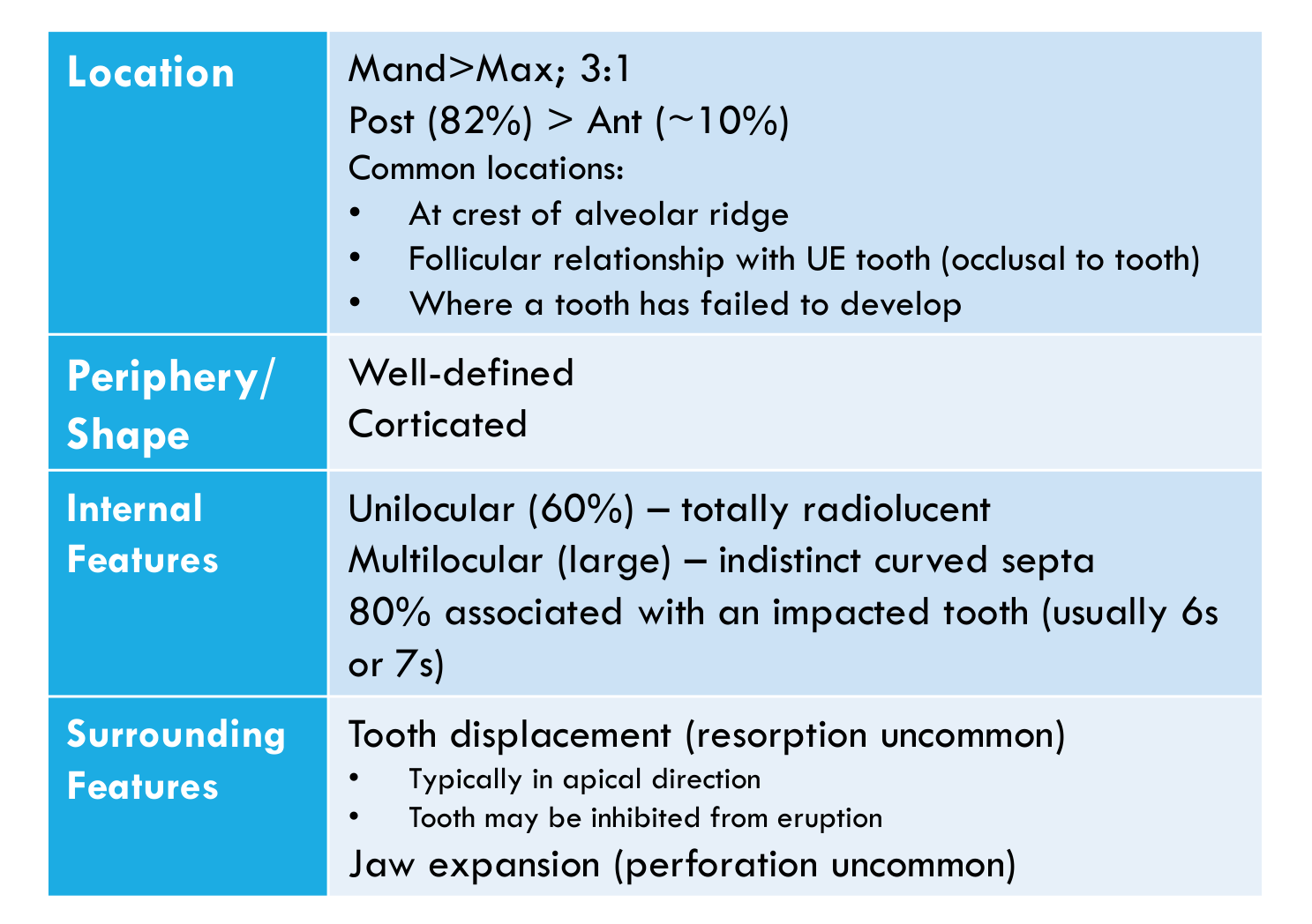
- Mand>Max; 3:1
- Posterior (82%) > Anterior (~10%)
- Common locations:
- At crest of alveolar ridge
- Follicular relationship with unerupted tooth (occlusal to tooth)
- Where a tooth has failed to develop
 | 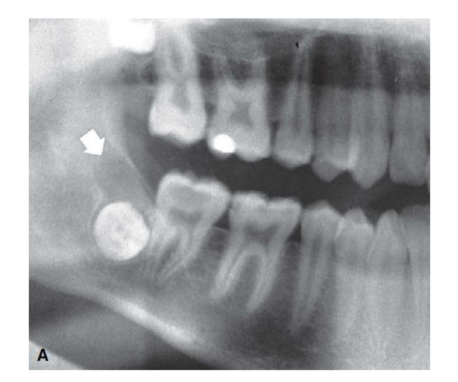 |
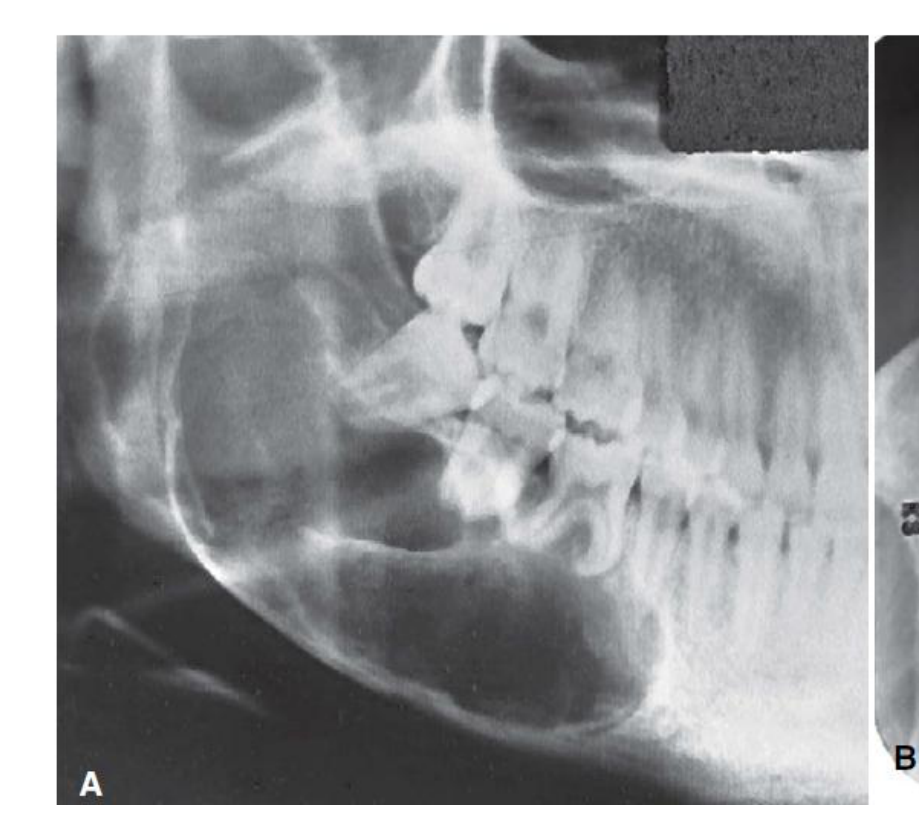 | 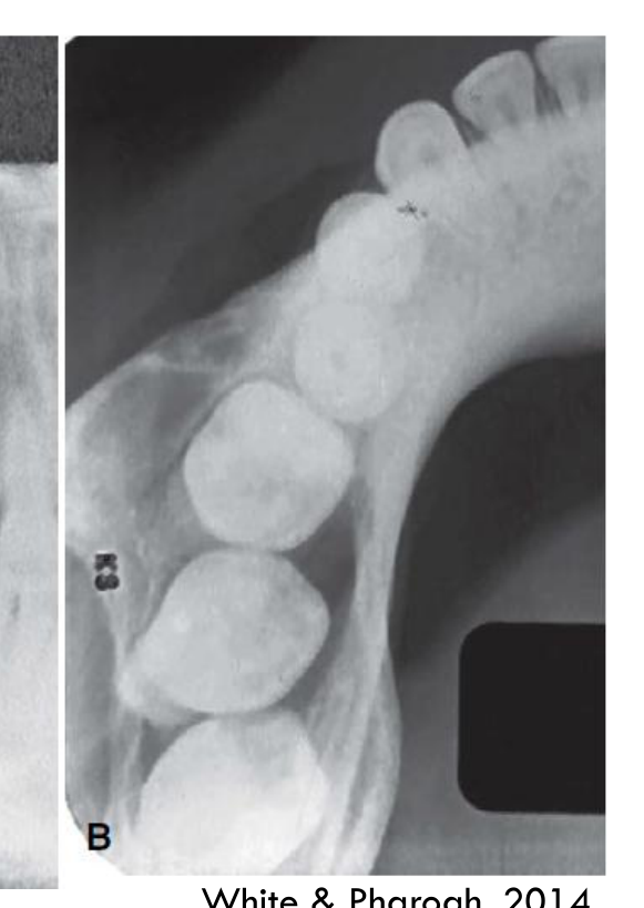 |
Radiographic Features
- Periphery/Shape: Well-defined; corticated
- Internal features:
- Unilocular (60%) – totally radiolucent
- Multilocular (large) – indistinct curved septa
- 80% associated with an impacted tooth (usually 6s or 7s)
- Surrounding features:
- Tooth displacement (resorption uncommon)
- Typically in apical direction
- Tooth may be inhibited from eruption
- Jaw expansion (perforation uncommon)
- Tooth displacement (resorption uncommon)
Lecturer — Cortical Perforation
Cortical perforation is relatively uncommon because the slow growth of ameloblastic fibroma permits periosteal new bone formation.
White & Pharoah, 2014
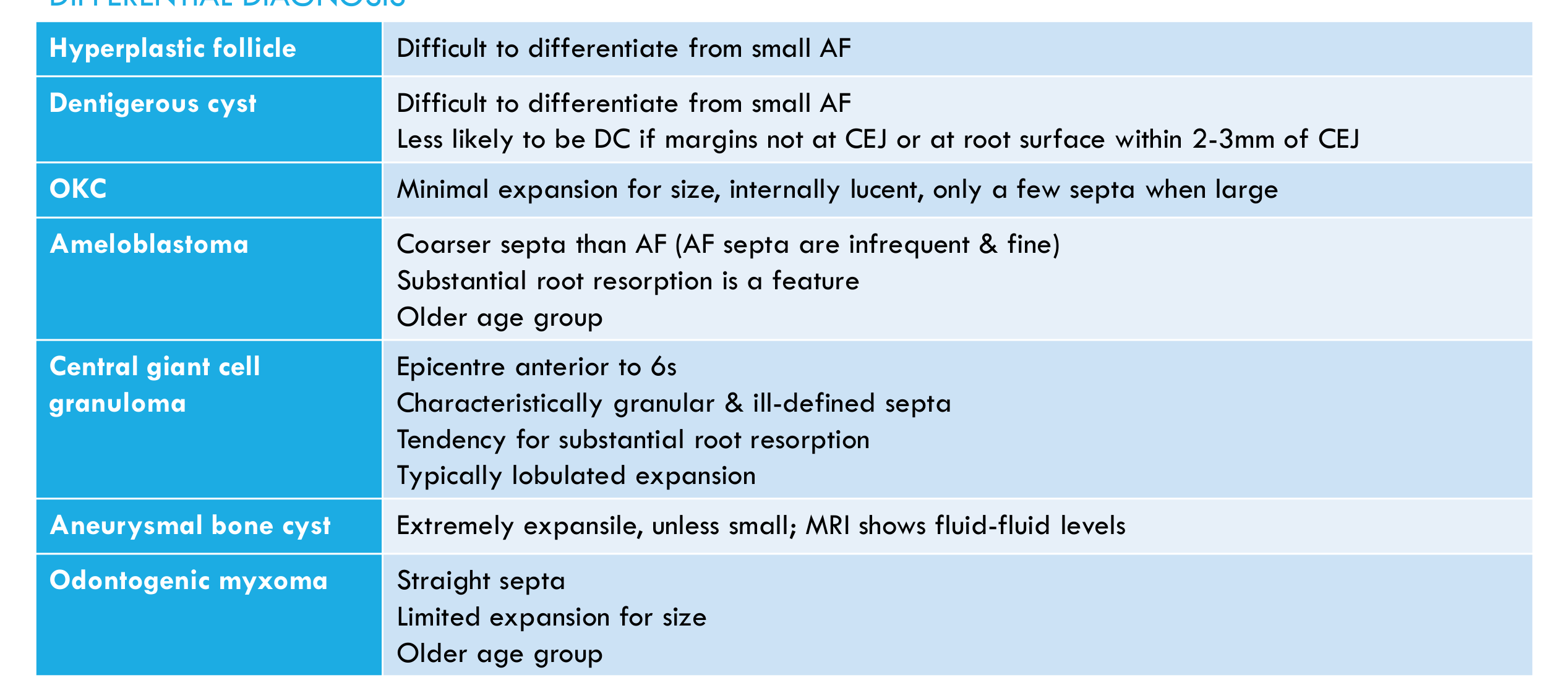
Differential Diagnosis41
- Hyperplastic follicle — Difficult to differentiate from small AF
- Dentigerous cyst — Difficult to differentiate from small AF; less likely to be DC if margins not at CEJ or at root surface within 2-3mm of CEJ
- OKC — Minimal expansion for size, internally lucent, only a few septa when large
- Ameloblastoma —
- Coarser septa than AF (AF septa are infrequent & fine)
- Substantial root resorption is a feature
- Older age group
- Central giant cell granuloma —
- Epicentre anterior to 6s
- Characteristically granular & ill-defined septa
- Tendency for substantial root resorption
- Typically lobulated expansion
- Aneurysmal bone cyst — Extremely expansile, unless small; MRI shows fluid-fluid levels
- Odontogenic myxoma — Straight septa; limited expansion for size; older age group
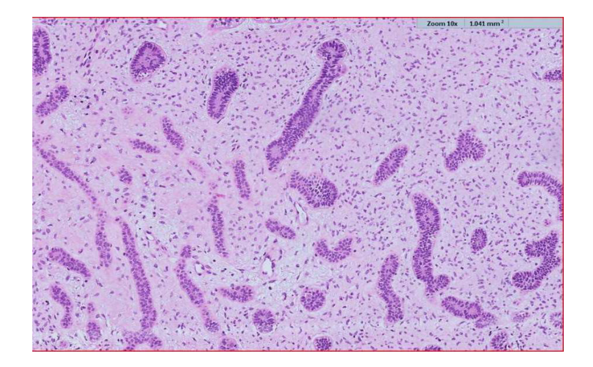
Histopathology42
- Mesenchymal component: myxoid and evenly hypercellular; resembles the dental papilla of the tooth bud
- Epithelial component: forms long narrow cords and strands of bilaminar cuboidal to columnar palisaded cells with occasional thickenings
- No dental hard tissue is normally present but extensive sampling may reveal small foci
Lecturer — Mesenchymal Component
The mesenchymal component was described as a myxoid, evenly hypocellular matrix resembling the dental papilla.
Management, Prognosis and Recurrence
- 19% recurrence with conservative removal
- Extensive, destructive and recurrent tumours should be treated radically
- Rare sarcomatous transformation
Ameloblastic Fibro-Odontoma
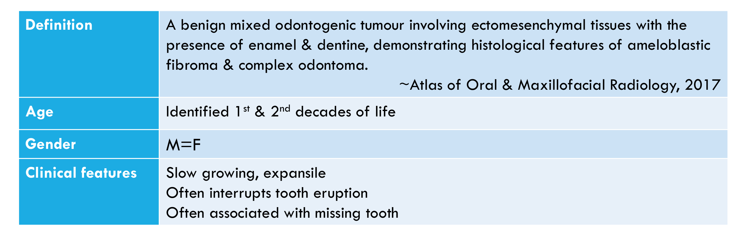
Definition43
A benign mixed odontogenic tumour involving ectomesenchymal tissues with the presence of enamel & dentine, demonstrating histological features of ameloblastic fibroma & complex odontoma.
Lecturer — AFO Classification
Ameloblastic fibro-odontoma is not officially recognised by the WHO as a separate lesion. Its status remains debated: it may represent an intermediate lesion, an ameloblastic fibroma containing hard tissue, or a separate entity. Its classification as a developing stage of an odontoma has been questioned because it may show BRAFpV600E mutation, locally aggressive behaviour and neoplastic recurrence.
~ Atlas of Oral & Maxillofacial Radiology, 2017
Clinical Presentation
- Age: Identified in the 1st & 2nd decades of life
- Gender: M = F
- Clinical features:
- Slow growing, expansile
- Often interrupts tooth eruption
- Often associated with missing tooth
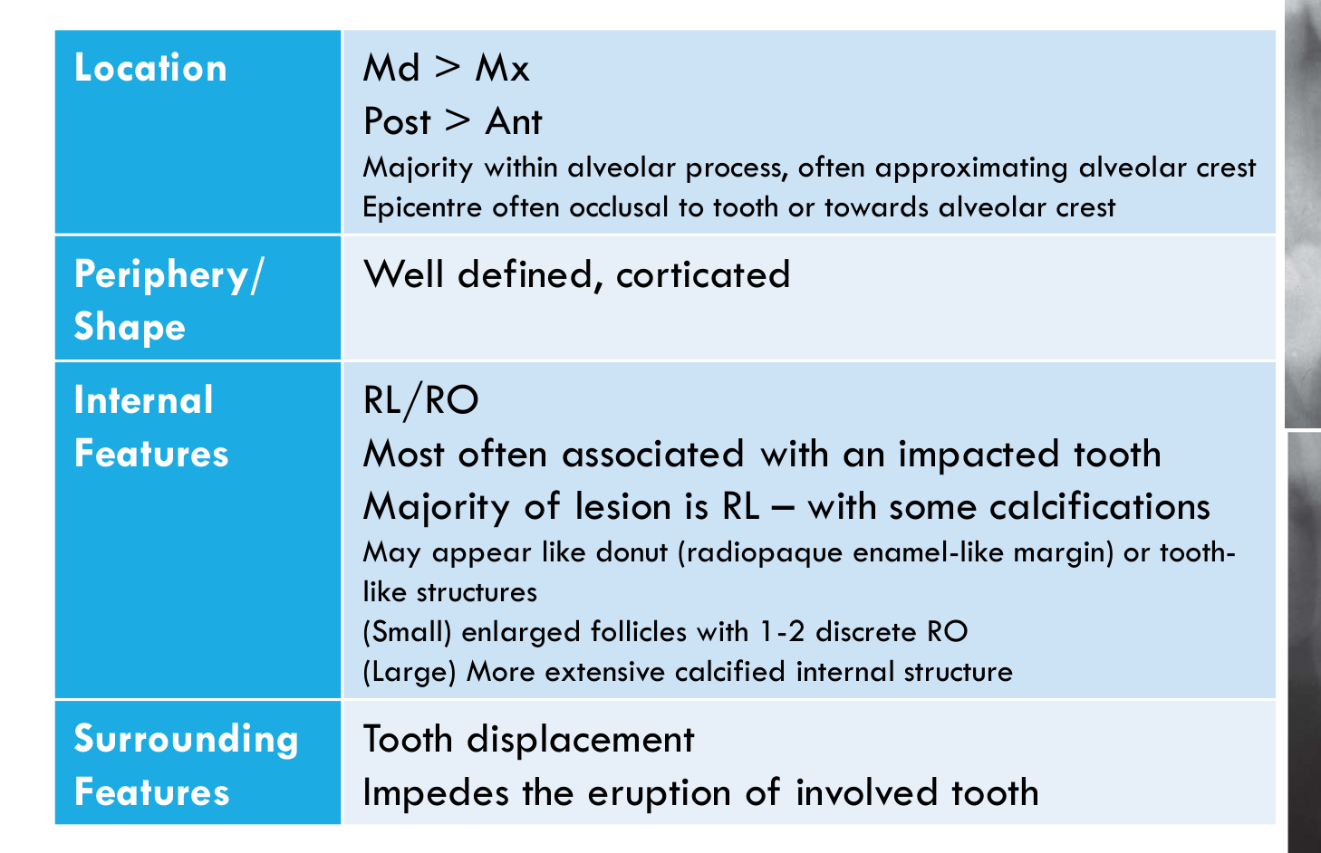
Radiographic Features44
- Location:
- Md > Mx
- Posterior > Anterior
- Majority within the alveolar process, often approximating the alveolar crest
- Epicentre often occlusal to the tooth or towards the alveolar crest
- Periphery/Shape:
- Well defined, corticated
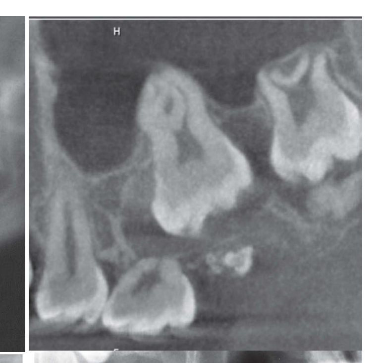
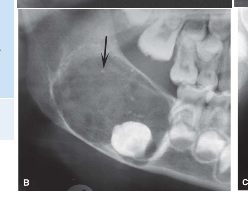
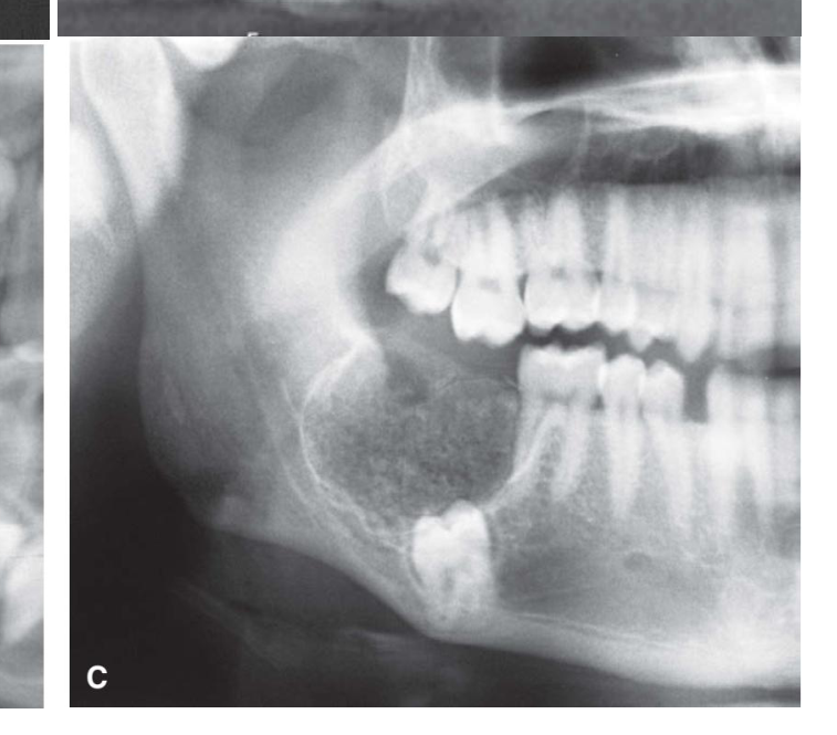
- Internal Features:
- RL/RO
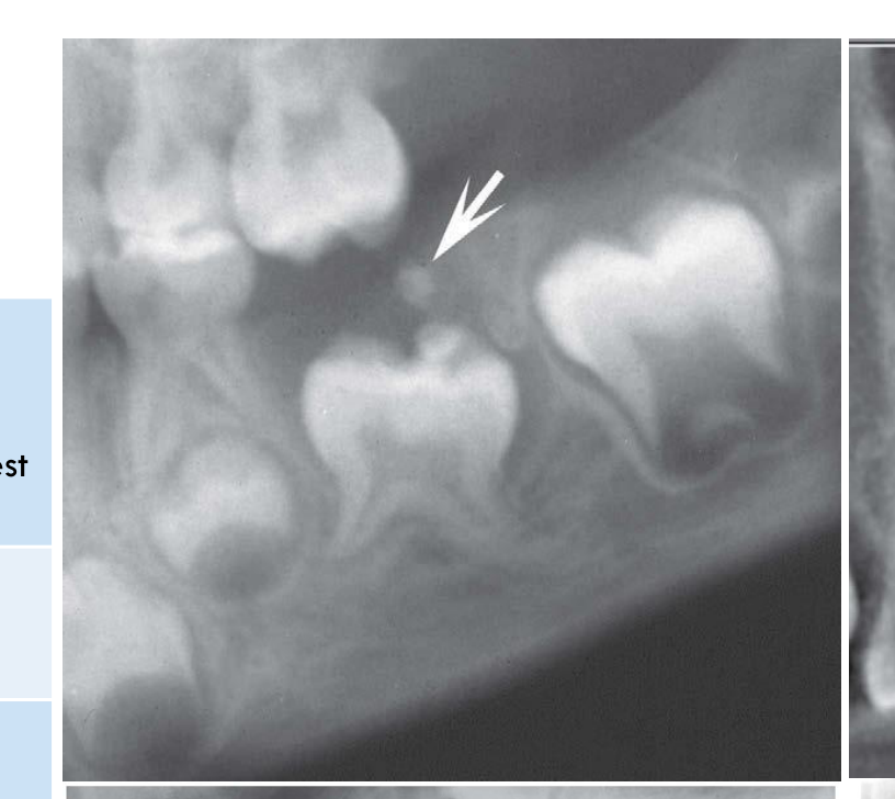
- Most often associated with an impacted tooth
- Majority of the lesion is RL — with some calcifications
- May appear like a donut (radiopaque enamel-like margin) or tooth-like structures
- (Small) enlarged follicles with 1–2 discrete RO
- (Large) more extensive calcified internal structure
- Surrounding Features:
- Tooth displacement
- Impedes the eruption of the involved tooth
 |  |  |
 |  |
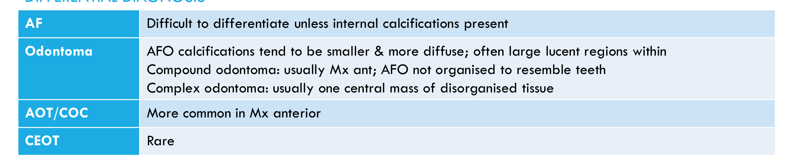
Differential Diagnosis45
- AF: Difficult to differentiate unless internal calcifications are present
- Odontoma:
- AFO calcifications tend to be smaller & more diffuse; often large lucent regions within
- Compound odontoma: usually Mx anterior; AFO is not organised to resemble teeth
- Complex odontoma: usually one central mass of disorganised tissue
- AOT/COC: More common in the Mx anterior
- CEOT: Rare
Histopathology
- Resembles AF & complex odontoma
Management/Prognosis/Recurrence
- Low recurrence with conservative excision & enucleation
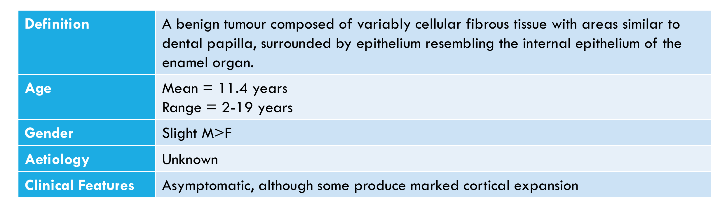
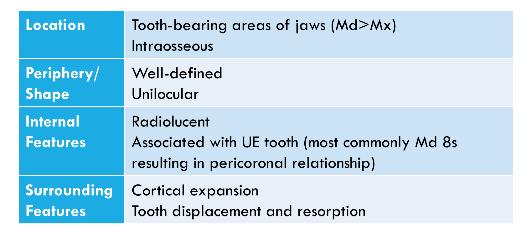
Primordial Odontogenic Tumour4647
| Feature | Details |
|---|---|
| Definition | A benign tumour composed of variably cellular fibrous tissue with areas similar to dental papilla, surrounded by epithelium resembling the internal epithelium of the enamel organ. |
| Age | Mean = 11.4 years; Range = 2–19 years |
| Gender | Slight M>F |
| Aetiology | Unknown |
| Clinical Features | Asymptomatic, although some produce marked cortical expansion |
| Feature | Details |
|---|---|
| Location | Tooth-bearing areas of jaws (Md>Mx); intraosseous |
| Periphery/Shape | Well-defined; unilocular |
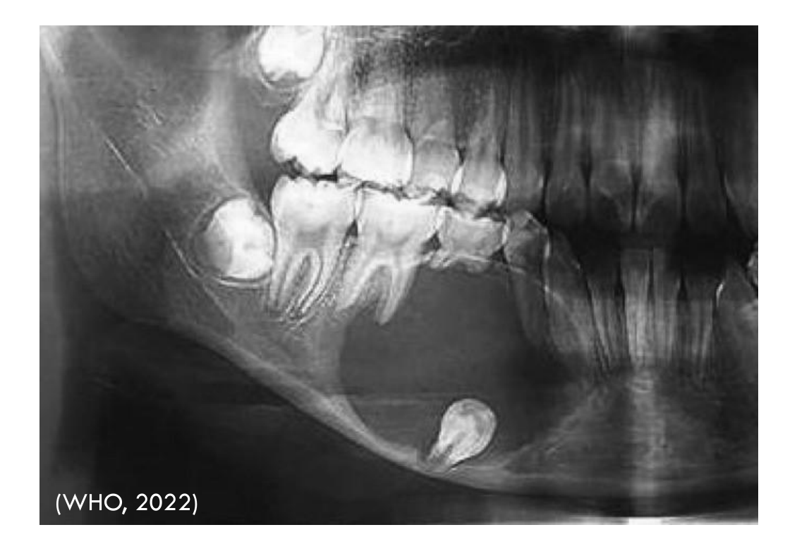
| Internal Features | Radiolucent; associated with UE tooth (most commonly Md 8s, resulting in a pericoronal relationship) |
| Surrounding Features | Cortical expansion; tooth displacement and resorption |
 |  |
 |
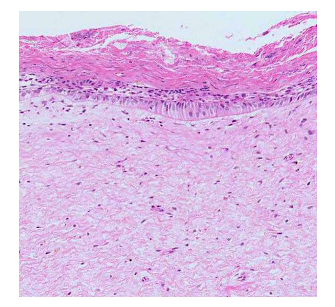
Histopathology48
- Loose or myxoid fibrous tissue with variable numbers of fusiform and stellate fibroblasts, with areas of cellular odontogenic mesenchyme
- Periphery is covered by a single layer of columnar or cuboidal epithelium enclosed by a thin fibrous capsule
Management
- Conservative excision
Prognosis/Recurrence
- Do not recur
Dentinogenic Ghost Cell Tumour
Definition49
A benign but locally infiltrative odontogenic tumour characterized by ameloblastoma-like sheets and islands of epithelium with prominent ghost cell keratinization and varying amounts of dentinoid in the stroma. Both the ghost cells and dentinoid may mineralise.
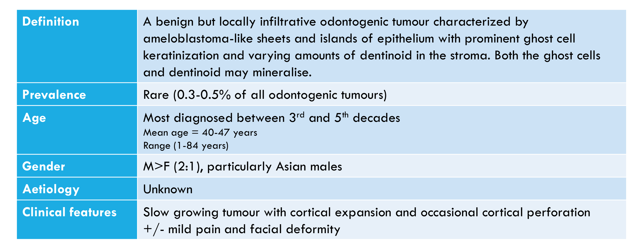
Prevalence
- Rare (0.3–0.5% of all odontogenic tumours)
Age
- Most diagnosed between 3rd and 5th decades
- Mean age = 40–47 years
- Range: 1–84 years
Gender
- M > F (2:1), particularly Asian males
Aetiology
- Unknown
Clinical Features
- Slow growing tumour with cortical expansion and occasional cortical perforation, +/- mild pain and facial deformity
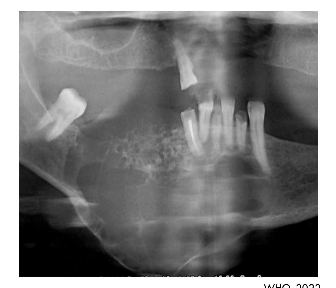
Location50
- Mandible = Maxilla
- Posterior > Anterior
- 25% are extraosseous (gingiva or alveolar ridge mucosa)
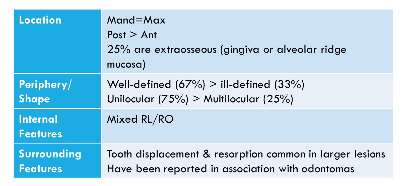 |  |
Periphery / Shape
- Well-defined (67%) > ill-defined (33%)
- Unilocular (75%) > multilocular (25%)
Internal Features
- Mixed RL/RO
Surrounding Features
- Tooth displacement and resorption common in larger lesions
- Have been reported in association with odontomas
WHO, 2022
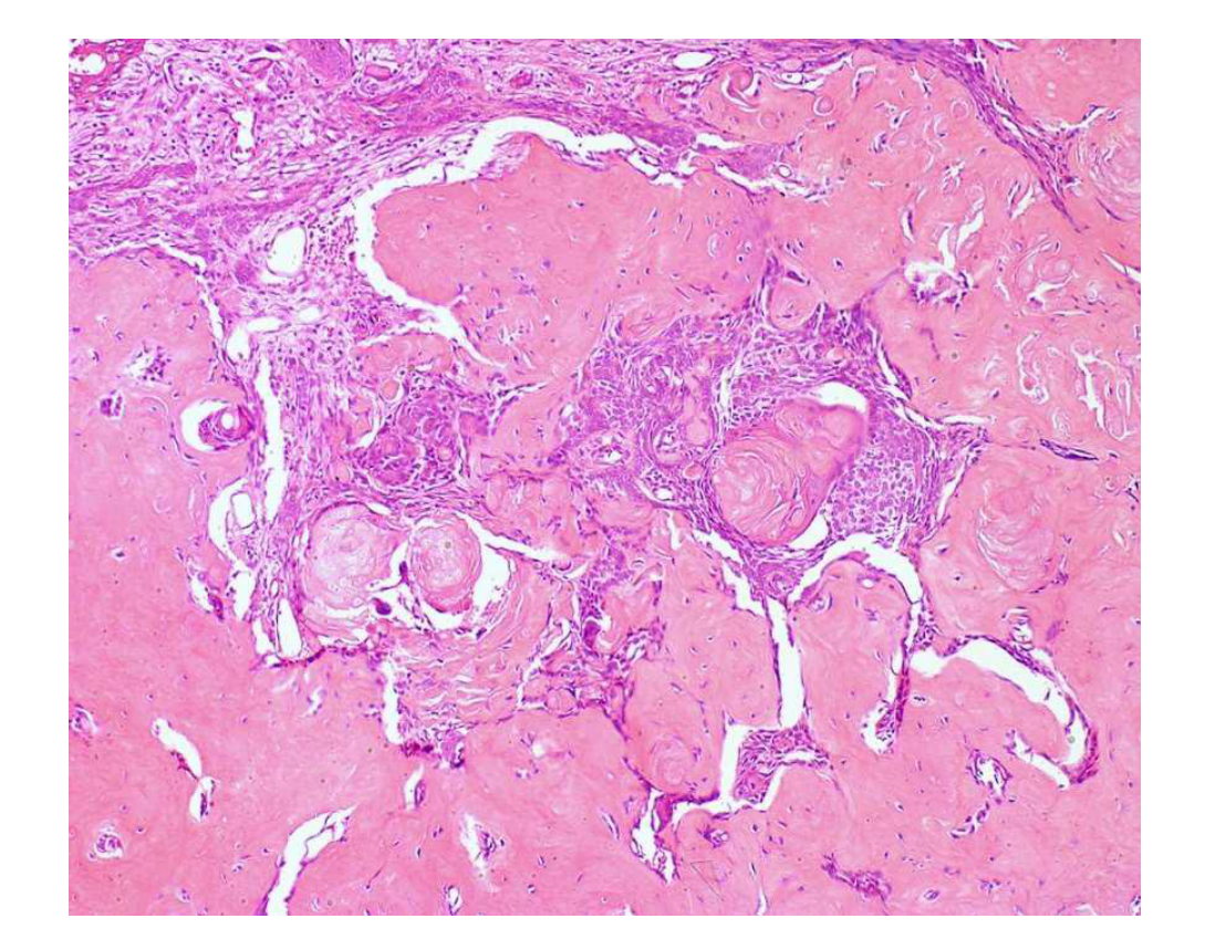
Histopathology51
- Nests, islands, and sheets of odontogenic epithelium resembling conventional ameloblastoma, with cuboidal to columnar hyperchromatic basal cells that have reverse nuclear polarity
- +/- cystic degeneration
- Characteristic ghost cells which may undergo mineralisation
Management / Prognosis / Recurrence
-
73% recurrence with conservative surgery
-
33% recurrence with radical surgery
-
Long term follow-up recommended
- Recurrence has been reported 1–20 years after treatment
-
Radical surgery may include marginal or segmental resection.
Part 3: Benign Mesenchymal Odontogenic Tumours52
- Odontogenic myxoma/myxofibroma
- Odontogenic fibroma
- Cementoblastoma
- Cemento-ossifying fibroma
Odontogenic Myxoma
Definition53
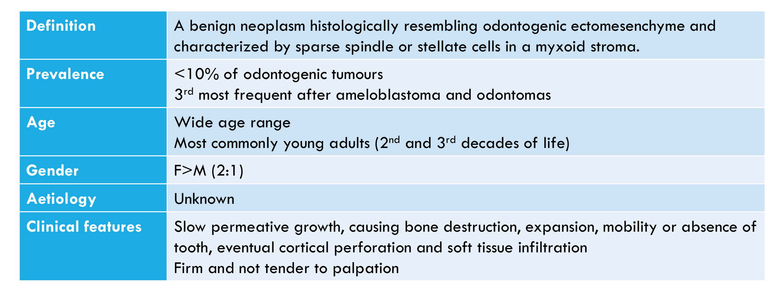
A benign neoplasm histologically resembling odontogenic ectomesenchyme and characterized by sparse spindle or stellate cells in a myxoid stroma.
Prevalence
- <10% of odontogenic tumours
- 3rd most frequent after ameloblastoma and odontomas
Age
- Wide age range
- Most commonly young adults (2nd and 3rd decades of life)
Gender
F > M (2:1)
Aetiology
Unknown
Clinical Features
- Slow permeative growth, causing bone destruction, expansion, mobility or absence of tooth, eventual cortical perforation and soft tissue infiltration
- Firm and not tender to palpation
Location54
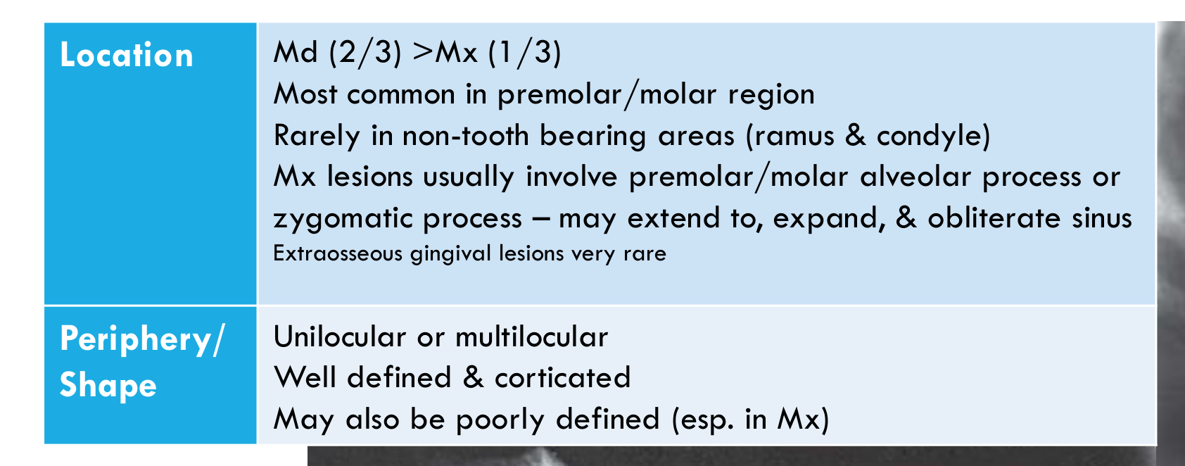
- Md (2/3) > Mx (1/3)
- Most common in premolar/molar region
- Rarely in non-tooth bearing areas (ramus & condyle)
- Mx lesions usually involve premolar/molar alveolar process or zygomatic process — may extend to, expand, & obliterate sinus
- Extraosseous gingival lesions very rare
 |  |
 |
Periphery / Shape
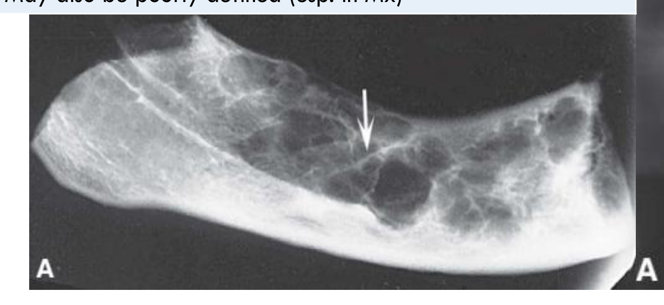
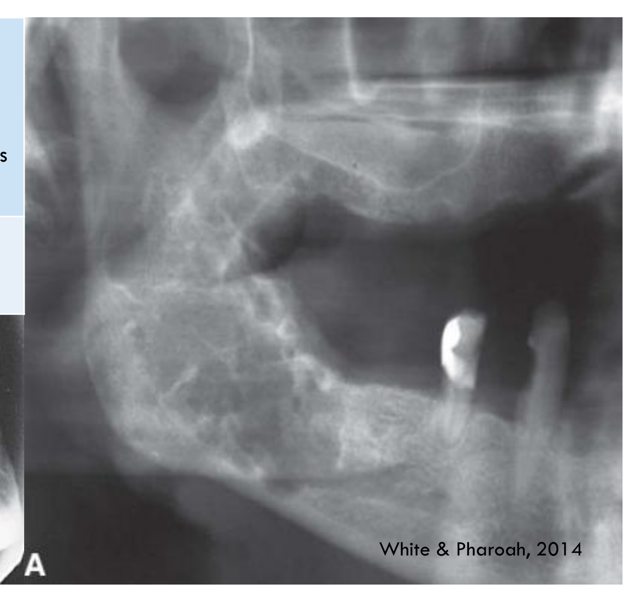
- Unilocular or multilocular
- Well defined & corticated
- May also be poorly defined (esp. in Mx)
White & Pharoah, 2014
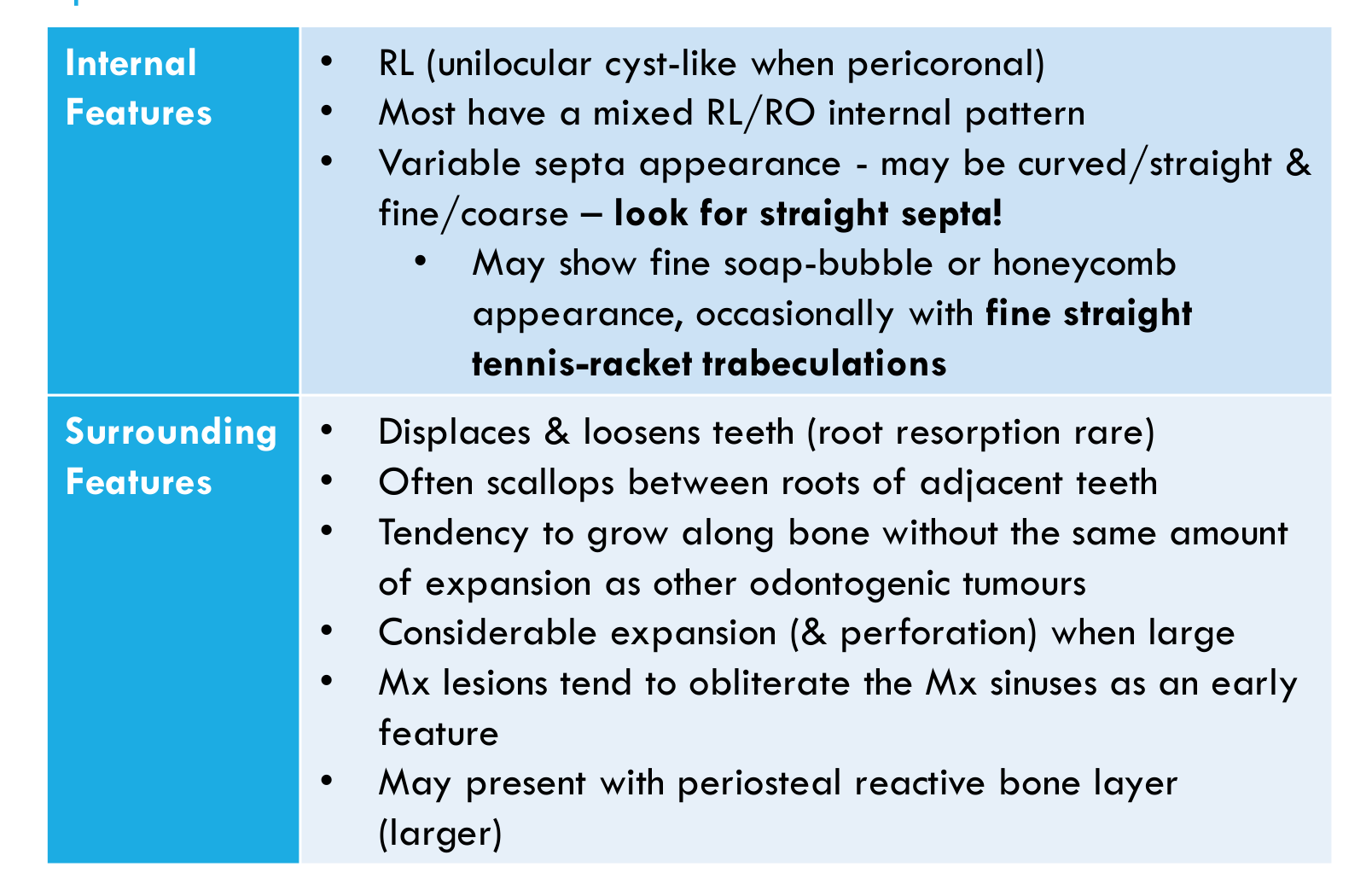
Internal Features55
-
RL (unilocular cyst-like when pericoronal)
-
Most have a mixed RL/RO internal pattern
-
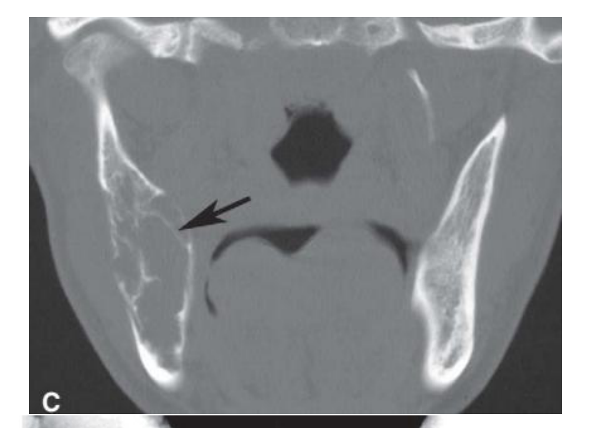
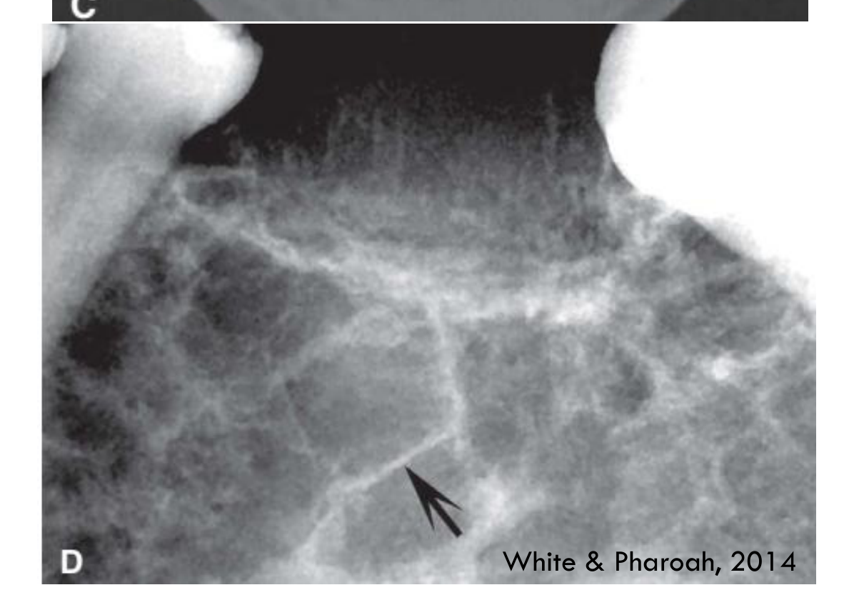
Variable septa appearance — may be curved/straight & fine/coarse — look for straight septa!
-
May show fine soap-bubble or honeycomb appearance, occasionally with fine straight tennis-racket trabeculations
-
At least one or two straight septa are usually present, which helps distinguish odontogenic myxoma from ameloblastoma.
 |  |
 |
Surrounding Features
- Displaces & loosens teeth (root resorption rare)
- Often scallops between roots of adjacent teeth
- Tendency to grow along bone without the same amount of expansion as other odontogenic tumours
- Considerable expansion (& perforation) when large
- Mx lesions tend to obliterate the Mx sinuses as an early feature
- May present with periosteal reactive bone layer (larger)
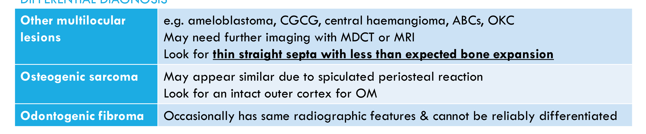
Other Multilocular Lesions56
- e.g. ameloblastoma, CGCG, central haemangioma, ABCs, OKC
- May need further imaging with MDCT or MRI
- Look for thin straight septa with less than expected bone expansion
Osteogenic Sarcoma
- May appear similar due to spiculated periosteal reaction
- Look for an intact outer cortex for OM
Lecturer — Myxoma Histopathology
Histopathological correlation is required when odontogenic fibroma cannot be distinguished radiographically from odontogenic myxoma.
Odontogenic Fibroma5758
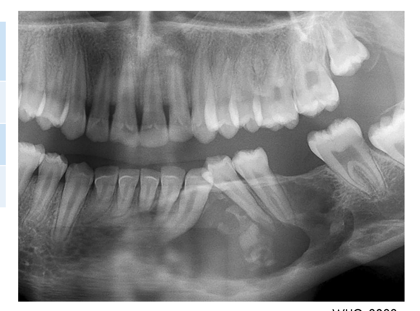
- Occasionally has same radiographic features & cannot be reliably differentiated
 |  |
 |
Histopathology59
- Predominantly loose myxoid stroma with a sparse population of stellate to spindle cells
- Small inactive rests of odontogenic epithelium are present in a small minority and are few in number
- Unencapsulated with permeative spread in medullary bone
- Appears histologically similar to the dental follicle or developing dental papilla (but clinical and radiologic features are different)
Management / Prognosis / Recurrence
- Lack of capsule and permeative spread account for recurrence rates = 10 to 43%
- It is unclear whether recurrence increases with more conservative treatment methods
- Maxillary lesions are more likely to recur than mandibular lesions
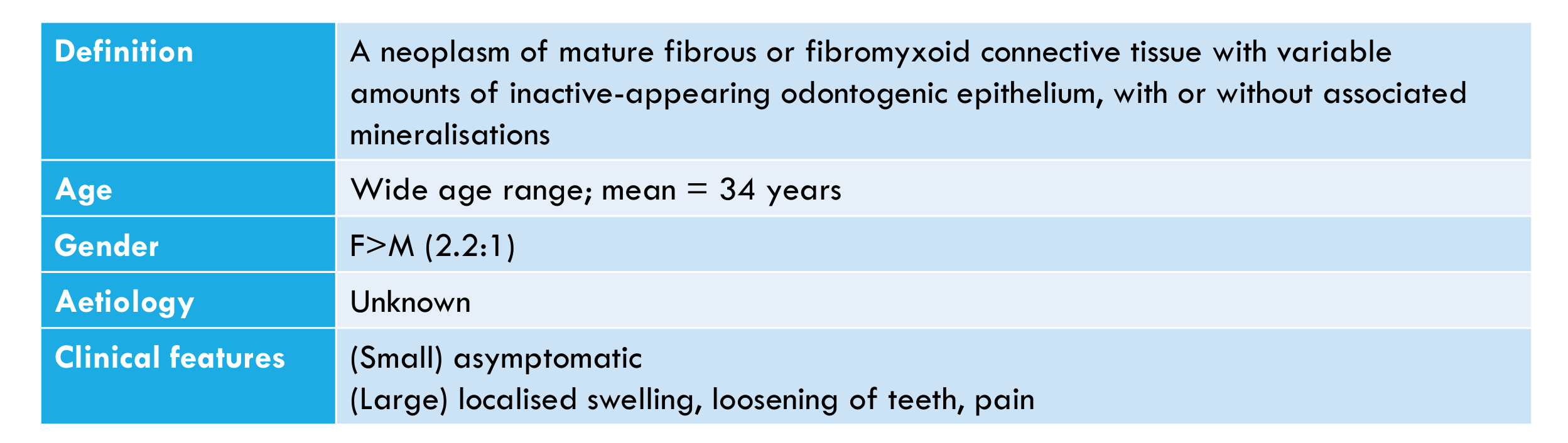
Odontogenic Fibroma
| Feature | Description |
|---|---|
| Definition | A neoplasm of mature fibrous or fibromyxoid connective tissue with variable amounts of inactive-appearing odontogenic epithelium, with or without associated mineralisations |
| Age | Wide age range; mean = 34 years |
| Gender | F>M (2.2:1) |
| Aetiology | Unknown |
| Clinical features | (Small) asymptomatic; (Large) localised swelling, loosening of teeth, pain |
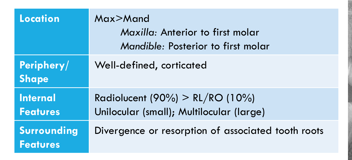
| Feature | Description |
|---|---|
| Location | Max>Mand; Maxilla: anterior to first molar; Mandible: posterior to first molar |
| Periphery/Shape | Well-defined, corticated |
| Internal Features | Radiolucent (90%) > RL/RO (10%); unilocular (small); multilocular (large) |
| Surrounding Features | Divergence or resorption of associated tooth roots |
WHO, 2022
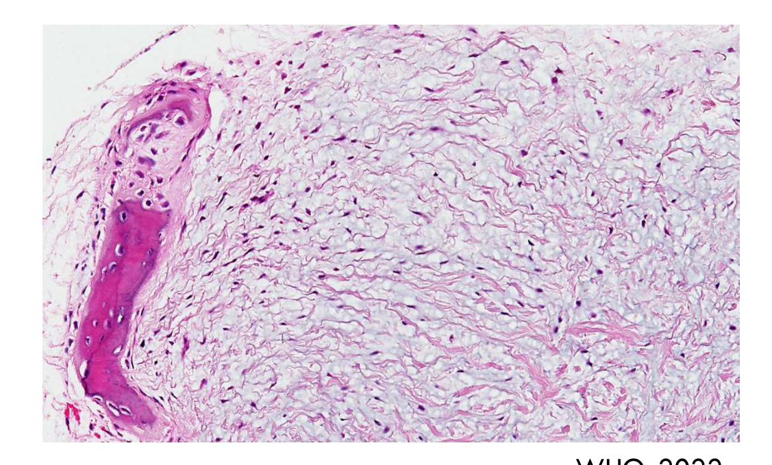
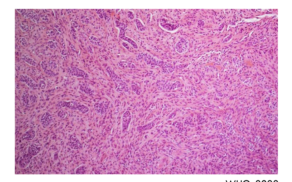
Histopathology60
- Moderately cellular bland fibrous tissue with moderate to dense collagen content, accompanied by varying amounts of dispersed inactive-appearing odontogenic epithelial nests and cords
- +/- minor hard tissue formation
Management/Prognosis/Recurrence
- Almost never recur after enucleation and curettage
Cementoblastoma
Definition61
A benign odontogenic neoplasm that forms a rounded mass of cementum on the root of a tooth.
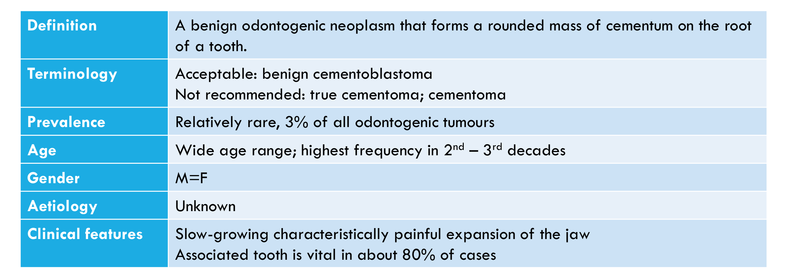
Terminology
- Acceptable: benign cementoblastoma
- Not recommended: true cementoma; cementoma
Prevalence
Relatively rare, 3% of all odontogenic tumours.
Age
Wide age range; highest frequency in 2nd – 3rd decades.
Gender
M = F
Aetiology
Unknown.
Clinical Features
- Slow-growing characteristically painful expansion of the jaw
- Associated tooth is vital in about 80% of cases
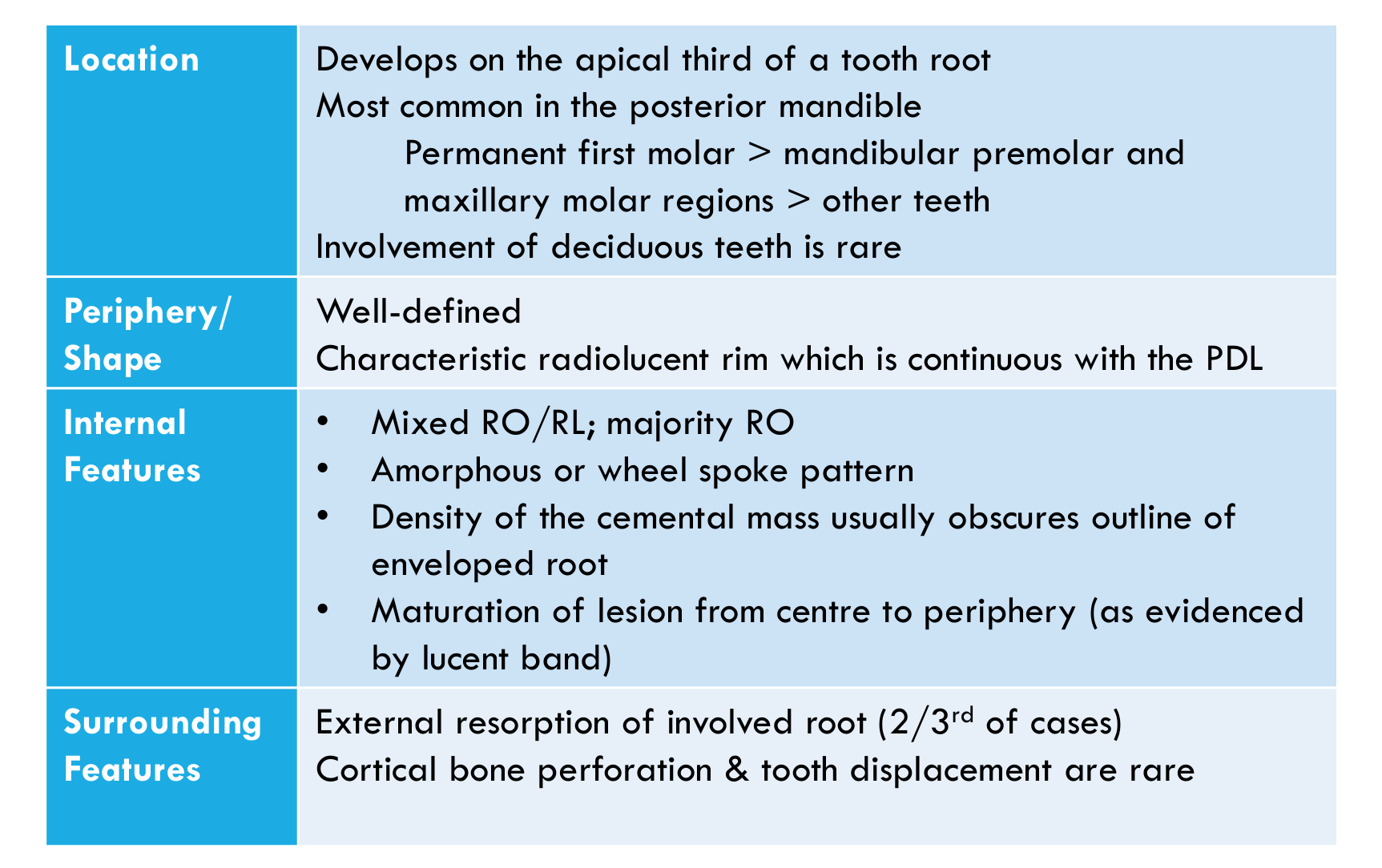
Location62
- Develops on the apical third of a tooth root
- Most common in the posterior mandible
- Permanent first molar > mandibular premolar and maxillary molar regions > other teeth
- Involvement of deciduous teeth is rare
 |  |
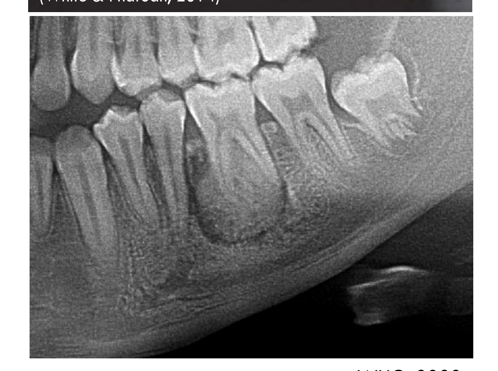 |
Periphery / Shape
- Well-defined
- Characteristic radiolucent rim which is continuous with the PDL
Internal Features
- Mixed RO/RL; majority RO
- Amorphous or wheel spoke pattern
- Density of the cemental mass usually obscures outline of enveloped root
- Maturation of lesion from centre to periphery (as evidenced by lucent band)
Surrounding Features
- External resorption of involved root (2/3rd of cases)
- Cortical bone perforation & tooth displacement are rare
WHO, 2022
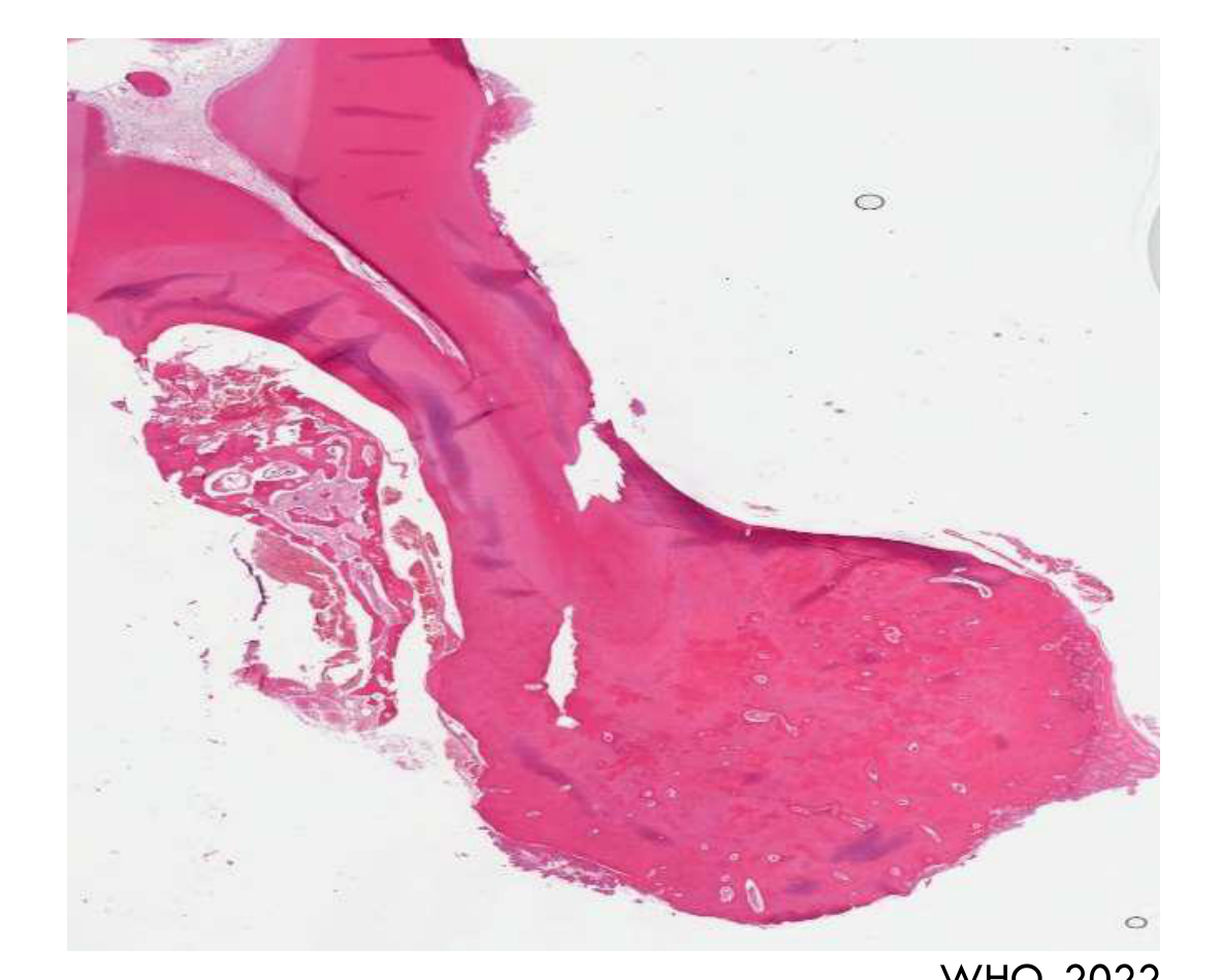
Histopathology63
-
A mass of dense cellular cementum resembling bone, often with prominent reversal lines, fused with the resorbed surface of the tooth root.
-
At the periphery there are radiating finger-like trabeculae of newly formed matrix, often associated with plump cementoblasts and cementoclasts and vascular immature fibrous tissue.
-
The periphery is least mature because growth continues there.
Management / Prognosis / Recurrence
-
Does not usually recur after extraction of the associated tooth and curettage
-
Recurrence usually results from incomplete removal or attempted conservation of the tooth. WHO, 2022
Cemento-Ossifying Fibroma
Definition64
A benign odontogenic fibro-osseous neoplasm arising in the jaws and characterised by production of bone and cementum-like calcifications in a fibrous stroma.
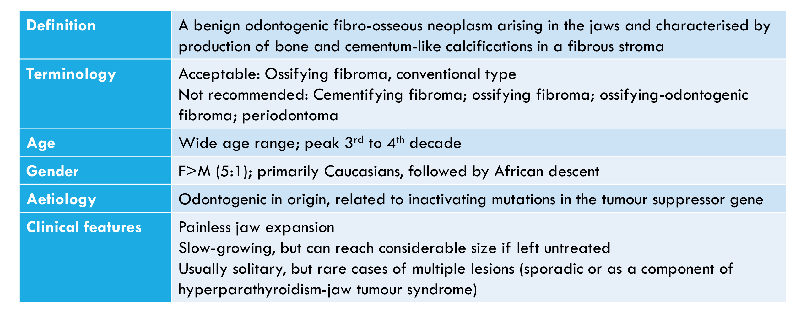
Terminology
- Acceptable: Ossifying fibroma, conventional type
- Not recommended: Cementifying fibroma; ossifying fibroma; ossifying-odontogenic fibroma; periodontoma
Age
Wide age range; peak in the 3rd to 4th decade.
Gender
F > M (5:1); primarily Caucasians, followed by African descent.
Aetiology
Odontogenic in origin, related to inactivating mutations in the tumour suppressor gene.
Clinical Features
-
Painless jaw expansion
-
Slow-growing, but can reach considerable size if left untreated
-
Usually solitary, but rare cases of multiple lesions (sporadic or as a component of hyperparathyroidism-jaw tumour syndrome)
-
Hyperparathyroidism-jaw tumour syndrome is a rare autosomal dominant disorder characterised by parathyroid adenomas or carcinomas, bilateral kidney cysts, hamartomas or Wilms tumours.
Location65
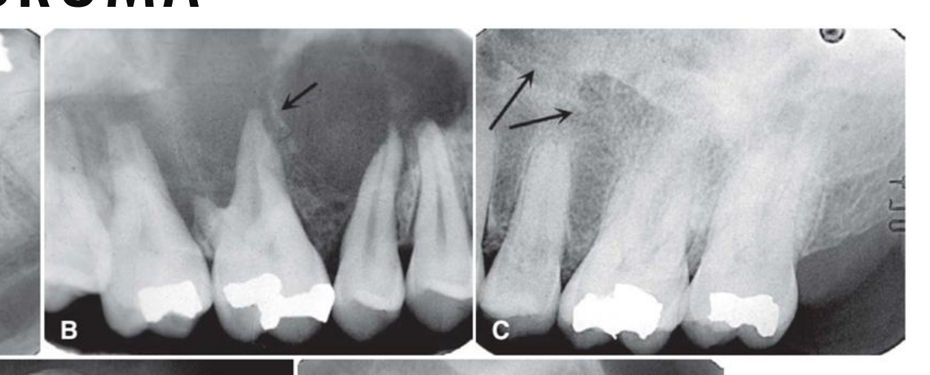
Mandible > Maxilla (particularly premolar-molar region).
 |  |  |
 |  |
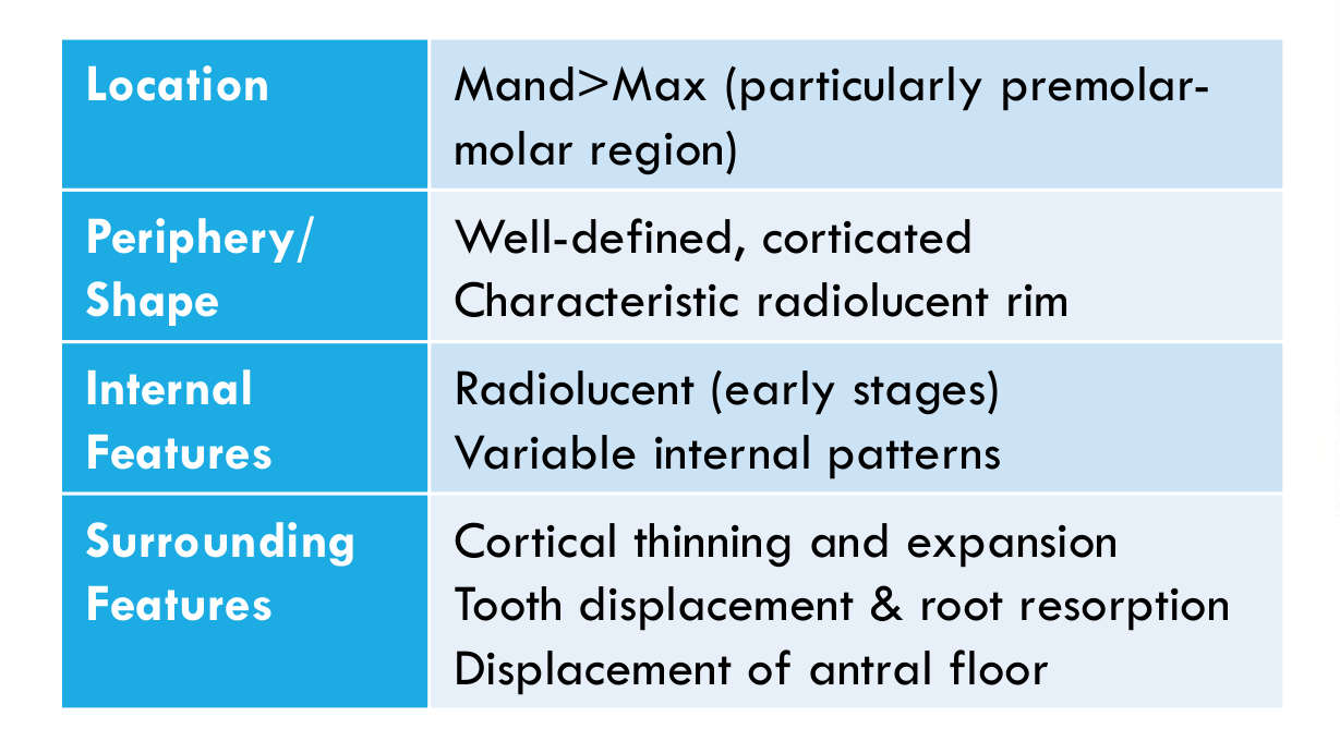
Periphery / Shape
- Well-defined, corticated
- Characteristic radiolucent rim
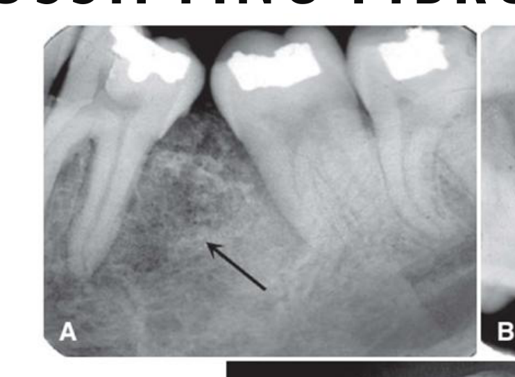
Internal Features
- Radiolucent (early stages)
- Variable internal patterns
Surrounding Features
- Cortical thinning and expansion
- Tooth displacement & root resorption
- Displacement of antral floor
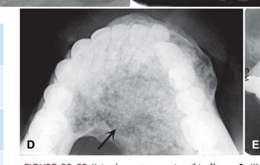
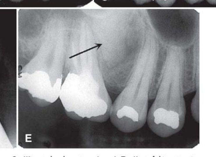
Figure 22-52: Various Bone Patterns Seen in Ossifying Fibroma
- A: Wispy trabecular pattern (arrow)
- B: Most of this pattern is radiolucent with a few wispy trabeculae (arrow)
- C: Fibrous dysplasia granular-like pattern (arrows)
- D: Flocculent pattern with larger tufts of bone formation (arrow)
- E: Solid, radiopaque, cementum-like pattern (arrow)
White & Pharoah, 2014
Histopathology66
- Variable proportions of fibrous and mineralised tissue, more heavily mineralised centrally and with a thin fibrous capsule or well demarcated margin from surrounding normal bone
- A fibro-osseous appearance of condensation of woven bone from stroma must be present at least focally
- Osteoblastic rimming is frequent and there is bone remodelling in the lesion with infrequent osteoclasts
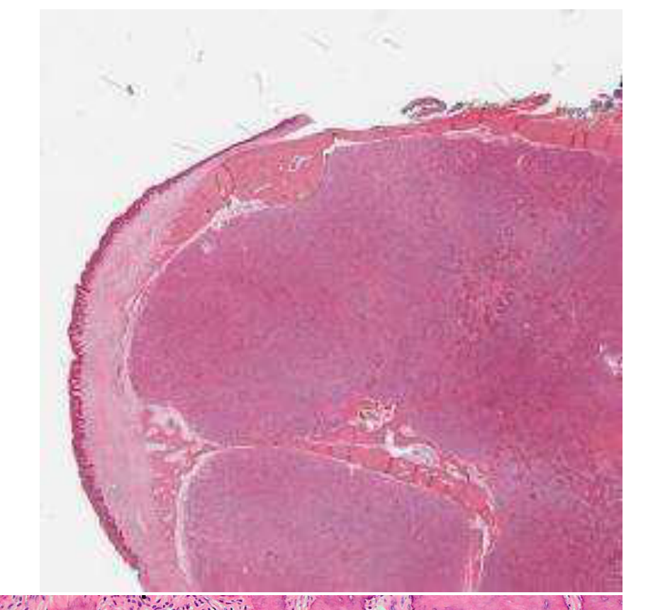 | 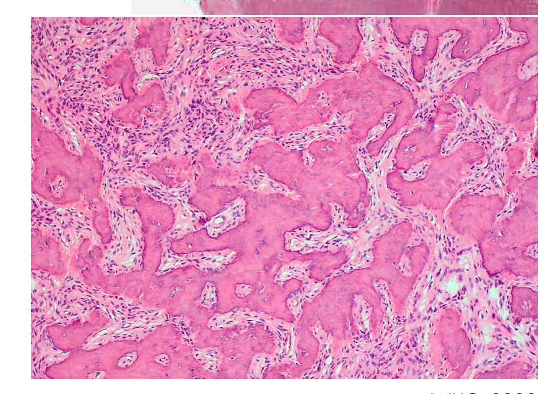 |
Management / Prognosis / Recurrence
- Rarely recurs with enucleation and curettage
WHO, 2022
References67
WHO Classification of Tumours Editorial Board. Head and neck tumours. Lyon (France): International Agency for Research on Cancer; 2022 (WHO classification of tumours series, 5th ed.; vol. 9).
Koong, B. (2017). Atlas of Oral and Maxillofacial Radiology. Chichester, UK: John Wiley and Sons.
White, and Pharoah. (2014). Oral radiology : Principles and interpretation (7th ed.). St. Louis: Elsevier/Mosby.
Audio Appendix
Additional Audio Content
The following sections from the lecture audio did not correspond to any heading in the main document.
Recommended Further Reading
The lecture recommended three references for further reading on benign tumours of the head and neck. Their specific titles were not stated in the transcript.
Clinical Features
- Benign tumours have specific anatomical predilections based on their tissue of origin.
- Odontogenic tumours arise from odontogenic epithelium and therefore occur in the alveolar processes, or tooth-bearing regions, of the jaws above the inferior alveolar canal.
- Cartilaginous tumours occur where residual cartilaginous cells are present, such as the mandibular condyle.
Clinical Cases
Case: Ameloblastoma with coarse curved septa
Question
Scenario: Radiograph of an ameloblastoma.
What’s shown: Coarse curved septa within the lesion.
Consider: What do these septa represent and what tumor are they characteristic of?
Answer
Observations:
- Coarse curved septa are visible within the lesion.
Reasoning: These septa represent residual bone trapped inside the tumor that has been remodeled into a curved shape by internal cystic structures. This appearance is highly characteristic of ameloblastomas.
Takeaway: Coarse curved septa in a multilocular radiolucency are a hallmark radiographic feature of ameloblastoma, representing remodeled residual bone.
Case: Ameloblastoma causing root resorption
Question
Scenario: Radiograph demonstrating the effects of an ameloblastoma on adjacent teeth.
What’s shown: Blunted appearance of tooth roots.
Consider: How does the root resorption caused by this benign tumor compare to that caused by cysts?
Answer
Observations:
- Tooth roots exhibit a blunted appearance due to resorption.
Reasoning: While both cysts and benign tumors can cause root resorption, benign tumors like ameloblastomas are much more likely to cause significant root resorption compared to cysts.
Takeaway: Benign odontogenic tumors are more likely to cause pronounced root resorption (blunting) than cystic lesions.
Case: Maxillary versus mandibular ameloblastoma borders
Question
Scenario: Radiographs comparing an ameloblastoma in the maxilla with those in the mandible.
What’s shown: Ill-defined borders in the maxillary lesion compared to well-defined borders in the mandibular lesions.
Consider: How does the anatomical location affect the radiographic visibility of the tumor’s borders?
Answer
Observations:
- The maxillary ameloblastoma has ill-defined borders.
- The mandibular ameloblastomas have well-defined, corticated borders.
Reasoning: Maxillary lesions are generally more ill-defined and harder to see the borders of compared to mandibular lesions, likely due to the anatomical structures and bone density differences in the maxilla.
Takeaway: Ameloblastomas in the maxilla tend to have ill-defined borders, making them harder to delineate radiographically than those in the mandible.
Case: Anterior versus posterior ameloblastoma locule size
Question
Scenario: Radiographs of ameloblastomas located in the anterior and posterior regions of the jaw.
What’s shown: Smaller locules in the anterior lesion and larger locules in the posterior lesion.
Consider: How does the size of the locules vary depending on the location of the ameloblastoma?
Answer
Observations:
- The anterior lesion exhibits smaller locules.
- The posterior lesion exhibits larger locules.
Reasoning: The internal multilocular appearance, specifically the size of the locules, tends to vary by location, with anterior lesions typically having smaller locules and posterior lesions having larger ones.
Takeaway: Locule size in ameloblastomas varies by location, being smaller in anterior lesions and larger in posterior lesions.
Case: Ameloblastoma with cortical perforation and tooth displacement
Question
Scenario: Radiograph of an ameloblastoma demonstrating mass effect.
What’s shown: Missing white cortical plate, blunted roots, and teeth angled in different directions.
Consider: What are the space-occupying effects demonstrated by this lesion on the surrounding structures?
Answer
Observations:
- Perforation of the cortex (the white cortical plate is no longer visible).
- Root resorption with blunting of the roots.
- Tooth displacement with teeth angled in different directions.
Reasoning: As a space-occupying lesion, the ameloblastoma exerts pressure, leading to cortical expansion and eventual perforation when periosteal new bone cannot keep up. It also displaces teeth and causes root resorption.
Takeaway: Ameloblastomas demonstrate classic mass effect features, including cortical perforation, root resorption, and significant tooth displacement.
Case: Unicystic ameloblastoma mimicking a radicular cyst
Question
Scenario: Radiograph of a lesion that clinically and radiographically resembled a radicular cyst, leading to endodontic treatment.
What’s shown: A unilocular radiolucency resembling a radicular cyst.
Consider: What was the actual histopathological diagnosis despite the initial clinical appearance and treatment?
Answer
Observations:
- The lesion appears radiographically identical to a radicular cyst.
- Endodontic treatment was initially carried out.
Reasoning: Unicystic ameloblastomas can occur in periapical locations and mimic radicular cysts both clinically and radiographically. Definitive diagnosis requires histopathological correlation, which in this case confirmed a unicystic ameloblastoma.
Takeaway: Unicystic ameloblastomas can mimic radicular cysts radiographically, necessitating histopathological examination for a definitive diagnosis.
Case: Unicystic ameloblastoma with pseudo-septa
Question
Scenario: Radiograph of a unicystic ameloblastoma.
What’s shown: Apparent septa within a unilocular lesion.
Consider: Are these true septa, and what do they actually represent?
Answer
Observations:
- The unilocular lesion appears to have internal septa.
Reasoning: Although it may look like there are true septa as seen in conventional multilocular ameloblastomas, these are not true septa. They are simply areas of residual bone within the unilocular cyst cavity.
Takeaway: Apparent septa in a unicystic ameloblastoma are not true septa but rather areas of residual bone.
Case: Adenomatoid odontogenic tumor surrounding a tooth
Question
Scenario: Radiograph of an adenomatoid odontogenic tumor (AOT) associated with an unerupted tooth.
What’s shown: A radiolucency surrounding the greater part of the tooth, including the root and part of the crown.
Consider: How does the attachment of this lesion to the tooth differ from a dentigerous cyst?
Answer
Observations:
- The lesion surrounds almost the entire root and part of the crown.
- It does not attach at the cementoenamel junction (CEJ).
Reasoning: Unlike a dentigerous cyst which typically attaches at the CEJ, an AOT tends to surround the greater part of the tooth, extending apically past the CEJ to encompass the root.
Takeaway: An AOT often surrounds the entire root and part of the crown without attaching at the CEJ, distinguishing it from a dentigerous cyst.
Case: Adenomatoid odontogenic tumor with corticated border and tooth displacement
Question
Scenario: Radiographs of adenomatoid odontogenic tumors.
What’s shown: Well-defined unilocular radiolucencies with a thin white corticated border and associated tooth displacement.
Consider: What are the characteristic border features and effects on surrounding structures for this tumor?
Answer
Observations:
- A thin white corticated or sclerotic border is present.
- The tumor causes displacement of the adjacent tooth.
Reasoning: AOTs typically present as well-defined, unilocular radiolucencies with a distinct corticated border. While they cause tooth displacement, root resorption is quite rare.
Takeaway: AOTs characteristically display a well-defined corticated border and cause tooth displacement, but rarely cause root resorption.
Case: Squamous odontogenic tumor on a 2D image
Question
Scenario: 2D radiograph of a squamous odontogenic tumor.
What’s shown: A triangular or semicircular unilocular radiolucency along tooth roots.
Consider: Why can’t cortical bone expansion and perforation be appreciated on this specific image?
Answer
Observations:
- A triangular/semicircular unilocular radiolucency is visible along the roots.
- Cortical expansion and perforation are not visible.
Reasoning: The image is a 2D radiograph, which lacks the cross-sectional detail needed to appreciate buccal or lingual cortical bone expansion and perforation.
Takeaway: 2D radiographs may fail to show cortical expansion and perforation in squamous odontogenic tumors; cross-sectional imaging is often needed to evaluate these features.
Case: Calcifying epithelial odontogenic tumor preventing tooth eruption
Question
Scenario: Radiograph of a mixed density lesion in the jaw.
What’s shown: A mixed radiolucent-radiopaque lesion preventing the eruption of a canine, with cortical expansion not appreciable on the 2D OPG.
Consider: What is the effect of this lesion on the adjacent tooth and cortex?
Answer
Observations:
- A mixed density lesion is present.
- The canine has not erupted due to the lesion.
- Cortical expansion is present but not visible on the 2D OPG.
Reasoning: Calcifying epithelial odontogenic tumors act as space-occupying lesions that can prevent tooth eruption. While they typically cause cortical expansion, this may not be appreciable on a 2D panoramic radiograph.
Takeaway: Calcifying epithelial odontogenic tumors can prevent tooth eruption and cause cortical expansion, though expansion may be missed on 2D imaging.
Case: Compound odontoma in the maxillary anterior region
Question
Scenario: Radiograph of a compound odontoma.
What’s shown: Distinct tooth-like structures (denticles) in the maxillary anterior region.
Consider: What is the typical location and radiographic appearance of this lesion?
Answer
Observations:
- Multiple distinct tooth-like structures (denticles) are visible.
- The lesion is located in the maxillary anterior region.
Reasoning: Compound odontomas are more common in the maxillary anterior region and radiographically present as multiple distinct, rudimentary tooth-like structures.
Takeaway: Compound odontomas typically occur in the maxillary anterior region and appear as multiple distinct tooth-like structures (denticles).
Case: Odontoma with a lucent band and corticated periphery
Question
Scenario: Radiograph of an odontoma.
What’s shown: Tooth-like structures surrounded by a lucent band and a corticated periphery.
Consider: What are the characteristic border features surrounding the calcified structures in this lesion?
Answer
Observations:
- A lucent band surrounds the tooth-like structures.
- A corticated periphery is present.
Reasoning: Odontomas typically present as well-defined mixed radiopaque structures surrounded by a narrow radiolucent band (representing the follicular capsule) and a corticated border.
Takeaway: Odontomas are characteristically surrounded by a radiolucent band and a corticated periphery.
Case: Compound odontoma histology
Question
Scenario: Histopathological image of a compound odontoma.
What’s shown: Individual denticles composed of dentine and enamel.
Consider: What dental hard tissues are visible in this histological specimen?
Answer
Observations:
- Individual denticles are visible.
- The structures contain dentine and enamel.
Reasoning: Histologically, compound odontomas contain multiple rudimentary teeth exhibiting dentine and enamel matrix, though the dentine may show irregularity of the tubular structure.
Takeaway: Histologically, compound odontomas consist of multiple rudimentary teeth (denticles) made of dentine and enamel.
Case: Ameloblastic fibroma with tooth displacement and jaw expansion
Question
Scenario: Radiographs (including an occlusal view) of an ameloblastic fibroma.
What’s shown: Posterior displacement of the root of tooth 47, and expansion of both buccal and lingual cortices.
Consider: What are the mass effect features demonstrated in this case?
Answer
Observations:
- The root of tooth 47 is displaced posteriorly.
- There is expansion of both the buccal and lingual cortices.
Reasoning: Ameloblastic fibromas demonstrate mass effect features such as tooth displacement and jaw expansion. Root resorption is less common, and cortical perforation is rare due to the slow growth allowing periosteal new bone formation.
Takeaway: Ameloblastic fibromas typically cause tooth displacement and cortical expansion, but rarely cause root resorption or cortical perforation.
Case: Ameloblastic fibro-odontoma with calcifications
Question
Scenario: Radiographs of ameloblastic fibro-odontomas.
What’s shown: Predominantly lucent internal components with areas of calcification, some resembling an outer enamel rim.
Consider: How do the calcifications in this mixed density lesion present?
Answer
Observations:
- The internal component is mostly lucent.
- There are areas of calcification, some with an outer enamel-like rim.
Reasoning: Ameloblastic fibro-odontomas are mixed density lesions. When small, they may have only one or two radiopacities, but as they enlarge, the calcified material becomes more extensive, sometimes forming tooth-like structures with an enamel rim.
Takeaway: Ameloblastic fibro-odontomas present as mixed density lesions with varying amounts of calcification that can become extensive and sometimes resemble tooth-like structures.
Case: Odontogenic myxoma with spiculated periosteal reaction
Question
Scenario: Radiograph of an odontogenic myxoma.
What’s shown: A periosteal reactive bone layer with a spiculated appearance.
Consider: What is causing this spiculated periosteal reaction?
Answer
Observations:
- A spiculated periosteal reactive bone layer is visible.
Reasoning: The spiculated appearance is a result of the reaction of the periosteum to the lesion. While it can mimic malignant tumors like osteogenic sarcoma, the presence of an intact outer cortex indicates a benign process like odontogenic myxoma.
Takeaway: Odontogenic myxomas can cause a spiculated periosteal reaction, but an intact outer cortex helps differentiate it from malignant bone-forming tumors.
Case: Odontogenic fibroma with panoramic artifact
Question
Scenario: OPG of an odontogenic fibroma.
What’s shown: An anterior border that appears ill-defined.
Consider: Why does the anterior border appear ill-defined on this specific radiograph?
Answer
Observations:
- The anterior border of the lesion appears ill-defined.
Reasoning: The ill-defined appearance is an artifact from the panoramic technique (OPG) rather than a true feature of the lesion, which typically has well-defined corticated margins.
Takeaway: Panoramic radiograph artifacts can create the illusion of ill-defined borders in lesions that are actually well-corticated.
Case: Cementoblastoma fused to a tooth root
Question
Scenario: Radiograph/histology of a cementoblastoma.
What’s shown: A cemental mass fused to a partially resorbed tooth root.
Consider: What is the relationship between the radiopaque mass and the tooth root?
Answer
Observations:
- A radiopaque cemental mass is fused to the tooth root.
- The tooth root is partially resorbed.
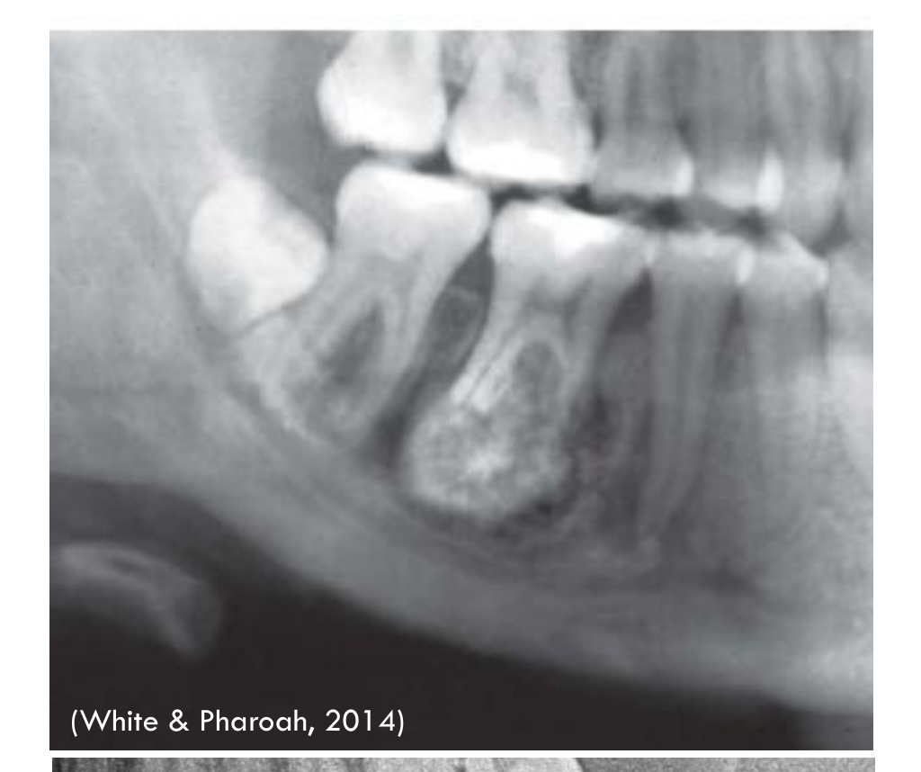
Reasoning: Cementoblastomas develop at the apical third of a tooth root and are characteristically fused to it. The density of the cemental mass often obscures the root outline, and root resorption occurs in about two-thirds of cases.
Takeaway: Cementoblastomas are characteristically fused to the affected tooth root and frequently cause root resorption.
Case: Cemento-ossifying fibroma with jaw expansion
Question
Scenario: Occlusal radiograph of a cemento-ossifying fibroma.
What’s shown: Significant expansion of the jaw.
Consider: What effect does this lesion have on the jawbone as seen on this view?
Answer
Observations:
- Marked expansion of the jaw is visible.
Reasoning: Cemento-ossifying fibromas typically demonstrate cortical thinning and significant expansion, which is clearly appreciable on an occlusal radiograph.
Takeaway: Cemento-ossifying fibromas cause significant jaw expansion and cortical thinning, which is well-demonstrated on occlusal radiographs.
Footnotes
-
Original PDF page 1: L3.2 - Benign Tumours of the Jaws Part 1, p.1 ↩
-
Original PDF page 2: L3.2 - Benign Tumours of the Jaws Part 1, p.2 ↩
-
Original PDF page 3: L3.2 - Benign Tumours of the Jaws Part 1, p.3 ↩
-
Original PDF page 4: L3.2 - Benign Tumours of the Jaws Part 1, p.4 ↩
-
Original PDF page 5: L3.2 - Benign Tumours of the Jaws Part 1, p.5 ↩
-
Original PDF page 6: L3.2 - Benign Tumours of the Jaws Part 1, p.6 ↩
-
Original PDF page 7: L3.2 - Benign Tumours of the Jaws Part 1, p.7 ↩
-
Original PDF page 8: L3.2 - Benign Tumours of the Jaws Part 1, p.8 ↩
-
Original PDF page 9: L3.2 - Benign Tumours of the Jaws Part 1, p.9 ↩
-
Original PDF page 10: L3.2 - Benign Tumours of the Jaws Part 1, p.10 ↩
-
Original PDF page 11: L3.2 - Benign Tumours of the Jaws Part 1, p.11 ↩
-
Original PDF page 12: L3.2 - Benign Tumours of the Jaws Part 1, p.12 ↩
-
Original PDF page 13: L3.2 - Benign Tumours of the Jaws Part 1, p.13 ↩
-
Original PDF page 14: L3.2 - Benign Tumours of the Jaws Part 1, p.14 ↩
-
Original PDF page 15: L3.2 - Benign Tumours of the Jaws Part 1, p.15 ↩
-
Original PDF page 16: L3.2 - Benign Tumours of the Jaws Part 1, p.16 ↩
-
Original PDF page 17: L3.2 - Benign Tumours of the Jaws Part 1, p.17 ↩
-
Original PDF page 18: L3.2 - Benign Tumours of the Jaws Part 1, p.18 ↩
-
Original PDF page 19: L3.2 - Benign Tumours of the Jaws Part 1, p.19 ↩
-
Original PDF page 20: L3.2 - Benign Tumours of the Jaws Part 1, p.20 ↩
-
Original PDF page 21: L3.2 - Benign Tumours of the Jaws Part 1, p.21 ↩
-
Original PDF page 22: L3.2 - Benign Tumours of the Jaws Part 1, p.22 ↩
-
Original PDF page 23: L3.2 - Benign Tumours of the Jaws Part 1, p.23 ↩
-
Original PDF page 24: L3.2 - Benign Tumours of the Jaws Part 1, p.24 ↩
-
Original PDF page 25: L3.2 - Benign Tumours of the Jaws Part 1, p.25 ↩
-
Original PDF page 26: L3.2 - Benign Tumours of the Jaws Part 1, p.26 ↩
-
Original PDF page 27: L3.2 - Benign Tumours of the Jaws Part 1, p.27 ↩
-
Original PDF page 28: L3.2 - Benign Tumours of the Jaws Part 1, p.28 ↩
-
Original PDF page 29: L3.2 - Benign Tumours of the Jaws Part 1, p.29 ↩
-
Original PDF page 30: L3.2 - Benign Tumours of the Jaws Part 1, p.30 ↩
-
Original PDF page 31: L3.2 - Benign Tumours of the Jaws Part 1, p.31 ↩
-
Original PDF page 32: L3.2 - Benign Tumours of the Jaws Part 1, p.32 ↩
-
Original PDF page 33: L3.2 - Benign Tumours of the Jaws Part 1, p.33 ↩
-
Original PDF page 34: L3.2 - Benign Tumours of the Jaws Part 1, p.34 ↩
-
Original PDF page 35: L3.2 - Benign Tumours of the Jaws Part 1, p.35 ↩
-
Original PDF page 36: L3.2 - Benign Tumours of the Jaws Part 1, p.36 ↩
-
Original PDF page 37: L3.2 - Benign Tumours of the Jaws Part 1, p.37 ↩
-
Original PDF page 38: L3.2 - Benign Tumours of the Jaws Part 1, p.38 ↩
-
Original PDF page 39: L3.2 - Benign Tumours of the Jaws Part 1, p.39 ↩
-
Original PDF page 40: L3.2 - Benign Tumours of the Jaws Part 1, p.40 ↩
-
Original PDF page 41: L3.2 - Benign Tumours of the Jaws Part 1, p.41 ↩
-
Original PDF page 42: L3.2 - Benign Tumours of the Jaws Part 1, p.42 ↩
-
Original PDF page 43: L3.2 - Benign Tumours of the Jaws Part 1, p.43 ↩
-
Original PDF page 44: L3.2 - Benign Tumours of the Jaws Part 1, p.44 ↩
-
Original PDF page 45: L3.2 - Benign Tumours of the Jaws Part 1, p.45 ↩
-
Original PDF page 46: L3.2 - Benign Tumours of the Jaws Part 1, p.46 ↩
-
Original PDF page 47: L3.2 - Benign Tumours of the Jaws Part 1, p.47 ↩
-
Original PDF page 48: L3.2 - Benign Tumours of the Jaws Part 1, p.48 ↩
-
Original PDF page 49: L3.2 - Benign Tumours of the Jaws Part 1, p.49 ↩
-
Original PDF page 50: L3.2 - Benign Tumours of the Jaws Part 1, p.50 ↩
-
Original PDF page 51: L3.2 - Benign Tumours of the Jaws Part 1, p.51 ↩
-
Original PDF page 52: L3.2 - Benign Tumours of the Jaws Part 1, p.52 ↩
-
Original PDF page 53: L3.2 - Benign Tumours of the Jaws Part 1, p.53 ↩
-
Original PDF page 54: L3.2 - Benign Tumours of the Jaws Part 1, p.54 ↩
-
Original PDF page 55: L3.2 - Benign Tumours of the Jaws Part 1, p.55 ↩
-
Original PDF page 56: L3.2 - Benign Tumours of the Jaws Part 1, p.56 ↩
-
Original PDF page 58: L3.2 - Benign Tumours of the Jaws Part 1, p.58 ↩
-
Original PDF page 59: L3.2 - Benign Tumours of the Jaws Part 1, p.59 ↩
-
Original PDF page 57: L3.2 - Benign Tumours of the Jaws Part 1, p.57 ↩
-
Original PDF page 60: L3.2 - Benign Tumours of the Jaws Part 1, p.60 ↩
-
Original PDF page 61: L3.2 - Benign Tumours of the Jaws Part 1, p.61 ↩
-
Original PDF page 62: L3.2 - Benign Tumours of the Jaws Part 1, p.62 ↩
-
Original PDF page 63: L3.2 - Benign Tumours of the Jaws Part 1, p.63 ↩
-
Original PDF page 64: L3.2 - Benign Tumours of the Jaws Part 1, p.64 ↩
-
Original PDF page 65: L3.2 - Benign Tumours of the Jaws Part 1, p.65 ↩
-
Original PDF page 66: L3.2 - Benign Tumours of the Jaws Part 1, p.66 ↩
-
Original PDF page 67: L3.2 - Benign Tumours of the Jaws Part 1, p.67 ↩