Benign Tumours of the Jaws (Part 2 — Non-Odontogenic Tumours)1
Dr. May Lam
Oral and Maxillofacial Radiologist
Senior lecturer
BDSc (Hons) FRACDS (GDP) DClinDent (DMFR)
MRACDS (DMFR)
WHO Classification of Head and Neck Tumours
Odontogenic Benign Tumours2
Benign epithelial odontogenic tumours
- Ameloblastoma
- Ameloblastoma, conventional
- Ameloblastoma, unicystic type
- Ameloblastoma, extraosseous/peripheral type
- Adenoid ameloblastoma
- Metastasising ameloblastoma
- Adenomatoid odontogenic tumour
- Squamous odontogenic tumour
- Calcifying epithelial odontogenic tumour
Benign mixed epithelial & mesenchymal odontogenic tumours
- Odontoma
- Ameloblastic fibroma
- Primordial odontogenic tumour
- Dentinogenic ghost cell tumour
Benign mesenchymal odontogenic tumours
- Odontogenic myxoma/myxofibroma
- Odontogenic fibroma
- Cementoblastoma
- Cemento-ossifying fibroma
Non-Odontogenic Benign Tumours
Benign maxillofacial bone and cartilage tumours
Lecturer — Lecture Scope
The previous lecture covered odontogenic benign tumours.
- Osteoma
- Osteochondroma
- Osteoblastoma
- (Osteoid osteoma)
- Chondroblastoma
- Chondromyxoid fibroma
- Desmoplastic fibroma of bone
Soft tissue tumours
Lecturer — Non-Odontogenic Tumours
This lecture covers selected, relatively common non-odontogenic benign tumours. The list is non-exhaustive and includes tumours encountered in the head and neck.
- The lecture is divided into benign maxillofacial bone and cartilage tumours.
- It also covers soft tissue tumours, with emphasis on lesions occurring within the bones of the head and neck.
- Vascular tumours
- Haemangioma
- Peripheral nerve sheath tumours
- Neurofibroma
- Schwannoma
- Neuroma
Part 1: Benign Maxillofacial Bone and Cartilage Tumours3
- Osteoma
- Osteochondroma
- Osteoblastoma (Osteoid osteoma)
- Chondroblastoma
- Chondromyxoid fibroma
- Desmoplastic fibroma of bone
Osteoma45
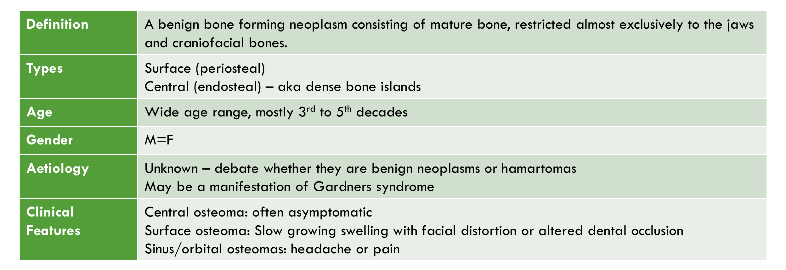
- Definition: A benign bone forming neoplasm consisting of mature bone, restricted almost exclusively to the jaws and craniofacial bones.
- Types:
- Surface (periosteal)
- Central (endosteal) — aka dense bone islands
- Age: Wide age range, mostly 3rd to 5th decades
- Gender: M=F
- Aetiology:
- Unknown — debate whether they are benign neoplasms or hamartomas
- May be a manifestation of Gardner’s syndrome
- Clinical Features:
- Central osteoma: often asymptomatic
- Surface osteoma: slow growing swelling with facial distortion or altered dental occlusion
- Sinus/orbital osteomas: headache or pain
Lecturer — Gardner Syndrome
Gardner syndrome may present with multiple osteomas or dense bone islands before other features are recognised.
- The syndrome includes multiple unerupted supernumerary or permanent teeth, odontomas, epidermoid cysts, subcutaneous desmoid tumours, and intestinal polyps.
- The intestinal polyps may become malignant at an average age of 39, so osteomas or dense bone islands should not be overlooked as a possible first clinical manifestation.
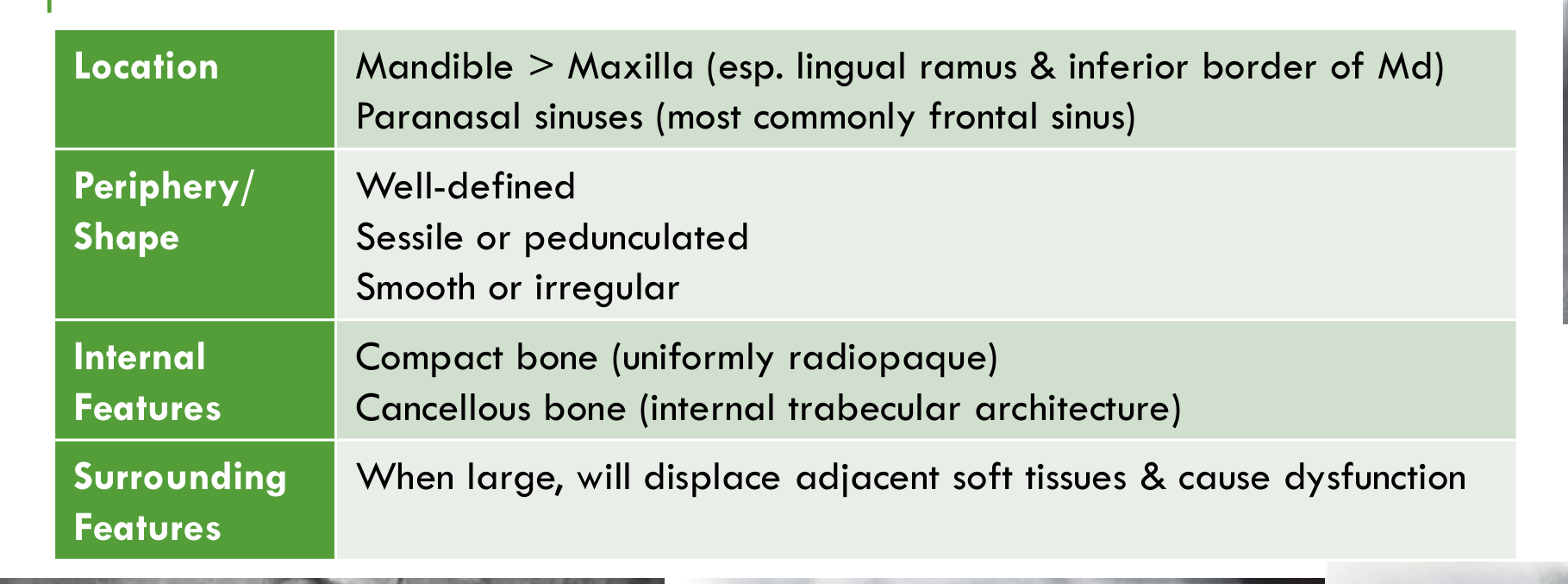
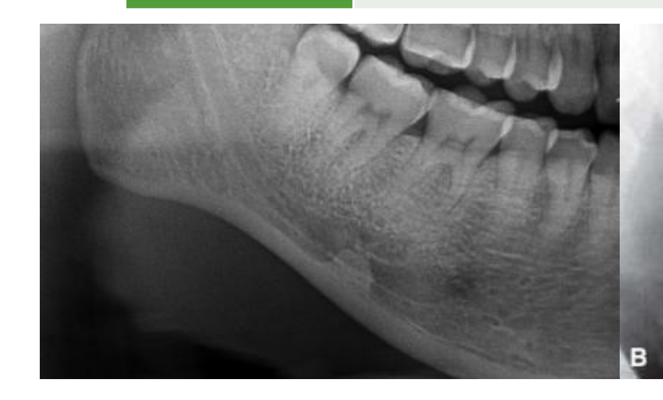
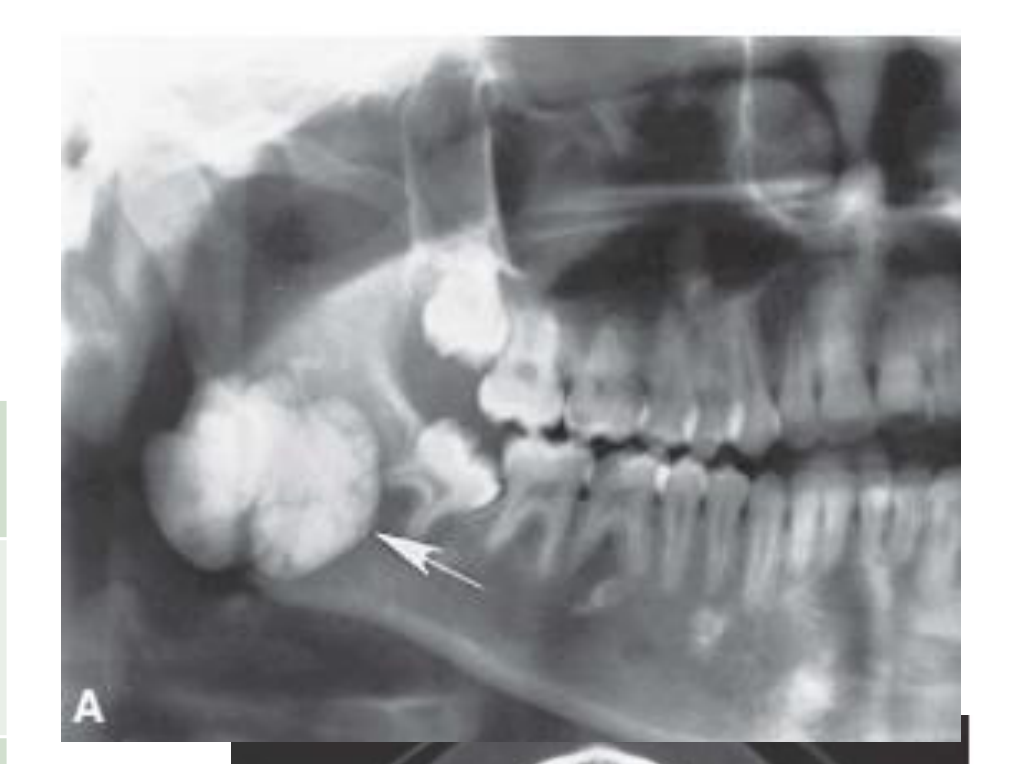
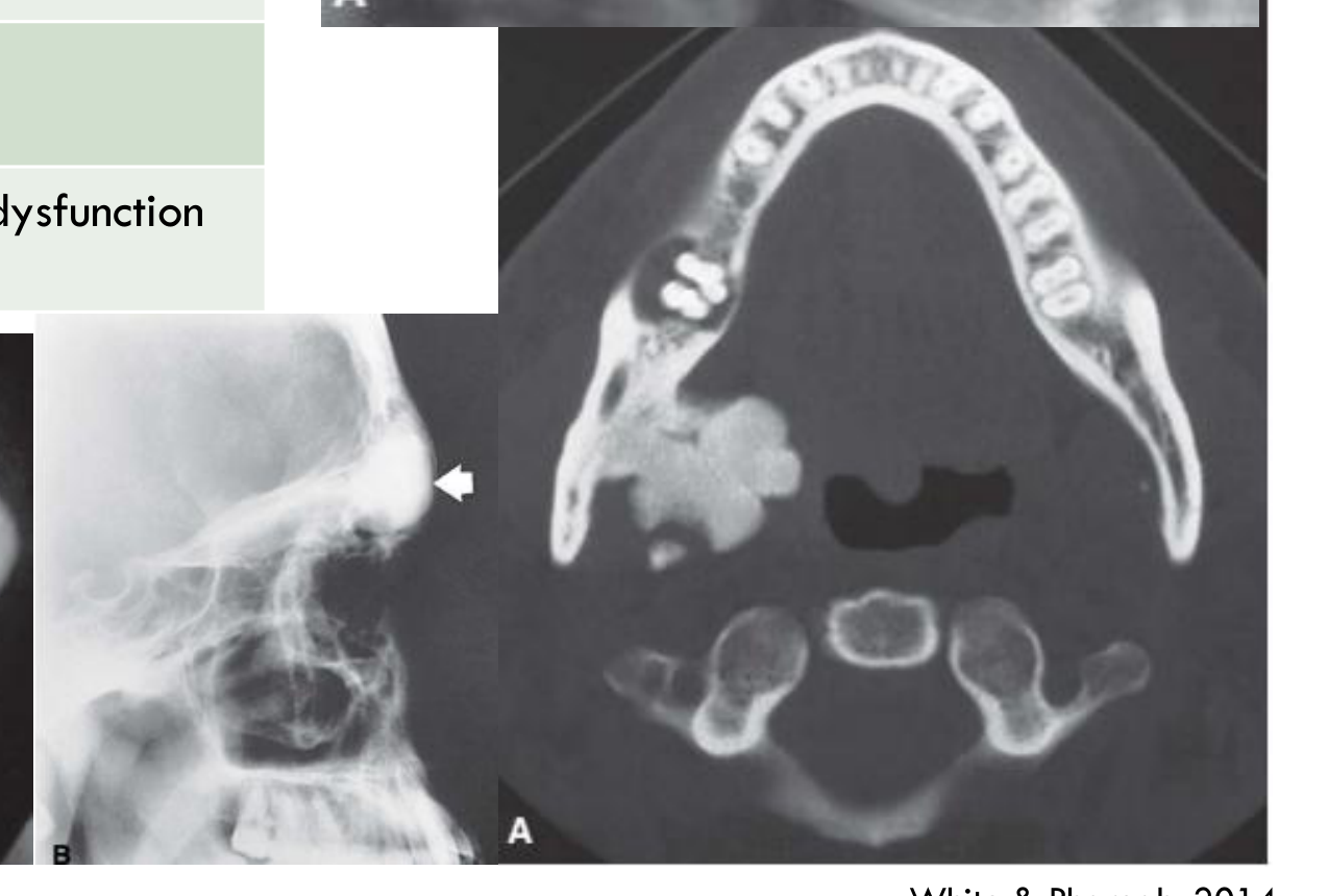
- Location: Mandible > Maxilla (esp. lingual ramus & inferior border of Md); paranasal sinuses (most commonly frontal sinus)
- Periphery/Shape: Well-defined; sessile or pedunculated; smooth or irregular
- Internal Features: Compact bone (uniformly radiopaque); cancellous bone (internal trabecular architecture)
- Surrounding Features: When large, will displace adjacent soft tissues & cause dysfunction
Lecturer — Osteoma Location
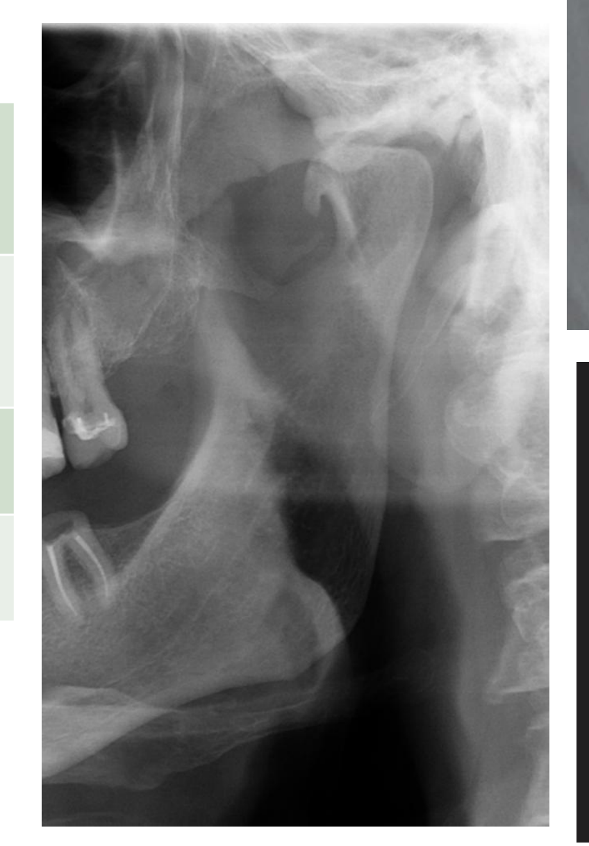
On CT, the precise buccal or lingual location of an osteoma may be demonstrated, including lesions arising from the lingual aspect of the ramus. A lesion near the ramus can affect the medial pterygoid muscle, tonsil, and parapharyngeal space.
White & Pharoah, 2014
 |  |  |
 |  |  |
Histopathology6
- Composed mostly of lamellar bone (compact or trabecular) in which osteoblasts and osteoclasts are inconspicuous
- +/- osteoblastoma-like areas (thought related to remodelling process rather than aggressive behaviour)
- Similar appearance to fibro-osseous lesions, sclerosing osteomyelitis, and ossification of a fibrous epulis — correlate with clinical and radiographic findings for definitive diagnosis
Management/Prognosis/Recurrence
- Slow growth — most are monitored & resection only for symptomatic lesions
- Recurrence is unusual
Osteochondroma789
-
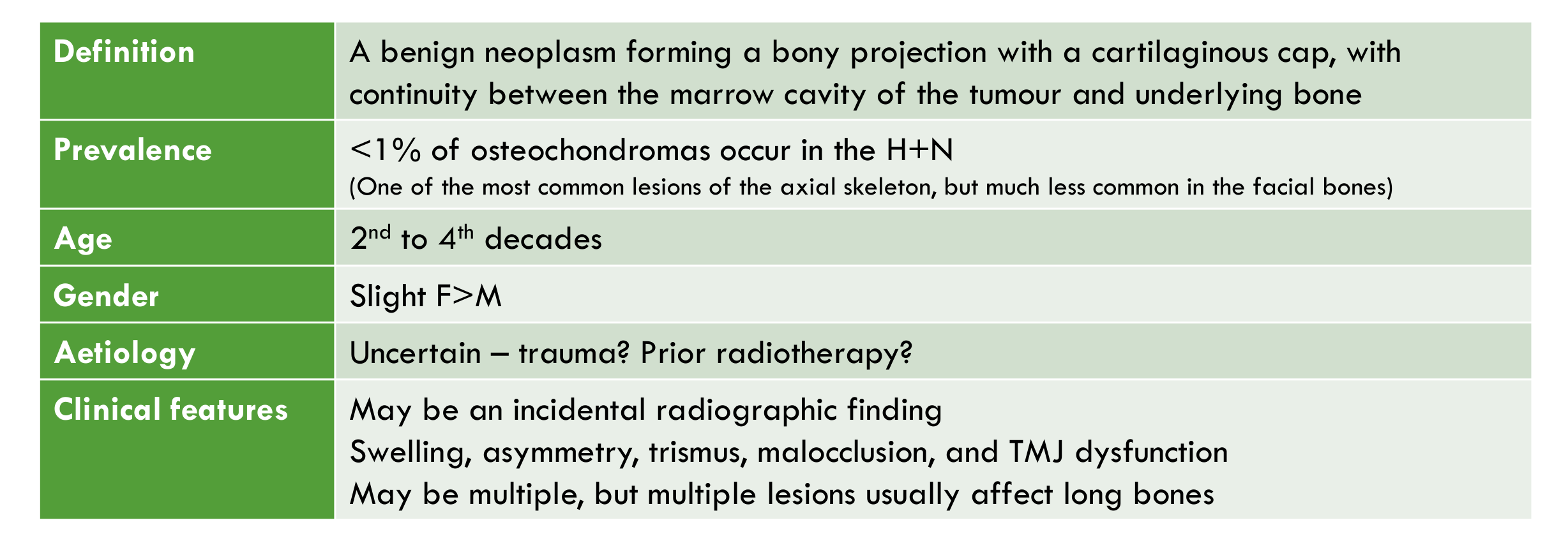
Definition: A benign neoplasm forming a bony projection with a cartilaginous cap, with continuity between the marrow cavity of the tumour and underlying bone
-
Prevalence: <1% of osteochondromas occur in the H+N (one of the most common lesions of the axial skeleton, but much less common in the facial bones)
-
Age: 2nd to 4th decades
-
Gender: Slight F>M
-
Aetiology: Uncertain — trauma? Prior radiotherapy?
-
Clinical Features:
- May be an incidental radiographic finding
- Swelling, asymmetry, trismus, malocclusion, and TMJ dysfunction
- May be multiple, but multiple lesions usually affect long bones
-
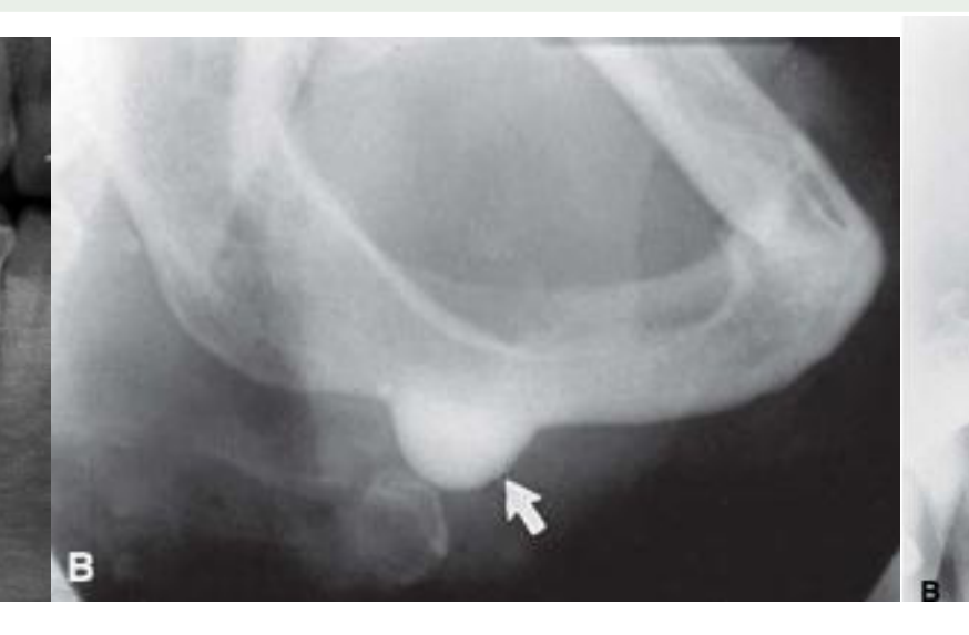
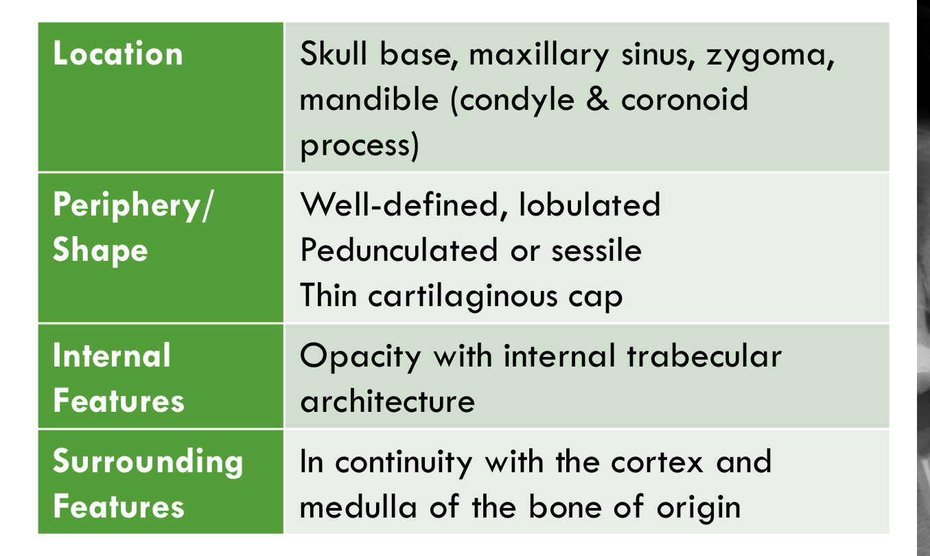
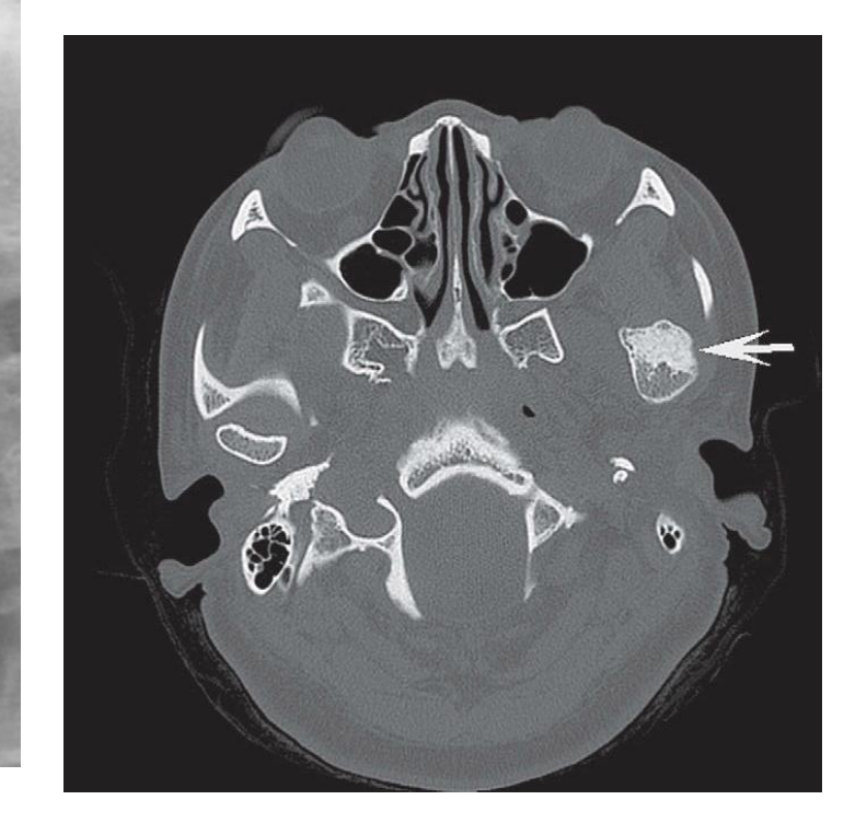
Location: Skull base, maxillary sinus, zygoma, mandible (condyle & coronoid process)
-
Periphery/Shape: Well-defined, lobulated; pedunculated or sessile; thin cartilaginous cap
-
Internal Features: Opacity with internal trabecular architecture
-
Surrounding Features: In continuity with the cortex and medulla of the bone of origin
-
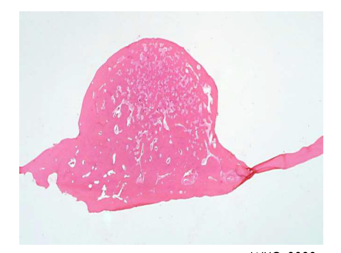
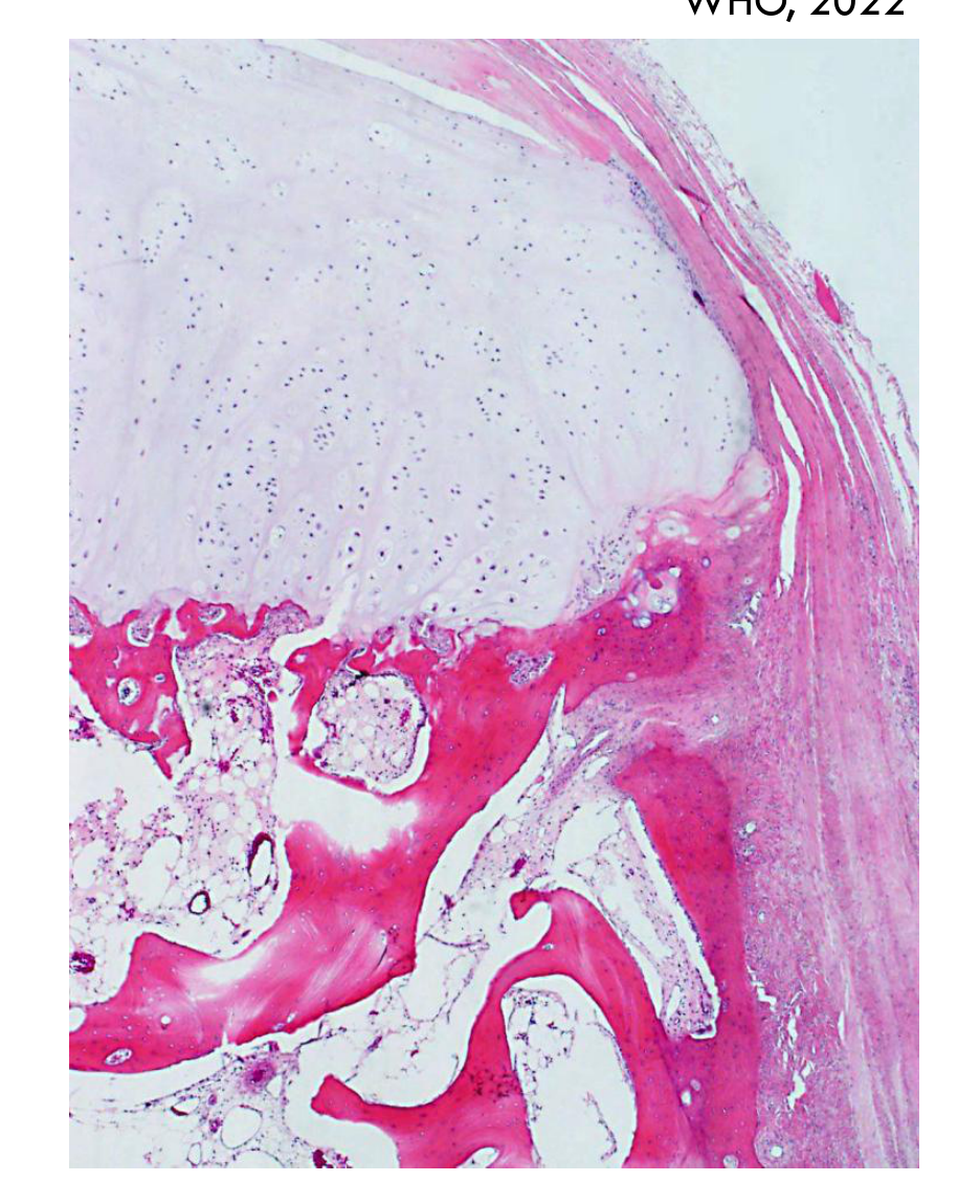
A cartilage capped bony projection from the external bone surface showing cortical and medullary continuity with the parent bone
-
- surface layer of fibrous tissue
-
- middle layer which resembles growth plate cartilage
-
May not need intervention if asymptomatic
-
Recurrence following excision is rare, unless incompletely removed
-
Malignant transformation is very rare
Lecturer — Condylar Osteochondroma
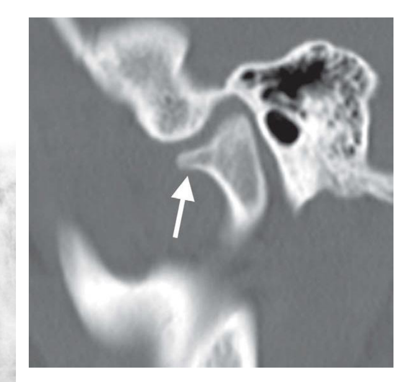
Head and neck osteochondromas may continue growing slowly after puberty and symptoms usually result from progressive enlargement of the condyle.
- Condylar lesions may cause prognathic deviation of the chin, contralateral crossbite, or an open bite on the affected side; pain is uncommon.
- Unlike condylar hyperplasia, which diffusely enlarges the entire condylar head, an osteochondroma produces uneven enlargement or protrusion from only one part of the condylar head.
WHO, 2022
Osteoblastoma1011121314
-
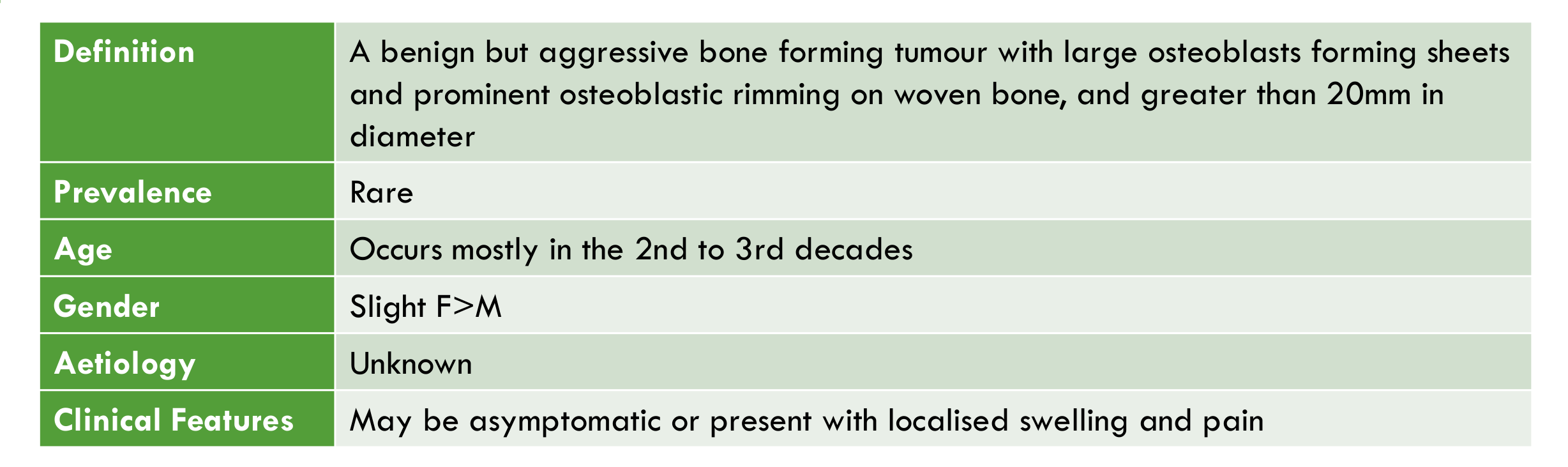
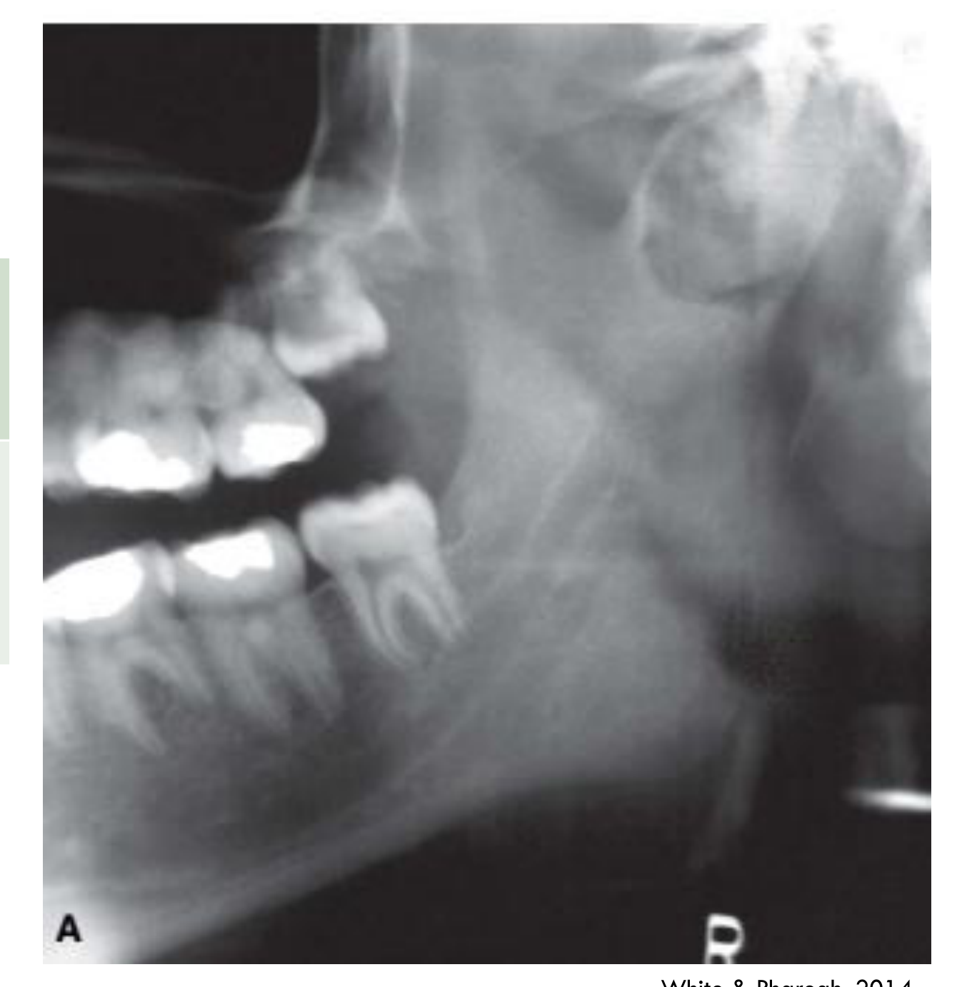
Definition: A benign but aggressive bone forming tumour with large osteoblasts forming sheets and prominent osteoblastic rimming on woven bone, and greater than 20mm in diameter
-
Prevalence: Rare
-
Age: Occurs mostly in the 2nd to 3rd decades
-
Gender: Slight F>M
-
Aetiology: Unknown
-
Clinical Features: May be asymptomatic or present with localised swelling and pain
-
Location:
- 10% in the craniofacial bones, most often in the body of the mandible
- May be intra-osseous or periosteal
-
Periphery/Shape:
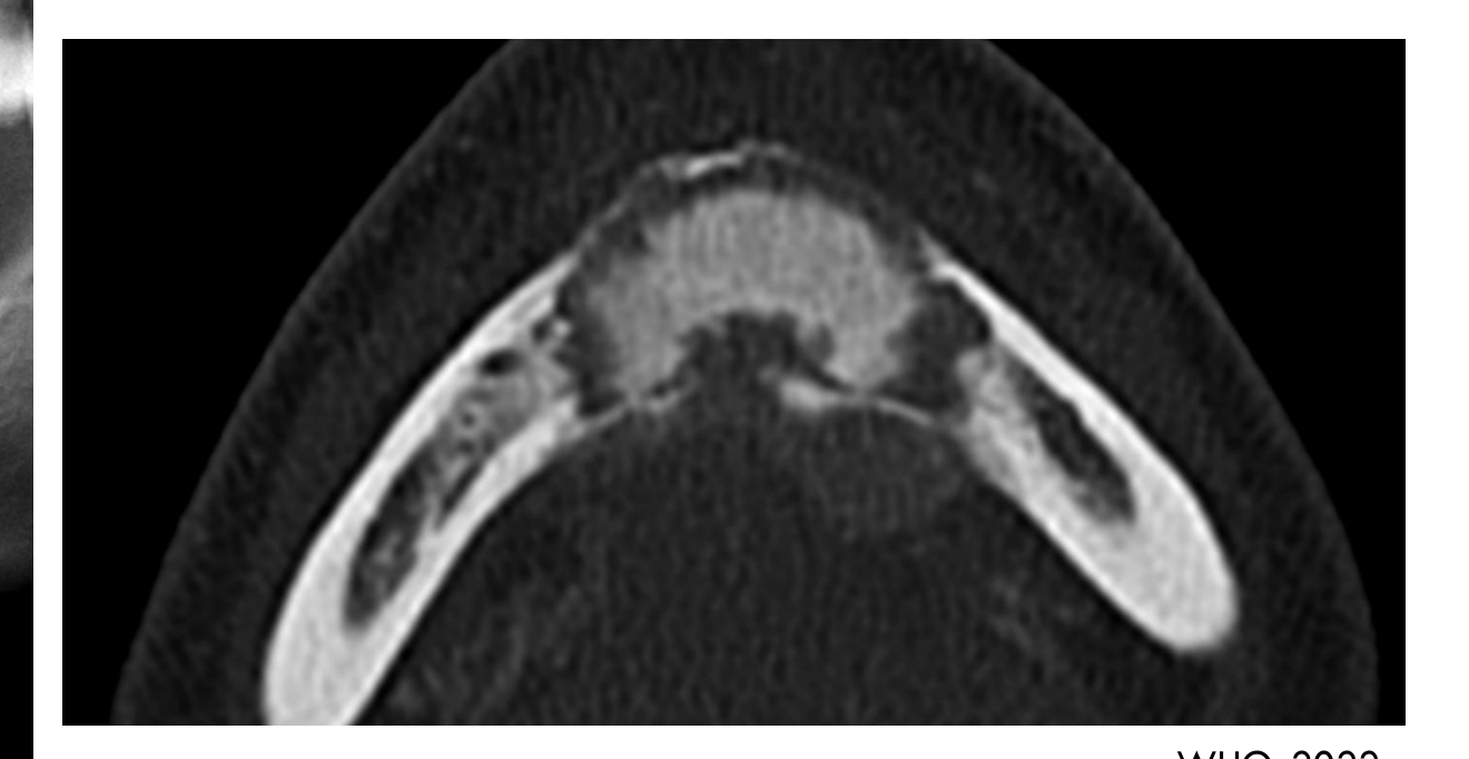
- Well circumscribed, round/oval shaped
- Usually no sclerotic border
-
20mm in size
WHO, 2022
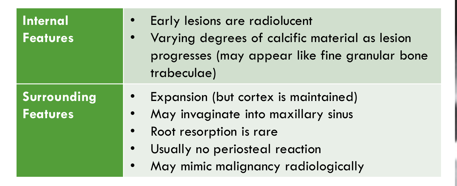
- Internal Features:
- Early lesions are radiolucent
- Varying degrees of calcific material as lesion progresses (may appear like fine granular bone trabeculae)
- Surrounding Features:
- Expansion (but cortex is maintained)
- May invaginate into maxillary sinus
- Root resorption is rare
- Usually no periosteal reaction
- May mimic malignancy radiologically
White & Pharoah, 2014
-
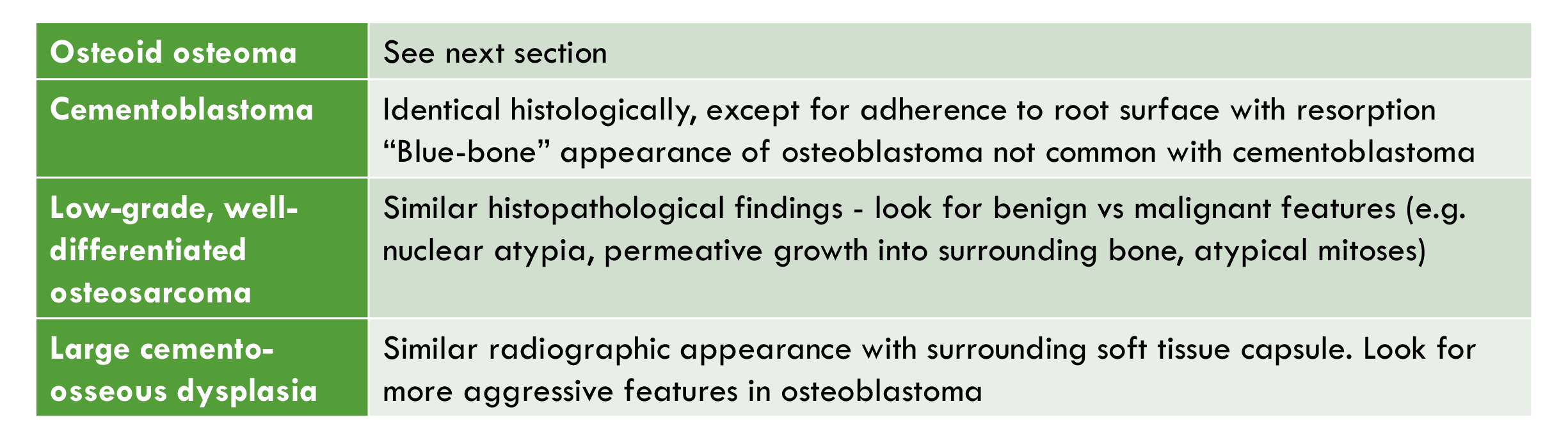
Osteoid osteoma — see next section
-
Cementoblastoma — identical histologically, except for adherence to root surface with resorption; “blue-bone” appearance of osteoblastoma not common with cementoblastoma
-
Low-grade, well-differentiated osteosarcoma — similar histopathological findings; look for benign vs malignant features (e.g. nuclear atypia, permeative growth into surrounding bone, atypical mitoses)
-
Large cemento-osseous dysplasia — similar radiographic appearance with surrounding soft tissue capsule; look for more aggressive features in osteoblastoma
-
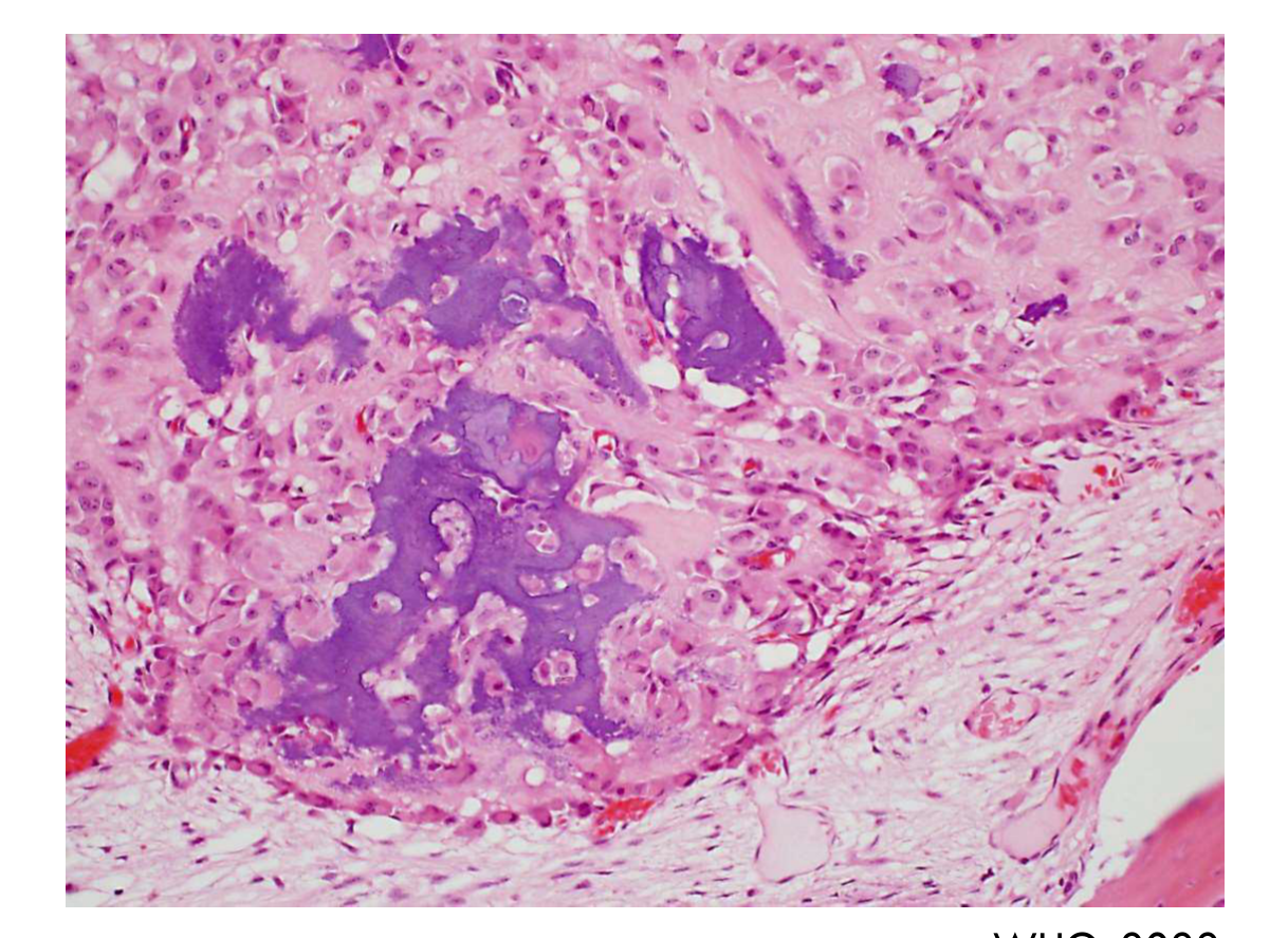
Anastomosing trabeculae of bone and osteoid, rimmed by plump osteoblasts within a loose richly vascular fibrous stroma
-
Characteristic “blue-bone” appearance
-
Some osteoblasts may appear larger and epithelioid, but this does not indicate a clinically aggressive course
-
Recurrence may follow curettage or incomplete removal
WHO, 2022
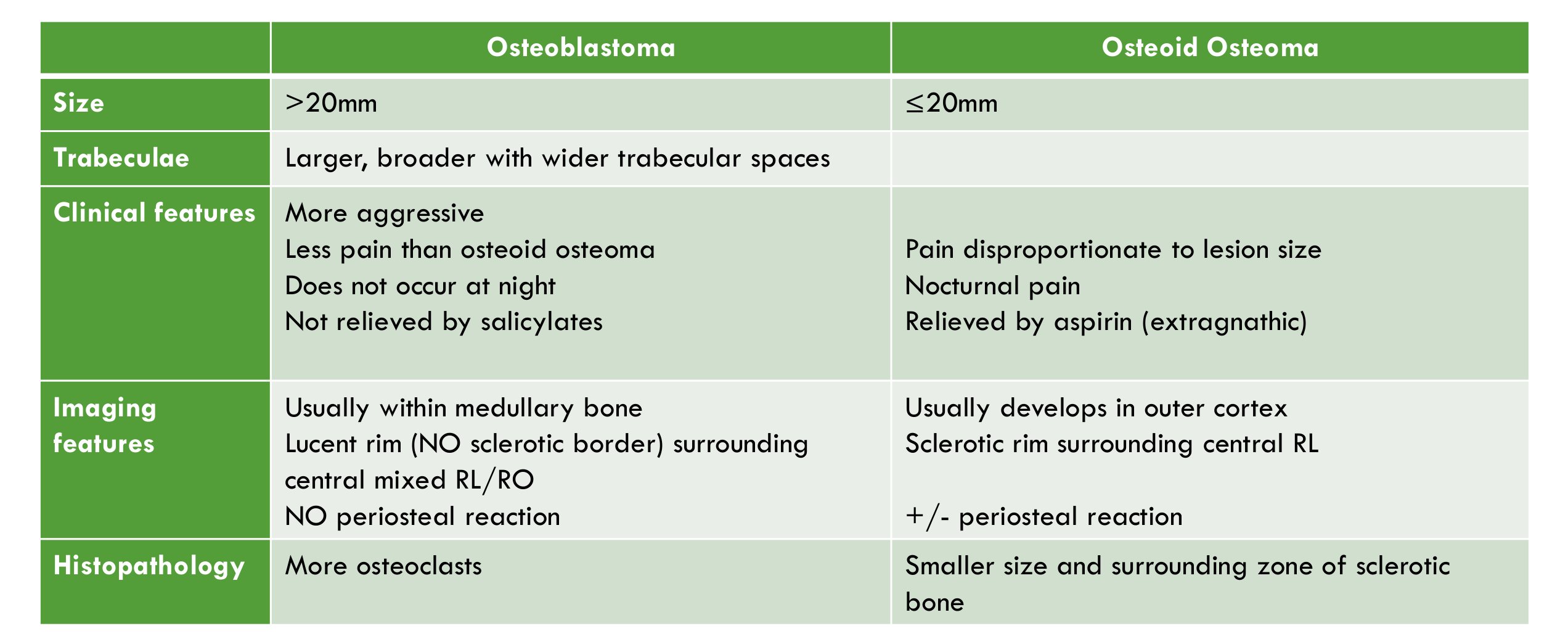
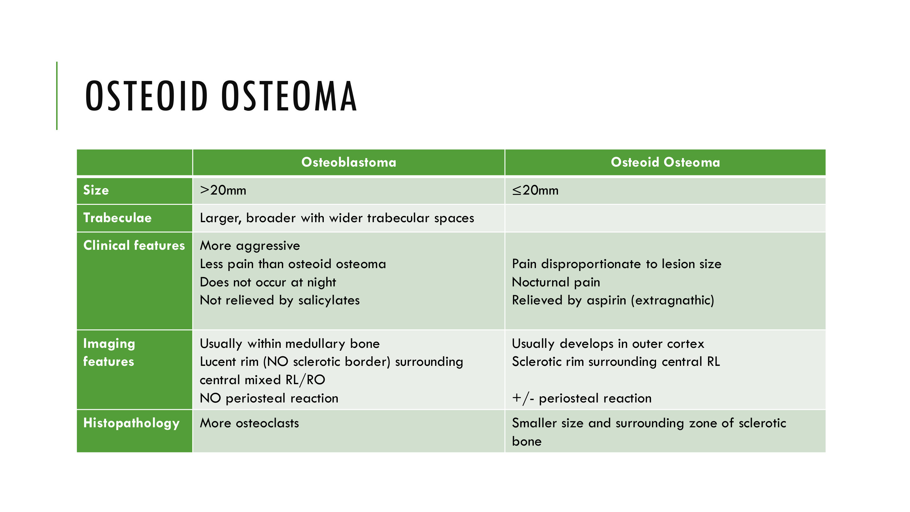
| Osteoblastoma | Osteoid Osteoma | |
|---|---|---|
| Size | >20mm | ≤20mm |
| Trabeculae | Larger, broader with wider trabecular spaces | |
| Clinical features | More aggressive; less pain than osteoid osteoma; does not occur at night; not relieved by salicylates | Pain disproportionate to lesion size; nocturnal pain; relieved by aspirin (extragnathic) |
| Imaging features | Usually within medullary bone; lucent rim (NO sclerotic border) surrounding central mixed RL/RO; NO periosteal reaction | Usually develops in outer cortex; sclerotic rim surrounding central RL; +/- periosteal reaction |
| Histopathology | More osteoclasts | Smaller size and surrounding zone of sclerotic bone |
Lecturer — Osteoid Osteoma
Osteoid osteoma is not listed as a separate entity in the WHO classification, and some authors consider it the same entity as osteoblastoma.
- The principal distinctions are lesion size and the surrounding zone of sclerotic bone; osteoid osteoma is 20 mm or smaller and usually causes more severe nocturnal pain relieved by non-steroidal anti-inflammatory drugs, particularly aspirin.
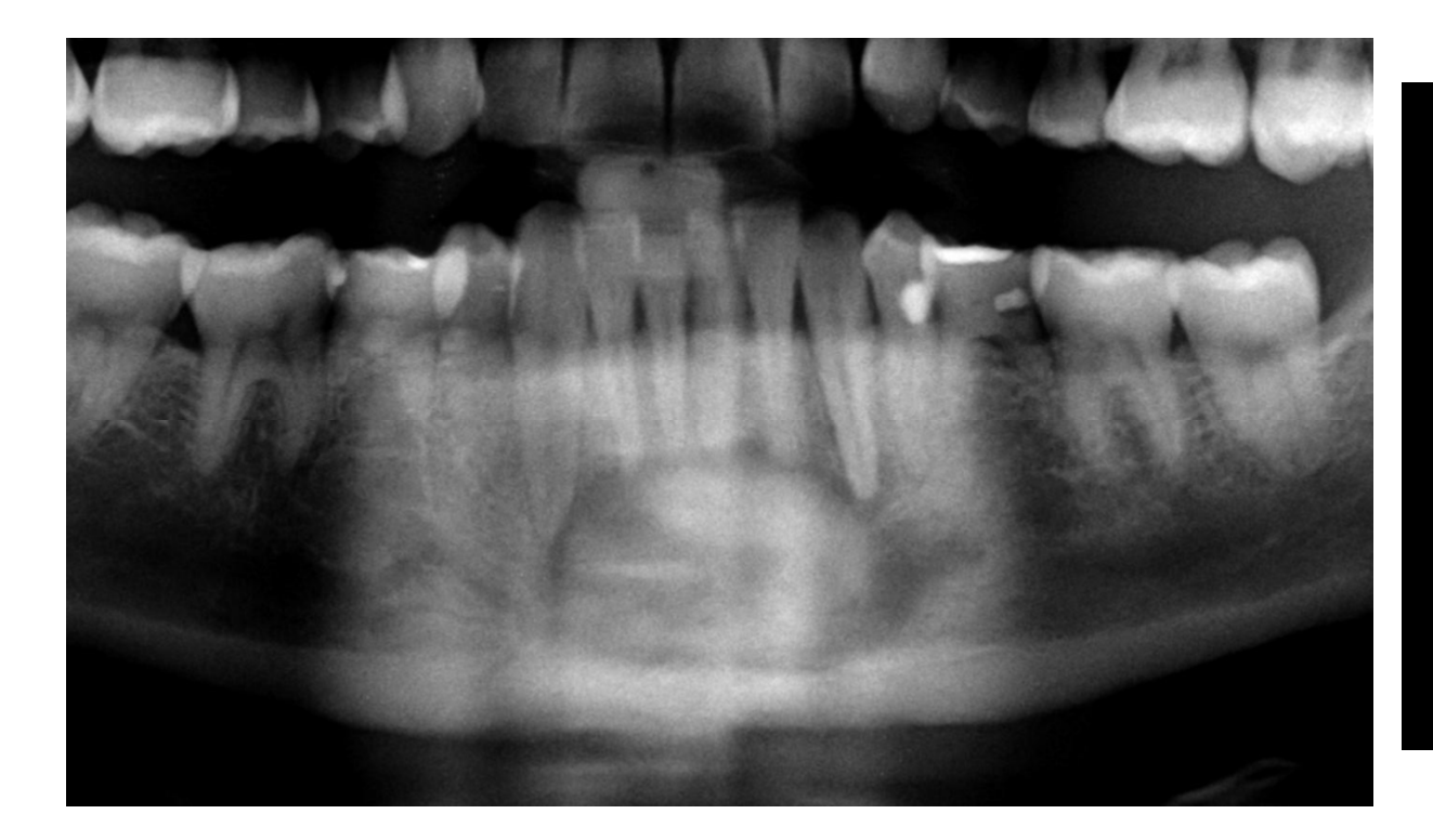
- A radiographic example showed altered bony trabeculae increasing in size over time with buccal and lingual cortical expansion.

Osteoid Osteoma1516
White & Pharoah, 2014
 | 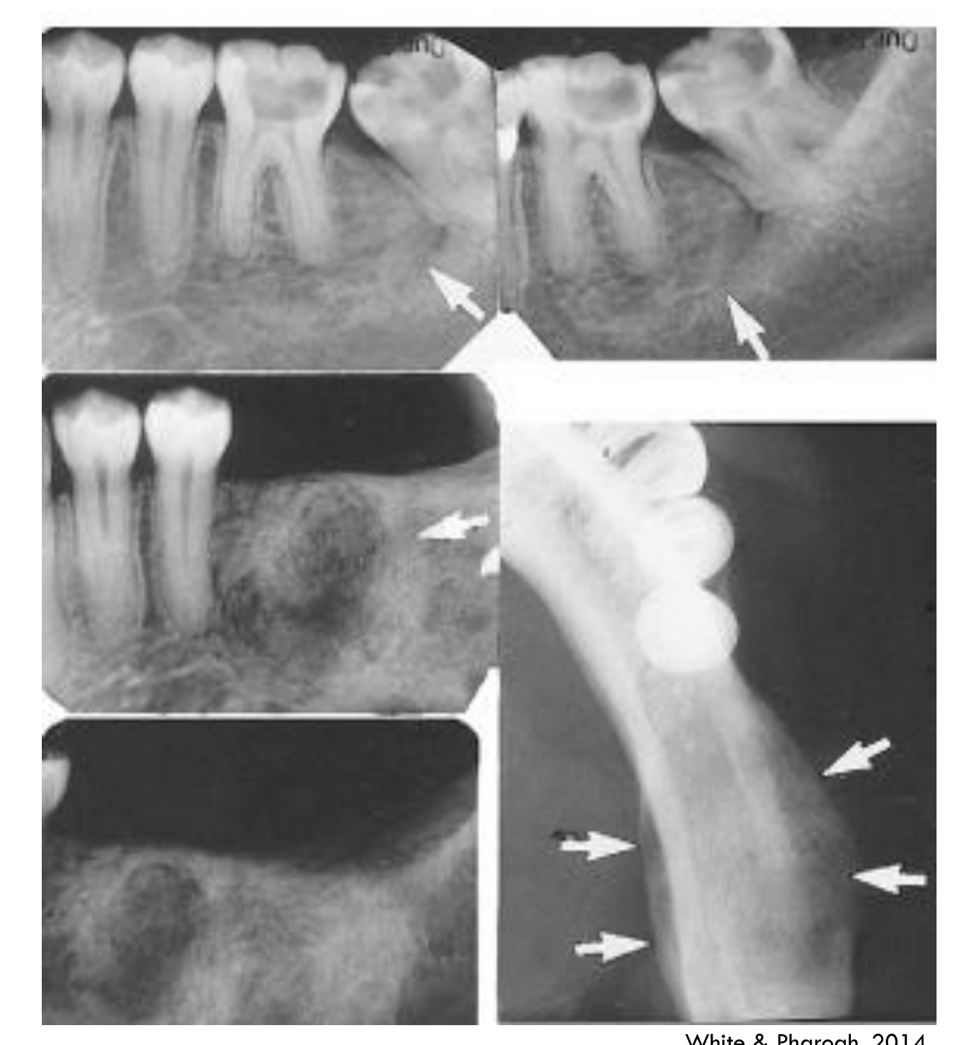 |
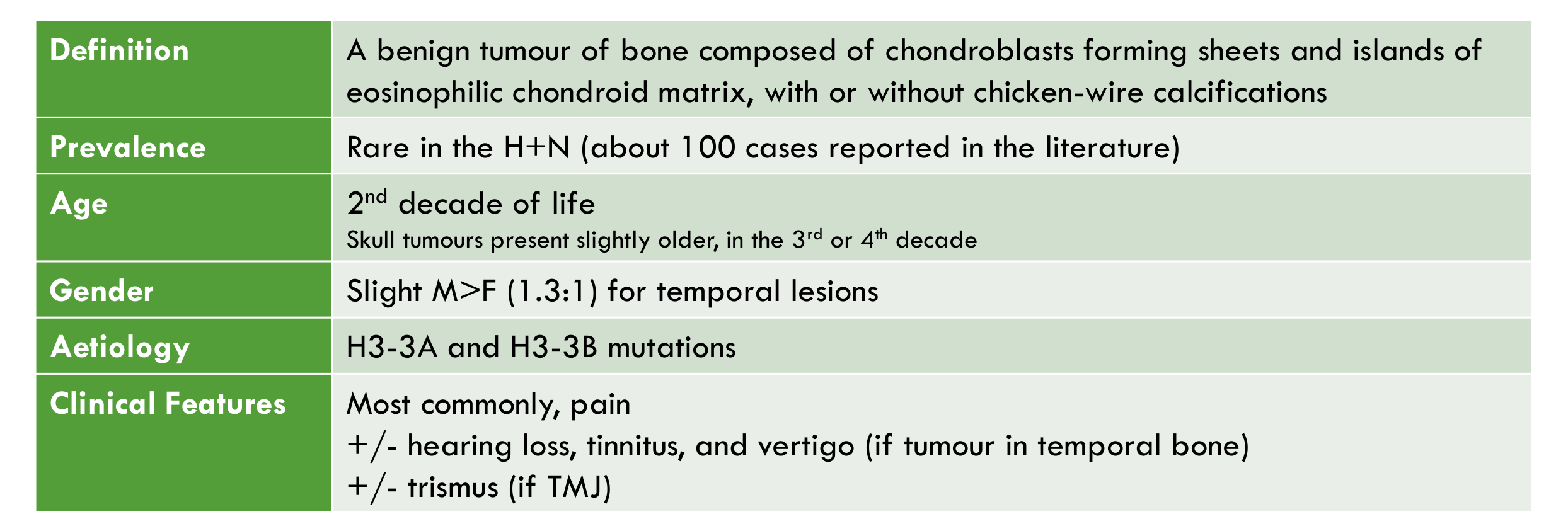
Chondroblastoma171819
-
Definition: A benign tumour of bone composed of chondroblasts forming sheets and islands of eosinophilic chondroid matrix, with or without chicken-wire calcifications
-
Prevalence: Rare in the H+N (about 100 cases reported in the literature)
-
Age: 2nd decade of life; skull tumours present slightly older, in the 3rd or 4th decade
-
Gender: Slight M>F (1.3:1) for temporal lesions
-
Aetiology: H3-3A and H3-3B mutations
-
Clinical Features:
- Most commonly, pain
- +/- hearing loss, tinnitus, and vertigo (if tumour in temporal bone)
- +/- trismus (if TMJ)
-
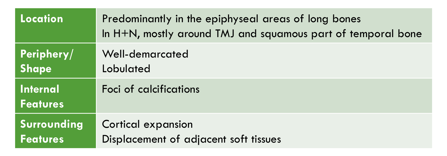
Location: Predominantly in the epiphyseal areas of long bones; in H+N, mostly around TMJ and squamous part of temporal bone
-
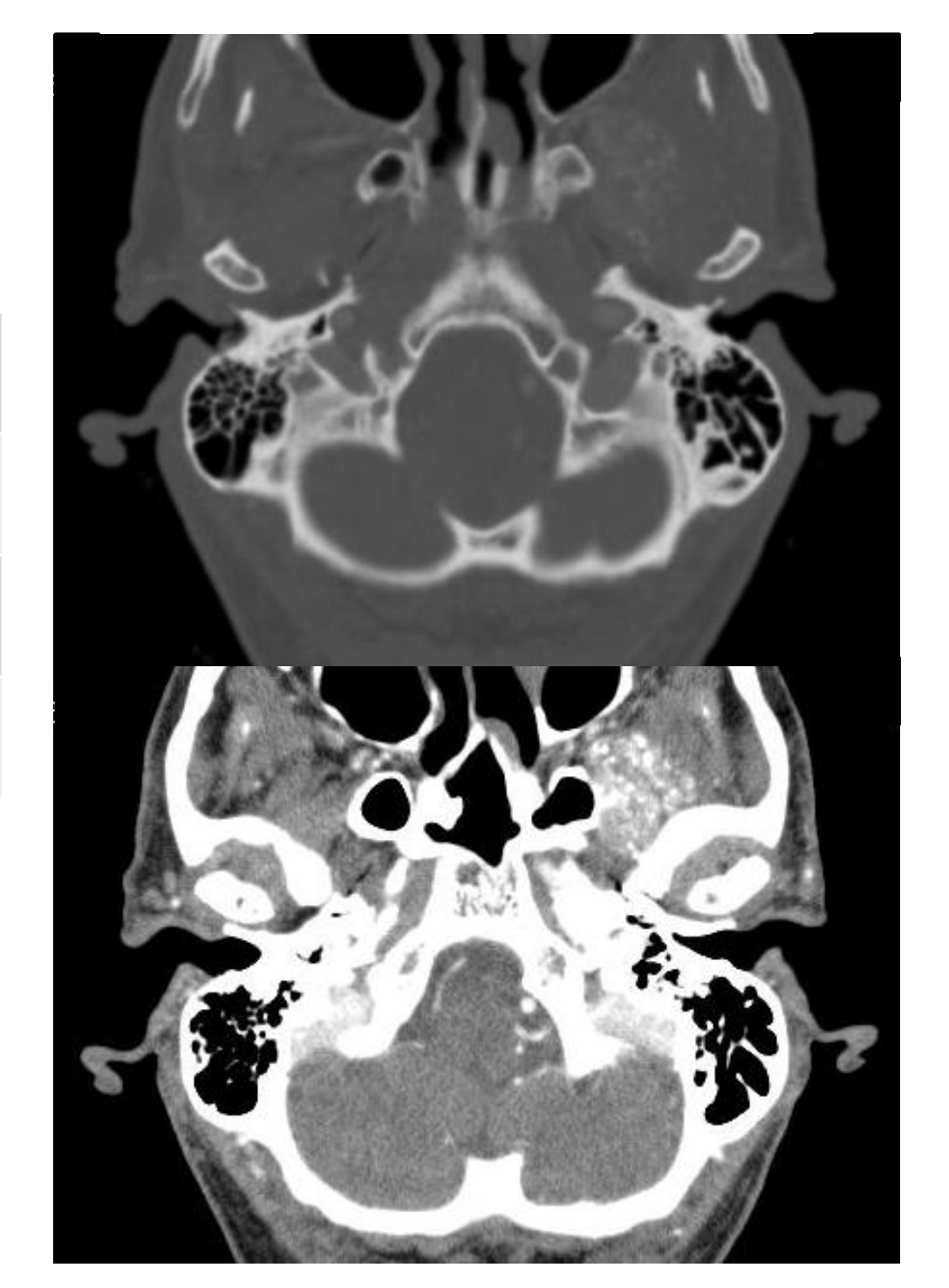
Periphery/Shape: Well-demarcated; lobulated
-
Internal Features: Foci of calcifications
-
Surrounding Features: Cortical expansion; displacement of adjacent soft tissues
Lecturer — Chondroblastoma Imaging
Foci of calcification in chondroblastoma are better appreciated on soft tissue window CT images. Adjacent soft tissues that may be displaced include the lateral pterygoid muscle posteriorly and medially and the temporalis muscle.
WHO, 2022
-
Uniform, eosinophilic and polygonal cells intermingled with an amorphous eosinophilic matrix with osteoclast-like giant cells and varying amounts of ‘chicken-wire’ calcification
-
+/- cystic haemorrhagic degeneration
-
Up to 50% of cases recur
-
Metastasis has been reported only rarely (<1%)
WHO, 2022
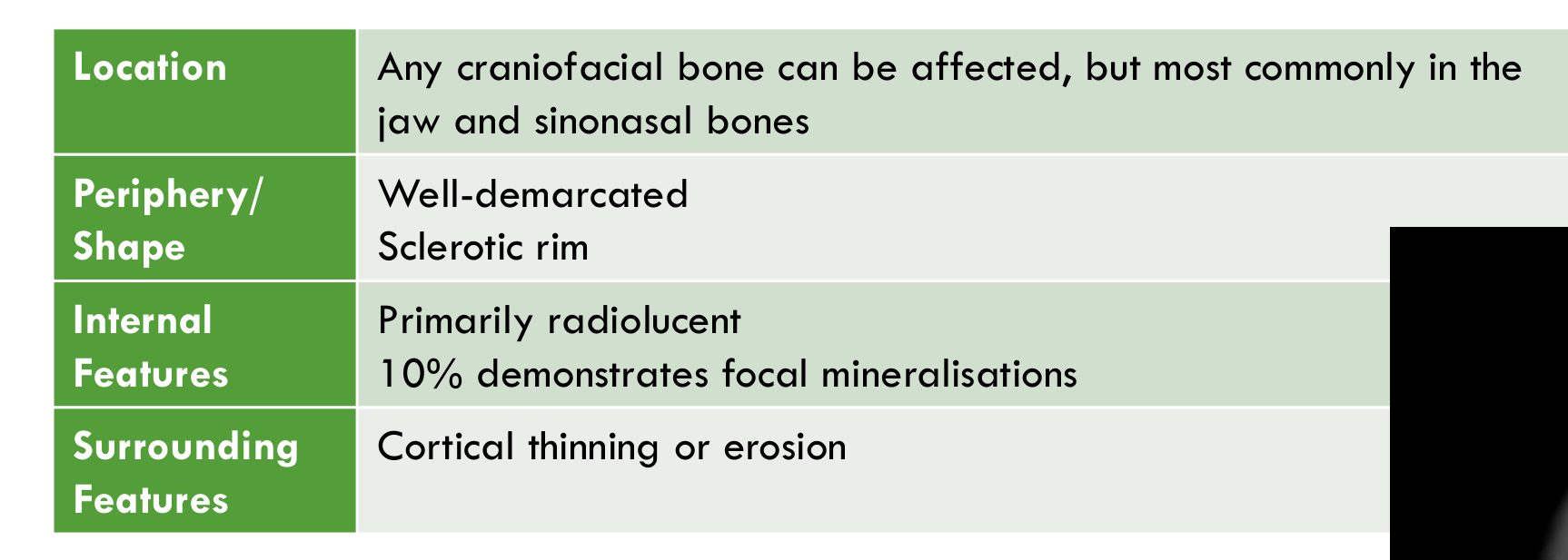
Chondromyxoid Fibroma202122
-
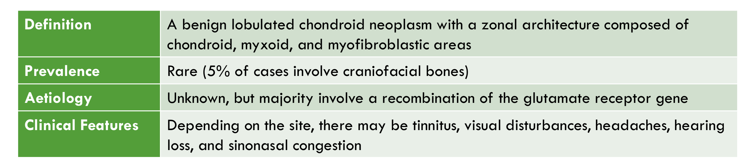
Definition: A benign lobulated chondroid neoplasm with a zonal architecture composed of chondroid, myxoid, and myofibroblastic areas
-
Prevalence: Rare (5% of cases involve craniofacial bones)
-
Aetiology: Unknown, but majority involve a recombination of the glutamate receptor gene
-
Clinical Features: Depending on the site, there may be tinnitus, visual disturbances, headaches, hearing loss, and sinonasal congestion
-
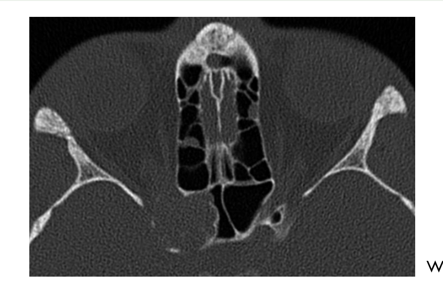
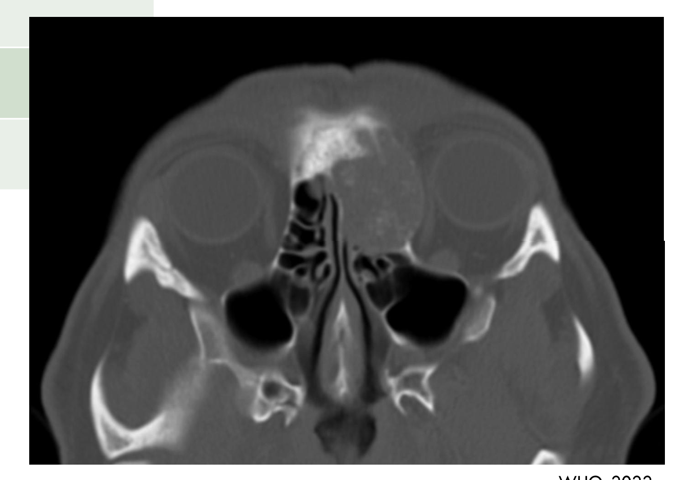
Location: Any craniofacial bone can be affected, but most commonly in the jaw and sinonasal bones
-
Periphery/Shape: Well-demarcated; sclerotic rim
-
Internal Features: Primarily radiolucent; 10% demonstrates focal mineralisations
-
Surrounding Features: Cortical thinning or erosion
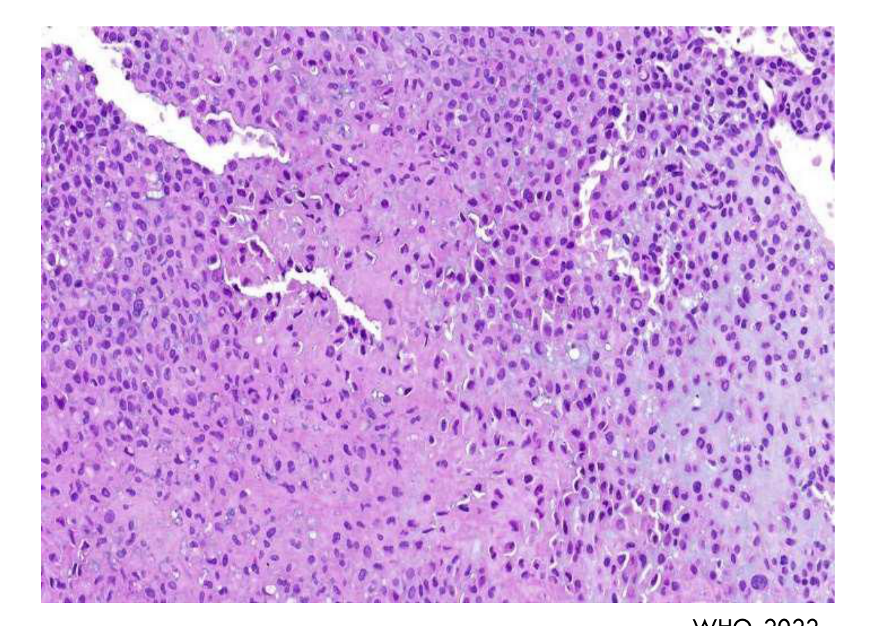
WHO, 2022
-
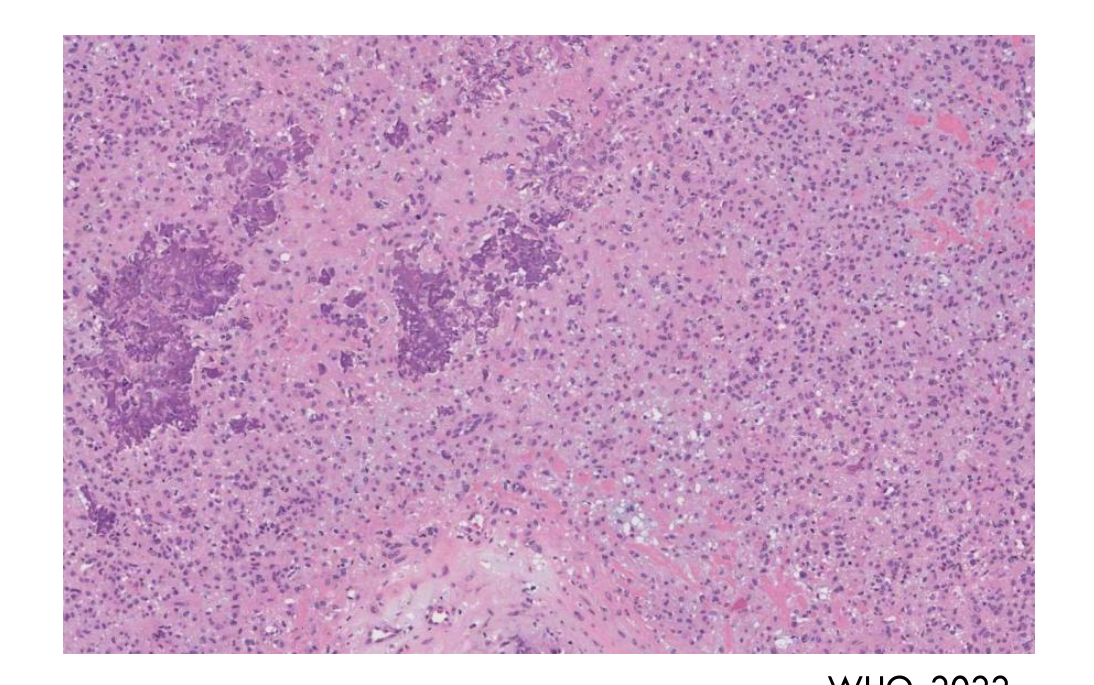
A lobular proliferation of spindled and stellate cells with abundant eosinophilic cytoplasm and a chondromyxoid background
-
1/3 cases demonstrate scattered coarse calcifications
-
Excellent prognosis, even for recurrent tumours
-
Recurrence more common in craniofacial lesions, due to challenges obtaining clear surgical margins
Desmoplastic Fibroma of Bone2324
-
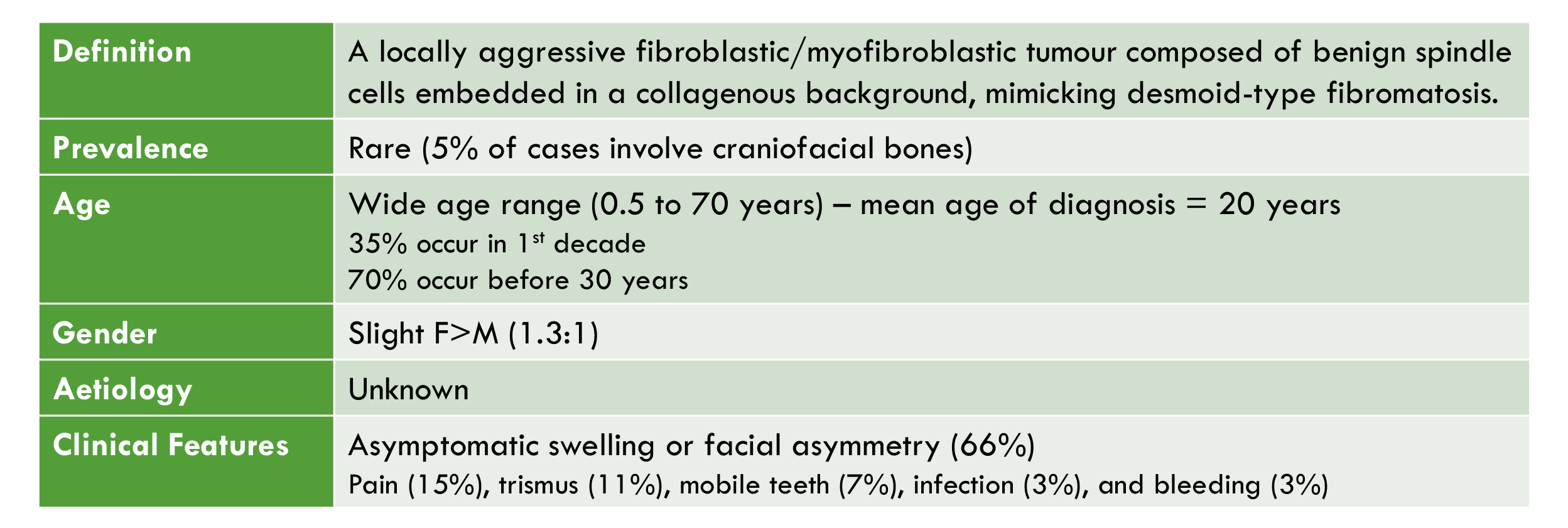
Definition: A locally aggressive fibroblastic/myofibroblastic tumour composed of benign spindle cells embedded in a collagenous background, mimicking desmoid-type fibromatosis.
-
Prevalence: Rare (5% of cases involve craniofacial bones)
-
Age: Wide age range (0.5 to 70 years) — mean age of diagnosis = 20 years; 35% occur in 1st decade; 70% occur before 30 years
-
Gender: Slight F>M (1.3:1)
-
Aetiology: Unknown
-
Clinical Features:
- Asymptomatic swelling or facial asymmetry (66%)
- Pain (15%), trismus (11%), mobile teeth (7%), infection (3%), and bleeding (3%)
-
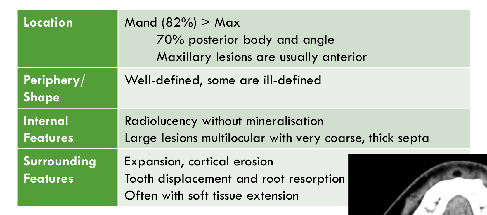
Location: Mandible (82%) > Maxilla; 70% posterior body and angle; maxillary lesions are usually anterior
-
Periphery/Shape: Well-defined, some are ill-defined
-
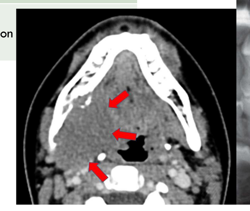
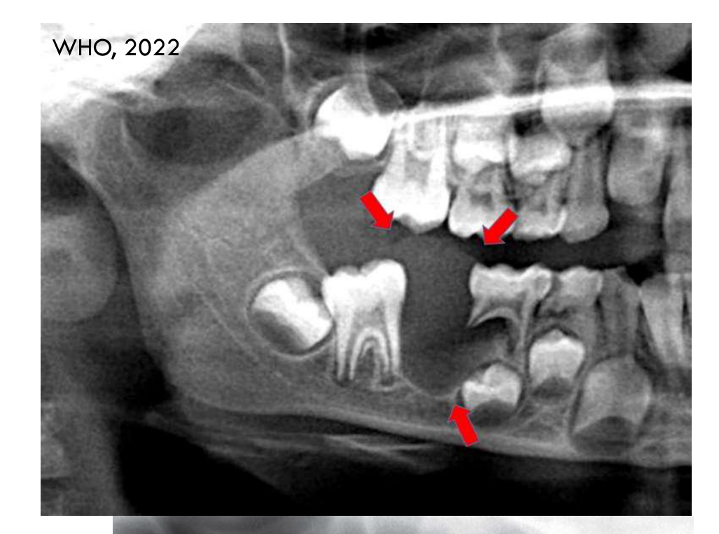
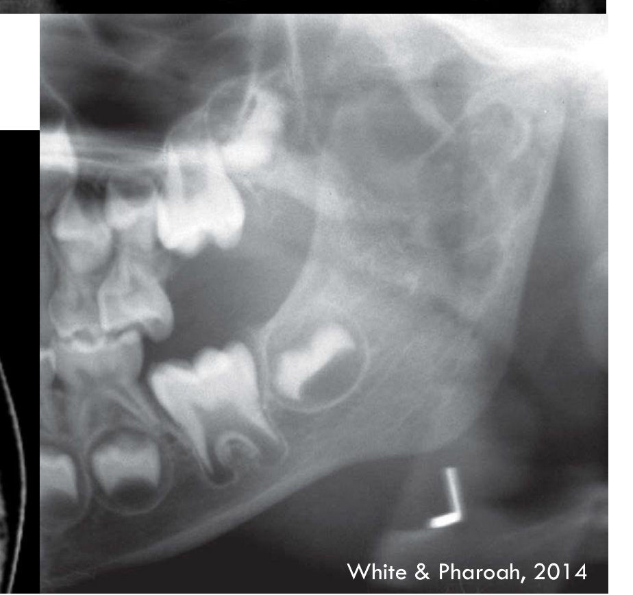
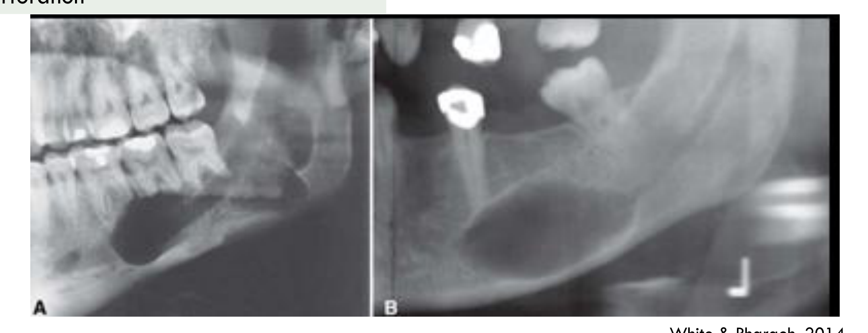
Internal Features: Radiolucency without mineralisation; large lesions multilocular with very coarse, thick septa
-
Surrounding Features:
- Expansion, cortical erosion
- Tooth displacement and root resorption
- Often with soft tissue extension
-
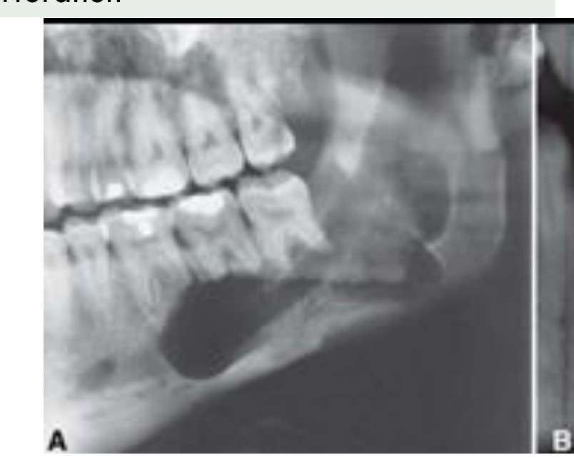
Large lesions may have very coarse, thick septa that are straight or irregular.
 |  |  |
 |  |
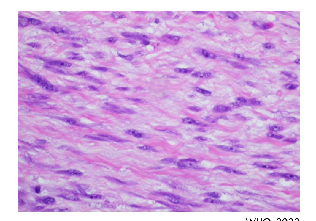
Histopathology25
- Uniform benign spindle cells with slender tapering nuclei, arranged in intertwining fascicles, without atypia or pleomorphism and only rare mitoses
- Due to the infiltrative margin, entrapped residual bone trabeculae may be present
Management/Prognosis/Recurrence
- Frequently recurs after curettage (31%) or enucleation (25%)
- Approximately 10% recurrence after resection
- Chemotherapy may be considered if excision is not feasible
- Radiotherapy is not recommended
Part 2: Soft Tissue Tumours26
-
Vascular tumours
- Haemangioma
-
Peripheral nerve sheath tumours
- Neurofibroma
- Schwannoma
- Neuroma
Central Haemangioma2728
| Feature | Details |
|---|---|
| Definition | A benign vascular neoplasm |
| Age | First decade, although may occur later in life |
| Gender | F > M (2:1) |
| Aetiology | Trauma or developmental in origin |
| Clinical Features | Slow, non-tender, bony hard expansion of the jaw over several months/years +/- pain, compressible and pulsatile (bruit on auscultation) +/- anaesthesia of skin supplied by the mental nerve +/- loosening & migration of teeth (Grade III mobility) +/- bleeding around neck of affected teeth |
| Management | Should be treated without delay due to the risk of lethal exsanguination. May involve one or a combination of embolisation, surgery, and/or sclerosing techniques. |
Lecturer — Haemangioma Context
Haemangiomas are more common in the soft tissues of the head and neck than in bone.
- Common soft-tissue sites include the gingiva and lips.
- The central type occurs within bone and is the focus of the radiological discussion.
 |  |
 |
Radiographic Features29
| Feature | Details |
|---|---|
| Location | More common in vertebrae or skull. Rarely in jaws. Posterior mandible — body, ramus, or within the IAC |
| Periphery / Shape | Well-defined and corticated or ill-defined (simulating malignancy) |
| Internal Features | When small, unilocular and lucent. Variably multilocular with honeycomb pattern with coarse, dense, well-defined trabeculae If involving the IAC, the whole canal is enlarged with serpiginous shape |
Lecturer — Haemangioma Imaging
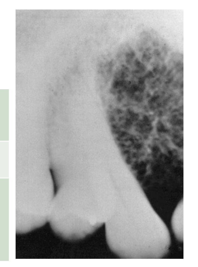
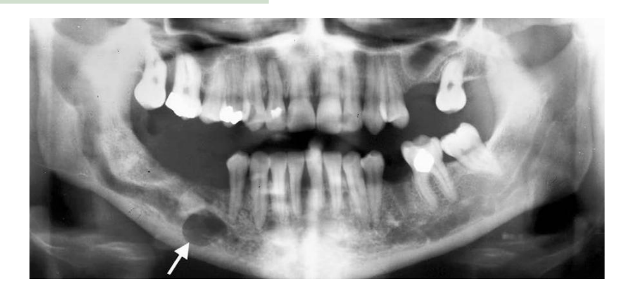
Small lesions may resemble an enlarged marrow space, while larger lesions become variably multilocular according to the amount of entrapped bone.
- When the inferior alveolar canal is involved, the mental foramen may also be enlarged.
- The enlarged canal may become serpiginous or snake-like.
White & Pharoah, 2014
 |  |
 |
Surrounding Features30
- Tooth displacement and root resorption
- IAC and associated foramina are often enlarged with serpiginous shape
- Involved bone may be enlarged, with coarse trabeculae
- Enlarged teeth with earlier eruption
- Small, channel-like perforations through the cortices
- May show spiculated, sun-ray-like periosteal response when it perforates the cortex and involves the periosteum
- Intralesional phleboliths may be present
- Presence of phleboliths raise the suspicion of a vascular malformation, typically venous type
- Well-defined, round calcifications with targetoid appearance
Lecturer — Haemangioma Effects
A soft-tissue vascular malformation may apply pressure to the mandible and cause resorption of the jawbone.
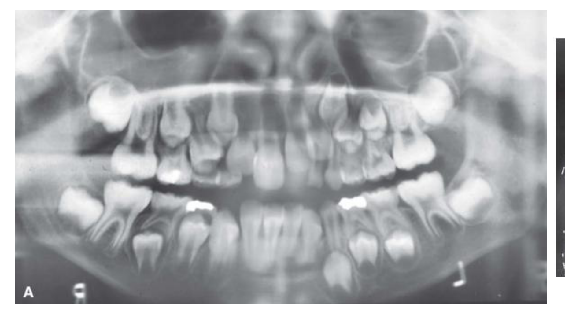
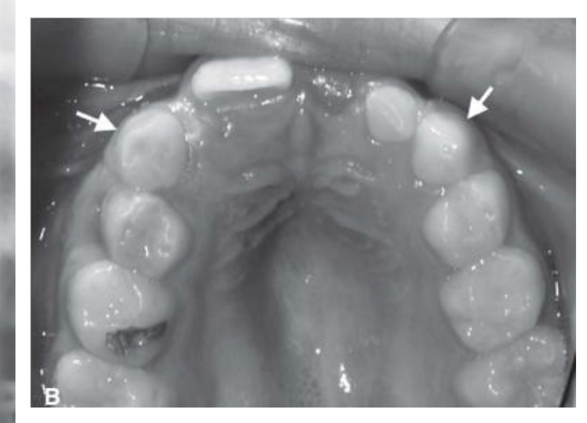
- A soft-tissue haemangioma may cause more advanced root development and eruption of the canine and premolars on the affected side.
- An associated primary tooth may be enlarged compared with the contralateral tooth.
White & Pharoah, 2014
White & Pharoah, 2014
 |  |
 |
Neurofibroma31
| Feature | Details |
|---|---|
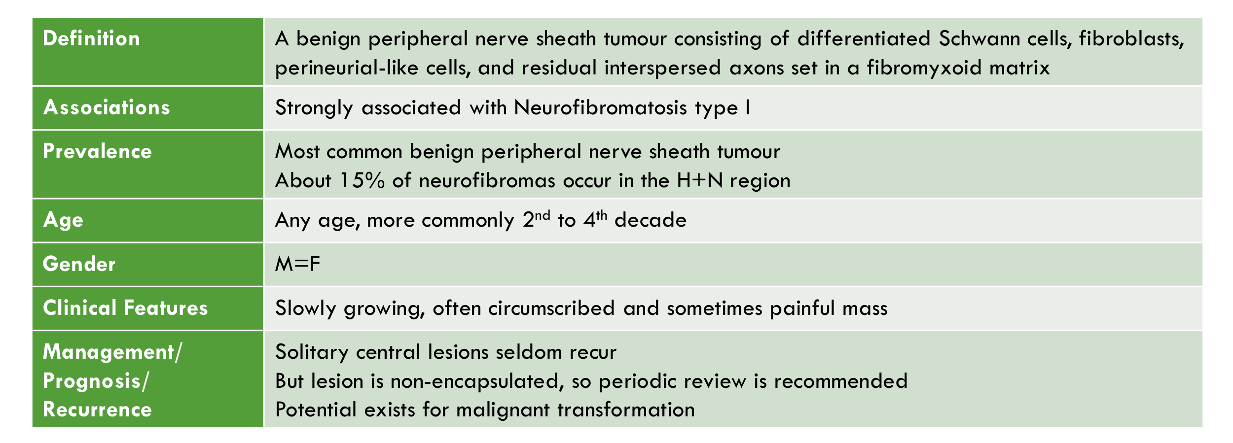
| Definition | A benign peripheral nerve sheath tumour consisting of differentiated Schwann cells, fibroblasts, perineurial-like cells, and residual interspersed axons set in a fibromyxoid matrix |
| Associations | Strongly associated with Neurofibromatosis type I |
| Prevalence | Most common benign peripheral nerve sheath tumour About 15% of neurofibromas occur in the H+N region |
| Age | Any age, more commonly 2nd to 4th decade |
| Gender | M = F |
| Clinical Features | Slowly growing, often circumscribed and sometimes painful mass |
| Management / Prognosis / Recurrence | Solitary central lesions seldom recur But lesion is non-encapsulated, so periodic review is recommended Potential exists for malignant transformation |
Radiographic Features32
| Feature | Details |
|---|---|
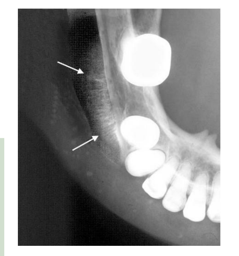
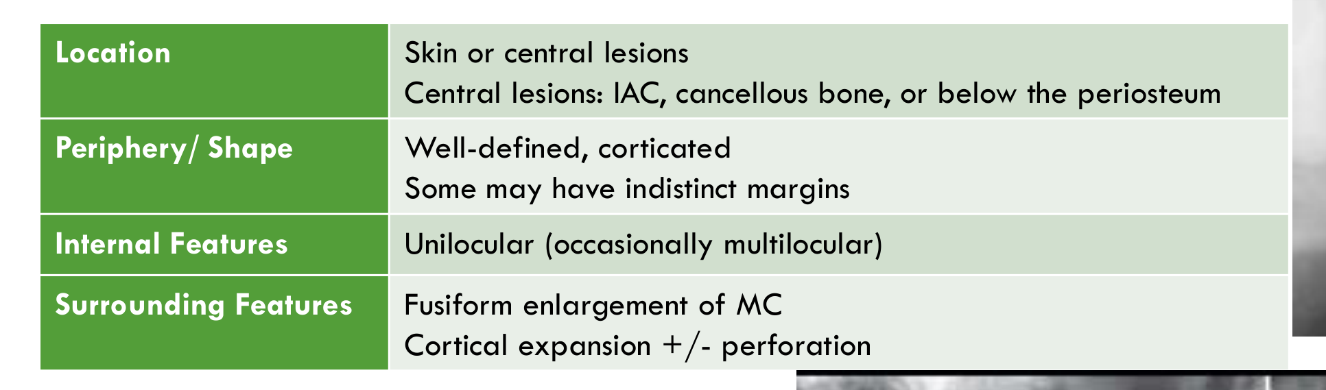
| Location | Skin or central lesions Central lesions: IAC, cancellous bone, or below the periosteum |
| Periphery / Shape | Well-defined, corticated Some may have indistinct margins |
| Internal Features | Unilocular (occasionally multilocular) |
| Surrounding Features | Fusiform enlargement of MC Cortical expansion +/- perforation |
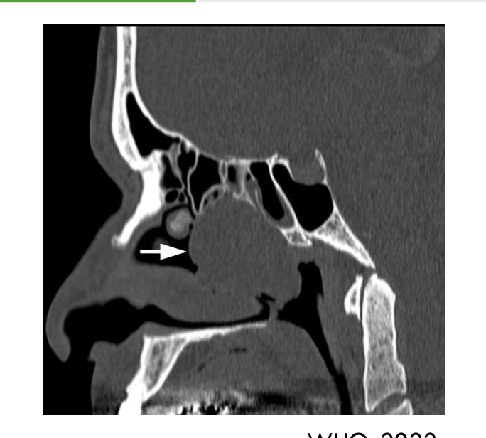
Lecturer — Neurofibroma Margins
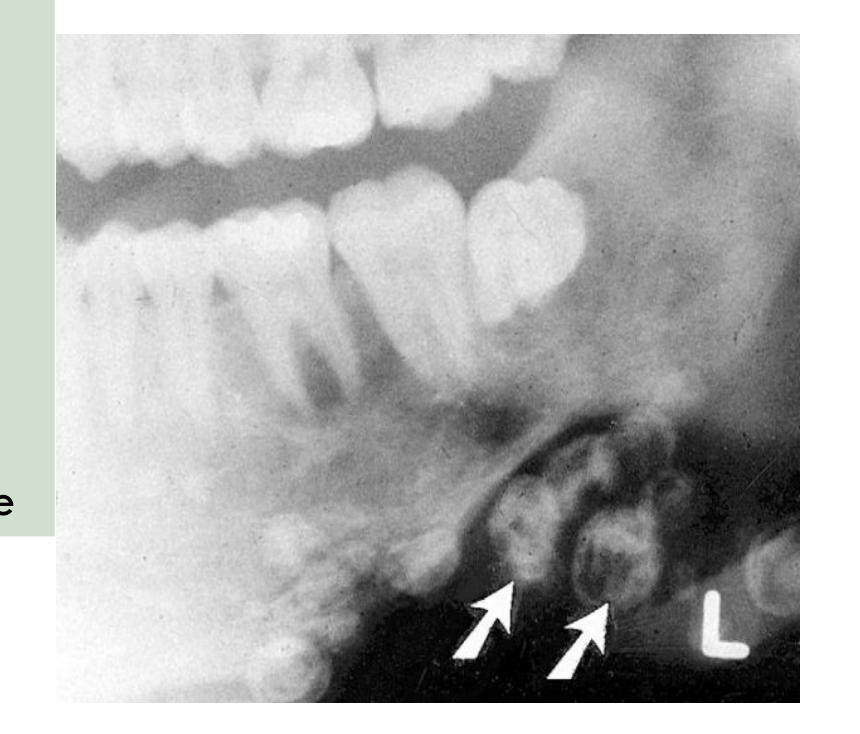
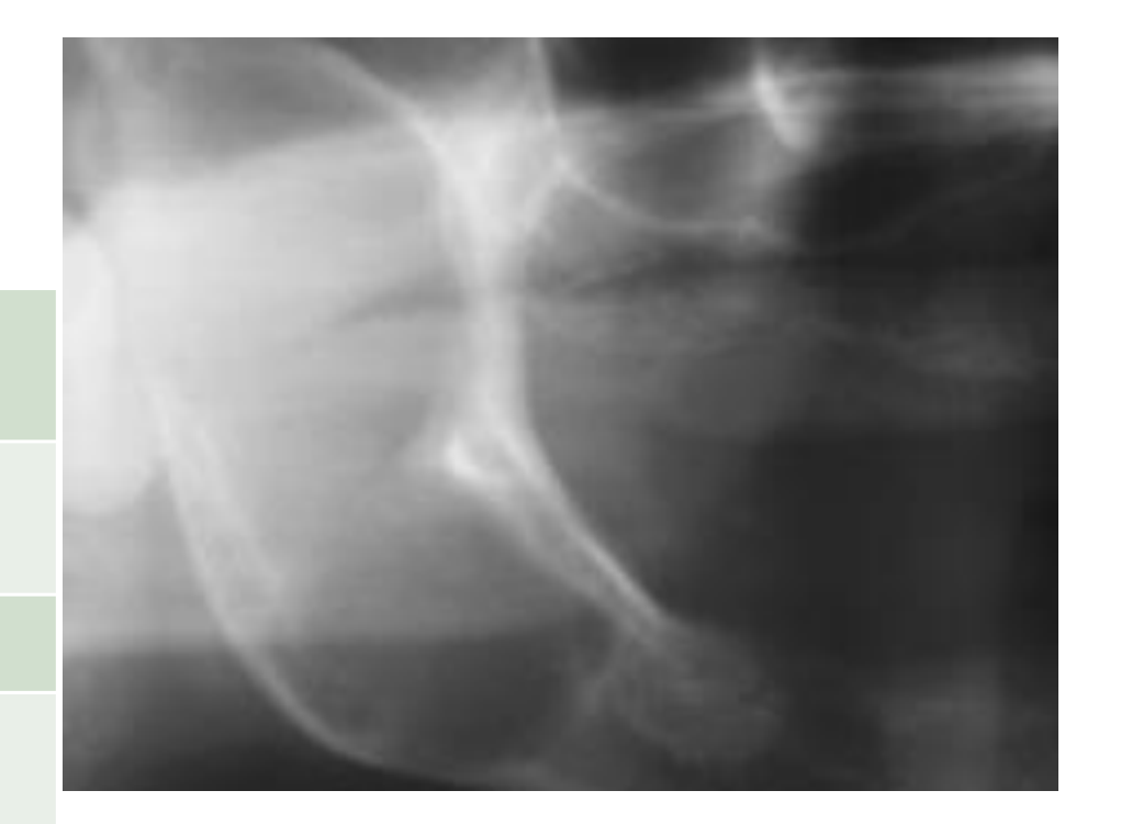
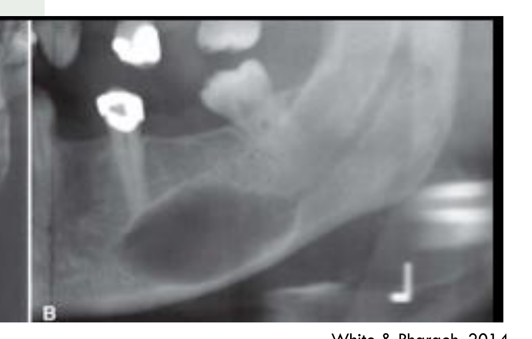
A sinonasal neurofibroma may have poorly defined bony margins around much of the lesion.
White & Pharoah, 2014
 |  |  |
 |  |  |
Schwannoma33
| Feature | Details |
|---|---|
| Definition | A nerve sheath tumour composed entirely or nearly entirely of differentiated neoplastic Schwann cells. |
| Prevalence | Up to 40% of schwannomas occur in the head and neck |
| Age | 2nd to 5th decades |
| Gender | M = F |
| Aetiology | Most lesions are sporadic. Possible association with neurofibromatosis type 2 and schwannomatosis |
| Clinical Features | Slowly growing sometimes painful mass Other signs depend largely on size, nerve of origin, and localization |
| Management / Prognosis / Recurrence | Schwannomas are benign Multifocality may mimic recurrence or malignancy |
Lecturer — Schwannoma Features
Schwannoma was formerly called neurolemmoma, but that term is no longer recommended.
- Malignant transformation is unheard of.
- Trigeminal nerve involvement may cause neuralgia, numbness in the relevant nerve distribution, or local mass effect.
- Cranial nerve VII or VIII involvement may cause sensorineural hearing loss or non-pulsatile tinnitus.
- Intracranial schwannomas account for approximately 6–8% of all intracranial tumours; the commonly involved nerves include cranial nerves VIII and VII and nerves IX, X, and XI within the jugular foramen.
Radiographic Features34
| Feature | Details |
|---|---|
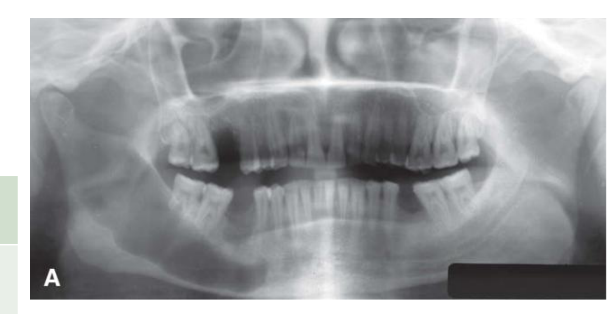
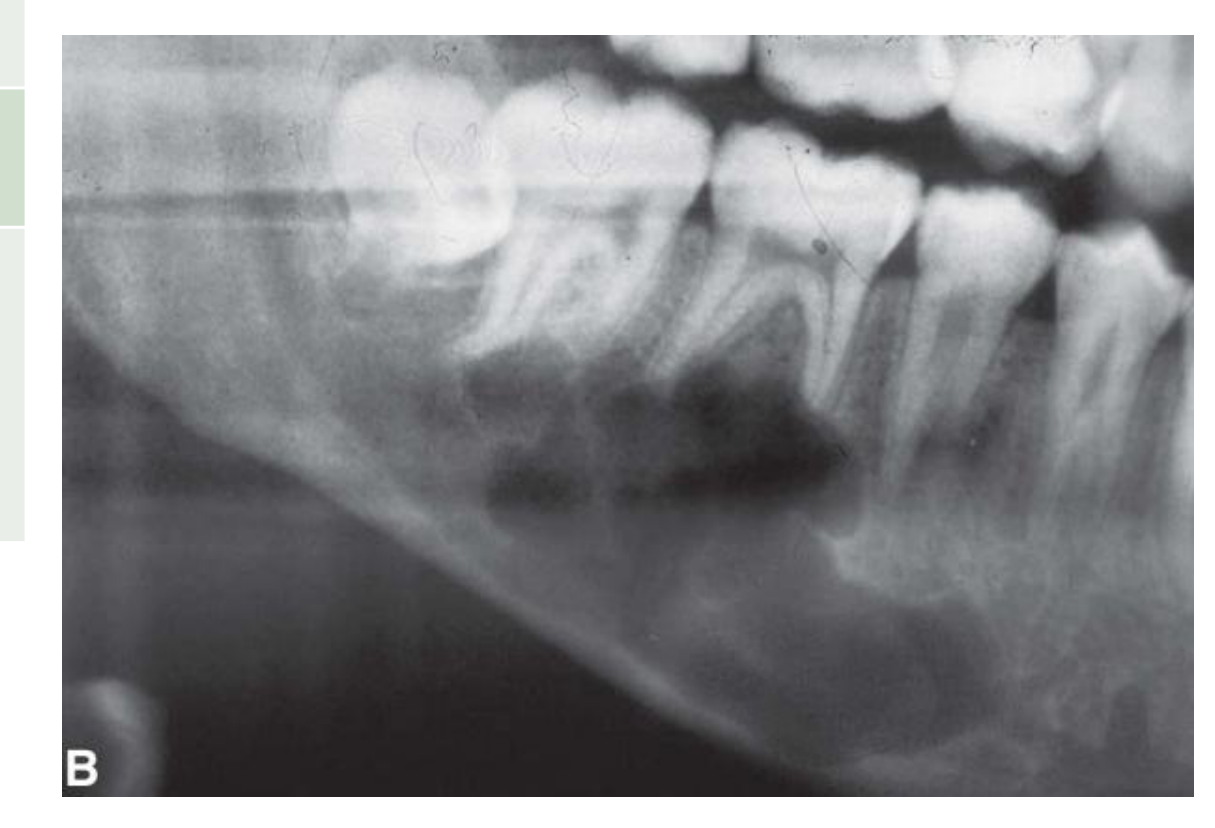
| Location | Intracranial (almost all involve cranial nerves) Mandible — most often within the IAC |
| Periphery / Shape | Well-circumscribed, corticated. Small lesions are cyst-like, but tend to be fusiform in shape Large lesions demonstrate scalloping outline |
| Internal Features | Typically lucent. Calcifications rare. Larger lesions demonstrate more heterogeneity |
| Surrounding Features | Enlargement of the mandibular/mental foramen Outer cortex of canal is maintained Expansion of canal is localised with a definite epicentre unless lesion is large +/- root resorption |
White & Pharoah, 2014
 |  |
 |
Neuroma35
| Feature | Details |
|---|---|
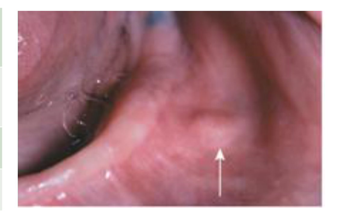
| Definition | Comprise a diverse group of peripheral nerve sheath tumors, some reactive and hyperplastic, including the Traumatic neuroma (TN). |
| Aetiology | Accidental or iatrogenic nerve injury or amputation. |
| Clinical Features | Slow-growing, reactive hyperplasias, seldom >1cm in diameter Severe pain (as the mass applies pressure within its bony cavity or a result of external trauma), reflex neuralgia, referred pain to eyes, face, and head. |
| Management / Prognosis / Recurrence | Simple excision when symptomatic. Recurrence is uncommon. |
Lecturer — Traumatic Neuroma
Following nerve injury, severed nerve fibres attempt to regenerate and overgrow, producing an abnormal peripheral scar.
- The scar contains a disorganised collection of axons, perineural connective tissue, Schwann cells, and scar tissue.
- Other types of neuroma occur in soft tissues and are not seen radiographically.
- Pain may be referred to other regions of the head as the lesion enlarges.
Radiographic Features36
| Feature | Details |
|---|---|
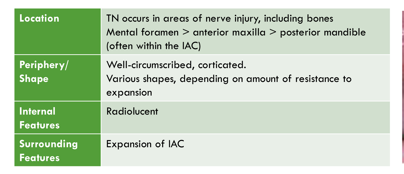
| Location | TN occurs in areas of nerve injury, including bones Mental foramen > anterior maxilla > posterior mandible (often within the IAC) |
| Periphery / Shape | Well-circumscribed, corticated. Various shapes, depending on amount of resistance to expansion |
| Internal Features | Radiolucent |
| Surrounding Features | Expansion of IAC |
Lecturer — Neuroma Imaging
Radiographic detection of a traumatic neuroma is rare.
 |  |
References37
-
WHO Classification of Tumours Editorial Board. Head and neck tumours. Lyon (France): International Agency for Research on Cancer; 2022 (WHO classification of tumours series, 5th ed.; vol. 9).
-
Koong, B. (2017). Atlas of Oral and Maxillofacial Radiology. Chichester, UK: John Wiley and Sons.
-
White, and Pharoah. (2014). Oral radiology : Principles and interpretation (7th ed.). St. Louis: Elsevier/Mosby.
Clinical Cases
Case: Mandibular Lingual Ramus Lesion
Question
Scenario: A patient presents with a lesion in the mandible, specifically around the ramus region.
What’s shown: Multi-slice CT imaging demonstrating a lesion on the lingual aspect of the ramus. The lesion has an internal trabecular architecture and is located near the medial pterygoid muscle, tonsil, and parapharyngeal space.
Consider: Identify the specific location of the lesion, its internal characteristics, and its potential impact on the surrounding anatomical structures.
Answer
Observations:
- The lesion is located on the lingual aspect of the mandibular ramus.
- It exhibits an internal trabecular architecture.
- It is situated in close proximity to the medial pterygoid muscle, tonsil, and parapharyngeal space.
Reasoning: The multi-slice CT clarifies the lingual position, which might be hard to appreciate on other views. The internal trabecular pattern is characteristic of certain bone-forming lesions. Its large size and location allow it to impact adjacent soft tissues like the medial pterygoid muscle and parapharyngeal space, potentially causing dysfunction.
Takeaway: Osteomas can have internal trabecular architecture and, when sufficiently large in the lingual ramus region, can displace and impact adjacent soft tissues such as the medial pterygoid muscle and parapharyngeal space.
Case: Mandibular Condyle Bony Outgrowth
Question
Scenario: A patient presents with a bony outgrowth on the mandibular condyle.
What’s shown: CT images showing a well-defined lobulated bony outgrowth that is in continuity with the cortex and medulla of the native condylar head. The enlargement is uneven, protruding from one part of the condylar head.
Consider: Differentiate the appearance of this lesion from diffuse condylar enlargement and identify the key structural features of the outgrowth.
Answer
Observations:
- A well-defined, lobulated bony outgrowth is present.
- The outgrowth is continuous with the cortex and medulla of the native bone.
- The enlargement is uneven and protrudes from only one part of the condylar head, rather than involving the entire condyle.
Reasoning: The continuity of the marrow cavity and cortex with the native bone is a hallmark of this specific benign neoplasm. The uneven, localized protrusion distinguishes it from condylar hyperplasia, which involves diffuse growth of the entire condylar head.
Takeaway: Osteochondromas of the mandibular condyle present as localized, uneven bony outgrowths continuous with the native bone’s cortex and medulla, distinguishing them from the diffuse enlargement seen in condylar hyperplasia.
Case: Progressive Bony Trabecular Alteration
Question
Scenario: A patient has a bony lesion that is being monitored over time.
What’s shown: A PA radiograph showing an area of altered bony trabecular appearance that has increased in size over time, accompanied by buccal and lingual cortical expansion.
Consider: Identify the radiographic progression and the specific cortical changes associated with this lesion.
Answer
Observations:
- There is an area of altered bony trabecular appearance.
- The lesion has increased in size over time.
- Buccal and lingual cortical expansion is present.
Reasoning: The progressive increase in size and the specific pattern of cortical expansion (buccal and lingual) are characteristic radiographic features of this small bone-forming tumor, which typically develops in the outer cortex.
Takeaway: Osteoid osteomas can present radiographically with an area of altered bony trabeculae that increases in size over time and causes buccal and lingual cortical expansion.
Case: TMJ/Temporal Bone Lesion with Muscle Displacement
Question
Scenario: A patient presents with a lesion in the TMJ or squamous part of the temporal bone region.
What’s shown: A soft tissue window of a multislice CT showing a well-demarcated lobulated lesion with internal foci of calcifications. The lesion demonstrates cortical expansion and displaces the lateral pterygoid muscle posteriorly and medially, as well as displacing the temporalis muscle.
Consider: Identify the internal characteristics of the lesion and its mass effect on the surrounding masticatory muscles.
Answer
Observations:
- A well-demarcated, lobulated lesion with internal foci of calcifications.
- Cortical expansion is present.
- The lateral pterygoid muscle is displaced slightly posteriorly and medially.
- The temporalis muscle is also slightly displaced.
Reasoning: The presence of calcifications within a lobulated, well-demarcated lesion in the TMJ/temporal bone region is characteristic of a chondroid tumor. The mass effect clearly demonstrates the displacement of adjacent muscles of mastication.
Takeaway: Chondroblastomas in the TMJ/temporal bone region appear as well-demarcated lobulated lesions with calcifications that can cause cortical expansion and displace adjacent muscles like the lateral pterygoid and temporalis.
Case: Craniofacial Bone Lesion with Focal Mineralizations
Question
Scenario: A patient presents with a well-demarcated lesion in a craniofacial bone.
What’s shown: Imaging demonstrating a well-demarcated lesion with a sclerotic border. The lesion is primarily radiolucent internally but contains small focal mineralizations.
Consider: Identify the border characteristics and the specific internal mineralization pattern of this space-occupying lesion.
Answer
Observations:
- The lesion is well-demarcated with a sclerotic border.
- It is primarily radiolucent internally.
- Small focal mineralizations are present within the lesion.
Reasoning: The well-demarcated nature with a sclerotic border indicates a benign, slow-growing process. The primarily radiolucent internal appearance with small focal mineralizations is a specific feature seen in a minority of these chondroid neoplasms.
Takeaway: Chondromyxofibromas are well-demarcated, primarily radiolucent lesions with a sclerotic border, and a subset of cases will demonstrate small focal internal mineralizations.
Case: Multilocular Mandibular Lesion with Tooth Displacement
Question
Scenario: A patient presents with a large, multilocular lesion in the posterior mandible.
What’s shown: Imaging showing a multilocular radiolucency with very coarse, thick septa. There is destruction of the lingual cortex of the mandible. Tooth 46 is displaced distally, and tooth 85 is displaced mesially, with evidence of root resorption.
Consider: Identify the septal pattern, the extent of cortical destruction, and the specific effects on the adjacent dentition.
Answer
Observations:
- A multilocular radiolucency with coarse, thick septa (straight or irregular).
- Destruction and effacement of the lingual cortex.
- Distal displacement of tooth 46 and mesial displacement of tooth 85.
- Root resorption is present.
Reasoning: The coarse, thick septa and multilocular appearance indicate a locally aggressive fibroblastic process. The destruction of the lingual cortex and the significant displacement and resorption of adjacent teeth highlight its aggressive, space-occupying nature within the bone.
Takeaway: Desmoplastic fibromas of bone can present as multilocular radiolucencies with coarse thick septa, causing cortical destruction and significant tooth displacement and root resorption.
Case: Mandibular Lesion with Honeycomb Trabeculae and Enlarged Canal
Question
Scenario: A patient presents with a vascular lesion within the mandible.
What’s shown: Imaging showing a honeycomb pattern with coarse, dense, well-defined trabeculae. The inferior alveolar canal is enlarged and serpiginous (snake-like), with an enlarged mental foramen. Lucent regions are visible in the cortex.
Consider: Identify the trabecular pattern, the morphology of the inferior alveolar canal, and the cortical findings.
Answer
Observations:
- A honeycomb pattern with coarse, dense, well-defined trabeculae.
- An enlarged, serpiginous (snake-like) inferior alveolar canal.
- An enlarged mental foramen.
- Lucent regions in the cortex representing vascular channels.
Reasoning: The honeycomb trabecular pattern and the specific serpiginous enlargement of the inferior alveolar canal and mental foramen are classic radiographic signs of a central vascular lesion within the bone. The cortical lucencies correspond to vascular channels perforating the cortex.
Takeaway: Central hemangiomas of the jaw often present with a honeycomb trabecular pattern and cause serpiginous enlargement of the inferior alveolar canal and mental foramen, with lucent vascular channels visible in the cortex.
Case: Soft Tissue Vascular Malformation with Mandibular Resorption
Question
Scenario: A patient presents with a soft tissue vascular malformation adjacent to the mandible.
What’s shown: Imaging showing a soft tissue mass applying pressure to the mandible, resulting in resorption of the jawbone. Calcified structures are visible within the soft tissue mass.
Consider: Identify the mechanism of bone resorption and the nature of the calcified structures within the soft tissue lesion.
Answer
Observations:
- A soft tissue mass applying pressure to the mandible.
- Resorption of the adjacent jawbone.
- Calcified structures (phleboliths) within the soft tissue mass.
Reasoning: The soft tissue vascular malformation exerts pressure on the adjacent bone, leading to pressure resorption. The calcified structures represent phleboliths, which are calcified thrombi typically found within venous vascular malformations.
Takeaway: Soft tissue venous vascular malformations can cause pressure resorption of adjacent jawbone and often contain phleboliths (calcified thrombi).
Case: Soft Tissue Hemangioma Affecting Dentition
Question
Scenario: A pediatric patient presents with a soft tissue hemangioma on one side of the jaw.
What’s shown: A panoramic radiograph showing advanced root development and eruption of the right canine and premolars compared to the contralateral side. An occlusal photo shows that tooth 53 is noticeably enlarged compared to tooth 63.
Consider: Identify the effects of the soft tissue hemangioma on the developing dentition and tooth morphology.
Answer
Observations:
- Advanced root development and eruption of the right canine and premolars on the affected side.
- Enlargement of tooth 53 compared to the contralateral tooth 63.
Reasoning: The increased vascularity and local tissue changes caused by the hemangioma stimulate the developing dental follicles, leading to accelerated root development and eruption. The same hyperemic effect can result in macrodontia (enlarged tooth size) on the affected side.
Takeaway: Soft tissue hemangiomas in the jaw can accelerate root development and eruption of the local dentition and may cause enlargement of the affected teeth.
Case: Inferior Alveolar Canal Lesion
Question
Scenario: A patient presents with a lesion located within the inferior alveolar canal.
What’s shown: Imaging showing a fusiform enlargement of the inferior alveolar canal, which is not circular. Cortical expansion is present, with or without perforation.
Consider: Identify the shape of the canal enlargement and the associated cortical changes.
Answer
Observations:
- Fusiform (spindle-shaped) enlargement of the inferior alveolar canal.
- The enlargement is not circular.
- Cortical expansion is present.
Reasoning: The fusiform shape of the canal enlargement is characteristic of a peripheral nerve sheath tumor growing along the nerve within the canal. The expansion reflects the slow-growing, space-occupying nature of the lesion within the confined bony canal.
Takeaway: Neurofibromas occurring within the inferior alveolar canal typically present as a fusiform, non-circular enlargement of the canal with cortical expansion.
Case: Sinonasal Nerve Sheath Lesion
Question
Scenario: A patient presents with a lesion in the sinonasal region.
What’s shown: A sagittal slice of a multislice CT showing a lesion in the sinonasal area where the bony margins are not fully present around much of the lesion.
Consider: Identify the relationship of the lesion to the surrounding bony margins in the sinonasal region.
Answer
Observations:
- A lesion in the sinonasal region.
- Bony margins are absent or not fully present around much of the lesion.
Reasoning: The lack of complete bony margins indicates that the lesion has expanded or remodeled the surrounding sinonasal bone, which is characteristic of a slow-growing nerve sheath tumor in this anatomical region.
Takeaway: Sinonasal neurofibromas can present on CT with absent or incomplete bony margins due to bone remodeling and expansion by the tumor.
Footnotes
-
Original PDF page 1: L3.3 Benign tumours of the jaws Part 2, p.1 ↩
-
Original PDF page 2: L3.3 Benign tumours of the jaws Part 2, p.2 ↩
-
Original PDF page 3: L3.3 Benign tumours of the jaws Part 2, p.3 ↩
-
Original PDF page 4: L3.3 Benign tumours of the jaws Part 2, p.4 ↩
-
Original PDF page 5: L3.3 Benign tumours of the jaws Part 2, p.5 ↩
-
Original PDF page 6: L3.3 Benign tumours of the jaws Part 2, p.6 ↩
-
Original PDF page 7: L3.3 Benign tumours of the jaws Part 2, p.7 ↩
-
Original PDF page 8: L3.3 Benign tumours of the jaws Part 2, p.8 ↩
-
Original PDF page 9: L3.3 Benign tumours of the jaws Part 2, p.9 ↩
-
Original PDF page 10: L3.3 Benign tumours of the jaws Part 2, p.10 ↩
-
Original PDF page 11: L3.3 Benign tumours of the jaws Part 2, p.11 ↩
-
Original PDF page 12: L3.3 Benign tumours of the jaws Part 2, p.12 ↩
-
Original PDF page 13: L3.3 Benign tumours of the jaws Part 2, p.13 ↩
-
Original PDF page 14: L3.3 Benign tumours of the jaws Part 2, p.14 ↩
-
Original PDF page 15: L3.3 Benign tumours of the jaws Part 2, p.15 ↩
-
Original PDF page 16: L3.3 Benign tumours of the jaws Part 2, p.16 ↩
-
Original PDF page 17: L3.3 Benign tumours of the jaws Part 2, p.17 ↩
-
Original PDF page 18: L3.3 Benign tumours of the jaws Part 2, p.18 ↩
-
Original PDF page 19: L3.3 Benign tumours of the jaws Part 2, p.19 ↩
-
Original PDF page 20: L3.3 Benign tumours of the jaws Part 2, p.20 ↩
-
Original PDF page 21: L3.3 Benign tumours of the jaws Part 2, p.21 ↩
-
Original PDF page 22: L3.3 Benign tumours of the jaws Part 2, p.22 ↩
-
Original PDF page 23: L3.3 Benign tumours of the jaws Part 2, p.23 ↩
-
Original PDF page 24: L3.3 Benign tumours of the jaws Part 2, p.24 ↩
-
Original PDF page 25: L3.3 Benign tumours of the jaws Part 2, p.25 ↩
-
Original PDF page 26: L3.3 Benign tumours of the jaws Part 2, p.26 ↩
-
Original PDF page 27: L3.3 Benign tumours of the jaws Part 2, p.27 ↩
-
Original PDF page 30: L3.3 Benign tumours of the jaws Part 2, p.30 ↩
-
Original PDF page 28: L3.3 Benign tumours of the jaws Part 2, p.28 ↩
-
Original PDF page 29: L3.3 Benign tumours of the jaws Part 2, p.29 ↩
-
Original PDF page 31: L3.3 Benign tumours of the jaws Part 2, p.31 ↩
-
Original PDF page 32: L3.3 Benign tumours of the jaws Part 2, p.32 ↩
-
Original PDF page 33: L3.3 Benign tumours of the jaws Part 2, p.33 ↩
-
Original PDF page 34: L3.3 Benign tumours of the jaws Part 2, p.34 ↩
-
Original PDF page 35: L3.3 Benign tumours of the jaws Part 2, p.35 ↩
-
Original PDF page 36: L3.3 Benign tumours of the jaws Part 2, p.36 ↩
-
Original PDF page 37: L3.3 Benign tumours of the jaws Part 2, p.37 ↩