Trauma Imaging1
Dr Dayea Oh — Oral & Maxillofacial Radiologist
 |  |
 |
Objectives2
-
Understand imaging goals in facial trauma
-
Review relevant maxillofacial anatomy on imaging
-
Compare CT, CBCT, and I/O radiographs
-
Review imaging appearances of dental and facial injuries
-
Compare periapical, occlusal, CBCT, and multislice CT imaging
Contents
Dento-alveolar trauma
- Injuries to dental structures
- Injuries to supporting structures
When to use Cone Beam CT
Facial Trauma & Multislice CT
Dento-Alveolar Trauma3
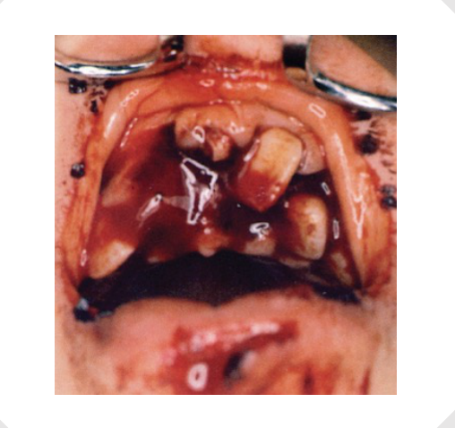
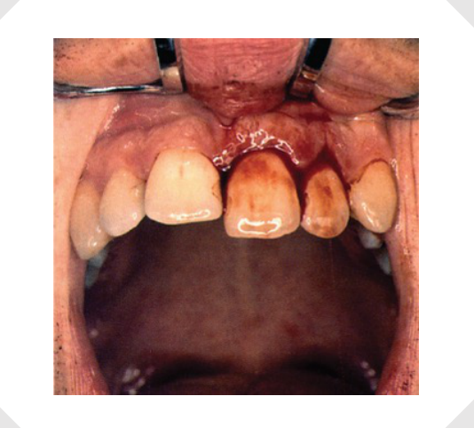
Epidemiology and Risk Factors45
- ~5% of all bodily injuries (all ages)
- 15.5% of patients aged between 7 and 20 had at least one traumatic dental injury to permanent dentition (Eilert-Petersson, 1997)
- Primary dentition injury prevalence 22.7% (Petti, Glendor and Andersson, 2018)
- If trauma occurs at an age below 9 years, there is 8x risk for new trauma (Glendor, 2007)
Lecturer — Epidemiology Details
The 1997 global study included more than 250,000 patients. A more recent global study reported a 15.2% prevalence of dental trauma in permanent dentition, based on a sample of 1 billion.
- Increased overjet with protrusion of upper incisors
- Insufficient lip closure
- Behaviour
- Risk-taking
- Peer relationship problems
- Attention-deficit hyperactivity disorder (ADHD)
- Stress
- Peer relationship problems may include bullying
Lecturer — Trauma Context
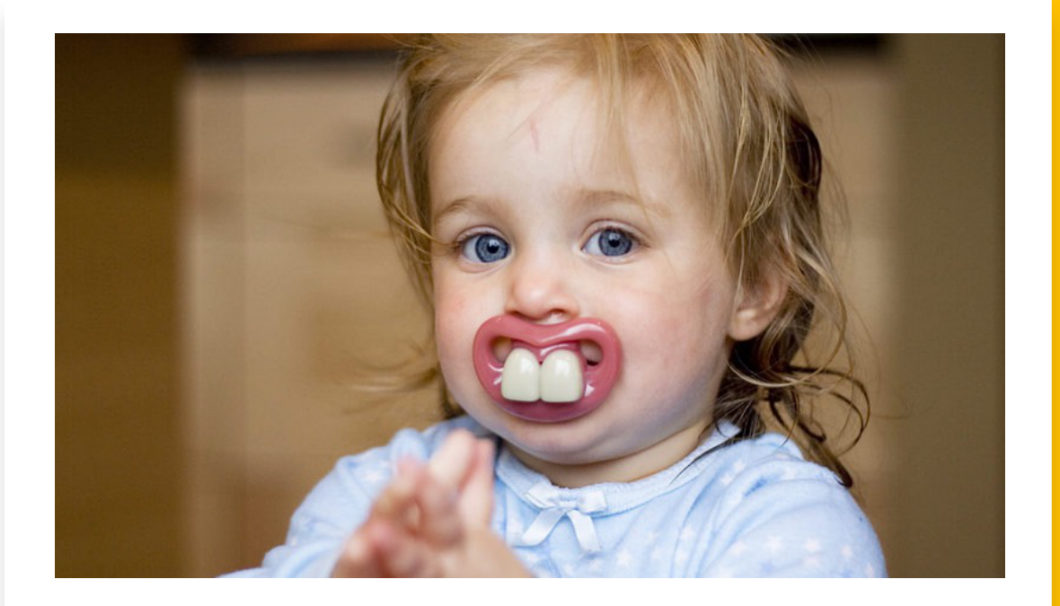
Dento-alveolar trauma involves injury to the teeth, periodontal tissues, alveolar bone, and adjacent soft tissues. It is common in young populations and is usually associated with accidents or interpersonal violence.
 |  |
Classification Systems6
- More than 50 classifications (Andersson 2022)
- Modified Andreasen’s (by WHO) in 1994
- Most frequently used
- Applies to both primary and permanent dentitions
- International Association of Dental Traumatology (IADT)
- American Association of Endodontists (AAE)
Lecturer — Classification History
The original Andreasen classification was published in 1981, and a later version was adopted by the WHO in 2022. The International Association of Dental Traumatology and the American Association of Endodontists have adopted this classification for their trauma guidance.
WHO (Andreasen’s)7
I. Injuries to the hard dental tissues and the pulp II. Injuries to the periodontal tissues III. Injuries to the supporting bone IV. Injuries to gingiva or oral mucosa
- The simplified classification combines periodontal tissues, supporting bone, and gingiva or oral mucosa into the second category
 |  |
NOW
I. Hard tissues and Pulp Injuries II. Periodontal Injuries
Reference Sources
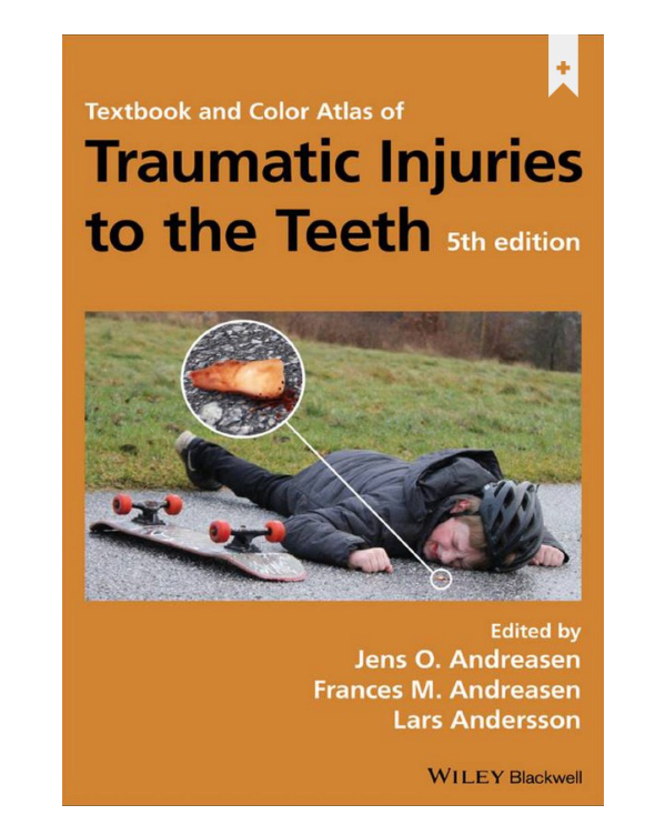
Textbook and Color Atlas of Traumatic Injuries to the Teeth, 5th edition. Edited by Jens O. Andreasen, Frances M. Andreasen, Lars Andersson. WILEY Blackwell.
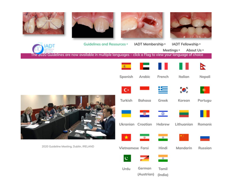
Dental Traumatology. SHORT COMMUNICATION | Open Access | CC. NA0D – The new Traumatic Dental Injury classification of the World Health Organization. Stefano Petti, Jens Ove Andreasen, Ulf Glendor, Lars Andersson. First published: 28 April 2022 | https://doi.org/10.1111/edt.12753
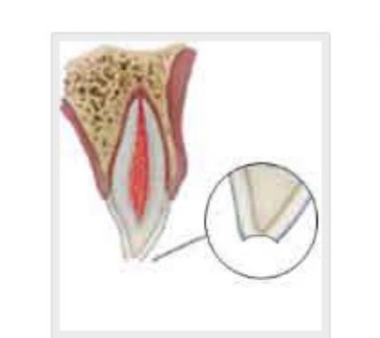
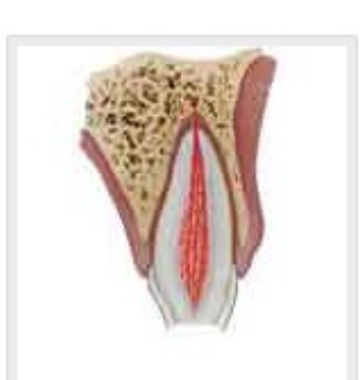
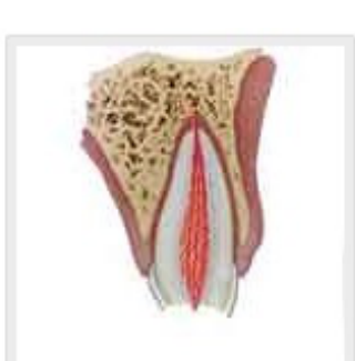
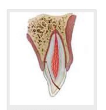
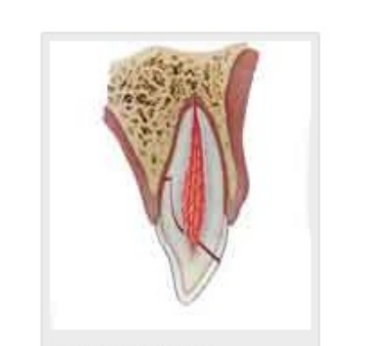
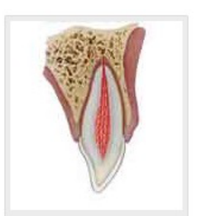
Hard Tissue and Pulp Injuries8
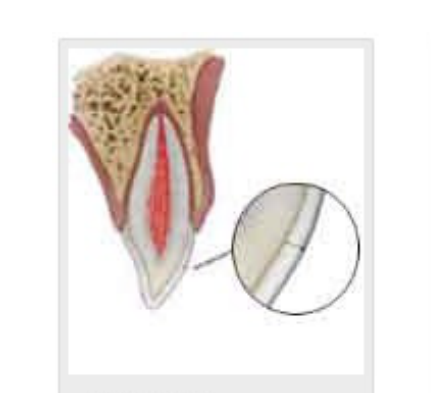
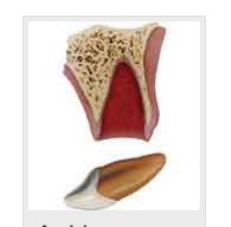
I. Injuries to the hard dental tissues and the pulp
 |  |  |
 |  |  |
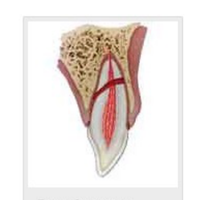 |
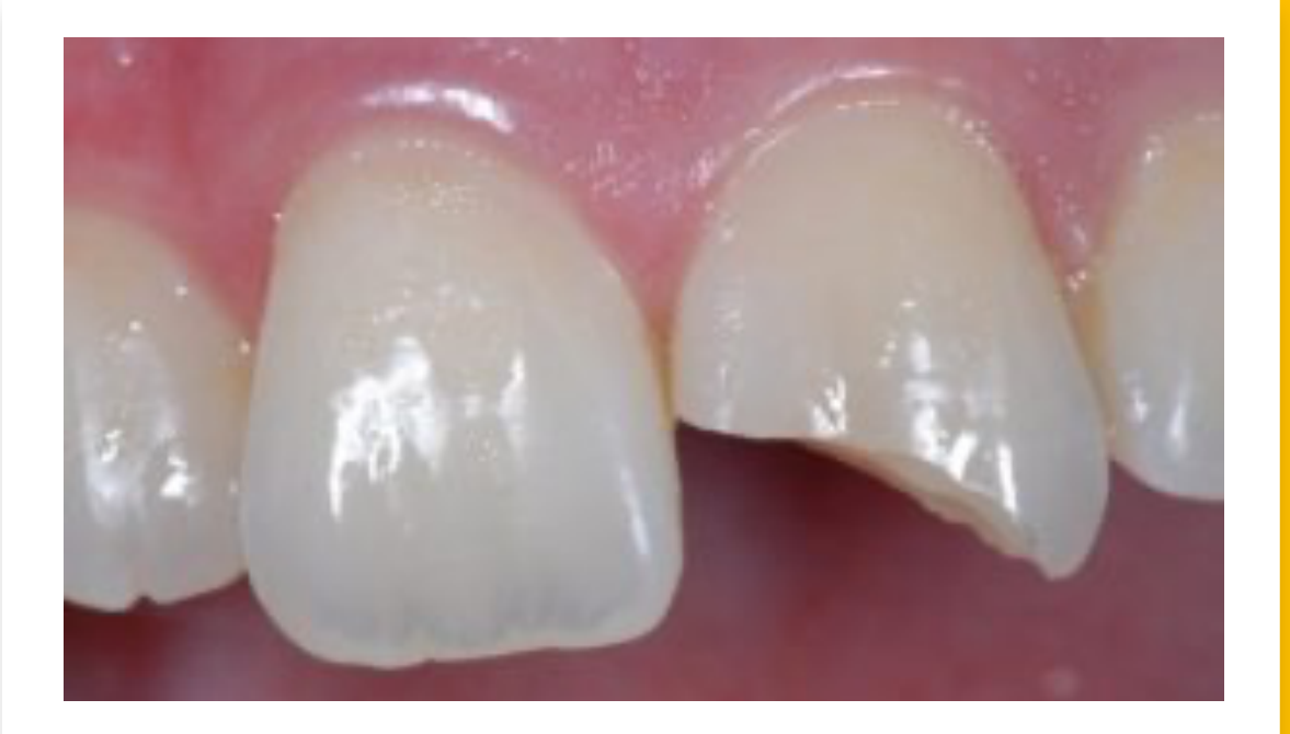
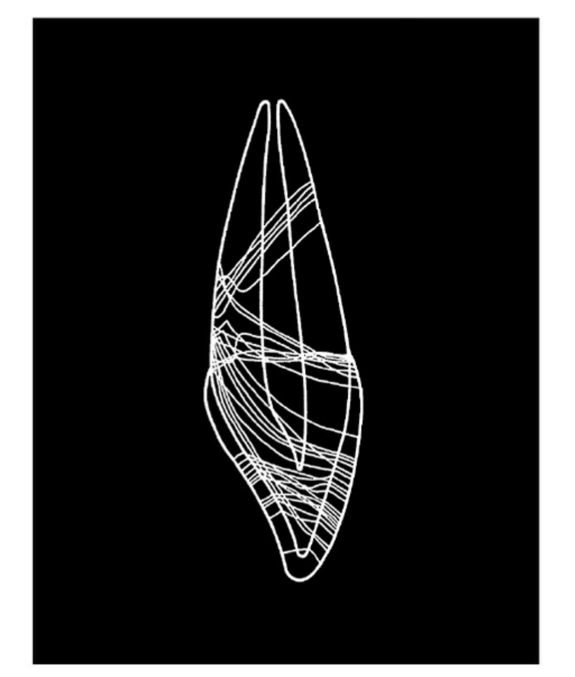
Tooth Fracture Patterns9
- Analysis of 33 fracture lines (of anterior teeth under direct frontal impacts)
- Four common fracture patterns:
- Horizontal crown fracture
- Horizontal fracture at the neck of the tooth
- Oblique crown-root fracture
- Oblique root fracture
Lecturer — Fracture Detection
Two-dimensional imaging has reduced sensitivity for oblique fractures, which are more readily identified on CBCT. When the lips absorb part of the impact force, the forces may be distributed across multiple teeth and can result in concussion, subluxation, lateral luxation, or intrusion.
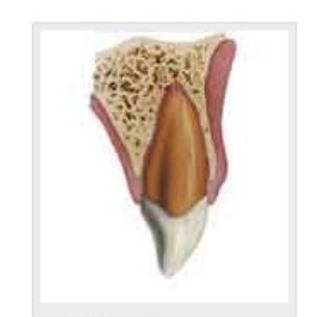
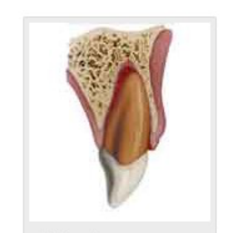
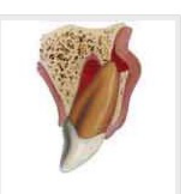
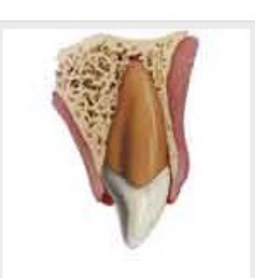
Periodontal Tissue Injuries101112
II. Injuries to the periodontal tissues
“Radiographs are necessary to make a thorough diagnosis of dental injuries. Tooth root and bone fractures, for instance, may occur without any clinical signs or symptoms and are frequently undetected when only one radiographic view is used.” (IADT, 2020)
International Association of Dental Traumatology
Dental Traumatology. COMPREHENSIVE REVIEW | Open Access. International Association of Dental Traumatology guidelines for the management of traumatic dental injuries: 1. Fractures and luxations. Cecilia Bourguignon, Nestor Cohenca, Eva Lauridsen, Marie Therese Flores, Anne C. O’Connell, Peter F. Day, Georgios Tsilingaridis, Paul V. Abbott, Ashraf F. Fouad, Lamar Hicks et al. First published: 31 May 2020 | https://doi.org/10.1111/edt.12578
Since maxillary central incisors are the most frequently affected teeth, the radiographs listed below are recommended to thoroughly examine the injured area:
- One parallel periapical radiograph aimed through the midline to show the two maxillary central incisors.
- One parallel periapical radiograph aimed at the maxillary right lateral incisors (should also show the right canine and central incisor).
- One parallel periapical radiograph aimed at the maxillary left lateral incisor (should also show the left canine and central incisor).
- One maxillary occlusal radiograph.
- At least one parallel periapical radiograph of the lower incisors centered on the two mandibular centrals. However, other radiographs may be indicated if there are obvious injuries of the mandibular teeth (eg, similar periapical radiographs as above for the maxillary teeth, mandibular occlusal radiograph).
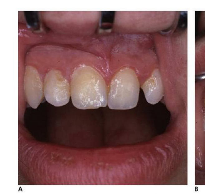
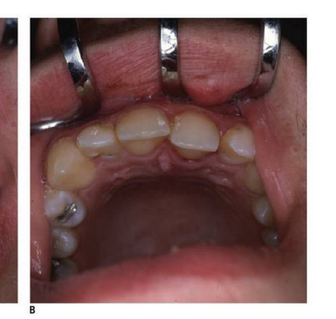
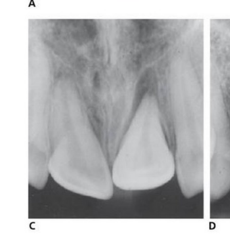
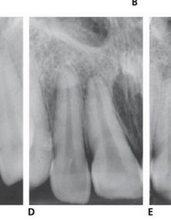
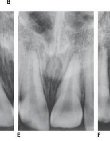
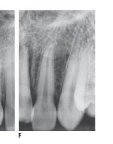
Maxillary Incisor Imaging Example
Clinical examination showed displacement of the left central incisor. The imaging series included one occlusal radiograph and three periapical radiographs; the occlusal view clearly demonstrated the displaced tooth and its socket, whereas one periapical view did not clearly demonstrate the displacement.
 |  |  |
 |  |  |
 |  |  |
 |  |  |
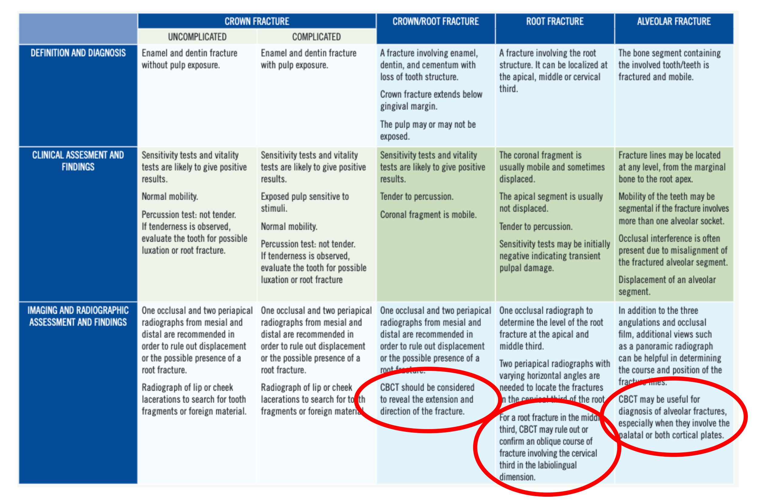
Fracture Injuries — Definition, Clinical Findings, and Imaging13
| CROWN FRACTURE (UNCOMPLICATED) | CROWN FRACTURE (COMPLICATED) | CROWN/ROOT FRACTURE | ROOT FRACTURE | ALVEOLAR FRACTURE | |
|---|---|---|---|---|---|
| Definition and diagnosis | Enamel and dentin fracture without pulp exposure. | Enamel and dentin fracture with pulp exposure. | A fracture involving enamel, dentin, and cementum with loss of tooth structure. Crown fracture extends below gingival margin. The pulp may or may not be exposed. | A fracture involving the root structure. It can be localized at the apical, middle or cervical third. | The bone segment containing the involved tooth/teeth is fractured and mobile. |
| Clinical assessment and findings | Sensitivity tests and vitality tests are likely to give positive results. Normal mobility. Percussion test: not tender. If tenderness is observed, evaluate the tooth for possible luxation or root fracture. | Sensitivity tests and vitality tests are likely to give positive results. Exposed pulp sensitive to stimuli. Normal mobility. Percussion test: not tender. If tenderness is observed, evaluate the tooth for possible luxation or root fracture. | Sensitivity tests and vitality tests are likely to give positive results. Tender to percussion. Coronal fragment is mobile. | The coronal fragment is usually mobile and sometimes displaced. The apical segment is usually not displaced. Tender to percussion. Sensitivity tests may be initially negative indicating transient pulpal damage. | Fracture lines may be located at any level, from the marginal bone to the root apex. Mobility of the teeth may be segmental if the fracture involves more than one alveolar socket. Occlusal interference is often present due to misalignment of the fractured alveolar segment. Displacement of an alveolar segment. |
| Imaging and radiographic assessment and findings | One occlusal and two periapical radiographs from mesial and distal are recommended in order to rule out displacement or the possible presence of a root fracture. Radiograph of lip or cheek lacerations to search for tooth fragments or foreign material. | One occlusal and two periapical radiographs from mesial and distal are recommended in order to rule out displacement or the possible presence of a root fracture. Radiograph of lip or cheek lacerations to search for tooth fragments or foreign material. | One occlusal and two periapical radiographs from mesial and distal are recommended in order to rule out displacement or the possible presence of a root fracture. CBCT should be considered to reveal the extension and direction of the fracture. | One occlusal radiograph to determine the level of the root fracture at the apical and middle third. Two periapical radiographs with varying horizontal angles are needed to locate the fractures in the cervical third of the root. For a root fracture in the middle third, CBCT may rule out or confirm an oblique course of fracture involving the cervical third in the labiolingual dimension. | In addition to the three angulations and occlusal film, additional views such as a panoramic radiograph can be helpful in determining the course and position of the fracture lines. CBCT may be useful for diagnosis of alveolar fractures, especially when they involve the palatal or both cortical plates. |
Lecturer — CBCT Recommendations
The American Association of Endodontists recommends CBCT for crown-root fractures, root fractures, and alveolar fractures. It also suggests considering CBCT for most injury types, particularly luxation injuries and avulsions where alveolar fractures or other complications are more common.
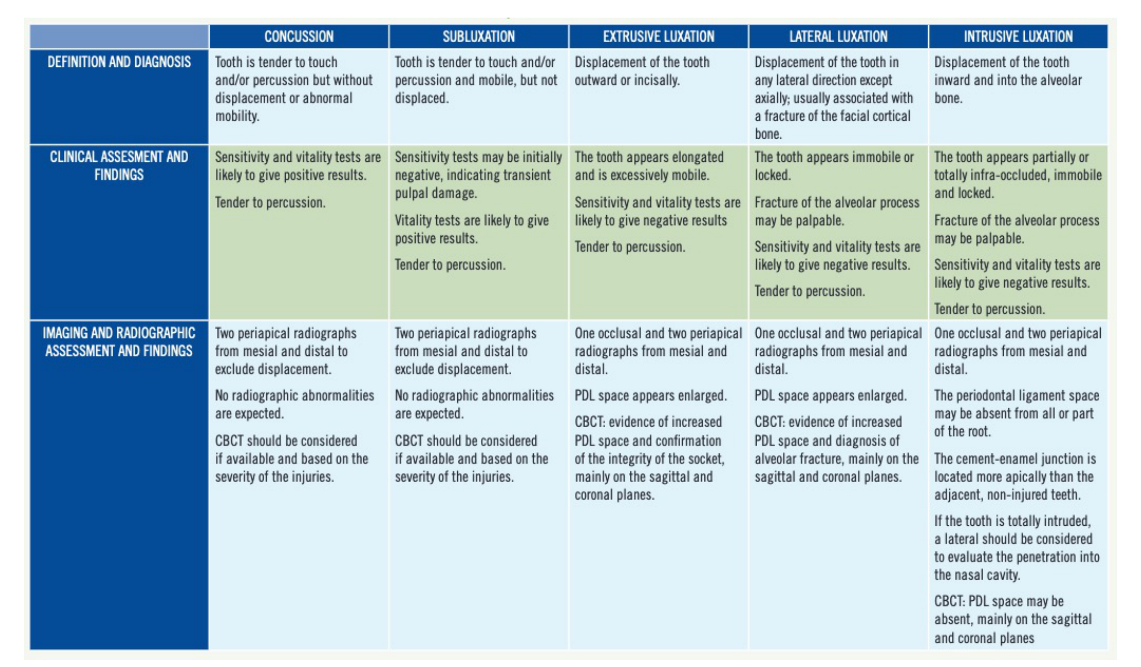
Luxation Injuries — Definition, Clinical Findings, and Imaging14
| CONCUSSION | SUBLUXATION | EXTRUSIVE LUXATION | LATERAL LUXATION | INTRUSIVE LUXATION | |
|---|---|---|---|---|---|
| Definition and diagnosis | Tooth is tender to touch and/or percussion but without displacement or abnormal mobility. | Tooth is tender to touch and/or percussion and mobile, but not displaced. | Displacement of the tooth outward or incisally. | Displacement of the tooth in any lateral direction except axially; usually associated with a fracture of the facial cortical bone. | Displacement of the tooth inward and into the alveolar bone. |
| Clinical assessment and findings | Sensitivity and vitality tests are likely to give positive results. Tender to percussion. | Sensitivity tests may be initially negative, indicating transient pulpal damage. Vitality tests are likely to give positive results. Tender to percussion. | The tooth appears elongated and is excessively mobile. Sensitivity and vitality tests are likely to give negative results. Tender to percussion. | The tooth appears immobile or locked. Fracture of the alveolar process may be palpable. Sensitivity and vitality tests are likely to give negative results. Tender to percussion. | The tooth appears partially or totally infra-occluded, immobile and locked. Fracture of the alveolar process may be palpable. Sensitivity and vitality tests are likely to give negative results. Tender to percussion. |
| Imaging and radiographic assessment and findings | Two periapical radiographs from mesial and distal to exclude displacement. No radiographic abnormalities are expected. CBCT should be considered if available and based on the severity of the injuries. | Two periapical radiographs from mesial and distal to exclude displacement. No radiographic abnormalities are expected. CBCT should be considered if available and based on the severity of the injuries. | One occlusal and two periapical radiographs from mesial and distal. PDL space appears enlarged. CBCT: evidence of increased PDL space and confirmation of the integrity of the socket, mainly on the sagittal and coronal planes. | One occlusal and two periapical radiographs from mesial and distal. PDL space appears enlarged. CBCT: evidence of increased PDL space and diagnosis of alveolar fracture, mainly on the sagittal and coronal planes. | One occlusal and two periapical radiographs from mesial and distal. The periodontal ligament space may be absent from all or part of the root. The cement-enamel junction is located more apically than the adjacent, non-injured teeth. If the tooth is totally intruded, a lateral should be considered to evaluate the penetration into the nasal cavity. CBCT: PDL space may be absent, mainly on the sagittal and coronal planes. |
Lecturer — Luxation Imaging
Intraoral imaging has poor sensitivity for minimal displacement, root fractures, alveolar fractures, labial-palatal displacement, luxation injuries, and oblique or vertical fractures. CBCT can demonstrate displacement, periodontal ligament space changes, and associated bony injury more effectively.
Avulsion
| IMAGING AND RADIOGRAPHIC ASSESSMENT AND FINDINGS | |
|---|---|
| Avulsion | Two periapical radiographs from mesial and distal. CBCT should be considered to confirm the reposition of the tooth and rule out alveolar bone fracture(s). |
Lecturer — Avulsion Follow-Up
For avulsion injuries, the IADT recommends radiographs immediately after replantation and during follow-up. The guideline does not explicitly recommend CBCT for every avulsion, whereas the AAE suggests considering it when alveolar fractures or other complications are suspected.
CBCT in Dental Trauma
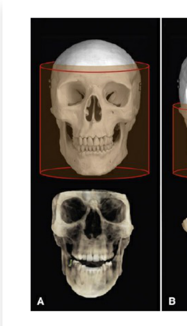
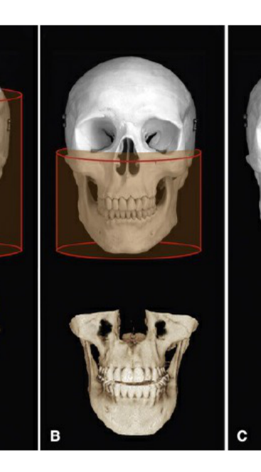
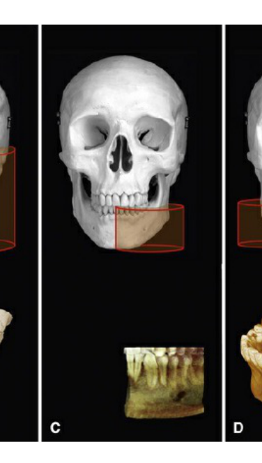
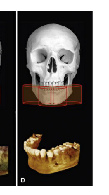
Advantages of CBCT
- No superimposition of anatomical structures
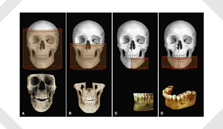
- Dimensionally accurate (isotropic voxel)
- Excellent spatial resolution
- Small FOV + Voxel size (less than 0.1 mm ideally)
- Intraoral imaging has poor sensitivity in detecting minimal tooth displacement and root & alveolar fractures, especially:
- Labial / palatal tooth displacement (lateral luxation injuries)
- Oblique & vertical root fractures
Advantages of CBCT15
Voxel size 0.2 mm · Voxel size 0.3 mm · Voxel size 0.4 mm (panels A, B, C, D)
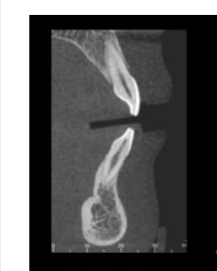 | 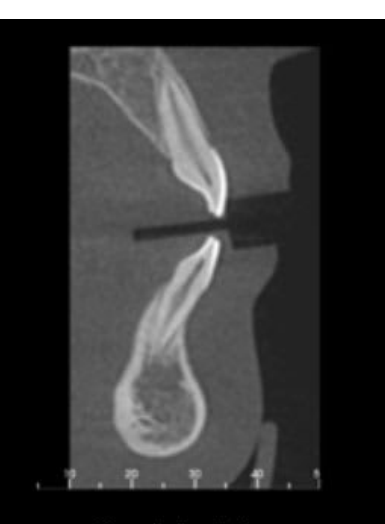 | 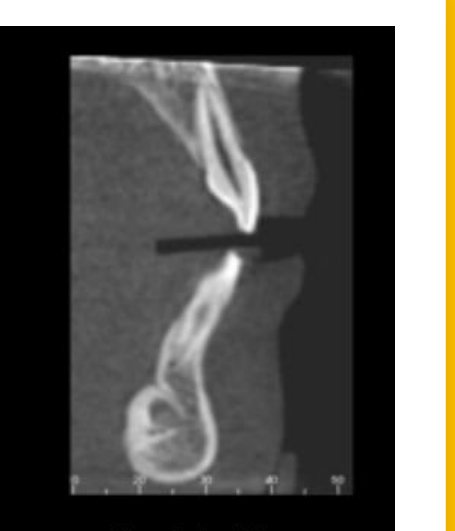 |
 |  |  |
 |
CBCT Versus Periapical Radiography16
 |  |  |
 | 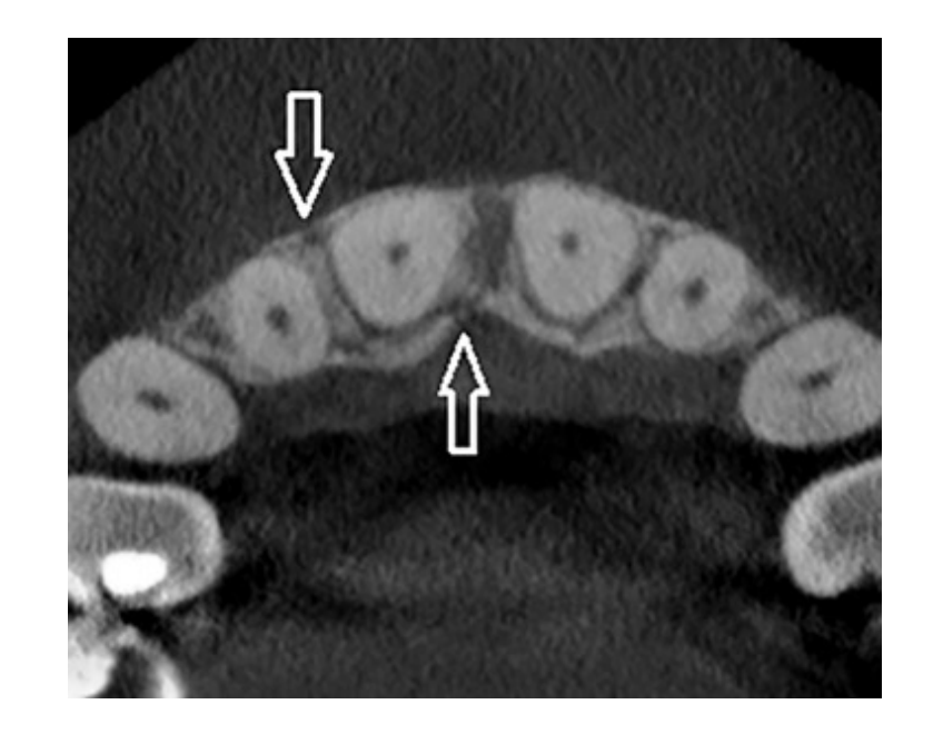 |
CBCT vs PA in Horizontal Root Fractures?
Li, F., Li, J., Zhang, D., and Wu, F. Role of Computed Tomography Scan in Dental Trauma: A Cross-Sectional Study. Dose Response. 2018 Jul-Sep; 16(3): 1559325818789837. Published online 2018 Aug 23. doi: 10.1177/1559325818789837. PMCID: PMC6108019. PMID: 30150907.
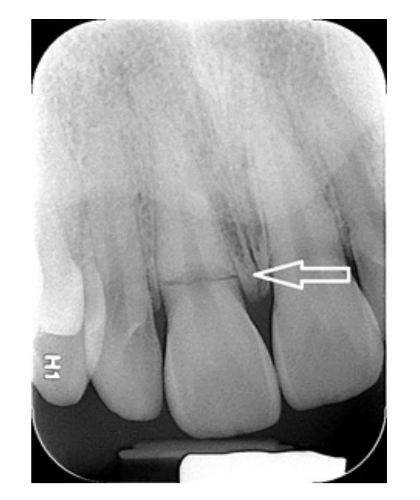
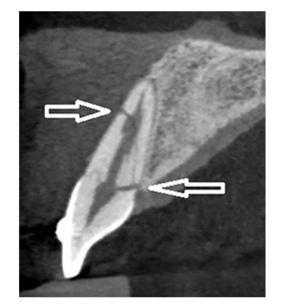
CBCT Versus Periapical Radiography17
Hashemia, S. M., Jahadi, S., Shaygannia, S., Hekmatian, E., Habibollahi, A., and Ghazizadeh, M. Cone Beam Computed Tomography and Digital Periapical Radiography in Determining Horizontal Root Fractures: An In-vitro Comparative Study. Dentistry 2018, 8:9. DOI: 10.4172/2161-1122.1000513.
Lecturer — Periapical Limitations
Periapical radiography may fail to show complex or multi-plane injuries.
- Oblique fractures and associated alveolar injury may be missed.
- Internal resorption may also be difficult to identify.
- CBCT demonstrates the three-dimensional relationship between the tooth, root fracture, alveolar bone, periodontal ligament space, and root canal.
-
In vivo study #1 → CBCT has 98.8% sensitivity & 95.6% specificity (100 microns) & PA has 97.2% sensitivity & 78.5% specificity
- Author states CBCT should be used for detection of horizontal root fractures
-
The 2018 study involved 250 cases.
-
In vitro study #2 → CBCT (100 microns) vs PA
- “The sensitivity and specificity of CBCT radiography was 0.97 and 0.93, and periapical was 0.83 and 0.885 respectively, indicating that the CBCT technique was preferred to the periapical to detect the horizontal fracture of the root”
- “Although CBCT is an accurate imaging modality for detection of root fractures but based on ALARA (as low as reasonably achievable) it must be used only for symptomatic patients that periapical images did not show any fractures”
PA vs CBCT18
- CBCT Application in Dentoalveolar Trauma
- February 14, 2018
- by Yosef Nahmias, DDS, MSc; Ali Fatemi, DDS, MSc, FRCD(C)
Lecturer — Periapical Projections
Periapical radiographs remain important for initial and follow-up imaging.
- Multiple periapical projections and an occlusal projection are recommended because a fracture or displacement may be visible in one view but not another.
- CBCT provides additional information when two-dimensional imaging is inconclusive or a more complex injury is suspected.
 |  |
Indications for CBCT
Summary: When to Take CBCT?
- Acute Stage (compliant patient only)
- Symptomatic patients that periapical images did not show any fractures
- Accurate assessment of root fractures & luxation injuries
- Late Stage
- CBCT also assists with crown-root fractures and alveolar fractures, including luxation or avulsion injuries with possible bony complications.
- Review healing
- “clinical and 2D radiographic examinations may not be sufficient to determine the presence or absence of pulp and periodontium healing”
- Necrotic Pulp
- Root resorption
Cohenca, N., and Silberman, A. Contemporary imaging for the diagnosis and treatment of traumatic dental injuries: A review. First published: 20 March 2017. https://doi.org/10.1111/edt.12339. - At follow-up, CBCT may also show ankylosis, inflammation, and infection.
CBCT Case Examples
CY Yu, PV Abbott. School of Dentistry, The University of Western Australia, Western Australia, Australia.
Table 1. Possible responses of the pulp and root canal system, the periradicular tissues, and the soft tissues following trauma to a tooth19
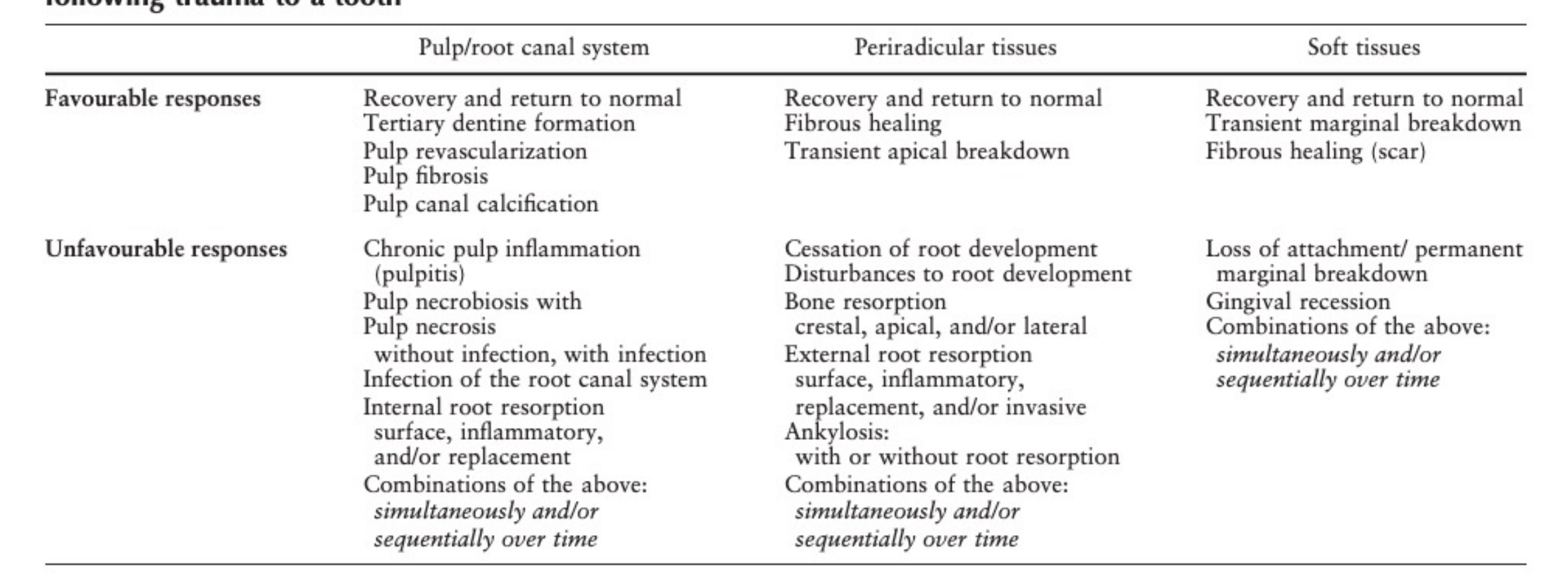
Pulp and Periradicular Responses
| Pulp/root canal system | Periradicular tissues | Soft tissues | |
|---|---|---|---|
| Favourable responses | Recovery and return to normal; tertiary dentine formation; pulp revascularization; pulp fibrosis; pulp canal calcification | Recovery and return to normal; fibrous healing; transient apical breakdown | Recovery and return to normal; transient marginal breakdown; fibrous healing (scar) |
| Unfavourable responses | Chronic pulp inflammation (pulpitis); pulp necrobiosis with pulp necrosis — without infection, with infection; infection of the root canal system; internal root resorption — surface, inflammatory, and/or replacement; combinations of the above: simultaneously and/or sequentially over time | Cessation of root development; disturbances to root development; bone resorption — crestal, apical, and/or lateral; external root resorption — surface, inflammatory, replacement, and/or invasive; ankylosis — with or without root resorption; combinations of the above: simultaneously and/or sequentially over time | Loss of attachment / permanent marginal breakdown; gingival recession; combinations of the above: simultaneously and/or sequentially over time |
Crown and Root Fracture Cases2021
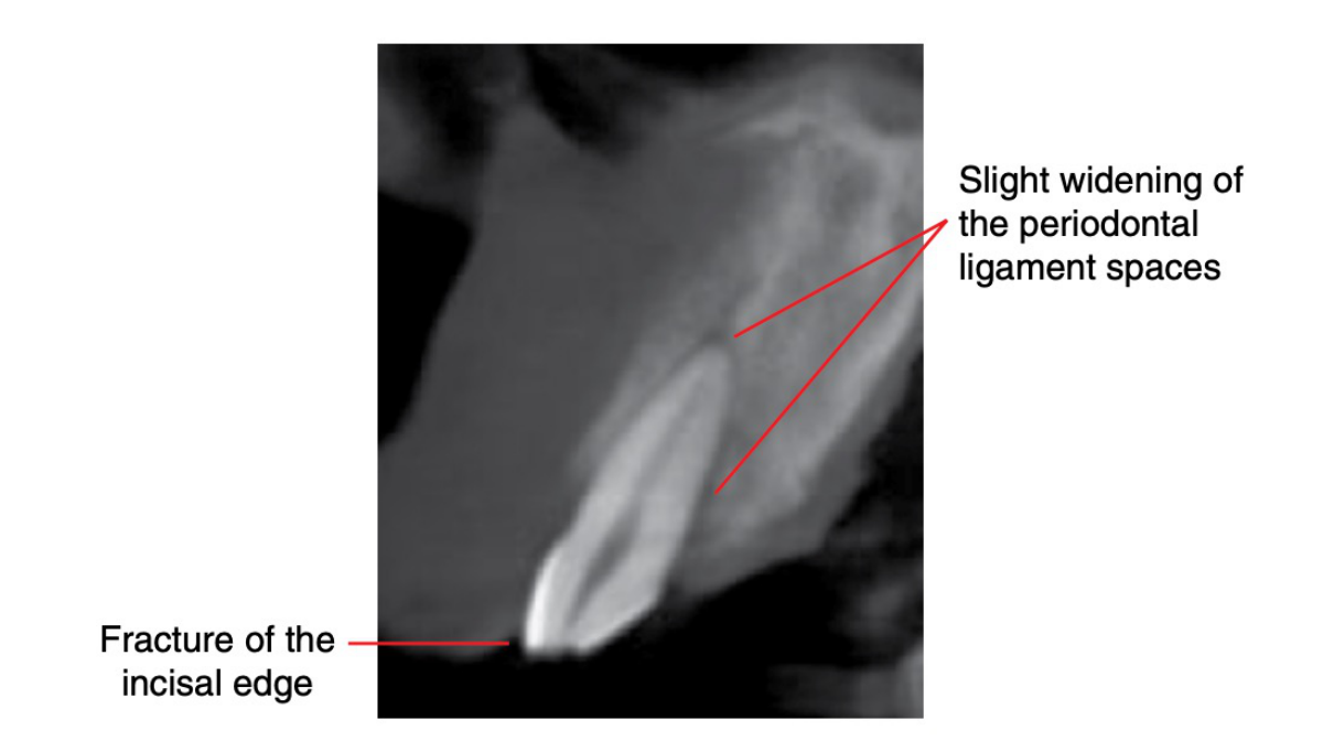
CBCT case: identify crown/root numbers and injuries to periodontal structures.
- Slight widening of the periodontal ligament spaces
- Fracture of the incisal edge
Ref: Atlas of Oral and Maxillofacial Radiology, Bernard Koong.
Lecturer — Subluxation Findings
Trauma assessment should identify both dental fractures and periodontal injuries. In one example, an uncomplicated crown fracture without pulp involvement was associated with widened periodontal ligament spaces, particularly palatally and apically, but no visible alveolar fracture. These findings were consistent with an uncomplicated fracture associated with subluxation.
MPR — trauma to 22.
 | 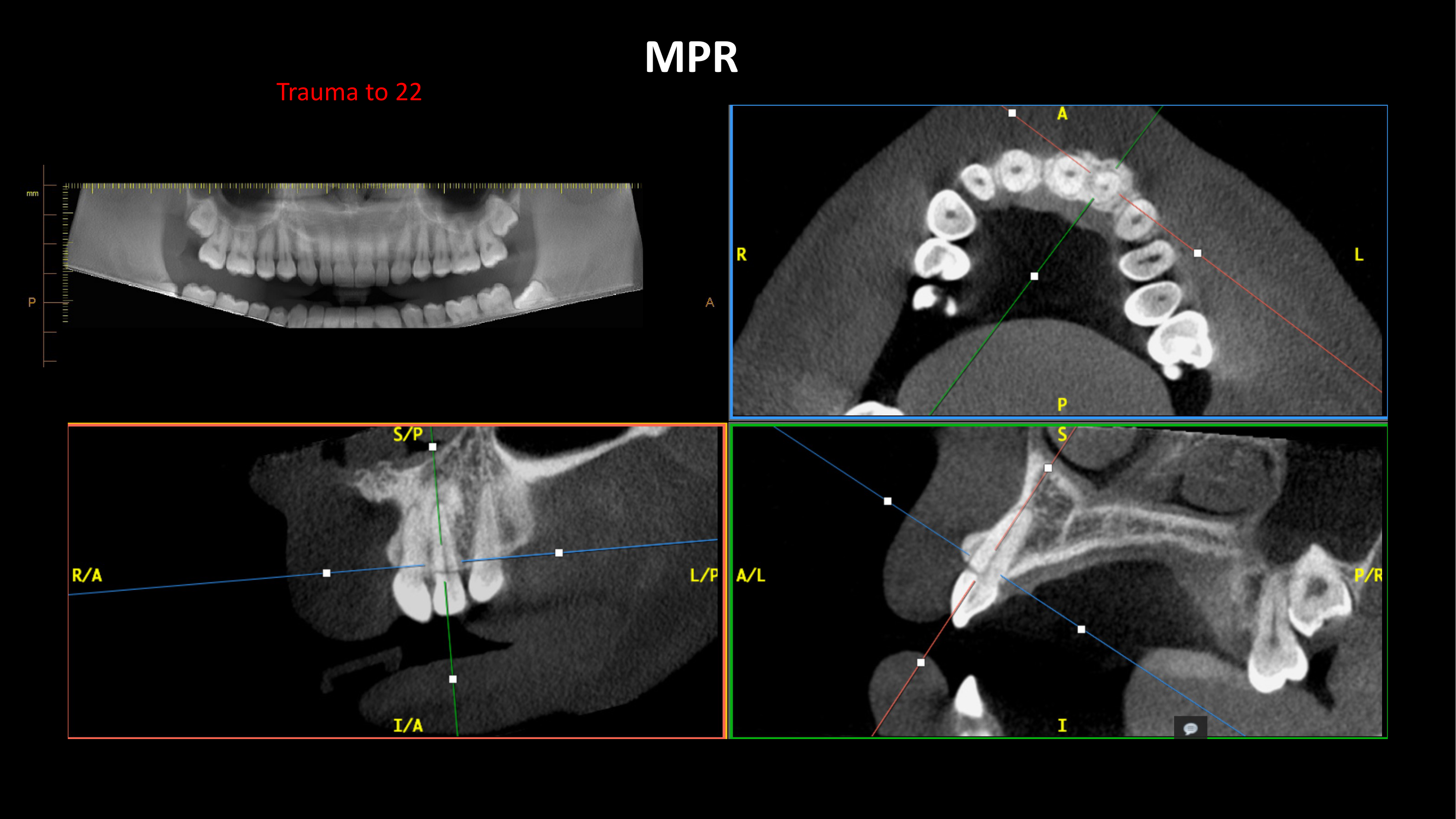 |
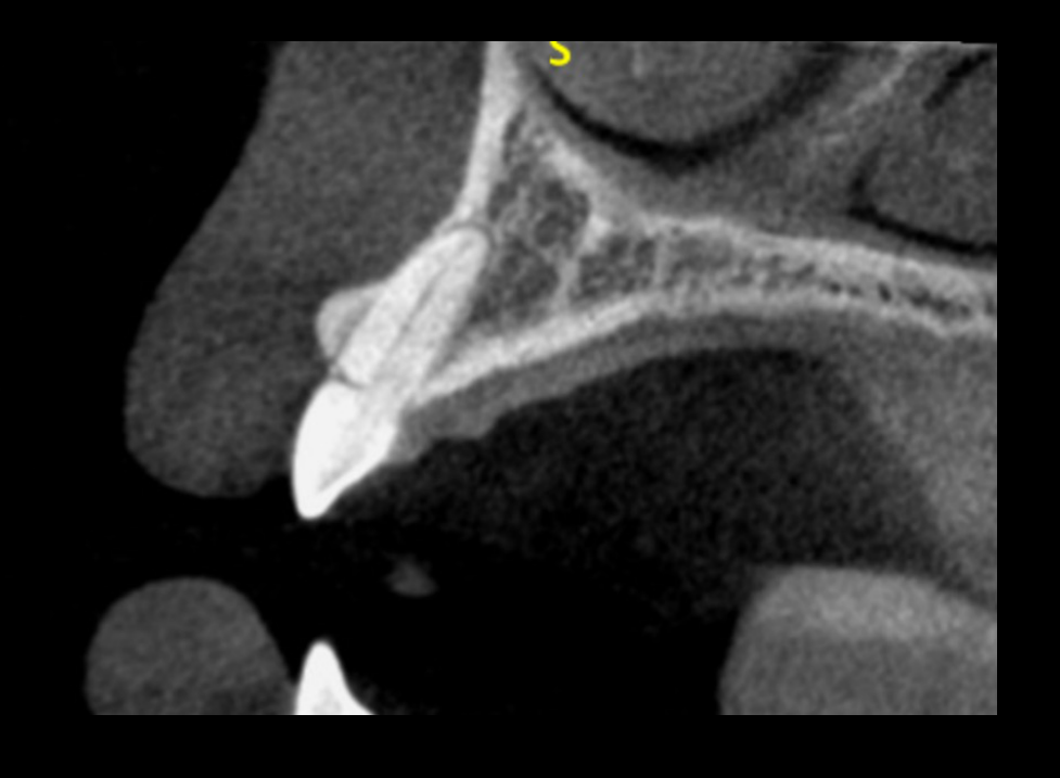
Crown/Root Fracture?22
- Horizontal / oblique / vertical?
- Extension?
- Complicated or uncomplicated?
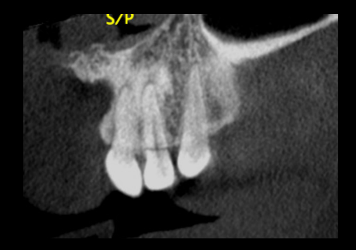
Changes to the PDL space? Bony fracture?
Lecturer — Fracture Assessment
CBCT may clarify whether an apparent crown fracture extends into the root and whether the pulp is involved. Corrected planes are necessary to assess the relationship between the fracture line and the tooth’s long axis.
 |  |
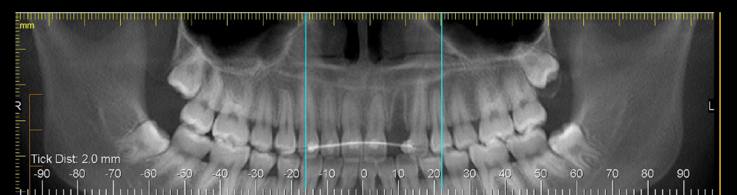
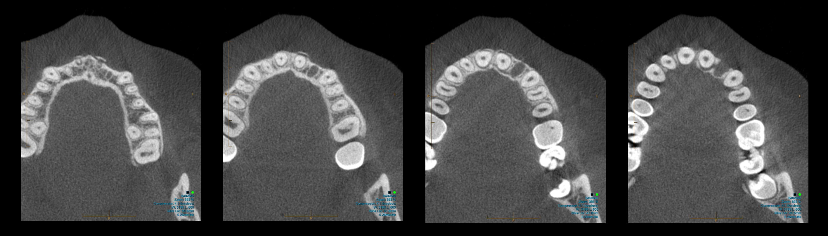
Anterior Maxillary Trauma Cases2324252627
Anterior maxillary trauma (11 injured). Orientation markers: R / L.
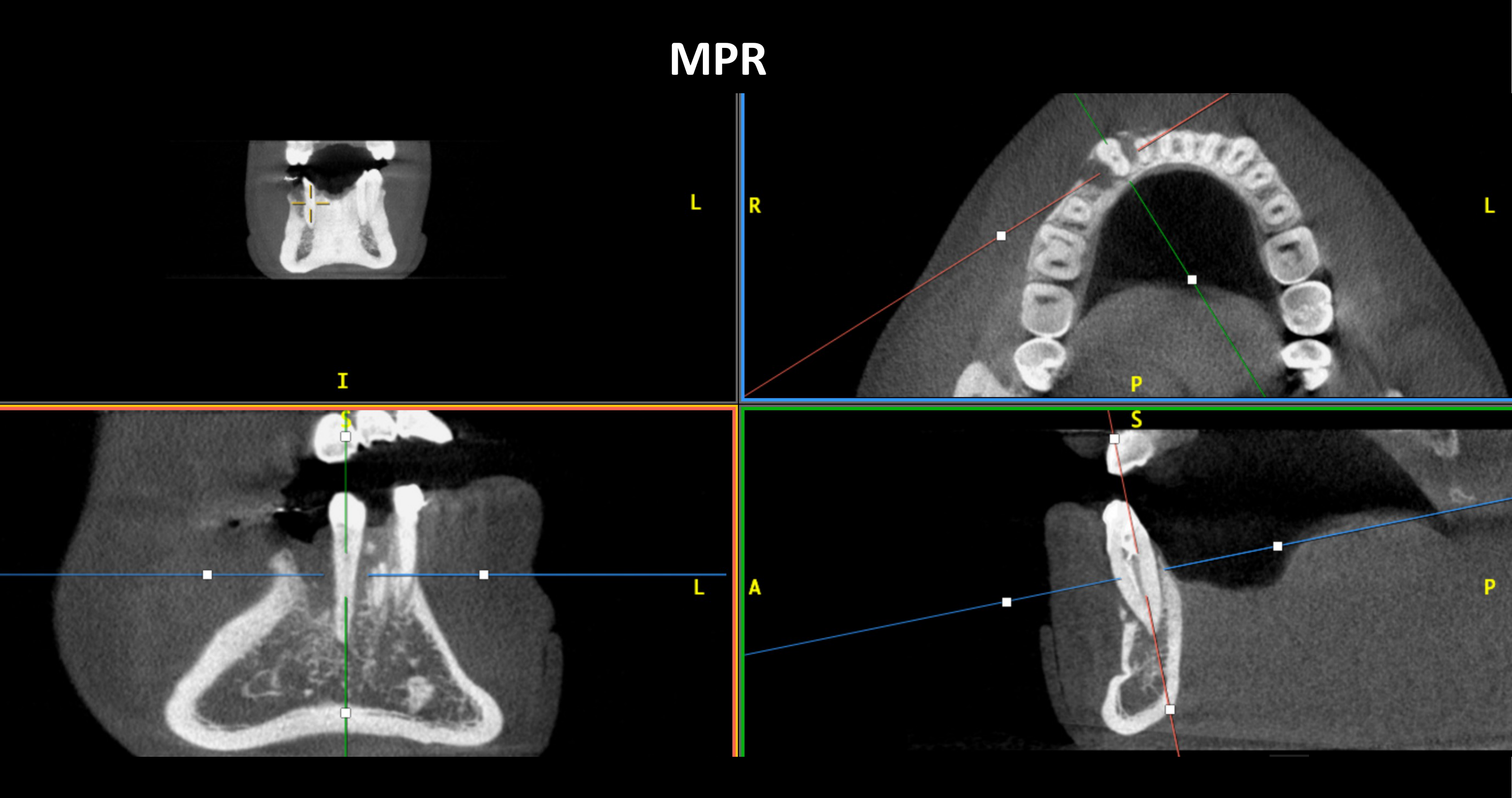
MPR view. Orientation markers: I, S, P, A; R / L.
11 vs 21:
- Crown/root fracture?
- Changes to the PDL space?
- Bony fracture?
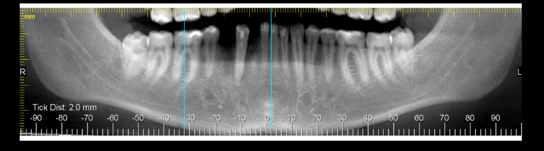
Measurement: Tick Dist: 2.0 mm. Scale (mm): -90 to 90 in increments of 10. Orientation markers: R / L.
Complex Root Fractures
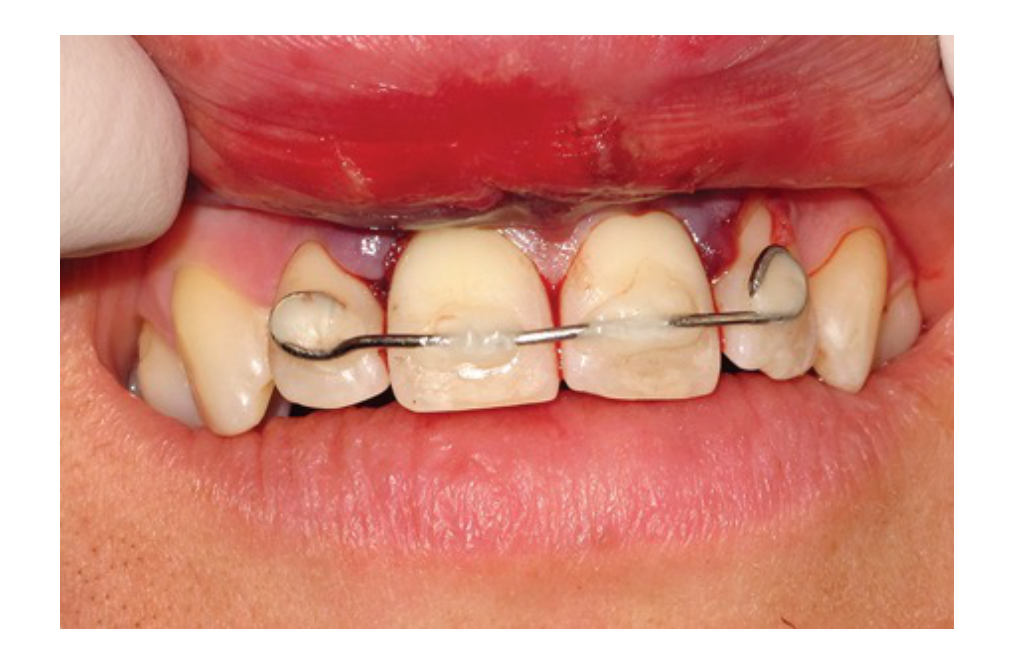
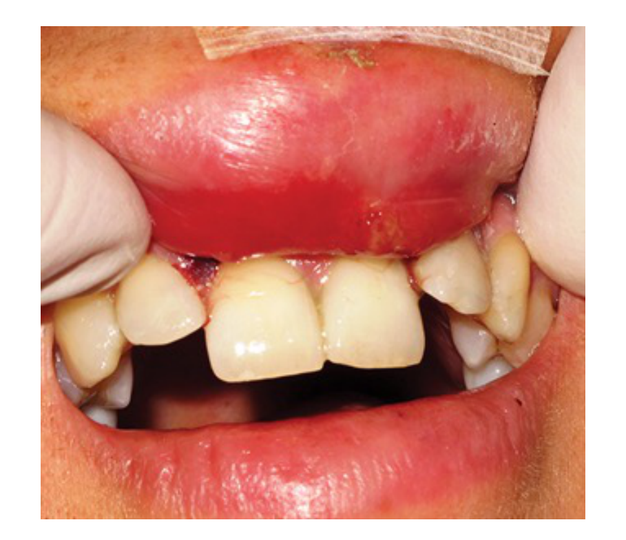
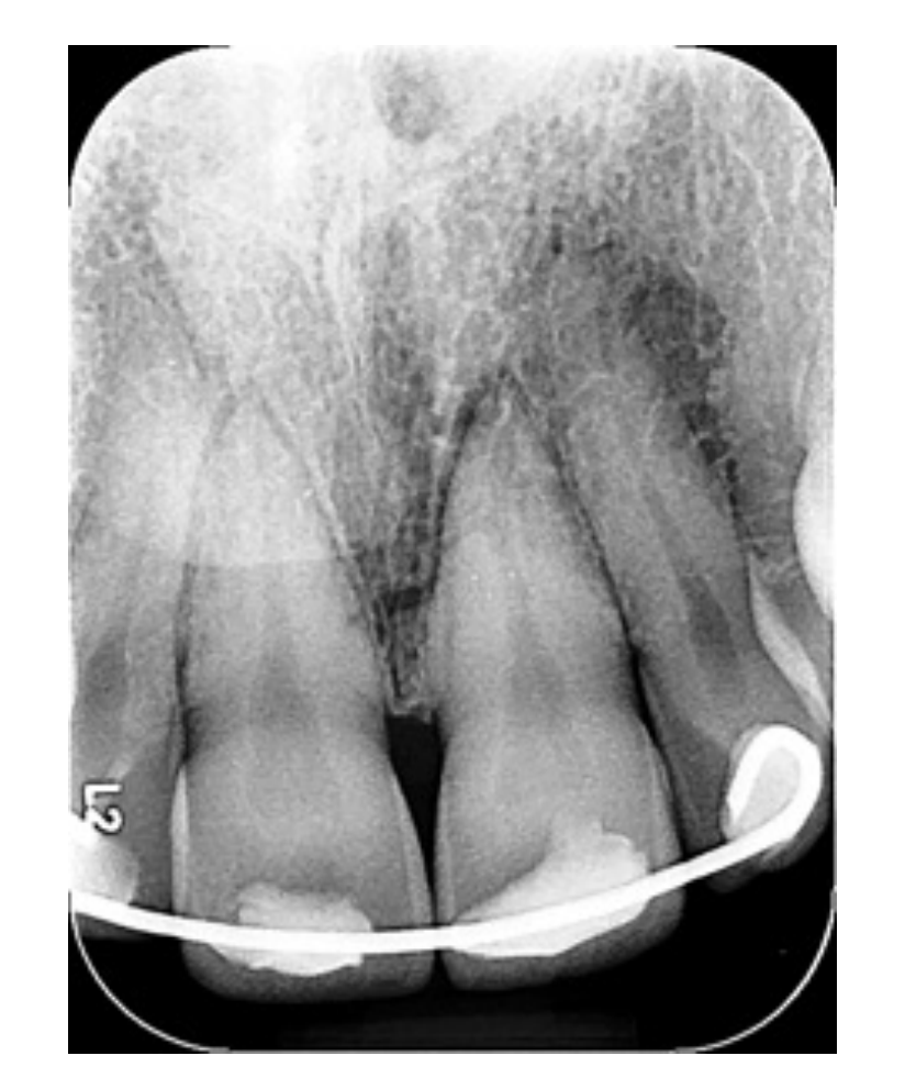
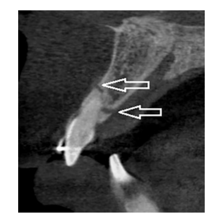
A 21-year-old patient presented with a loose maxillary incisor after trauma. The tooth was mobile, tender to palpation and percussion, and non-responsive to cold testing. Initial periapical radiography showed a horizontal fracture at the cervical third of the root; the tooth was splinted, but the patient missed the six-week review and returned four months later with pain.
- CBCT showed an incomplete horizontal fracture at the alveolar crest and an oblique fracture extending from the palatal surface, sharing the horizontal fracture line and extending apically to the labial surface near the junction of the middle and apical thirds.
- Internal resorption involved the canal, and the internal resorption and complex fracture pattern were not evident on the initial periapical radiograph.
Assess tooth by tooth: 11? 21? 22?
Palatal Displacement and Alveolar Fracture
A 47-year-old woman fell and traumatized the maxillary central and lateral incisors. Both teeth were displaced palatally and non-responsive to sensibility testing; soft tissue lacerations were sutured immediately, and radiographs were obtained after repositioning and splinting.
- Periapial imaging showed whitening of the periodontal ligament space, so CBCT was ordered to exclude dento-alveolar fractures.
- CBCT demonstrated a mildly displaced bicortical alveolar fracture extending from the palatal cortex to the labial cortex and involving the periodontal ligament spaces.
- No root fractures were identified at four weeks, but the teeth remained non-responsive, became discolored, and underwent root canal therapy.
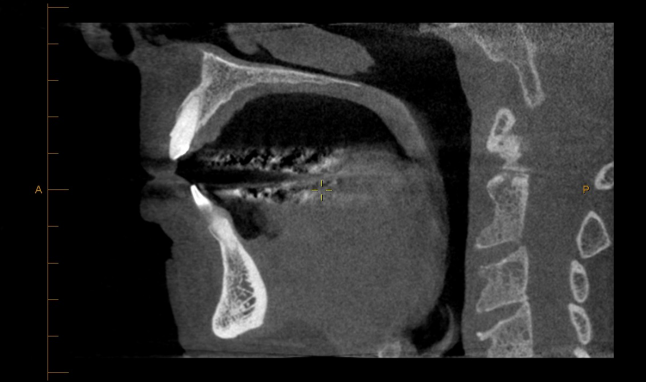
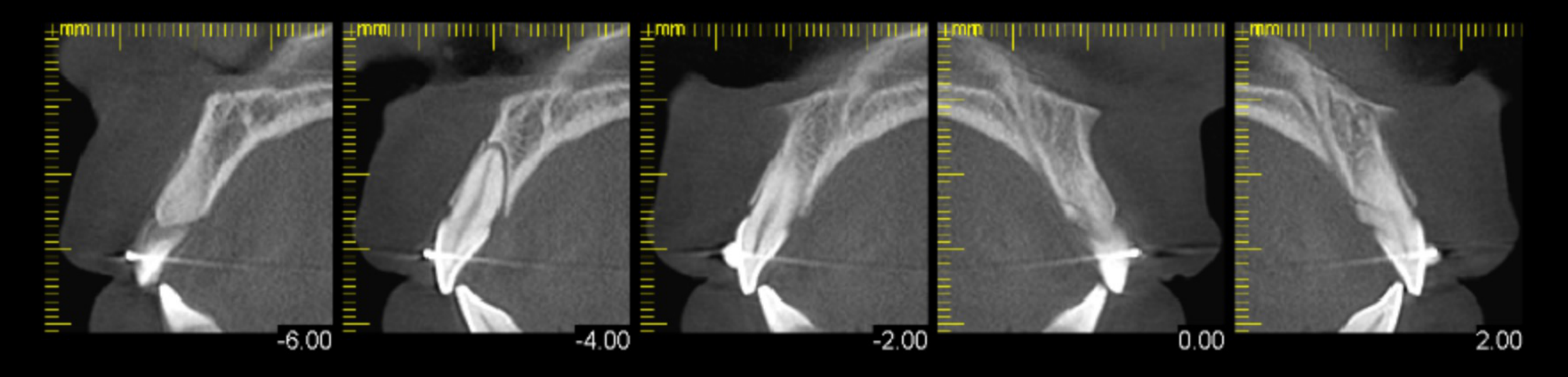
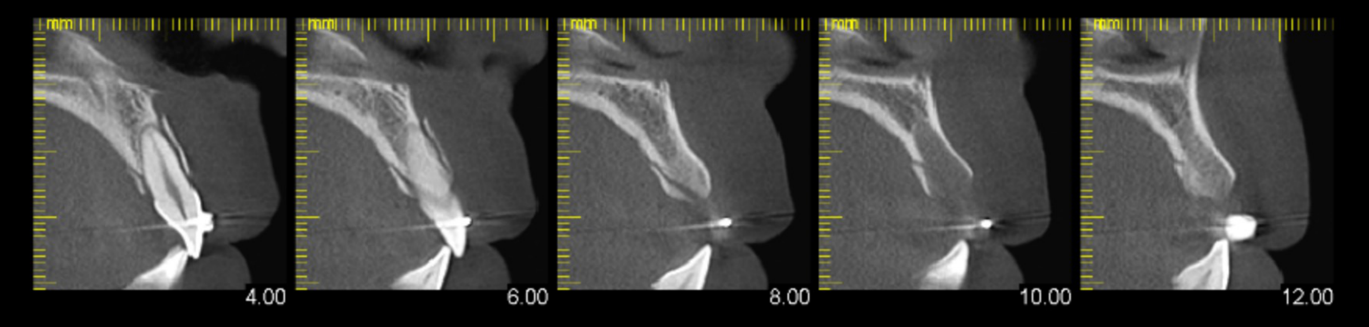
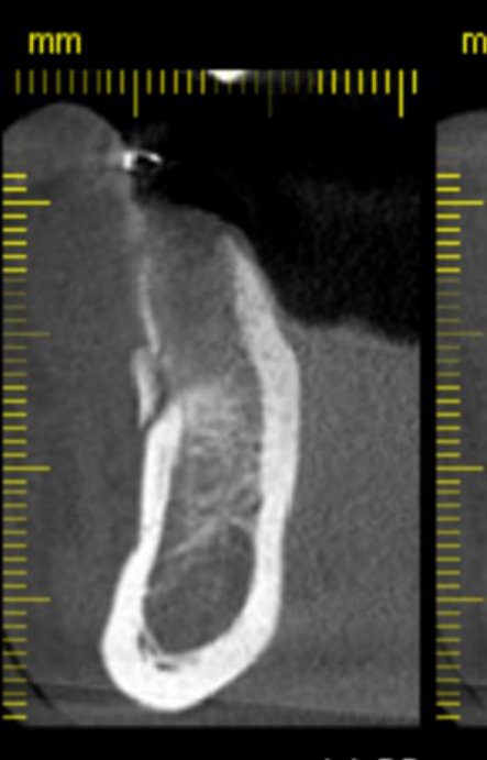
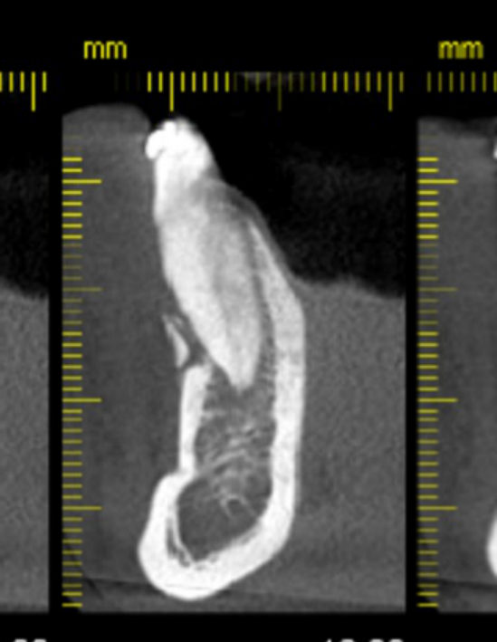
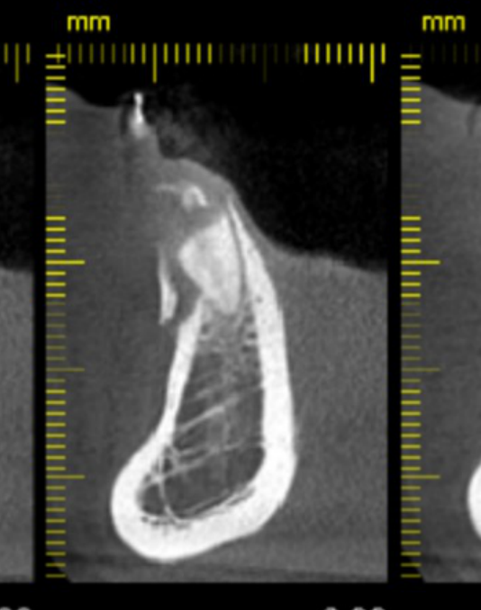
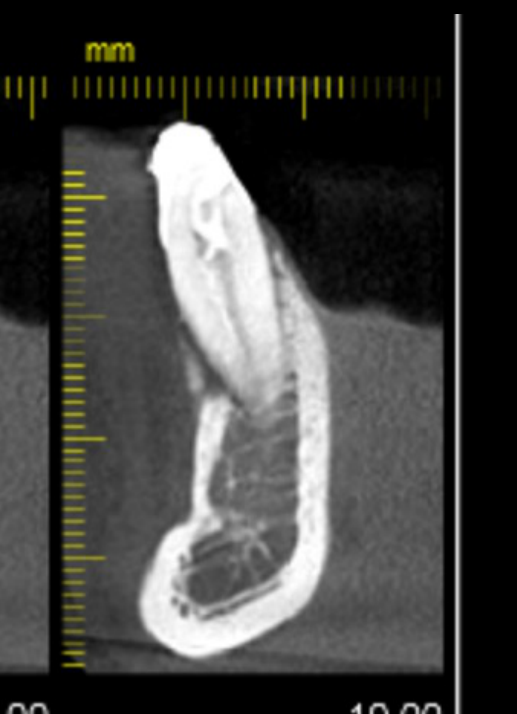
Additional MPR Case Reviews28293031323334
MPR view. Orientation markers: I, P, S; L, A, P; L / R.
Scale: -20.00, -18.00, -16.00, -14.00, -12.00, -10.00, -8.00, -6.00, -4.00, -2.00
Lecturer — MPR Alignment
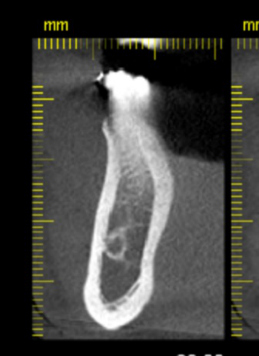
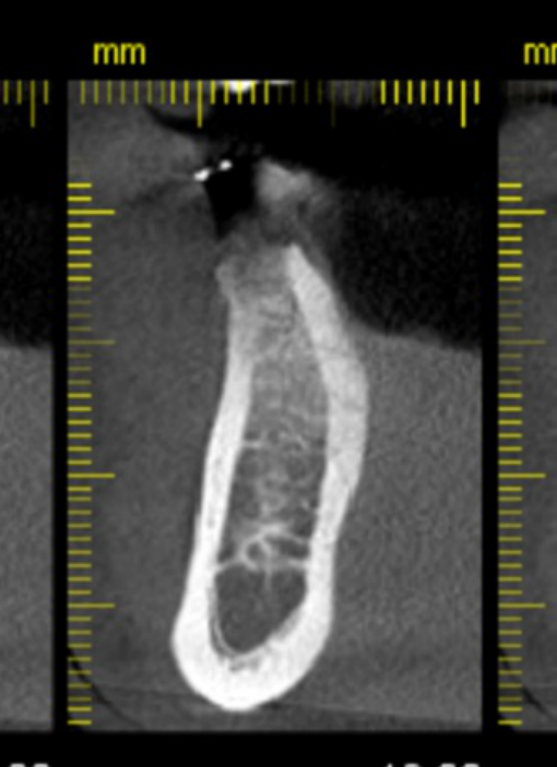
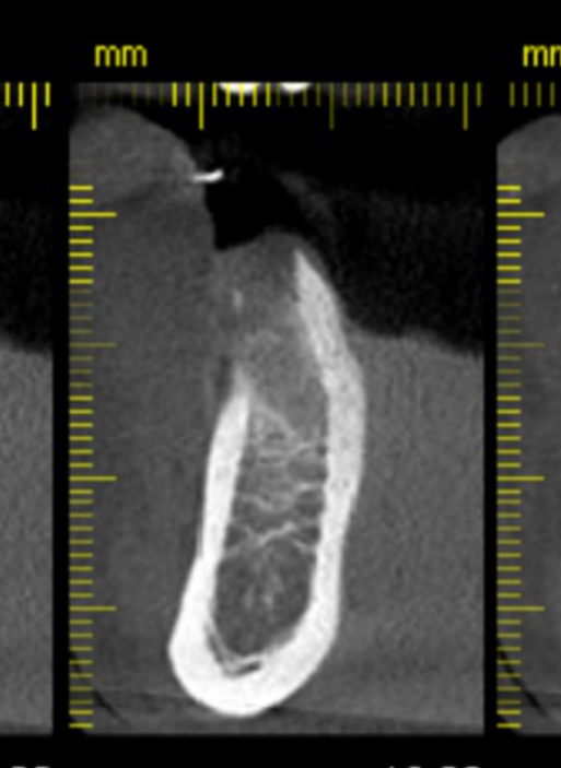
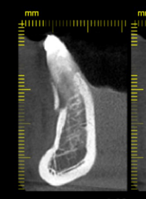
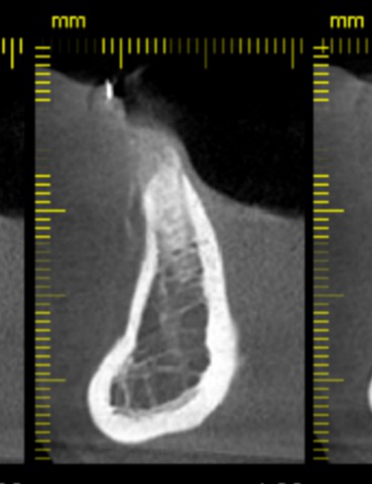
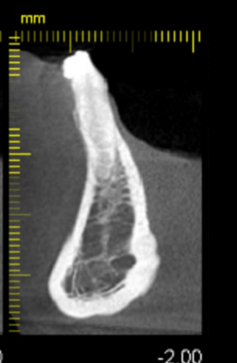
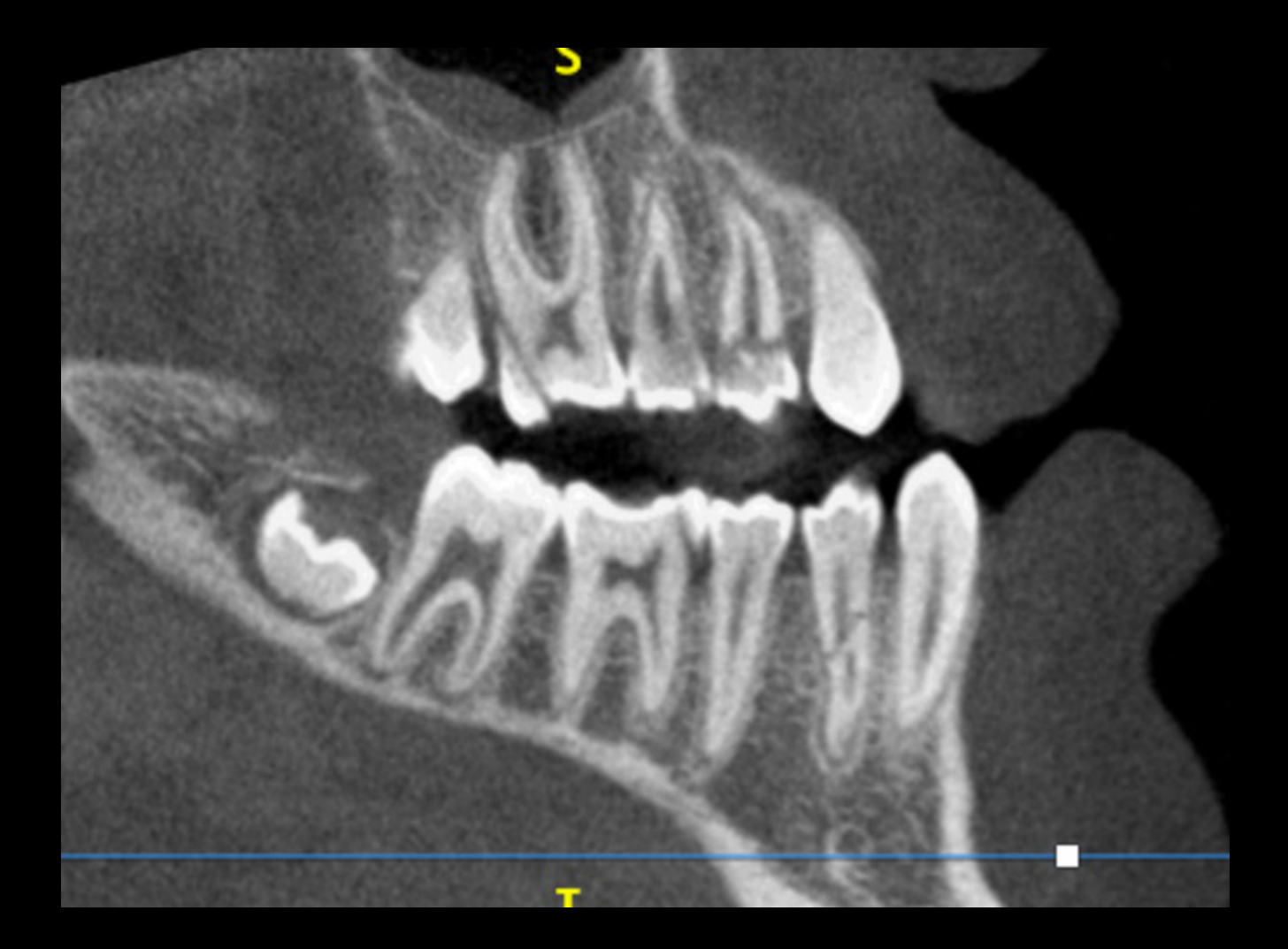
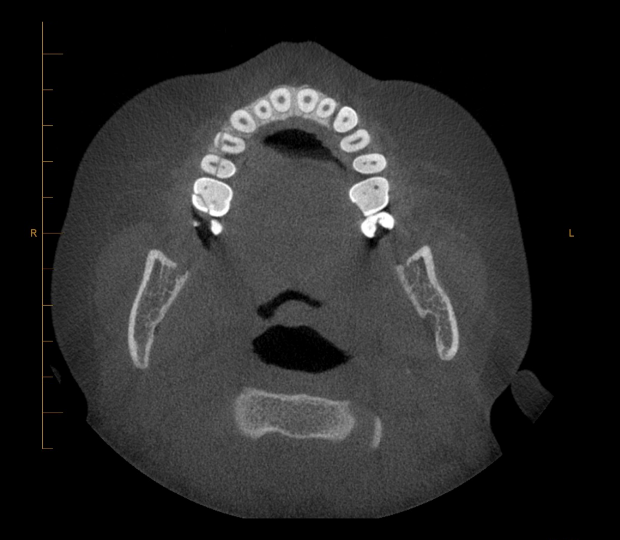
Multiplanar reformatting is important for dental trauma assessment, and the CBCT data should be manipulated to obtain the appropriate angle for the clinical question. For a specific tooth, the MPR planes are aligned with the long axis by moving and rotating the cross-cursor. The corrected coronal and sagittal views follow the tooth’s long axis, with the coronal view resembling a traditional periapical radiograph.
Incomplete Lateral Incisor Fracture
Corrected MPR views of the lateral incisor showed an incomplete horizontal root fracture at the cementoenamel junction extending from the labial surface to the pulp. The periodontal ligament spaces were normal, and no bony fracture was visible in the corrected sagittal or coronal views.
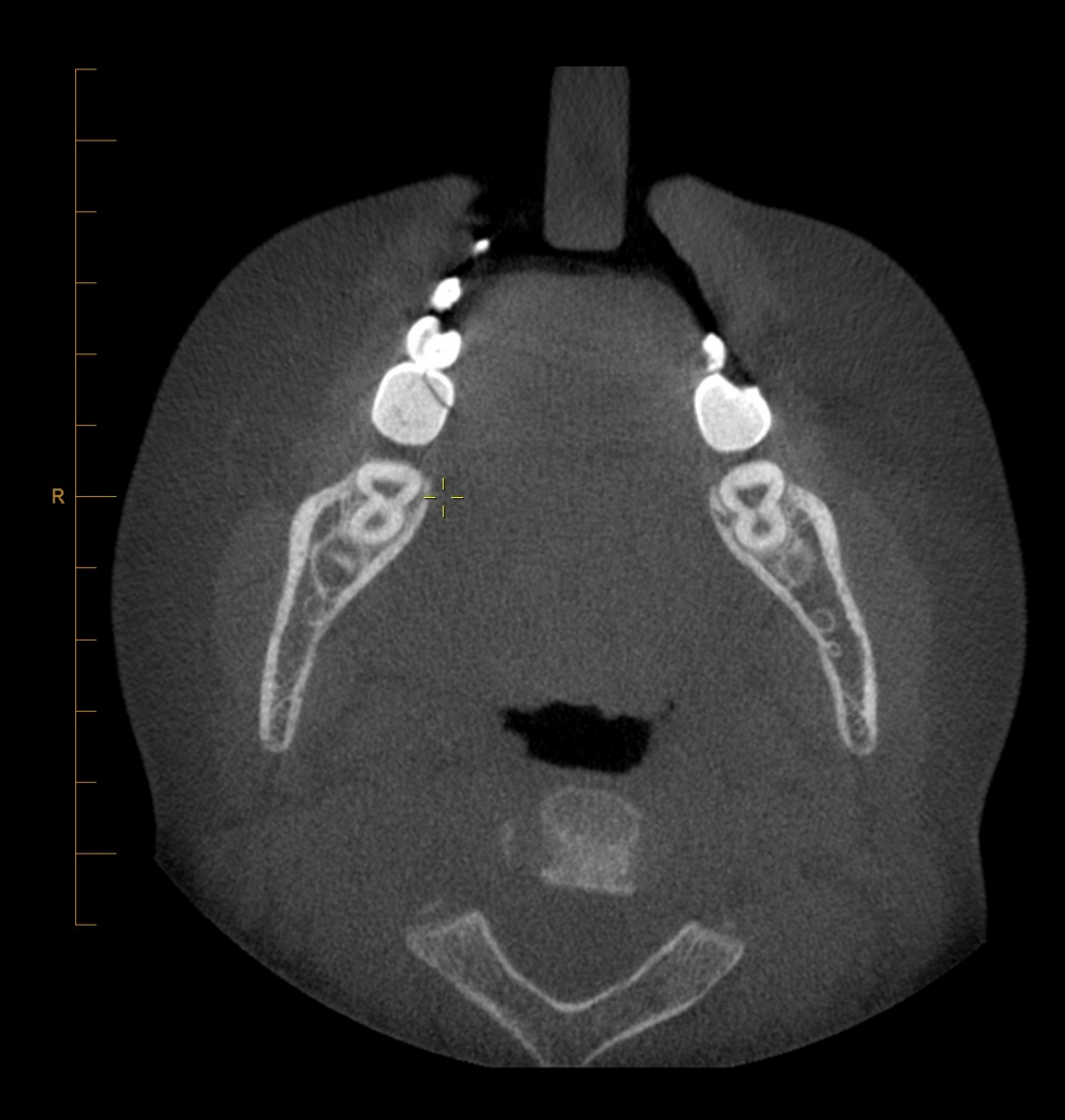
Facial Bone Trauma
CT is the gold standard for imaging facial bone trauma.
Lecturer — CT and CBCT
CT was described as the gold standard for maxillofacial trauma because it provides a large field of view and excellent resolution of hard and soft tissues.
- CBCT is ideal for dento-alveolar and jaw fractures, but has a restricted field of view, slower or more limited scanning considerations, poorer soft tissue evaluation, and more noise than CT.
- Facial trauma commonly results from road traffic accidents, assault, and falls.
- Plain radiographs remain useful in some situations, but complex trauma is limited by superimposition, low sensitivity, and two-dimensional imaging.
CT as the Gold Standard35
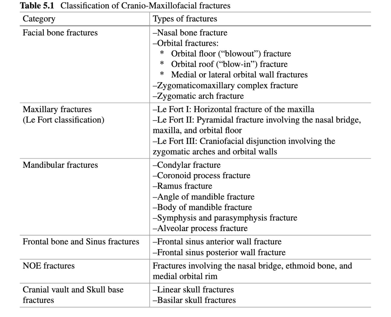
Classification of Cranio-Maxillofacial Fractures36
Table 5.1 — Classification of Cranio-Maxillofacial Fractures
| Category | Types of fractures |
|---|---|
| Facial bone fractures | - Nasal bone fracture - Orbital fractures: - Orbital floor (“blowout”) fracture - Orbital roof (“blow-in”) fracture - Medial or lateral orbital wall fractures - Zygomaticomaxillary complex fracture - Zygomatic arch fracture |
| Maxillary fractures (Le Fort classification) | - Le Fort I: Horizontal fracture of the maxilla - Le Fort II: Pyramidal fracture involving the nasal bridge, maxilla, and orbital floor - Le Fort III: Craniofacial disjunction involving the zygomatic arches and orbital walls |
| Mandibular fractures | - Condylar fracture - Coronoid process fracture - Ramus fracture - Angle of mandible fracture - Body of mandible fracture - Symphysis and parasymphysis fracture - Alveolar process fracture |
| Frontal bone and sinus fractures | - Frontal sinus anterior wall fracture - Frontal sinus posterior wall fracture |
| NOE fractures | Fractures involving the nasal bridge, ethmoid bone, and medial orbital rim |
| Cranial vault and skull base fractures | - Linear skull fractures - Basilar skull fractures |
Lecturer — Le Fort Classification
Le Fort fractures are classified according to separation of the maxilla from the skull base, and a fracture of the pterygoid plates is required as a hallmark of a Le Fort injury. These high-impact injuries describe patterns of midfacial skeletal dissociation.
- Le Fort I produces a floating palate through horizontal separation above the teeth.
- Le Fort II is a pyramidal fracture involving the nasal bridge and infraorbital rim.
- Le Fort III detaches the midface from the cranial base, and the higher the fracture line, the more severe the injury.
 | 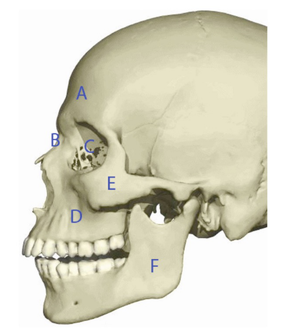 |
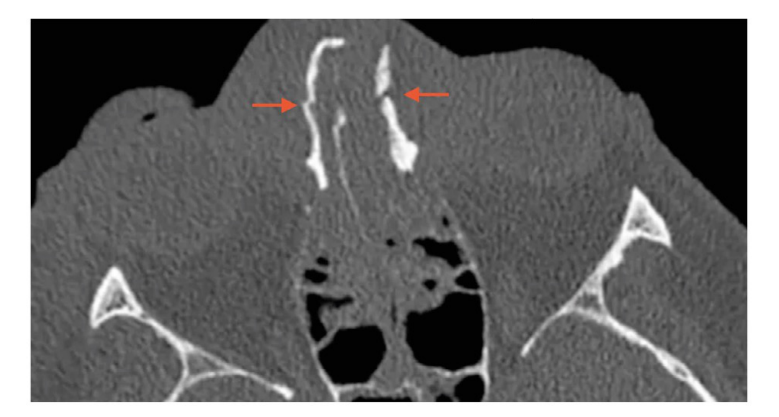
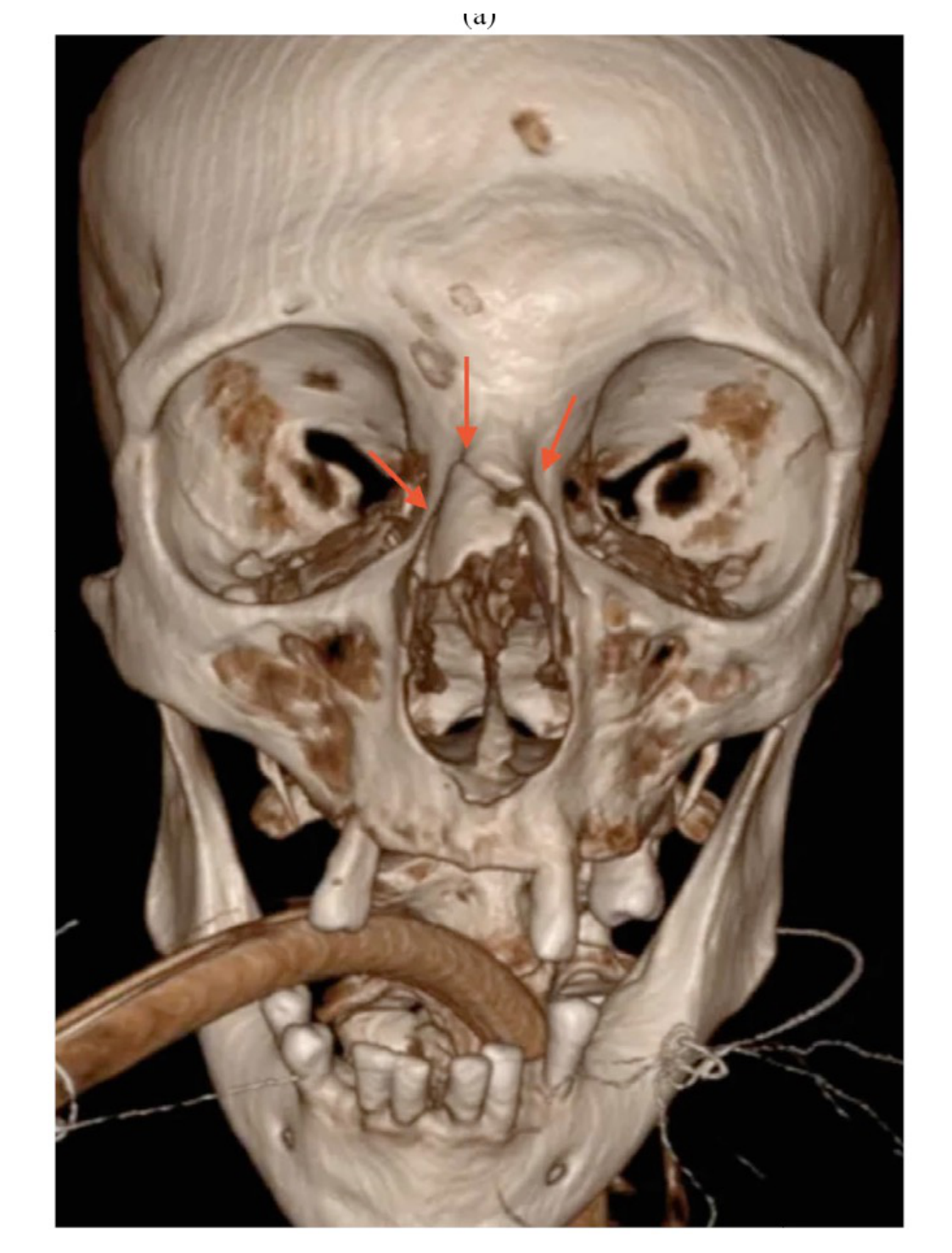
Nasal Fractures37
 |  |
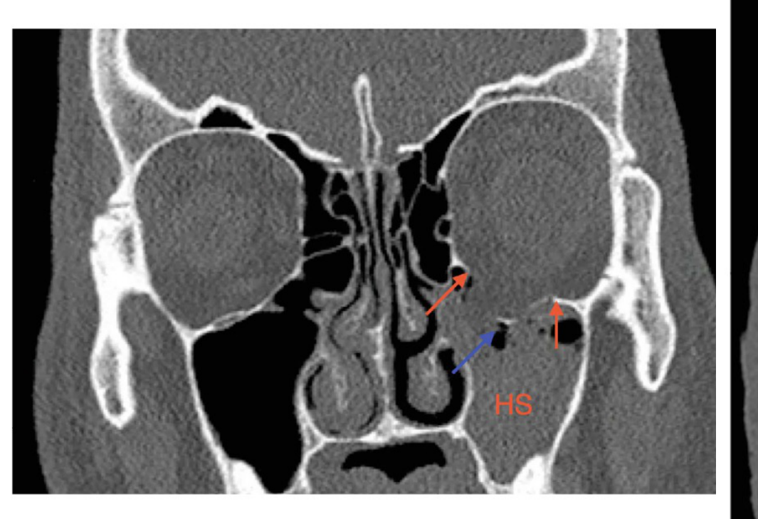
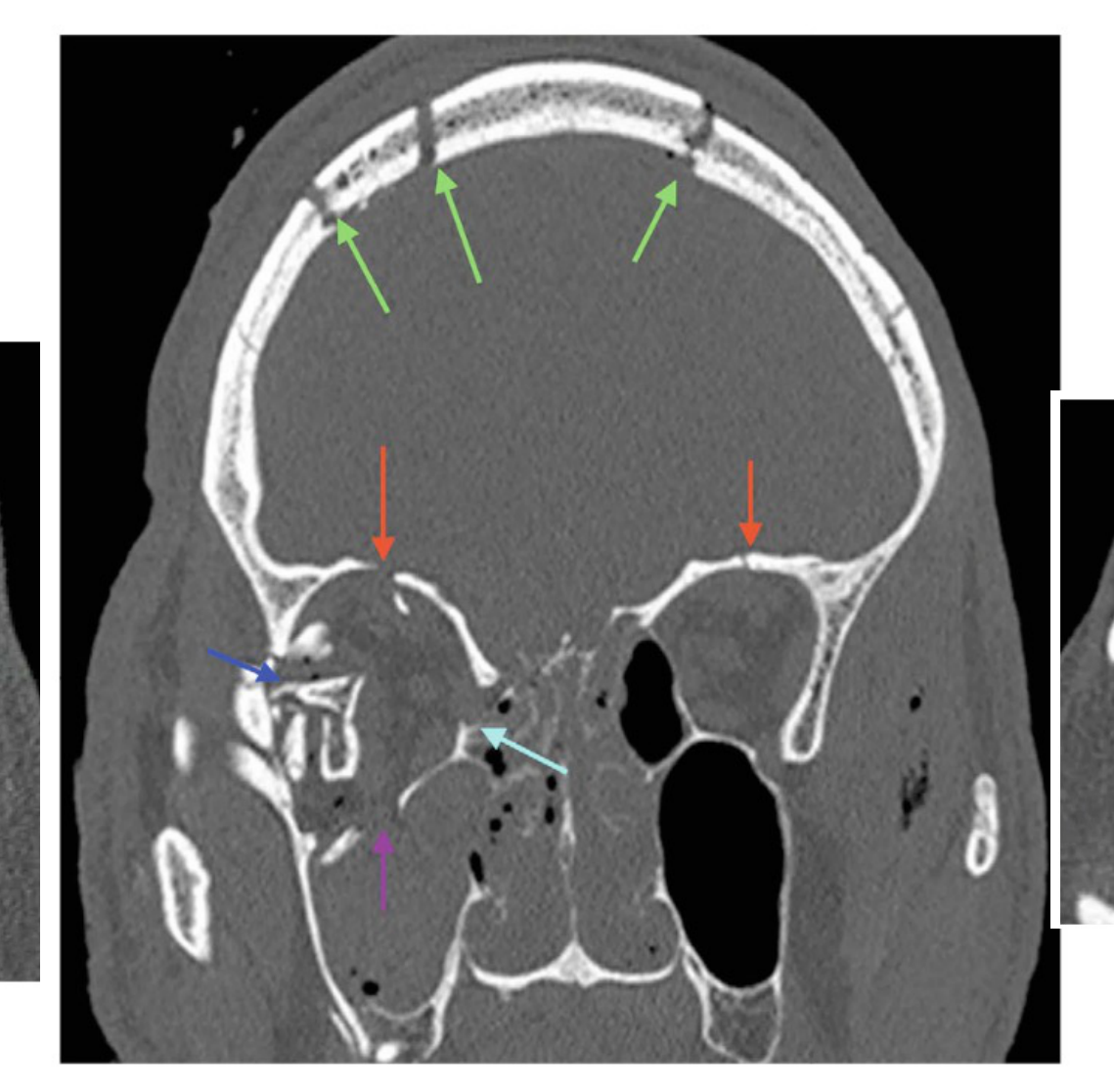
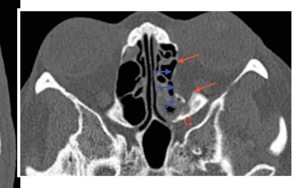
Orbital Fractures38
 |  |
 |
Le Fort Fractures39
Le Fort fractures (midface / maxilla), shown by level:
- Le Fort III level
- Le Fort II level
- Le Fort I level
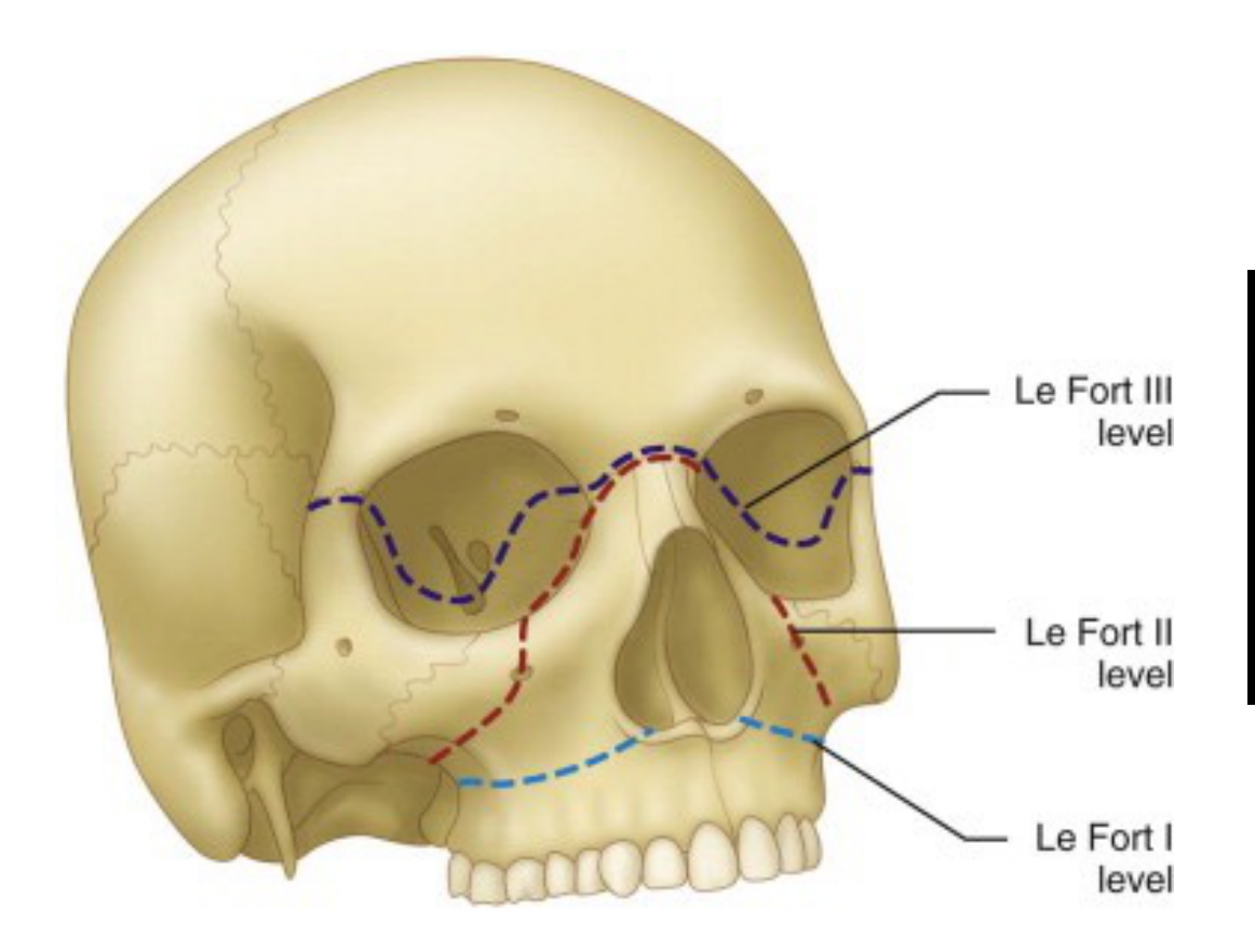 | 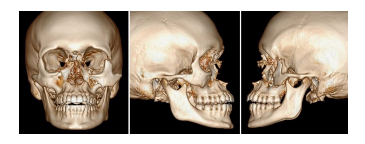 |
Mandibular Fractures
Frequency by Location
- Coronoid process: 2%
- Condyle: 30%
- Ramus: 3%
- Angle: 25%
- Body: 25%
- Parasymphysis / mental: 15%
F Gaillard, 2009, @Radiopedia.org
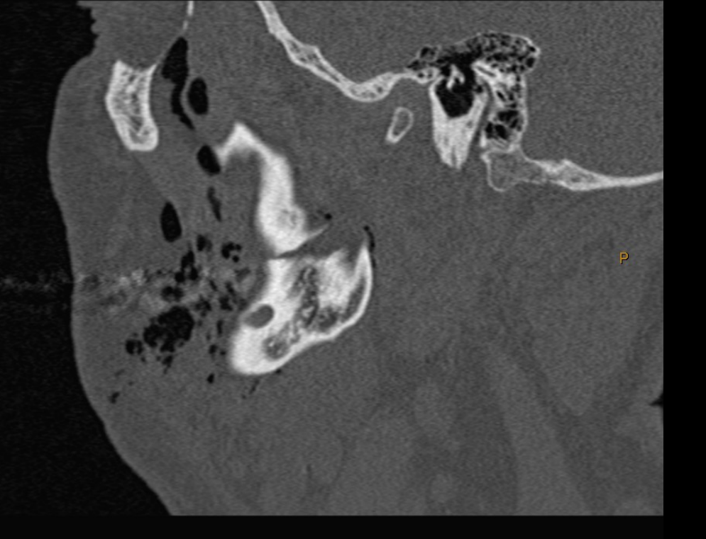
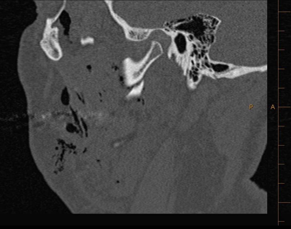
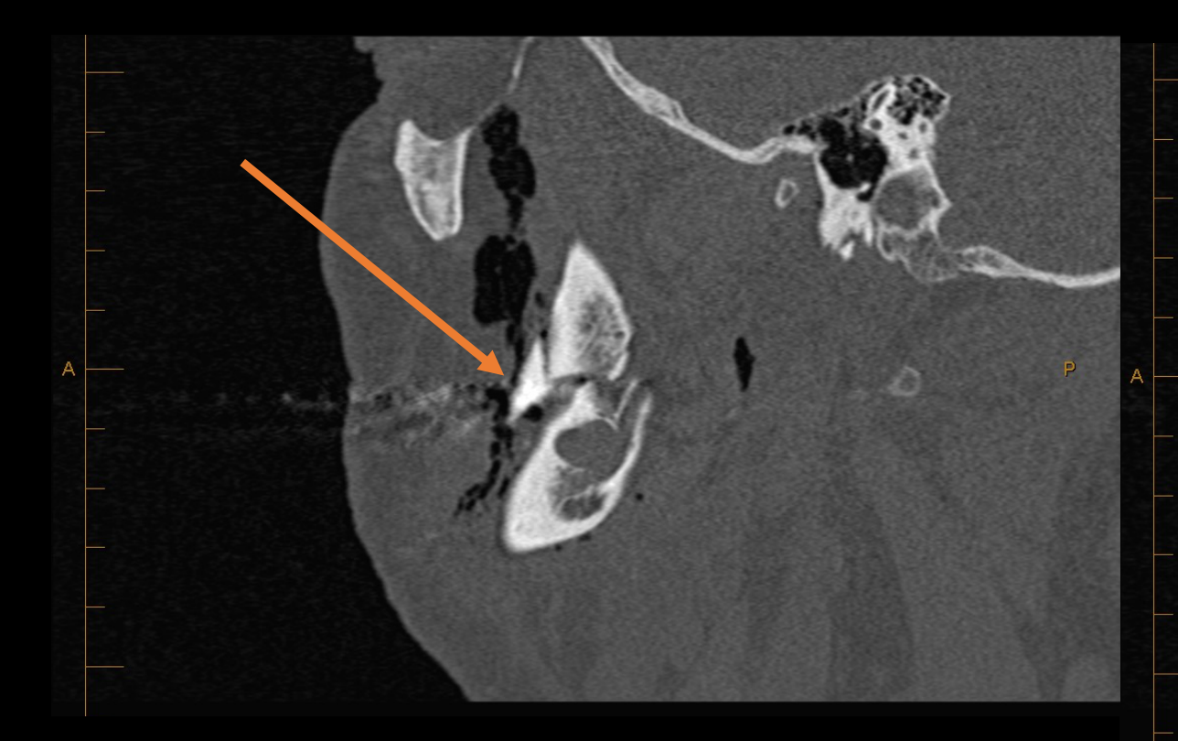
Displaced Comminuted Ramus Fracture
CT showed a displaced oblique comminuted fracture of the right mandibular ramus involving the retromolar triangle.
- Axial images showed the fracture, air around it, and traumatic socket changes.
- Sagittal images showed the fracture extending posteriorly toward the anterior region, an associated socket, a triangular bony fragment up to 13 mm, and involvement of the 48 socket without a root fragment.
- Coronal images showed the displaced fracture extending anteriorly toward the socket.
- Soft tissue windows demonstrated air bubbles, subcutaneous emphysema, and involvement of the masticator and parapharyngeal spaces.
- Hard tissue windows demonstrate bone more clearly, whereas soft tissue windows demonstrate soft tissue involvement more clearly.
CT volume-rendered images (H 36s\3), HFS orientation.
- Image 1: VR Mag: 1.05 — R / L markers; 120.00 kV, 27.00 mA, Tilt: 0.00, FOV: 167.00, Thickness: 0.60 mm; 10.00 mm/div; LeftButton: Rotate; JPEG Lossy: 75
- Image 2: VR Mag: 1.85 — P / A markers; 120.00 kV, 27.00 mA, Tilt: 0.00, FOV: 167.00, Thickness: 0.60 mm; 5.00 mm/div; LeftButton: Rotate; JPEG Lossy: 75
CT: 120.0 kVp, 38 mA, Tilt: 0.0 deg; R / L markers.
AI in Trauma Imaging4041
- AI applications:
- Automated fracture detection (mandible, midface)
- Triage in emergency settings
- 3D reconstruction assistance
- Deep learning (CNNs) improves detection accuracy
- Limitations:
- Training dataset bias
- Limited generalizability
REVIEW article. Front. Artif. Intell., 05 January 2024. Sec. Medicine and Public Health, Volume 6 - 2023 | https://doi.org/10.3389/frai.2023.1278529. A review on artificial intelligence for the diagnosis of fractures in facial trauma imaging. Tuan D. Pham, S. Simon B. Holmes, Paul Coulthard. Barts and The London School of Medicine and Dentistry, Queen Mary University of London, London, United Kingdom.
Lecturer — AI Performance and Limits
The lecturer emphasized that AI performance varies by fracture type and dataset.
- AI has demonstrated high sensitivity for detecting maxillofacial fractures on CT.
- Some studies show performance approaching that of experienced radiologists, particularly for mandibular fractures.
- In emergency triage, AI may flag suspected fractures for rapid review.
- Accuracy may be reduced in complex or comminuted fractures, and AI should support rather than replace clinical judgment.
Audio Appendix
Additional Audio Content
The following sections from the lecture audio did not correspond to any heading in the main document.
When to use Cone Beam CT
- Assessment of root fractures, crown-root fractures, luxation injuries, alveolar fractures, and unfavorable follow-up changes.
- Use when intraoral radiographs are inconclusive or demonstrate a possible fracture in the middle third of the root.
Facial Trauma & Multislice CT
- CT as the preferred imaging modality for maxillofacial trauma.
- Assessment of nasal, orbital, midfacial, and mandibular fractures.

Clinical Cases
Case: Anterior Maxillary Trauma Imaging
Question
Scenario: A patient presents with anterior maxillary trauma, and clinical examination reveals displacement of the left central incisor.
What’s shown: An occlusal radiograph and three periapical radiographs (right side, midline, and left side).
Consider: What do the different radiographic views demonstrate regarding the displacement of the tooth, and why are multiple views necessary?
Answer
Observations:
- The occlusal view clearly demonstrates the displacement of the left central incisor, showing the empty socket.
- The periapical views do not necessarily show the displacement of the tooth as clearly as the occlusal view.
Reasoning: Different intraoral radiographs provide different angles and perspectives of the teeth and surrounding structures. Relying on a single view may miss critical details like tooth displacement, which is why multiple views (one occlusal and three periapicals) are required to fully assess the injury.
Takeaway: Multiple intraoral radiographic views are essential in dental trauma to capture different angles and accurately diagnose displacements or fractures that may be missed on a single image.
Case: Maxillary Incisor Trauma Follow-up
Question
Scenario: A 21-year-old patient presents with a loose, mobile, and tender maxillary incisor (tooth 11) following trauma. The tooth is non-responsive to cold testing. An initial periapical radiograph is taken, a splint is placed, and the patient is scheduled for a 6-week follow-up but returns after 4 months due to pain.
What’s shown: An initial periapical radiograph and a follow-up CBCT scan taken at 4 months.
Consider: What are the findings on the initial periapical radiograph compared to the 4-month follow-up CBCT?
Answer
Observations:
- The initial periapical radiograph revealed a horizontal fracture at the cervical third of the root.
- The 4-month follow-up CBCT showed multiple complex root fractures: an incomplete horizontal fracture at the alveolar crest, and an oblique fracture extending from the palatal surface to the labial surface at the junction of the middle and apical thirds.
- Internal resorption involving the canal was also present on the CBCT.
Reasoning: The initial 2D periapical radiograph only captured the horizontal cervical fracture. The 3D CBCT scan, taken later due to persistent pain, revealed a much more complex fracture pattern and internal resorption that were not evident on the initial 2D imaging.
Takeaway: CBCT is highly beneficial for detecting complex, oblique, or multiple root fractures and complications like internal resorption that may be missed or not fully appreciated on initial 2D intraoral radiographs.
Case: Palatal Displacement of Central Incisors
Question
Scenario: A 47-year-old female falls and presents with trauma to teeth 11 and 21. Both teeth are displaced palatally, non-responsive to sensibility tests, and associated with soft tissue lacerations that are sutured. Radiographs are taken after the teeth are repositioned and splinted.
What’s shown: A periapical radiograph and a CBCT scan.
Consider: What do the periapical radiograph and CBCT reveal about the periodontal and alveolar structures, and what is the subsequent clinical course?
Answer
Observations:
- The periapical radiograph shows whitening of the periodontal ligament (PDL) spaces of teeth 11 and 21.
- The CBCT reveals a mildly displaced bicortical alveolar fracture extending from the palatal to the labial cortex, involving the PDL spaces, with no root fractures identified.
- At the 4-week recall, teeth 11 and 21 remain non-responsive to sensibility tests and become discolored.
Reasoning: While the 2D radiograph suggested PDL widening, the CBCT provided a definitive diagnosis of a bicortical alveolar fracture. The lack of response to sensibility tests and subsequent discoloration at the 4-week follow-up indicated pulp necrosis, necessitating root canal therapy.
Takeaway: CBCT is valuable for ruling in or out dentoalveolar fractures following trauma, and clinical follow-up is critical to monitor for pulp necrosis requiring endodontic intervention.
Case: Tooth 22 Fracture and MPR Alignment
Question
Scenario: A patient presents with a fractured tooth 22. The initial assessment suggests an uncomplicated crown fracture with subluxation.
What’s shown: CBCT images utilizing Multiplanar Reformatting (MPR) aligned specifically to the long axis of tooth 22, showing corrected coronal and sagittal views.
Consider: What specific fracture and periodontal findings are identified when the 3D data is manipulated to align with the long axis of the tooth?
Answer
Observations:
- The corrected views reveal an incomplete horizontal fracture of the tooth 22 root at the cementoenamel junction (CEJ), extending from the labial surface to the pulp.
- The PDL spaces appear normal.
- No bony fractures are identified in the corrected sagittal or coronal views.
Reasoning: By using MPR to align the imaging planes with the long axis of the specific tooth, the resulting corrected coronal view resembles a traditional periapical radiograph, allowing for precise diagnosis of the root fracture extent and assessment of the PDL spaces without the distortion of standard axial/coronal/sagittal planes.
Takeaway: Multiplanar reformatting (MPR) aligned to the long axis of a tooth is essential in CBCT assessment to accurately diagnose root fractures and evaluate periodontal spaces.
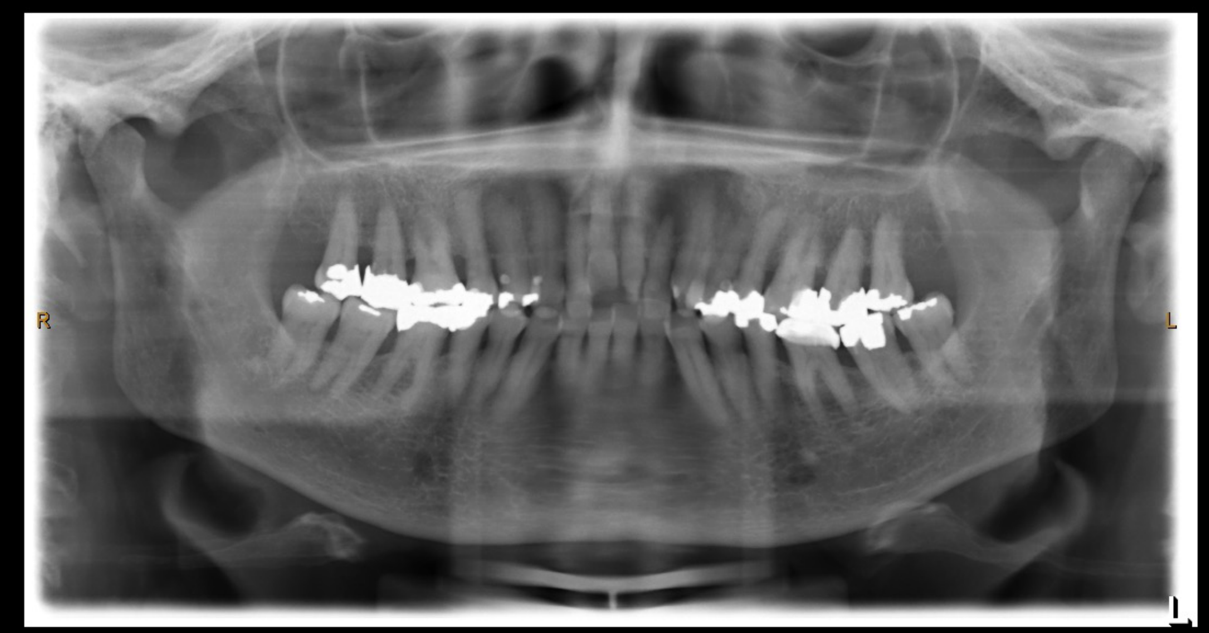
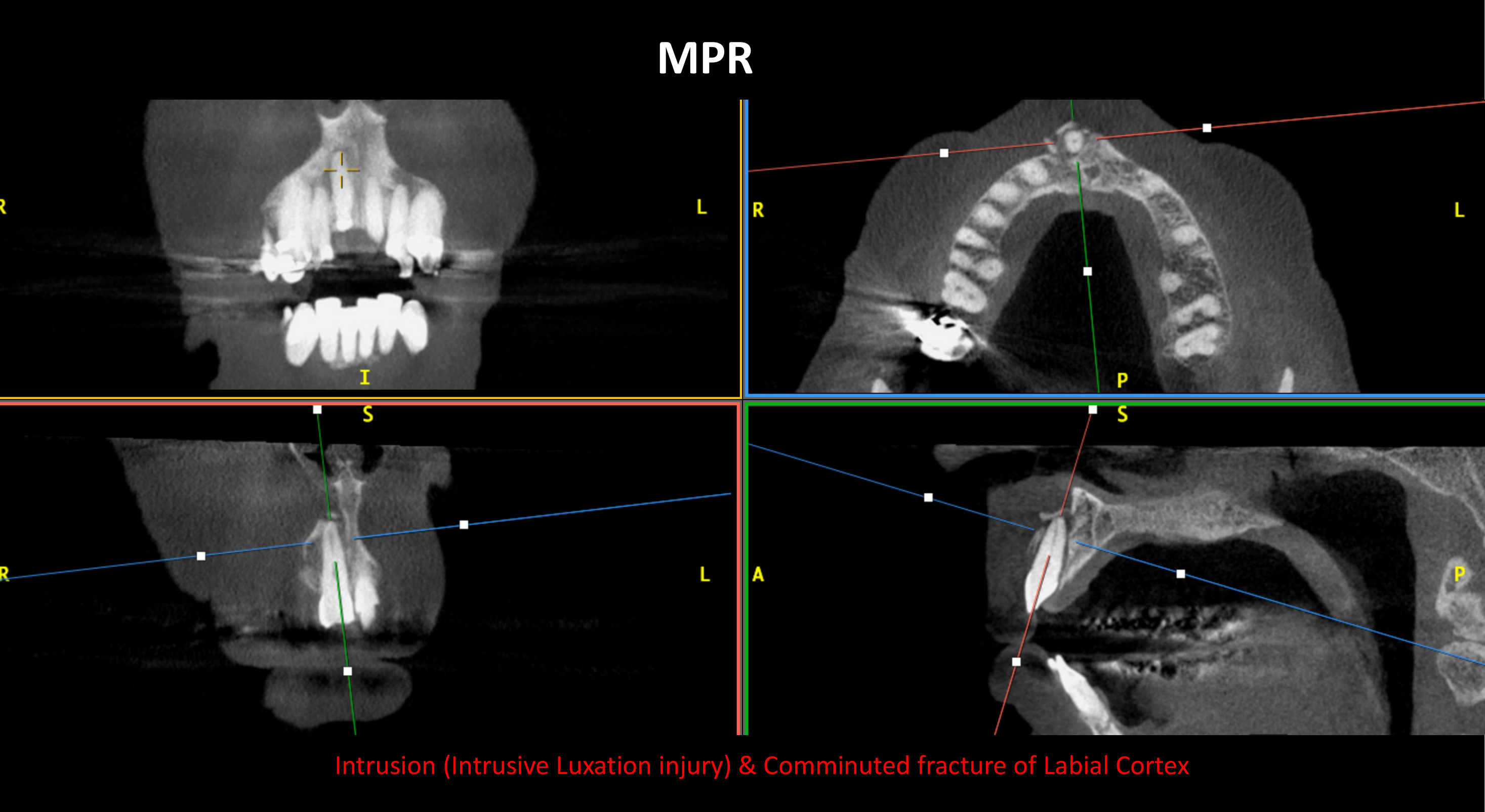
Case: Intrusive Injury to Tooth 11 and Crown Defect on Tooth 21
Question
Scenario: A patient presents with trauma to tooth 11. An initial OPG is taken, followed by a CBCT with MPR for both teeth 11 and 21.
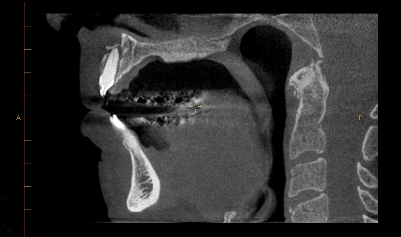
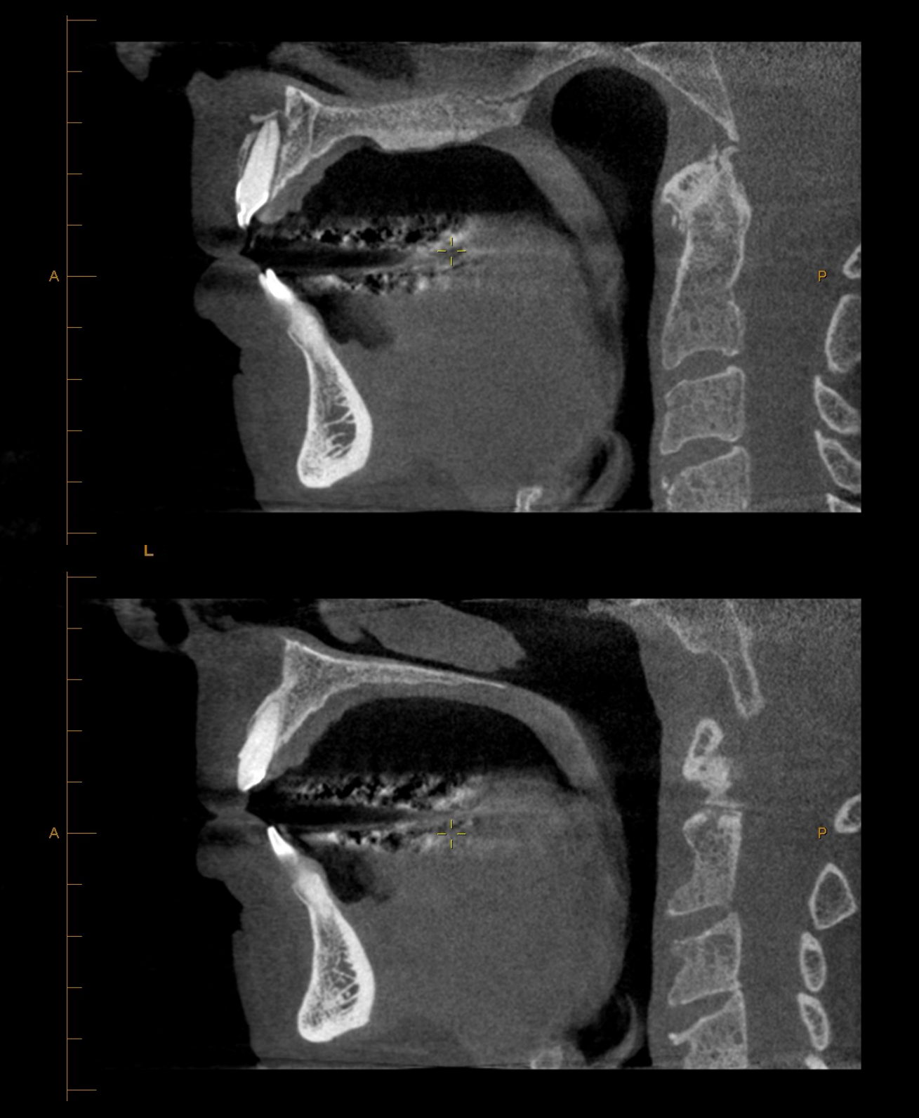
What’s shown: An initial OPG and follow-up CBCT with corrected coronal and sagittal planes for teeth 11 and 21, as well as a volume-rendered image.
Consider: What do the OPG and CBCT reveal regarding the injury to tooth 11, and what is observed regarding the crown structure of tooth 21?
Answer
Observations:
- The initial OPG shows tooth 11 located slightly superiorly with partly missing coronal tooth structure.
- The CBCT volume-rendered and corrected views confirm an intrusive injury of tooth 11 with a mild uncomplicated crown fracture at the tip, widened PDL spaces, and a comminuted labial alveolar fracture with multiple bone fragments.
- The corrected sagittal view of tooth 21 shows a missing crown structure resembling a missing veneer rather than a shear fracture, with no PDL space whitening or bony fracture.
Reasoning: The OPG provided a preliminary view of the intrusion and coronal loss, but the CBCT with MPR allowed for precise evaluation of the intrusive displacement, the exact nature of the crown fracture, and the associated alveolar bone comminution. The appearance of tooth 21 requires clinical correlation to differentiate between a veneer loss and a true shear fracture.
Takeaway: CBCT with MPR provides critical 3D detail for intrusive injuries and associated alveolar fractures that cannot be fully appreciated on a 2D OPG, while also aiding in the assessment of adjacent teeth.
Case: Anterior Maxilla Trauma and Avulsion
Question
Scenario: A patient presents with trauma to the upper anterior teeth, specifically involving tooth 22.
What’s shown: A reconstructed panoramic CBCT image, axial views of the maxilla, and corrected sagittal views of the upper anterior teeth.
Consider: What is the extent of the bony fractures and the status of the teeth in the anterior maxilla based on the multiplanar CBCT views?
Answer
Observations:
- The panoramic and axial views show that tooth 22 is missing (avulsed), leaving an empty socket.
- There is a labial fracture superior to the apex of tooth 21, continuing inferiorly to involve the PDL space of 21 and extending to the 22 avulsion socket.
- A separate palatal fracture starts in the 21 region and extends to the 22 socket.
- Tooth 11 shows PDL space widening and a labial fracture.
Reasoning: By utilizing panoramic, axial, and corrected sagittal views, the full extent of the complex alveolar fractures can be traced. The fractures involve both the labial and palatal cortices, connecting the injuries around teeth 11 and 21 to the avulsion site of tooth 22.
Takeaway: Multiplanar CBCT imaging is necessary to trace the full extent of complex alveolar fractures and their relationship to avulsed and adjacent teeth in the anterior maxilla.
Case: Mandibular Avulsion and Compound Fracture
Question
Scenario: A patient presents with mandibular dental trauma involving the 42 to 44 region.
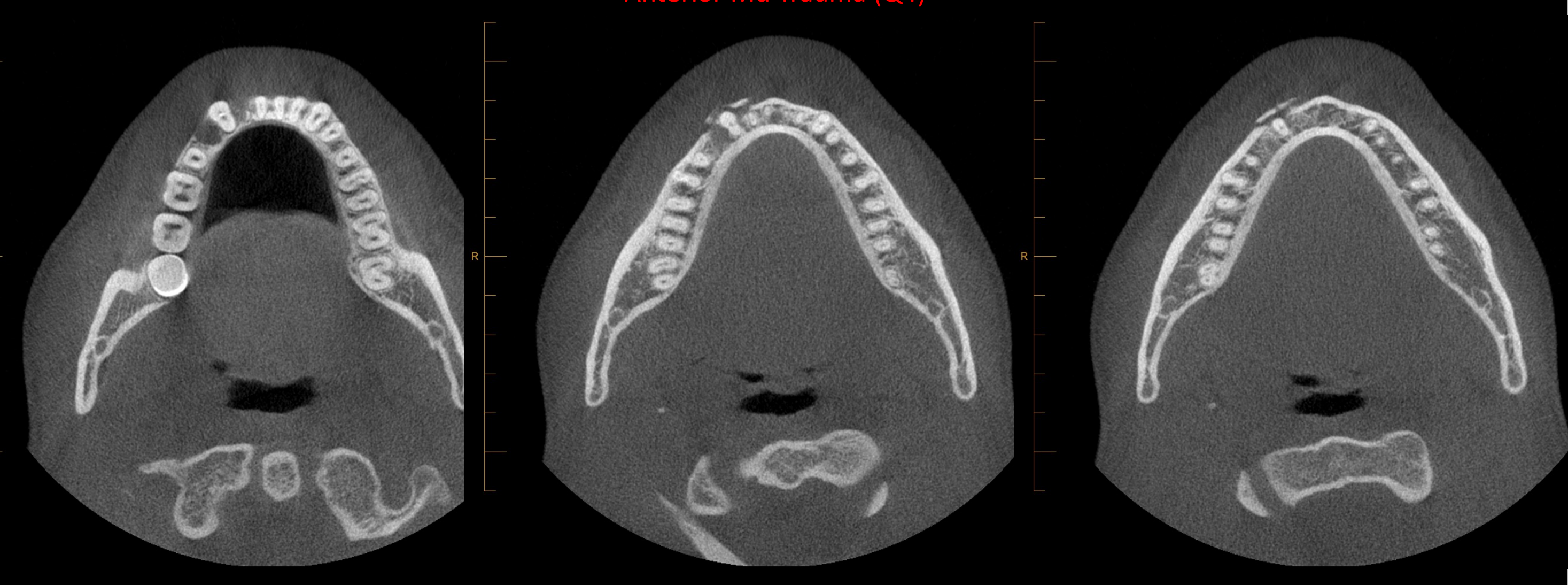
What’s shown: CBCT cross-sectional slices and MPR (corrected coronal and sagittal views) of the right mandible (quadrant 4).
Consider: What are the findings regarding the status of teeth 42, 43, and 44, and what type of fracture is present?
Answer
Observations:
- Teeth 42 and 44 are missing, compatible with avulsion injuries. Tooth 42 has a root remnant, and tooth 44 is an empty socket with a small bone piece.
- Tooth 43 has undergone endodontic treatment, with no root or crown fracture, but shows widened labial PDL space and PDL space whitening.
- There is a comminuted labial bone fracture involving the labial PDL space of tooth 43 and extending from the 44 to 42 region.
Reasoning: The cross-sectional slices and MPR clearly identify the avulsed teeth and root remnants. The involvement of the PDL (a soft tissue) by the bony fracture classifies this as a compound fracture.
Takeaway: CBCT is highly effective in identifying avulsed teeth, root remnants, and compound fractures where bony fractures extend into the periodontal soft tissues.
Case: Complex Dental Alveolar Trauma
Question
Scenario: A patient presents with complex dental alveolar trauma involving multiple teeth on the right side of the face (16, 15, 14, 44, 46).
What’s shown: A CBCT-generated panoramic view, corrected sagittal views, and axial views.
Consider: What specific types of fractures are identified on the different CBCT planes for the involved teeth?
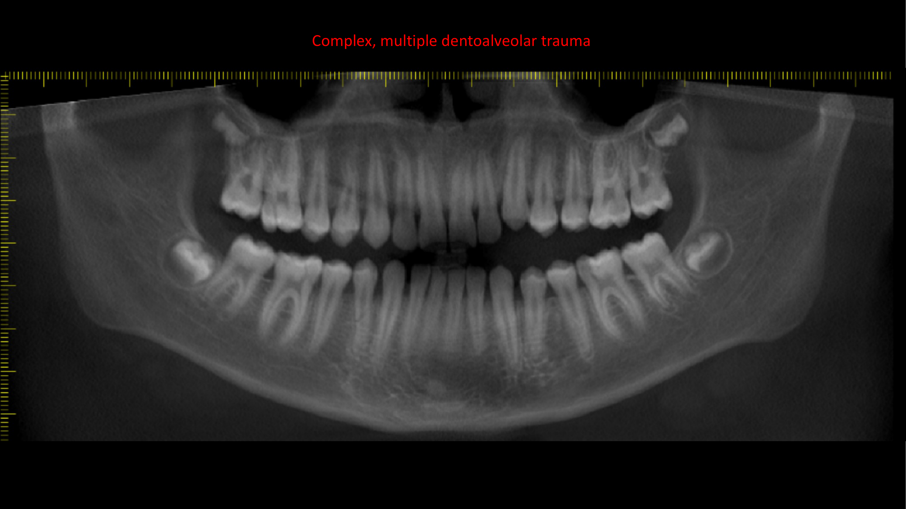
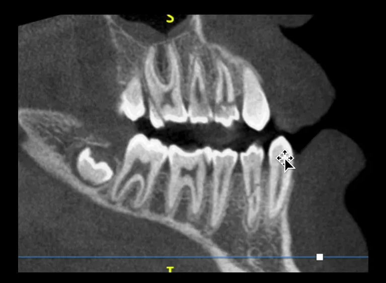
Answer
Observations:
- Tooth 16: Cuspal fracture of the distobuccal cusp.
- Tooth 15: Vertical fracture running mesiodistally, involving the pulp (not seen on initial sagittal views).
- Tooth 14: Horizontal/labial fracture near the pulp extending towards the mandible.
- Tooth 44: Oblique fracture.
- Tooth 46: Mesiolingual cuspal fracture.
Reasoning: The panoramic and sagittal views initially show definite fractures, but the axial views provide crucial additional information, revealing the exact orientation (e.g., vertical vs. horizontal) and pulp involvement of the fractures that were not fully appreciated on the other planes.
Takeaway: In complex multi-tooth trauma, axial CBCT views are essential to determine the precise orientation, extent, and pulp involvement of fractures that may be ambiguous on panoramic or sagittal views.
Case: Displaced Nasal Bone Fracture
Question
Scenario: A patient presents with facial trauma.
What’s shown: A CT scan of the nasal region.
Consider: What are the findings regarding the nasal bone and adjacent structures?
Answer
Observations:
- There is a visible fracture line with rotation of the nasal bone, indicating a displaced nasal bone fracture.
- Mucosal changes are present in the adjacent ethmoid air cells.
Reasoning: CT imaging clearly delineates the bony disruption and displacement of the nasal bone, as well as the secondary soft tissue/mucosal changes in the adjacent sinuses resulting from the trauma.
Takeaway: CT is the gold standard for evaluating nasal bone fractures, allowing for the assessment of both bony displacement and associated mucosal changes.
Case: Orbital Floor Blowout Fracture
Question
Scenario: A patient presents with facial trauma involving the orbit.
What’s shown: A CT image of the orbital region.
Consider: What are the findings regarding the orbital floor and the paranasal sinuses?
Answer
Observations:
- There is a defect in the orbital floor (blowout fracture).
- There is hemisinus (blood accumulation) in both the left and right maxillary sinuses.
Reasoning: The CT scan demonstrates the structural defect in the orbital floor, which can allow orbital contents to herniate into the sinus. The presence of bilateral hemisinus indicates bleeding into the sinuses, a common associated finding in orbital floor fractures.
Takeaway: CT imaging is essential for identifying orbital floor blowout fractures and associated complications like hemisinus, which helps in assessing the severity and potential for muscle entrapment.
Case: Right Mandibular Ramus Fracture
Question
Scenario: A patient presents with trauma to the right mandible.
What’s shown: CT axial, sagittal, coronal, and soft tissue window views of the right mandibular ramus.
Consider: What are the characteristics of the fracture, and what soft tissue complications are visible on the soft tissue window?
Answer
Observations:
- There is a displaced, oblique, comminuted bony fracture of the right mandibular ramus involving the retromolar triangle.
- A small triangular bony fragment measuring 13 mm is present.
- The fracture involves the tooth 48 socket without a root fragment.
- The soft tissue window reveals subcutaneous emphysema (air bubbles) involving the masticator and parapharyngeal spaces.
Reasoning: The hard tissue windows clearly define the complex, comminuted nature of the fracture and the involvement of the third molar socket. Switching to the soft tissue window is crucial for identifying the presence of air (subcutaneous emphysema) in the fascial spaces, indicating a communication with the oral cavity or airway.
Takeaway: Utilizing both hard and soft tissue CT windows is critical in mandibular trauma to evaluate the bony fracture details and identify soft tissue complications like subcutaneous emphysema.
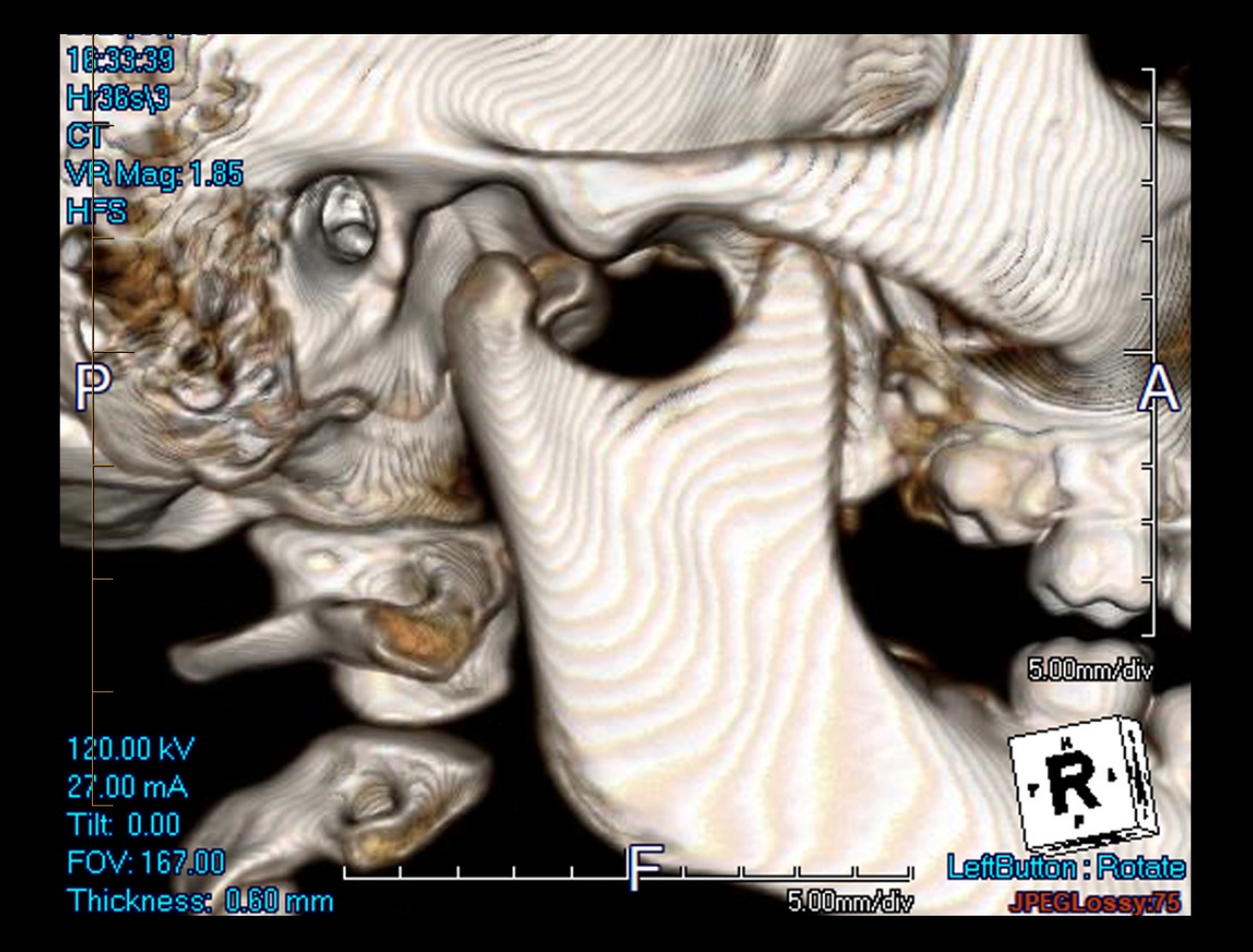
Case: Displaced Condylar Fracture
Question
Scenario: A patient presents with mandibular trauma and dental malocclusion.
What’s shown: CT axial, coronal, and sagittal views of the mandibular condyle.
Consider: What is the position of the fractured condylar head, and are there any associated findings?
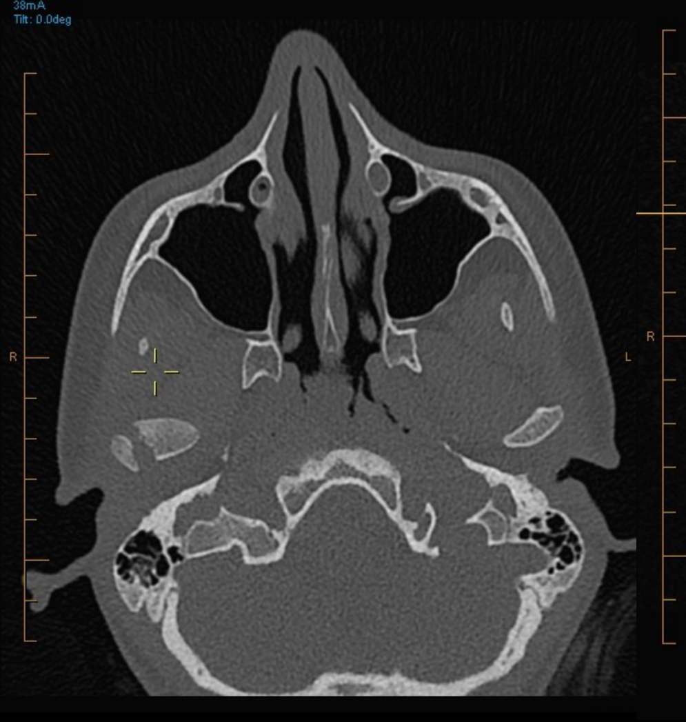
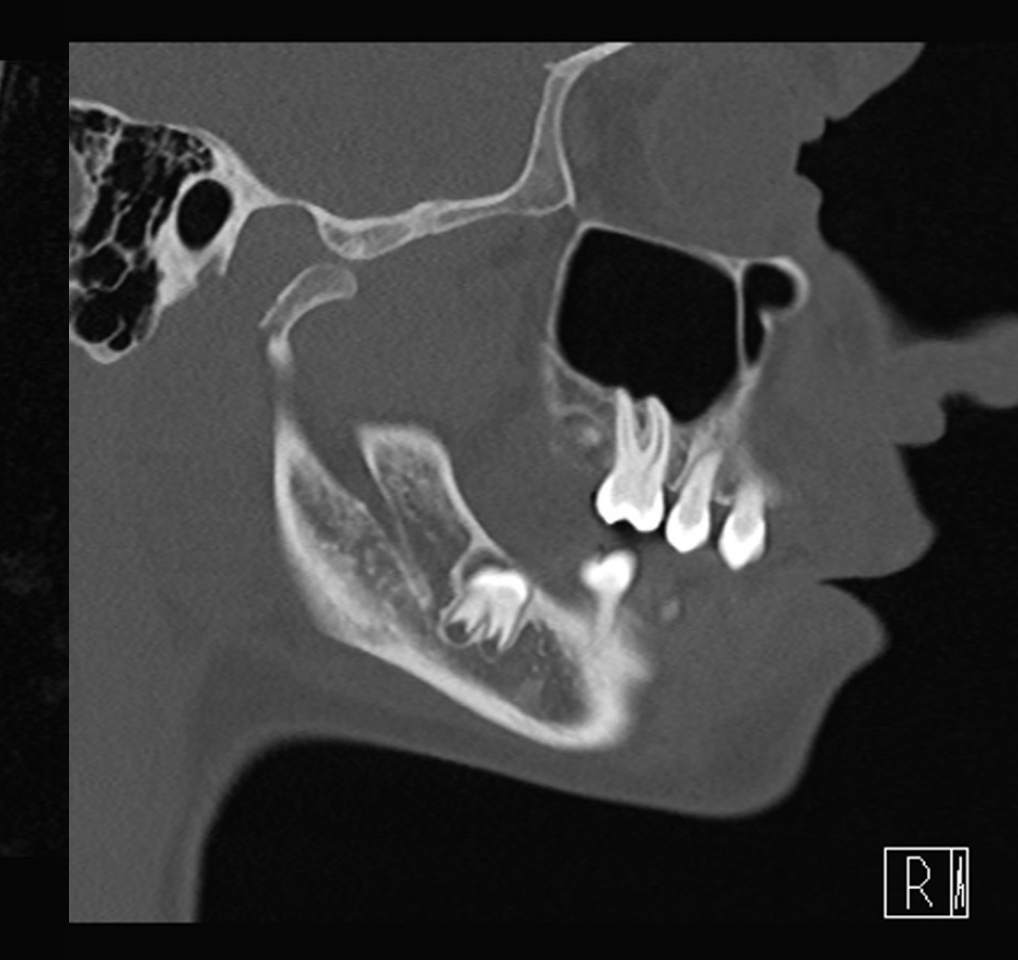
Answer
Observations:
- The fractured condylar head is displaced anteriorly (seen on axial), medially (seen on coronal), and inferior to the articular eminence (seen on sagittal).
- There is minimal contact at the middle aspect of the condylar neck.
- The mandibular symphysis is deviated to the left.
- There is no contralateral fracture.
Reasoning: Multiplanar CT imaging allows for precise 3D localization of the displaced condylar fragment. The displacement explains the symphysis deviation and the clinical finding of dental malocclusion. The absence of a contralateral fracture is noted as a rare but important finding, as mandibular fractures often occur in two locations.
Takeaway: Multiplanar CT is necessary to accurately determine the 3D displacement of condylar fractures and assess for associated deviations or contralateral injuries.
Footnotes
-
Original PDF page 1: L4 - Imaging for Trauma, p.1 ↩
-
Original PDF page 2: L4 - Imaging for Trauma, p.2 ↩
-
Original PDF page 3: L4 - Imaging for Trauma, p.3 ↩
-
Original PDF page 4: L4 - Imaging for Trauma, p.4 ↩
-
Original PDF page 5: L4 - Imaging for Trauma, p.5 ↩
-
Original PDF page 6: L4 - Imaging for Trauma, p.6 ↩
-
Original PDF page 7: L4 - Imaging for Trauma, p.7 ↩
-
Original PDF page 8: L4 - Imaging for Trauma, p.8 ↩
-
Original PDF page 9: L4 - Imaging for Trauma, p.9 ↩
-
Original PDF page 10: L4 - Imaging for Trauma, p.10 ↩
-
Original PDF page 11: L4 - Imaging for Trauma, p.11 ↩
-
Original PDF page 12: L4 - Imaging for Trauma, p.12 ↩
-
Original PDF page 13: L4 - Imaging for Trauma, p.13 ↩
-
Original PDF page 14: L4 - Imaging for Trauma, p.14 ↩
-
Original PDF page 15: L4 - Imaging for Trauma, p.15 ↩
-
Original PDF page 18: L4 - Imaging for Trauma, p.18 ↩
-
Original PDF page 16: L4 - Imaging for Trauma, p.16 ↩
-
Original PDF page 17: L4 - Imaging for Trauma, p.17 ↩
-
Original PDF page 20: L4 - Imaging for Trauma, p.20 ↩
-
Original PDF page 21: L4 - Imaging for Trauma, p.21 ↩
-
Original PDF page 22: L4 - Imaging for Trauma, p.22 ↩
-
Original PDF page 23: L4 - Imaging for Trauma, p.23 ↩
-
Original PDF page 24: L4 - Imaging for Trauma, p.24 ↩
-
Original PDF page 25: L4 - Imaging for Trauma, p.25 ↩
-
Original PDF page 26: L4 - Imaging for Trauma, p.26 ↩
-
Original PDF page 27: L4 - Imaging for Trauma, p.27 ↩
-
Original PDF page 28: L4 - Imaging for Trauma, p.28 ↩
-
Original PDF page 29: L4 - Imaging for Trauma, p.29 ↩
-
Original PDF page 30: L4 - Imaging for Trauma, p.30 ↩
-
Original PDF page 31: L4 - Imaging for Trauma, p.31 ↩
-
Original PDF page 32: L4 - Imaging for Trauma, p.32 ↩
-
Original PDF page 33: L4 - Imaging for Trauma, p.33 ↩
-
Original PDF page 34: L4 - Imaging for Trauma, p.34 ↩
-
Original PDF page 35: L4 - Imaging for Trauma, p.35 ↩
-
Original PDF page 36: L4 - Imaging for Trauma, p.36 ↩
-
Original PDF page 37: L4 - Imaging for Trauma, p.37 ↩
-
Original PDF page 38: L4 - Imaging for Trauma, p.38 ↩
-
Original PDF page 39: L4 - Imaging for Trauma, p.39 ↩
-
Original PDF page 40: L4 - Imaging for Trauma, p.40 ↩
-
Original PDF page 48: L4 - Imaging for Trauma, p.48 ↩
-
Original PDF page 49: L4 - Imaging for Trauma, p.49 ↩