Imaging for Facial Deformity1
Dr Dayea Oh Oral and Maxillofacial Radiologist
Objectives2
-
Understand types of facial deformity
-
Compare imaging modalities
-
Recognise key radiographic features relevant to treatment planning
-
Understand functional and surgical implications
-
Facial deformities can affect both function and aesthetics.
-
Assessment and treatment often require a multidisciplinary approach involving orthodontists, maxillofacial surgeons, and radiologists.
-
Imaging supports diagnosis, surgical simulation, and postoperative evaluation in addition to treatment planning.
-
Precise knowledge of craniofacial skeletal anatomy is essential for identifying deformities, interpreting imaging, and planning surgery.
-
Modern 3D imaging and digital workflows are contributing to more predictable and personalized outcomes.
Skull Anatomy
Bones in the Skull3
Paired bones (11):
- Parietal
- Temporal
- Zygomatic
- Maxilla
- Palatine
- Nasal
- Lacrimal
- Inferior nasal concha
- Malleus
- Incus
- Stapes
Single bones (6):
- Frontal
- Occipital
- Sphenoid
- Ethmoid
- Vomer
- Mandible
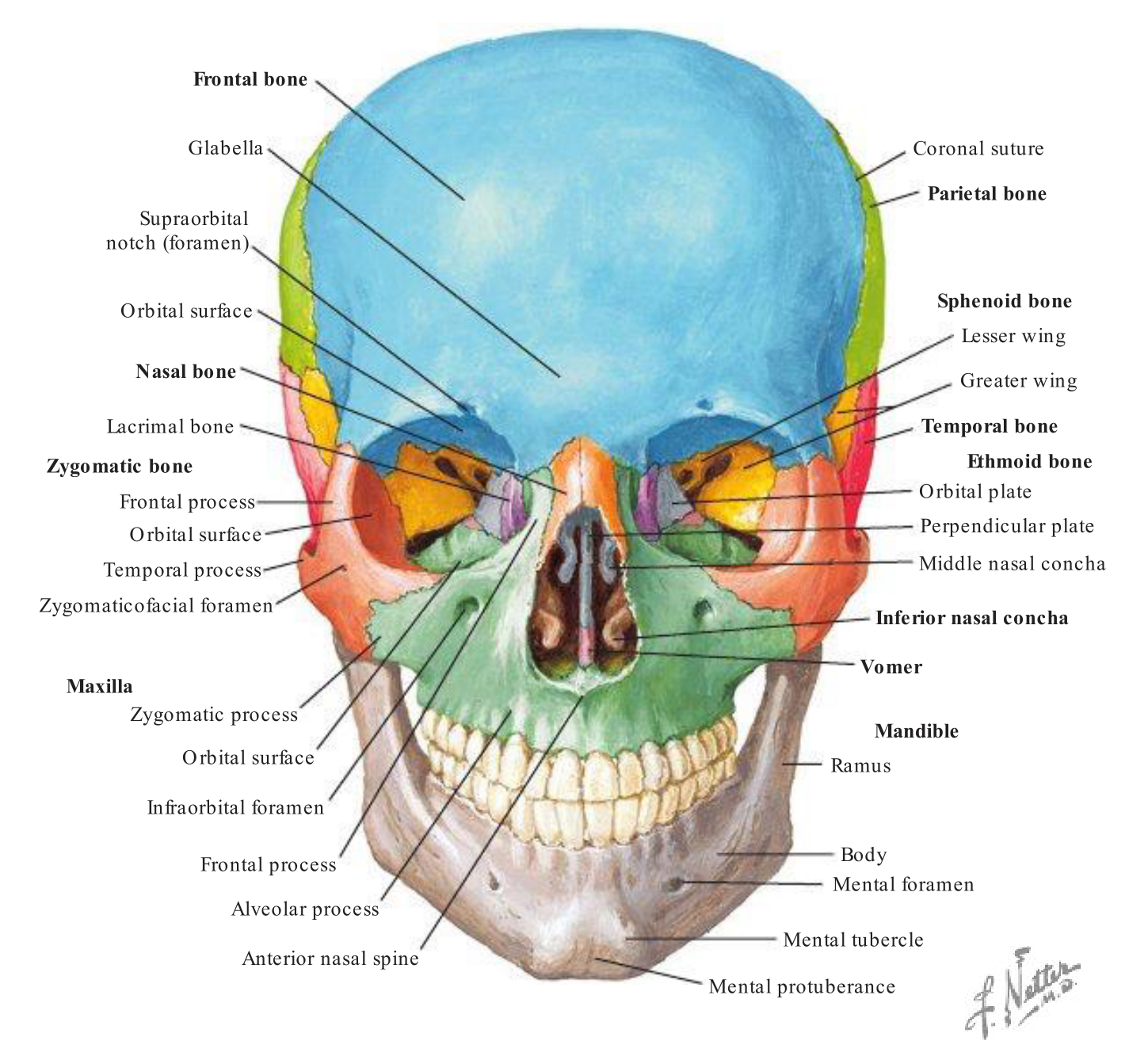
Anterior View — Labeled Structures
- Frontal bone
- Glabella
- Supraorbital notch (foramen)
- Orbital surface
- Nasal bone
- Lacrimal bone
- Zygomatic bone
- Frontal process
- Orbital surface
- Temporal process
- Zygomaticofacial foramen
- Maxilla
- Zygomatic process
- Orbital surface
- Frontal process
- Alveolar process
- Anterior nasal spine
- Infraorbital foramen
- Coronal suture
- Parietal bone
- Sphenoid bone
- Lesser wing
- Greater wing
- Temporal bone
- Ethmoid bone
- Orbital plate
- Perpendicular plate
- Middle nasal concha
- Inferior nasal concha
- Vomer
- Mandible
- Ramus
- Body
- Mental foramen
- Mental tubercle
- Mental protuberance
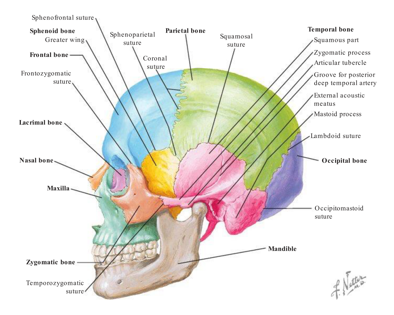
Lateral View — Labeled Structures4
- Sphenofrontal suture
- Sphenoid bone
- Greater wing
- Frontal bone
- Frontozygomatic suture
- Lacrimal bone
- Nasal bone
- Maxilla
- Zygomatic bone
- Temporozygomatic suture
- Coronal suture
- Parietal bone
- Squamosal suture
- Temporal bone
- Squamous part
- Zygomatic process
- Articular tubercle
- Groove for posterior deep temporal artery
- External acoustic meatus
- Mastoid process
- Lambdoid suture
- Occipital bone
- Occipitomastoid suture
- Mandible
- Sphenoparietal suture
 | 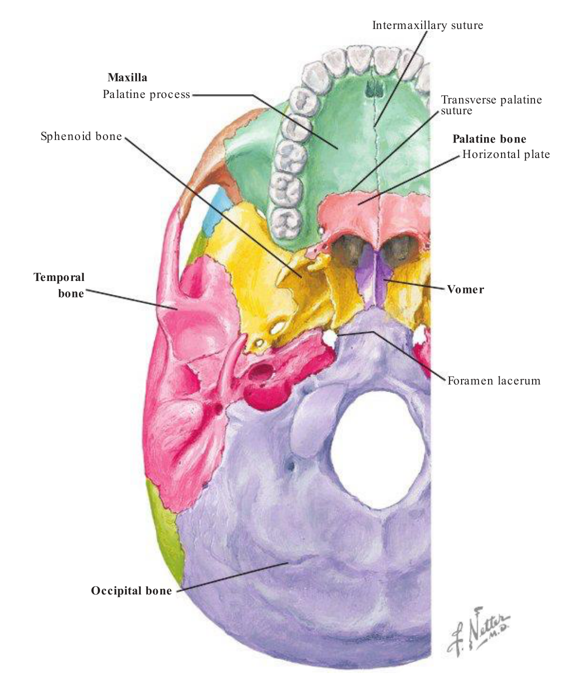 |
Inferior View — Labeled Structures
- Intermaxillary suture
- Maxilla
- Palatine process
- Transverse palatine suture
- Palatine bone
- Horizontal plate
- Vomer
- Foramen lacerum
- Occipital bone
- Sphenoid bone
Basal Structures — Labeled
- Maxilla
- Palatine process
- Palatine bone
- Horizontal plate
- Temporal bone
- Vomer
- Occipital bone
- Sphenoid bone
- Intermaxillary suture
- Transverse palatine suture
Lecturer — Anatomy Knowledge
Precise knowledge of craniofacial skeletal anatomy is fundamental to accurate diagnosis, imaging interpretation, and surgical planning.
Craniofacial Development5
- Migratory cranial neural crest cells
- Abnormal quantity or migration of NCC cells can result in craniofacial syndromes
- External facial structures develop between 4th and 6th week
- Palate develops between 6th and 8th week, and completes at the 12th week
Lecturer — Developmental Disruption
Craniofacial development is tightly regulated, begins early in embryogenesis, and continues throughout adolescence.
- Neural crest cells migrate into the developing branchial or pharyngeal arches and contribute to the bones, cartilage, and connective tissues of the face and neck.
- Developmental disruption may result from genetic mutations, environmental factors, or vascular disturbances.
- Potential features include facial asymmetry, hypoplasia of facial structures, ear abnormalities, and mandibular abnormalities.
- These disturbances can produce asymmetry, skeletal discrepancies, and syndromic conditions.
Facial Deformity Classification6
Hard tissue only, Soft tissue only or BOTH
- Congenital
- Birth defects eg. cleft palate & various craniofacial syndromes
- Developmental
- Growth-related eg. bilateral or unilateral hypoplasia & hyperplasia (of the jaws)
- Acquired
- Trauma, cysts, tumours, bone dysplasias, post-surgery etc.
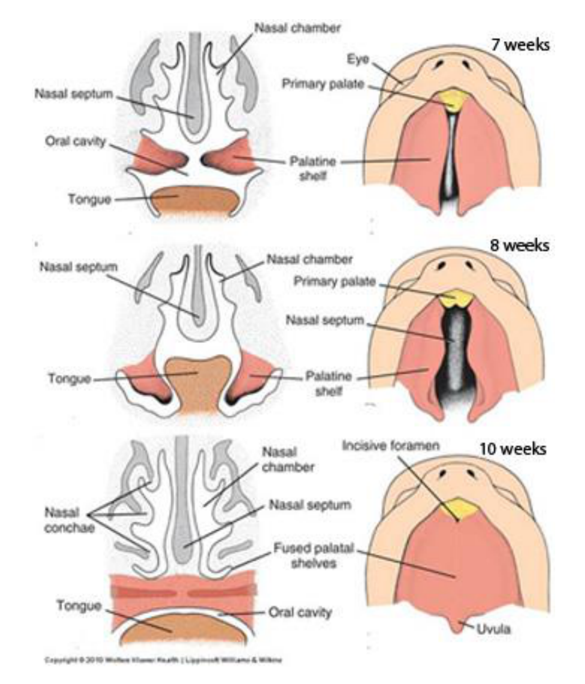
Palatal Development Timeline7
Palatal development — labelled figure views:
- 7 weeks: Nasal chamber, Eye, Primary palate, Nasal septum, Oral cavity, Palatine shelf, Tongue
- 8 weeks: Nasal septum, Nasal chamber, Primary palate, Tongue, Palatine shelf
- 10 weeks: Nasal chamber, Incisive foramen, Nasal conchae, Nasal septum, Fused palatal shelves, Tongue, Oral cavity, Uvula
Copyright © 2010 Wolters Kluwer Health | Lippincott Williams & Wilkins
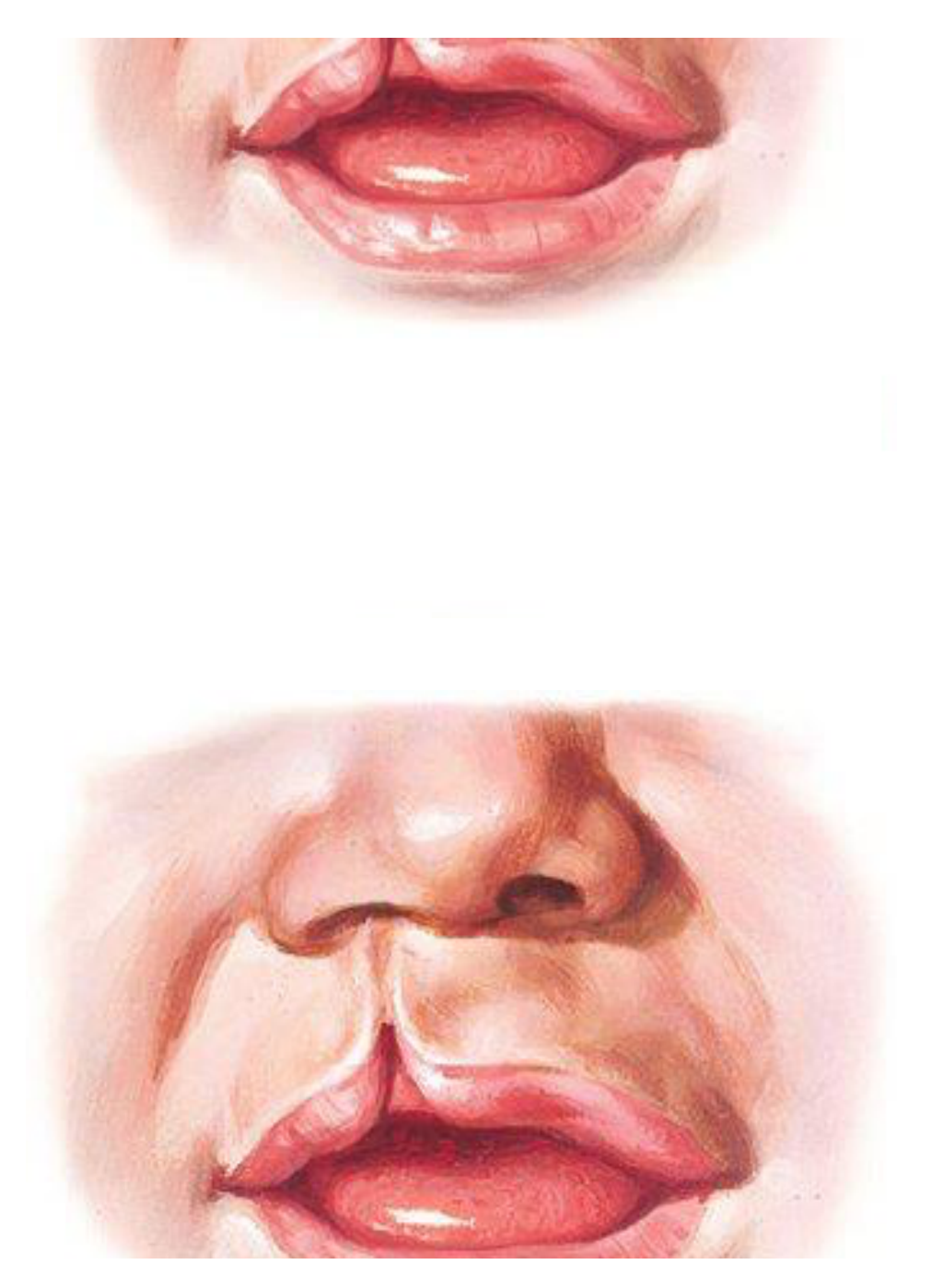
Failure of the medial and lateral nasal processes to fuse with the maxillary process
 |  |
Craniofacial Syndromes
- Many are branchial arch syndromes related to first and second branchial/pharyngeal arches →
- Hemifacial Microsomia (Goldenhar)
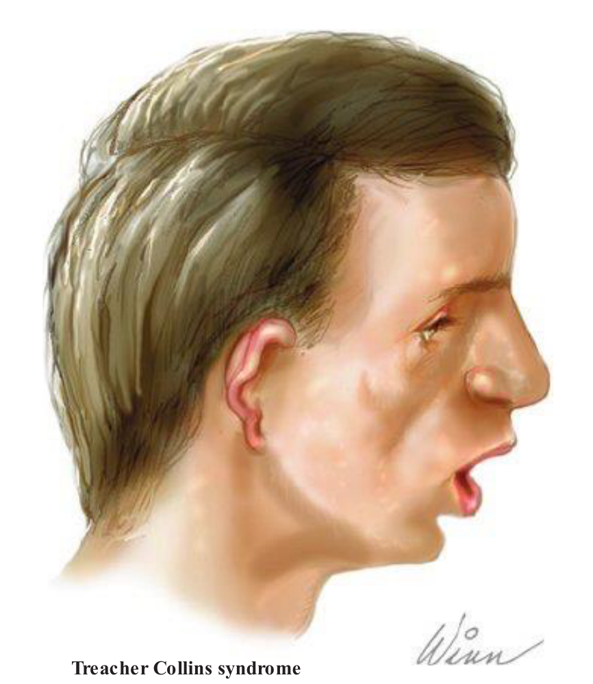
- Treacher Collins
- Manifest a spectrum of hypoplasia and aplasia of the structures composing the facial bones and ear
- Severity depends on the alteration of gene-expression profiles
Lecturer — Syndrome Involvement
Craniofacial syndromes may involve multiple hard- and soft-tissue structures, and their severity and symptoms vary between conditions.
- The spectrum of hypoplasia and aplasia may involve the facial bones, jaws, ears, zygomatic bones, and lateral orbital regions.
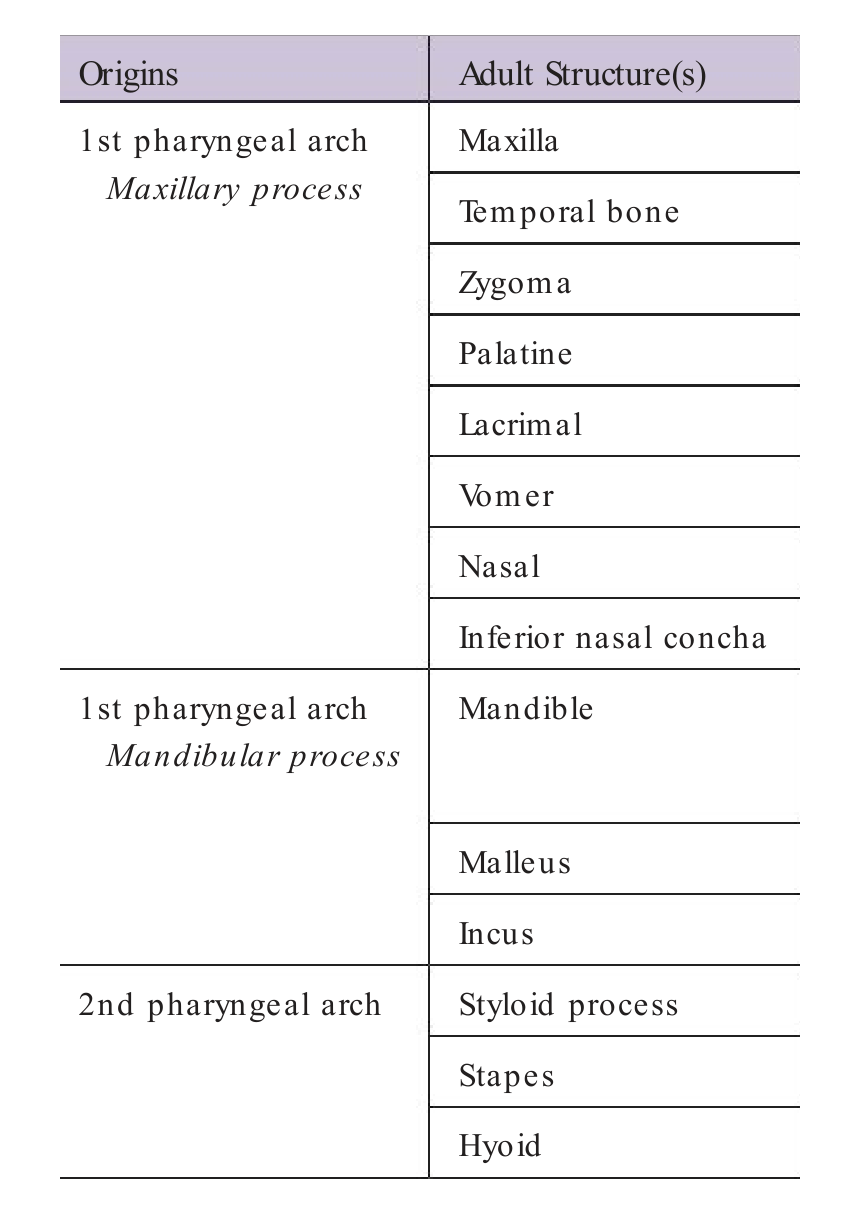
Pharyngeal Arch Origins of Adult Structures8
| Origins | Adult Structure(s) |
|---|---|
| 1st pharyngeal arch — Maxillary process | Maxilla, Temporal bone, Zygoma, Palatine, Lacrimal, Vomer, Nasal, Inferior nasal concha |
| 1st pharyngeal arch — Mandibular process | Mandible, Malleus, Incus |
| 2nd pharyngeal arch | Styloid process, Stapes, Hyoid |
 |  |
Anatomy Image Plates9101112131415
A
B
C
D
Continued
A
B
C
D
E
a
b
c
d
A B C D
E F G H
I J K L
B
Pfieffer

Imaging for Congenital Facial Deformity16
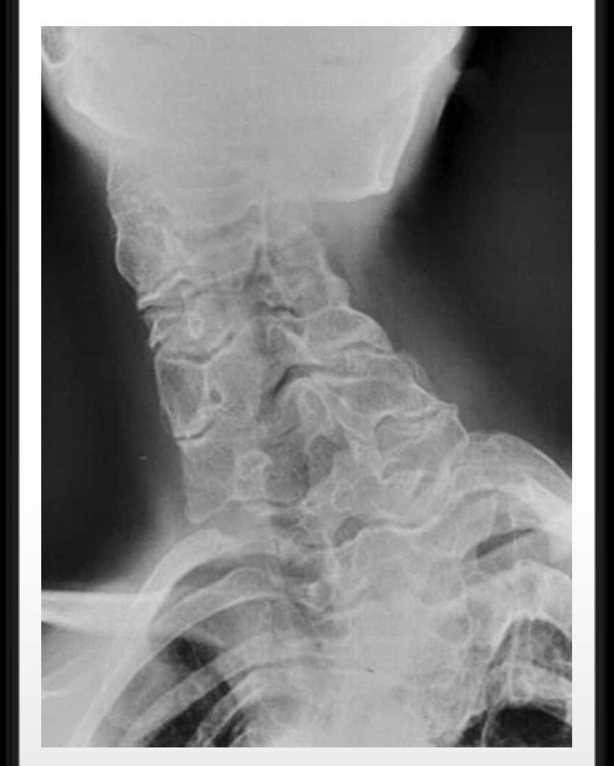
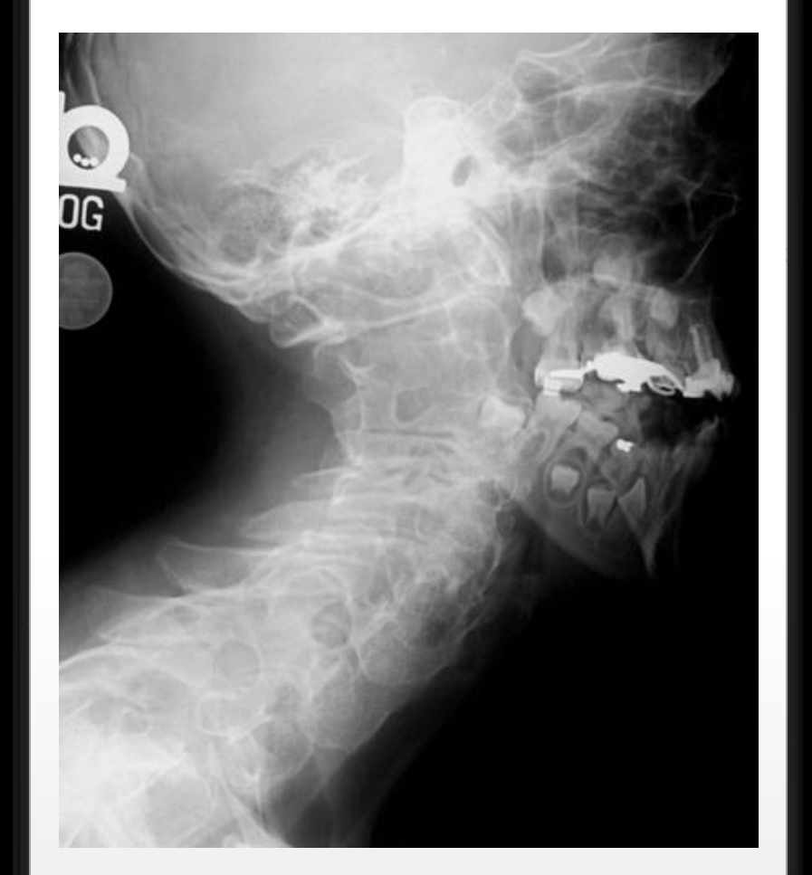
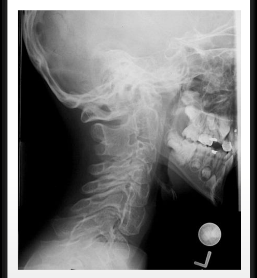
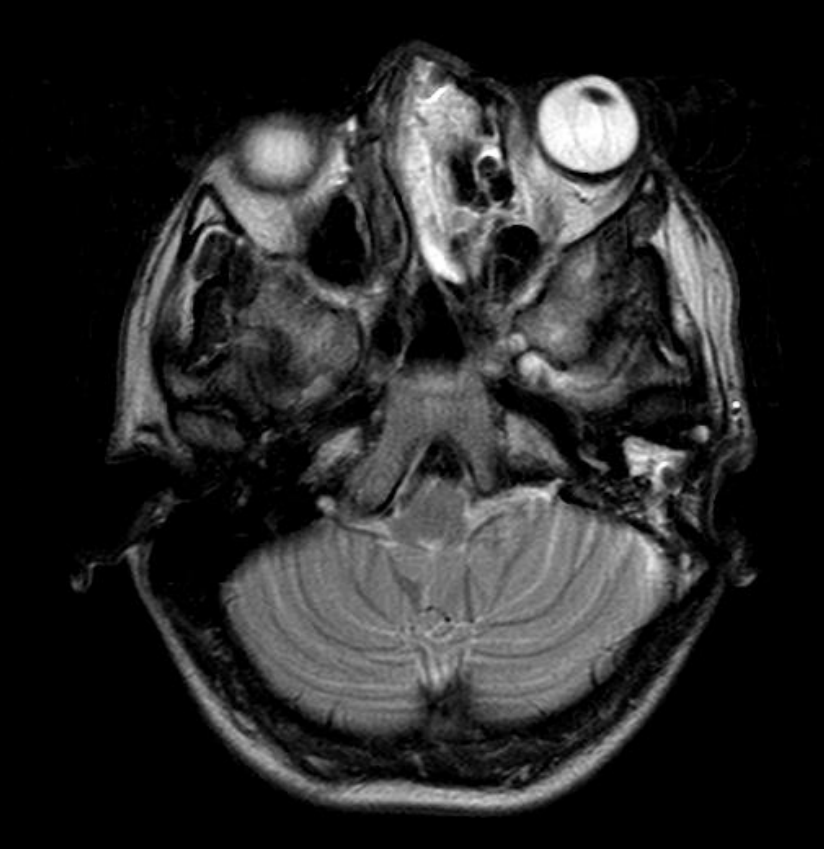
- Craniofacial syndromes involve multiple structures with varying degrees of severity and symptoms
- CT +/- MRI are the imaging modality of choice
- Detailed assessment of hard & soft tissues in 3D, especially pre-surgery
- Assess for any intracranial abnormalities
- OPG may be useful for overview of dental complications
Lecturer — Imaging Limitations
CT was described as the gold standard for imaging congenital facial deformities.
- Traditional skull views have limitations when demonstrating complex facial deformities.
- Conventional skull views may nevertheless remain useful for initial assessment because they are quick, cost-effective, and associated with low radiation exposure.
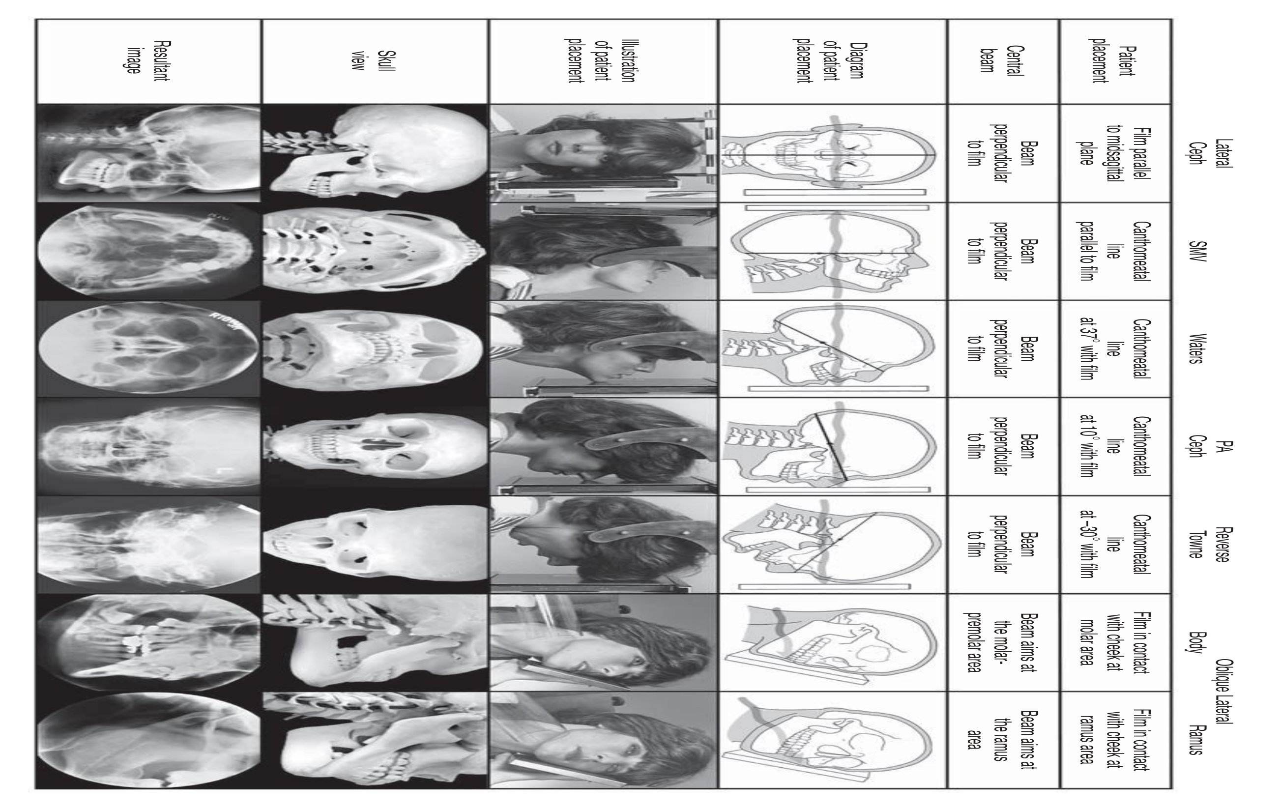
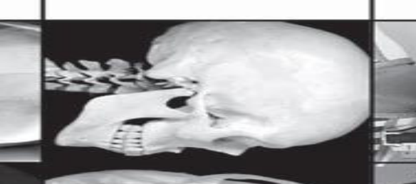
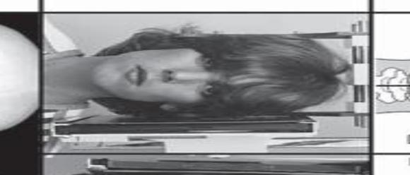
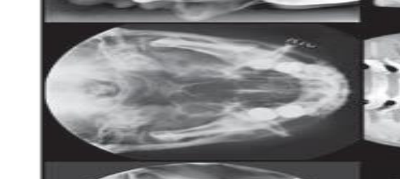
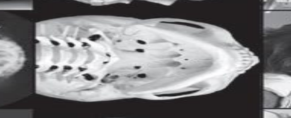
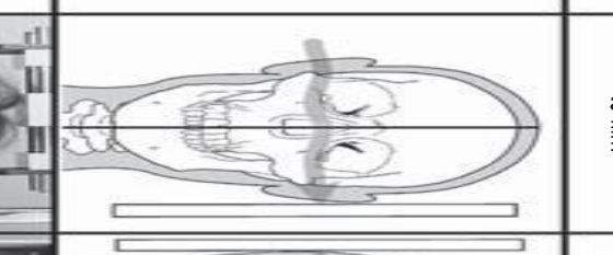
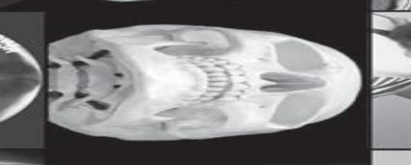
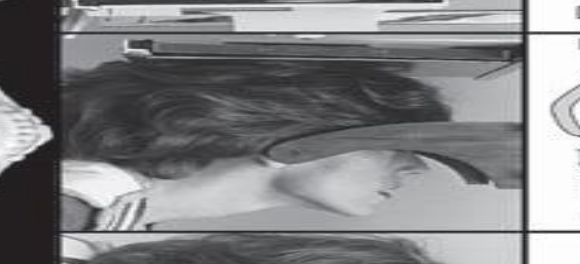
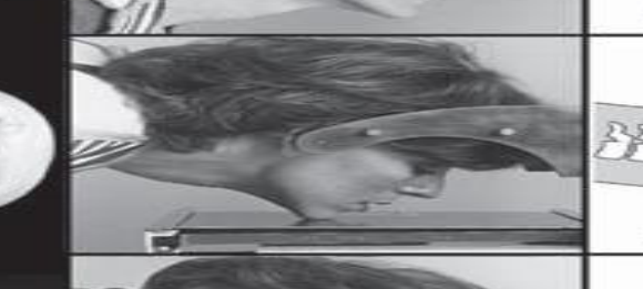
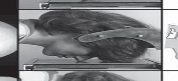
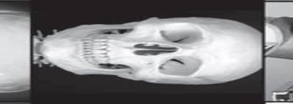
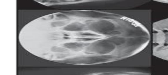
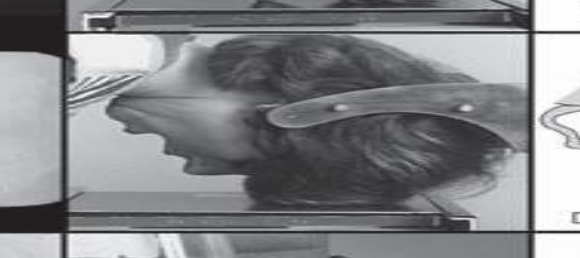
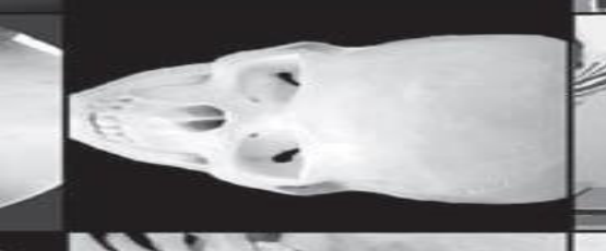
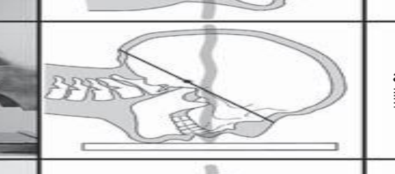
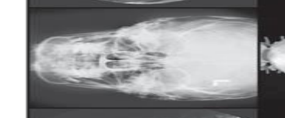
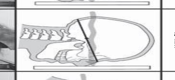
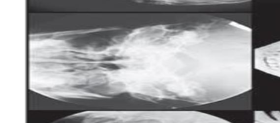
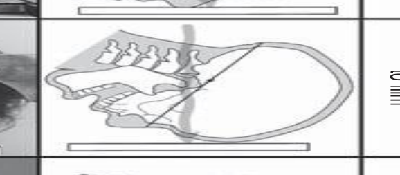
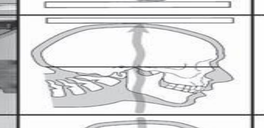
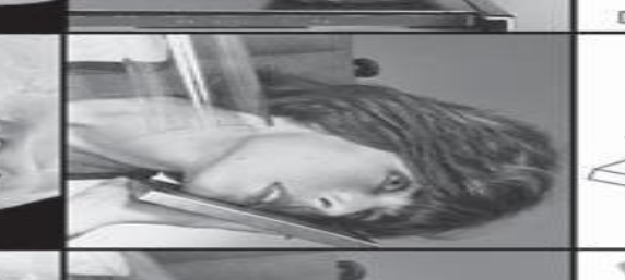
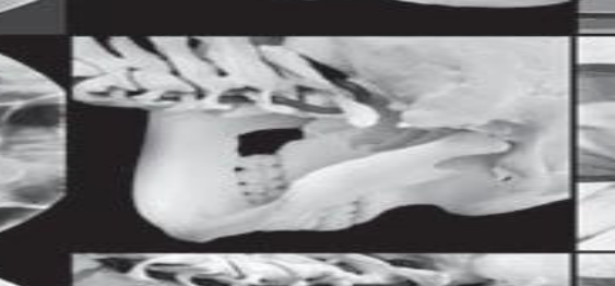
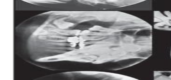
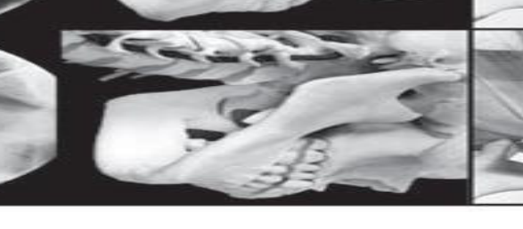
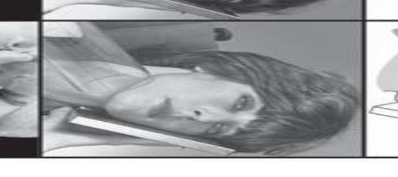
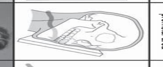
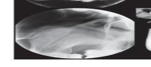
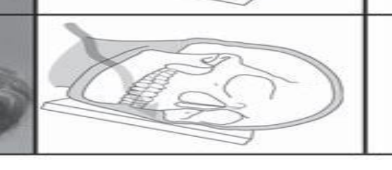
Skull View Projections17
| Skull view | Central beam | Patient placement |
|---|---|---|
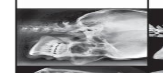
| Lateral Ceph | Beam perpendicular to film | Film parallel to midsagittal plane |
| SMV | Beam perpendicular to film | Canthomeatal line parallel to film |
| Waters | Beam perpendicular to film | Canthomeatal line at 37° with film |
| PA Ceph | Beam perpendicular to film | Canthomeatal line at 10° with film |
| Reverse Towne | Beam perpendicular to film | Canthomeatal line at –30° with film |
| Oblique Lateral Body | Beam aims at the molar-perimolar area | Film in contact with cheek at molar area |
| Ramus | Beam aims at the ramus area | Film in contact with cheek at ramus area |
 |  |  |
 |  |  |
 |  |  |
 |  |  |
 |  |  |
 |  |  |
 |  |  |
 |  |  |
 |  |  |
 |  |
Cases18192021222324252627
Imaging of Facial Deformity
CCD
Developmental Dentofacial Deformity28
- Prognathism or retrognathism (A-P)
- Transverse jaw discrepancy
- Increased or decreased vertical dimension of the jaws
→ Can affect quality of life, e.g. function (chewing, breathing & swallowing), aesthetics & speech
- Developmental deformities occur during growth and are commonly seen in orthodontic and orthognathic patients
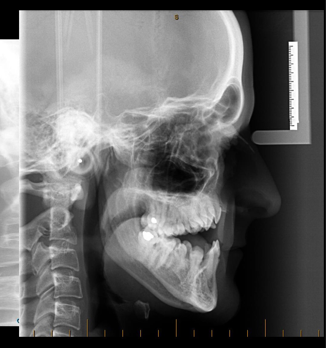
- Retrognathism may produce a skeletal Class II relationship and is associated with a retrognathic mandible
- Skeletal Class III deformity may involve a prognathic mandible or a deficient maxilla
- These are among the most common deformities encountered in dental and maxillofacial practice
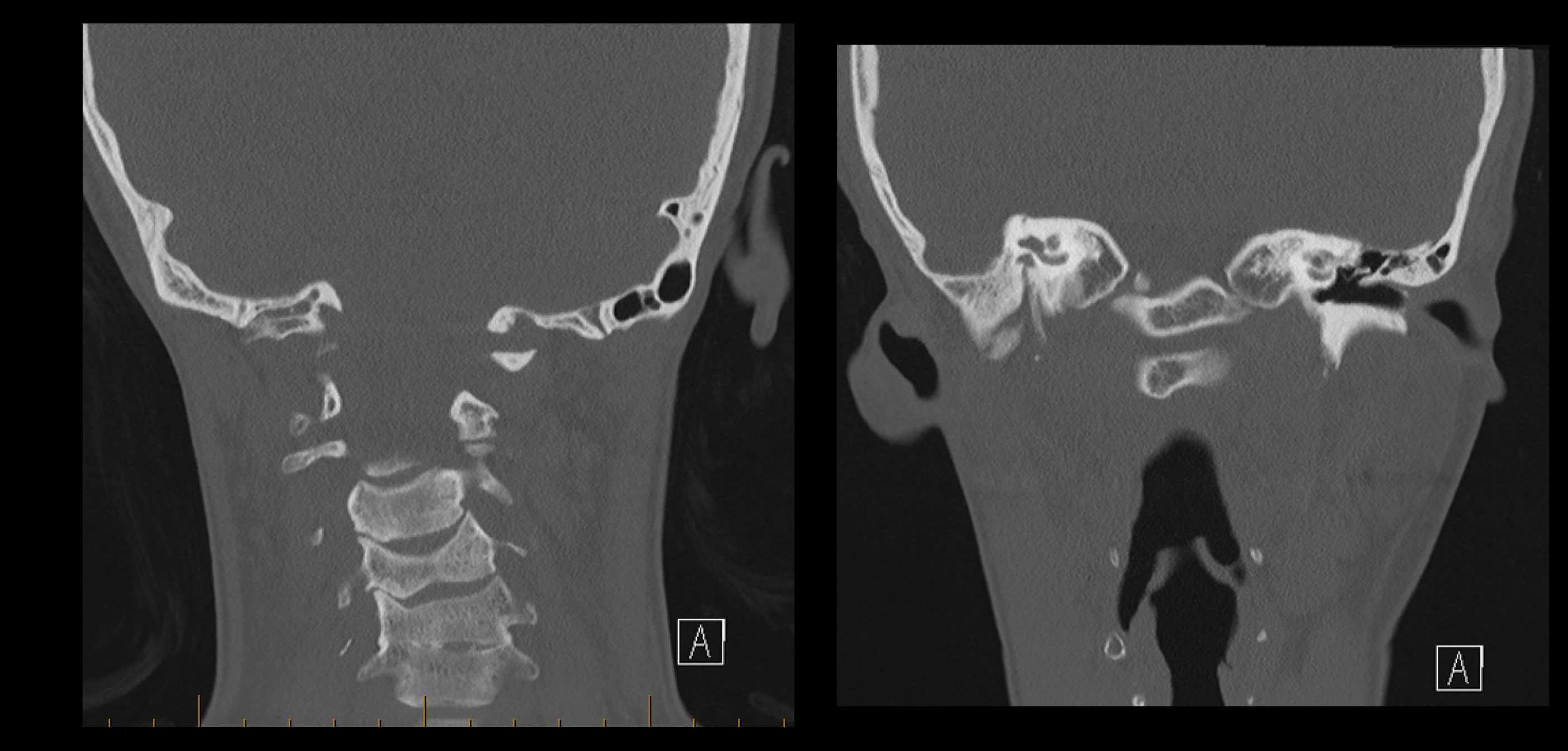
Imaging for Orthognathic Surgery2930313233343536
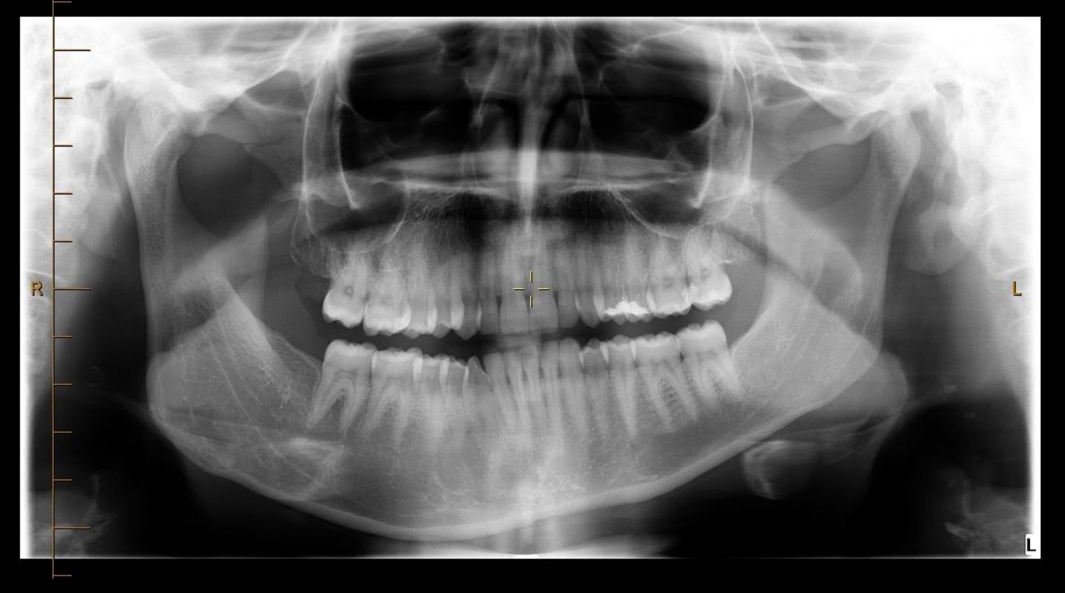
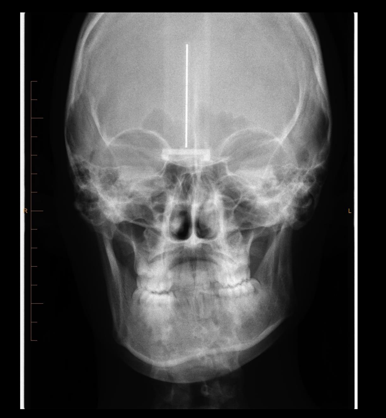
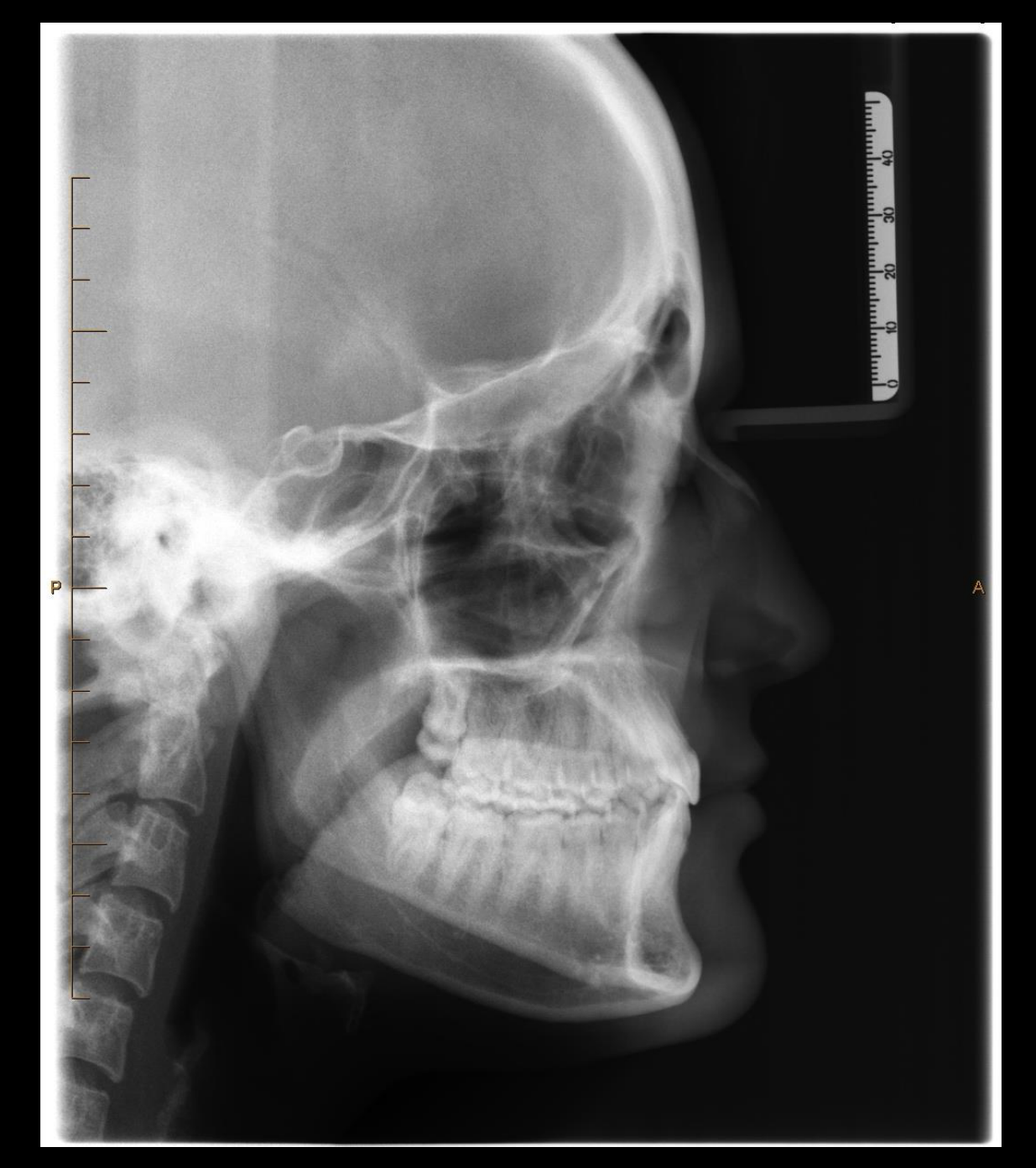
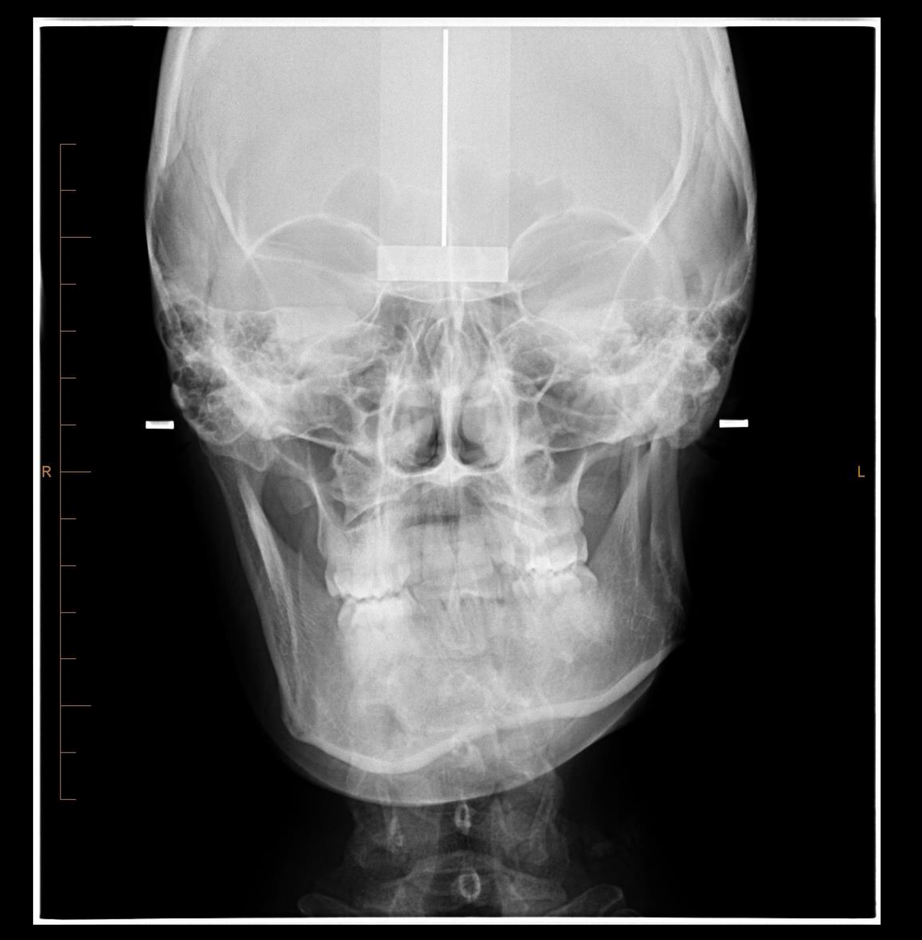
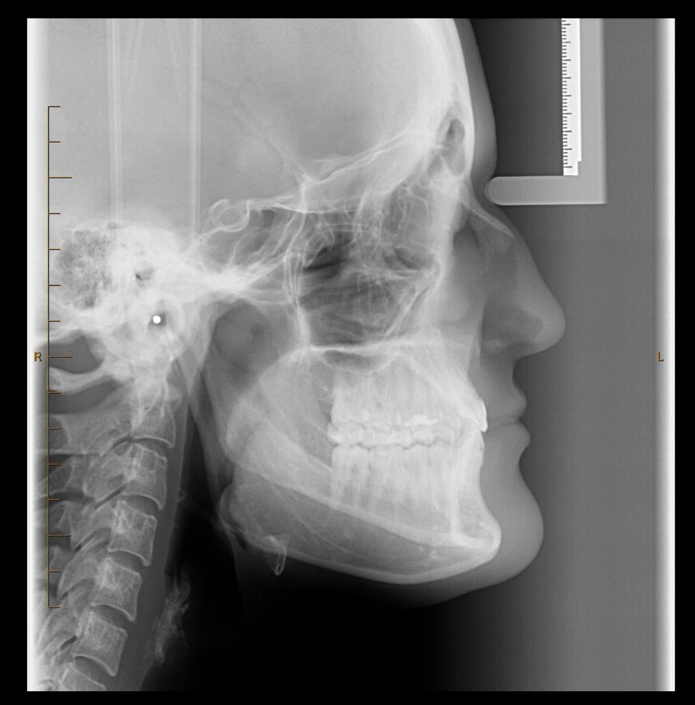
- OPG, Lat Ceph & PA Ceph
- CBCT (orofacial scan)
- CT (head)
Lecturer — Surgical Planning
The imaging protocol is similar to that used for orthodontic treatment planning.
- Three-dimensional imaging is essential for modern surgical planning.
- Current surgical planning relies on virtual surgical planning.
- Bimaxillary surgery may be selected when treatment must address both jaws to achieve functional improvement and aesthetic balance.
Lecturer — Acquired Causes
Acquired deformities develop after birth due to external or pathological causes.
- Paget’s disease of bone
- Iatrogenic causes, including postsurgical changes
Acquired Facial Deformity37
- Trauma
- Benign cysts & tumours
- Infection
- Malignancy
- Other bony lesions
- Fibro-osseous lesions
- Giant cell lesions
- Post-surgery (for removal of pathological lesions)
Depends on the nature of the abnormality.
 |  |
Imaging for Acquired Facial Deformity38394041
- OPG (good overview)
- CBCT
- CT
- MRI
- NMI, especially bone scans
Lecturer — Imaging Modalities
The choice of imaging depends on the abnormality, with CT serving as the main modality for acquired facial deformity.
- CT can assess both hard and soft tissues and is useful for evaluating the morphology of affected bones and condyles.
- Orthopantomograms provide an overall view of the jaws and dentition but have inherent artifacts and limited skeletal accuracy.
- CBCT provides accurate three-dimensional visualization of bony structures, but high image noise, a low contrast-to-noise ratio, and poor soft-tissue contrast can limit its use when soft-tissue information is needed for surgical planning.
- MRI provides excellent soft-tissue detail and is particularly useful for tumors, cysts, and vascular malformations; it is generally an adjunct to CT.
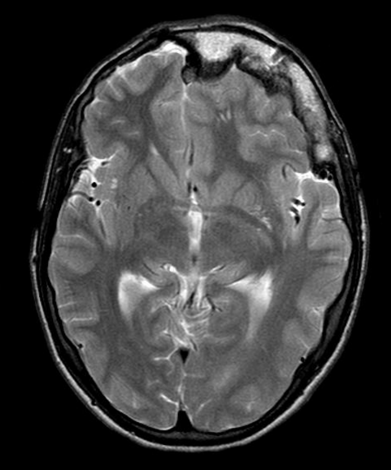
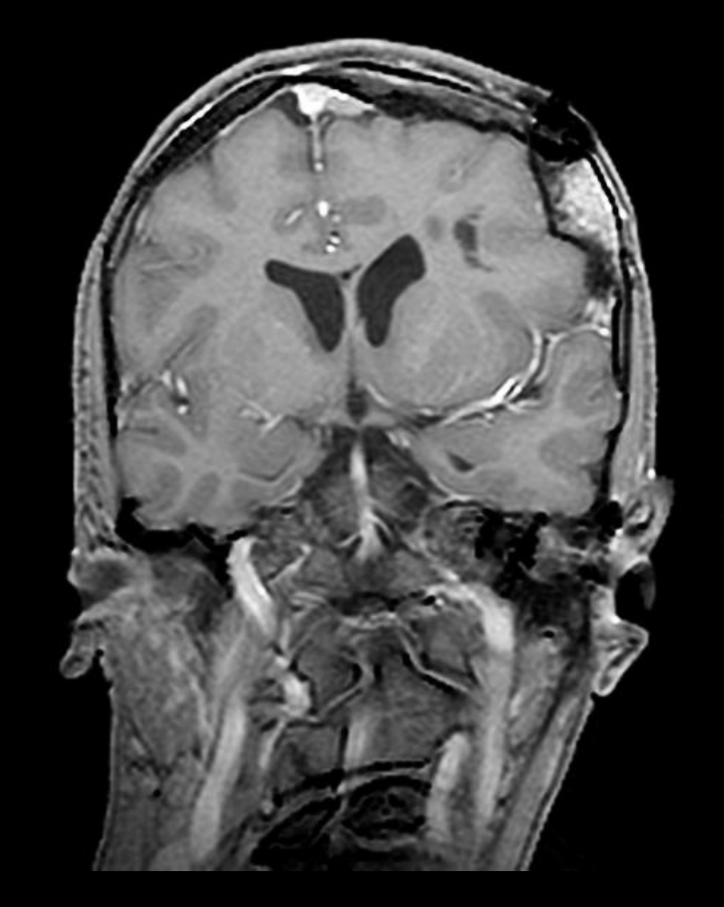
- MRI is especially valuable for identifying vascular malformations associated with conditions such as Proteus syndrome.
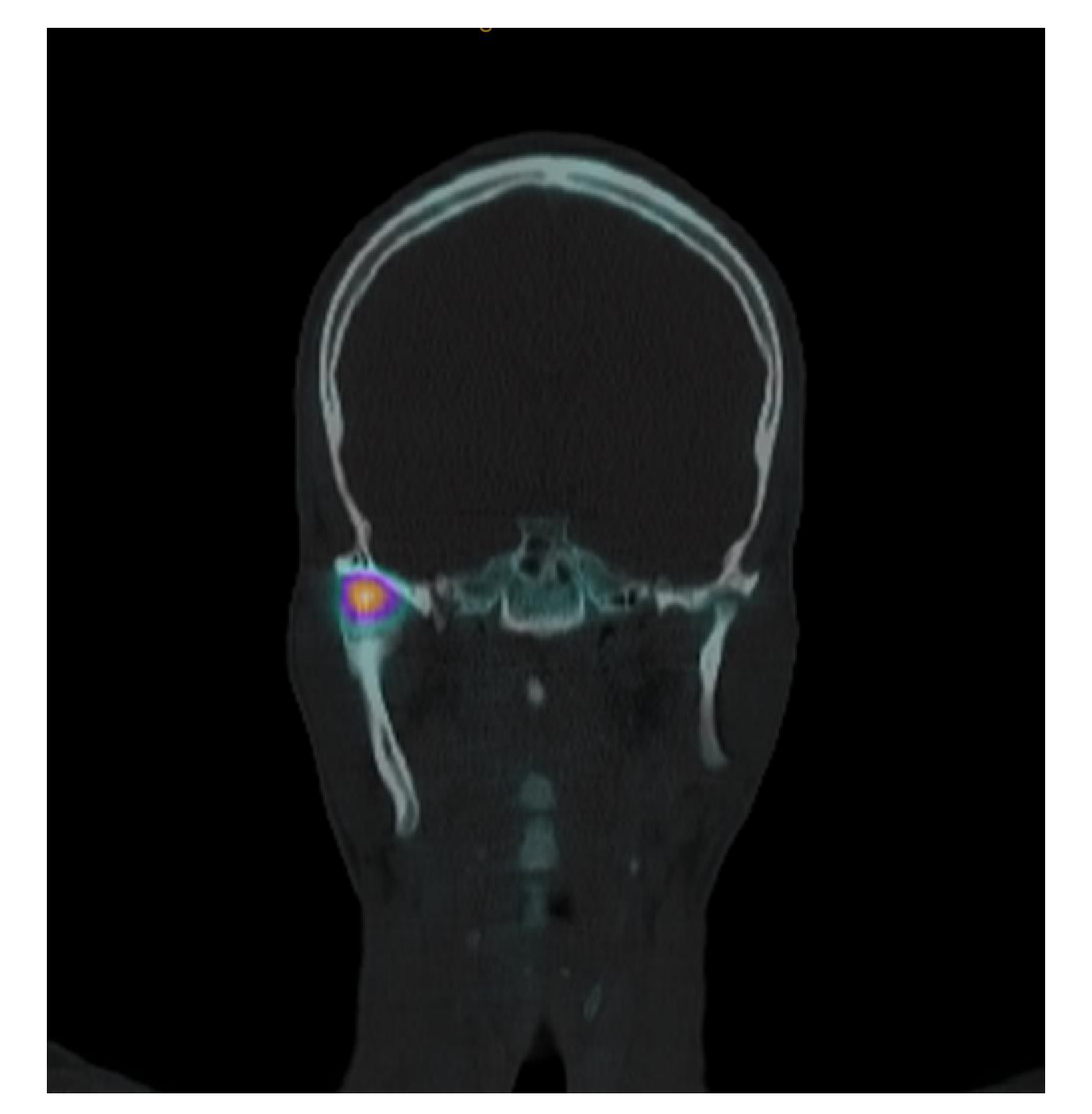
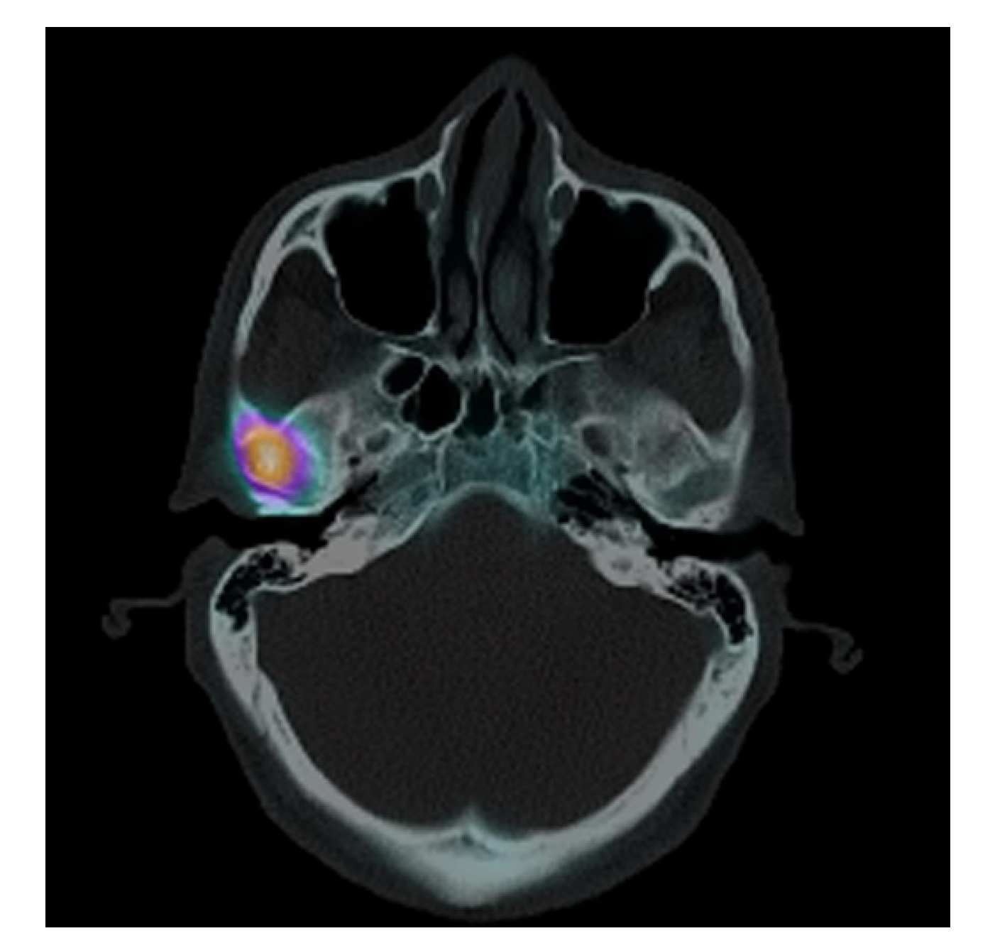
- Nuclear medicine imaging is primarily functional rather than morphological and can determine whether a deformity or lesion is progressive or stable, helping determine the timing of surgery, particularly in unilateral condylar hyperplasia.
Marcelo Medeiros¹, Gabriela Granja Porto², Jose Rodrigues Laureano Filho³, Luís Portela⁴, Ricardo Holanda Vasconcellos⁵. CASE REPORT, DOI: 10.1016/S1808-8694(15)30589-9, Open Access.
References4243
(b) (c)
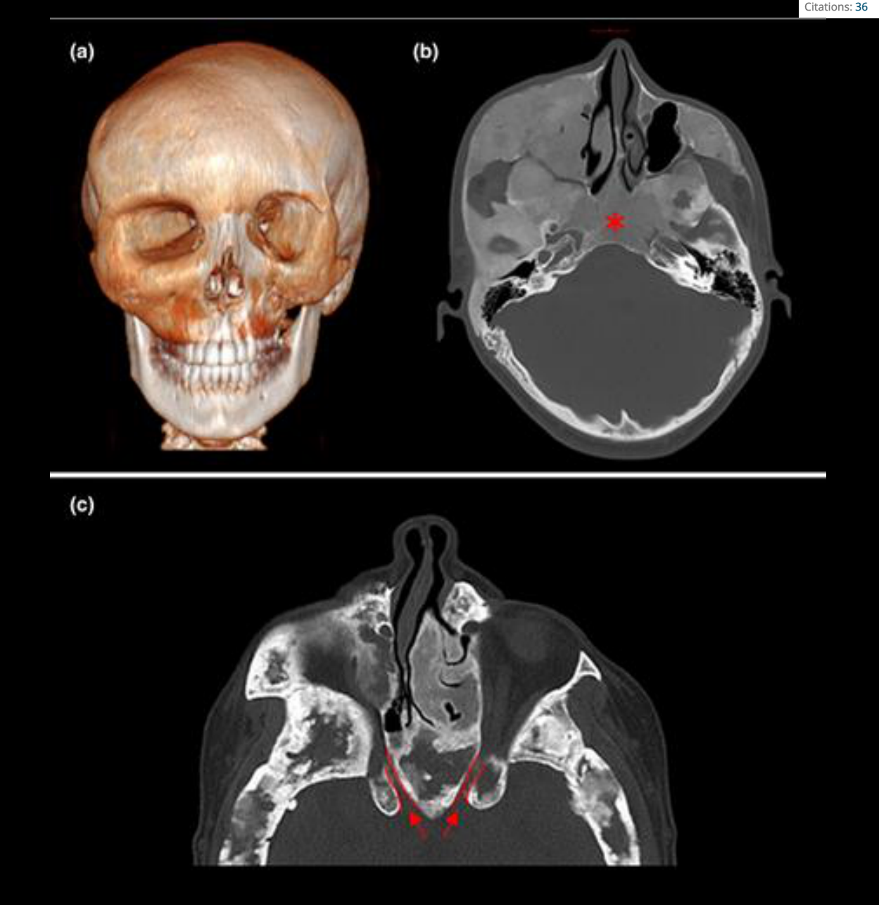
AB Burke, MT Collins, AM Boyce. Fibrous dysplasia of bone: craniofacial and dental implications. Oral Diseases, Volume 23, Issue 6, p. 697-708. Invited Medical Review | Free Access. First published: 05 August 2016. https://doi.org/10.1111/odi.12563. Citations: 36.
Audio Appendix
Additional Audio Content
The following sections from the lecture audio did not correspond to any heading in the main document.
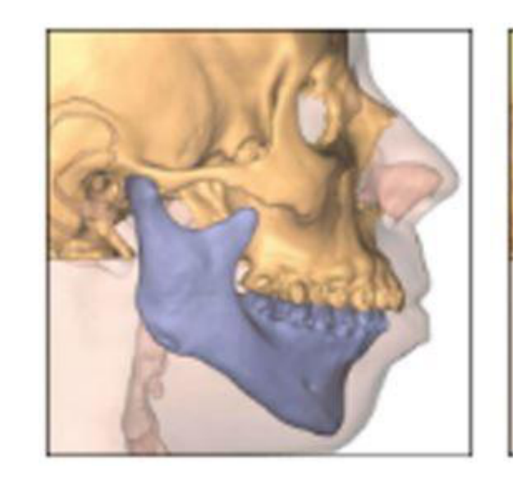
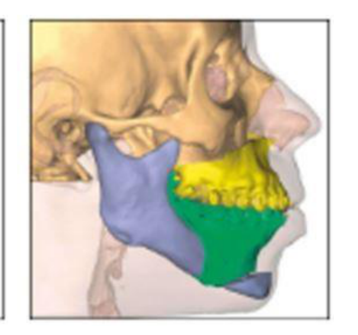
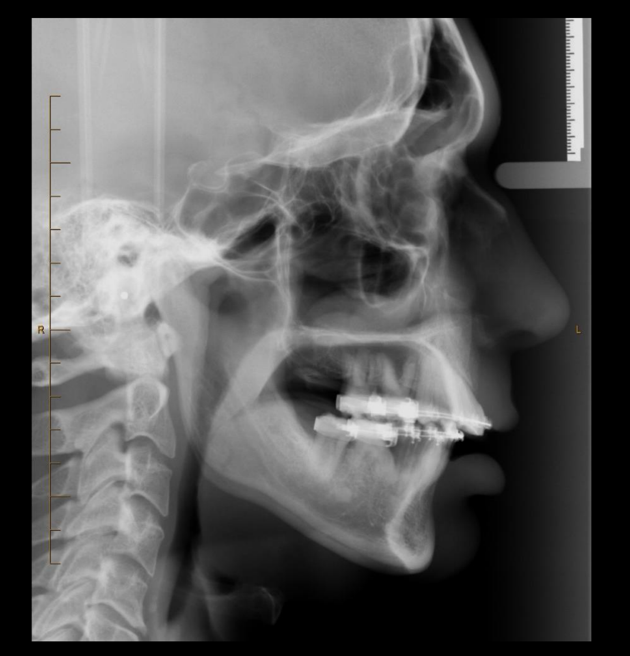
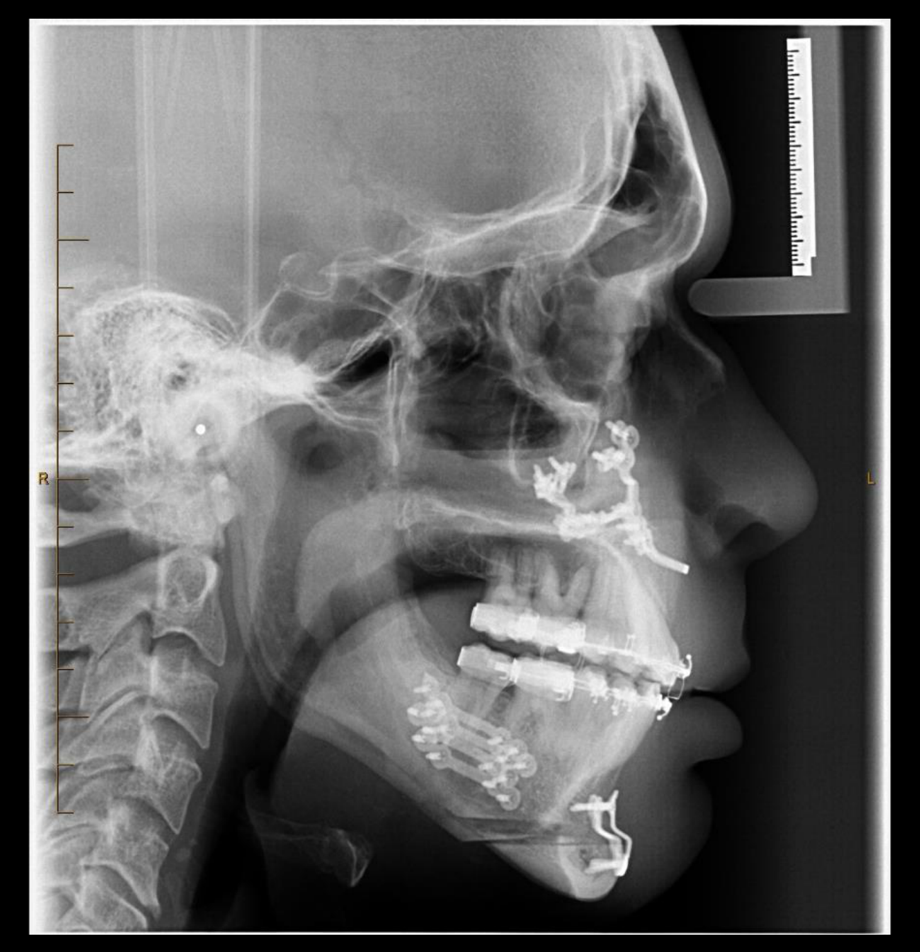
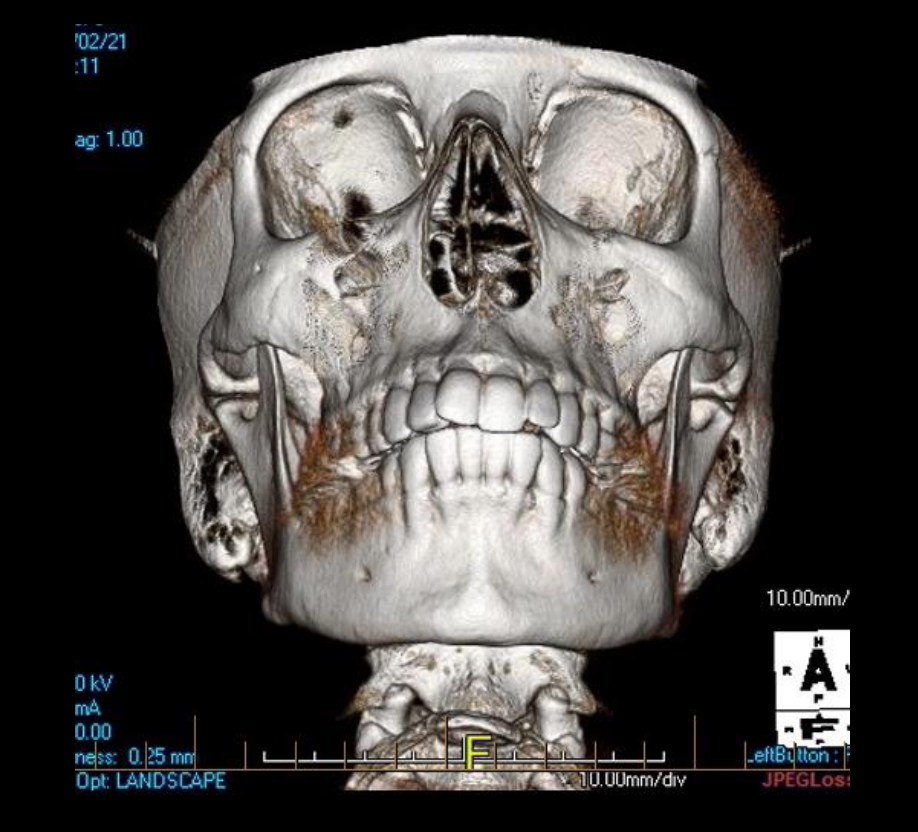
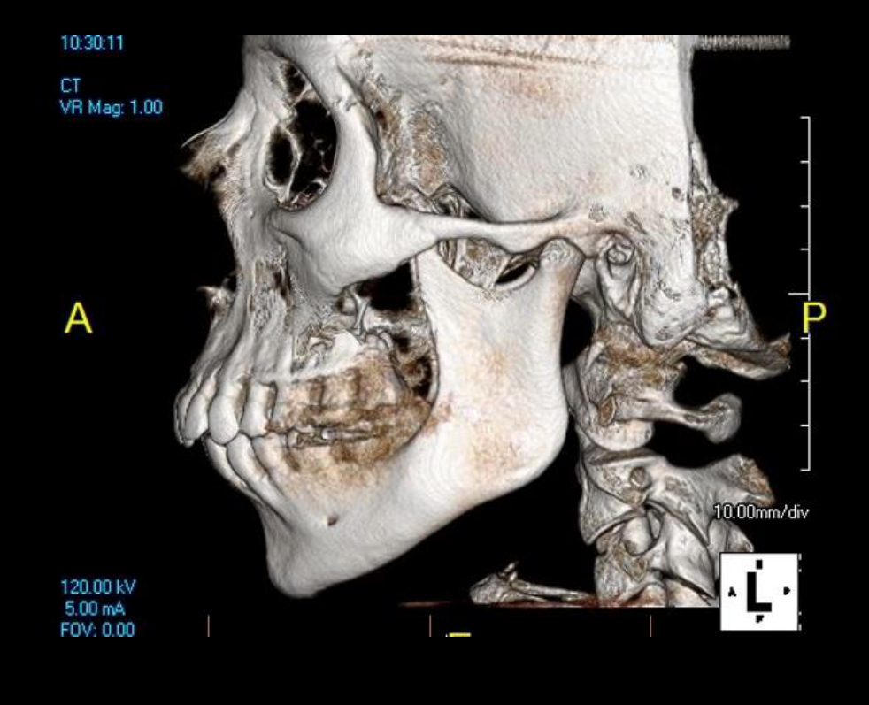
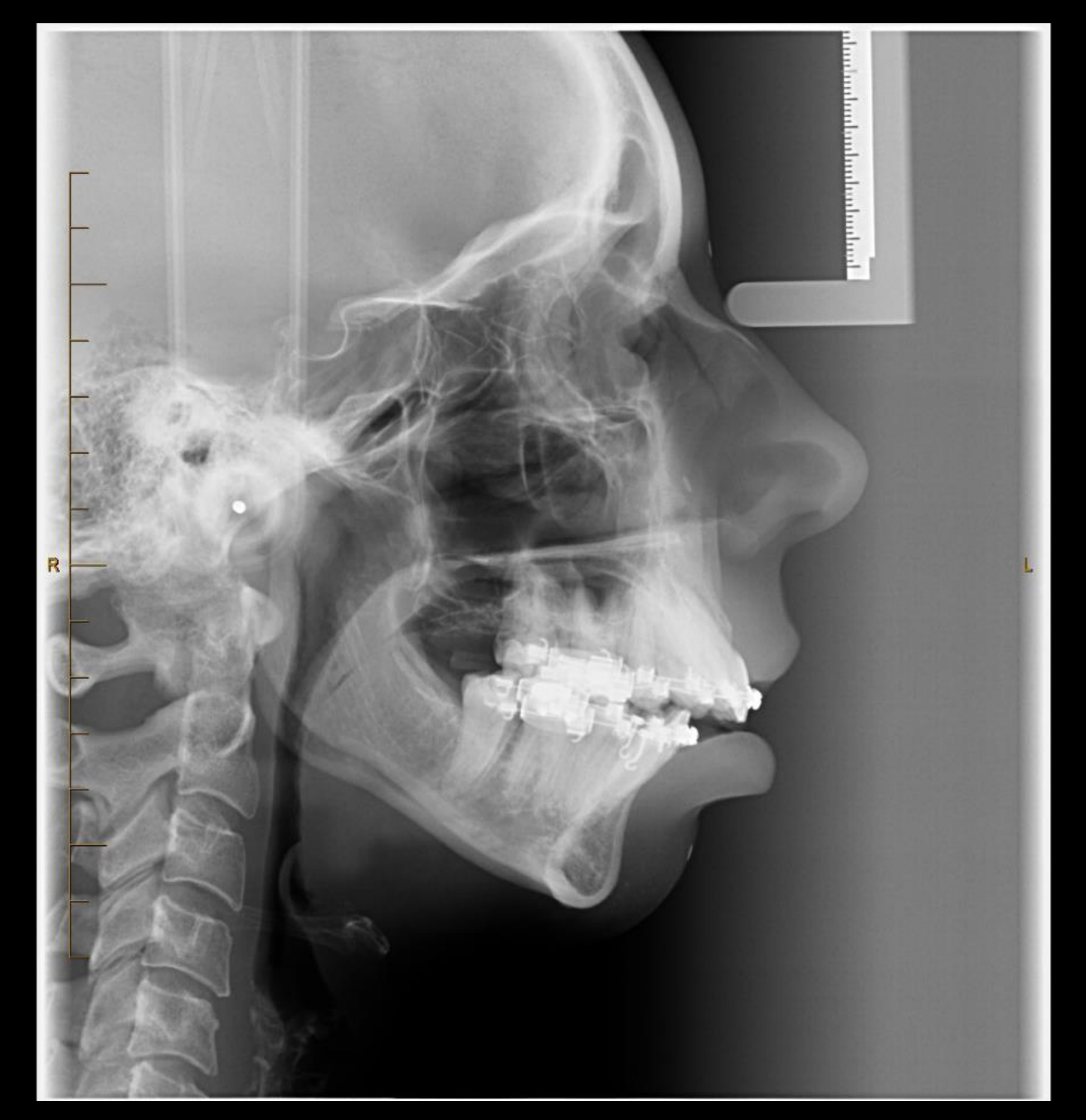
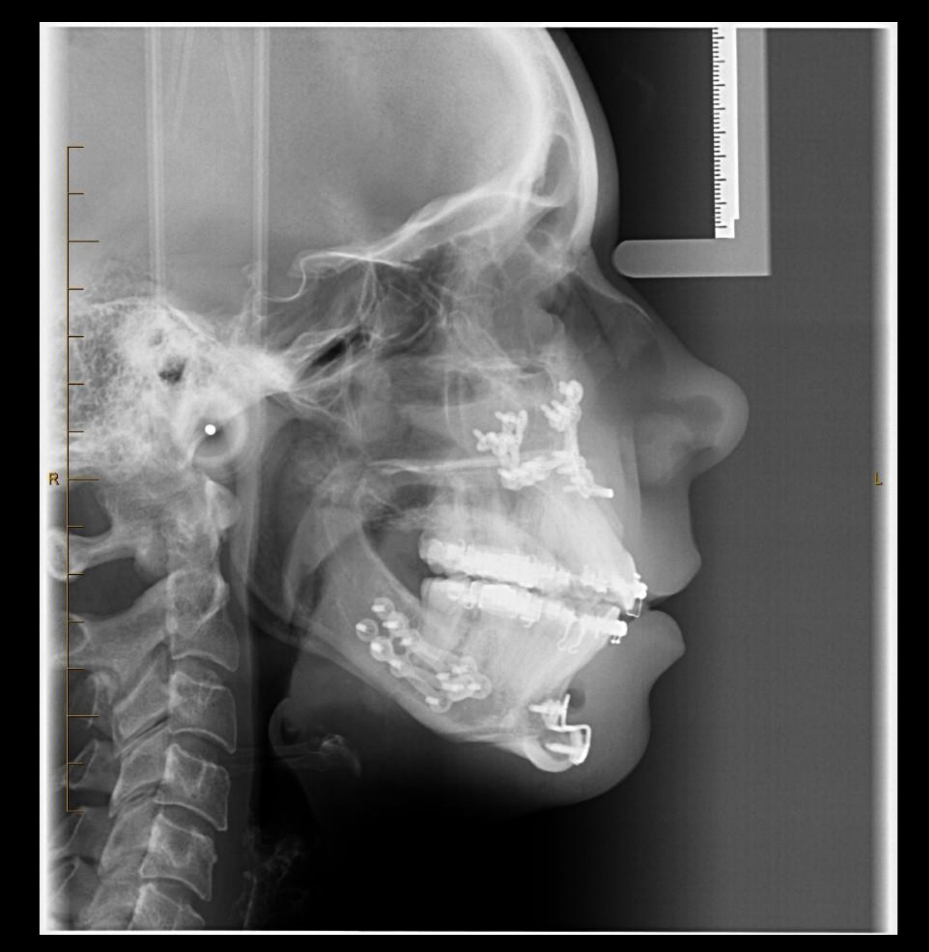
Case Study: Retrognathism and Bimaxillary Osteotomy
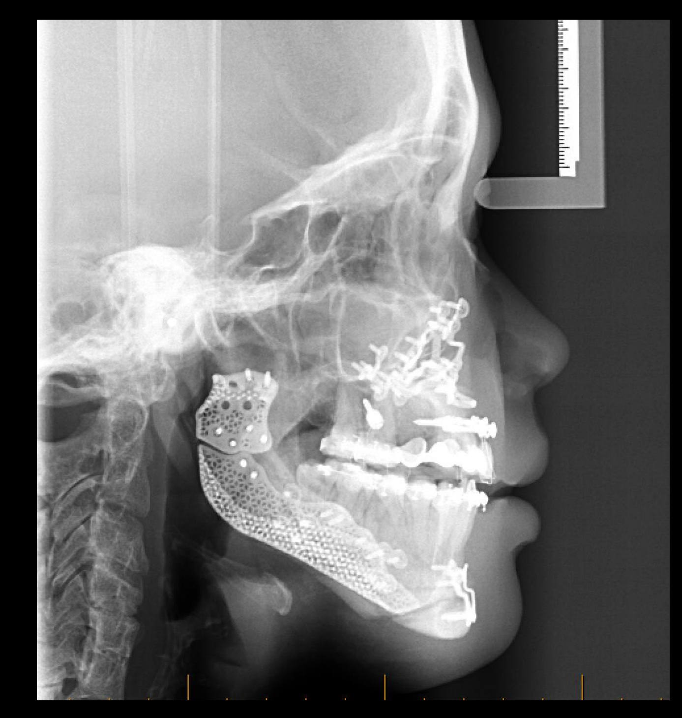
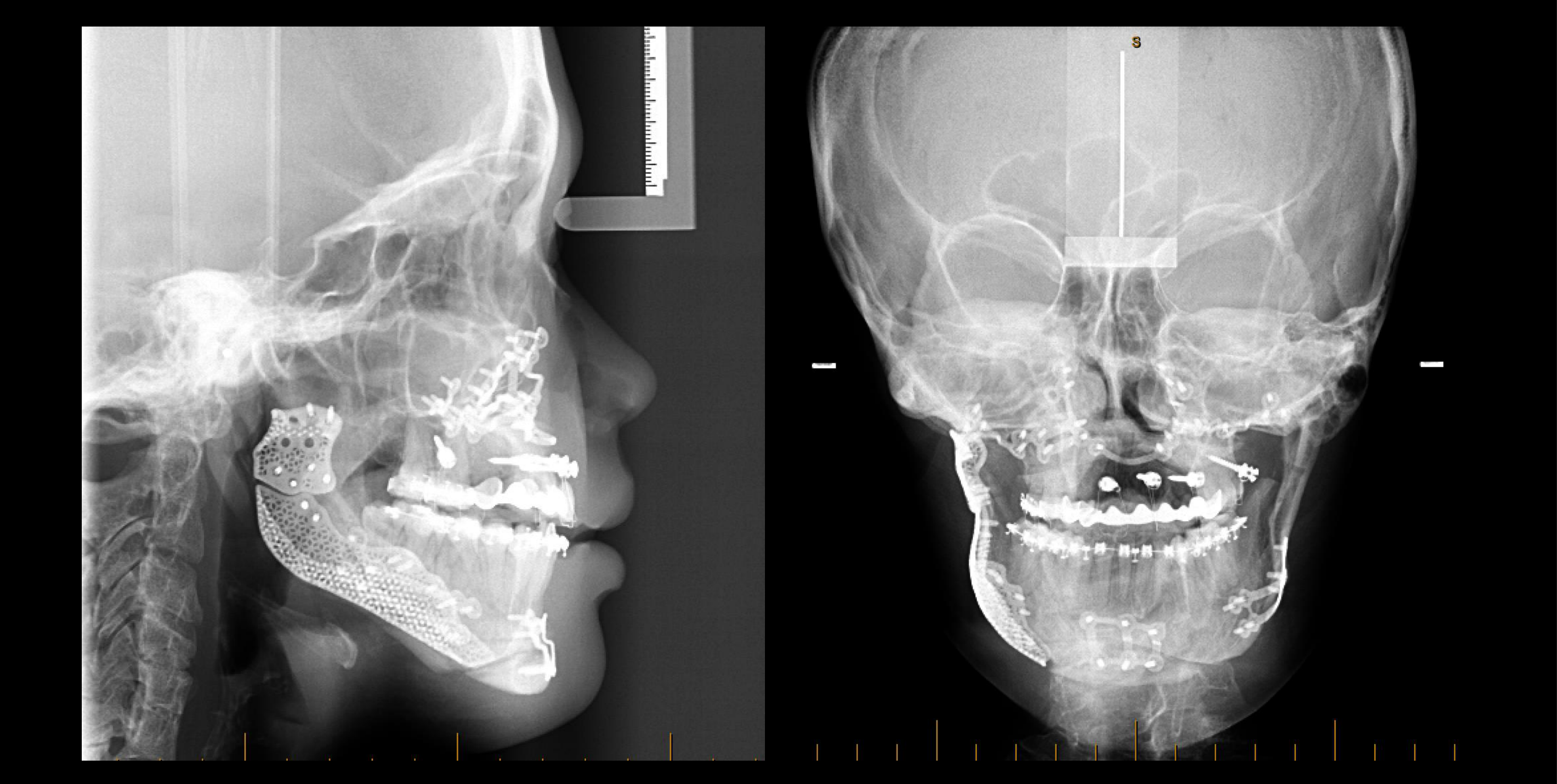
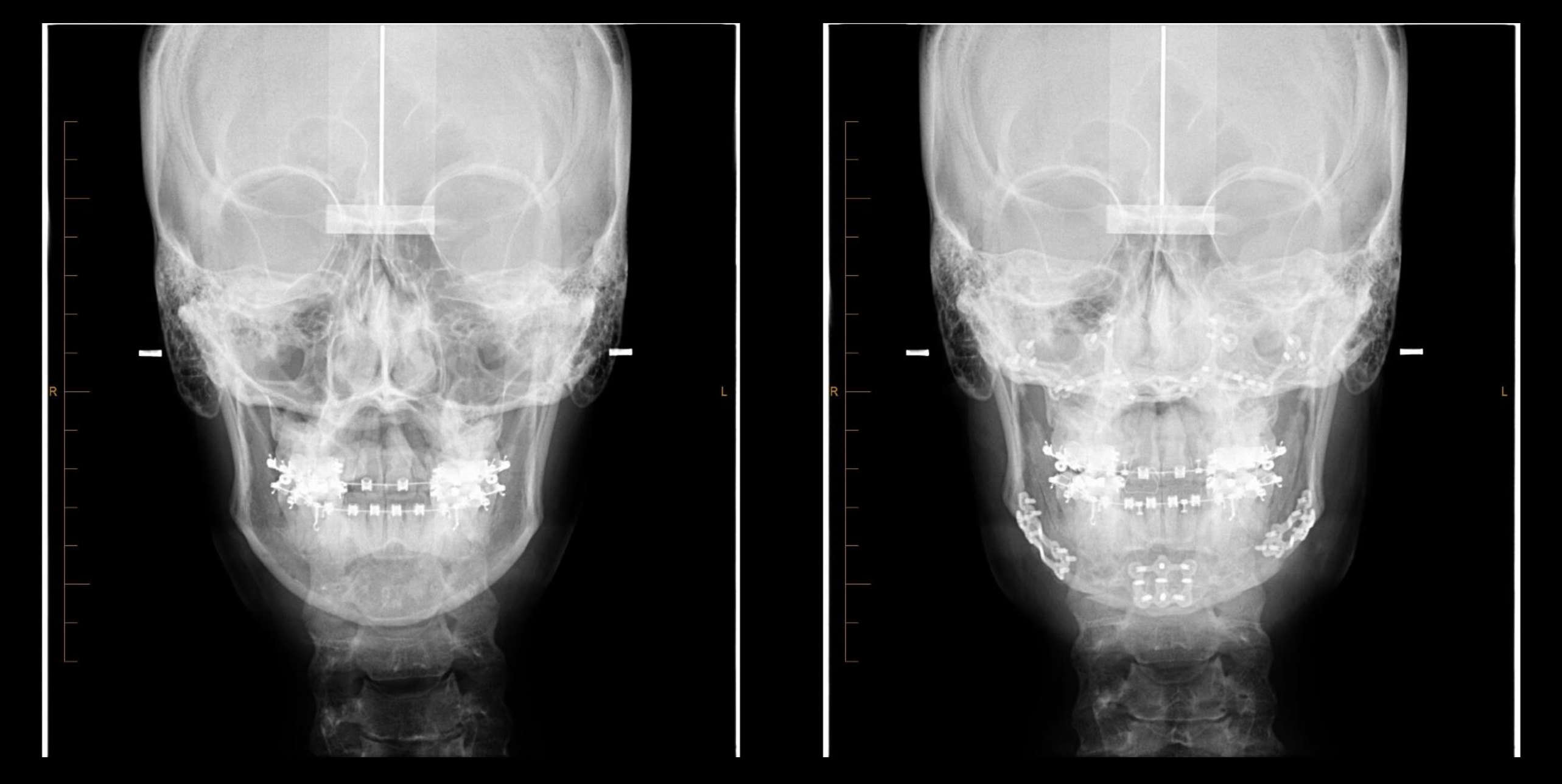
- The patient had retrognathism with a skeletal Class II relationship.
- There was severe anterior overjet before surgery.
- The right-side image showed the result after orthognathic surgery.
- The procedure was a bimaxillary osteotomy involving both the maxilla and mandible.
- A bimaxillary approach may be used to achieve both:
- Functional improvement
- Aesthetic balance
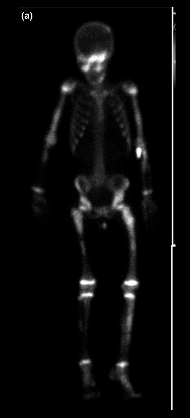
Case Study: Fibrous Dysplasia
- Fibrous dysplasia may cause significant facial asymmetry.
- It typically affects:
- The maxilla
- The mandible
- The skull base
- It may cause:
- Vision problems
- Hearing problems
- Malocclusion
- It is usually diagnosed during adolescence.
- Management may involve surgery.
- Bone scans are performed before surgery to assess whether the lesion is stable.
Hemifacial Microsomia
- Hemifacial microsomia is a first- and second-pharyngeal-arch syndrome.
- It usually has unilateral involvement.
- It was described as the second most common congenital facial deformity after cleft lip and palate.
- It causes poor development of one side of the face.
- The mandible is often the most severely affected structure.
- When the spine is also involved, the condition belongs to the broader spectrum of Goldenhar syndrome.
- Hemifacial microsomia and Goldenhar syndrome are part of the continuum known as the oculo-auriculo-vertebral spectrum.
Treacher Collins Syndrome
- Treacher Collins syndrome is a rare example of bilateral facial deformity and facial hypoplasia.
- It affects the jaws and may also involve:
- The ears
- The zygomatic bones
- The lateral aspects of the orbits
- It was described as much rarer than cleft lip and palate.
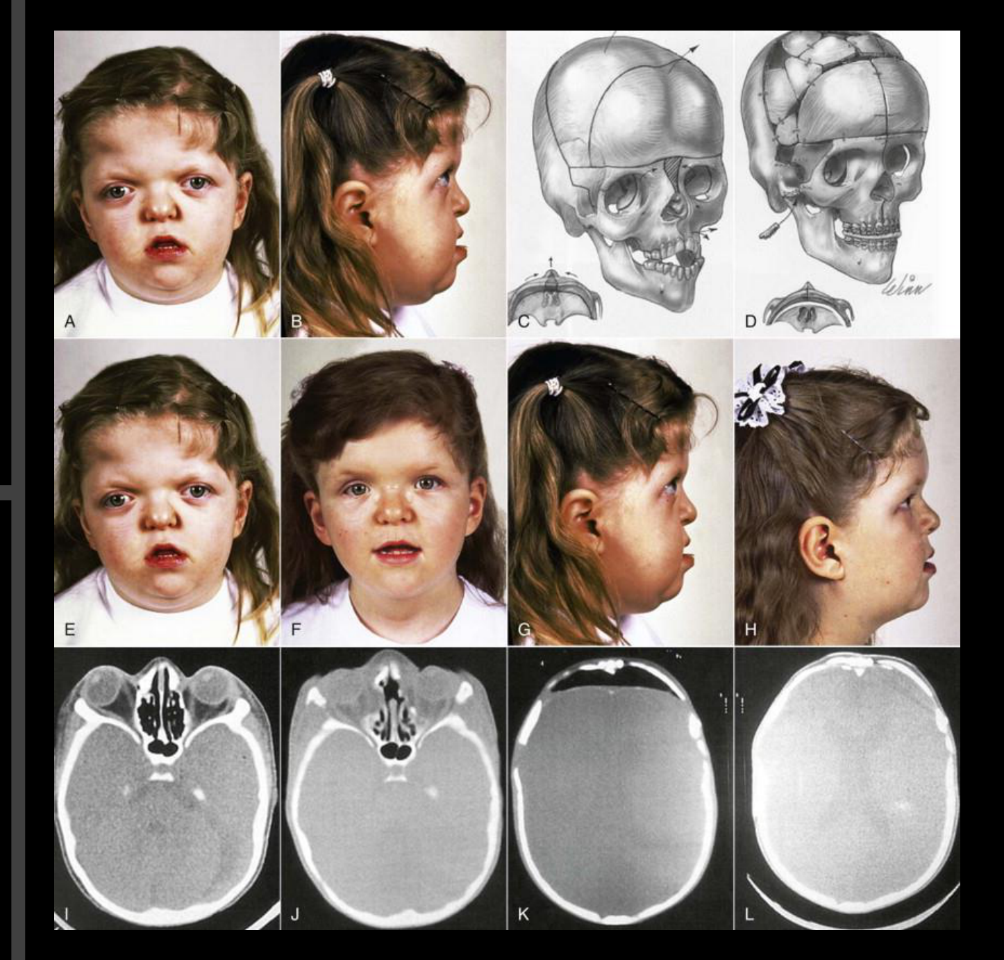
Crouzon Syndrome
- Crouzon syndrome involves premature fusion of the cranial sutures.
- Findings may include:
- Reduced anteroposterior dimension of the cranial base
- A copper-beaten appearance on skull imaging
- Hypoplastic jaws
- Shallow orbits
- Proptosis
- A skeletal Class III relationship
- The skeletal Class III relationship was associated with maxillary hypoplasia.
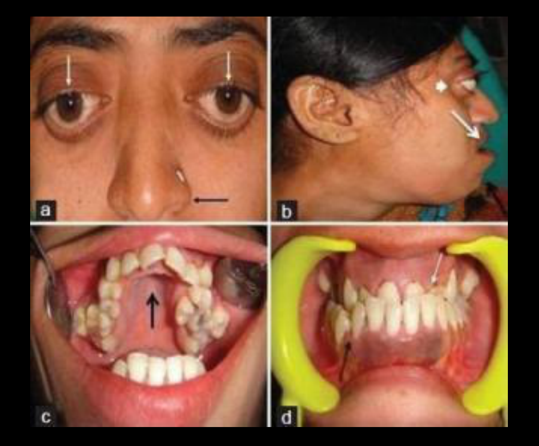
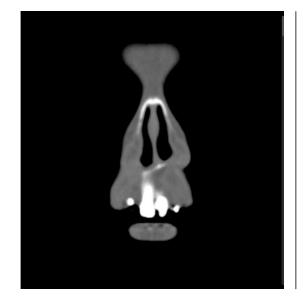
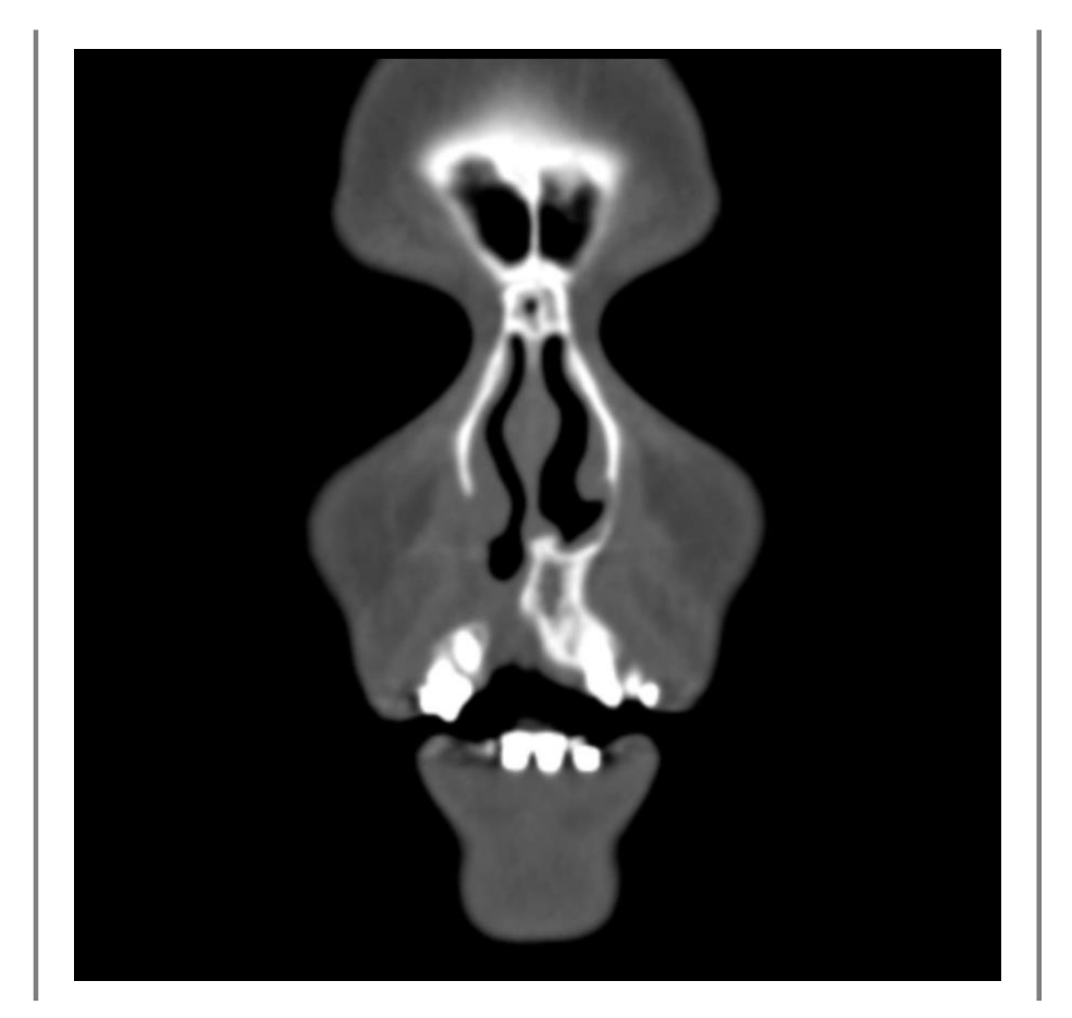
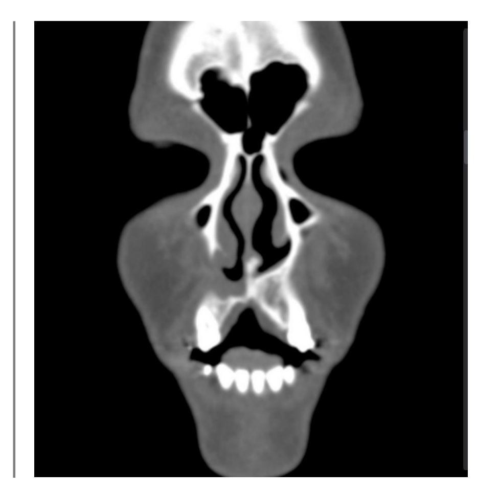
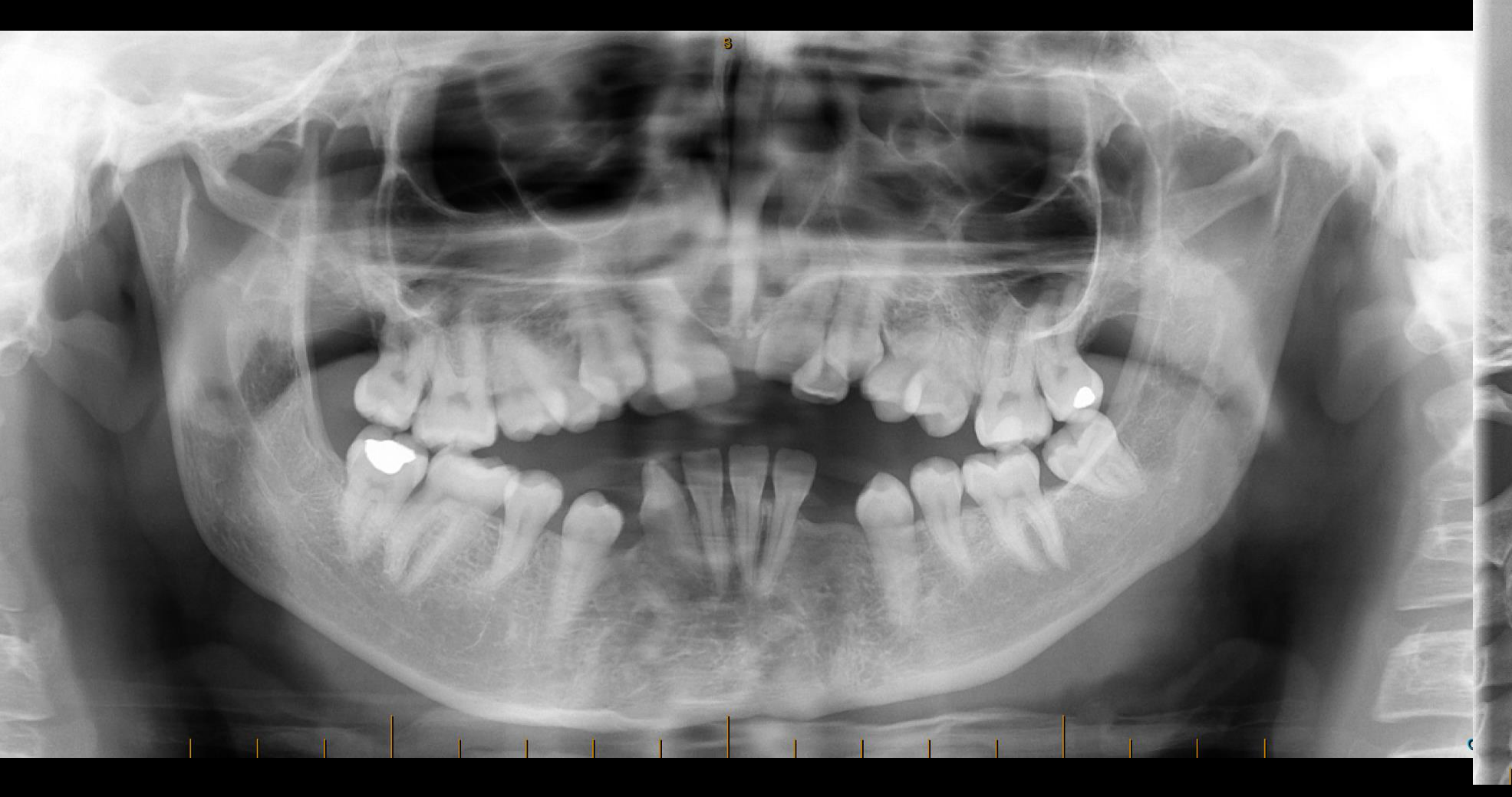
Case Study: Unilateral Right-Sided Cleft Lip and Palate
- The case demonstrated a unilateral right-sided cleft lip and palate.
- A bony defect was visible in the right maxilla at the region of tooth 12.
- The defect involved the nasal cavity.
Case Study: Midline Cleft Lip and Palate
- A midline cleft lip and palate was described as extremely rare.
- It may be part of a larger syndrome, including:
- Frontonasal dysplasia
- Orofacial digital syndrome type 1
- CHARGE association
- The patient demonstrated significant facial deformity, including:
- Maxillary hypoplasia
- Nasal hypoplasia
- Severe skeletal Class III malocclusion
- A concave facial profile
- An anterior open bite
Case Study: Severe Right-Sided Hemimandibular Hyperplasia
- This was a more severe case of right-sided hemimandibular hyperplasia.
- The mandible was rotated.
- The symphysis was positioned to the left of the facial skeletal midline.
- There was a dental midline shift.
- A skeletal Class III relationship was present without a reverse anterior overjet.
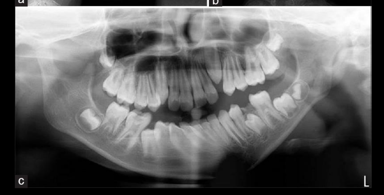
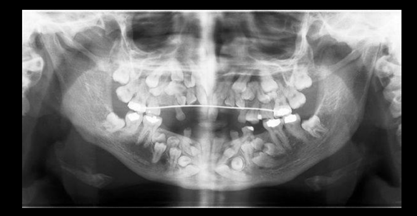
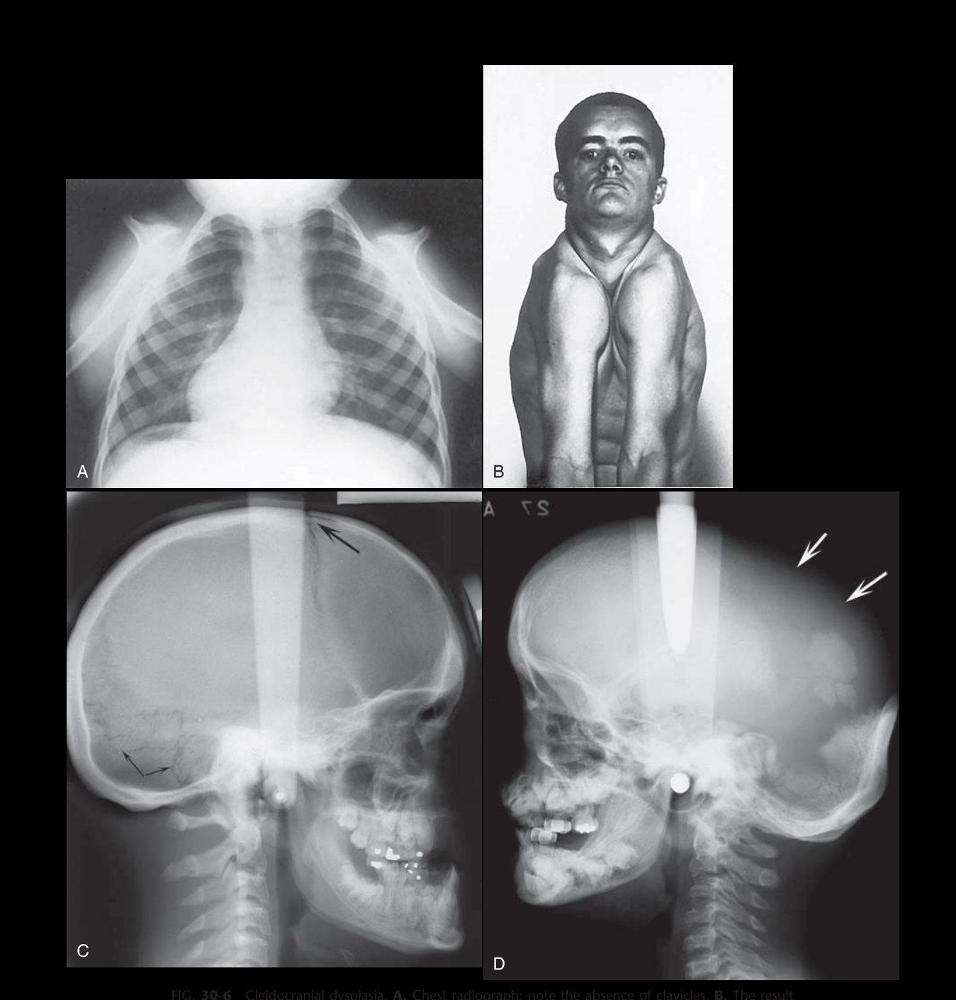
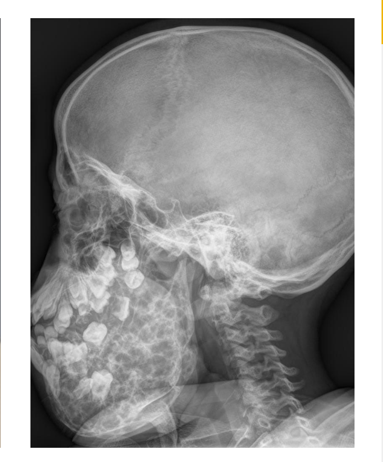
Cleidocranial Dysplasia
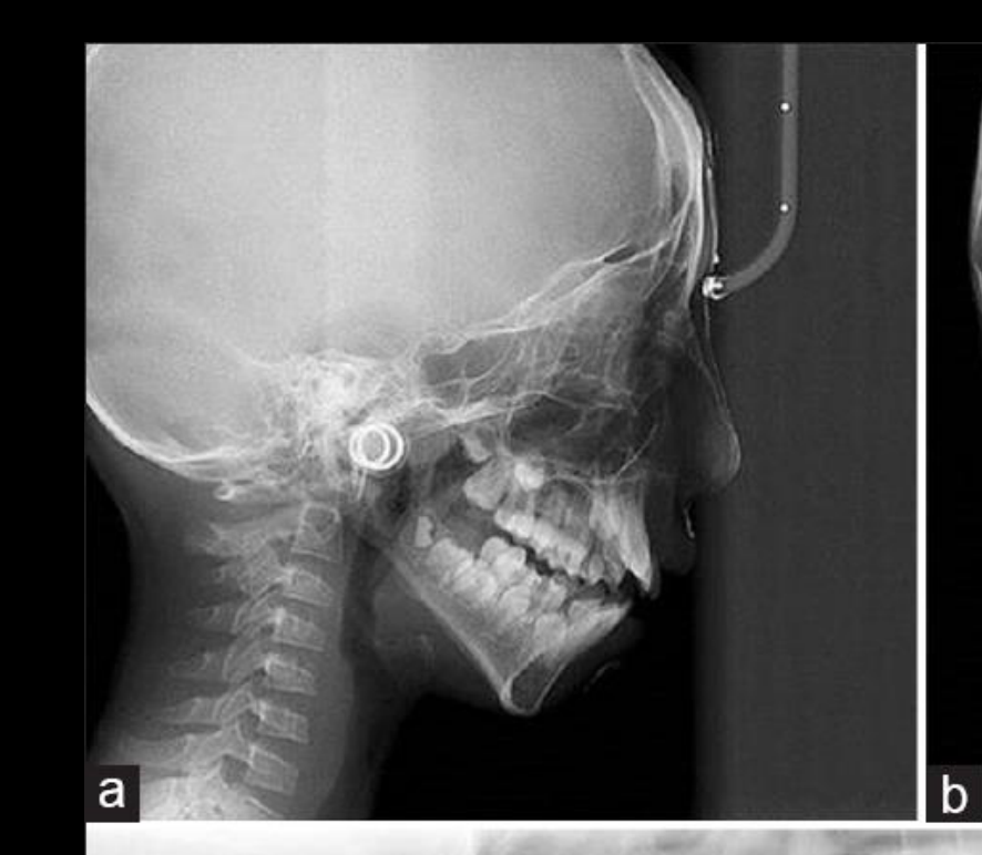
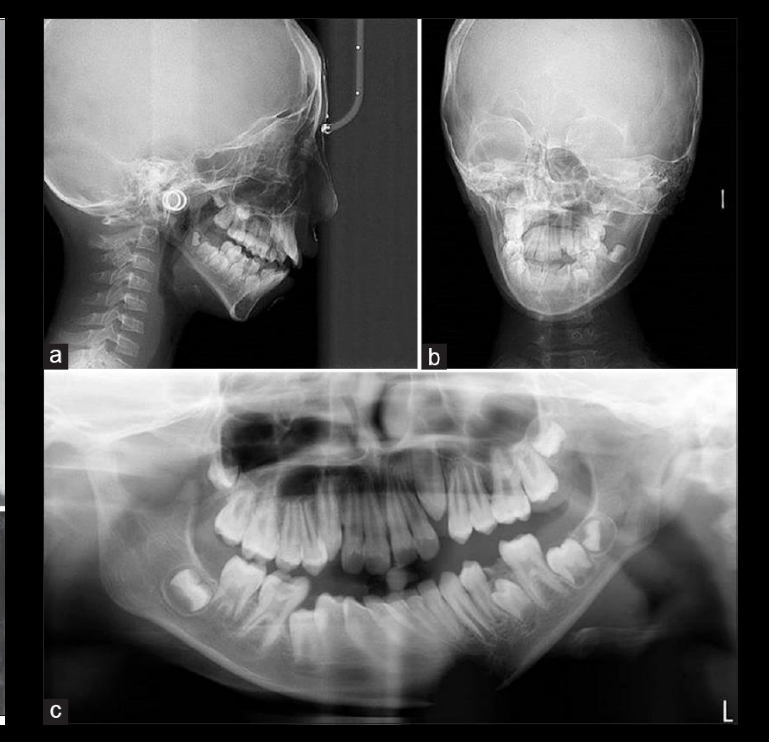
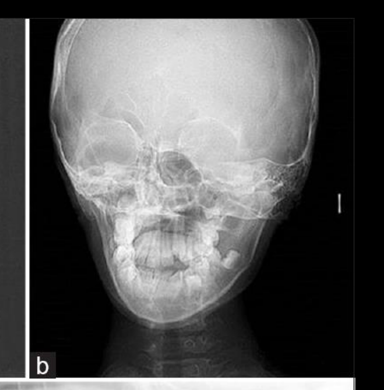
- Cleidocranial dysplasia shares some features with other facial deformity syndromes but has distinguishing findings:
- Absent or hypoplastic clavicles
- Persistent open cranial sutures
- Supernumerary teeth
- Prolonged retention of primary teeth
- Multiple impacted permanent teeth
- Maxillary hypoplasia
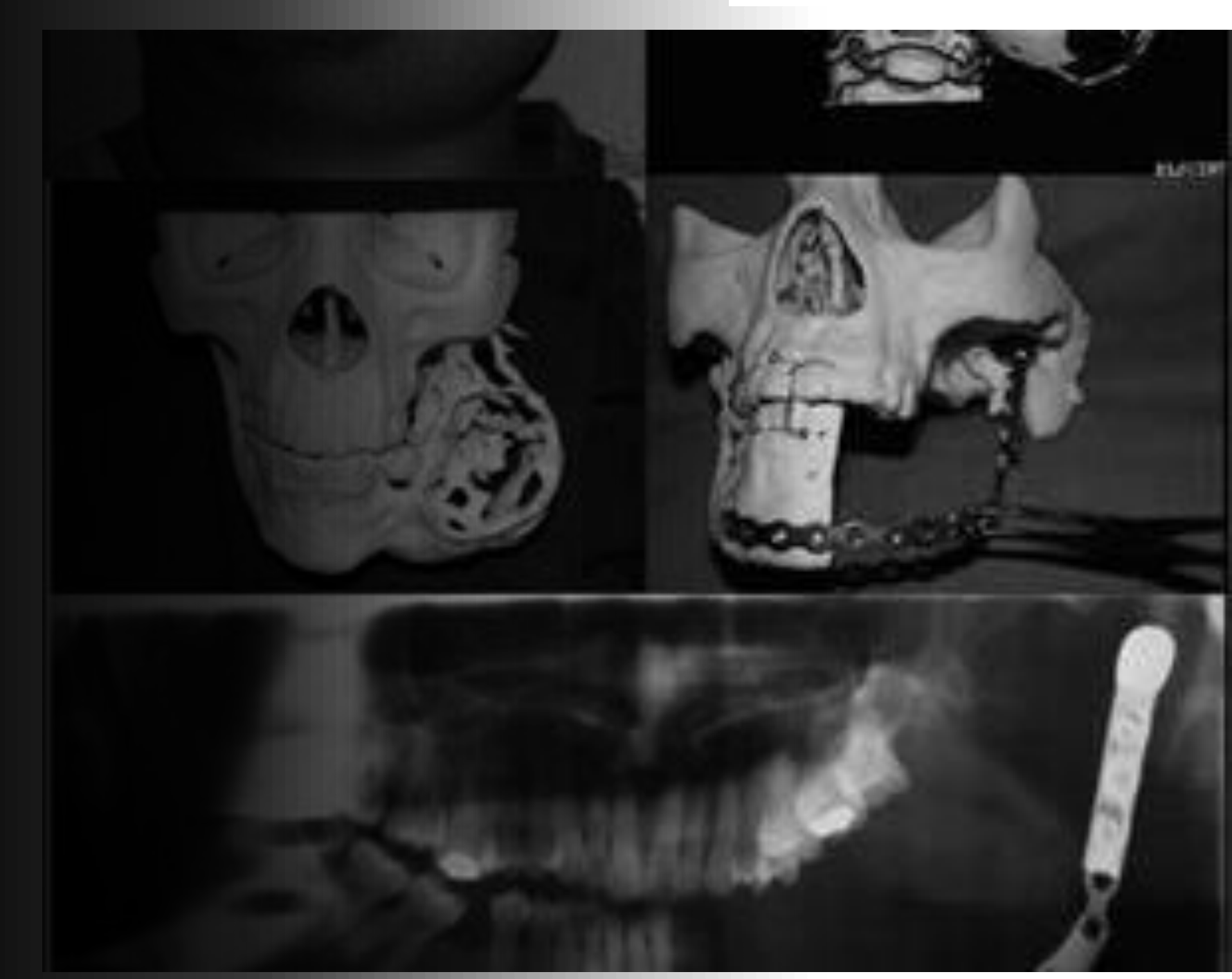
Case Study: Hemifacial Microsomia
- The patient had poor development of the right side of the face.
- The mandible was the most severely affected structure.
- Involvement of the spine would place the condition within the broader spectrum of Goldenhar syndrome.
- The condition is also part of the oculo-auriculo-vertebral spectrum.
- After surgery, the facial asymmetry, particularly mandibular asymmetry, was corrected.
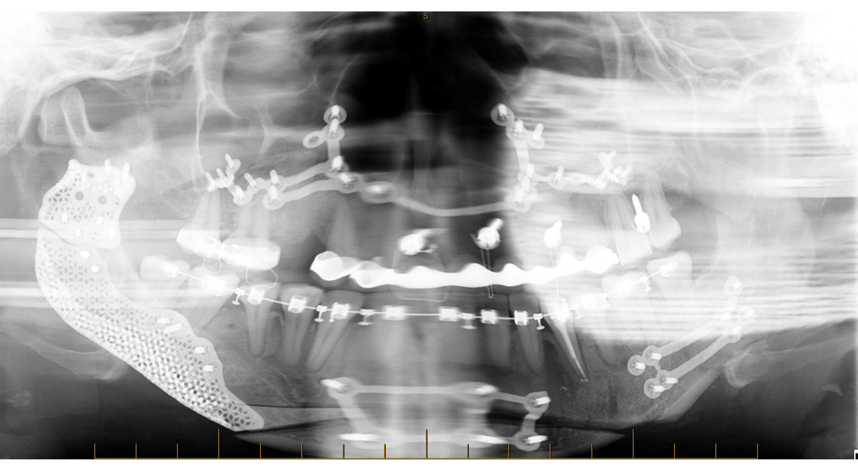
- Numerous artifacts were visible on the postoperative orthopantomogram, making it almost unusable for assessment.
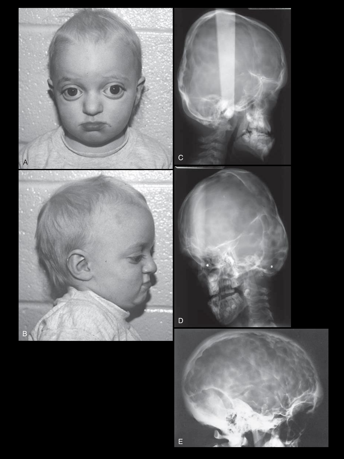
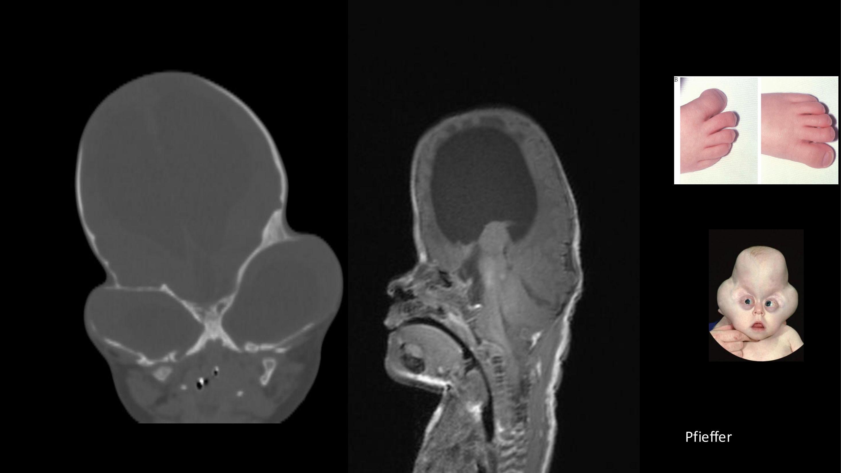
Pfeiffer Syndrome
- Pfeiffer syndrome is another syndrome associated with craniosynostosis.
- It differs from Apert and Crouzon syndromes through:
- Variable widening of the thumbs and toes
- Extreme skull deformity
- The skull shape may be described radiologically as a “cloverleaf” appearance.
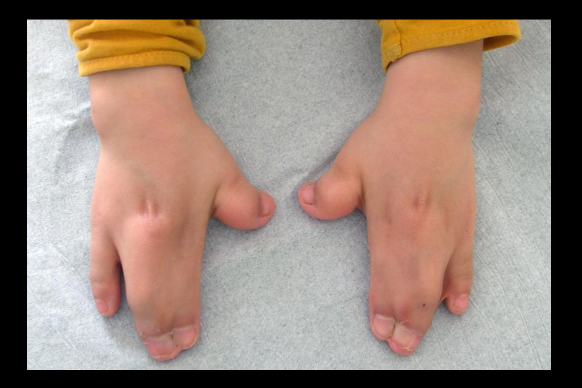
Apert Syndrome
- Apert syndrome is associated with early fusion of the cranial sutures, known as craniosynostosis.
- Its primary distinguishing feature compared with Crouzon syndrome is syndactyly.
- Syndactyly is a congenital condition in which the fingers or toes are webbed or fused.
- Apert syndrome is also associated with an increased chance of intellectual disability.
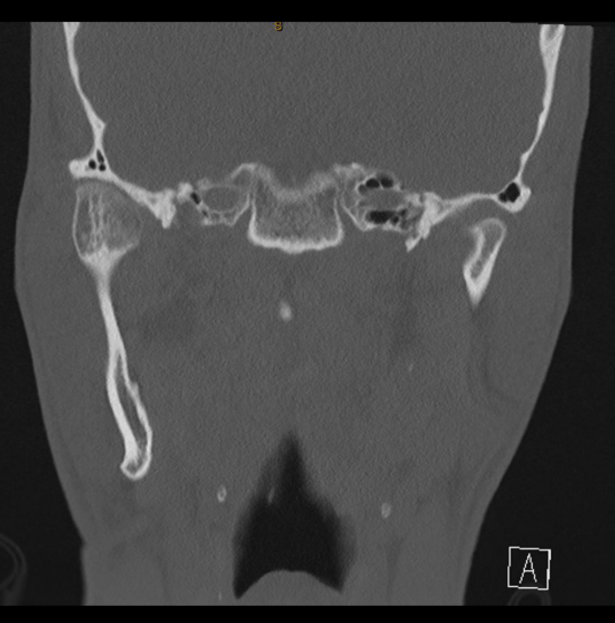
Case Study: Condylar Tumor-Related Hyperplasia
- The patient had unilateral right condylar hyperplasia.
- Imaging demonstrated overgrowth of the right condyle.
- The enlargement was related to a tumor involving the condyle rather than simple developmental enlargement.
- Functional nuclear medicine imaging with SPECT demonstrated increased radionuclide uptake in the right condyle.
- The increased uptake indicated that the benign tumor was still active.
- Because the lesion was still growing, surgery needed to be delayed.
- CT was useful for evaluating the morphology of the affected condyle.
- The condyle was enlarged and had lost its normal morphology.
- Developmental condylar hyperplasia generally preserves normal condylar morphology while increasing its size.
- Tumor-related enlargement may instead demonstrate:
- Irregular morphology
- Loss of the normal trabecular bone pattern
- Expansile growth
Case Study: Ameloblastoma
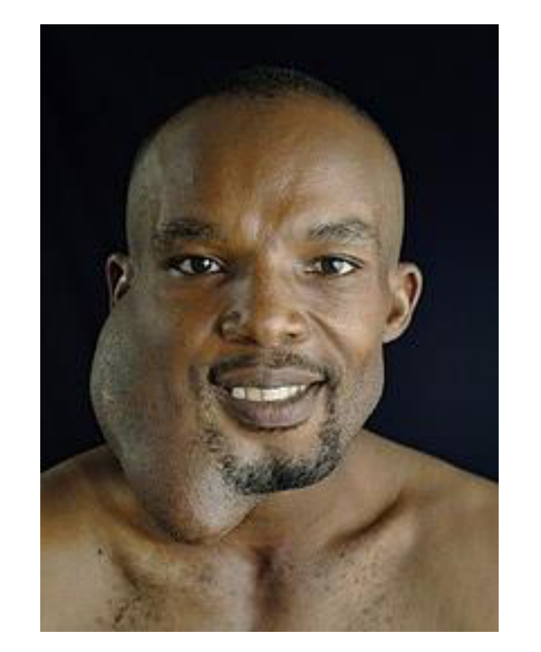
- The patient had an ameloblastoma.
- There was severe right-sided facial deformity caused by an expansile multilocular tumor.
- The tumor involved the right hemimandible.
Case Study: Bilateral Cleft Lip and Palate
- Cleft lip and palate was described as the most common congenital facial deformity.
- Most clefts are unilateral, and approximately 70% of unilateral cases are on the left.
- The case demonstrated bilateral cleft lip and palate using a reconstructed CBCT panoramic image.
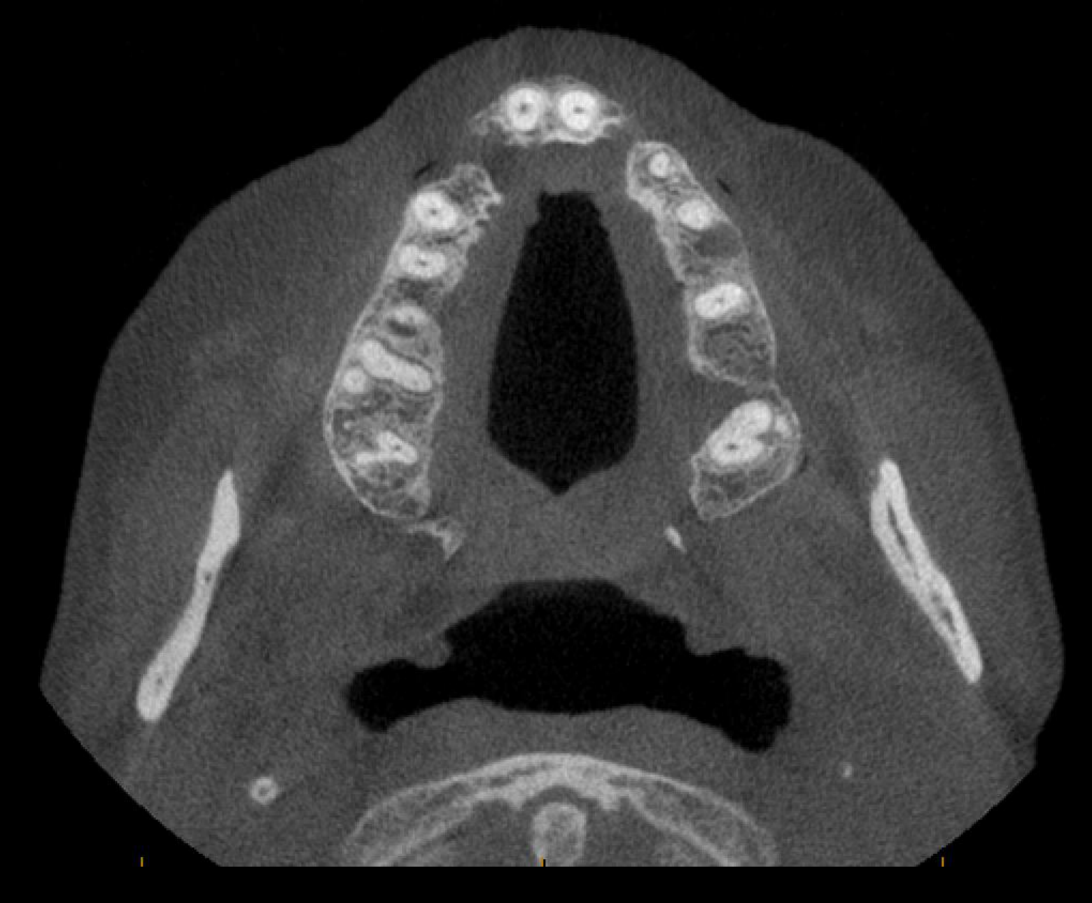
- On the axial view, bony defects were present at the regions corresponding to teeth 12 and 22.
- The areas of bony cleft may also lack the associated teeth.
- In this patient, both teeth 12 and 22 were missing.
Proteus Syndrome
- Proteus syndrome is an extremely rare and progressive genetic disorder.
- It causes asymmetrical overgrowth rather than undergrowth.
- Although it is congenital, its features may not be apparent at birth.
- Findings usually begin to appear between approximately six and 18 months and progressively worsen.
- Facial findings may include asymmetrical facial hypertrophy.
- The condition may also be associated with:
- Tumors
- Vascular malformations
- More complex imaging protocols involving CT and MRI may be required.
- MRI is useful for assessing associated vascular malformations.
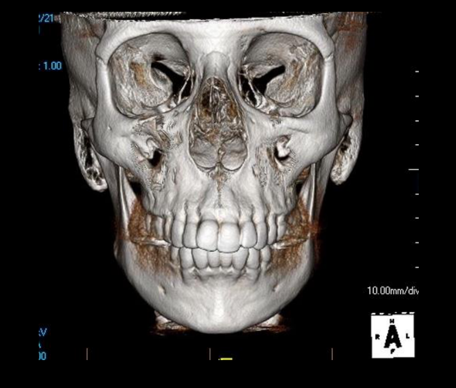
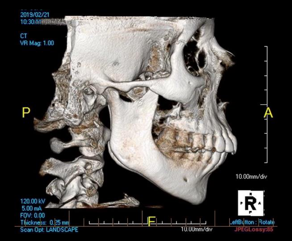
Case Study: Protrusive Maxilla and Retrognathic Mandible
- This rare case involved both:
- A protrusive maxilla
- A retrognathic mandible
- The result was a skeletal Class II relationship.
- The patient had a very convex facial profile and severe anterior overjet.
- Bimaxillary surgery corrected the preoperative Class II relationship and convex profile.
- The frontal view showed that the vertical facial dimension was not altered by the surgery.
Congenital Deformities
- Congenital deformities are present at birth and are often related to developmental disturbances.
- Examples include:
- Cleft lip and palate
- Craniosynostosis-associated syndromes
- Hemifacial microsomia
- Cleft lip and palate is the most common congenital facial deformity discussed.
- It involves both soft and hard tissues and may affect:
- The airway
- Speech
- Feeding
Case Study: Right-Sided Mandibular Hyperplasia
- The condition involved persistent growth of the right mandibular condyle.
- Progressive facial asymmetry resulted.
- Characteristic features included:
- Outward bowing of the mandibular body and ramus
- Downward growth of the affected side
- Only the mandible was affected.
- Growth was primarily in the vertical direction.
- The symphysis was only slightly left of the facial skeletal midline.
- The dental midline was coincident.
- There was a skeletal Class III relationship without a reverse anterior overjet.
 |  |
Clinical Cases
Case: Treacher Collins syndrome
Question
Scenario: A diagram is presented to illustrate a rare bilateral facial deformity syndrome affecting the first and second branchial arches.
What’s shown: Bilateral facial hypoplasia affecting the jaws, ears, zygomatic bones, and lateral aspects of the orbits.
Consider: Identify the syndrome and its characteristic anatomical features based on the diagram.
Answer
Observations:
- Bilateral facial hypoplasia
- Involvement of the jaws, ears, zygomatic bones, and lateral orbits
Reasoning: The bilateral nature of the deformity and the specific involvement of first and second branchial arch derivatives (jaws, ears, zygoma, orbits) are characteristic of this rare syndrome, distinguishing it from unilateral conditions.
Outcome: None stated.
Takeaway: Treacher Collins syndrome is a rare first and second branchial arch syndrome characterized by bilateral facial hypoplasia affecting the jaws, ears, zygomatic bones, and lateral orbits.
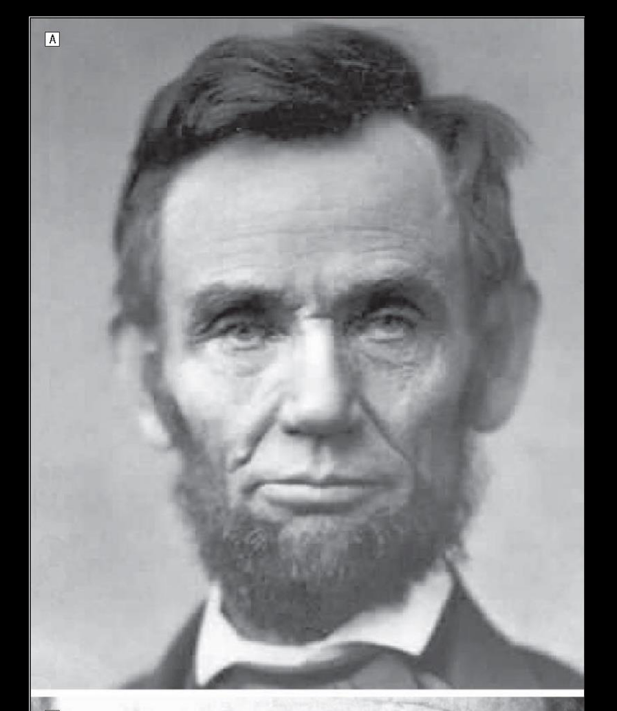
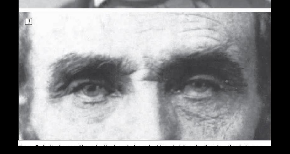
Case: Hemifacial microsomia (Abraham Lincoln example)
Question
Scenario: A historical example is used to illustrate a first and second branchial arch syndrome.
What’s shown: Unilateral involvement of facial structures.
Consider: Identify the condition and its laterality based on the historical presentation.
Answer
Observations:
- Unilateral facial involvement
- First and second branchial arch syndrome
Reasoning: Unlike bilateral syndromes, this condition presents with unilateral involvement. It is noted as the second most common congenital facial deformity after cleft lip and palate.
Outcome: None stated.
Takeaway: Hemifacial microsomia (craniofacial microsomia) is a first and second branchial arch syndrome characterized by unilateral facial involvement.
Case: Cleidocranial dysplasia
Question
Scenario: A specific condition is highlighted using a pop culture reference to illustrate its distinct skeletal and dental features.
What’s shown: Absent or hypoplastic clavicles, persistent open cranial sutures, supernumerary teeth, prolonged retention of primary teeth, multiple impacted permanent teeth, and a hypoplastic maxilla.
Consider: Identify the syndrome and its distinguishing dental and skeletal features.
Answer
Observations:
- Absent or hypoplastic clavicles
- Persistent open cranial sutures
- Supernumerary teeth, prolonged retention of primary teeth, multiple impacted permanent teeth
- Hypoplastic maxilla
Reasoning: These specific skeletal and dental findings distinguish this condition from other facial deformity syndromes, particularly the combination of clavicular hypoplasia and multiple dental anomalies.
Outcome: None stated.
Takeaway: Cleidocranial dysplasia is distinguished by absent or hypoplastic clavicles, open cranial sutures, and multiple dental anomalies including supernumerary and impacted teeth.
Case: Crouzon syndrome
Question
Scenario: A syndrome characterized by premature cranial suture fusion is presented.
What’s shown: Decreased anterior-posterior dimension at the cranial base, copper-beaten appearance on skull imaging, hypoplastic jaws, shallow orbits resulting in proptosis, and a class three skeletal relationship.
Consider: Identify the syndrome and its cranial, orbital, and skeletal manifestations.
Answer
Observations:
- Premature fusion of cranial sutures (craniosynostosis)
- Decreased AP dimension and copper-beaten skull appearance
- Hypoplastic jaws and shallow orbits with proptosis
- Class three skeletal relationship due to maxillary hypoplasia
Reasoning: The combination of craniosynostosis with a copper-beaten skull appearance, shallow orbits causing proptosis, and maxillary hypoplasia leading to a class three relationship points to this specific syndrome.
Outcome: None stated.
Takeaway: Crouzon syndrome features craniosynostosis with a copper-beaten skull appearance, shallow orbits causing proptosis, and maxillary hypoplasia resulting in a class three skeletal relationship.
Case: Apert syndrome
Question
Scenario: A syndrome similar to Crouzon but with distinct limb findings is presented.
What’s shown: Early cranial suture fusion (craniosynostosis) and syndactyly (webbed or fused fingers/toes).
Consider: Differentiate this syndrome from Crouzon syndrome based on the clinical features.
Answer
Observations:
- Craniosynostosis (early cranial suture fusion)
- Syndactyly
- Increased chance of intellectual disability
Reasoning: While similar to Crouzon syndrome in having craniosynostosis, the primary distinguishing feature is the presence of syndactyly, along with a higher risk of intellectual disability.
Outcome: None stated.
Takeaway: Apert syndrome is distinguished from Crouzon syndrome by the presence of syndactyly and an increased risk of intellectual disability.
Case: Pfeiffer syndrome
Question
Scenario: Another syndrome associated with craniosynostosis is presented, featuring distinct digit and skull abnormalities.
What’s shown: Variable thumb and toe widening, extreme skull deformity described as a “clover leaf brain” shape.
Consider: Identify the syndrome based on the specific digit and skull morphology.
Answer
Observations:
- Variable thumb and toe widening
- Extreme skull deformity (clover leaf brain shape)
- Craniosynostosis
Reasoning: The combination of craniosynostosis with variable thumb and toe widening and the classic “clover leaf” skull deformity is characteristic of this syndrome.
Outcome: None stated.
Takeaway: Pfeiffer syndrome is characterized by craniosynostosis, variable thumb and toe widening, and a classic “clover leaf” skull deformity.
Case: Proteus syndrome
Question
Scenario: A rare, progressive genetic disorder causing asymmetrical overgrowth is presented.
What’s shown: Asymmetrical facial hypertrophy, various tumors, and vascular malformations visible on MRI.
Consider: Identify the condition and the imaging modality used to assess its soft tissue and vascular components.
Answer
Observations:
- Asymmetrical facial hypertrophy (overgrowth, not undergrowth)
- Associated tumors and vascular malformations
- Vascular malformations visible on MRI
Reasoning: Unlike other congenital deformities that cause hypoplasia, this condition causes progressive asymmetrical overgrowth. The complex soft tissue and vascular components require MRI for detailed assessment.
Outcome: None stated.
Takeaway: Proteus syndrome causes progressive asymmetrical overgrowth and is associated with tumors and vascular malformations, which are best evaluated with MRI.
Case: Bilateral cleft lip and palate
Question
Scenario: A patient with the most common congenital facial deformity is presented using a reconstructed CBCT panorama image.
What’s shown: Bony defects at the 12 and 22 sites on the axial view, with missing teeth in the cleft areas.
Consider: Identify the type of cleft and the associated dental anomalies seen on the CBCT.
Answer
Observations:
- Bilateral cleft lip and palate
- Bony defects at the 12 and 22 sites
- Missing teeth 12 and 22
Reasoning: The CBCT clearly shows bilateral bony defects in the maxilla corresponding to the lateral incisor regions, which are also missing the associated teeth, a common finding in clefts.
Outcome: None stated.
Takeaway: Bilateral cleft lip and palate often presents with bony defects in the maxilla and missing teeth in the cleft areas, clearly visualized on CBCT.
Case: Unilateral right-sided cleft lip and palate
Question
Scenario: A patient with a unilateral cleft is presented.
What’s shown: A bony defect in the right maxilla at the 12 region involving the nasal cavity.
Consider: Identify the location and extent of the bony defect in this unilateral cleft.
Answer
Observations:
- Unilateral right-sided cleft lip and palate
- Bony defect in the right maxilla at the 12 region
- Involvement of the nasal cavity
Reasoning: The defect is localized to the right side of the maxilla, extending into the nasal cavity, which is typical for a unilateral cleft.
Outcome: None stated.
Takeaway: Unilateral cleft lip and palate presents with a localized bony defect in the maxilla that can involve the nasal cavity.
Case: Midline cleft lip and palate
Question
Scenario: An extremely rare midline cleft case is presented, often associated with larger syndromes.
What’s shown: Significant facial deformities including maxillary and nasal hypoplasia, leading to a severe Class 3 malocclusion, concave facial profile, and anterior open bite.
Consider: Identify the associated facial deformities and skeletal relationships resulting from this rare midline cleft.
Answer
Observations:
- Midline cleft lip and palate
- Maxillary and nasal hypoplasia
- Severe Class 3 malocclusion with a concave facial profile
- Anterior open bite
Reasoning: The midline cleft is associated with severe hypoplasia of the maxilla and nose, which directly causes the severe Class 3 skeletal relationship and anterior open bite.
Outcome: None stated.
Takeaway: Midline cleft lip and palate is extremely rare and often associated with severe maxillary and nasal hypoplasia, resulting in a Class 3 malocclusion and anterior open bite.
Case: Hemifacial microsomia and Goldenhar syndrome
Question
Scenario: A patient with poor development of the right side of the face is presented, showing pre- and post-surgical images.
What’s shown: Severe mandibular asymmetry pre-operatively, and post-operative OPG with significant artifacts.
Consider: Identify the condition, its broader spectrum classification, and the limitations of post-operative OPG imaging.
Answer
Observations:
- Poor development of the right side of the face, severely affecting the mandible
- Post-operative OPG shows significant artifacts, making it almost useless
- Condition belongs to the ocular auricular vertebral spectrum (Goldenhar syndrome) when the spine is involved
Reasoning: The unilateral mandibular hypoplasia is characteristic of hemifacial microsomia. When spinal involvement is present, it is classified as Goldenhar syndrome. Post-surgical hardware causes severe artifacts on OPG.
Outcome: Surgical correction of mandibular asymmetry was performed.
Takeaway: Hemifacial microsomia severely affects the mandible; when spinal involvement is present, it is part of Goldenhar syndrome, and post-operative OPGs are often limited by artifacts.
Case: Right-sided mandibular hyperplasia
Question
Scenario: A patient with persistent growth of the right condyle leading to progressive facial asymmetry is presented.
What’s shown: Outward bowing and downward growth of the mandibular body and ramus on the right side. Growth is mainly in the vertical vector, symphysis slightly left of midline, coincidental dental midline, and a Class 3 skeletal relationship without reverse anterior overjet.
Consider: Identify the condition and characterize the vector of growth and skeletal relationship.
Answer
Observations:
- Persistent growth of the right condyle
- Outward bowing and downward growth of the right mandibular body and ramus
- Vertical vector growth, symphysis slightly left of midline
- Coincidental dental midline
- Class 3 skeletal relationship without reverse anterior overjet
Reasoning: The persistent condylar growth causes characteristic outward and downward bowing. The vertical vector of growth shifts the symphysis slightly but keeps the dental midline coincidental, resulting in a Class 3 relationship.
Outcome: None stated.
Takeaway: Right-sided mandibular hyperplasia features persistent condylar growth causing outward and downward bowing, primarily in a vertical vector, leading to a Class 3 skeletal relationship.
Case: Severe right-sided hemimandibular hyperplasia
Question
Scenario: A more severe case of right-sided hyperplasia is presented.
What’s shown: Rotation of the mandible, symphysis shifted to the left of the facial skeletal midline, dental midline shift, and a Class 3 skeletal relationship without reversed anterior overjet.
Consider: Differentiate this severe case from the previous one based on mandibular rotation and midline shifts.
Answer
Observations:
- Rotation of the mandible
- Symphysis shifted to the left of the facial skeletal midline
- Dental midline shift
- Class 3 skeletal relationship without reversed anterior overjet
Reasoning: Unlike the previous case, the severe hyperplasia causes actual rotation of the mandible, leading to a shift in both the facial symphysis and the dental midline.
Outcome: None stated.
Takeaway: Severe hemimandibular hyperplasia causes mandibular rotation, resulting in shifts of both the facial symphysis and the dental midline.
Case: Retrognathism (Class 2 skeletal relationship)
Question
Scenario: A patient with a Class 2 skeletal relationship is presented with pre- and post-surgical images.
What’s shown: Severe anterior overjet pre-operatively, and the post-operative result following a bimaxillary osteotomy.
Consider: Identify the skeletal discrepancy and the surgical approach used to correct it.
Answer
Observations:
- Class 2 skeletal relationship (retrognathism)
- Severe anterior overjet pre-operatively
- Correction achieved via bimaxillary osteotomy (maxilla and mandible)
Reasoning: The severe anterior overjet and Class 2 relationship are corrected by operating on both jaws to achieve functional improvement and aesthetic balance.
Outcome: Bimaxillary osteotomy performed to correct the deformity.
Takeaway: Bimaxillary osteotomy is often used to correct severe Class 2 retrognathism and anterior overjet, achieving both functional and aesthetic balance.
Case: Protrusive maxilla and retrognathic mandible
Question
Scenario: A rare case of a specific Class 2 skeletal discrepancy is presented with pre- and post-surgical views.
What’s shown: Very convex profile, severe anterior overjet, and Class 2 relationship pre-operatively. Post-operatively, the profile and overjet are corrected, but the vertical dimension of the face is unaltered.
Consider: Identify the specific skeletal discrepancy and note the effect of the surgery on the vertical facial dimension.
Answer
Observations:
- Protrusive maxilla and retrognathic mandible
- Very convex profile and severe anterior overjet
- Correction of profile and overjet post-surgery
- Vertical dimension of the face remains unaltered
Reasoning: The combination of a protrusive maxilla and retrognathic mandible creates a severe convex profile. Bimax surgery corrects the AP discrepancy without altering the vertical facial dimension.
Outcome: Bimax surgery performed to correct the Class 2 relationship and convex profile.
Takeaway: Bimax surgery can correct the severe convex profile and anterior overjet of a protrusive maxilla and retrognathic mandible without altering the vertical facial dimension.
Case: Ameloblastoma
Question
Scenario: A patient with a specific benign tumor is presented.
What’s shown: Severe right-sided facial deformity caused by an expansile multilocular tumor involving the right hemimandible.
Consider: Identify the tumor and its effect on the facial skeleton based on the imaging.
Answer
Observations:
- Expansile multilocular tumor
- Involvement of the right hemimandible
- Severe right-sided facial deformity
Reasoning: The expansile, multilocular nature of the tumor in the mandible causing severe facial deformity is characteristic of an ameloblastoma.
Outcome: None stated.
Takeaway: Ameloblastoma presents as an expansile multilocular tumor that can cause severe facial deformity when involving the mandible.
Case: Unilateral condylar hyperplasia and osteochondroma
Question
Scenario: A patient with overgrowth of the right condyle is evaluated to determine if the lesion is active.
What’s shown: SPECT nuclear medicine imaging showing increased radionuclide uptake in the right condyle, and CT showing an enlarged condyle with lost original morphology and irregular trabecular bone pattern.
Consider: Determine the activity status of the condylar lesion and differentiate it from developmental condylar hyperplasia based on imaging.
Answer
Observations:
- Increased radionuclide uptake in the right condyle on SPECT
- Enlarged right condyle with lost original morphology on CT
- Irregular trabecular bone pattern
Reasoning: The increased uptake on SPECT indicates the benign tumor (osteochondroma) is still active. Unlike developmental condylar hyperplasia which maintains normal morphology, tumorous activity causes irregular morphology and loss of normal trabecular pattern.
Outcome: Surgery must be delayed because the tumor is still active and growing.
Takeaway: SPECT imaging determines the activity of condylar tumors; active lesions show increased uptake and irregular morphology on CT, indicating surgery should be delayed until they burn out.
Case: Fibrous dysplasia
Question
Scenario: A condition causing significant facial asymmetry is presented.
What’s shown: Lesion typically affecting the maxilla, mandible, or skull base. Bone scans are mentioned as being taken before surgery.
Consider: Identify the condition and the purpose of the bone scan in its management.
Answer
Observations:
- Significant facial asymmetry
- Typically affects maxilla, mandible, or skull base
- Bone scans taken before surgery
Reasoning: The condition causes asymmetry by affecting major craniofacial bones. Bone scans are utilized pre-operatively to ensure the lesion is stable before proceeding with surgical management.
Outcome: Managed with surgery after confirming stability with a bone scan.
Takeaway: Fibrous dysplasia causes facial asymmetry and requires bone scans prior to surgery to ensure the lesion is stable.
Footnotes
-
Original PDF page 1: L5 - Imaging for Facial Deformity, p.1 ↩
-
Original PDF page 2: L5 - Imaging for Facial Deformity, p.2 ↩
-
Original PDF page 3: L5 - Imaging for Facial Deformity, p.3 ↩
-
Original PDF page 4: L5 - Imaging for Facial Deformity, p.4 ↩
-
Original PDF page 5: L5 - Imaging for Facial Deformity, p.5 ↩
-
Original PDF page 6: L5 - Imaging for Facial Deformity, p.6 ↩
-
Original PDF page 7: L5 - Imaging for Facial Deformity, p.7 ↩
-
Original PDF page 8: L5 - Imaging for Facial Deformity, p.8 ↩
-
Original PDF page 9: L5 - Imaging for Facial Deformity, p.9 ↩
-
Original PDF page 10: L5 - Imaging for Facial Deformity, p.10 ↩
-
Original PDF page 11: L5 - Imaging for Facial Deformity, p.11 ↩
-
Original PDF page 12: L5 - Imaging for Facial Deformity, p.12 ↩
-
Original PDF page 13: L5 - Imaging for Facial Deformity, p.13 ↩
-
Original PDF page 14: L5 - Imaging for Facial Deformity, p.14 ↩
-
Original PDF page 15: L5 - Imaging for Facial Deformity, p.15 ↩
-
Original PDF page 16: L5 - Imaging for Facial Deformity, p.16 ↩
-
Original PDF page 17: L5 - Imaging for Facial Deformity, p.17 ↩
-
Original PDF page 18: L5 - Imaging for Facial Deformity, p.18 ↩
-
Original PDF page 19: L5 - Imaging for Facial Deformity, p.19 ↩
-
Original PDF page 20: L5 - Imaging for Facial Deformity, p.20 ↩
-
Original PDF page 21: L5 - Imaging for Facial Deformity, p.21 ↩
-
Original PDF page 22: L5 - Imaging for Facial Deformity, p.22 ↩
-
Original PDF page 23: L5 - Imaging for Facial Deformity, p.23 ↩
-
Original PDF page 24: L5 - Imaging for Facial Deformity, p.24 ↩
-
Original PDF page 25: L5 - Imaging for Facial Deformity, p.25 ↩
-
Original PDF page 26: L5 - Imaging for Facial Deformity, p.26 ↩
-
Original PDF page 27: L5 - Imaging for Facial Deformity, p.27 ↩
-
Original PDF page 28: L5 - Imaging for Facial Deformity, p.28 ↩
-
Original PDF page 29: L5 - Imaging for Facial Deformity, p.29 ↩
-
Original PDF page 30: L5 - Imaging for Facial Deformity, p.30 ↩
-
Original PDF page 31: L5 - Imaging for Facial Deformity, p.31 ↩
-
Original PDF page 32: L5 - Imaging for Facial Deformity, p.32 ↩
-
Original PDF page 33: L5 - Imaging for Facial Deformity, p.33 ↩
-
Original PDF page 34: L5 - Imaging for Facial Deformity, p.34 ↩
-
Original PDF page 35: L5 - Imaging for Facial Deformity, p.35 ↩
-
Original PDF page 36: L5 - Imaging for Facial Deformity, p.36 ↩
-
Original PDF page 37: L5 - Imaging for Facial Deformity, p.37 ↩
-
Original PDF page 38: L5 - Imaging for Facial Deformity, p.38 ↩
-
Original PDF page 39: L5 - Imaging for Facial Deformity, p.39 ↩
-
Original PDF page 40: L5 - Imaging for Facial Deformity, p.40 ↩
-
Original PDF page 41: L5 - Imaging for Facial Deformity, p.41 ↩
-
Original PDF page 42: L5 - Imaging for Facial Deformity, p.42 ↩
-
Original PDF page 43: L5 - Imaging for Facial Deformity, p.43 ↩