Malignant Diseases of the Jaws12
Lecturer — Jaw Malignancy Overview
Jaw malignancies are uncommon but tend to be aggressive. Early detection is important because it significantly affects prognosis.
 |  |
 |
Overview Of Malignant Jaw Lesions34
Malignant lesions in the jaws share common imaging features:
- Destructive & invasive/infiltrative (lytic and ill-defined)
Malignancies that can affect the jaws are divided into four categories:
- Carcinomas — lesions of epithelial origin
- Sarcomas — lesions of mesenchymal origin
- Malignancies of the hematopoietic system — lymphoma, leukemia, myeloma
- Metastatic lesions from distant sites
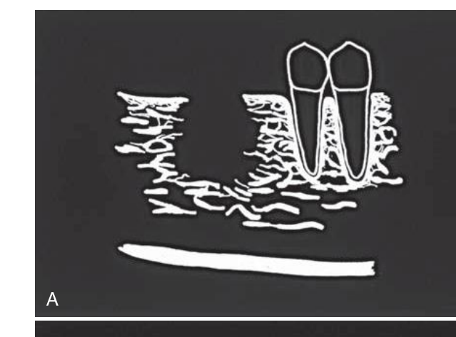
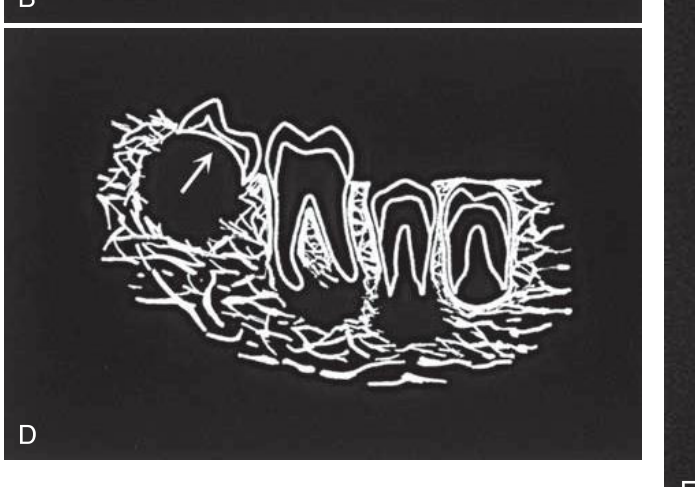
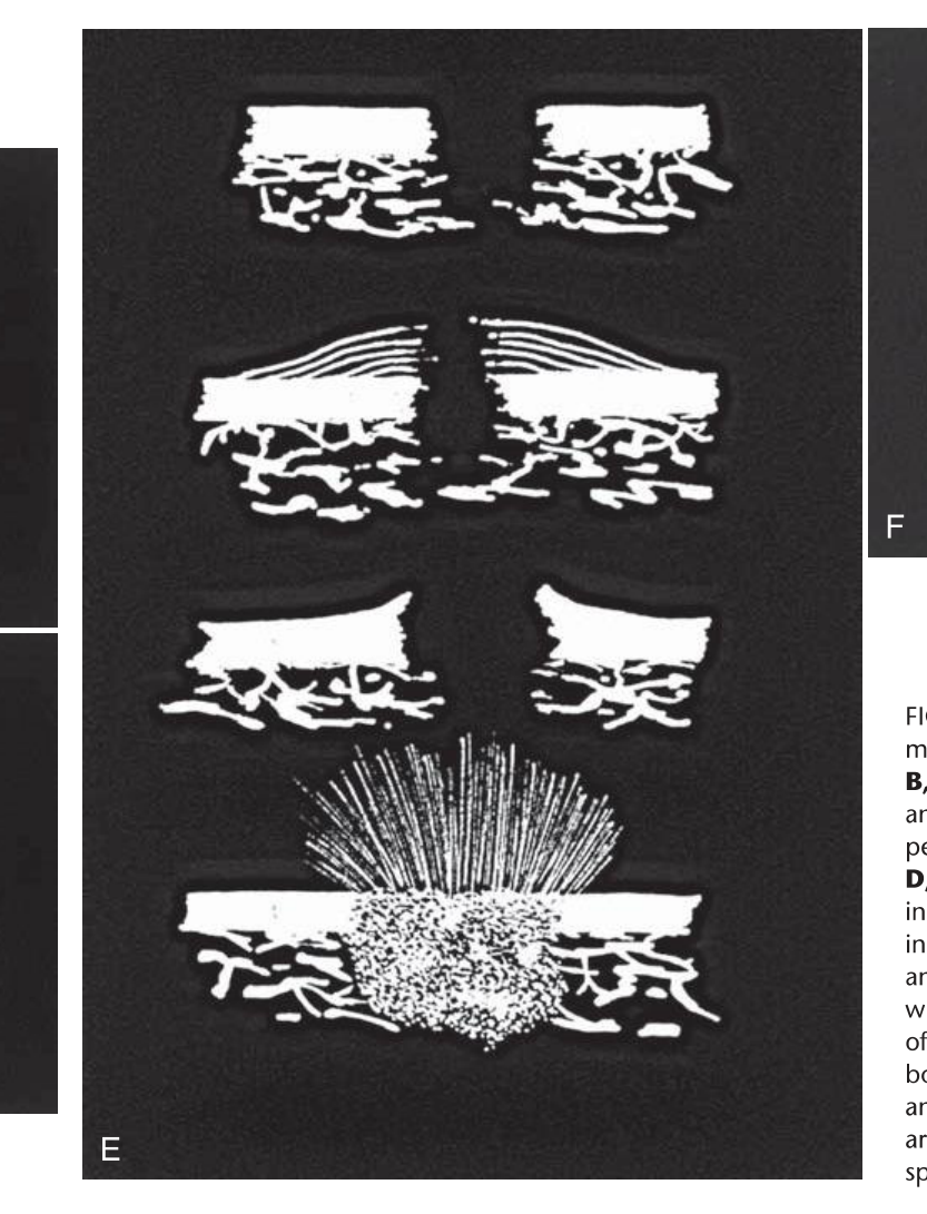
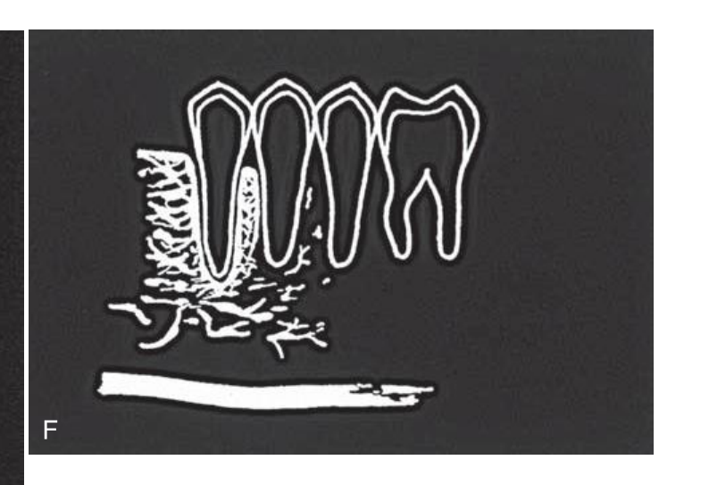
FIG. 23-1 Diagrammatic representation of radiologic features of oral malignancy.
- A — Ill-defined invasive borders followed by bone destruction.
- B — Destruction of the cortical boundary (floor of maxillary antrum) with an adjacent soft tissue mass (arrows).
- C — Tumor invasion along the periodontal membrane space causing irregular thickening of this space.
- D — Multifocal lesions located at root apices and in the papilla of a developing tooth destroying the crypt cortex and displacing the developing tooth in an occlusal direction (arrow).
- E — Four types of effects on cortical bone and periosteal reaction, from top to the bottom: cortical bone destruction without periosteal reaction, laminated periosteal reaction with destruction of the cortical bone and the new periosteal bone, destruction of cortical bone with periosteal reaction at the periphery forming Codman’s triangles, and a spiculated or sunray type of periosteal reaction.
- F — Bone destruction around existing teeth, producing an appearance of teeth floating in space.
 |  |
 |  |
Carcinomas
Squamous Cell Carcinoma5
 |  |
Squamous Cell Carcinoma (SCC)6
- SCC of the oral mucosa — the most common malignant tumour of the head and neck, originating from surface epithelium
- Primary intraosseous SCC — arises within the jawbones and has no original connection with the surface epithelium of the oral mucosa (rare)
- SCC originating in a cyst — the lining squamous epithelium of a cyst gives rise to the malignant neoplasm (rare)
Lecturer — Clinical Features
Oral mucosal squamous cell carcinoma may present with several clinical features.
- Ulceration
- A non-healing socket
- Pain
- Paresthesia
- Unexplained tooth mobility
 |  |
Radiographic Features on Dental Imaging (PA, OPG & CBCT)
-
Ill-defined hypodensity — “moth eaten”
-
Destruction of adjacent cortical structures such as cortical plates, lamina dura and antral floors
-
Irregular PDL space widening of affected teeth; if extensive, the teeth appear “floating”
-
Irregular MC widening
-
Irregular widening or loss of the mandibular canal
-
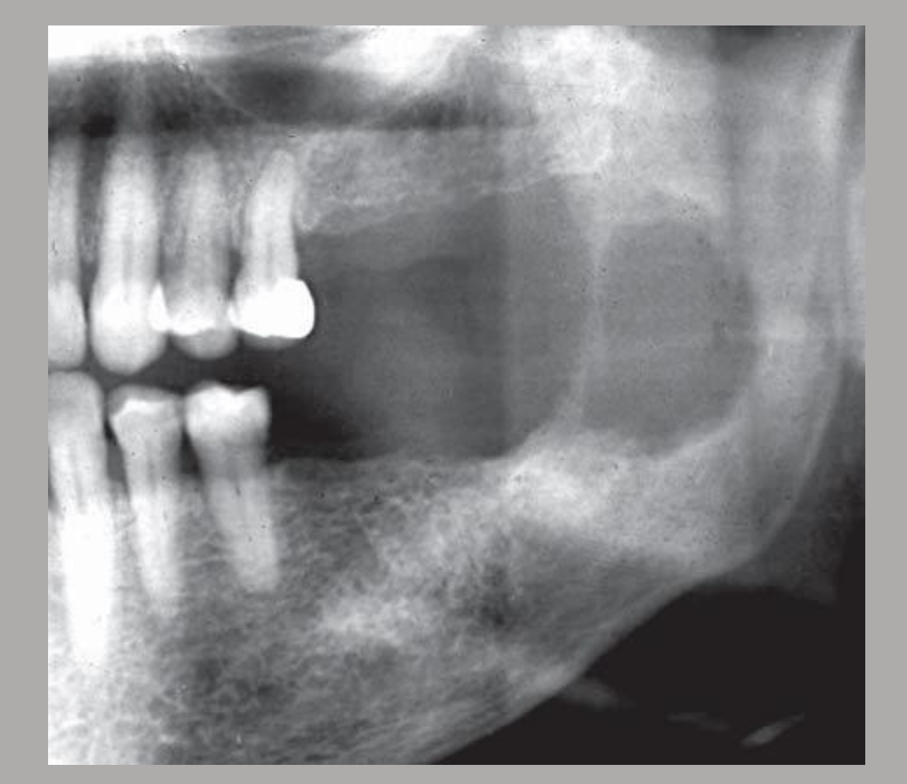
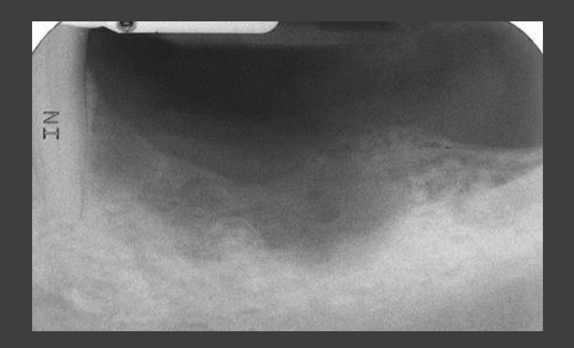
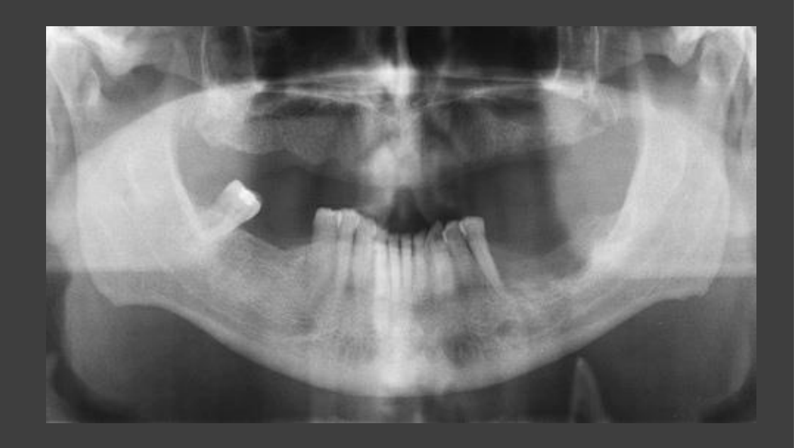
Soft tissue involvement FIG. 23-4 This primary intraosseous carcinoma in the left mandible exhibits no internal structure, a poorly defined periphery, and thinning of the overlying mandibular bone.
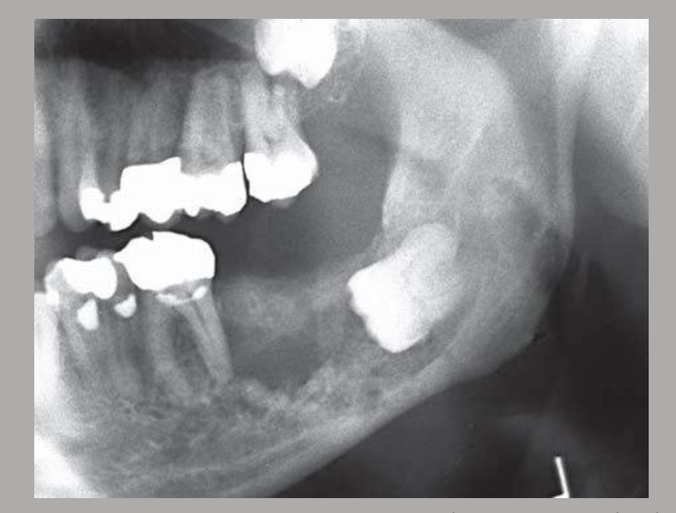
FIG. 23-5 Carcinoma arising in a preexisting dentigerous cyst related to the mandibular left third molar shows absence of a cyst cortex, invasion into adjacent bone, and ill-defined borders.
Other Rare Carcinomas Of The Jaws
Central Mucoepidermoid Carcinoma7
- Mucoepidermoid carcinoma is usually a salivary gland malignancy; the central type is rare
- Likely originates from pluripotential odontogenic epithelium or from a cyst lining
- Histologically indistinguishable from its soft tissue counterpart
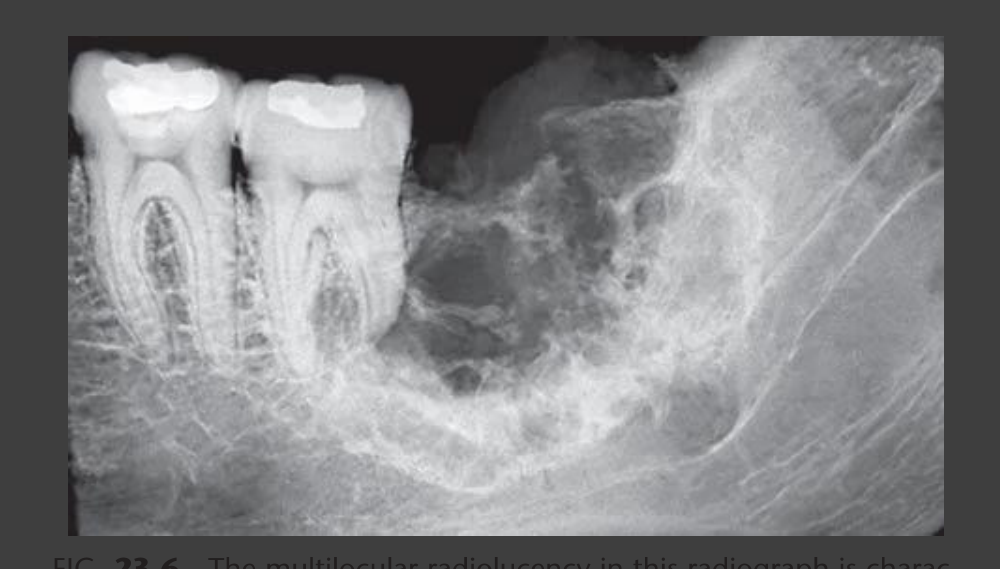
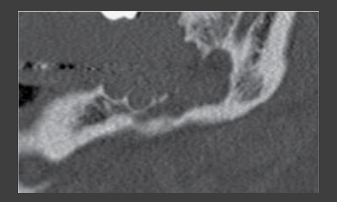
- Radiographically, a unilocular or multilocular expansile mass, often well defined and well corticated (ddx: ameloblastoma)
- Teeth remain largely unaffected by this disease, although adjacent lamina dura may be lost
Lecturer — Diagnostic Difficulty
Central mucoepidermoid carcinoma can closely resemble a benign multilocular lesion such as ameloblastoma. This similarity makes diagnosis difficult despite the lesion’s malignant behavior.
 |  |
Other Rare Carcinomas
- Malignant ameloblastoma, etc.
Further Imaging Required8910
- MSCT with IV contrast
- MRI with Gad (soft tissue details, perineural spread & nodal metastasis, etc)
- PET/CT for cancer staging
- Ultrasound → used for guided biopsy (lymph nodes in the neck)
Lecturer — Further Assessment
Aggressive bony features on primary imaging require urgent referral to an oral and maxillofacial surgeon.
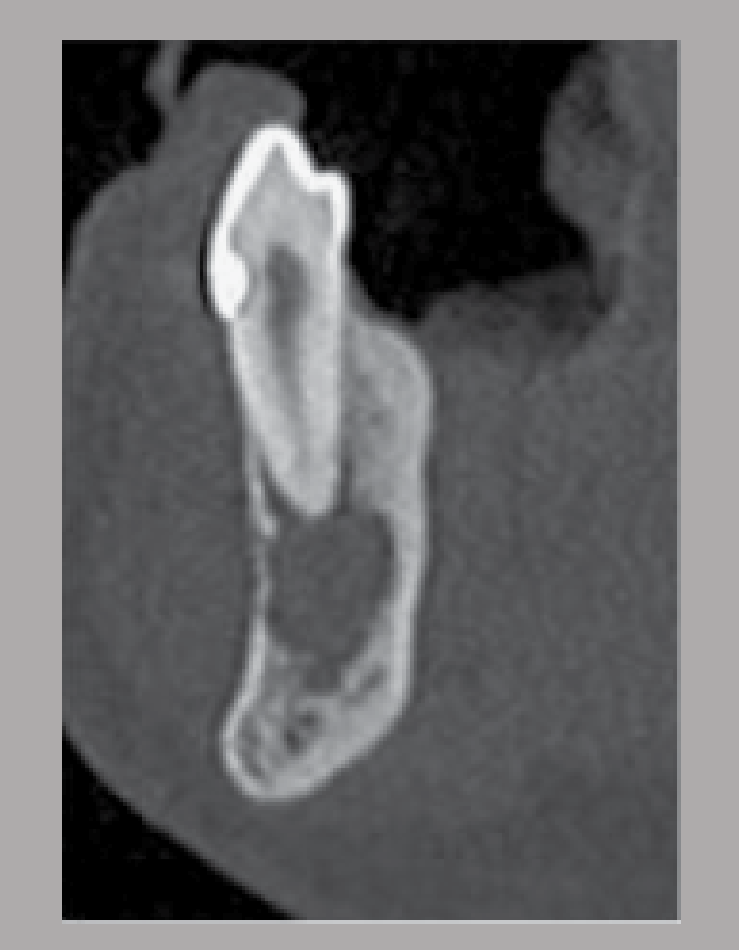
- CT assesses bulk bony destruction, dental involvement, nodal involvement, contrast enhancement, biopsy, and staging; contrast helps define malignant tumor margins when they are difficult to identify without it.
- MRI provides superior assessment of marrow and soft tissue involvement, tumor size, deep-space and muscle invasion, perineural spread, and nodal metastasis. It is sensitive to early marrow invasion and soft tissue changes that may precede cortical damage and was described as the principal imaging method for staging.
- PET/CT evaluates distant metastasis, while bone scans can assess bone metastasis; PET/CT has higher sensitivity and specificity for bone metastasis than bone scans.
- Ultrasound is useful for assessing cervical lymph nodes and detecting nodal metastasis, as well as guiding biopsy.
 |  |  |
 |  |  |
 |
Sarcomas11
- Osteosarcoma
- Osteoid is produced by the malignant stroma, i.e. osteoblastic tumour
- Chondrosarcoma
- Sarcoma of cartilaginous origin
- Ewing’s Sarcoma
- Arise in the medullary bone and spread to the endosteal and periosteal surfaces
- Fibrosarcoma, etc.
Lecturer — Sarcoma Overview
Sarcomas arise from connective tissues and are mesenchymal in origin. They tend to occur in younger patients than carcinomas.
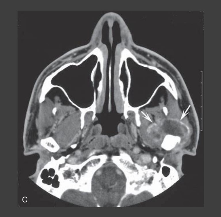
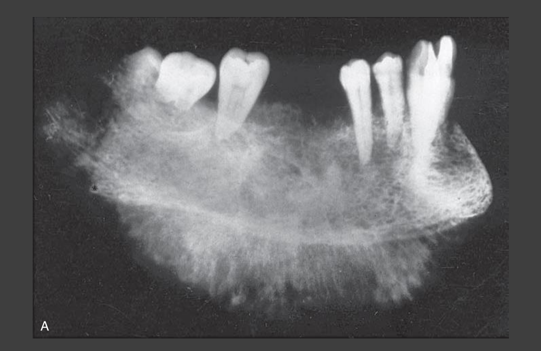
- Compared with carcinomas, sarcomas are destructive lesions that may also produce new bone formation.
- Carcinomas are more predominantly destructive and lytic, whereas sarcomas may show mixed-density appearances because of new bone production.
 |  |
Osteosarcoma Of The Jaws12
- Primary osteosarcoma: typically occurs in young patients, with 75% taking place before the age of 20
- Secondary osteosarcoma: occurs in the elderly, secondary to malignant degeneration of benign bone diseases, e.g. Paget disease, osteochondroma, etc
Radiographic Features13
-
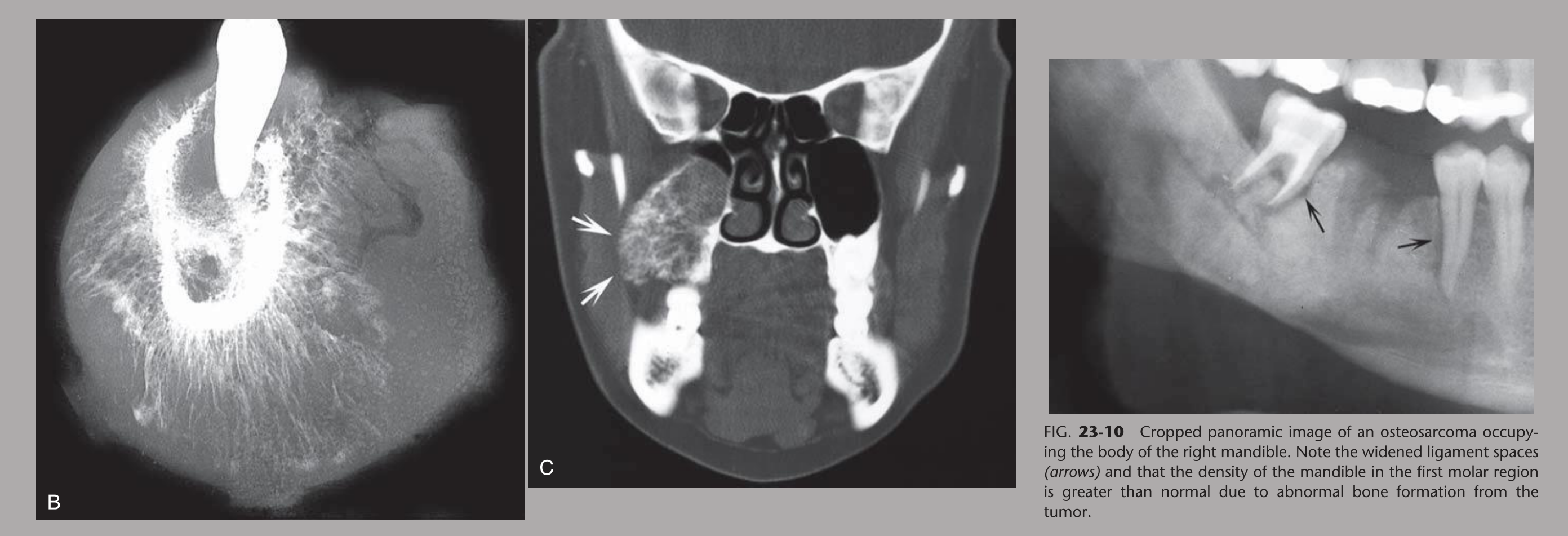
Expansile & destructive mass with ill-defined borders
-
Destruction of cortex
-
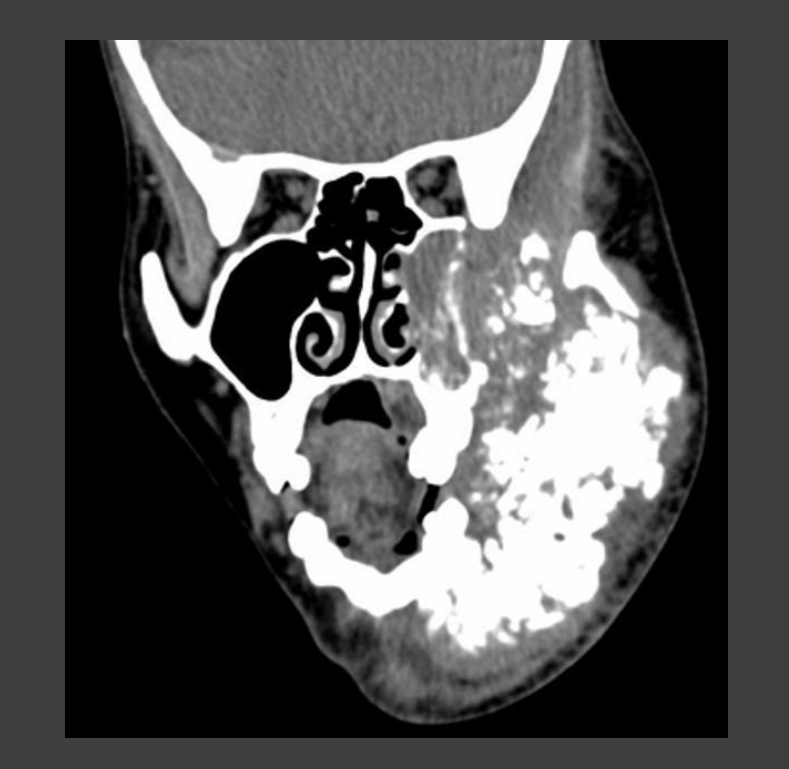
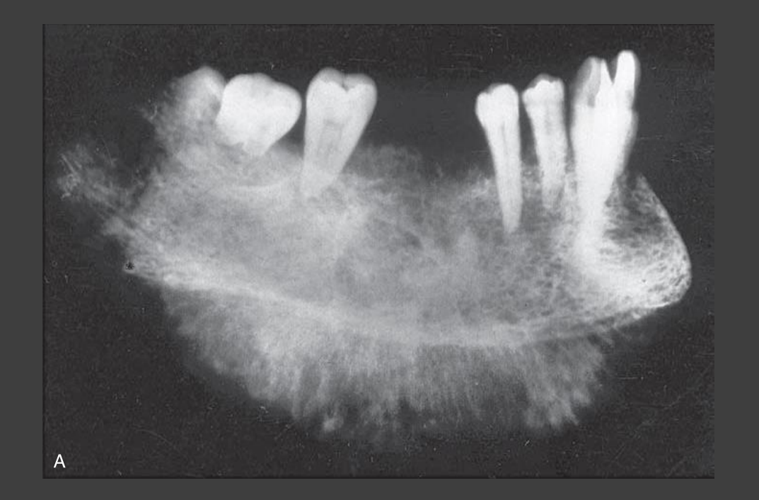
Internal “cloud-like” opacities — cotton balls
-
Sunburst & spiculated periosteal reaction
-
Enlarged neurovascular canals
-
Mixed density caused by new bone formation
-
Irregular periodontal ligament space widening
-
Loss or destruction of the lamina dura
-
Loss of the mandibular canal
Lecturer — Osteosarcoma vs SCC
Osteosarcoma may show increased density because of new bone formation, whereas SCC more typically produces lucency or decreased density. Both can show invasive cortical destruction, irregular periodontal ligament widening, and loss of the lamina dura or mandibular canal.
 |  |
Additional Imaging
-
MRI is used for local staging by assessing intraosseous tumour extension and soft tissue involvement.
-
Bone scans for distant sites
-
MRI can detect early marrow invasion and soft tissue changes before cortical damage is visible on CT
-
CT helps assess bony destruction, dental involvement, biopsy, and staging-related information
-
PET CT has higher sensitivity and specificity than bone scans for bone metastasis B
C
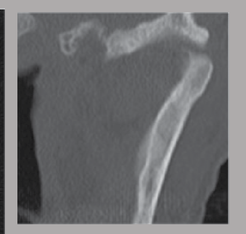
Chondrosarcoma14
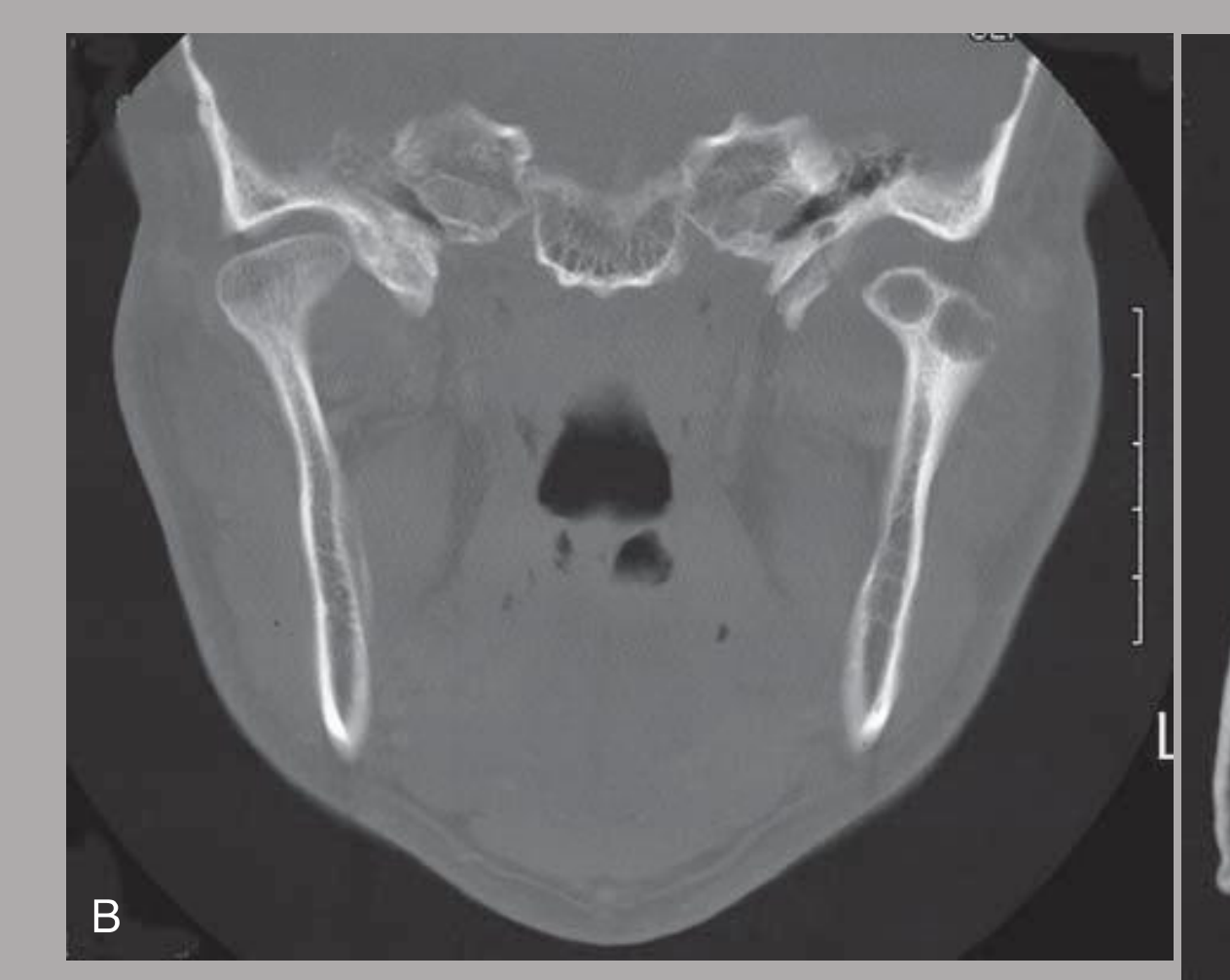
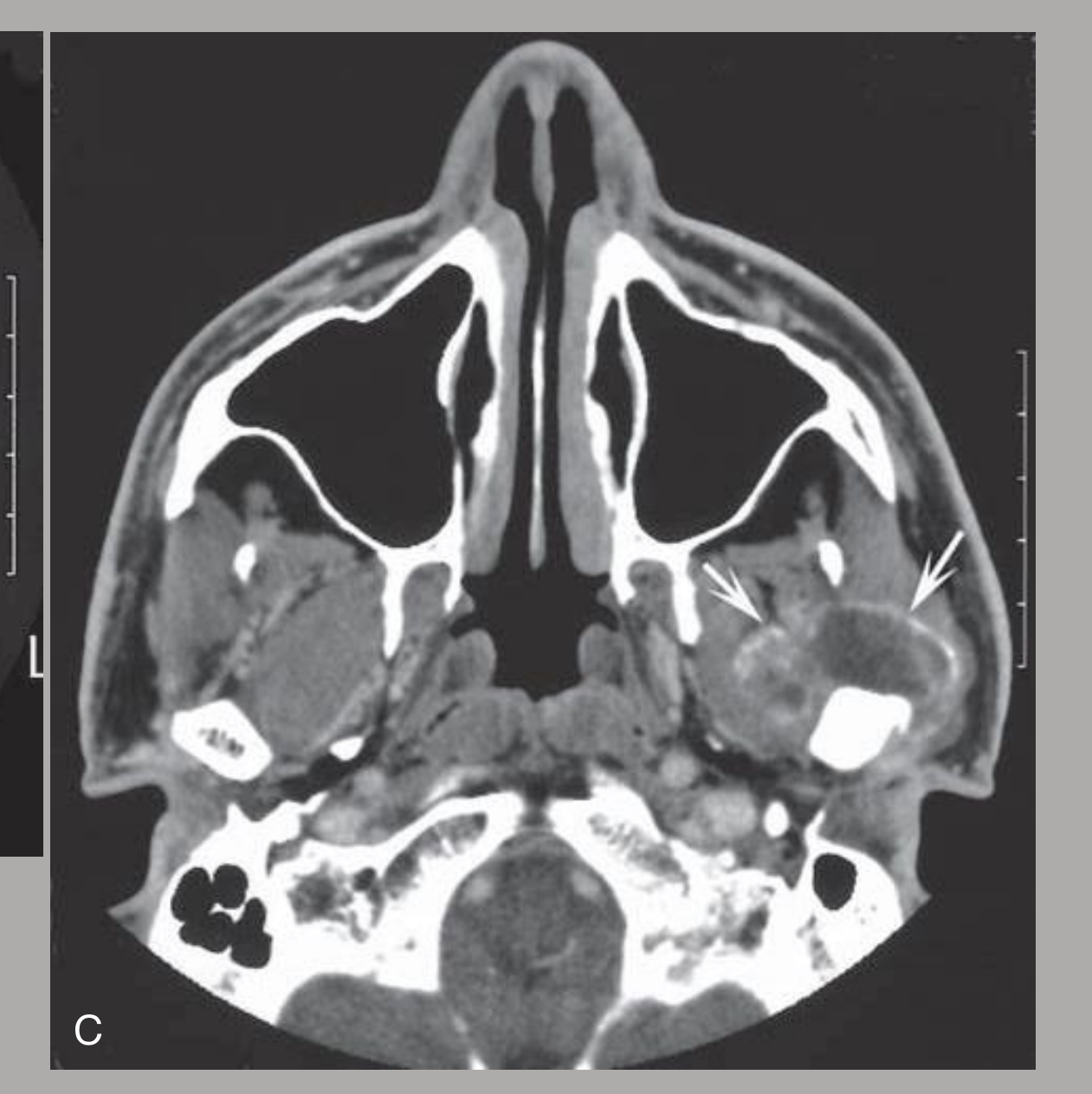
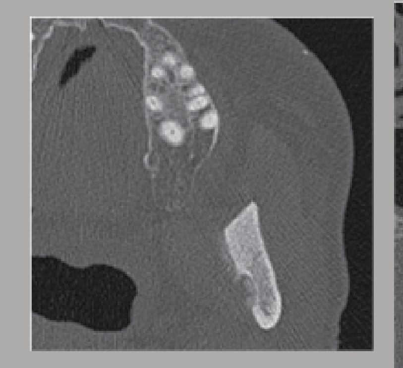
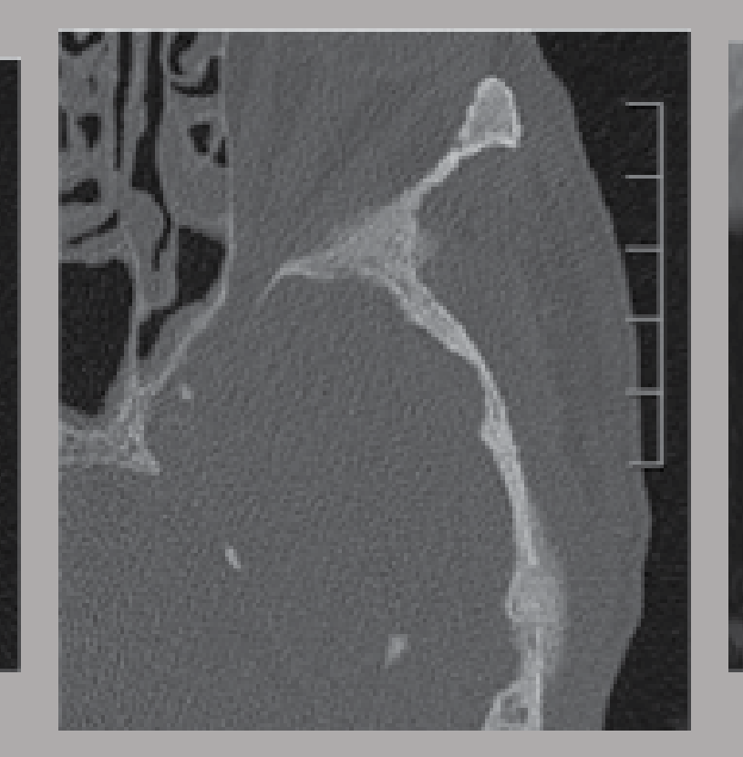
left condyle
Lecturer — Chondrosarcoma Imaging
Chondrosarcoma of the temporomandibular joint was described as extremely rare and involves cartilaginous tissue rather than only bone. Unlike osteosarcoma, it is predominantly lytic and may show ring-and-arc or popcorn calcification, with calcification around the borders of lytic lesions.
 |  |
Ewing’s Sarcoma1516
A B
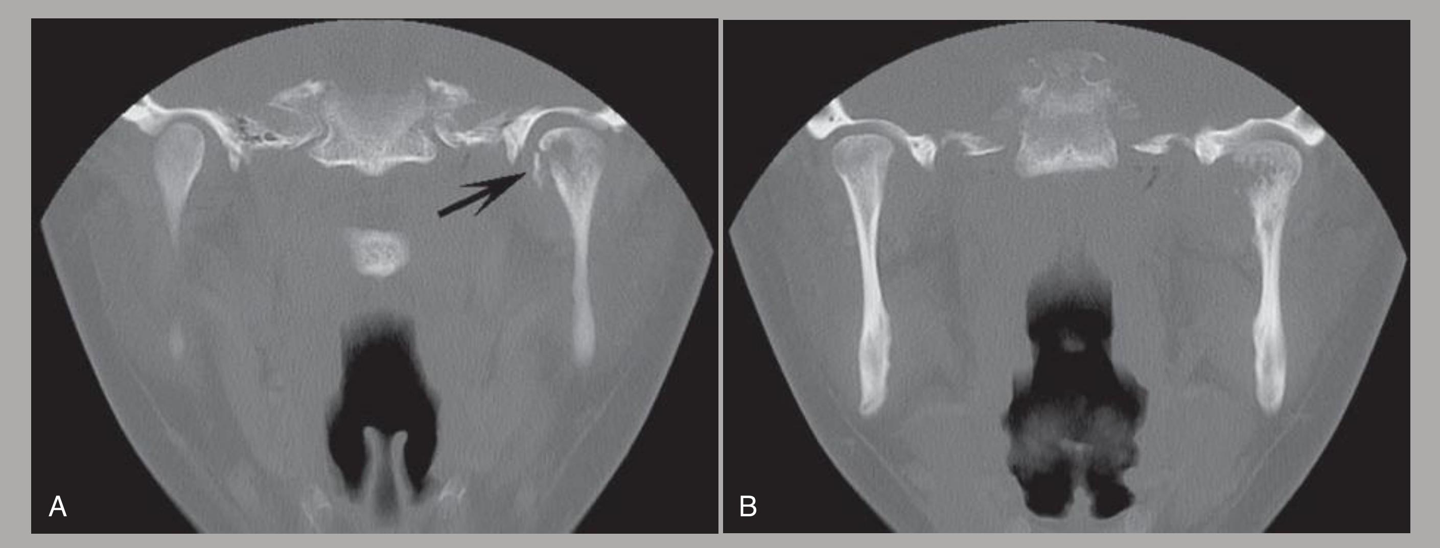
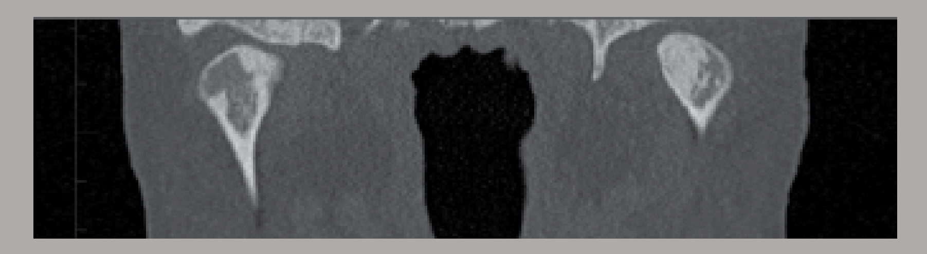
FIG. 23-12 A and B, Coronal CT images with bone algorithm demonstrating Ewing’s sarcoma involving the left mandibular condyle; note the irregular margins, destruction of the medial cortex of the condyle, and a small pathologic fracture (arrow).
Lecturer — Ewing Sarcoma
Ewing’s sarcoma was described as the second most common malignant primary bone tumor of childhood after osteosarcoma. It is closely related to Askin tumor and neuroepithelioma, which are collectively referred to as the Ewing sarcoma family of tumors.
- Imaging may show predominantly lytic lesions, ill-defined margins, and variable periosteal reactions.
- Misdiagnosis is common because it can mimic osteomyelitis and other malignant non-sarcoma tumors.
2
LEFT
 |  |
Fibrosarcoma17
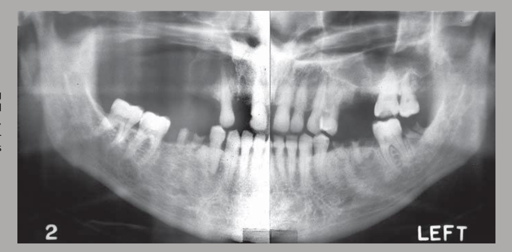
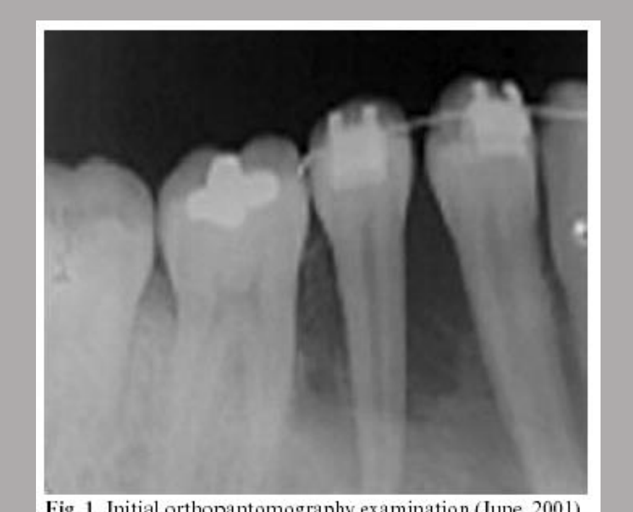
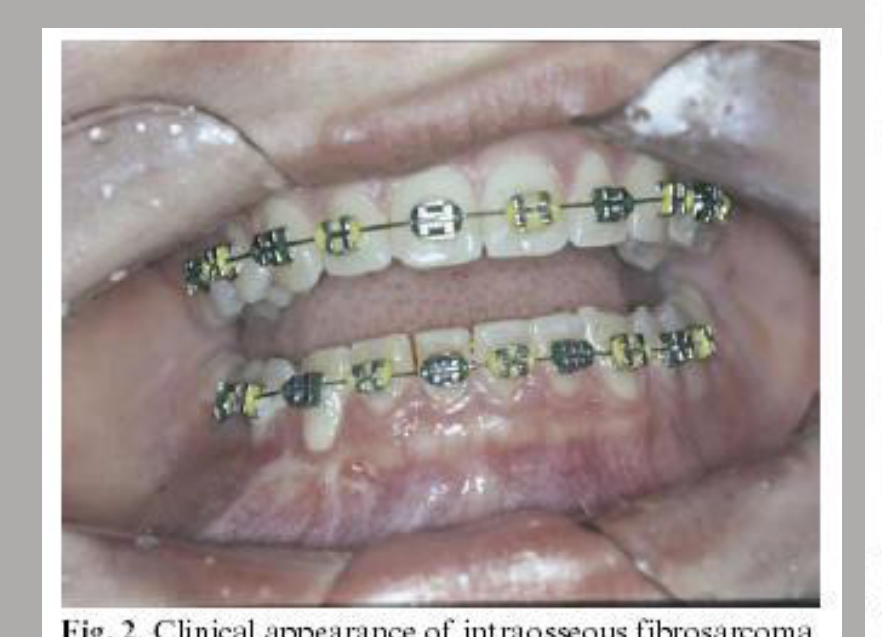
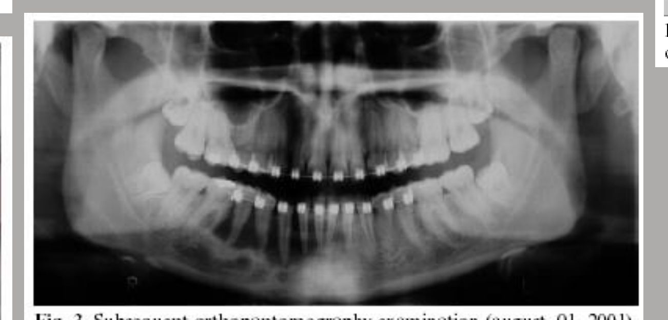
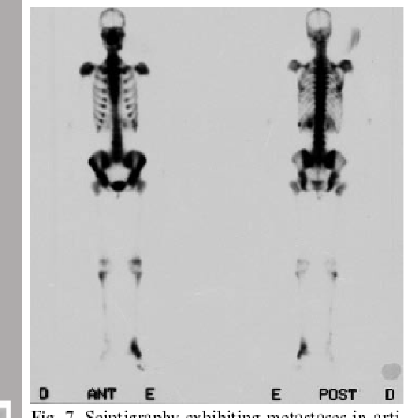
High-grade Fibrosarcoma of the MD with Mets
-
Fibrosarcoma of the jaws was described as very rare Fig. 7. Scintigraphy exhibiting metastases in articulations.
-
Hematopoietic malignancies arise from blood-forming tissues and may present differently from other solid tumors.
-
Multiple destructive areas may be present.
-
Periosteal changes may occur.
-
Normal trabecular pattern may be lost.
-
Cortical destruction may occur.
-
The mandibular canal may be destroyed or lost.
-
Teeth may be displaced.
-
Follicular spaces around unerupted teeth may be destroyed.
-
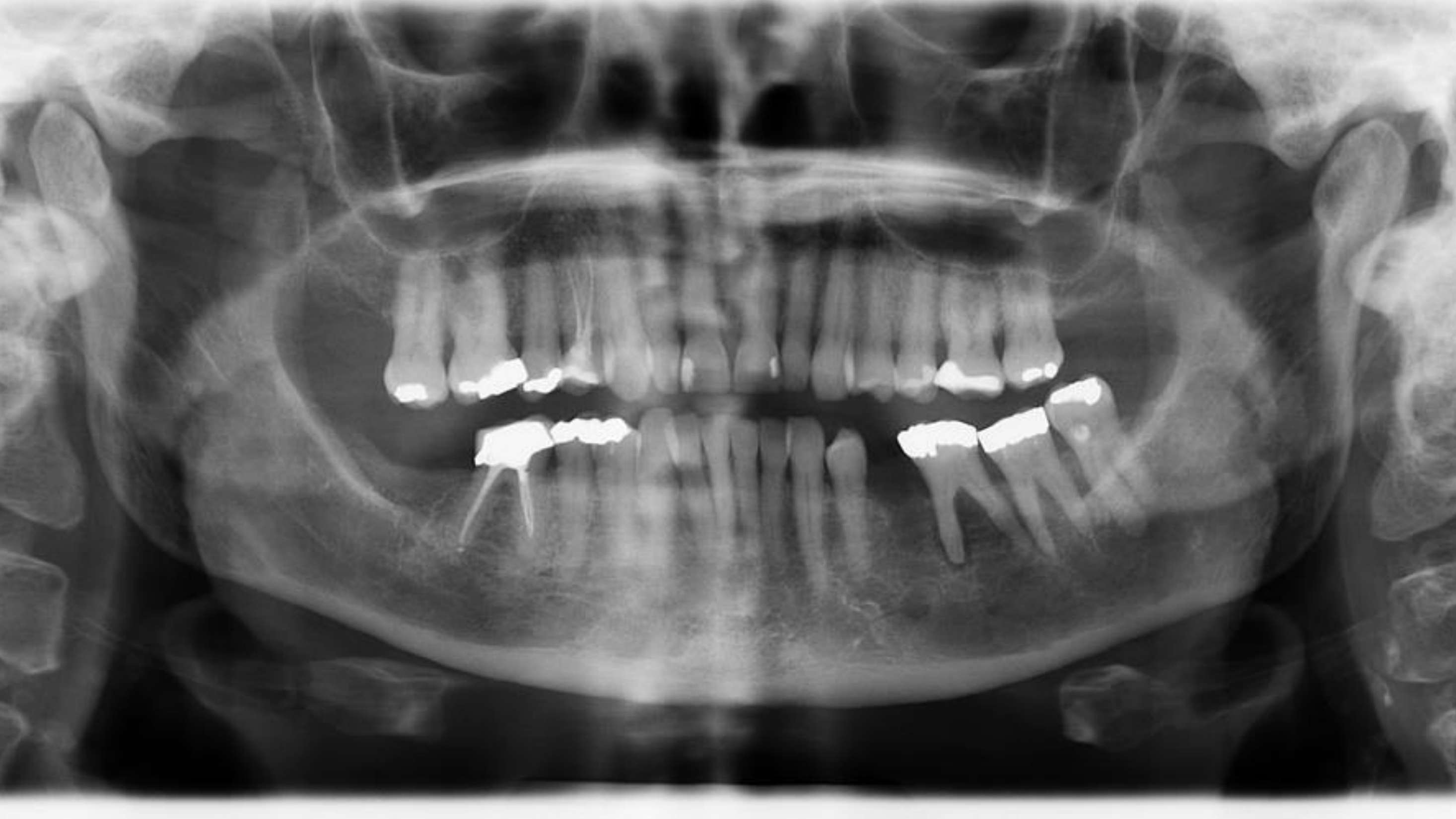
Sclerotic metastases may resemble bone islands, but periosteal change is not expected with bone islands.
-
Bone islands involving the condyles are rare.
-
Multiple sclerotic lesions in unusual locations should prompt assessment of other skeletal regions.
-
Metastasis should be considered particularly when there is a history of breast or prostate cancer.
-
The mandibular body is the most common site of involvement.
-
Multiple lesions involving the condyles, rami, and cervical spine are suspicious for malignancy.
 |  |
 |  |
Hematopoietic Malignancies18
- Multiple Myeloma
- Most common adult primary malignant bone cancer
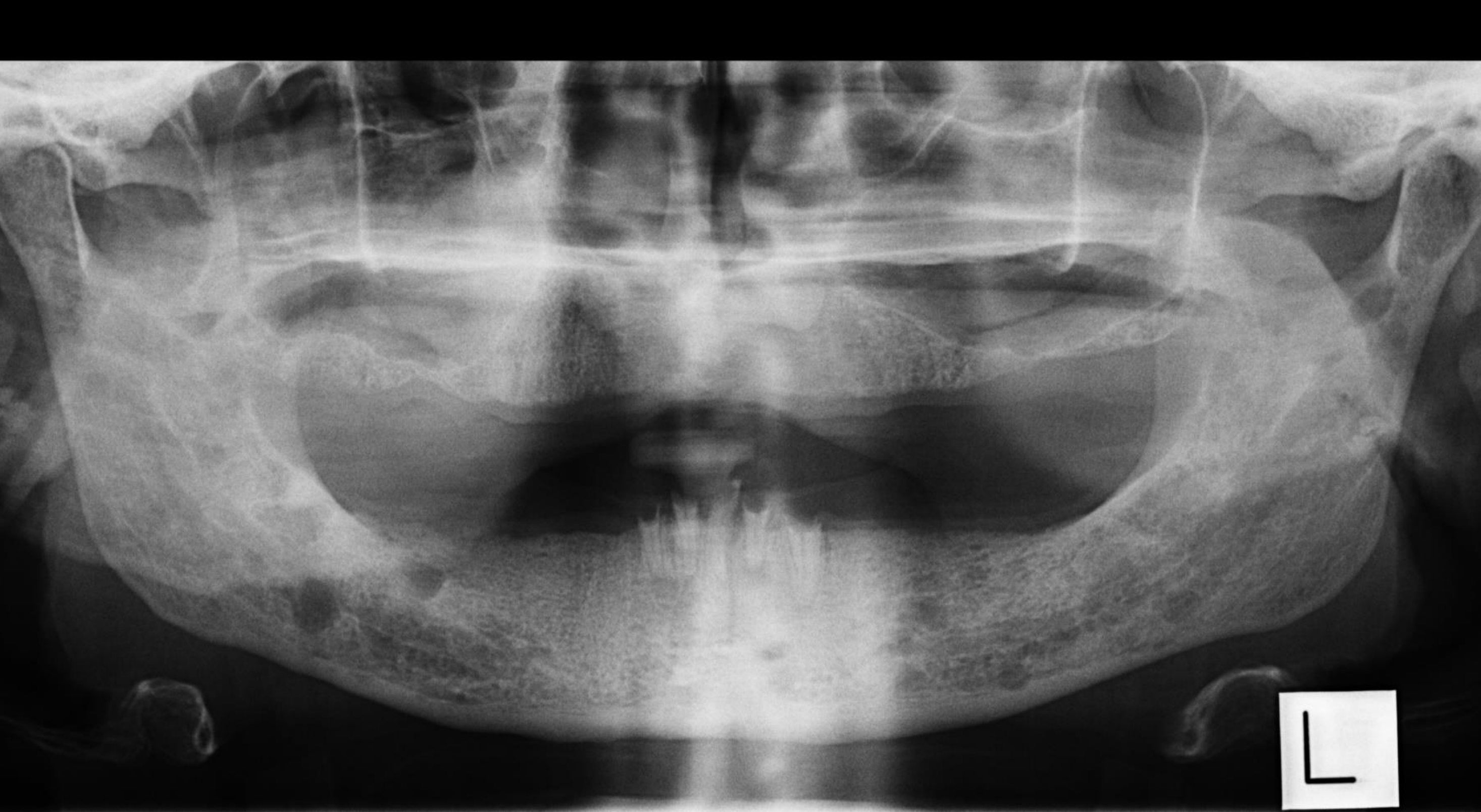
- Classic radiographic feature: multiple ‘punched-out’ lytic lesions
- Lymphoma
- Non-Hodgkin (most lymphomas involving the jaws) >>>> Hodgkin
- Lytic, destructive & invasive/infiltrative
- Leukemia
- ALL (most common leukemia in children), AML, CLL, CML
Atlas of Oral and Maxillofacial Radiology — Bernard Koong — Wiley Blackwell
Metastatic Lesions In The Jaws19
- Metastatic tumours represent the establishment of new malignant disease from a distant malignant tumour.
- Common primary sites include the breast, kidney, lung, prostate, thyroid, stomach.
Lecturer — Metastatic Spread
Metastatic lesions usually arise from a distant primary tumor through the blood vessels.
- Jaw metastases commonly arise from tumors located anatomically inferior to the clavicle.
- Paresthesia is an important clinical feature because of nerve involvement.
- Metastatic lesions may involve multiple regions because they travel through the body.
 |  |
Radiographic Features2021222324252627
- Sclerotic (breast & prostate mets) or lytic (renal & thyroid mets)
- Multiple foci
- Most are ill-defined
Audio Appendix
Additional Audio Content
The following sections from the lecture audio did not correspond to any heading in the main document.
Case Study: Young Patient With Aggressive Mandibular Changes
- The patient’s young age made leukemia a likely consideration.
- There was irregular periodontal ligament space widening around the 36.
- The lamina dura was lost.
- The left mandibular canal was not visible, although it could be seen on the opposite side.
- The findings were highly suspicious for malignancy rather than localized periodontal disease.
- Urgent referral was recommended.
- Acute jaw infections such as osteomyelitis can resemble aggressive lesions such as leukemia.
- Infection requires bacterial invasion and an identifiable cause, whereas leukemia and lymphoma represent tumor-related processes.
- Further assessment in a maxillofacial or children’s hospital setting was required.
Multiple Myeloma
Multiple myeloma was described as the most common adult primary malignant bone cancer.
- Its classic imaging appearance is multiple punched-out lytic lesions.
- A case showing multiple punched-out lesions was considered very typical of multiple myeloma.
Case Study: High-Grade Fibrosarcoma of the Mandible
- The patient was a 16-year-old girl.
- She had orthodontic braces.
- The lesion was lytic and involved the mandible.
- An initial PA image showed an ill-defined lesion in the 4–5 region.
- Swelling of the jaw was noted clinically.
- An OPG taken two months later demonstrated extensive progression.
- The rapid progression was consistent with the high-grade nature of the lesion.
- No CT image was available from the case report.
- The patient underwent total mandibulectomy, with removal of the whole mandible.
- A bone scan one year later demonstrated extensive spread throughout the body.
- Radiation therapy and chemotherapy were administered.
- The patient ultimately did not survive.
- The case illustrated how rapidly a small, high-grade fibrosarcoma of the jaw can progress.
Case Study: Lesion With Displacement of the 38
- There was a change in density in the left mandible with displacement of the 38.
- The displacement was not early eruption; it was displacement by the tumor.
- The follicular space was completely destroyed.
- The 37 had lost its lamina dura.
- The mandibular canal was no longer visible.
- These were considered typical malignant features.
Case Study: Lymphoma of the Right Maxilla
- An OPG was initially non-diagnostic.
- A cropped OPG obtained at another time showed unusual periodontal ligament space widening around the right maxillary teeth.
- The sinus floor was not clearly visible.
- Later CT demonstrated:
- Irregular periodontal ligament space widening.
- Lamina dura destruction.
- Buccal cortical destruction.
- Sinus floor destruction.
- The appearance was very similar to SCC.
- Imaging alone could not reliably distinguish between SCC and lymphoma in this case.
Case Study: Localized Lesion Suggestive of SCC
- The lesion appeared more localized and possibly related to the alveolar crest or gingiva.
- Its appearance was considered more suggestive of SCC.
- Further testing was still required urgently to establish the tumor type and guide management.
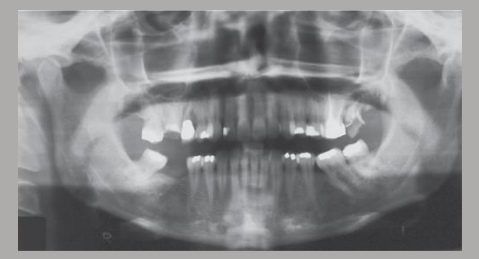
Case Study: Incidental Malignant-Appearing Lesion of the Left Mandible
- The OPG was obtained for treatment planning.
- It incidentally demonstrated:
- An altered trabecular pattern in the left mandible.
- Lytic change involving the inferior border.
- Destruction of the mandibular canal.
- Loss of cortical structures.
- Diffuse change without clear margins.
- The appearance was considered typical of malignancy.
- The specific tumor type could not be determined from the OPG alone. Possibilities included:
- SCC.
- Lymphoma.
- Metastasis.
- Further imaging and testing were required.
Common Clinical and Imaging Features
- Ill-defined margins are a key feature because malignant lesions tend to be invasive or infiltrative and do not respect anatomical boundaries.
- Other important findings include:
- Lytic, destructive, or permeative bone changes.
- Cortical destruction.
- Soft tissue extension.
- Unexplained tooth mobility.
- Loss or destruction of the lamina dura.
- Irregular widening of periodontal ligament spaces.
- Loss or irregular widening of the mandibular canal.
- Lesions may extend through periodontal ligament spaces.
- When unerupted teeth are involved, they may be displaced superiorly. Apparent early eruption in this context represents pathological displacement rather than normal eruption.
- Periosteal involvement can produce different patterns, including:
- Lamellated periosteal change.
- Sun-ray or sunburst periosteal change.
- Extensive dentoalveolar lesions may cause the teeth to appear to be “floating.”
Jaw malignancies were broadly classified into four groups:
- Carcinomas.
- Sarcomas.
- Hematopoietic malignancies.
- Metastatic lesions.
Imaging is important for:
- Recognizing malignant features.
- Assessing the extent of spread.
- Determining bone and soft tissue involvement.
- Planning treatment.
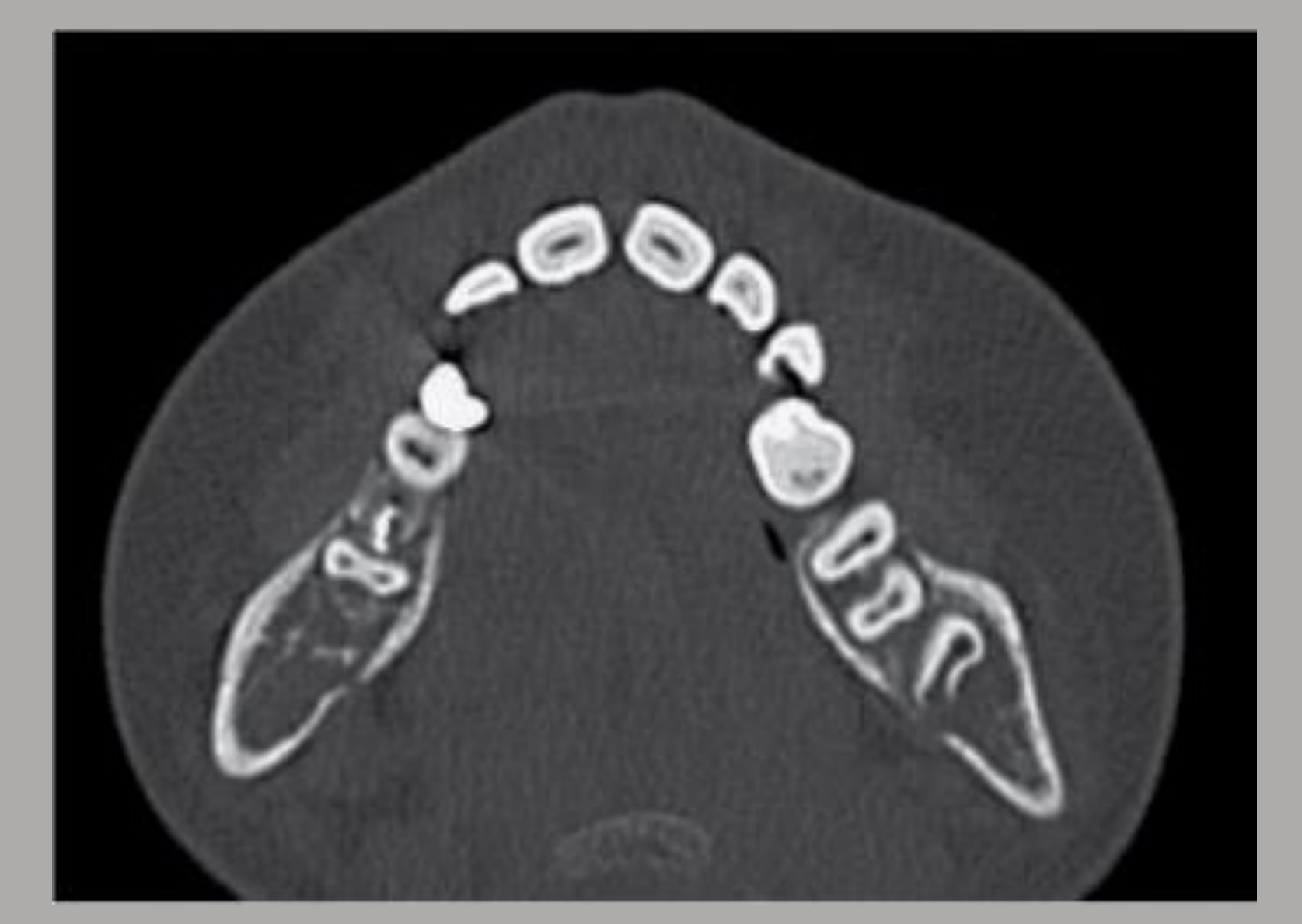
Case Study: Multiple Sclerotic Lesions
- The lesions were sclerotic and could resemble bone islands.
- The presence of periosteal changes favored metastatic disease rather than bone islands.
- In a male patient, the appearance would be more suggestive of prostate metastasis.
- In a female patient, it would be more suggestive of breast metastasis.
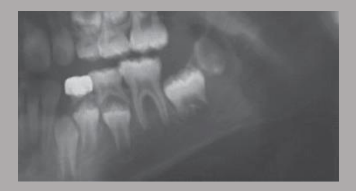
Case Study: Leukemia Involving the Left Mandible
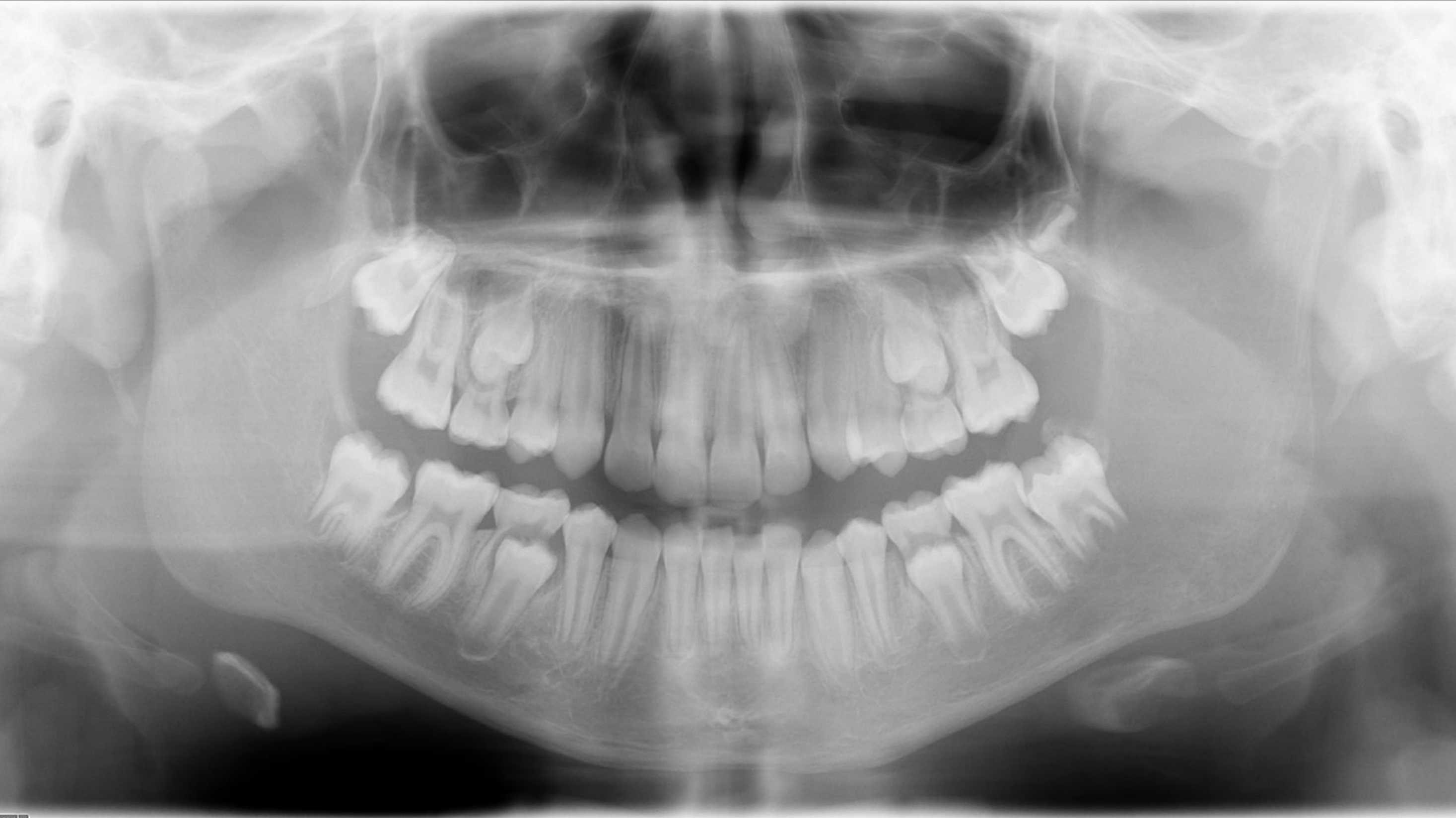
- The patient was young and had mixed dentition.
- The lesion was lytic and invasive.
- There was lamina dura destruction.
- Follicular spaces around unerupted teeth, including the 38, were destroyed or no longer had visible borders.
- The lucency was relatively subtle on the OPG because bone density is already low in children.
- Destructive changes were therefore more useful than changes in density.
Important findings to assess in children include:
- Periodontal ligament space widening.
- Lamina dura destruction.
- Loss of cortical structures.
- Displacement of unerupted teeth.
Aggressive jaw lesions in a child require urgent referral for further assessment.
Case Study: Osteosarcoma With Periosteal Change
- The lesion was an expansile mixed-density lesion.
- It demonstrated sun-ray periosteal change.
- The periosteal reaction was considered characteristic of osteosarcoma.
Case Study: Multiple Punched-Out Lesions
- The final OPG demonstrated multiple punched-out lesions.
- Although renal cancer metastasis or another malignancy could produce a similar pattern, the appearance was considered classic for multiple myeloma.
Early recognition and appropriate imaging are essential in jaw malignancies. Understanding radiographic patterns supports timely referral and management. Ill-defined lesions should be treated cautiously because missing a malignancy is considered more significant than over-investigating a benign lesion.
Case Study: Primary Intraosseous Carcinoma and Carcinoma Arising From a Dentigerous Cyst
- Primary intraosseous carcinoma was described as very rare.
- Carcinoma arising from a dentigerous cyst was also described as very rare.
- Both lesions demonstrated:
- Reduced density.
- Irregular margins.
- Destructive behavior.
- The intraosseous carcinoma appeared slightly more well defined.
- The other lesion had poorly visible margins and loss of part of the mandibular canal.
- Loss of the mandibular canal was emphasized as an important imaging feature of malignant mandibular lesions.
Leukemia and Lymphoma
Leukemia and lymphoma are blood cancers involving lymphocytes, but they differ in origin:
- Leukemia starts in the bone marrow and spreads through the blood and marrow.
- Lymphoma begins in lymph nodes.
- Non-Hodgkin lymphoma is more likely to involve the jaws than Hodgkin lymphoma.
- When lymphoma affects the jaws, it may present as an extranodal lesion with bony destruction.
- Hodgkin lymphoma rarely involves the jaws and more typically presents in the neck lymph nodes.
Jaw lymphoma may cause:
- Rapid growth.
- Swelling.
- Pain.
- Loosened teeth.
- Lytic, destructive, and invasive changes.
Leukemia involving the jaws is more commonly reported in acute leukemia, particularly acute lymphoblastic leukemia. Possible symptoms include:
- Pain.
- Swelling.
- Numbness.
- Tooth mobility.
The imaging appearance of leukemia may be similar to lymphoma and may consist of nonspecific aggressive malignant features.
Case Study: Subtle Osteosarcoma on OPG
- The lesion was relatively subtle on the OPG.
- Important warning features included:
- A non-healing socket.
- Destruction of adjacent cortical structures.
- Lamina dura destruction.
- Irregular periodontal ligament space widening.
- Increased density rather than the decreased density expected in SCC.
- Loss of the mandibular canal.
- The combination of these findings was considered more consistent with osteosarcoma.
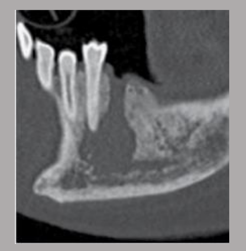
Case Study: SCC Involving a Non-Healing Left Mandibular Socket
- The lesion involved a non-healing left mandibular socket.
- It appeared more well defined than the maxillary lesion because of surrounding sclerosis.
- Anteriorly and inferiorly, the margins were invasive and ill defined.
- There was destruction of:
- The lamina dura of a premolar.
- The cortical borders of the mandibular canal.
- Irregular periodontal ligament space widening was also present.
Case Study: SCC Involving the Left Maxilla
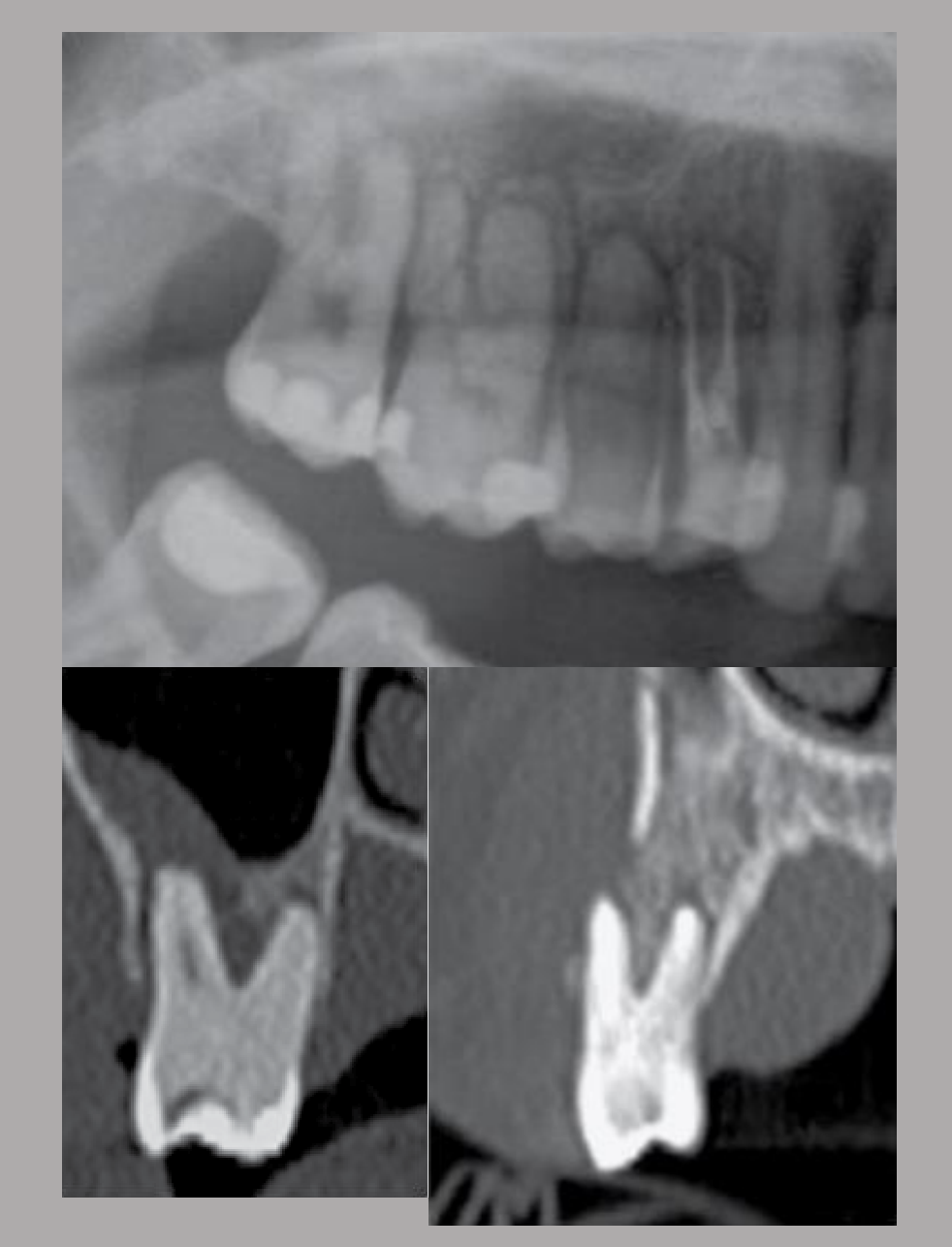
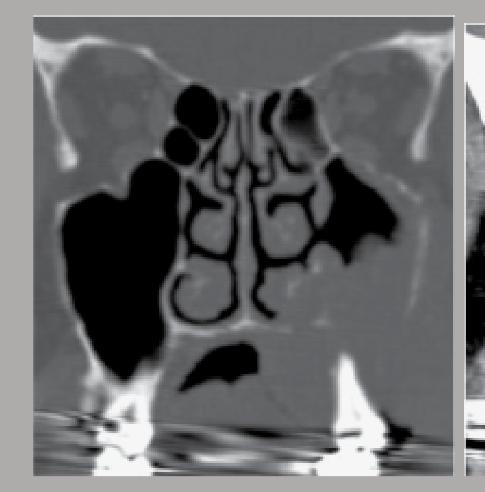
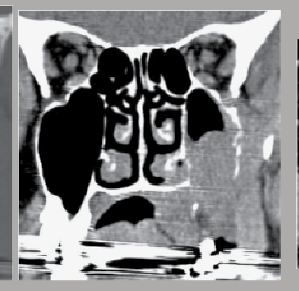
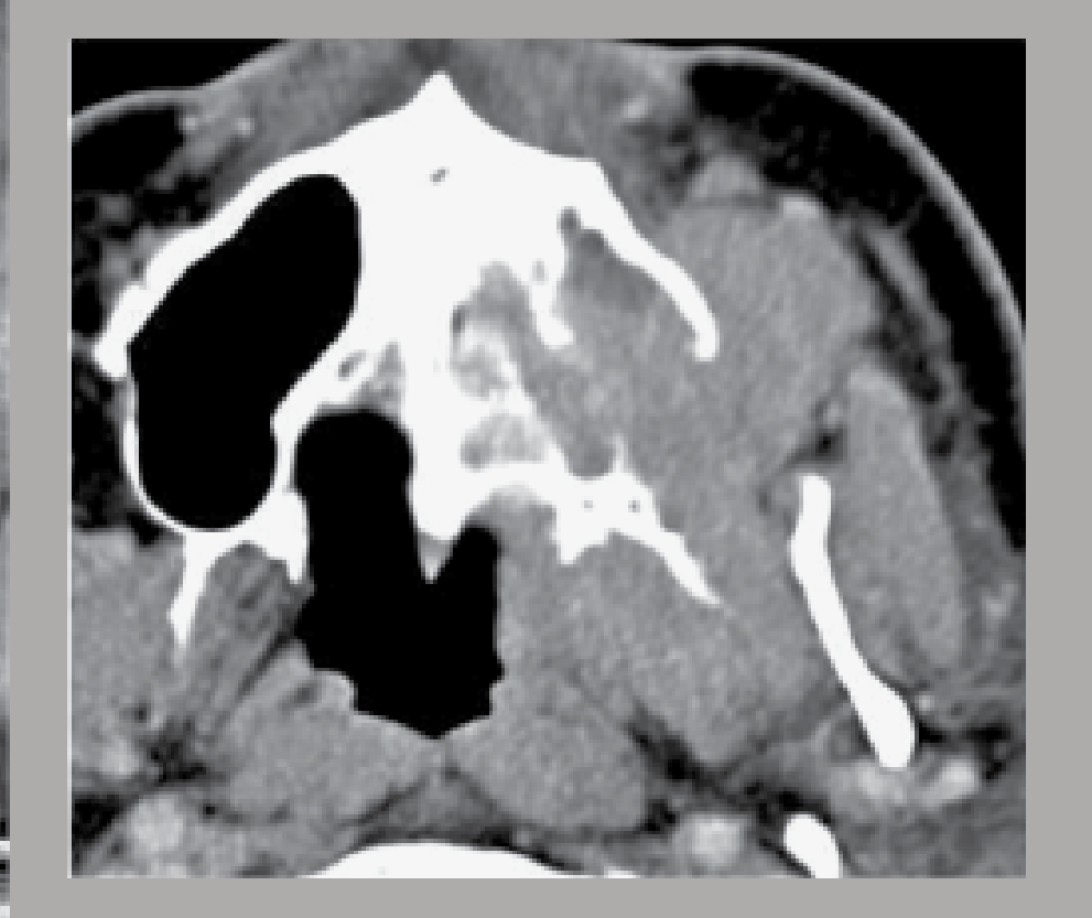
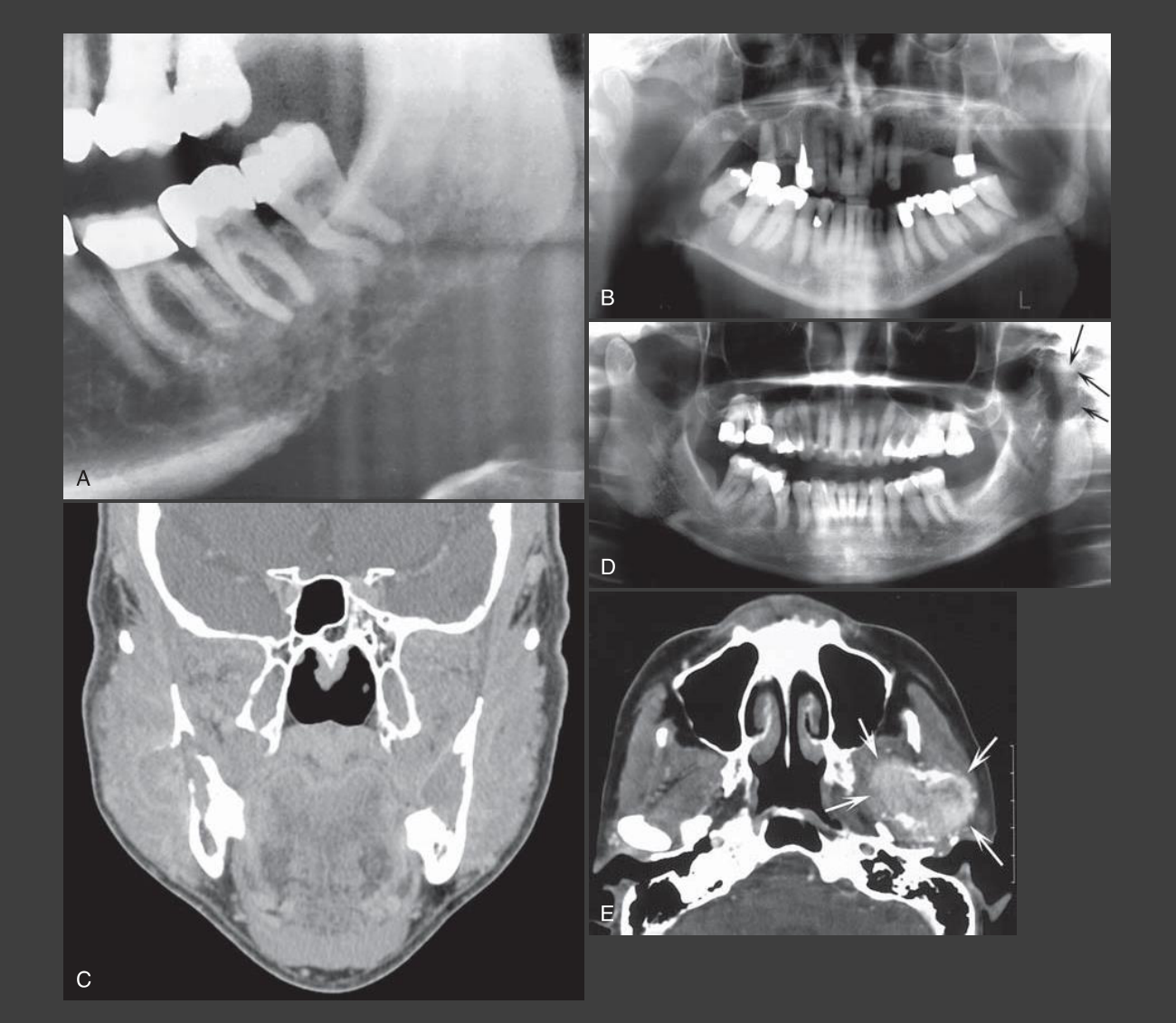
- CT demonstrated a large soft tissue mass involving the left maxilla.
- There was extensive destruction of the left maxillary bone.
- The associated teeth appeared to be floating.
- The lesion had ill-defined margins and was markedly erosive.
- There was extensive soft tissue replacement in an area where fat would normally be visible.

Clinical Cases
Case: Rare carcinomas of the jaws
Question
Scenario: Examples of rare carcinomas of the jaws, including primary intraosseous carcinoma and carcinoma arising from a dentigerous cyst.
What’s shown: Images showing decreased density with irregular margins. One lesion is slightly more well-defined, while the other has no visible margins and shows loss of the mandibular canal.
Consider: Identify the imaging features of these rare carcinomas.
Answer
Observations:
- Decreased density with irregular margins.
- One lesion is slightly more well-defined (intraosseous type).
- The other lesion has no visible margins and loss of the mandibular canal.
Reasoning: Primary intraosseous carcinoma and carcinoma arising from a dentigerous cyst are rare but share typical malignant imaging features. The lack of visible margins and the loss of anatomical boundaries, such as the mandibular canal, are key indicators of their aggressive, malignant behavior.
Takeaway: Rare carcinomas of the jaws exhibit typical malignant features such as ill-defined margins and the destruction of anatomical structures like the mandibular canal.
Case: SCC involving the left maxilla
Question
Scenario: A CT scan of a patient with squamous cell carcinoma (SCC) involving the left maxilla.
What’s shown: A large soft tissue mass with extensive destruction of the left maxilla and floating teeth. The margins are ill-defined and highly erosive, with soft tissue present where normal fat should be.
Consider: Identify the malignant features of this SCC.
Answer
Observations:
- Large soft tissue mass.
- Extensive destruction of the left maxilla.
- Floating teeth.
- Ill-defined and highly erosive margins.
- Soft tissue replacing normal fat spaces.
Reasoning: The lesion demonstrates classic malignant features, including ill-defined margins, extensive bony destruction, floating teeth, and soft tissue extension that replaces normal anatomical fat.
Takeaway: SCC of the maxilla can present with extensive bony destruction, floating teeth, and a soft tissue mass replacing normal fat spaces.
Case: SCC of a non-healing mandibular socket
Question
Scenario: A case of SCC involving a non-healing left mandibular socket.
What’s shown: A lesion with surrounding sclerosis making it appear more well-defined, but with invasive, ill-defined margins anteriorly and inferiorly. There is destruction of the lamina dura of a premolar and the cortical borders of the mandibular canal, along with irregular PDL space widening.
Consider: Identify the aggressive features of this SCC despite the surrounding sclerosis.
Answer
Observations:
- Surrounding sclerosis.
- Invasive and ill-defined margins anteriorly and inferiorly.
- Destruction of the lamina dura of a premolar.
- Destruction of the cortical borders of the mandibular canal.
- Irregular PDL space widening.
Reasoning: Although surrounding sclerosis can make a lesion appear more well-defined, the anterior and inferior margins remain invasive and ill-defined. The destruction of the lamina dura, mandibular canal borders, and irregular PDL widening are critical red flags for malignancy.
Takeaway: Surrounding sclerosis can mask the ill-defined margins of an SCC, but localized cortical destruction and irregular PDL widening remain critical indicators of malignancy.
Case: Osteosarcoma with sun-ray periosteal change
Question
Scenario: Examples of osteosarcomas of the jaws.
What’s shown: Expansile mixed-density lesions, with one demonstrating a sun-ray periosteal reaction.
Consider: Identify the characteristic imaging features of osteosarcoma.
Answer
Observations:
- Expansile mixed-density lesions.
- Sun-ray periosteal change.
Reasoning: Osteosarcomas are characterized by new bone formation giving a mixed, cloud-like density, expansile and destructive behavior with ill-defined borders, and classic periosteal reactions like the sun-ray or sunburst pattern.
Takeaway: Osteosarcomas of the jaws present as expansile mixed-density lesions with classic periosteal reactions such as a sun-ray or sunburst pattern.
Case: Subtle osteosarcoma on OPG
Question
Scenario: An osteosarcoma lesion that appears subtle on an OPG.
What’s shown: A non-healing socket with destruction of adjacent cortical structures (lamina dura), irregular PDL space widening, increased density (rather than decreased density), and loss of the mandibular canal.
Consider: Identify the red flags for osteosarcoma in this subtle presentation.
Answer
Observations:
- Non-healing socket.
- Destruction of adjacent cortical structures and lamina dura.
- Irregular PDL space widening.
- Increased density.
- Loss of the mandibular canal.
Reasoning: While the lesion is subtle on the OPG, the combination of a non-healing socket, cortical destruction, irregular PDL widening, and specifically increased density (unlike the lucency seen in SCC) along with loss of the mandibular canal points to osteosarcoma.
Takeaway: Osteosarcoma can present subtly on an OPG, but increased density combined with cortical destruction, irregular PDL widening, and loss of the mandibular canal are key diagnostic red flags.
Case: Chondrosarcoma of the TMJ
Question
Scenario: A rare case of chondrosarcoma involving the temporomandibular joint (TMJ).
What’s shown: Predominantly lytic lesions with rings and arcs calcification or popcorn calcification around the borders.
Consider: Identify the distinguishing calcification patterns of chondrosarcoma.
Answer
Observations:
- Predominantly lytic lesions.
- Rings and arcs calcification.
- Popcorn calcification around the borders.
Reasoning: Unlike osteosarcomas which show new bone formation, chondrosarcomas involve cartilaginous tissue and are predominantly lytic. The presence of rings and arcs or popcorn calcifications around the borders is characteristic of cartilaginous tumors.
Takeaway: Chondrosarcomas of the TMJ are predominantly lytic and characteristically display rings and arcs or popcorn calcifications.
Case: High-grade fibrosarcoma in a pediatric patient
Question
Scenario: A 16-year-old girl with ortho braces presenting with jaw swelling and a high-grade fibrosarcoma of the mandible.
What’s shown: Initial PA showing a lytic lesion with ill-defined margins in the 4, 5 region. A follow-up OPG two months later shows extensive, rapid progression of the lesion.
Consider: Evaluate the progression and outcome of this high-grade malignancy.
Answer
Observations:
- Lytic lesion with ill-defined margins in the 4, 5 region on initial PA.
- Extensive and rapid progression on OPG two months later.
Reasoning: The rapid and extensive progression over just two months aligns with the high-grade, aggressive nature of fibrosarcoma.
Outcome: The patient received a total mandibulectomy. A bone scan a year later showed extensive metastasis throughout the body. Despite radiation and chemotherapy, the patient did not survive.
Takeaway: High-grade fibrosarcomas of the jaw are extremely aggressive, rapidly progressive, and carry a poor prognosis with a high risk of extensive metastasis and mortality.
Case: Lymphoma of the right maxilla
Question
Scenario: A case of lymphoma involving the right maxilla.
What’s shown: An initial non-diagnostic OPG. A cropped OPG shows unusual PDL space widening of the right maxillary teeth. A subsequent CT shows irregular PDL space widening, destruction of the lamina dura, buccal cortex, and sinus floor.
Consider: Differentiate the imaging features of lymphoma from other malignancies like SCC.
Answer
Observations:
- Unusual PDL space widening.
- Destruction of the lamina dura.
- Destruction of the buccal cortex and sinus floor.
Reasoning: The imaging features of lymphoma, including irregular PDL widening and destruction of cortical structures like the lamina dura and sinus floor, are very similar to SCC, making it difficult to distinguish between the two based on imaging alone.
Takeaway: Lymphoma of the jaw can mimic SCC on imaging, presenting with irregular PDL widening and destruction of cortical structures like the lamina dura and sinus floor.
Case: Leukemia in a mixed dentition patient
Question
Scenario: A young patient with mixed dentition presenting with leukemia involving the left mandible.
What’s shown: A lytic and invasive lesion with lamina dura destruction and loss of follicular space borders of unerupted teeth (38). The lucency is subtle due to the naturally low bone density in children.
Consider: Identify the subtle signs of leukemia in a pediatric jaw radiograph.
Answer
Observations:
- Lytic and invasive lesion.
- Lamina dura destruction.
- Destruction and loss of follicular space borders of unerupted teeth.
- Subtle lucency.
Reasoning: In children, bone density is naturally low, making lucency subtle. Therefore, destructive changes such as PDL space widening, lamina dura destruction, loss of cortical structures, and displacement or follicular space destruction of unerupted teeth are more reliable indicators of leukemia.
Takeaway: In pediatric patients, leukemia may present with subtle lucency; clinicians should instead look for destructive changes like lamina dura loss, cortical destruction, and disruption of unerupted tooth follicular spaces.
Case: Sclerotic metastasis with periosteal changes
Question
Scenario: A case of sclerotic metastasis in the jaws.
What’s shown: Sclerotic lesions that resemble bone islands but are located in unusual spots like the condyles, accompanied by periosteal changes.
Consider: Differentiate sclerotic metastases from benign bone islands.
Answer
Observations:
- Sclerotic lesions resembling bone islands.
- Located in unusual areas like the condyles.
- Visible periosteal changes.
Reasoning: Sclerotic metastases (often from breast or prostate primary tumors) can look like bone islands. However, bone islands in the condyles are rare, and the presence of periosteal changes is a red flag that indicates malignancy rather than a benign bone island.
Takeaway: Sclerotic metastases can mimic bone islands, but their presence in unusual locations like the condyles and the presence of periosteal changes are key indicators of malignancy.
Case: Incidental suspected leukemia in a young patient
Question
Scenario: An incidental finding on an OPG taken for treatment planning in a young patient.
What’s shown: Altered trabecular pattern of the left mandible, lytic change of the inferior border, destruction of the mandibular canal, hyperdensity with displacement of tooth 38, loss of follicular space, and loss of lamina dura and mandibular canal around tooth 37.
Consider: Identify the malignant features and likely diagnosis based on the patient’s age.
Answer
Observations:
- Altered trabecular pattern.
- Lytic change of the inferior border.
- Destruction of the mandibular canal.
- Hyperdensity displacing tooth 38.
- Loss of follicular space.
- Loss of lamina dura around tooth 37.
Reasoning: The diffuse loss of cortical structures, trabecular pattern, and mandibular canal borders are typical of malignancy. Given the young age of the patient, these aggressive, destructive features are most likely indicative of leukemia rather than an infection or other solid tumor.
Takeaway: Diffuse loss of cortical and canal borders with altered trabecular patterns in a young patient is highly suspicious for leukemia and requires urgent referral.
Case: Suspected SCC involving the alveolar crest
Question
Scenario: A case presenting with localized malignant features in the mandible.
What’s shown: Irregular widening of the PDL space of tooth 36, loss of lamina dura, and loss of visibility of the left mandibular canal.
Consider: Determine the likely origin and type of this localized malignancy.
Answer
Observations:
- Irregular PDL space widening of tooth 36.
- Loss of lamina dura.
- Loss of the left mandibular canal.
Reasoning: The localized nature of the lesion, likely originating from the alveolar crest or gingiva, combined with irregular PDL widening and loss of the mandibular canal, is highly suspicious for a localized malignancy such as SCC.
Takeaway: Localized irregular PDL widening and loss of the mandibular canal originating from the alveolar crest are highly suspicious for SCC and require urgent referral.
Case: Multiple myeloma
Question
Scenario: An OPG showing classic features of multiple myeloma.
What’s shown: Multiple punched-out lytic lesions distributed throughout the jaws.
Consider: Identify the classic radiographic presentation of multiple myeloma.
Answer
Observations:
- Multiple punched-out lytic lesions.
Reasoning: The presence of multiple, distinct punched-out lytic lesions is the classic and highly characteristic imaging appearance of multiple myeloma in the jaws.
Takeaway: Multiple punched-out lytic lesions on a jaw radiograph are a classic and highly characteristic sign of multiple myeloma.
Footnotes
-
Original PDF page 1: L6 - Imaging for Jaw Malignancies, p.1 ↩
-
Original PDF page 18: L6 - Imaging for Jaw Malignancies, p.18 ↩
-
Original PDF page 2: L6 - Imaging for Jaw Malignancies, p.2 ↩
-
Original PDF page 3: L6 - Imaging for Jaw Malignancies, p.3 ↩
-
Original PDF page 5: L6 - Imaging for Jaw Malignancies, p.5 ↩
-
Original PDF page 4: L6 - Imaging for Jaw Malignancies, p.4 ↩
-
Original PDF page 6: L6 - Imaging for Jaw Malignancies, p.6 ↩
-
Original PDF page 7: L6 - Imaging for Jaw Malignancies, p.7 ↩
-
Original PDF page 8: L6 - Imaging for Jaw Malignancies, p.8 ↩
-
Original PDF page 9: L6 - Imaging for Jaw Malignancies, p.9 ↩
-
Original PDF page 10: L6 - Imaging for Jaw Malignancies, p.10 ↩
-
Original PDF page 12: L6 - Imaging for Jaw Malignancies, p.12 ↩
-
Original PDF page 11: L6 - Imaging for Jaw Malignancies, p.11 ↩
-
Original PDF page 13: L6 - Imaging for Jaw Malignancies, p.13 ↩
-
Original PDF page 14: L6 - Imaging for Jaw Malignancies, p.14 ↩
-
Original PDF page 15: L6 - Imaging for Jaw Malignancies, p.15 ↩
-
Original PDF page 16: L6 - Imaging for Jaw Malignancies, p.16 ↩
-
Original PDF page 17: L6 - Imaging for Jaw Malignancies, p.17 ↩
-
Original PDF page 19: L6 - Imaging for Jaw Malignancies, p.19 ↩
-
Original PDF page 20: L6 - Imaging for Jaw Malignancies, p.20 ↩
-
Original PDF page 21: L6 - Imaging for Jaw Malignancies, p.21 ↩
-
Original PDF page 22: L6 - Imaging for Jaw Malignancies, p.22 ↩
-
Original PDF page 23: L6 - Imaging for Jaw Malignancies, p.23 ↩
-
Original PDF page 24: L6 - Imaging for Jaw Malignancies, p.24 ↩
-
Original PDF page 25: L6 - Imaging for Jaw Malignancies, p.25 ↩
-
Original PDF page 26: L6 - Imaging for Jaw Malignancies, p.26 ↩
-
Original PDF page 27: L6 - Imaging for Jaw Malignancies, p.27 ↩