Why Do We Do Orthodontics?12
Contemporary Orthodontics
Chapter 1: Malocclusion and Dentofacial Deformity in Contemporary Society
From the UNC Undergraduate training course
Lecturer — Epidemiologic Context
The United States provides the epidemiologic statistics because comparable Australian data are not available.
- Slides marked with an asterisk have been associated with test questions.
- Prevalence / Treatment Need
Epidemiologic Data For Malocclusion
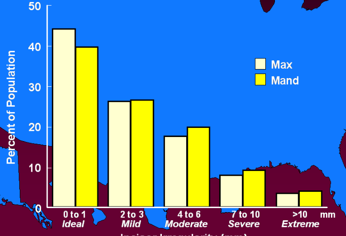
Class I Crowding3
- How many people (%) have well-aligned teeth?
- How many (%) have severe crowding?
- What does this graph tell us?
Lecturer — Crowding and Treatment
Class I crowding is the most common orthodontic problem encountered clinically, but severe and extreme crowding affect a much smaller proportion of the population.
- Approximately 30% of people have mild incisor irregularity in both the maxilla and mandible.
- Approximately 15% of the population is sufficiently crowded that extractions are probably required for treatment.
- Treating severe or extreme crowding without extractions may result in significant relapse, periodontal problems, bone loss, or dehiscence.
- Irregularity alone does not establish a need for treatment; crowding must be assessed with the other diagnostic findings before choosing extraction or non-extraction treatment.
Mandibular Incisor Irregularity (mm) — Percent of Population4
| Category | 0 to 1 mm (Ideal) | 2 to 3 mm (Mild) | 4 to 6 mm (Moderate) | 7 to 10 mm (Severe) | > 10 mm (Extreme) |
|---|---|---|---|---|---|
| Males | ~45% | ~25% | ~18% | ~8% | ~4% |
| Females | ~40% | ~25% | ~20% | ~10% | ~5% |
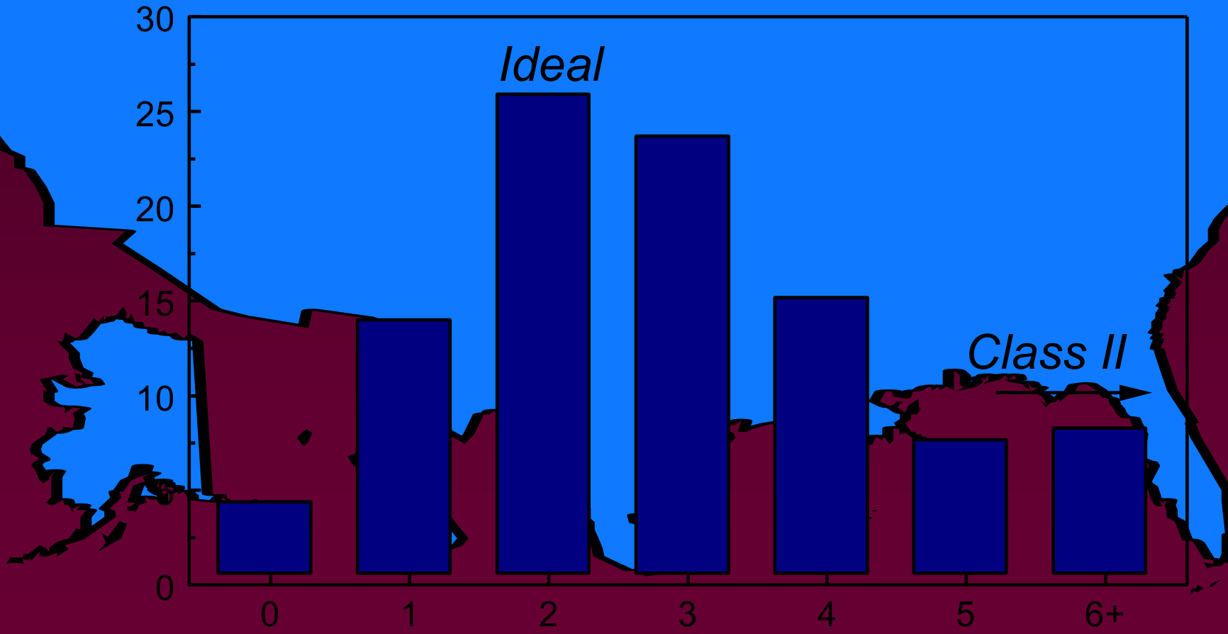
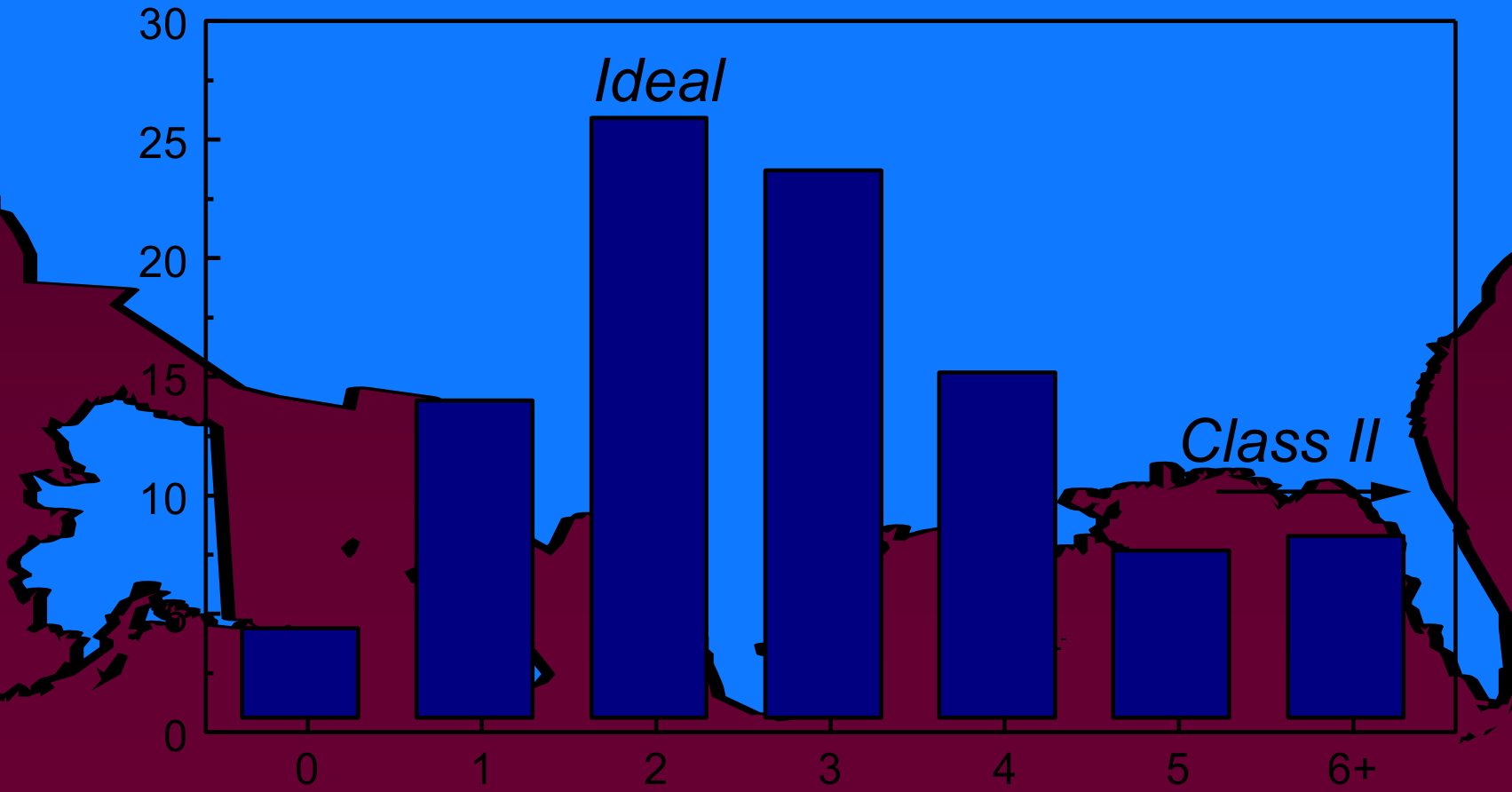
Class II Overjet56
- How many in our population are Class II?
- How many of those have a severe enough problem to cause problems in social interaction?
15% of the population are Class II; half of those have overjet >6 mm, severe enough to predispose to problems in social interactions.
Lecturer — Class II Treatment Need
Not every patient with a Class II relationship requires treatment.
- Some patients have a Class II relationship without a substantial overjet or functional problem.
- A patient may desire treatment without necessarily requiring treatment to achieve a reasonably normal occlusion.
 |  |
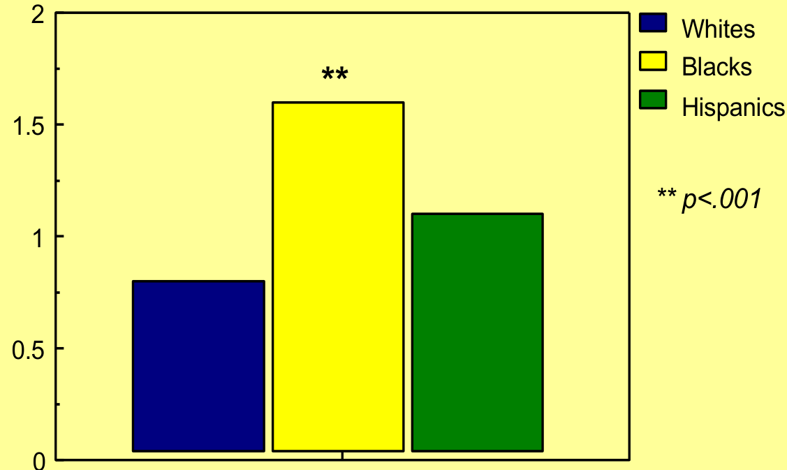
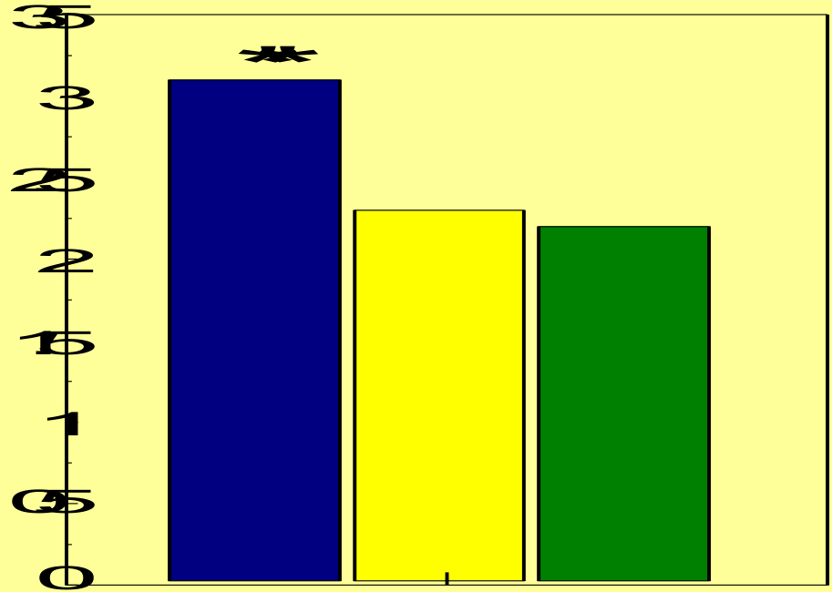
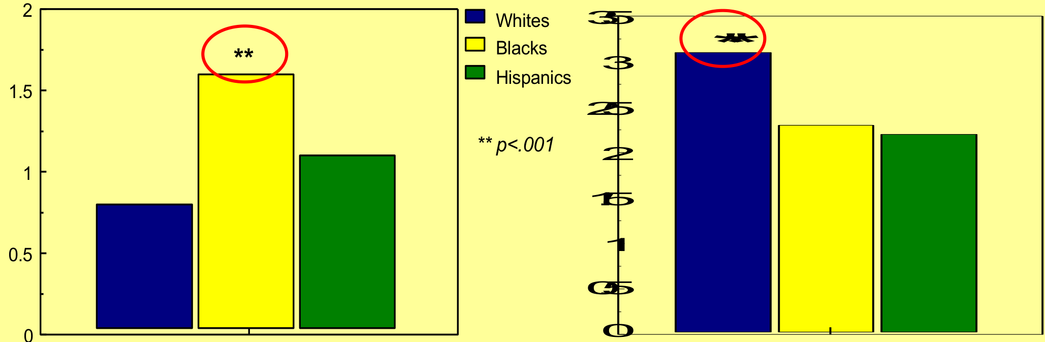
Ethnic Differences78
What’s the biggest racial / ethnic difference?
Whites / Blacks / Hispanics — **p<.001
- Open bite much more prevalent in Blacks; deep bite more prevalent in Whites
Whites / Blacks / Hispanics — **p<.001
 |  |
 |
Class III Reverse Overjet9
- Racial / ethnic differences: open bite much more prevalent in Blacks, deep bite more prevalent in Whites
- Other differences are modest: Whites / Blacks / Mex-Americans — Class III = 0.5%
But for Asians, Class III = up to 5%
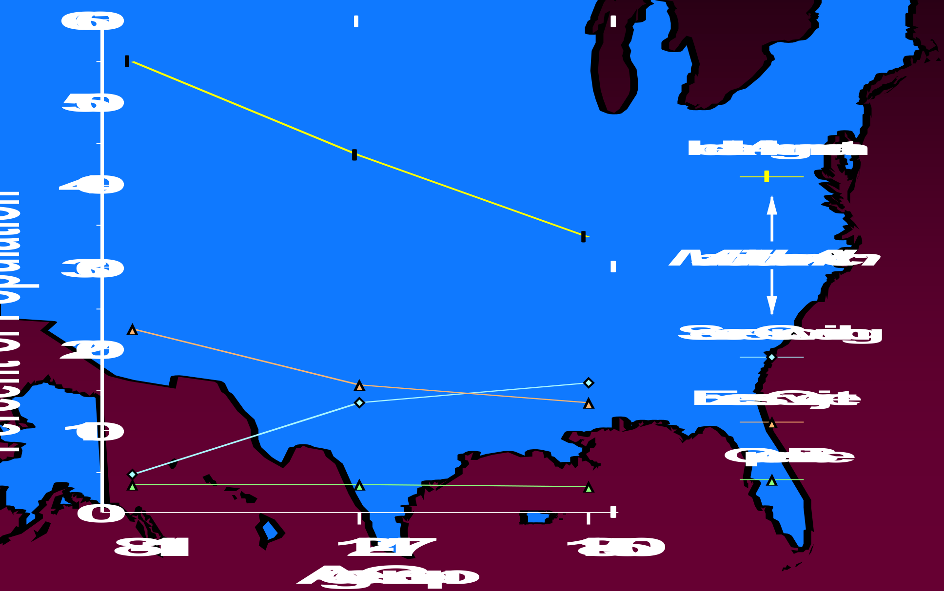
Changes From Child To Adolescent To Adult1011
What changes occur with age in the prevalence of types of malocclusion?
| Age Group | Ideal Alignment | Severe Crowding | Excess Overjet | Open Bite |
|---|---|---|---|---|
| 8-11 | ~55% | ~16% | ~1% | ~4% |
| 12-17 | ~39% | ~8% | ~11% | ~4% |
| 18-50 | ~23% | ~7% | ~13% | ~4% |
(Mandibular Arch; values are Percent of Population)
Crowding gets worse with age — other things don’t.
(Percent of Population, Mandibular Arch, by Age Group: Ideal Alignment, Severe Crowding, Excess Overjet, Open Bite)
Treatment Need In The USA
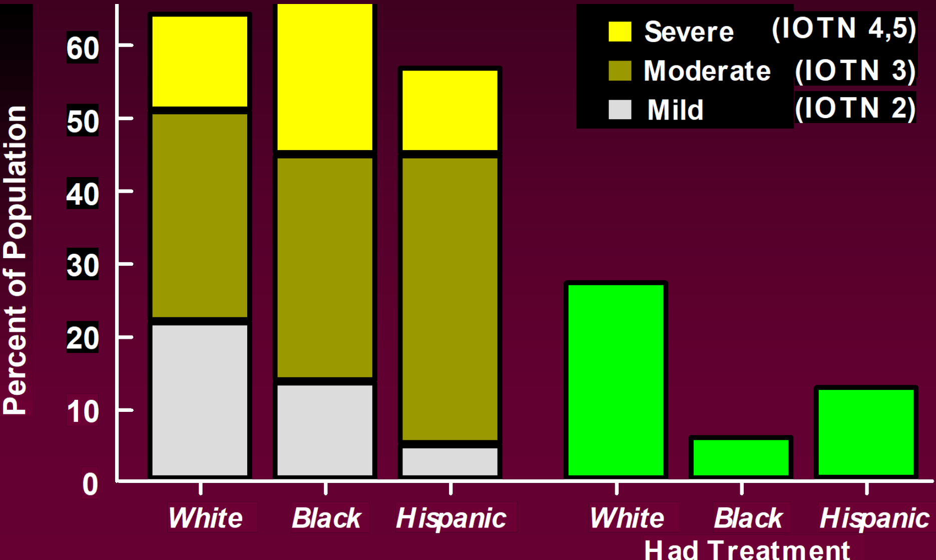
How many people need orthodontic treatment?
Treatment Need From IOTN Scores
Percent of Population12
| Category | White | Black | Hispanic |
|---|---|---|---|
| Mild (IOTN 2) | ~22% | ~14% | ~5% |
| Moderate (IOTN 3) | ~29% | ~31% | ~40% |
| Severe (IOTN 4,5) | ~13% | ~21% | ~12% |
| Total Need | ~64% | ~66% | ~57% |
Lecturer — IOTN Scores
The IOTN measures malocclusion severity to assess treatment need, rather than merely identifying an occlusal irregularity.
- In the white population represented in the data, approximately 20% had a mild treatment need and approximately 50% had a moderate treatment need.
- A group having fewer people with high treatment need does not necessarily mean that it receives less treatment.
| Had Treatment | White | Black | Hispanic |
|---|---|---|---|
| Percentage | ~27% | ~6% | ~13% |
Legend: Severe (IOTN 4,5); Moderate (IOTN 3); Mild (IOTN 2); Had Treatment
Treatment Received By Family Income13
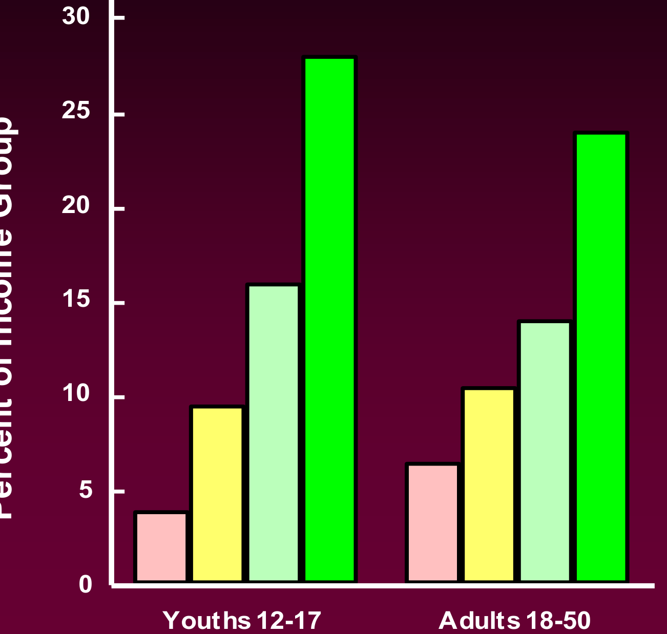
How many people receive orthodontic treatment?
Lecturer — Family Income
Orthodontic treatment received increases with family income and disposable income.
- The greater amount of treatment received by the white population was potentially attributed to greater concern about malocclusion and socioeconomic differences.
- The data were old, approximately from around 1990, although the general pattern was still considered relevant.
- In Western Australia, safety-net arrangements may reduce socioeconomic disparity through government assistance with treatment costs or through “Give a Smile,” in which an orthodontist provides treatment free of charge.
- Subsidized treatment remains limited because most treatment for patients with crooked teeth is concentrated in a restricted service area.
Treatment Of Crowding And Protrusion14
- Prevalence / Treatment Need
- Treatment of Crowding / Protrusion
Lecturer — Treatment Assessment
Treatment decisions should not be based solely on the fact that teeth overlap or appear crowded.
- Assess incisor position and inclination, facial profile, available space, and leeway space.
- Also evaluate the skeletal and dental relationships.
- Protrusion may result from mandibular protrusion, maxillary protrusion, or teeth positioned too far forward within the arch.
Case Study Jessica J Age 9-41516171819
Case moved — see Case: 9.4-year-old with crowding and missing primary canines (continued in Case: 9-year-old female with crowding and previous extractions)
Epidemiologic Data for Malocclusion — Class I: Crowding20
- About half the population have well-aligned incisors; 15% are so crowded that extractions probably required for treatment
Lecturer — Class I Crowding
The approximately 15% estimate does not mean that every such case must be treated with extractions. Some clinicians may accept incisor proclination, together with its associated risks of relapse, recession, and dehiscence, so treatment plans may differ between clinicians.
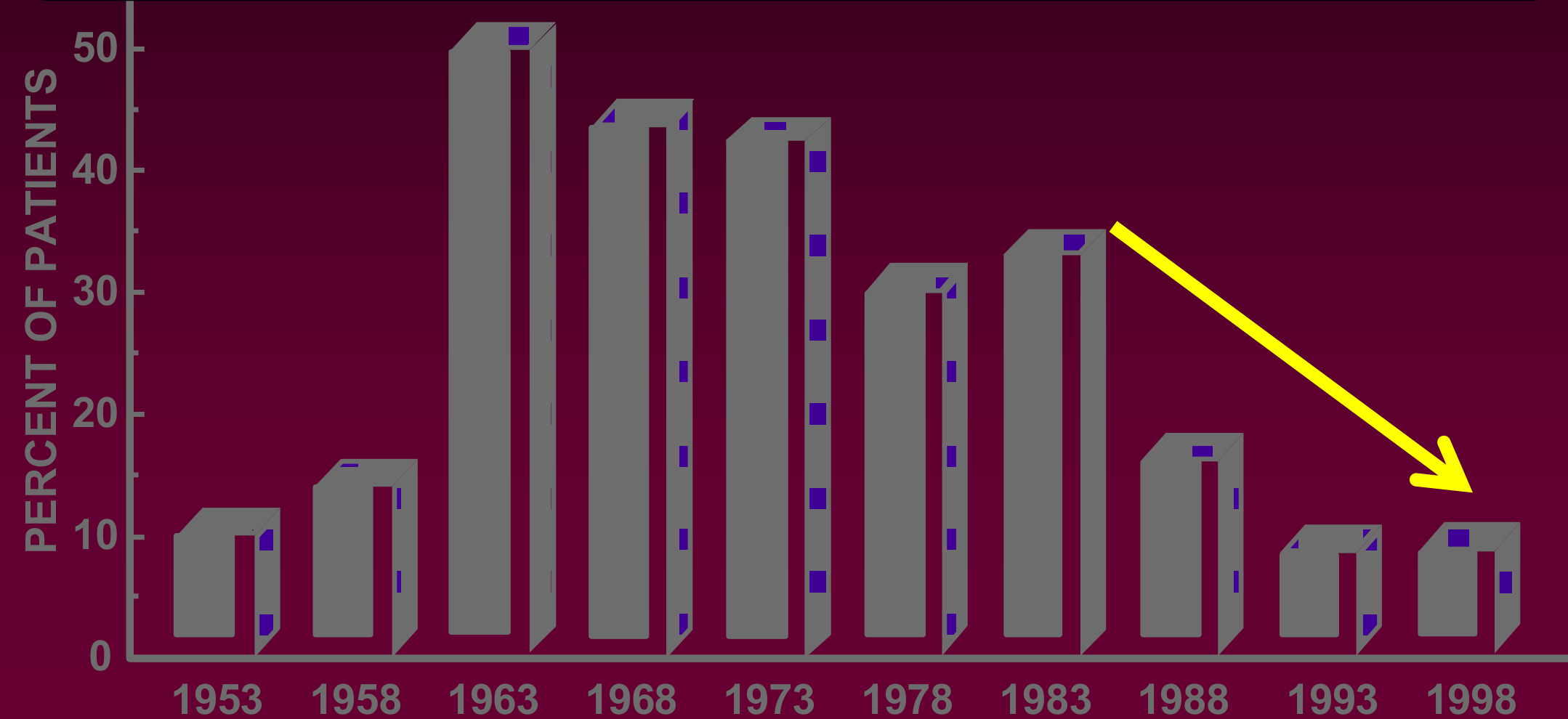
Changes In The Extraction Percentage 1953-199821
Why the increase in the 1960s?
Search for Stability — Technique-Related22
- Full rather than partial banding
Graph: PERCENT OF PATIENTS (Y-axis: 0, 10, 20, 30, 40, 50) vs. Years (X-axis: 1953, 1958, 1963, 1968, 1973, 1978, 1983, 1988, 1993, 1998)
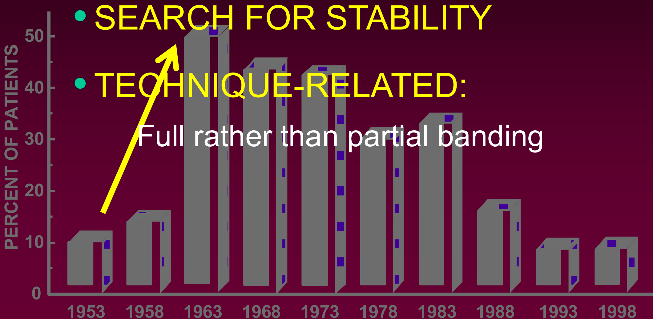
Why the decrease more recently?23
- Esthetic considerations
- Concerns about stability
- Bonding
- TMD accusations
- Treatment time
Graph: PERCENT OF PATIENTS (Y-axis: 0 to 50) vs. Years (X-axis: 1953, 1958, 1963, 1968, 1973, 1978, 1983, 1988, 1993, 1998)
McNamara, AAO Early Treatment Conference, Feb. 2002: 12-15% of Class I crowded cases had premolar extraction
Lecturer — Extraction Trends
Earlier non-extraction treatment frequently involved proclining teeth and expanding the arches, but many of these cases relapsed. Clinicians began extracting four bicuspids in some cases to improve stability, while full banding also became more common as treatment moved from only the upper six teeth against the first molars to banding all teeth.
Lecturer — Extraction Concerns
A United States legal case also influenced attitudes toward extraction. After two upper bicuspids were extracted and the spaces retracted, the patient developed temporomandibular joint problems; the lecturer stated that orthodontic extractions are not associated with temporomandibular disorders, and that symptoms began after third molar extraction in a patient considered susceptible to joint problems. The orthodontist was sued after the oral surgeon settled out of court, contributing to concern about extracting teeth.
Graph: PERCENT OF PATIENTS (Y-axis: 0, 10, 20, 30, 40, 50) vs. Years (X-axis: 1953, 1958, 1963, 1968, 1973, 1978, 1983, 1988, 1993, 1998)
Case Study Phyllis R Age 11-7242526272829
Case moved — see Case: Phyllis R., 11-year-old with Class II and deep bite
Class II Camouflage Treatment30
- Prevalence / Treatment Need
- Treatment of Crowding / Protrusion
- Camouflage of Skeletal Problems
- Class II
Lecturer — Camouflage Planning
Camouflage fits the teeth within the existing skeletal relationship rather than correcting the underlying skeletal relationship.
- Class II camouflage may involve retracting the upper teeth and positioning the teeth to produce an acceptable occlusion.
- Treatment planning depends on the patient’s age and remaining growth, amount of overjet, inclination of the lower incisors, facial profile and lip position, and amount of space required.
- Advancing the mandible with elastics, appliances, or other forces tends to procline the lower incisors, whereas distalizing or restraining the maxilla does not have the same effect.
- The clinician may therefore consider treating the maxilla rather than relying only on mandibular advancement.
What is camouflage of a skeletal problem?
How do you do that?
Case Study Lara W Age 12-931323334353637
Case moved — see Case: Lara W., 12-year-old with severe Class II and protrusion
Extraction Choices For Class II Camouflage38
If we extract 2 premolars to camouflage the Class II and reduce the overjet, which ones would we remove??
If we extract 4 premolars to camouflage the Class II and reduce the overjet, which ones would we remove??
- Class I Canines
- Class II Molars
- Class I Canines
- Class I Molars
Lecturer — Extraction Patterns
Extraction patterns are selected to allow the teeth to finish in an acceptable relationship rather than to alter the skeletal relationship.
Extracting the upper first premolars allows the upper canines to be retracted, establishing a Class I canine relationship while a functional Class II molar relationship remains.
Another option is extraction of the upper first premolars and lower second premolars; lower extractions may be considered when proclined lower incisors need to be retroclined while the Class II relationship is corrected.
Teeth may be extracted to camouflage the skeletal discrepancy even when the arches are not crowded.
Extracting upper teeth shortens the upper arch, so the remaining teeth must close the extraction spaces while maintaining the intended occlusion.
Lecturer — Poor Prognosis Molars
Extracting a single first molar with a poor prognosis is rarely preferred because space closure is difficult, significant asymmetry may result, and conventional orthodontic treatment can become very difficult or impossible. If both upper first molars have a poor prognosis, removing both may be considered, although treatment can be delayed because the second molars are needed for anchorage while the remaining teeth are retracted. Temporary anchorage devices improve management of difficult extraction patterns, but asymmetry can still make treatment challenging.
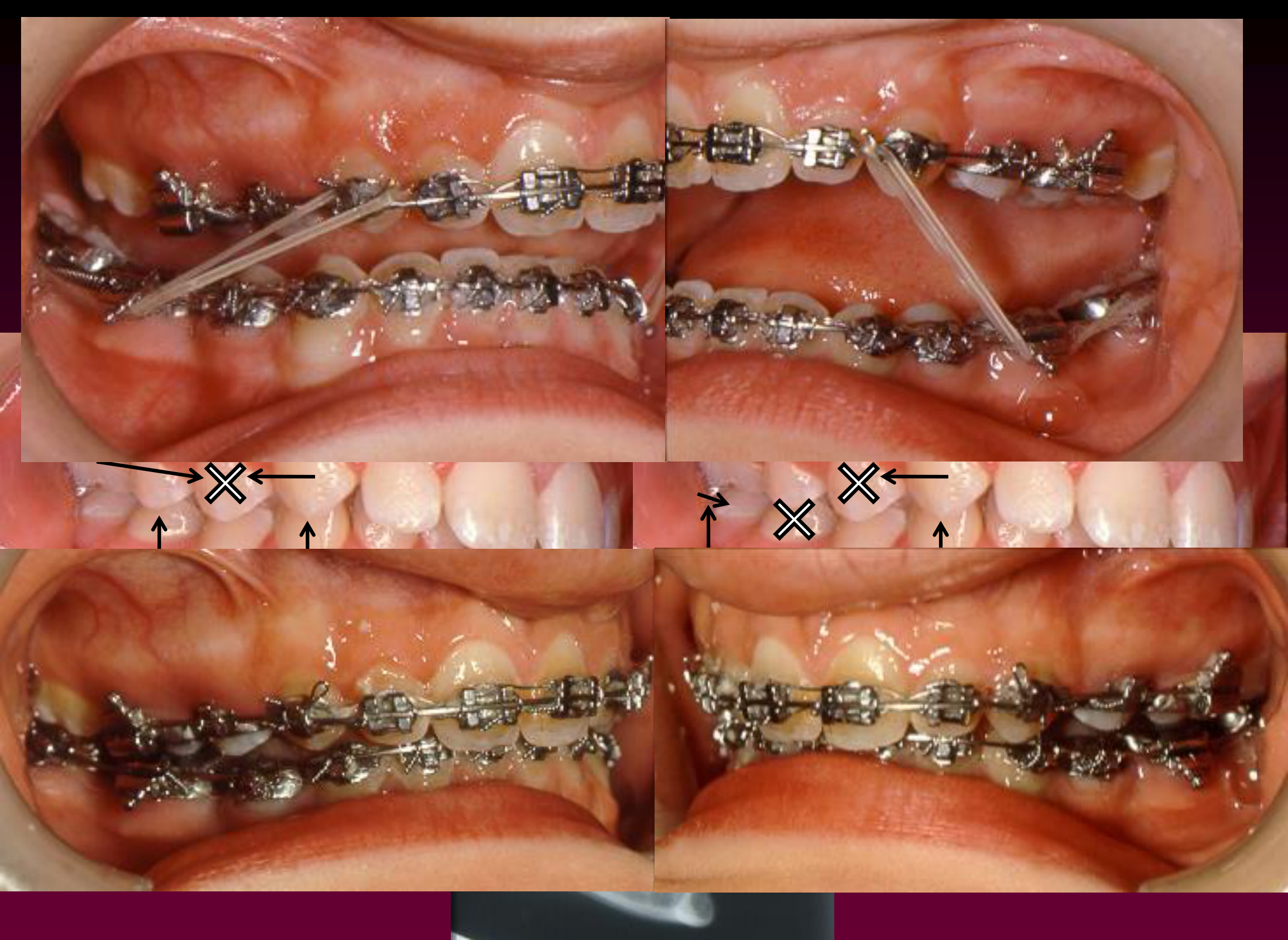
Treatment Outcome And Third Molar Considerations394041424344
Case moved — see Case: Lara W., 12-year-old with severe Class II and protrusion
Class III Camouflage Versus Surgery45
- Prevalence / Treatment Need
- Treatment of Crowding / Protrusion
- Camouflage
- Class II
- Class III
Lecturer — Class III Camouflage
Class III camouflage conceals the underlying skeletal discrepancy through dental movement, but patients are generally treated later because mandibular growth may continue.
- Continued mandibular growth after treatment may return the patient to a more severe Class III relationship.
- The classic extraction pattern is upper second premolars with lower first premolars.
- This allows the lower anterior teeth to move backward, the upper anterior teeth to remain relatively stable, and the posterior teeth to move forward.
- Extraction patterns must be adapted when teeth are already missing or have a poor prognosis.
Case Study Ebenezer E Age 20-104647484950
Case moved — see Case: Ebenezer E., 20-year-old with Class III skeletal relationship
Camouflage Or Surgery Decision51
Is he a candidate for camouflage or surgery?
Lecturer — Camouflage Decisions
The choice between camouflage and surgery depends on the severity of the skeletal discrepancy; in this case, both options were possible.
- Crowding is not required for a camouflage extraction plan: teeth may be extracted to compensate for a skeletal Class II or Class III relationship.
- Possible Class III approaches include upper second premolar and lower first premolar extractions, lower first premolar extractions alone for severe mandibular protrusion, or extraction of a single lower incisor when appropriate.
- The extraction choice must allow the remaining teeth to finish in a functional occlusion and should account for the patient’s large mandible and missing teeth.
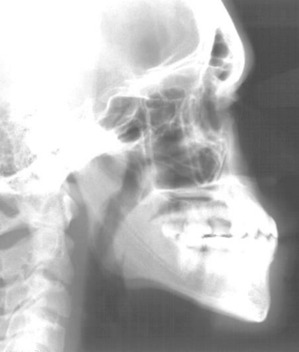
Treatment Outcome5253545556
Case moved — see Case: Adult with Class 3 skeletal relationship and missing teeth
Course Conclusion57
At the end of the Orthodontic course, you have neither the didactic or clinical knowledge to provide comprehensive orthodontic treatment.
Lecturer — Diagnostic Treatment Planning
Orthodontic treatment decisions require more than identifying crowding or an overjet; they require individualized diagnosis rather than applying one extraction or non-extraction rule to every patient.
- Important considerations include facial profile, incisor inclination, space analysis, leeway space, growth potential, skeletal and dental relationships, and the number and condition of permanent teeth.
- Non-extraction treatment may involve accepting proclination and its risks, including relapse, recession, and dehiscence.
- Class II correction generally benefits from growth, particularly when the mandible is retrognathic, whereas Class III patients are generally treated later because continued mandibular growth may worsen the relationship.
- Extraction patterns should reflect the desired final occlusion, existing missing teeth, tooth prognosis, anchorage, and facial considerations.
Clinical Cases
Case: 9.4-year-old with crowding and missing primary canines
Question
Scenario: A 9.4-year-old female patient (Jessica J.) is evaluated for dental crowding. Her parents are asking whether she will need teeth extracted to straighten them.
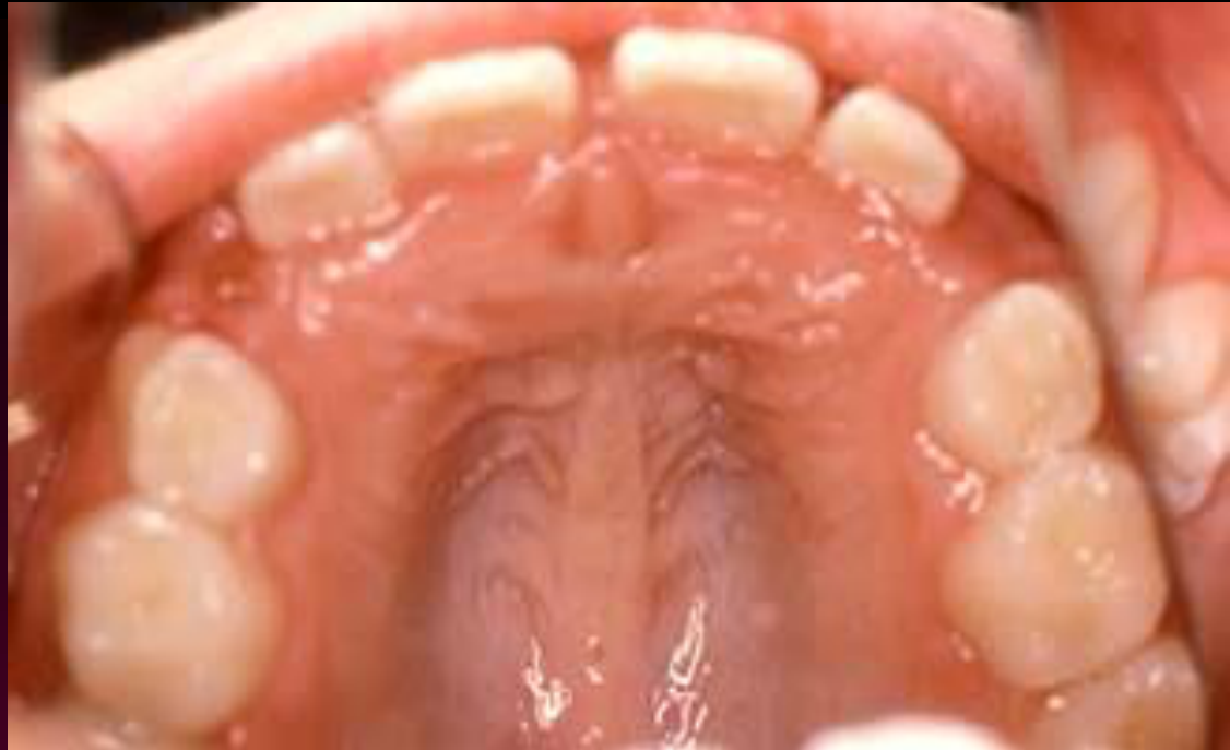
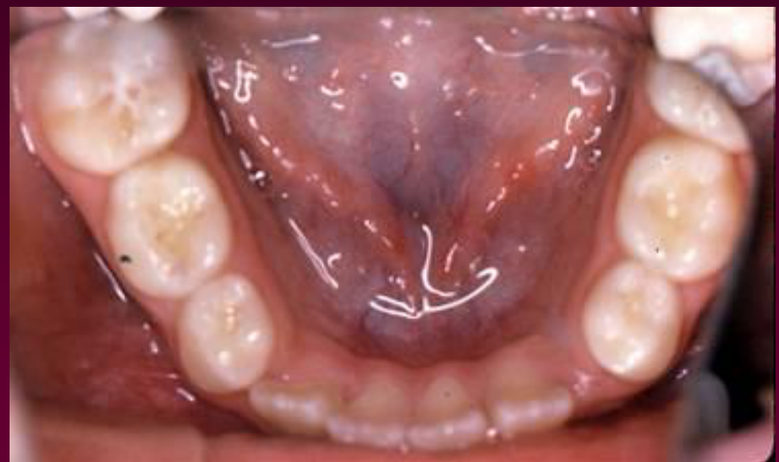
What’s shown: Intraoral photographs of the maxillary and mandibular arches showing first molars, remaining deciduous teeth, missing deciduous canines, a localized crossbite, and significant crowding. The primary canines were extracted the previous year.
Consider: What clinical problems are evident, and what additional diagnostic information is required to determine if extractions are necessary to resolve the crowding?
Answer
Observations:
- Presence of first molars and some deciduous teeth.
- Missing deciduous canines (extracted the previous year).
- A localized crossbite.
- Significant crowding in the dental arches.
Reasoning: The parents’ question regarding the need for extractions cannot be answered based on the clinical presentation alone. A definitive treatment decision requires comprehensive diagnostic records. These include a lateral cephalogram to assess the facial profile, and a space analysis. The space analysis must account for “leeway space” (the size difference between deciduous molars and the succeeding premolars). Additionally, the inclination of the incisors must be evaluated: if the lower incisors are retroclined, proclining them can gain space in the arch; however, if they are already proclined, further flaring risks relapse, root dehiscence, or gingival recession, meaning any space gain must come from the leeway space or extractions.
Outcome: The primary canines were previously extracted by a dentist to allow for the alignment of the permanent incisors and prevent future periodontal or soft tissue complications. This serves as an initial step, potentially leading to serial extraction, but a full diagnostic workup is needed to finalize the treatment plan.
Takeaway: Visual assessment of crowding is insufficient to decide on extractions; definitive orthodontic planning requires a full diagnostic workup including space analysis (incorporating leeway space), facial profile evaluation, and assessment of incisor inclination.
From the lecture slides
Jessica J. — age 9-4
Parents’ question: “Will she need to have teeth pulled to straighten them?”
Jessica J. — age 9-4
- Primary canines extracted last year by another dentist, to allow alignment of permanent incisors
- Does that mean you have to continue with serial extraction?
- Did the previous dentist fail to do something that probably he should have done?
- Is she likely to require extraction of premolars eventually?
- Do you need any further information?
Jessica J. — age 9-4
Do you still need any further information?
Age 8-5 to 9-4
Jessica J — Diagnostic Assessment
The deciduous canine extractions were common treatment and were not necessarily inappropriate. They could allow crowded incisors to erupt, reduce the risk of later periodontal, recession, and soft-tissue problems, and represent the beginning of a serial extraction approach.
- The lower arch was critical: the lower incisors were essentially retroclined and might be proclined to gain space, whereas proclination of already proclined incisors could increase relapse, recession, or dehiscence risks.
- Further assessment required a lateral cephalometric radiograph, space analysis, facial profile, incisor inclination, protrusion, smile arc, and lip position.
- The upper molars might be distalized to gain space, with headgear mentioned as one possible method.
- The patient should generally be referred to an orthodontist; the available information could not establish whether premolar extraction or a space maintainer was appropriate.
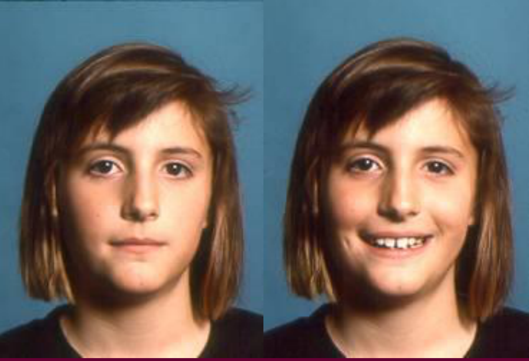
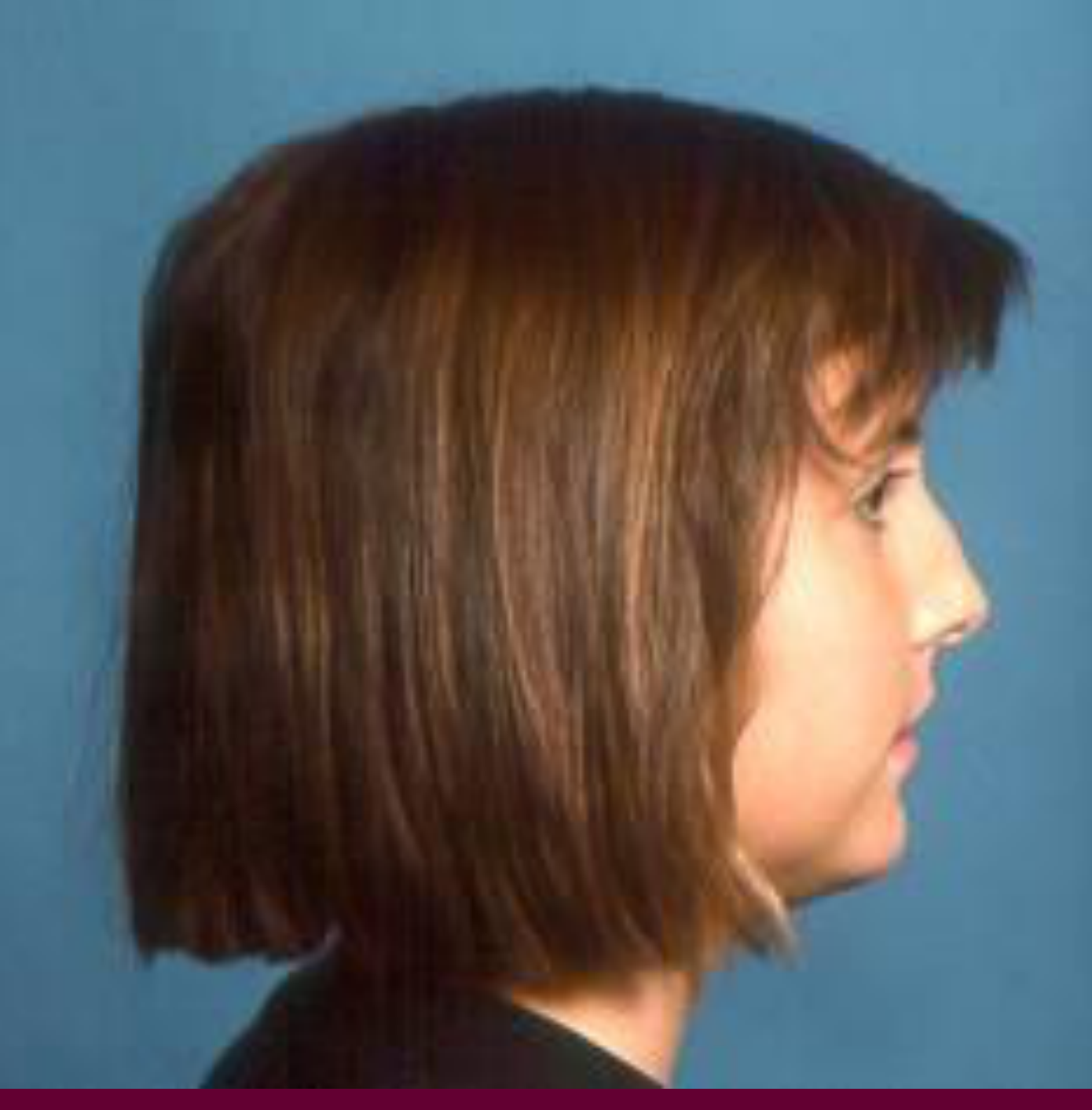
Case: 9-year-old female with crowding and previous extractions
Question
Scenario: A 9-year-old female patient (Jessica J.) is being evaluated for orthodontic treatment. She has a history of previous tooth extractions by another dentist. The lecturer raised the possibility that no space maintainer was placed, but said the available records don’t show this (“we don’t know”).
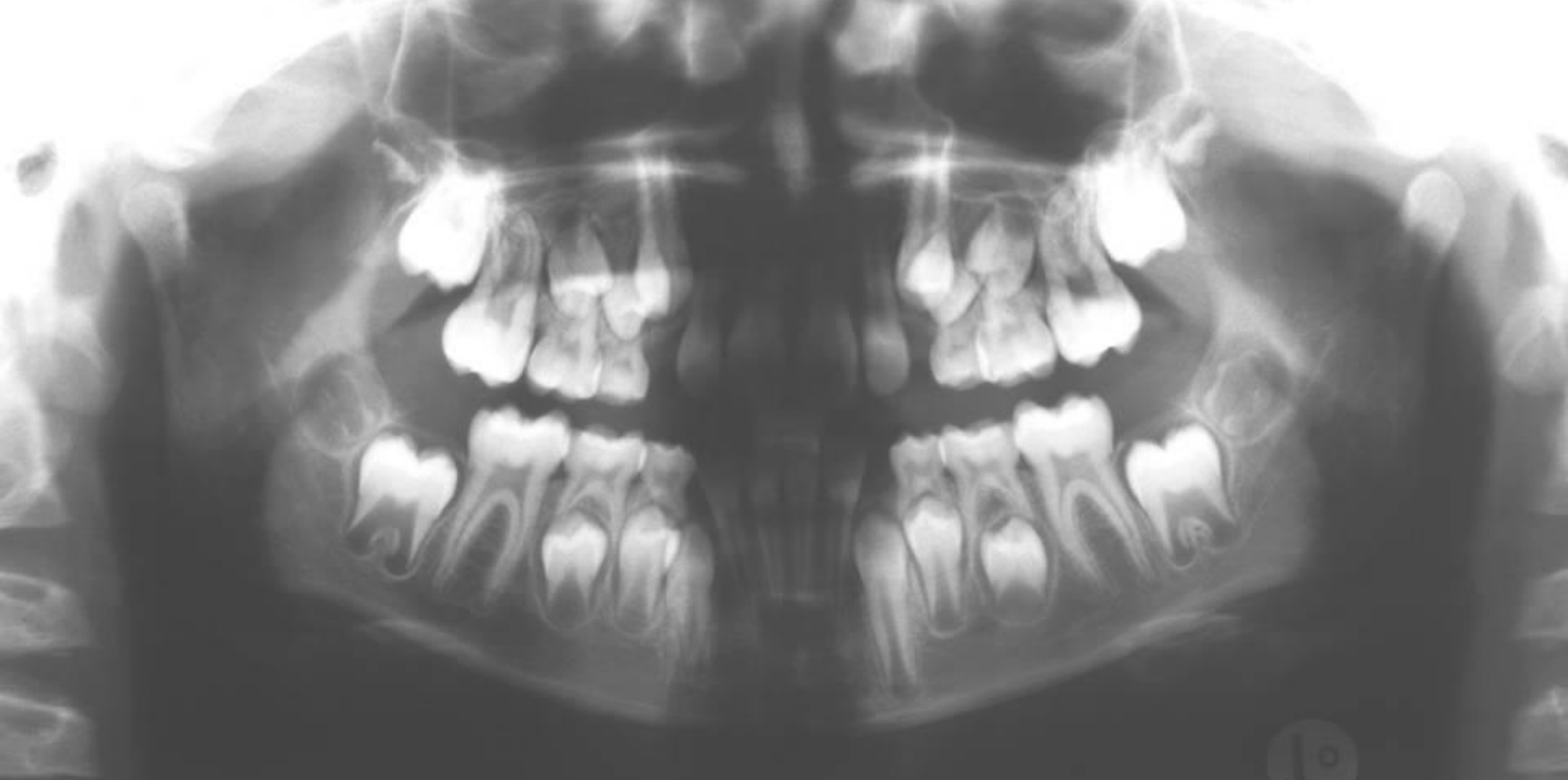
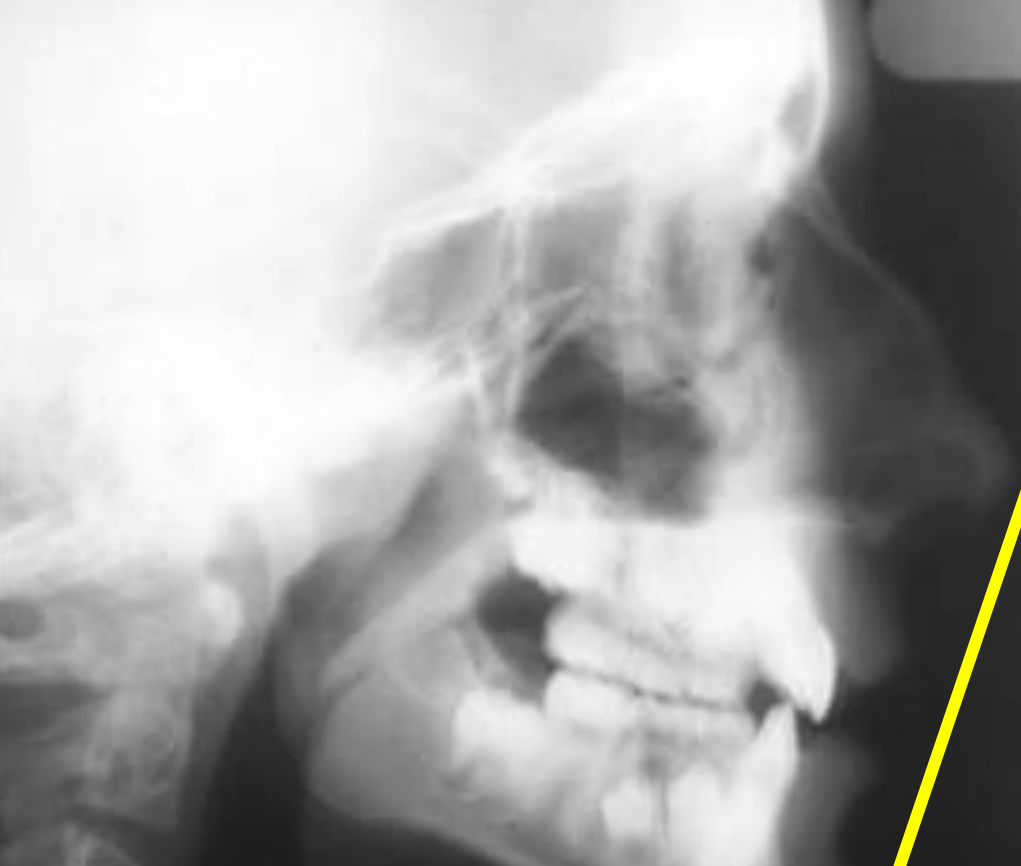
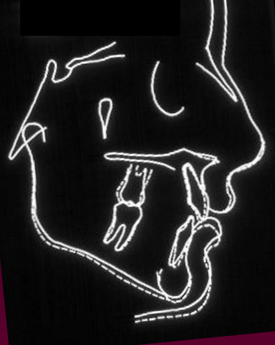
What’s shown: A lateral cephalometric radiograph with the E-line drawn, and a superimposed cephalometric tracing (age 8-5 to 9-4). Her intraoral photographs and panoramic radiograph are in the preceding case.
Consider: How to interpret the clinical and radiographic findings to determine if space can be gained without further extractions, and what the appropriate management plan is.
Answer
Observations:
- Intraoral photographs show crowding in the arches, with the lower incisors appearing retroclined.
- The panoramic radiograph shows all permanent teeth are present.
- The soft-tissue profile on the cephalogram shows the lips are not full and fall behind the E-line.
- The lateral cephalometric radiograph confirms the lower incisors are retroclined, tipped back relative to the mandibular plane at an angle of less than 90 degrees.
Reasoning: Because the lower incisors are retroclined, space can potentially be gained by proclining them, avoiding the need for premolar extractions. The facial profile, with lips behind the E-line, indicates that proclining the incisors would be aesthetically acceptable. The upper arch crowding can be managed by distalizing the molars to gain space. The lower arch is the critical factor in this treatment plan, and managing the incisor inclination and leeway space requires specialized care.
Outcome: The lecturer highly recommends referring the patient to an orthodontist for treatment rather than managing the case in general practice.
Takeaway: In patients with crowding, assessing the inclination of the lower incisors and the facial profile (such as the E-line) is essential to determine if space can be gained through proclination, which may prevent the need for extractions.
From the lecture slides
Do you think she will need premolar extraction?
If she has further orthodontic treatment:
- when would be the best time to do it?
- refer or treat yourself?
Case: Phyllis R., 11-year-old with Class II and deep bite
Question
Scenario: An 11-year-old female patient presents for orthodontic evaluation.
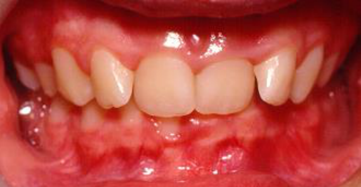
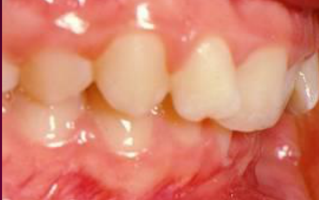
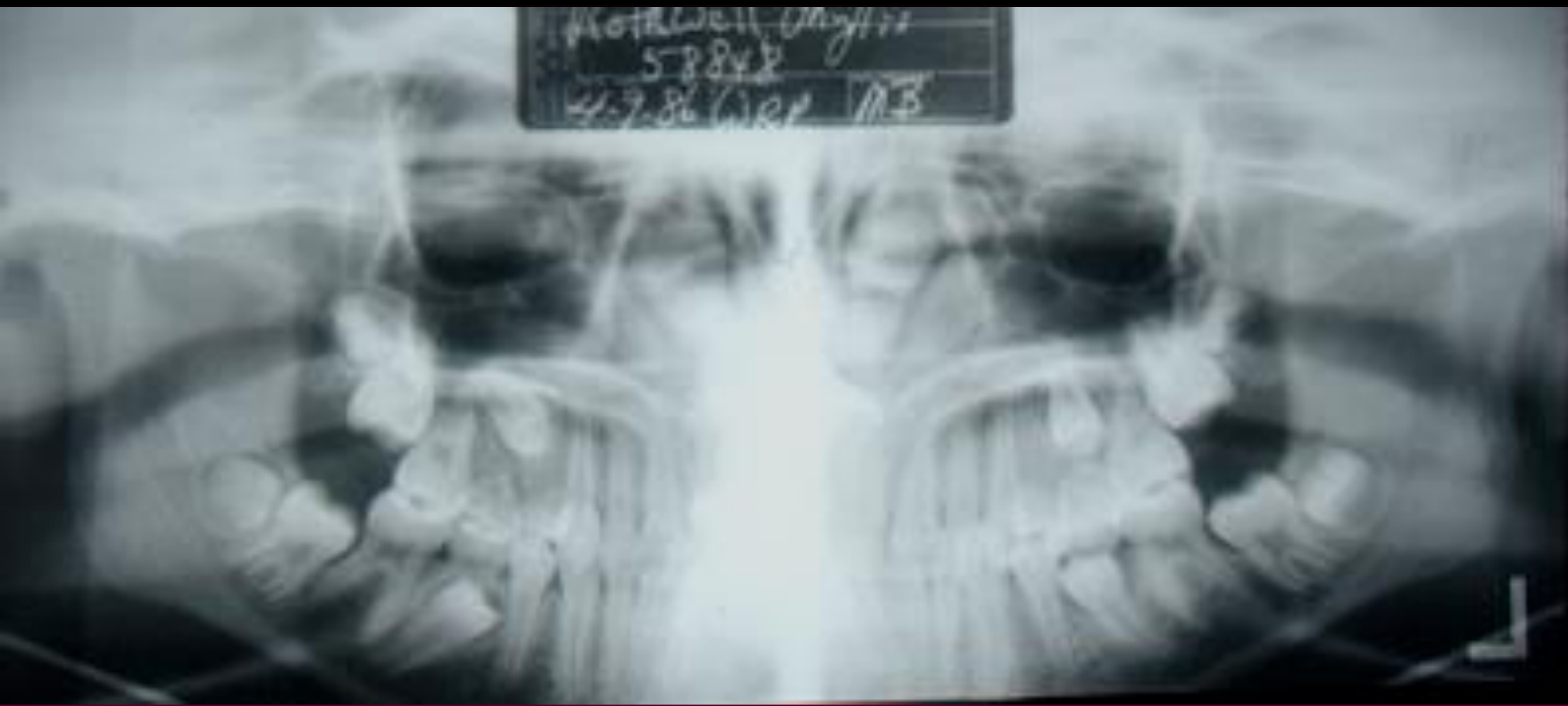
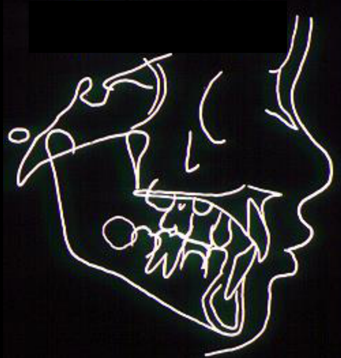
What’s shown: Intraoral photographs showing a deep bite, a slight Class II relationship, and anterior crowding with deciduous teeth still present posteriorly. A panoramic radiograph, lateral profile photograph, and lateral cephalometric radiograph with tracing are also evaluated.
Consider: Whether the patient is a candidate for orthodontic extractions or surgery, and the best timing for treatment given her growth potential.
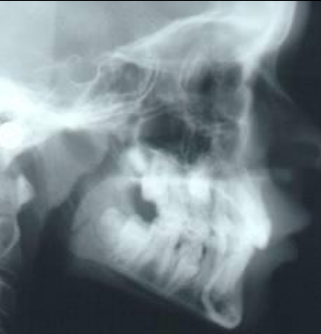
Answer
Observations:
- Deep bite, slight Class II relationship, and anterior crowding.
- Deciduous teeth are still present posteriorly.
- Facial profile and lateral cephalometric radiograph are assessed to determine growth potential and incisor inclination.
Reasoning: The lecturer notes that there is not enough information from the panoramic radiograph alone to decide on extractions or surgery. Evaluating the facial profile and cephalometric radiograph is necessary to assess the inclination of the incisors and the patient’s growth potential. For a Class II correction, growth is required, so treatment timing should coincide with the patient’s pubertal growth spurt.
Outcome: None stated; the case is used to illustrate the diagnostic process and timing for Class II treatment.
Takeaway: Treatment for Class II correction requires growth, so timing should align with the patient’s pubertal growth spurt, and comprehensive records (profile, cephalometric) are needed before deciding on extractions or surgery.
From the lecture slides
Phyllis R. — Age: 11-7
Is she a candidate for orthodontic extractions?
Phyllis R — Growth and Treatment
At 11 years and 7 months, further growth was likely, and the information provided was insufficient to decide whether orthodontic extractions or surgery were appropriate. Assessment still required the facial profile and features, incisor inclination, the amount of space required, and confirmation that all permanent teeth were present.
- For a Class II patient with a retrognathic mandible, correction should be coordinated with the pubertal growth spurt.
- Possible approaches included brackets and elastics, springs, or headgear.
- Class II correction generally requires growth, although skeletal Class II problems can also be camouflaged with tooth movement.
(trick question, be careful!)
Phyllis R. — Age: 11-7
Do you think she will need premolar extraction?
If she has further orthodontic treatment:
- is she likely to grow more?
- when would be the best time to do it?
- refer or treat yourself?
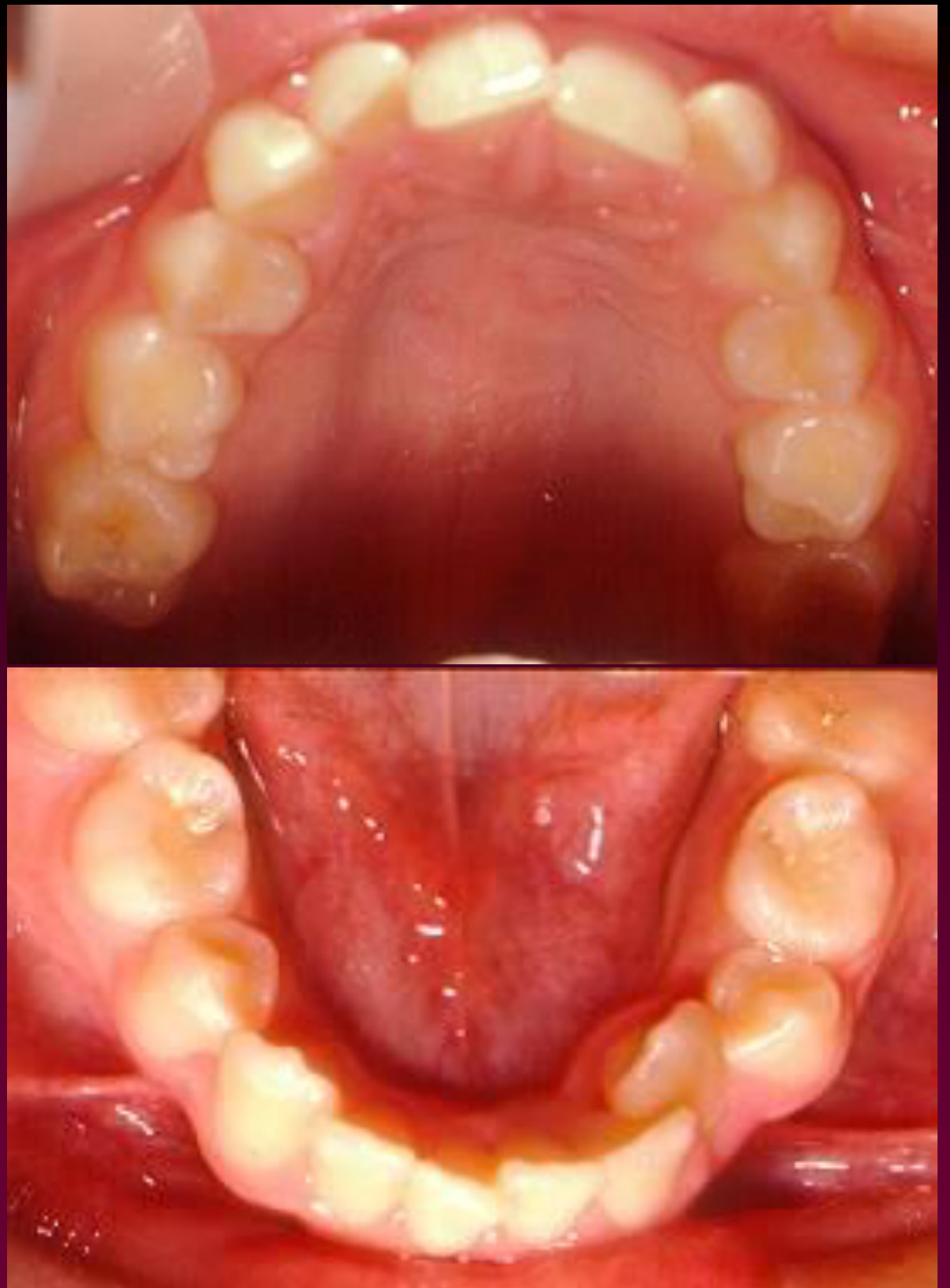
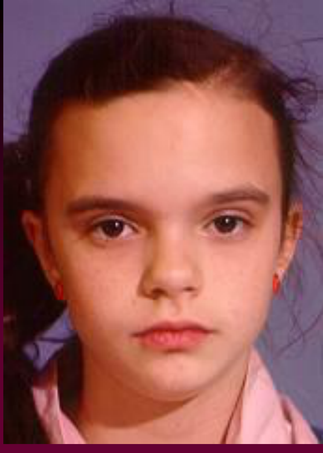
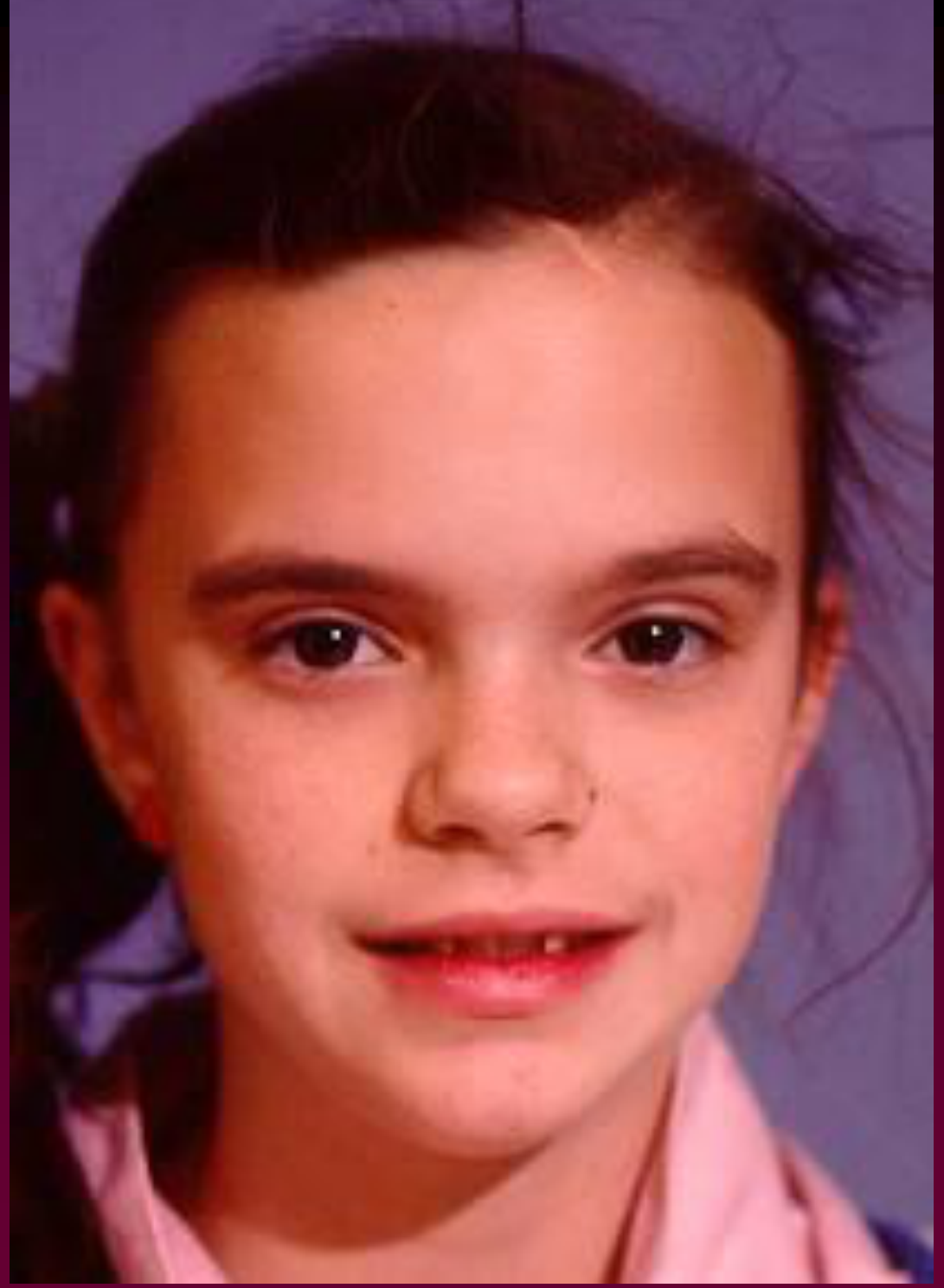
Case: Lara W., 12-year-old with severe Class II and protrusion
Question
Scenario: A 12-year-old female patient presents with a significant overjet and protrusion, and is unhappy with her profile.
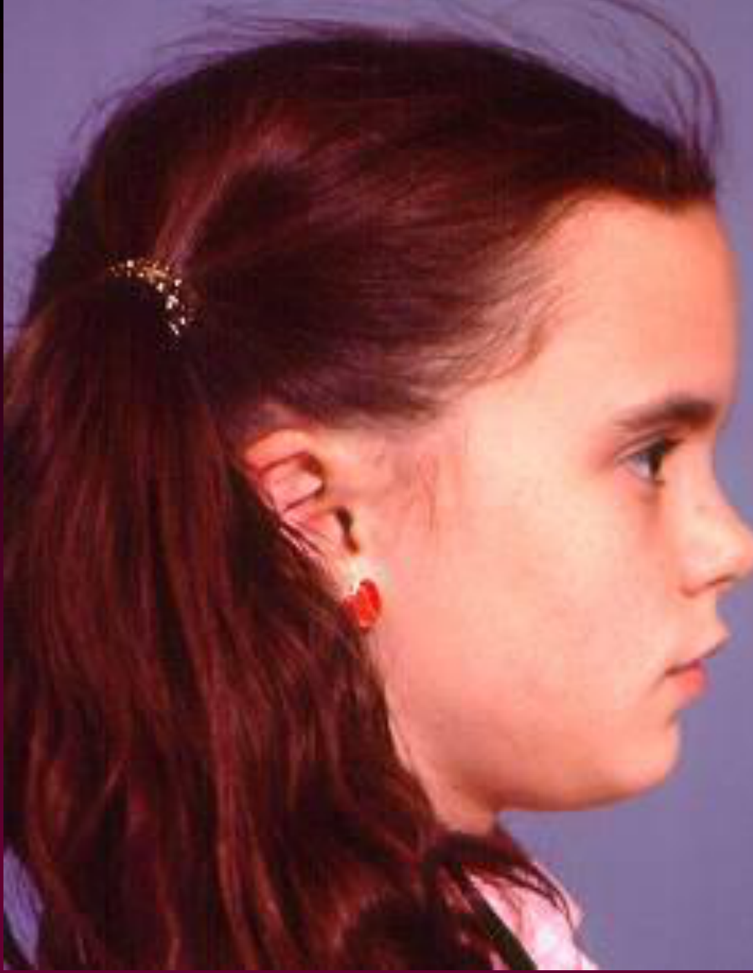
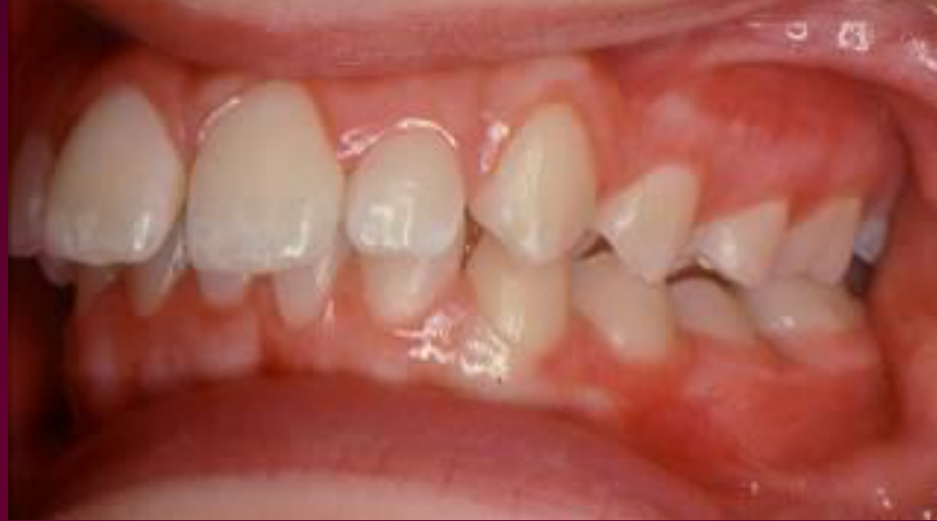
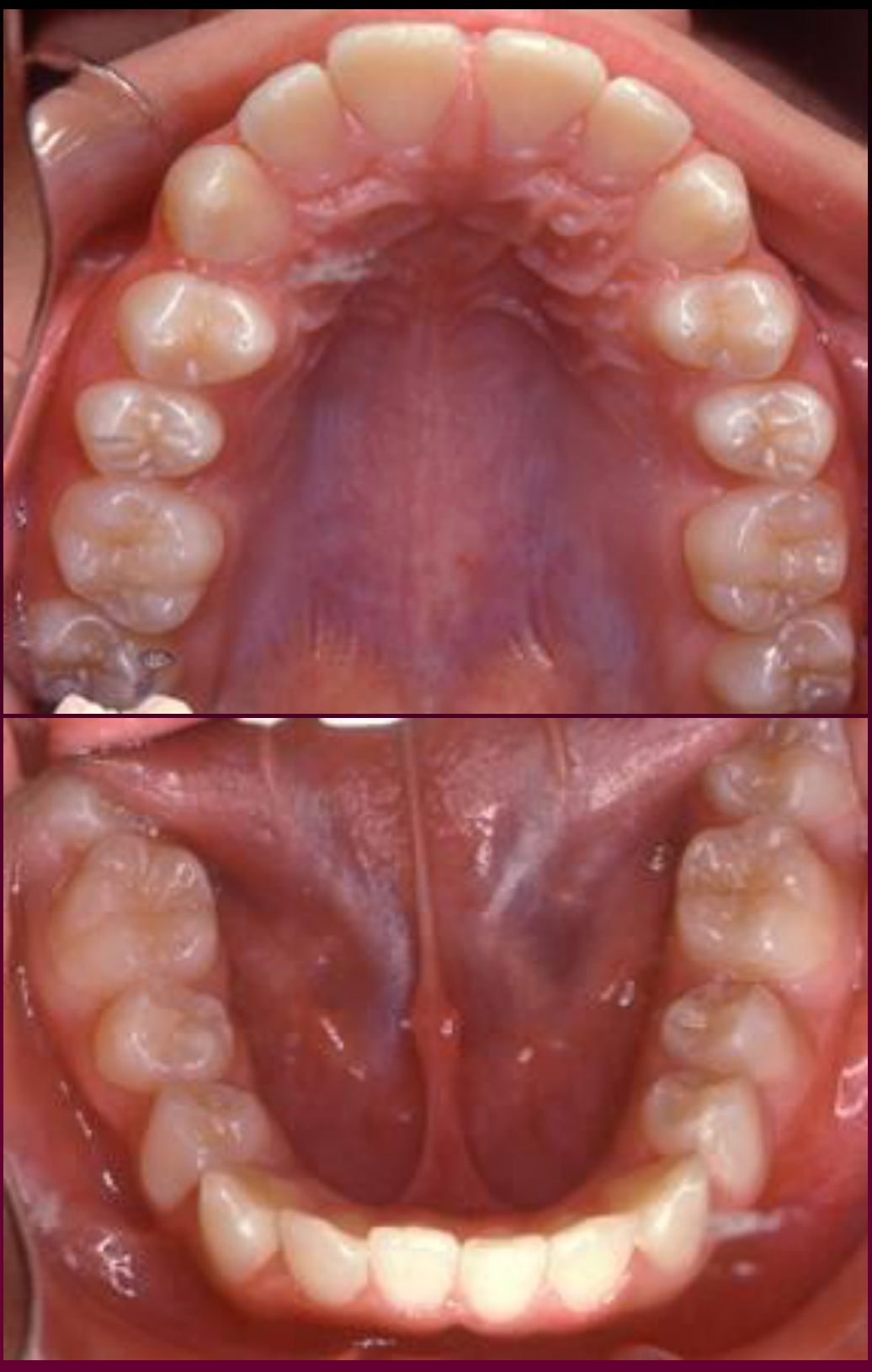
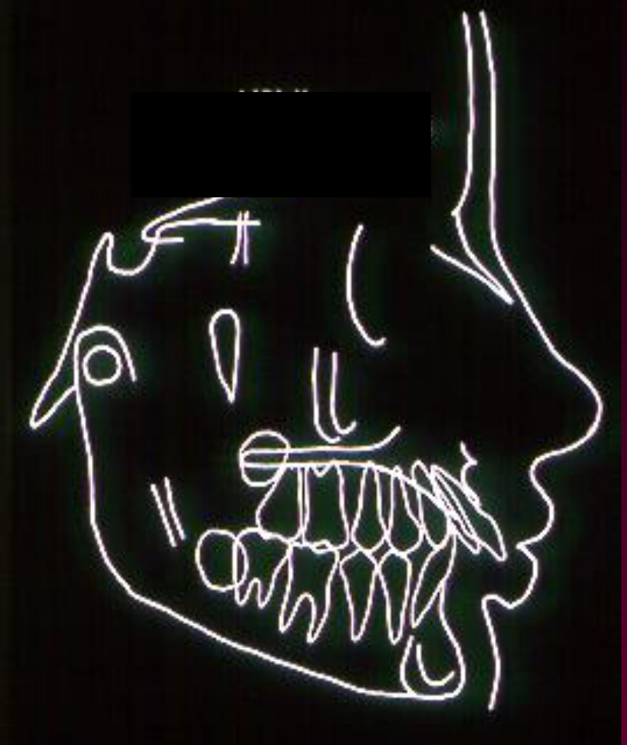
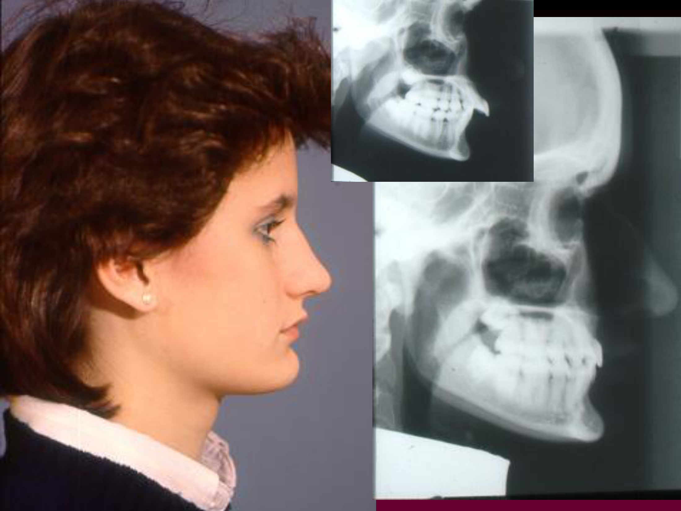
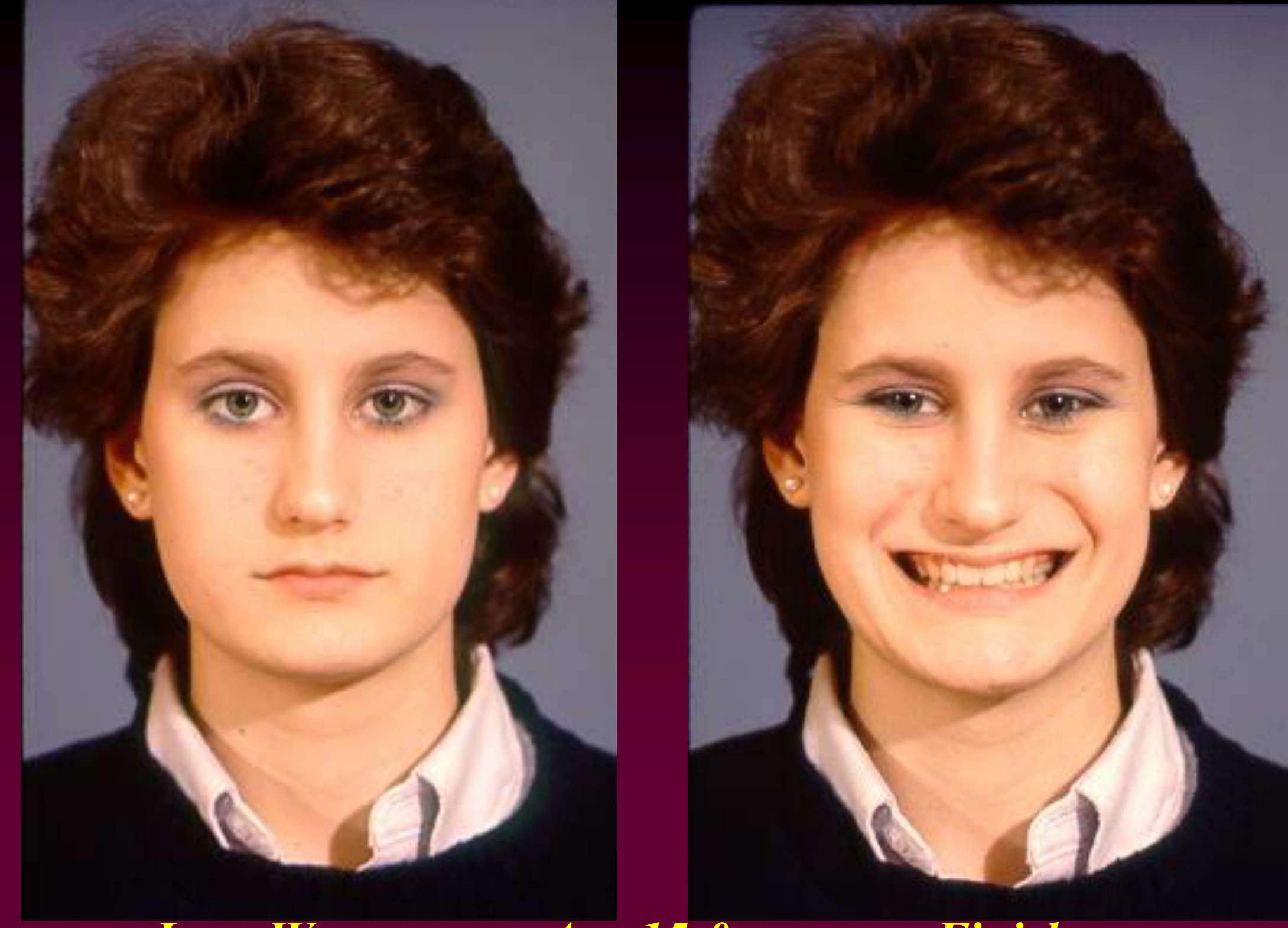
What’s shown: Clinical photographs showing a full-step Class II relationship, protruded upper incisors, and a hypodivergent facial profile with reasonably full lips. A lateral cephalometric tracing shows proclined lower incisors. Post-treatment photographs and a superimposed cephalometric tracing are also shown.
Consider: How to treat the severe Class II relationship and protrusion, considering her growth potential and lower incisor inclination, and what changes occurred as a result of the camouflage treatment.
Answer
Observations:
- Full-step Class II relationship with a large overjet and protruded upper incisors.
- Hypodivergent facial profile with full lips.
- Lower incisors are proclined relative to the mandibular plane.
- Post-treatment: well-aligned teeth, corrected overbite, Class I canine and molar relationships (upper first and lower second premolars extracted), residual space in the lower arch, and parallel roots.
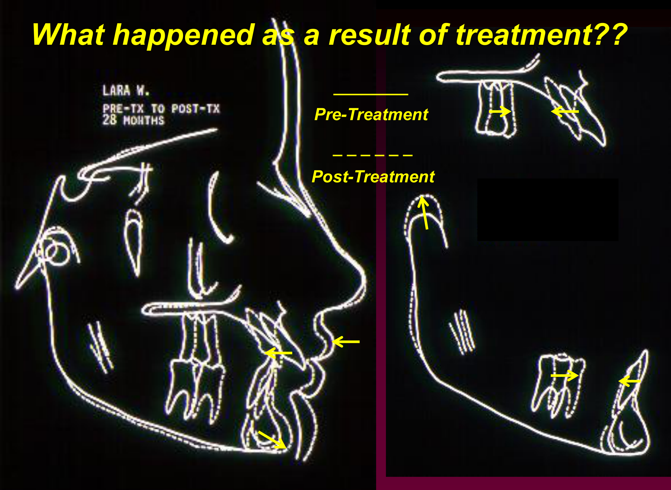
- Cephalometric superimposition shows the first molar moved forward, upper incisors retracted, lower incisors uprighted, and a change in chin position (developed a “chin button”) due to growth.
Reasoning: Because the lower incisors are already proclined, advancing the mandible with elastics or appliances would further procline them. Distalizing the maxilla or using camouflage extractions (upper first bicuspids and lower second bicuspids) are considered to fit the teeth within the existing skeletal relationship without changing it. The patient is near the end of her growth period, so treatment should proceed quickly.
Outcome: Treated with fixed appliances, elastics, and extraction of upper first premolars and lower second premolars (camouflage). Finished with a Class I molar and canine relationship, corrected overbite, and well-aligned teeth. Facial profile flattening was minimal, but the chin position changed significantly due to continued growth.
Takeaway: Camouflage orthodontic treatment for a Class II patient with proclined lower incisors and limited growth remaining can involve extracting upper first and lower second premolars to correct the overjet without altering the skeletal relationship.
From the lecture slides
Lara W. – age 12-9
“My upper front teeth stick out and have a gap between them.”
Lara W. — Age 12-9
Do you think she will need premolar extraction?
If she has further orthodontic treatment:
- is she likely to grow more?
- when would be the best time to do it?
- refer or treat yourself?
Lara W: Initial Assessment
Lara W. was a 12-year-and-9-month-old female with a large overjet, full-step Class II relationship, prominent upper incisors, reasonably full lips, a hypodivergent facial pattern, and apparently proclined lower incisors. She was unhappy with the protrusion.
- She was likely to have some additional growth, but not a large amount.
- Treatment needed to begin relatively soon because the overjet was substantial.
- The case was considered appropriate for referral to an orthodontist.
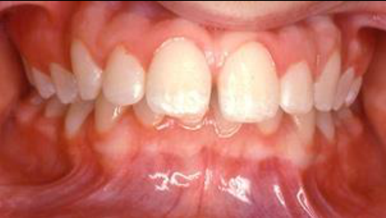
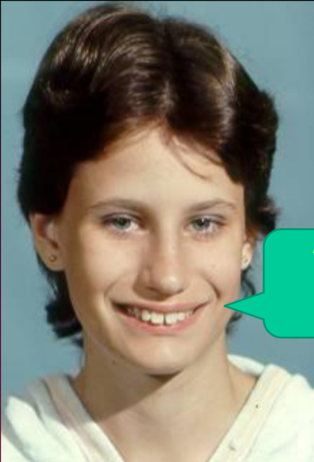
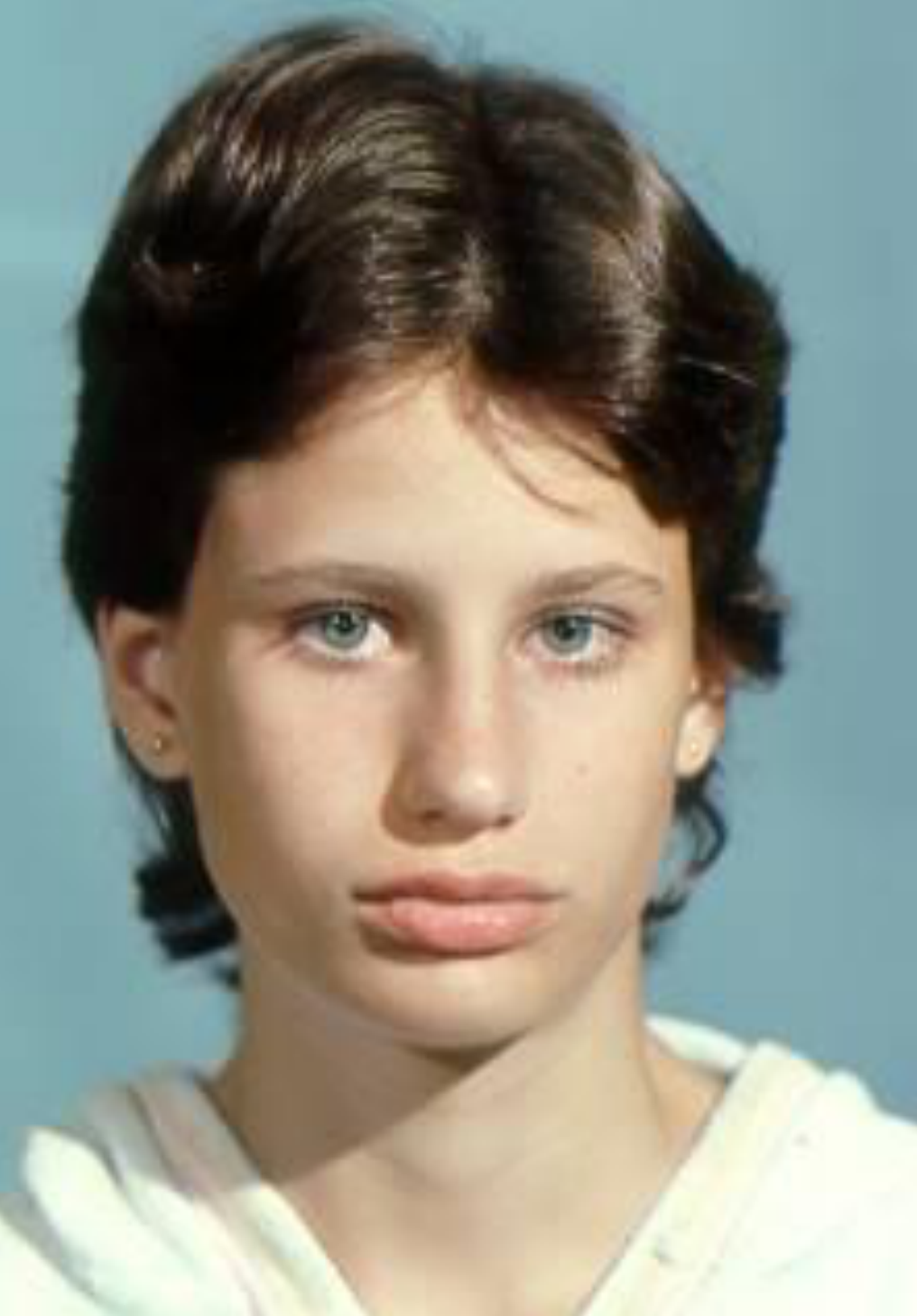
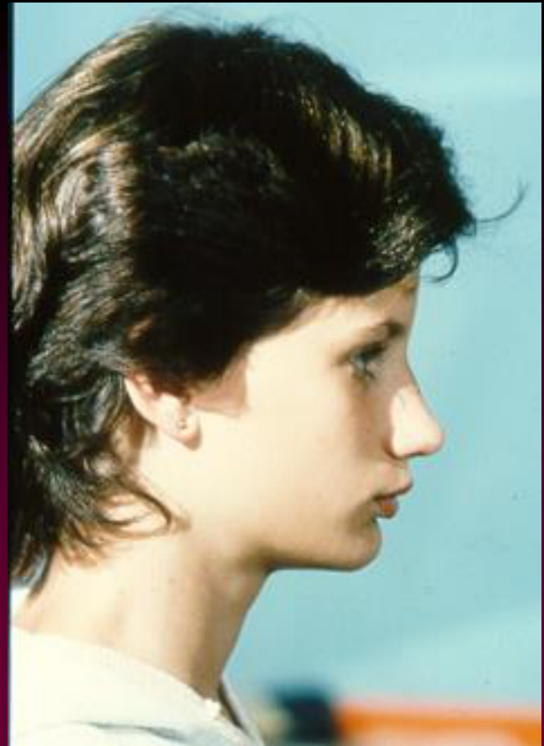
Lara W. — Age 15-0 — Finish
3rd molars and premolar extraction
Is this successful treatment?
How do we decide??
What happened as a result of treatment??
LARA W. PRE-TX TO POST-TX 28 MONTHS
Pre-Treatment
Post-Treatment
Lecturer — Treatment Outcome
The patient was treated with fixed appliances and elastics after extraction of the upper first premolars and lower second premolars. The result was considered good treatment.
- The outcome included Class I molar and canine relationships, corrected overbite, well-aligned teeth, slight residual lower-arch space, and generally parallel roots.
- Success was assessed by whether the patient was happy with the treatment and smile and whether the facial profile had been flattened.
- The facial profile probably did not flatten substantially; the changes were attributed more to chin development than to major alteration of lip position.
- Cephalometric superimposition showed forward movement of the first molar, retraction of the upper incisor, some uprighting of the lower incisors, a change in relative lip position, and significant chin growth.
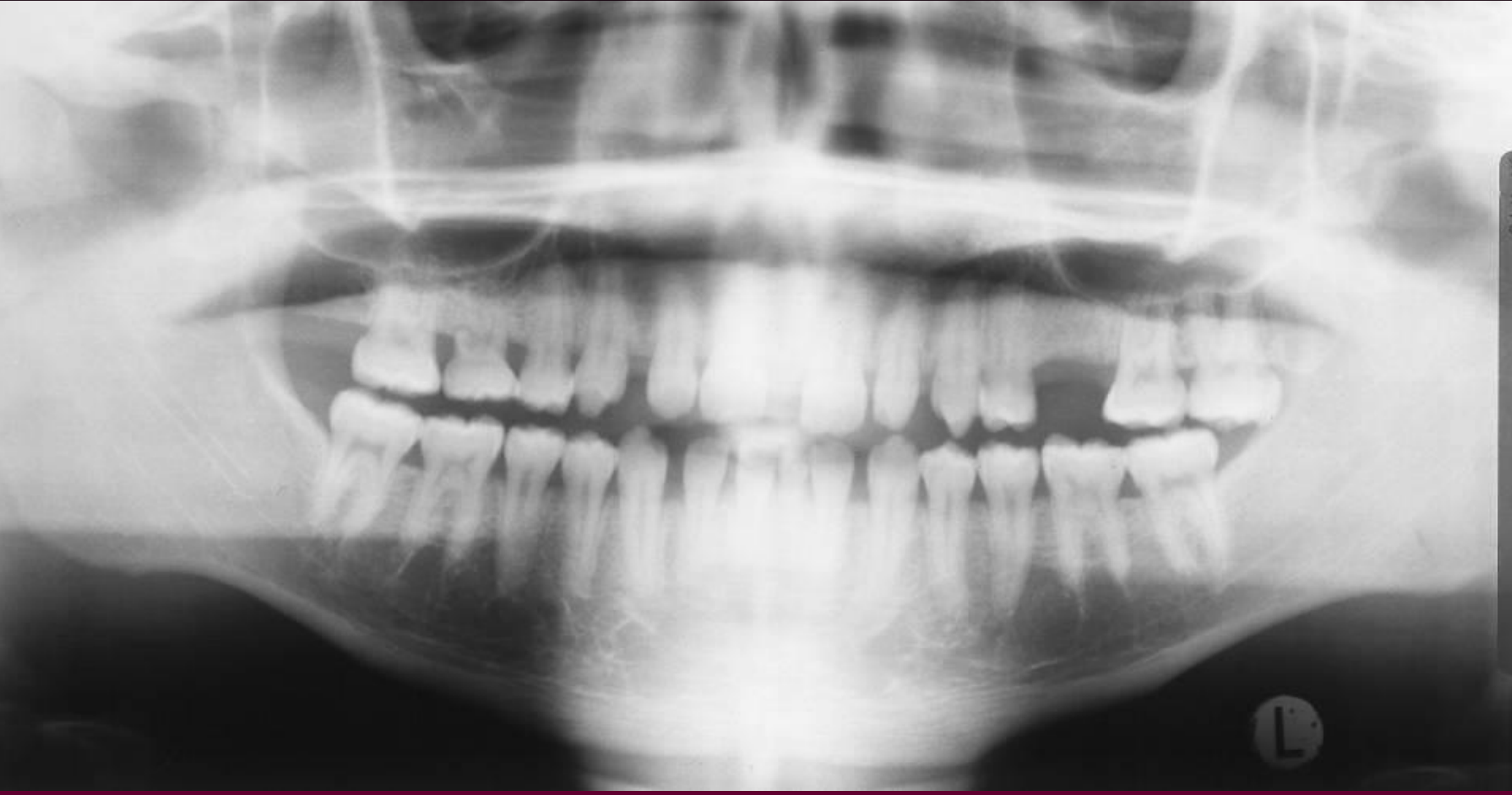
Case: Ebenezer E., 20-year-old with Class III skeletal relationship
Question
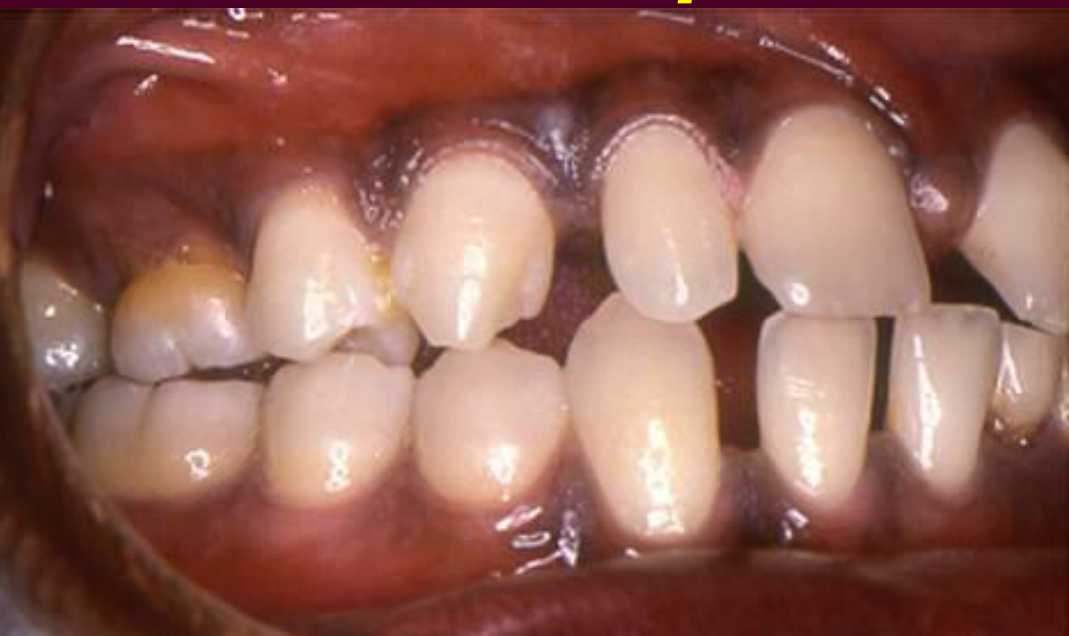
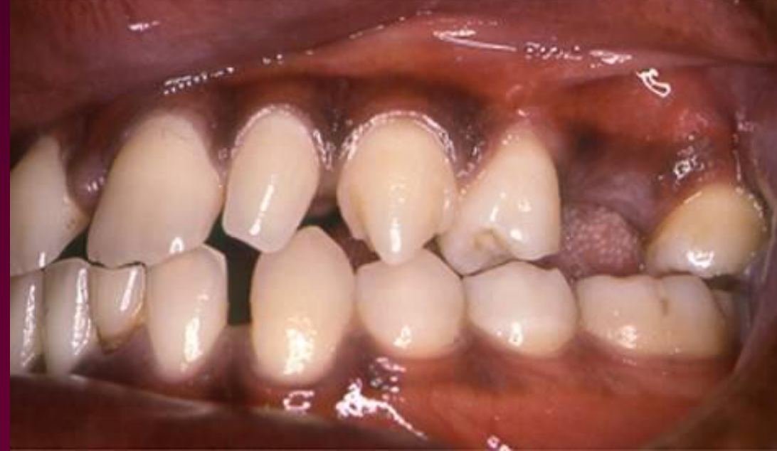
Scenario: An adult male patient presents with a Class III skeletal relationship and multiple dental issues.
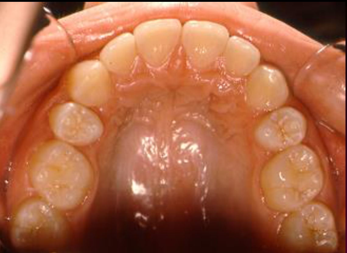
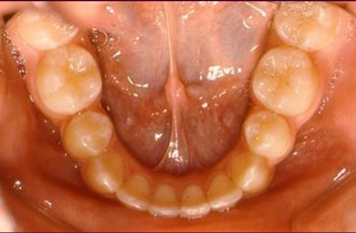
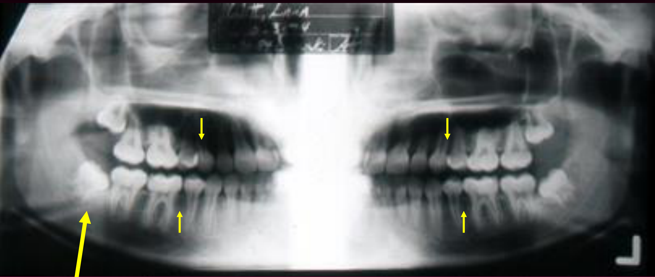
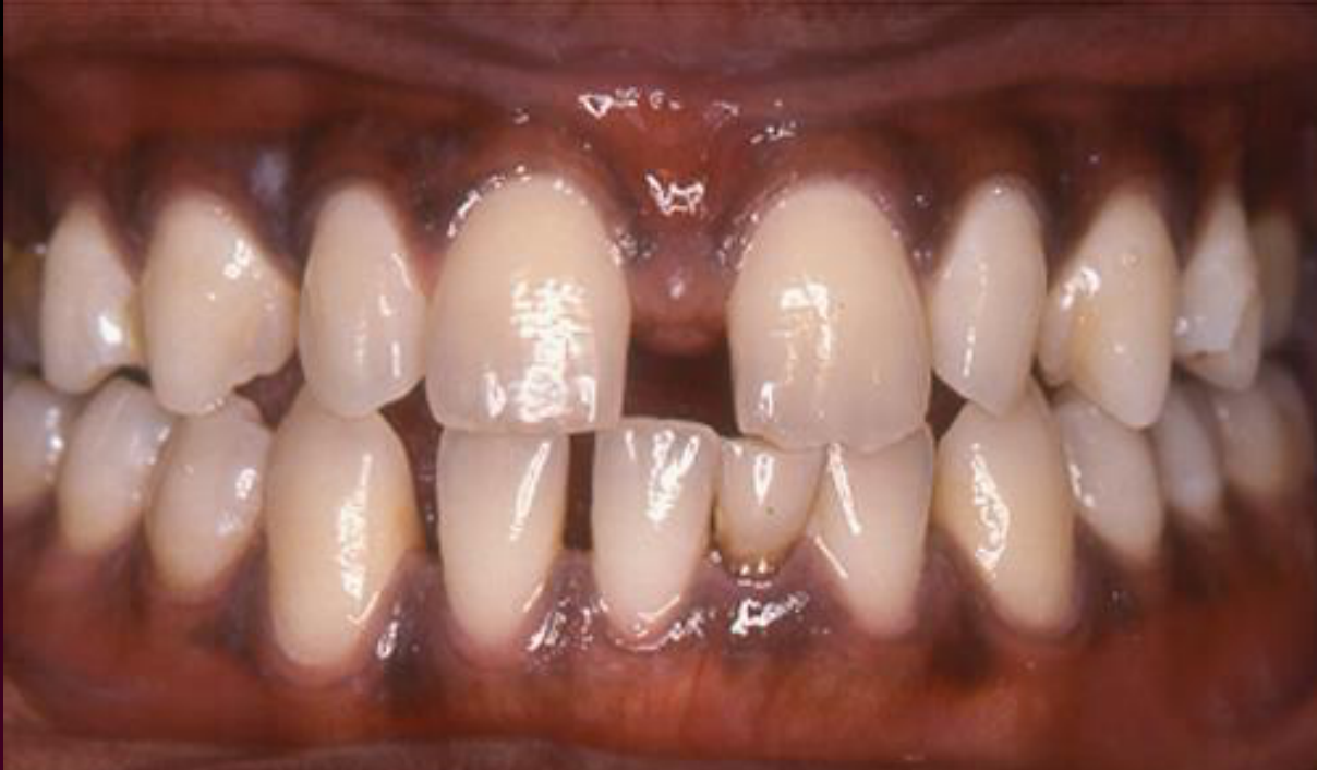
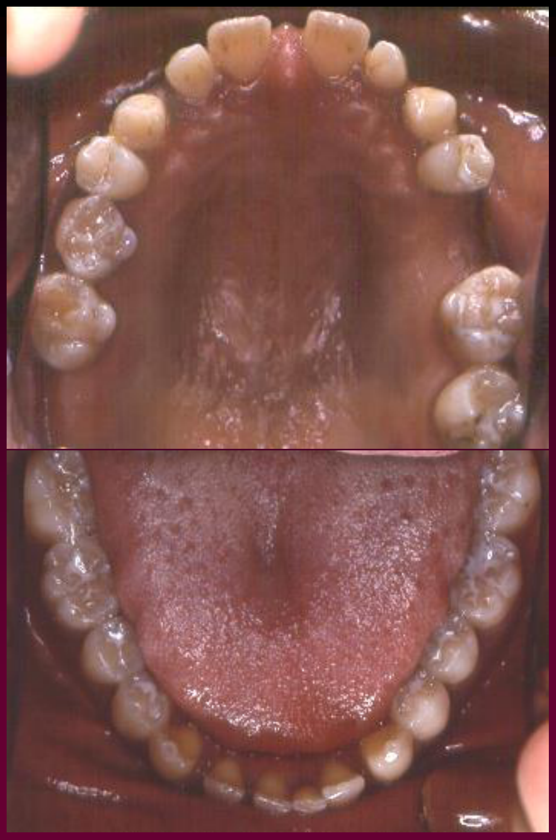
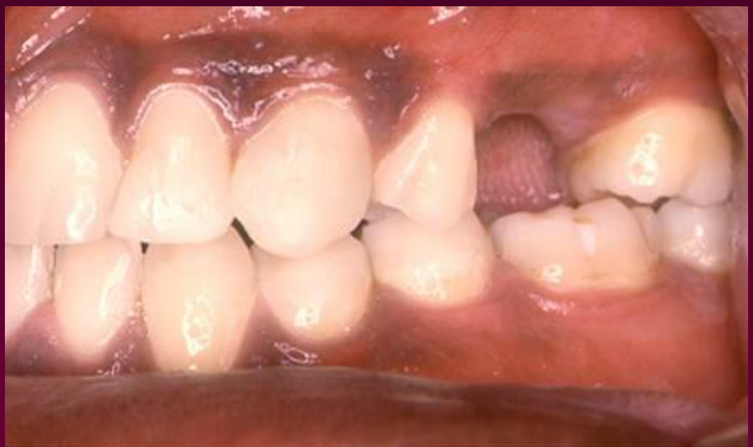
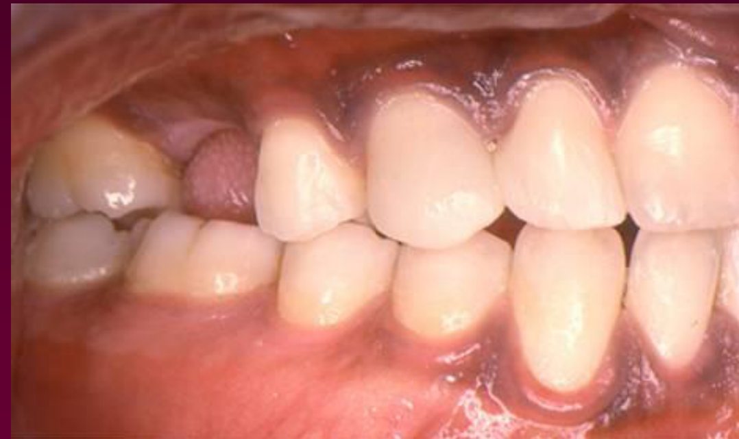
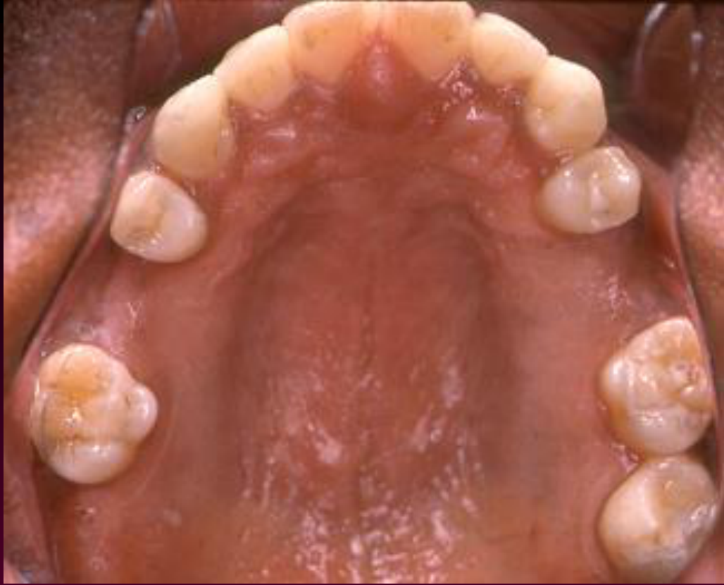
What’s shown: Intraoral photographs and a panoramic radiograph showing a Class III relationship, missing bicuspids in multiple quadrants, and retained deciduous teeth in the lower arch.
Consider: What the extraction pattern would be for camouflage treatment in a Class III patient, contrasting it with the Class II camouflage pattern.
Answer
Observations:
- Class III skeletal relationship.
- Missing bicuspids in multiple quadrants.
- Retained deciduous teeth in the lower arch.
Reasoning: The lecturer contrasts this with Class II camouflage (which typically involves extracting upper first and lower second premolars). For a Class III patient, the extraction pattern would intuitively be reversed to camouflage the skeletal discrepancy.
Outcome: None stated; the case is used to prompt discussion on Class III camouflage extraction patterns.
Takeaway: Camouflage extraction patterns for Class III patients are typically the reverse of those used for Class II patients to compensate for the skeletal discrepancy.
From the lecture slides
What problems do you see?
Ebenezer E.
Age: 20-10
Ebenezer E — Class III Camouflage Case
This adult patient was 20 years and 10 months old and had a skeletal Class III relationship with mandibular protrusion and lower incisors positioned in front of the upper incisors.
- The patient had missing teeth in multiple areas, a retained deciduous tooth, a missing bicuspid, and a large mandible.
- A conventional Class III extraction pattern was unsuitable because teeth were already missing and limited posterior tooth structure was available.
- The patient lost a deciduous tooth during treatment, although this was not part of the original plan, and space was maintained in one quadrant.
- Because the lower arch contained only three incisors rather than four, a single lower incisor could be extracted and the lower anterior teeth retracted into a Class I relationship.
- Either camouflage or surgery was possible because the Class III relationship was not sufficiently severe to require surgery alone.
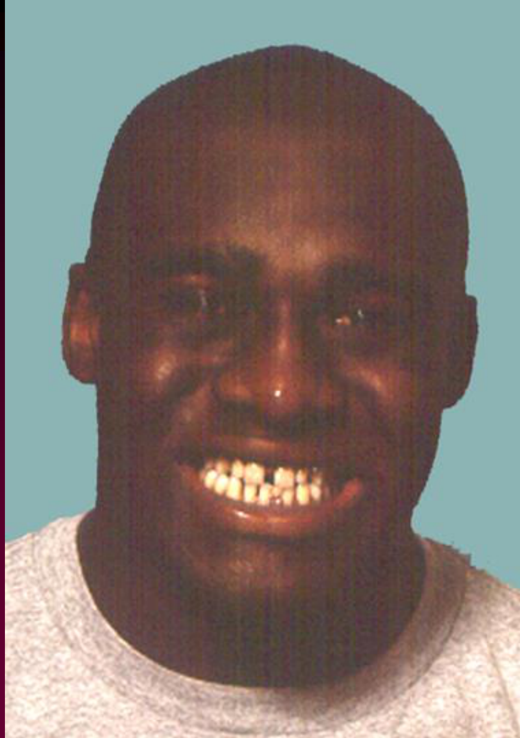
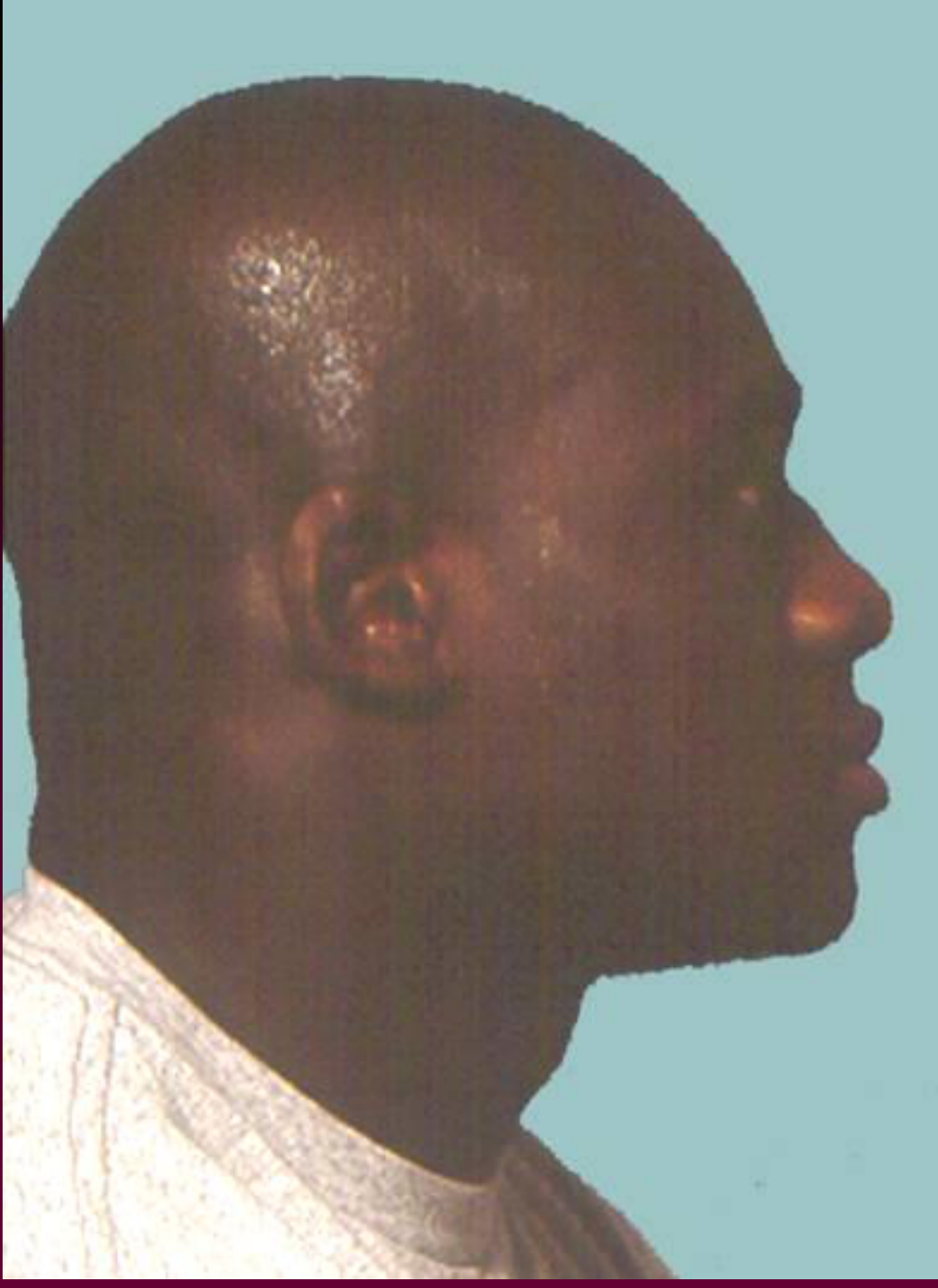
Case: Adult with Class 3 skeletal relationship and missing teeth
Question
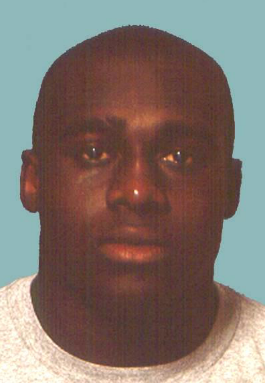
Scenario: An adult patient presents with a Class 3 skeletal relationship, mandibular protrusion, and missing teeth in the dental arches.
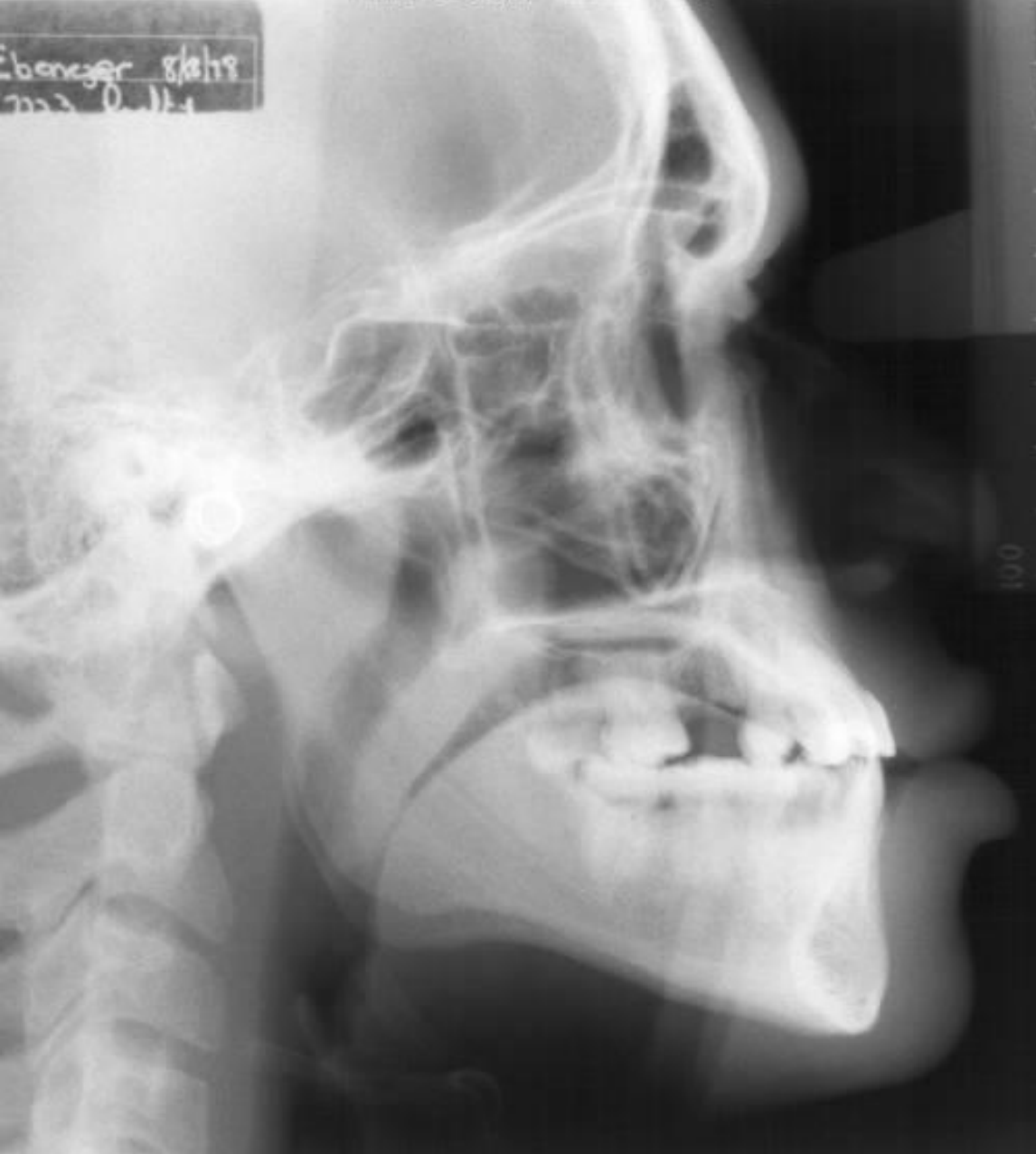
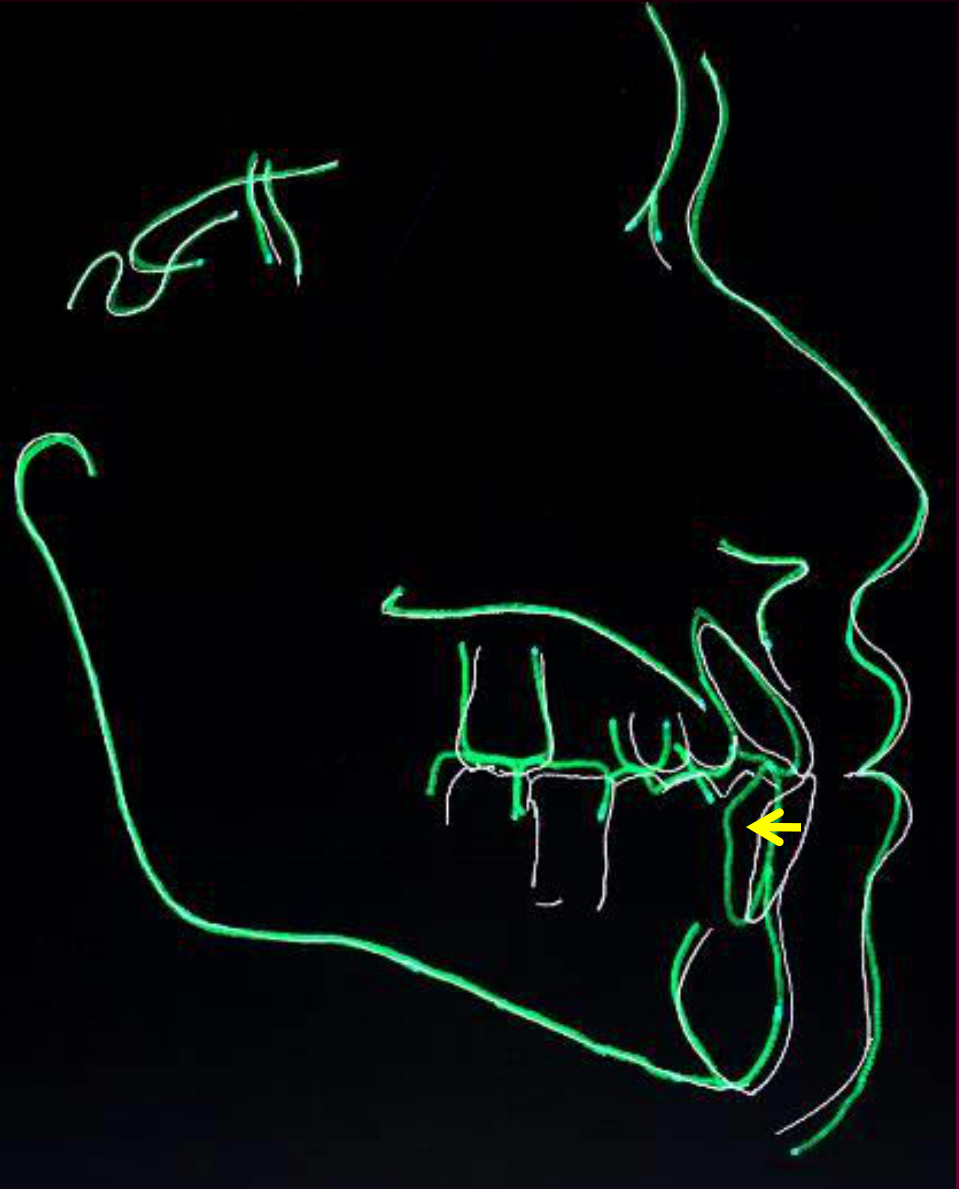
What’s shown: Intraoral clinical photographs, a panoramic radiograph, frontal and lateral facial profiles, and lateral cephalometric radiographs showing the initial and post-treatment skeletal and dental relationships.
Consider: What are the treatment options for this patient, and what specific camouflage approach was utilized to address the Class 3 relationship given the missing teeth?
Answer
Observations:
- Skeletal Class 3 pattern with the mandible positioned forward relative to the maxilla.
- Lower incisors are positioned in front of the upper incisors.
- Missing teeth in the arches, including a retained deciduous tooth and a missing bicuspid.
- Lateral cephalometric radiographs demonstrate the initial incisor protrusion and the final position after treatment.
Reasoning:
- The patient is a candidate for either orthognathic surgery or orthodontic camouflage because the Class 3 discrepancy is not extremely severe.
- Classic Class 3 camouflage (extracting upper second and lower first bicuspids) is not ideal here because the patient is already missing teeth and has a large mandible with limited posterior tooth structure.
- An alternative camouflage option is to extract a single lower incisor (the patient also lost a deciduous tooth) and retract the remaining lower anterior teeth to correct the Class 3 relationship.
Outcome:
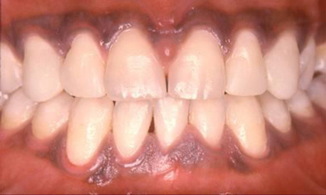
- The patient was treated with camouflage involving the extraction of a lower incisor and retraction of the lower anterior teeth.
- At the post-treatment review (age 22-8), the patient has good alignment of the teeth and a decent occlusion.
- The facial profile has not changed, and the patient is happy with his smile, though some areas require restorative treatment.
Takeaway:
- Camouflage treatment for a Class 3 skeletal relationship can involve extracting a single lower incisor to retract the lower anterior teeth, providing a viable alternative when patients have missing teeth or when surgery is not pursued.
From the lecture slides
Lateral cephalometric X-ray and tracing showing dental profile
Ebenezer E.
Age: 22-3
Finish
Ebenezer E.
Age: 22-3
Finish
Ebenezer E.
Age: 22-8
Lecturer — Treatment Outcome
The patient achieved good alignment and a reasonable occlusion and was happy with the smile, although restorative treatment was still required in some areas.
- The facial profile did not change substantially.
- The case demonstrates that there are multiple ways to camouflage a Class III relationship.
- In contrast, extractions in a Class I patient are generally performed because of crowding rather than to conceal a skeletal discrepancy.
Footnotes
-
Original PDF page 1: L1 - DENT5311 Orthodontics Lecture - Why We Do Orthodontics, p.1 ↩
-
Original PDF page 2: L1 - DENT5311 Orthodontics Lecture - Why We Do Orthodontics, p.2 ↩
-
Original PDF page 4: L1 - DENT5311 Orthodontics Lecture - Why We Do Orthodontics, p.4 ↩
-
Original PDF page 3: L1 - DENT5311 Orthodontics Lecture - Why We Do Orthodontics, p.3 ↩
-
Original PDF page 5: L1 - DENT5311 Orthodontics Lecture - Why We Do Orthodontics, p.5 ↩
-
Original PDF page 6: L1 - DENT5311 Orthodontics Lecture - Why We Do Orthodontics, p.6 ↩
-
Original PDF page 7: L1 - DENT5311 Orthodontics Lecture - Why We Do Orthodontics, p.7 ↩
-
Original PDF page 8: L1 - DENT5311 Orthodontics Lecture - Why We Do Orthodontics, p.8 ↩
-
Original PDF page 9: L1 - DENT5311 Orthodontics Lecture - Why We Do Orthodontics, p.9 ↩
-
Original PDF page 10: L1 - DENT5311 Orthodontics Lecture - Why We Do Orthodontics, p.10 ↩
-
Original PDF page 11: L1 - DENT5311 Orthodontics Lecture - Why We Do Orthodontics, p.11 ↩
-
Original PDF page 12: L1 - DENT5311 Orthodontics Lecture - Why We Do Orthodontics, p.12 ↩
-
Original PDF page 13: L1 - DENT5311 Orthodontics Lecture - Why We Do Orthodontics, p.13 ↩
-
Original PDF page 14: L1 - DENT5311 Orthodontics Lecture - Why We Do Orthodontics, p.14 ↩
-
Original PDF page 15: L1 - DENT5311 Orthodontics Lecture - Why We Do Orthodontics, p.15 ↩
-
Original PDF page 16: L1 - DENT5311 Orthodontics Lecture - Why We Do Orthodontics, p.16 ↩
-
Original PDF page 17: L1 - DENT5311 Orthodontics Lecture - Why We Do Orthodontics, p.17 ↩
-
Original PDF page 18: L1 - DENT5311 Orthodontics Lecture - Why We Do Orthodontics, p.18 ↩
-
Original PDF page 19: L1 - DENT5311 Orthodontics Lecture - Why We Do Orthodontics, p.19 ↩
-
Original PDF page 20: L1 - DENT5311 Orthodontics Lecture - Why We Do Orthodontics, p.20 ↩
-
Original PDF page 23: L1 - DENT5311 Orthodontics Lecture - Why We Do Orthodontics, p.23 ↩
-
Original PDF page 21: L1 - DENT5311 Orthodontics Lecture - Why We Do Orthodontics, p.21 ↩
-
Original PDF page 22: L1 - DENT5311 Orthodontics Lecture - Why We Do Orthodontics, p.22 ↩
-
Original PDF page 24: L1 - DENT5311 Orthodontics Lecture - Why We Do Orthodontics, p.24 ↩
-
Original PDF page 25: L1 - DENT5311 Orthodontics Lecture - Why We Do Orthodontics, p.25 ↩
-
Original PDF page 26: L1 - DENT5311 Orthodontics Lecture - Why We Do Orthodontics, p.26 ↩
-
Original PDF page 27: L1 - DENT5311 Orthodontics Lecture - Why We Do Orthodontics, p.27 ↩
-
Original PDF page 28: L1 - DENT5311 Orthodontics Lecture - Why We Do Orthodontics, p.28 ↩
-
Original PDF page 29: L1 - DENT5311 Orthodontics Lecture - Why We Do Orthodontics, p.29 ↩
-
Original PDF page 30: L1 - DENT5311 Orthodontics Lecture - Why We Do Orthodontics, p.30 ↩
-
Original PDF page 31: L1 - DENT5311 Orthodontics Lecture - Why We Do Orthodontics, p.31 ↩
-
Original PDF page 32: L1 - DENT5311 Orthodontics Lecture - Why We Do Orthodontics, p.32 ↩
-
Original PDF page 33: L1 - DENT5311 Orthodontics Lecture - Why We Do Orthodontics, p.33 ↩
-
Original PDF page 34: L1 - DENT5311 Orthodontics Lecture - Why We Do Orthodontics, p.34 ↩
-
Original PDF page 35: L1 - DENT5311 Orthodontics Lecture - Why We Do Orthodontics, p.35 ↩
-
Original PDF page 36: L1 - DENT5311 Orthodontics Lecture - Why We Do Orthodontics, p.36 ↩
-
Original PDF page 37: L1 - DENT5311 Orthodontics Lecture - Why We Do Orthodontics, p.37 ↩
-
Original PDF page 38: L1 - DENT5311 Orthodontics Lecture - Why We Do Orthodontics, p.38 ↩
-
Original PDF page 39: L1 - DENT5311 Orthodontics Lecture - Why We Do Orthodontics, p.39 ↩
-
Original PDF page 40: L1 - DENT5311 Orthodontics Lecture - Why We Do Orthodontics, p.40 ↩
-
Original PDF page 41: L1 - DENT5311 Orthodontics Lecture - Why We Do Orthodontics, p.41 ↩
-
Original PDF page 42: L1 - DENT5311 Orthodontics Lecture - Why We Do Orthodontics, p.42 ↩
-
Original PDF page 43: L1 - DENT5311 Orthodontics Lecture - Why We Do Orthodontics, p.43 ↩
-
Original PDF page 44: L1 - DENT5311 Orthodontics Lecture - Why We Do Orthodontics, p.44 ↩
-
Original PDF page 45: L1 - DENT5311 Orthodontics Lecture - Why We Do Orthodontics, p.45 ↩
-
Original PDF page 46: L1 - DENT5311 Orthodontics Lecture - Why We Do Orthodontics, p.46 ↩
-
Original PDF page 47: L1 - DENT5311 Orthodontics Lecture - Why We Do Orthodontics, p.47 ↩
-
Original PDF page 48: L1 - DENT5311 Orthodontics Lecture - Why We Do Orthodontics, p.48 ↩
-
Original PDF page 49: L1 - DENT5311 Orthodontics Lecture - Why We Do Orthodontics, p.49 ↩
-
Original PDF page 50: L1 - DENT5311 Orthodontics Lecture - Why We Do Orthodontics, p.50 ↩
-
Original PDF page 51: L1 - DENT5311 Orthodontics Lecture - Why We Do Orthodontics, p.51 ↩
-
Original PDF page 52: L1 - DENT5311 Orthodontics Lecture - Why We Do Orthodontics, p.52 ↩
-
Original PDF page 53: L1 - DENT5311 Orthodontics Lecture - Why We Do Orthodontics, p.53 ↩
-
Original PDF page 54: L1 - DENT5311 Orthodontics Lecture - Why We Do Orthodontics, p.54 ↩
-
Original PDF page 55: L1 - DENT5311 Orthodontics Lecture - Why We Do Orthodontics, p.55 ↩
-
Original PDF page 56: L1 - DENT5311 Orthodontics Lecture - Why We Do Orthodontics, p.56 ↩
-
Original PDF page 57: L1 - DENT5311 Orthodontics Lecture - Why We Do Orthodontics, p.57 ↩