UNC Level III - Unit B1
Space Management in Preadolescent Children2
- Discuss the indications and contraindications for the selection of space maintaining appliances.
- Discuss the indications and contraindications for the selection of various space regaining appliances.
- Discuss the construction of different appliances.
- Describe the timing and sequence of treatment.
- Identify the possible causes of midline diastemas and spacing.
- Discuss the relationship between spacing and protrusion.
Space Maintaining Appliances3
- Discuss the indications and contraindications for the selection of space maintaining appliances.
- Fixed
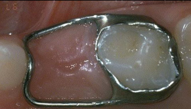
- Band and loop
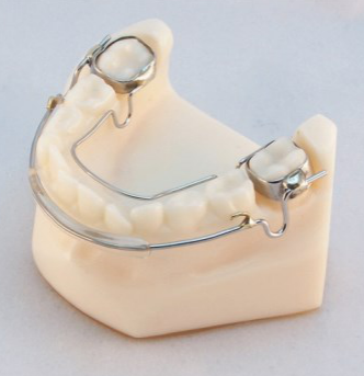
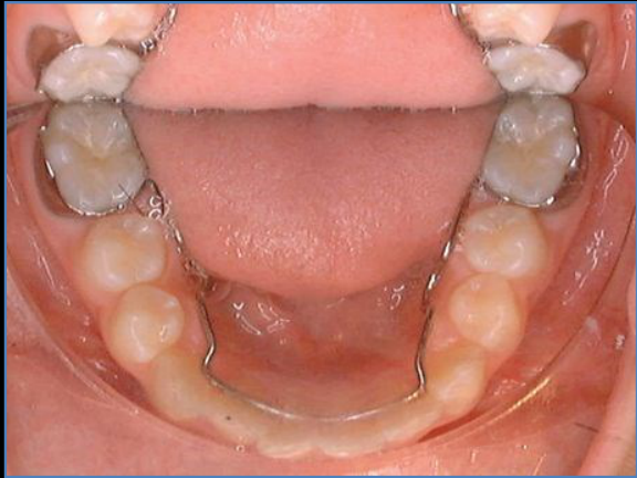
- Lingual arch
- Nance holding arch
- Distal shoe
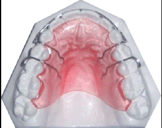
- Removable
- An acrylic removable plate is an alternative, but removable appliances require patient cooperation and younger patients need supervision from parents or carers.
- Fixed
Posterior Space Maintenance—Pathways of Care4
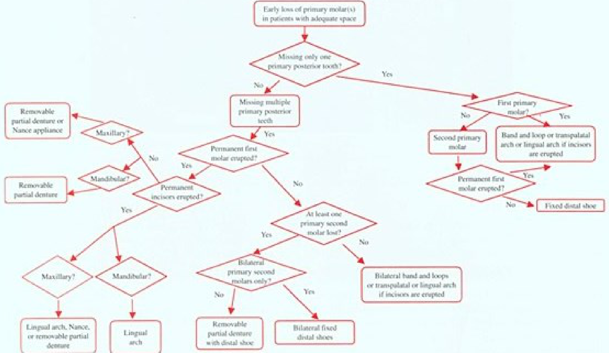
Early loss of primary molar(s) in patients with adequate space.
Lecturer — Space Decisions
The clinical decision should incorporate several features beyond whether space appears adequate.
- Assess the dental age, the amount of premolar root development, and whether a permanent tooth is absent.
- If space has been lost, space regaining rather than space maintenance may be considered.
- The yes-or-no pathways help connect diagnosis with the appropriate appliance and provide a useful clinical decision-making and revision tool.
Flowchart re-transcribed from the figure (2026-10-23)
The OCR scrambles these branches; this version was re-read directly from FIGURE 13-5 below (re-checked against the figure 2026-10-31).
Missing only one primary posterior tooth?
- Yes — First primary molar?
- Yes → Band and loop or transpalatal arch or lingual arch if incisors are erupted
- No — Second primary molar → Permanent first molar erupted?
- Yes → Band and loop or transpalatal arch or lingual arch if incisors are erupted
- No → Fixed distal shoe
Missing multiple primary posterior teeth
- Permanent first molar erupted?
- Yes — Permanent incisors erupted?
- Yes
- Maxillary? → Lingual arch, Nance, or removable partial denture
- Mandibular? → Lingual arch
- No
- Maxillary? → Removable partial denture or Nance appliance
- Mandibular? → Removable partial denture
- Yes
- No — At least one primary second molar lost?
- Yes — Bilateral primary second molars only?
- Yes → Bilateral fixed distal shoes
- No → Removable partial denture with distal shoe
- No → Bilateral band and loops or transpalatal or lingual arch if incisors are erupted
- Yes — Bilateral primary second molars only?
- Yes — Permanent incisors erupted?
Space Regaining Appliances5
- Discuss the indications and contraindications for the selection of various space regaining appliances.
Up to 3 mm
- Fixed
- Spring appliances with braces
- Active lingual arches
- Fixed distalising appliances
- Removable
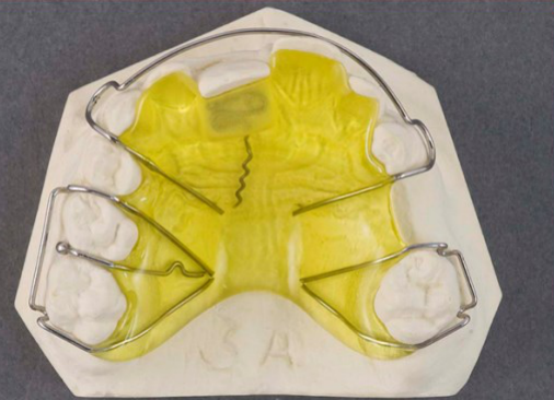
- URA with spring
- URA with screw
- Lip bumper
Lecturer — Space Regaining
Space loss of approximately 3 mm is often recoverable, whereas loss beyond 3 mm is more difficult to regain.
- Tipping of the permanent first molars commonly occurs during the first six months after early primary molar loss.
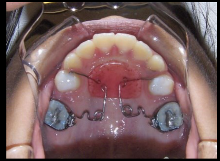
- An activated lingual arch can upright the molars, and a removable spring appliance can upright a lower first molar to recover space for the premolar.
- A lip bumper transfers force from the lower lip musculature to the molars and can assist with uprighting.
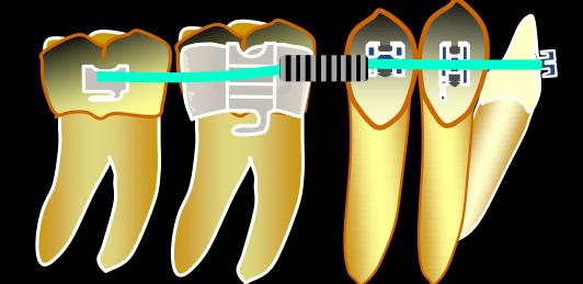
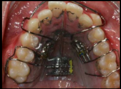
- A pendulum appliance distalizes both upper molars using springs engaged in the molar tubes; it is bonded to the premolars and supported by an acrylic palatal plate similar to a Nance button.
- Recovering approximately 3 mm may prevent extraction of a premolar.
 |  |  |
 |  |  |
Appliance Construction6
- Discuss the construction of different appliances.
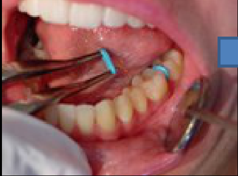
- Removable — Impression → Insertion
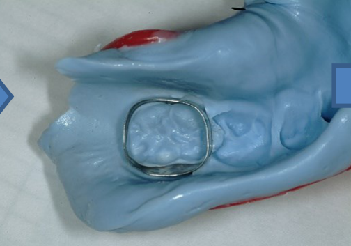
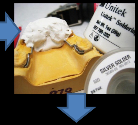
- Fixed — Separators → Fit bands → placed in impression → wire bent and soldered → Cemented in mouth
Lecturer — Lingual Arch Construction
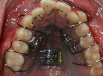
The lingual arch construction sequence includes transferring the bands to the impression and casting the model with the bands in their clinical positions.
- Separators are generally left in place for approximately one week.
- They remove enough contact to permit band placement, but the space closes quickly if separators are removed without proceeding with band placement.
- The wire is connected to the bands using silver soldering.
 |  |
 |  |
Timing and Sequence of Treatment7
- Describe the timing and sequence of treatment.
- URA
Lecturer — Treatment Timing
Timing depends on space availability, dental age, and the development of the unerupted permanent teeth.
- A premolar with more than two-thirds of its root formed is considered close to eruption; if adequate space is present, follow-up may be sufficient.
- With less than two-thirds of root formation, the permanent first molar may tip and block the premolar.
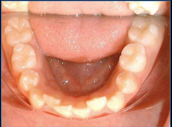
- In a younger patient whose premolar eruption is not expected until approximately 11 or 12 years of age, a lingual arch may prevent major molar tipping.
- Treatment planning must also account for the patient’s ability to attend appointments.
 |  |
 |  |
Causes of Midline Diastemas and Spacing8
- Identify the possible causes of midline diastemas and spacing.
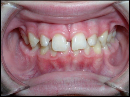
- Normal development (ugly duckling)
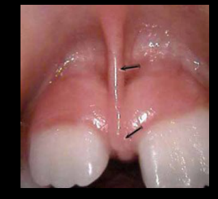
- Frenum
- Proclined teeth
- Small teeth (laterals)
- Missing laterals
- Mesiodens or pathology
Lecturer — Midline Spacing
During the ugly duckling phase, the lateral incisors may appear tilted because the permanent canines are positioned for later eruption, and the space may close spontaneously as the canines erupt.
- Immediate closure is not necessarily indicated.
- The root of a lateral incisor should not be moved against the crown of the canine because root resorption may result.
- A lower frenum may require surgical removal; spaces of approximately 2 mm may close spontaneously, whereas larger spaces require monitoring to determine the cause and need for treatment.
 |  |
Relationship Between Spacing and Protrusion9
- Discuss the relationship between spacing and protrusion.
Lecturer — Spacing and Protrusion
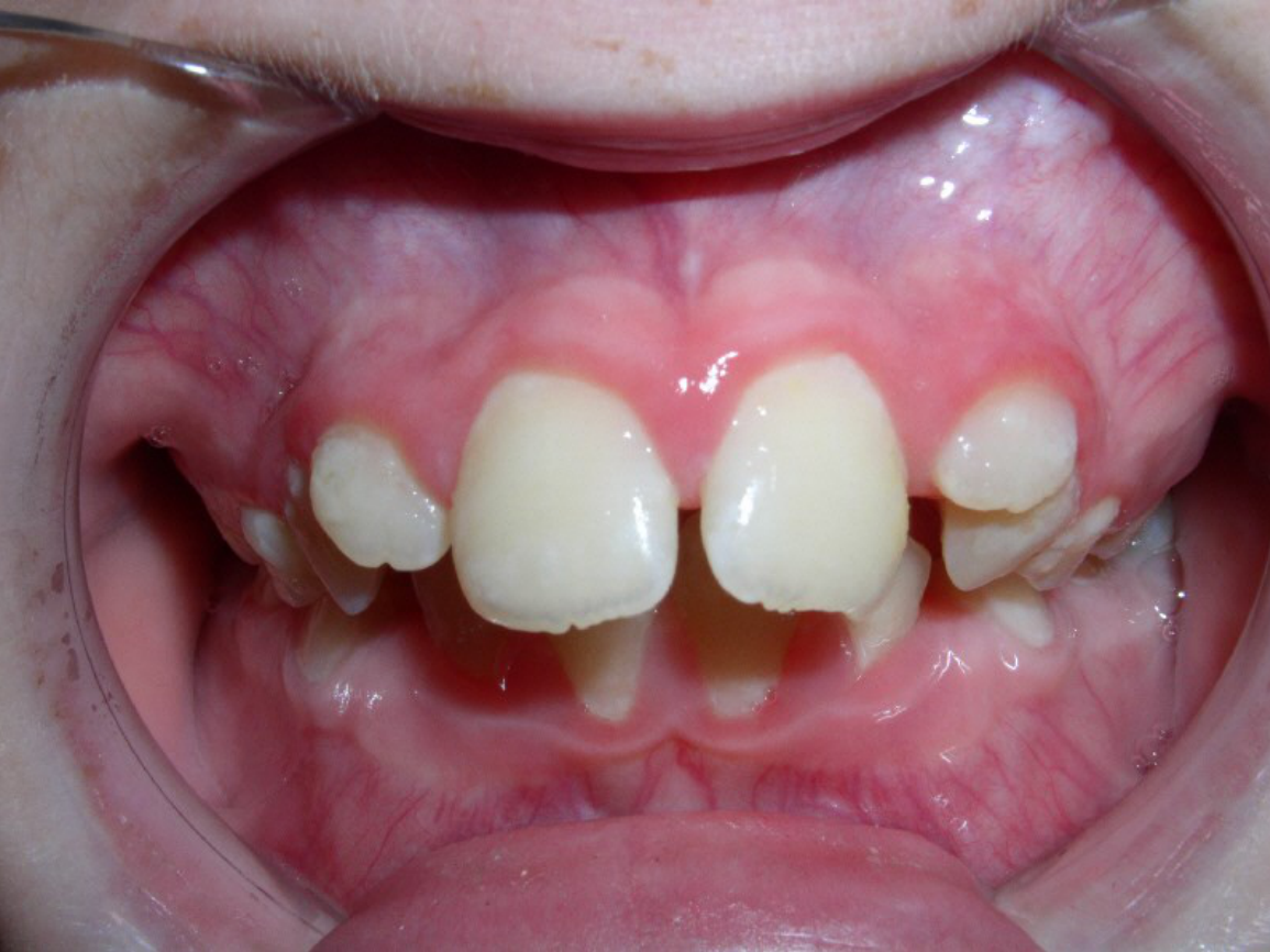
Proclined incisors can create a false impression of increased space.
- When the upper incisors are proclined beyond approximately 22 degrees, the available space may be overestimated.
- Uprighting the incisors or correcting the flaring can reveal the underlying lack of space.
Potential Crowding and Space Maintenance10
- Describe diagnostic information necessary to make decisions concerning space maintenance or management.
- Identify and discuss alternative space analysis procedures.
- Discuss the importance of determining the etiology of tooth loss.
- Discuss the phenomenon of space loss.
- List factors that weigh in the decision whether to maintain or regain space.
- Discuss the relevancy of the above factors to space management.
Diagnostic Information for Space Management11
- Describe diagnostic information necessary to make decisions concerning space maintenance or management.
-
If deciduous tooth lost early — when?
-
How much space loss?
-
Dental age and future eruption/growth changes
-
Agenesis?
-
Facial form — lip competence and fullness
-
Obtain an orthopantomogram and assess the amount of premolar root development.
-
Determine whether adequate space remains and whether permanent teeth are present.
-
These findings guide the choice between space maintenance, space regaining, and follow-up without immediate intervention.
Space Analysis Procedures12
- Identify and discuss alternative space analysis procedures.
-
Measuring radiographs and teeth
-
Using tables
-
Correlation tables, measuring incisors and predicting 345
-
Moyers
-
Tanaka and Johnston
-
The Tanaka-Johnston analysis uses the lower incisors.
-
Model analysis and prediction tables can help determine whether enough space will be available for unerupted teeth.
Etiology of Tooth Loss
- Discuss the importance of determining the etiology of tooth loss.
Big Teeth/Small Jaws13
- Expansion
- Space maintenance — leeway space
- Extraction
Lecturer — Lingual Arch
A lingual arch may be used even when no primary teeth have been lost.
- It prevents major mesial drift of the permanent first molars and preserves the leeway space.
- The preserved space can be transferred toward the anterior region to improve lower anterior crowding.
- Because the molars do not drift mesially into Class I, the upper molars may later need to be distalized to establish a Class I relationship.
- In one case, the primary teeth were large and the lower arch had approximately 3 mm of potential space on each side.
Early Loss (Crowding/Pathology)
-
Space regaining (drift)
-
Space maintenance
-
Extraction
-
Early loss of primary molars can cause mesial tipping of the permanent first molars, loss of premolar space, and blocked or delayed premolar eruption.
-
The decision to maintain or regain space depends on the amount lost and how close the premolar is to eruption.
The Phenomenon of Space Loss
- Discuss the phenomenon of space loss.
Crowding — Early Loss14
- Dental age — when do new teeth emerge
- E’s lose more, E’s > D > C
- Rate of loss: uppers > lowers
Timing of Space Loss
-
Lose space within 3 months
-
Faster in first 6 months, then slows
-
Approximately 3 mm is the practical threshold for space regaining: up to 3 mm may be recoverable, whereas recovery beyond 3 mm is more difficult.
-
If premolar root development is limited, the molar may need to be uprighted and space maintained or regained.
-
Treatment timing should account for the date of tooth loss, molar tipping, dental age, premolar root formation, and anticipated premolar eruption.
Factors in Deciding to Maintain or Regain Space15
- List factors that weigh in the decision whether to maintain or regain space.
-
When lost?
-
Why? Crowding? Pathology?
-
When will future teeth emerge? Agenesis?
-
How much crowding — lips/face
-
Also consider whether one or multiple primary molars were lost, the degree of premolar root development, the patient’s ability to cooperate, and regular appointment attendance.
Relevance of Factors to Space Management16
- Discuss the relevancy of the above factors to space management.
-
Minimal space loss and teeth soon to emerge — leave and observe
-
3mm crowding — refer
- Extraction/expansion — significant rx
-
Adequate space with a premolar close to eruption may require only follow-up.
-
Inadequate space with limited premolar root development may require space maintenance or regaining.
-
Early treatment may prevent further molar tipping and space regaining may prevent premolar extraction.
-
Travel distance and appointment availability should be considered when selecting an appliance.
Crossbites and Vertical Problems in Children17
- Identify skeletal contributions to anterior crossbites.
- Identify dental contributions to anterior crossbites.
- Identify the significance of functional shifts in anterior crossbites.
- Evaluate space, tooth orientation and position, intermaxillary relationships, and eruption timing and sequence regarding the teeth involved in the anterior crossbites as etiologic and treatment factors.
- State rationale for correcting anterior crossbites.
- Recommend appropriate treatment for anterior crossbites including timing and appliance design.
- Explain the relevance of facial form to posterior crossbite diagnosis.
- List the etiologic factors of posterior crossbite.
- Describe the clinical findings consistent with a bilateral maxillary constriction, a bilateral constriction accompanied by a mandibular shift, and a true unilateral maxillary constriction.
- Describe the rationale for correcting posterior crossbites.
- Describe the appropriate timing and appliance design for posterior crossbite treatment.
Skeletal Contributions to Anterior Crossbites1819202122
- Identify skeletal contributions to anterior crossbites.
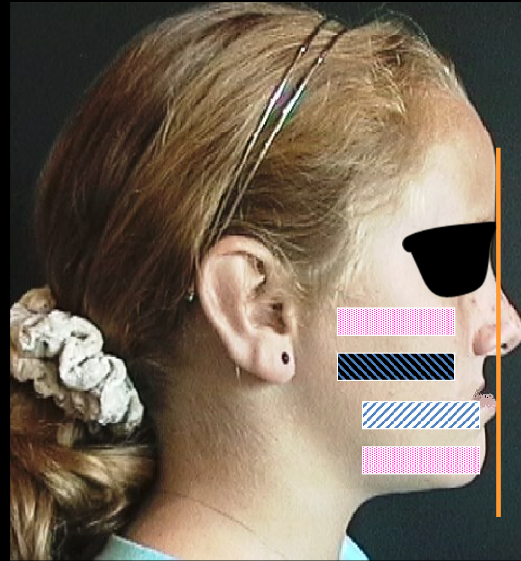
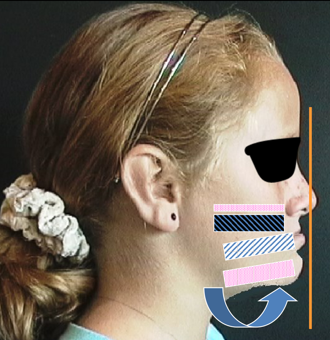
Maxilla back
-
Maxilla back
-
Mandible forward
-
Maxilla vertically deficient
-
Mandible rotated up and forward
Lecturer — Skeletal Patterns
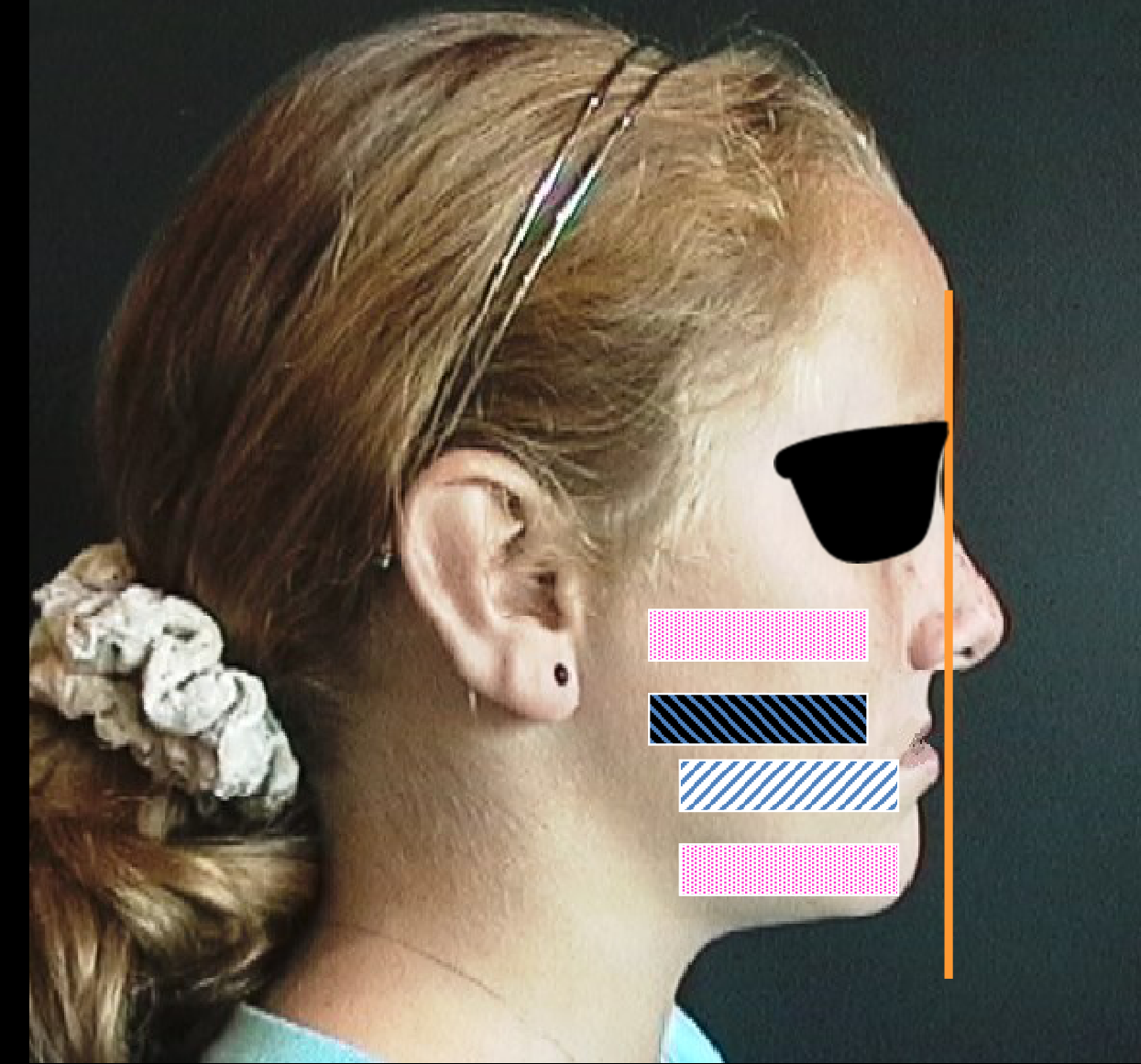
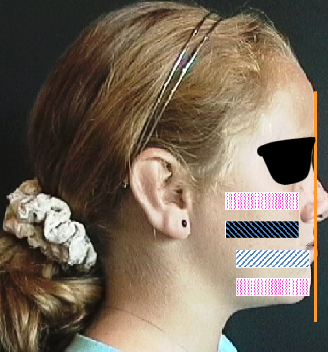
An anterior crossbite may result from skeletal discrepancies involving the maxilla, mandible, or both.
- A retruded or deficient maxilla
- A protruded mandible
- A combination of maxillary deficiency and mandibular protrusion
- When the maxilla is vertically deficient, the mandible may rotate anticlockwise to achieve tooth contact.
Dental Contributions to Anterior Crossbites23242526
- Identify dental contributions to anterior crossbites.
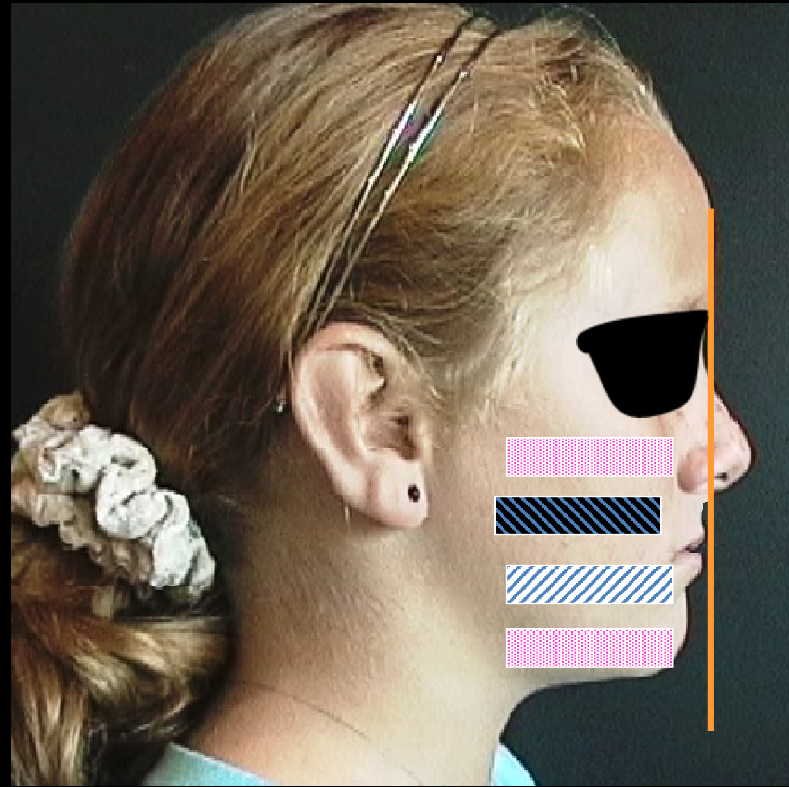
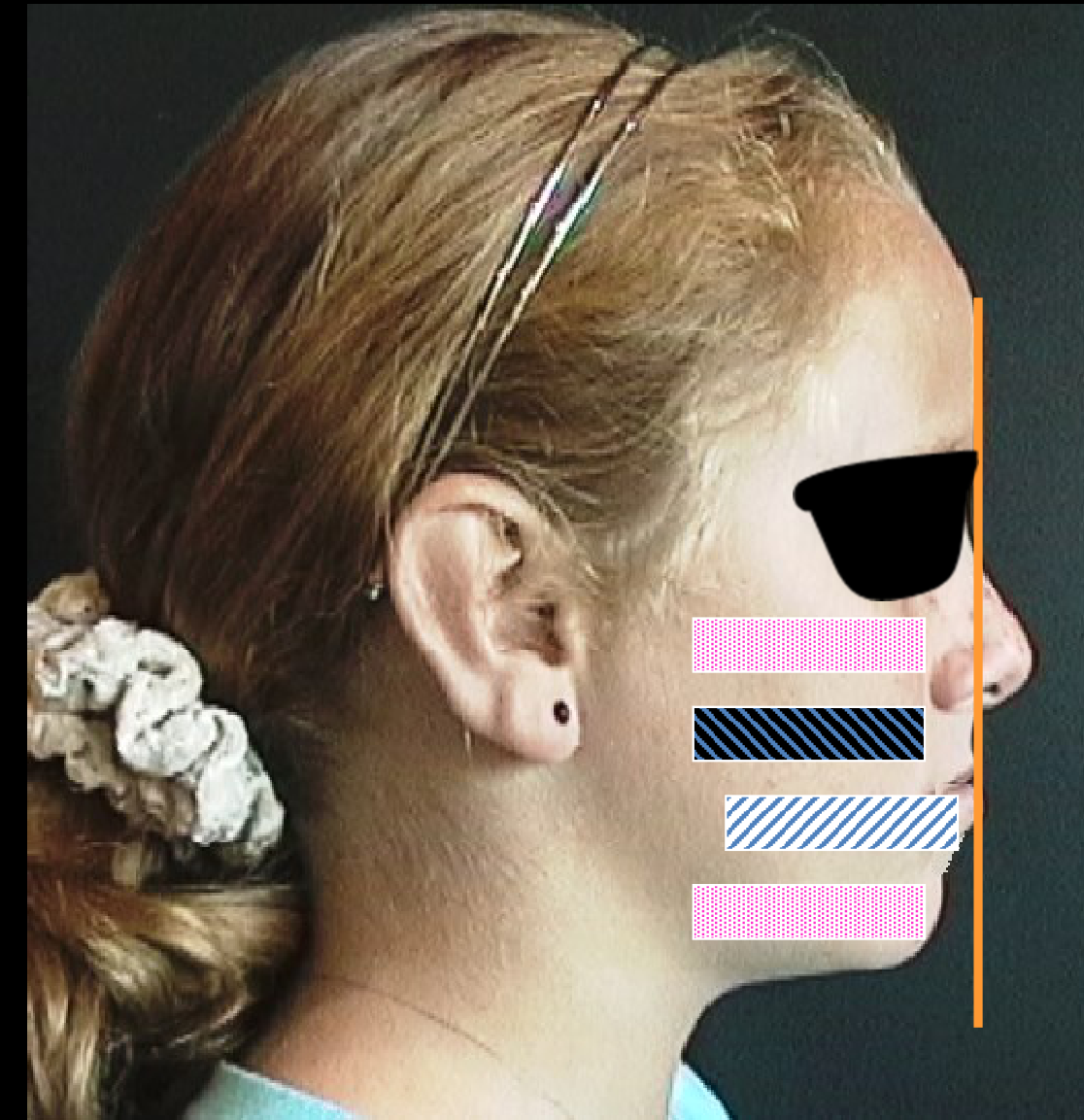
Maxillary teeth back
Mandibular teeth forward
- Maxillary teeth back
- Mandibular teeth forward
Lecturer — Dental Patterns
An anterior crossbite may occur despite a normal skeletal relationship.
- Retroclined upper incisors, as in a Class II Division 2 pattern
- Proclined lower incisors associated with lower anterior crowding
- A maxillary tooth that is tipped or positioned incorrectly
- The skeletal relationship may be Class I even though the dental relationship produces the anterior crossbite.
Significance of Functional Shifts in Anterior Crossbites27282930
- Identify the significance of functional shifts in anterior crossbites.
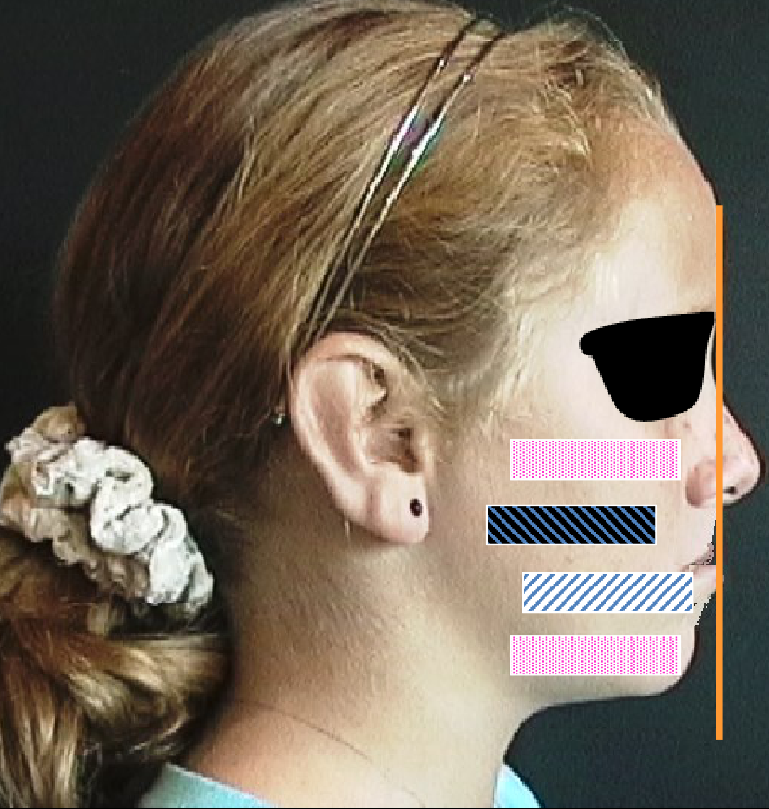
Pseudo Class III’s are special
- Teeth hit edge to edge in RCP
- Shift forward to ICP
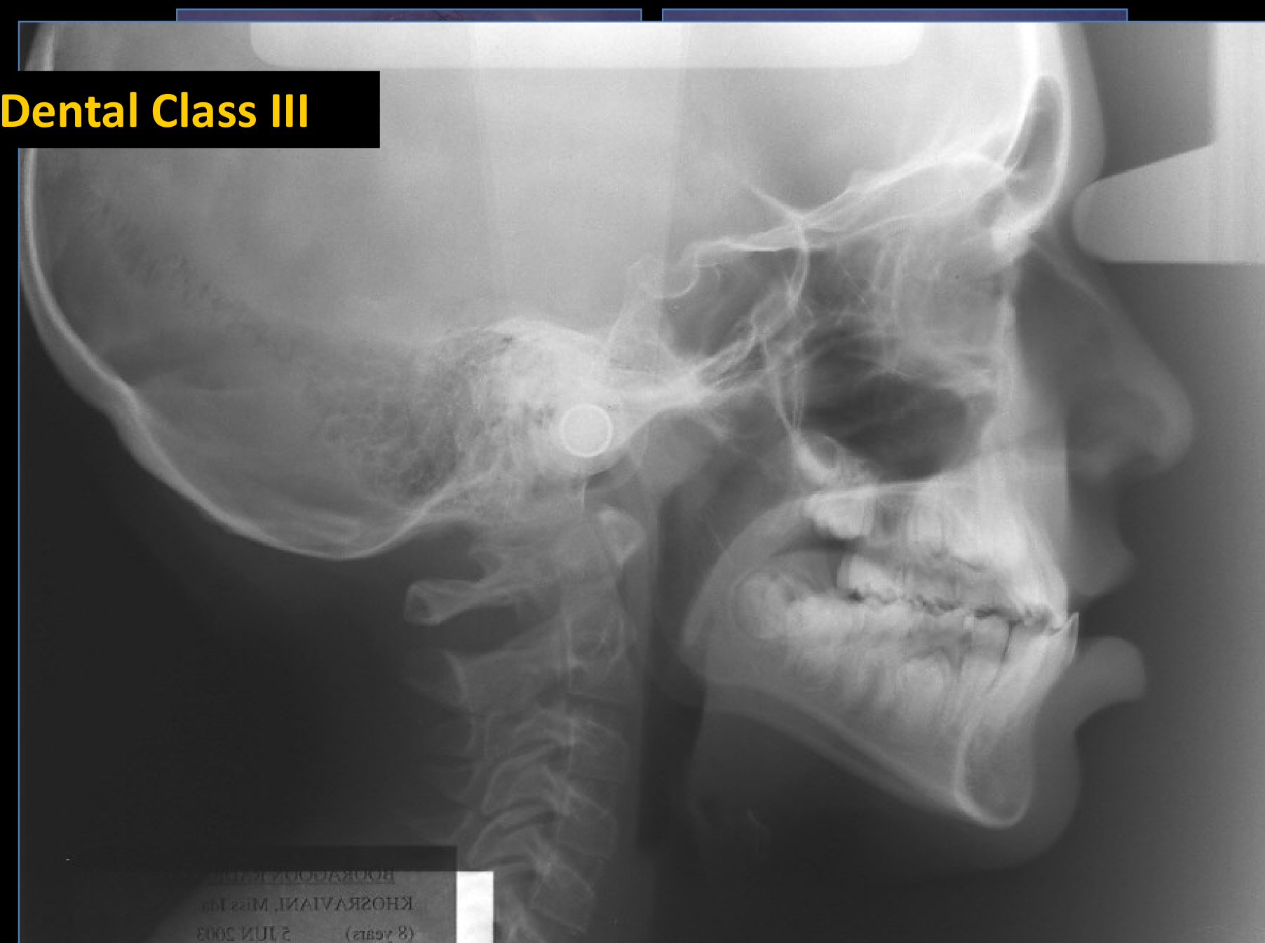
Dental Class III
Lecturer — Functional Shifts
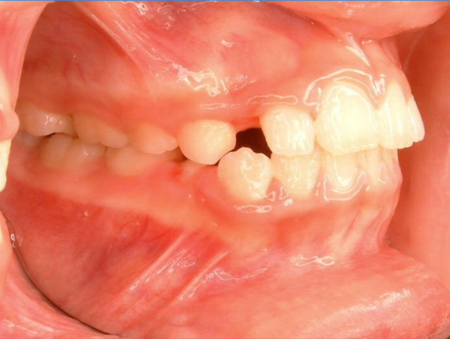
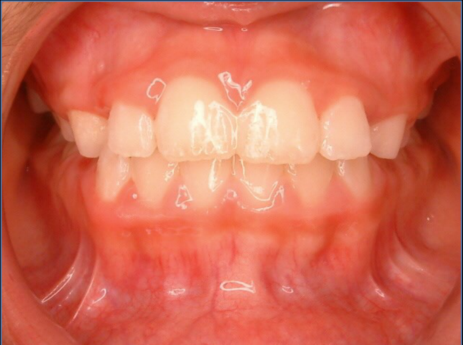
A functional shift may occur when the incisors meet edge to edge in the retruded contact position.
- The edge-to-edge position may be unstable or uncomfortable.
- The patient shifts the mandible forward to reach a more comfortable intercuspal position.
- The resulting anterior crossbite is termed a pseudo-Class III relationship.
- The molar relationship may remain Class I.
- Manipulation of the mandible is necessary to distinguish the retruded contact position from the intercuspal position.
FUJI AD MAMMO MEDIUM 290361 (8 years) 21UN 2003 KHOSRAVANI, Miss Ma ROURAGOON KATIN
 |  |  |
 |  |
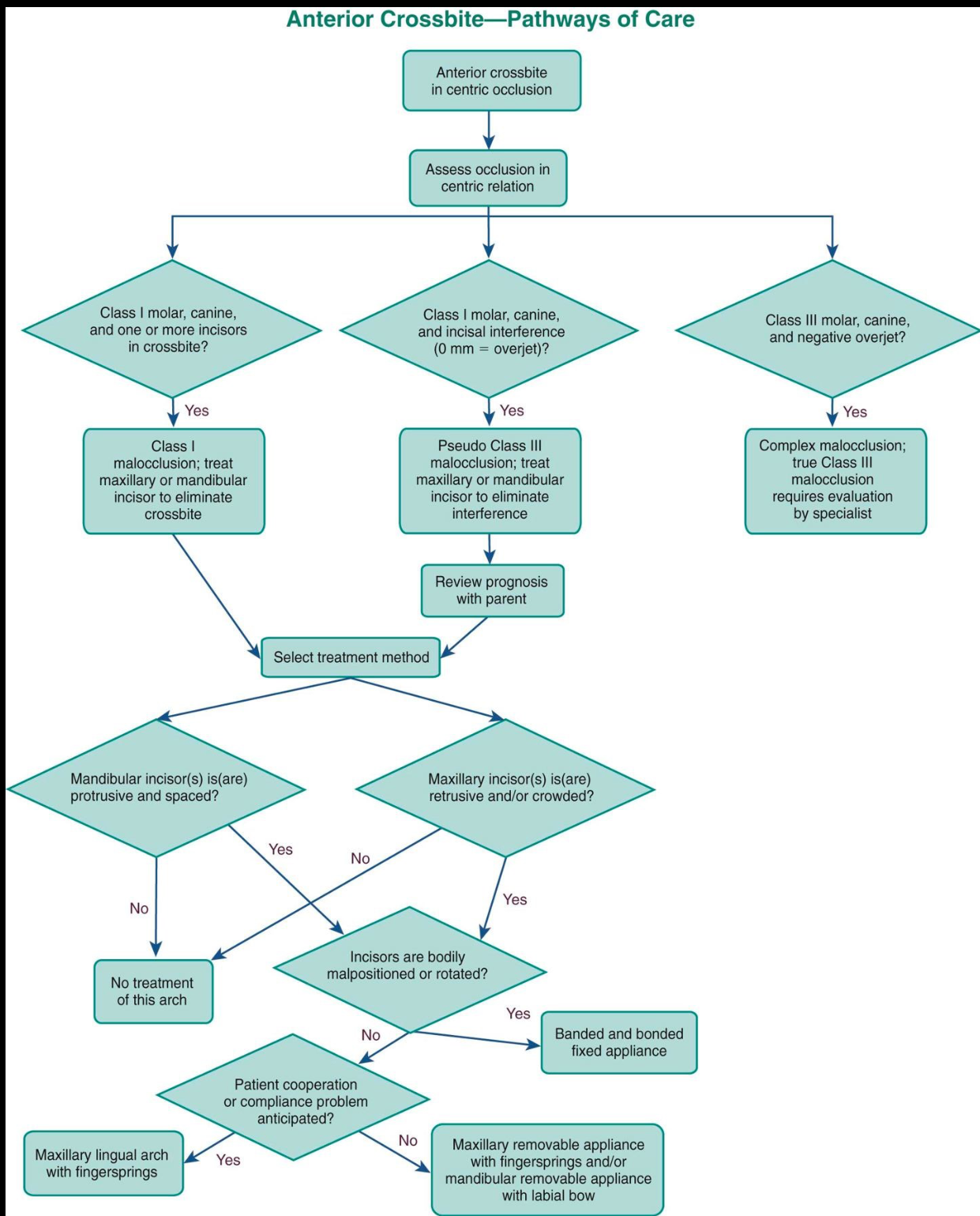
Evaluating Space, Tooth Position, and Eruption Timing in Anterior Crossbites3132
- Evaluate space, tooth orientation and position, intermaxillary relationships, and eruption timing and sequence regarding the teeth involved in the anterior crossbites as etiologic and treatment factors.
Lecturer — Diagnostic Assessment
Diagnosis requires assessment of the skeletal relationship on the cephalometric radiograph and the position and inclination of the incisors.
- Determine whether the crossbite involves one tooth or several teeth.
- Assess whether a functional anterior shift is present.
- Compare the retruded contact position with the intercuspal position.
- Assess the available space for alignment.
Rationale for Correcting Anterior Crossbites33
- State rationale for correcting anterior crossbites.
- Prevent displacement and aberrant movement patterns/wear
- Prevent perio problems
- Function RCP/MI problems
- Create space for alignment — expand anteriorly
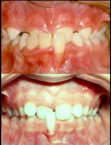
Lecturer — Incisor Trauma
When one lower incisor is crowded and pushed by the other incisors, it may progressively detach from the bone.
- Significant gingival recession may develop.
- The tooth may become displaced from the symphysis.
Treatment for Anterior Crossbites3435
- Recommend appropriate treatment for anterior crossbites including timing and appliance design.
- Single tooth — URA ASAP — Space? Strip, extract
- Multiple teeth? Class III? Shift
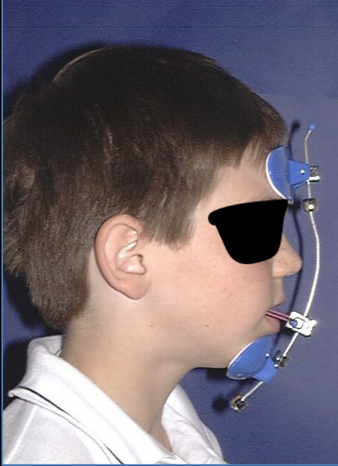
- URA or Facemask protraction
- Part bands — comprehensive treatment
Lecturer — Treatment Selection
Treatment depends on whether the problem is dental or skeletal.
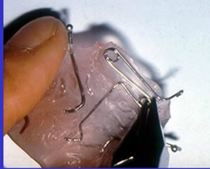
- A single dental crossbite may be treated with a spring on a removable upper appliance.
- Partial braces may bond the affected incisors and molars.
- Arch-wire activation can procline retroclined incisors.
- A skeletal problem may require maxillary protraction, guided by the cephalometric assessment.
Adult Class III
 |  |
 |
Compensation36
- Elastics
- Extractions
- TADS
Surgery
- Orthognathics
- Distraction
Lecturer — Class III Camouflage
In an adult skeletal Class III relationship that can be camouflaged, elastics may be used from the canine to the upper first molar when the problem is dental and suitable for camouflage.
- Retention is important because the corrected relationship may be unstable and prone to relapse.
- If the discrepancy cannot be corrected by compensation or camouflage, orthognathic surgery may be required.
Diagram of orthodontic elastics on teeth with brackets and colored lines.
Facial Form and Posterior Crossbite Diagnosis373839
- Explain the relevance of facial form to posterior crossbite diagnosis.
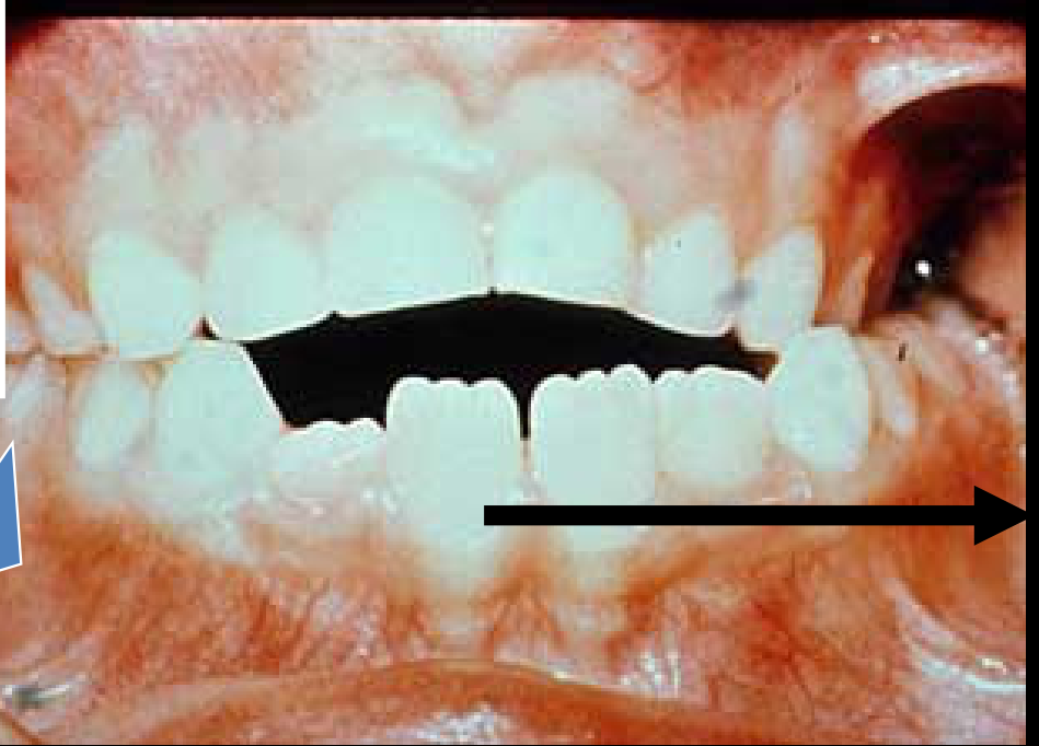
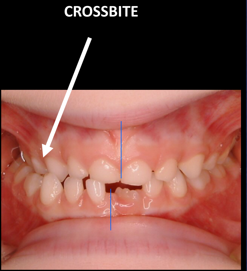
- End to end occlusion — RCP
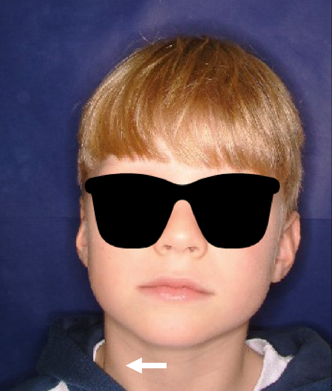
- Mandible has moved forwards on right
- Shift to right — ICP
CROSSBITE
Lecturer — Functional Asymmetry
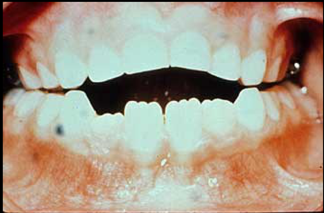
A unilateral posterior crossbite may result from a functional shift rather than a true skeletal asymmetry.
- The posterior teeth may meet edge to edge in the retruded contact position before the mandible shifts to one side for intercuspal contact.
- The chin and lower dental midline deviate toward the side of the shift.
- Diagnosis should compare the retruded contact position and intercuspal position, assess maxillary arch symmetry, and determine whether the crossbite is unilateral or bilateral.
 |  |
 |  |
Etiologic Factors of Posterior Crossbite40414243
- List the etiologic factors of posterior crossbite.
- Habits
- Breathing? Nasorespiratory function
- Genetics
- Class III
- Narrow maxilla/Broad mandible
Differential diagnosis
- Skeletal crossbite
- Dental crossbite (slide from Ackerman Proffit)
Lecturer — Posterior Etiology
Posterior crossbite may involve a narrow or constricted maxillary arch, a dental discrepancy in the posterior teeth, a functional mandibular shift, or a skeletal transverse discrepancy.
- If the upper arch is severely constricted, the upper first molars may be held inside the lower molars and produce no functional shift.
- A shift is more likely when the posterior teeth meet edge to edge.
Clinical Findings in Posterior Crossbites44
- Describe the clinical findings consistent with a bilateral maxillary constriction, a bilateral constriction accompanied by a mandibular shift, and a true unilateral maxillary constriction.
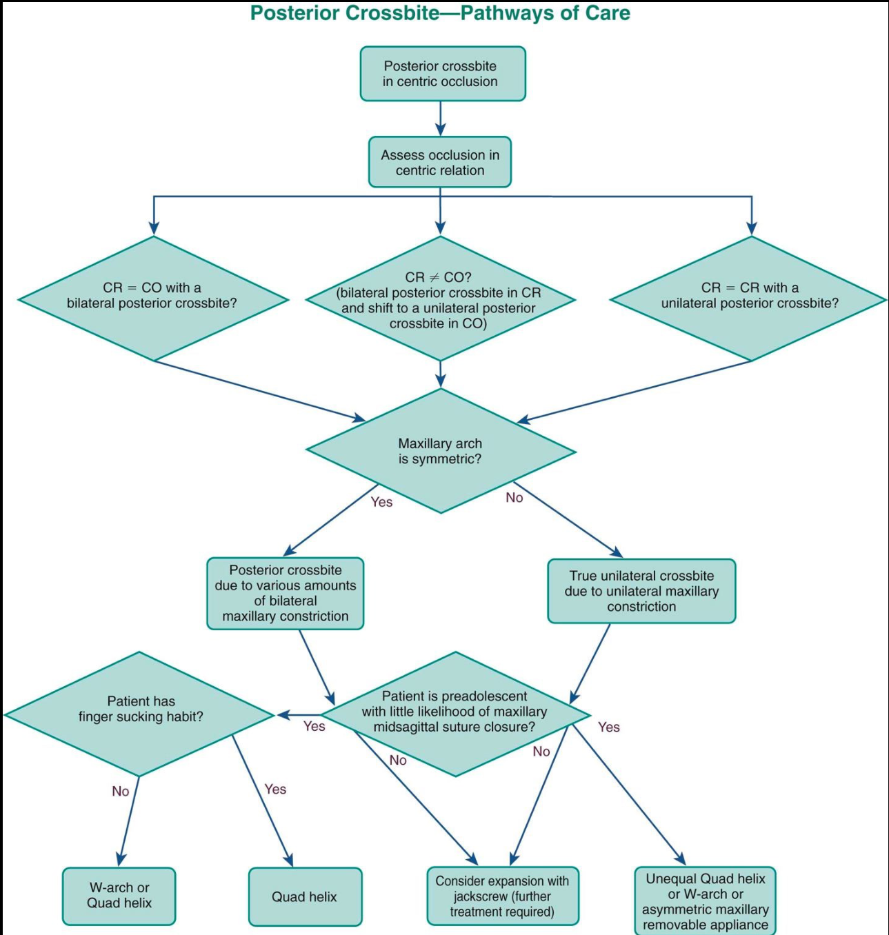
Posterior Crossbite — Pathways of Care45
- Posterior crossbite in centric occlusion → assess occlusion in centric relation.
- Assess occlusion in centric relation → three possibilities:
- CR = CO with a bilateral posterior crossbite?
- CR ≠ CO? (bilateral posterior crossbite in CR and shift to a unilateral posterior crossbite in CO)
- CR = CR with a unilateral posterior crossbite?
- All three pathways → is the maxillary arch symmetric?
- Yes → posterior crossbite due to various amounts of bilateral maxillary constriction.
- No → true unilateral crossbite due to unilateral maxillary constriction.
- Bilateral maxillary constriction → further questions:
- Patient has finger sucking habit?
- No → W-arch or Quad helix
- Yes → Quad helix
- Patient is preadolescent with little likelihood of maxillary midsagittal suture closure?
- Yes → finger sucking habit question (above)
- No → consider expansion with jackscrew (further treatment required)
- Patient has finger sucking habit?
- True unilateral maxillary constriction → patient is preadolescent with little likelihood of maxillary midsagittal suture closure?
- Yes → unequal Quad helix, or W-arch, or asymmetric maxillary removable appliance
- No → consider expansion with jackscrew (further treatment required)
Rationale for Correcting Posterior Crossbites46
- Describe the rationale for correcting posterior crossbites.
- Abnormal soft tissue growth modification
- Abnormal wear
- Dental compensation
- Insufficient space — narrow arches
Lecturer — Expansion Objectives
Expansion may eliminate an unstable edge-to-edge relationship and create a more stable intercuspal relationship.
- Remove the functional mandibular shift.
- Improve posterior intercuspation.
- Reduce apparent chin and midline deviations.
- Establish a more stable transverse relationship.
Timing and Appliance Design for Posterior Crossbite Treatment4748
- Describe the appropriate timing and appliance design for posterior crossbite treatment.
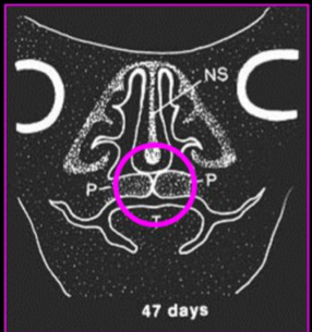
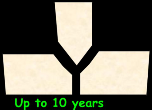
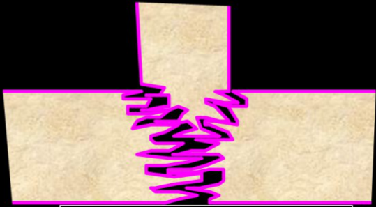
Suture morphology
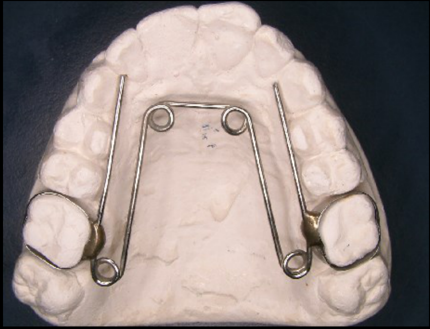
Options for appliance
Lecturer — Expansion Timing
The skeletal response to maxillary expansion decreases with age.
- At approximately 12 years of age, greater interdigitation of the palatal suture may reduce the skeletal effect of expansion.
- Treatment may therefore produce more dental expansion and less skeletal expansion.
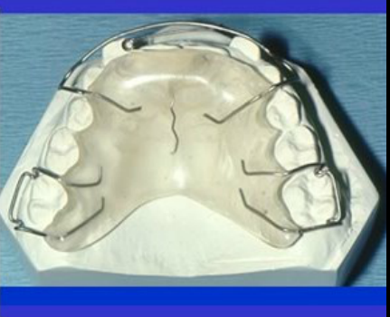
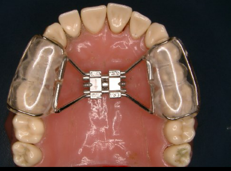
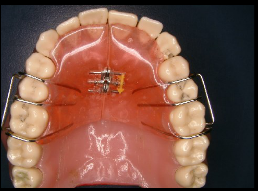
- A slow expansion plate may be used when a patient cannot cooperate with a fixed appliance.
- Activation may occur once per week or every second week.
- One-quarter of a millimetre of screw activation produces approximately 0.125 mm of expansion on each side.
- The appliance must be retained so that it remains in position.
 |  |  |
 |  |  |
Vertical Problems and Habits49
- Define vertical problem and its classification, i.e., dental, skeletal, and possible etiologies.
- Describe epidemiology relating to vertical problems and malocclusion.
- Identify skeletal patterns predisposing patients to deep bite and open bite.
- Explain oral-facial growth patterns and physiology as they relate to vertical problems.
- Identify treatment and timing for vertical problems.
Definition and Classification of Vertical Problems505152
Slide not transcribed — see PDF p.52
- Define vertical problem and its classification, i.e., dental, skeletal, and possible etiologies.
| Category | Type | |
|---|---|---|
| Open Bite | Skeletal | Both |
| Open Bite | Dental | Both |
| Deep Bite | Skeletal | Both |
| Deep Bite | Dental | Both |
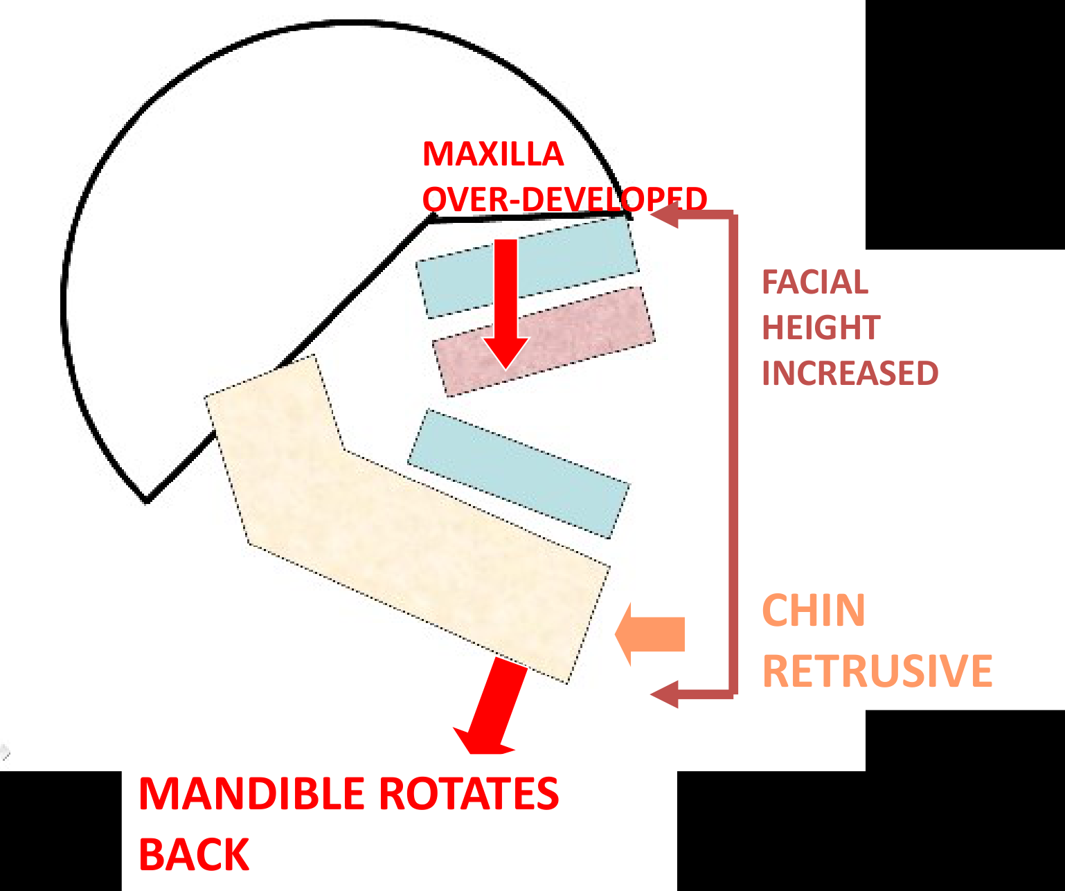
Maxilla over-developed — mandible rotates back, facial height increased, chin retrusive.
Maxilla over-developed → mandible rotates back, facial height increased, chin retrusive
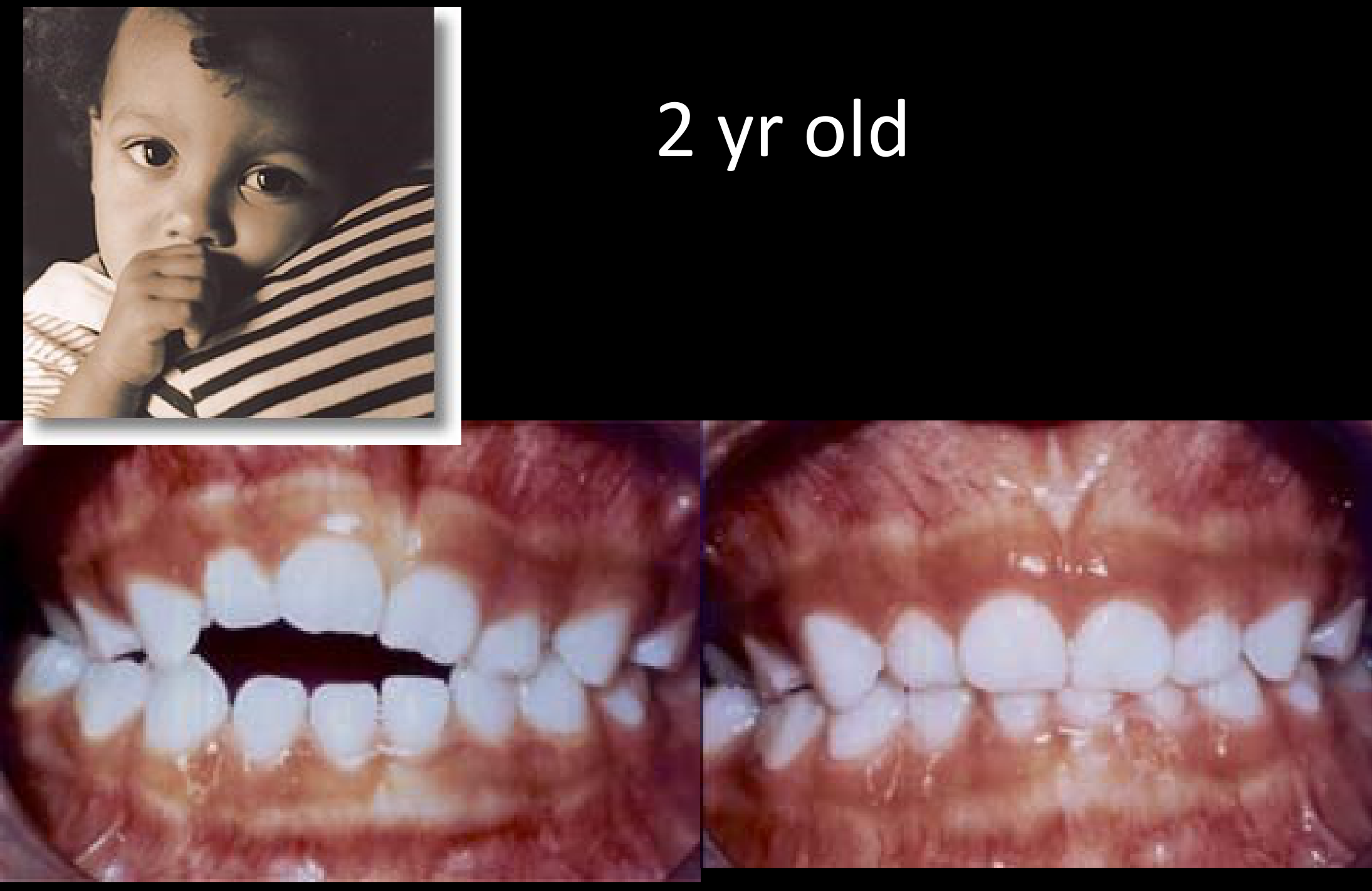
2 yr old
Lecturer — Open Bite Mechanism
Increased vertical development of the maxilla can produce an anterior open bite.
- It increases the height of the midface.
- The mandible rotates clockwise to accommodate the maxilla, making the chin appear retrusive.
- The increased facial height may be clinically apparent.
 |  |
Epidemiology of Vertical Problems and Malocclusion53
- Describe epidemiology relating to vertical problems and malocclusion.
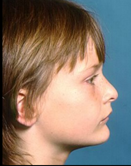
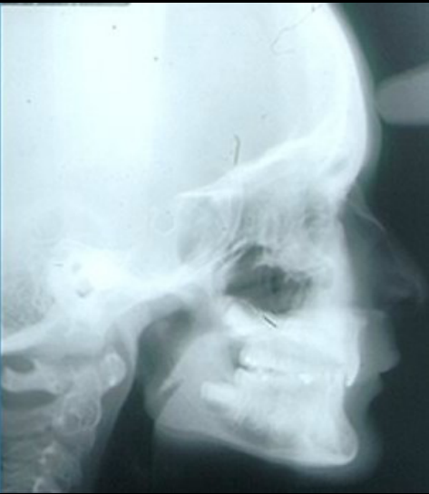
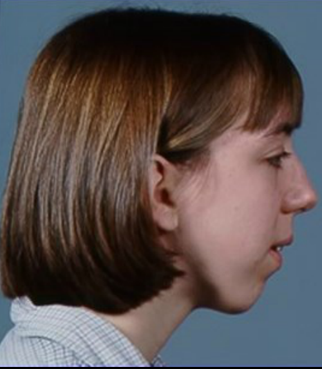
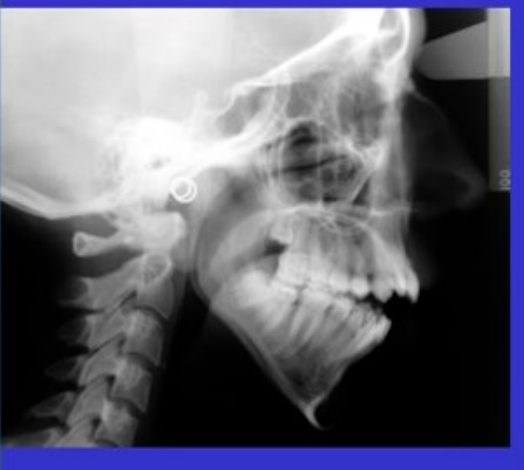
Skeletal Patterns Predisposing to Deep Bite and Open Bite54
- Identify skeletal patterns predisposing patients to deep bite and open bite.
 |  |
 |  |
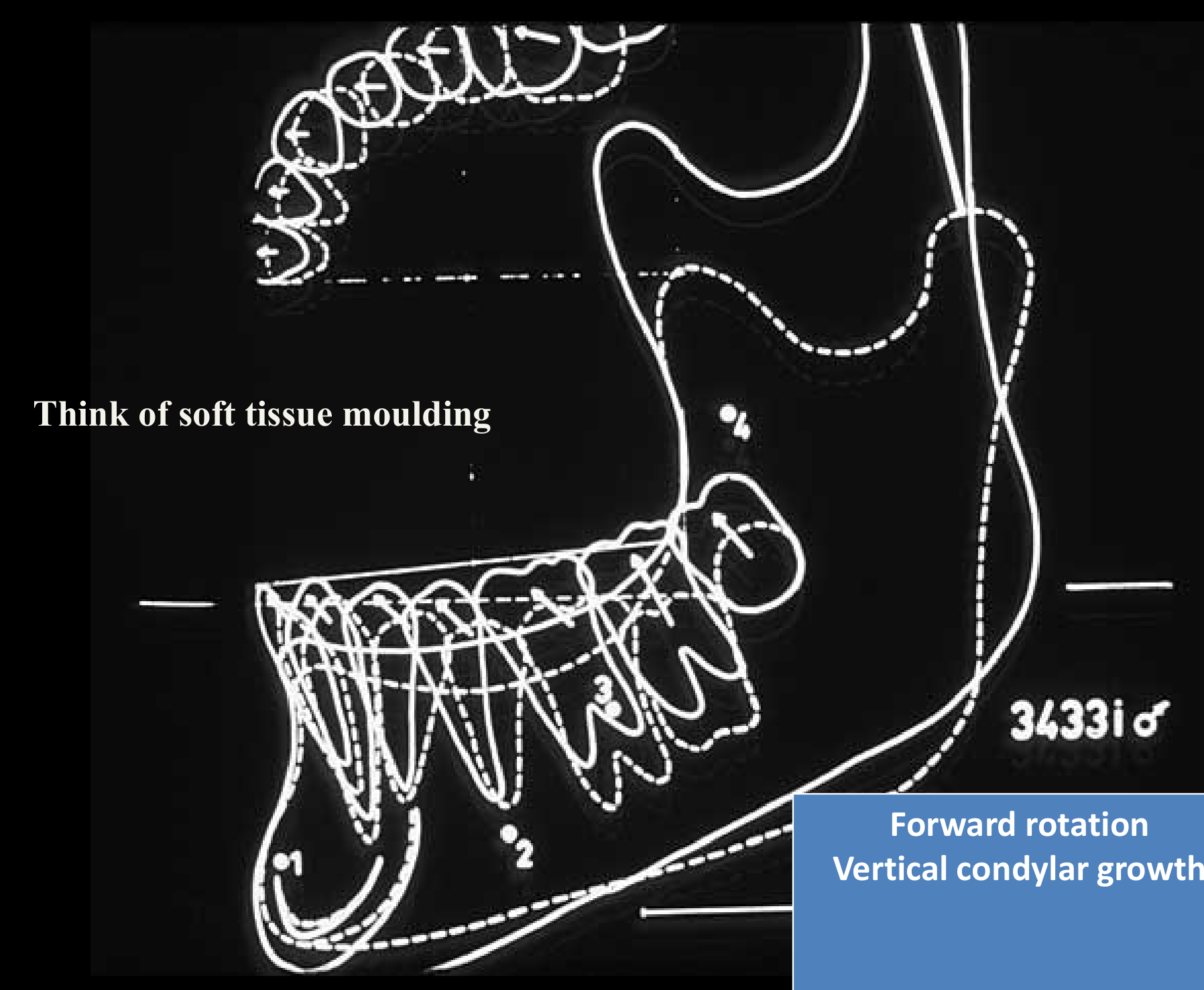
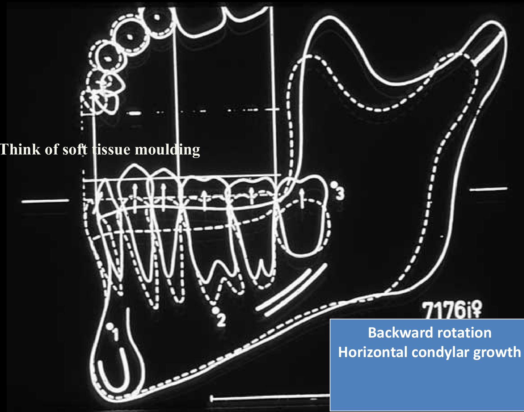
Oral-Facial Growth Patterns and Vertical Problems555657
- Explain oral-facial growth patterns and physiology as they relate to vertical problems.
Think of soft tissue moulding
- Forward rotation
- Vertical condylar growth
Think of soft tissue moulding
- Backward rotation
- Horizontal condylar growth
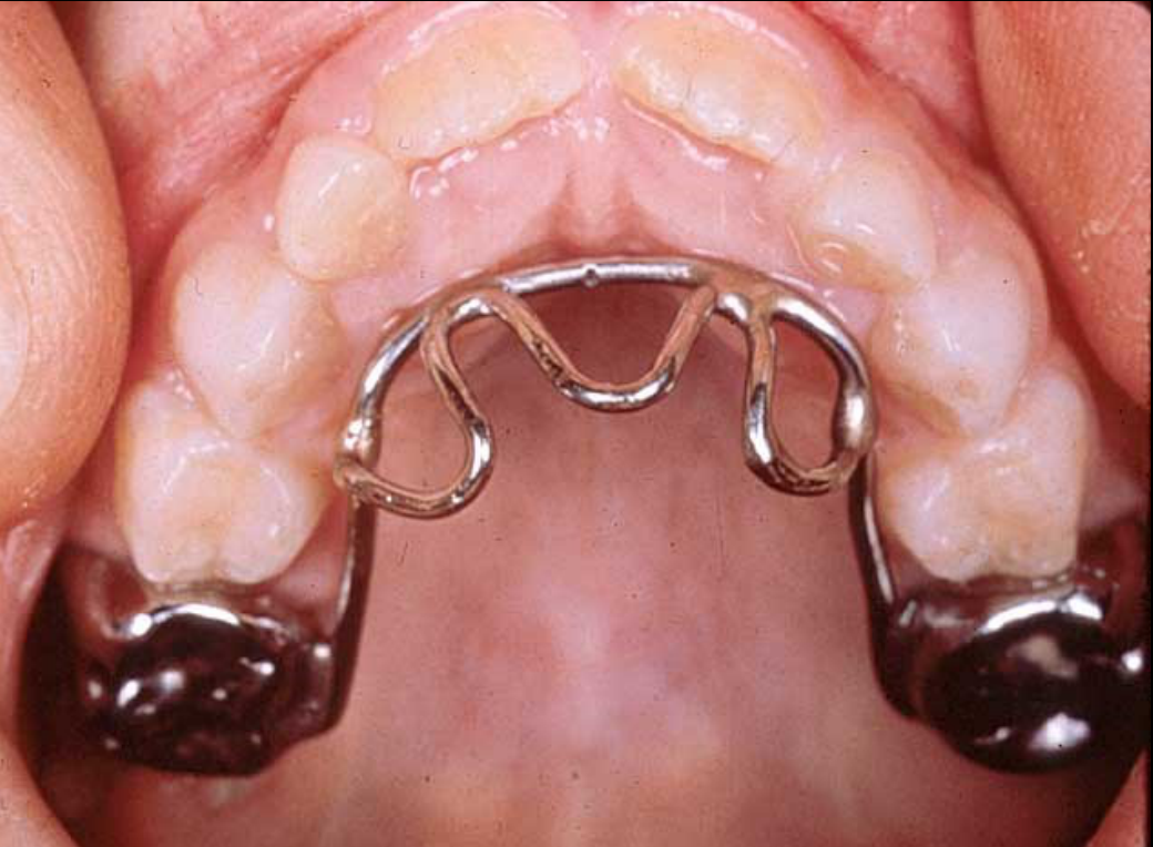
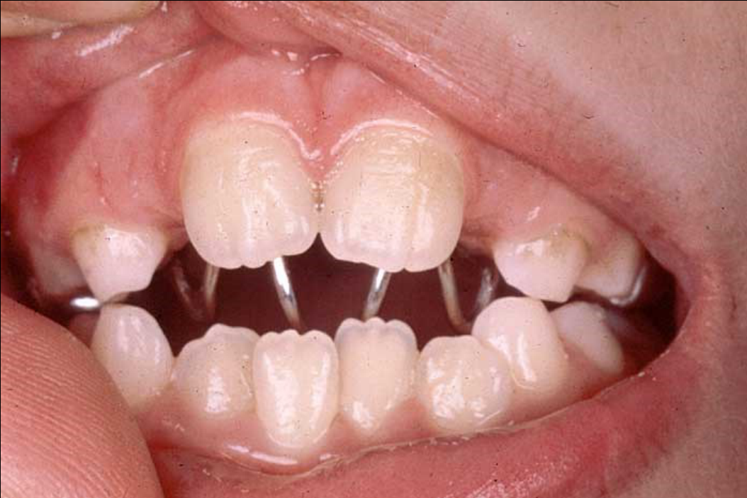
Lecturer — Tongue Habits
An anterior open bite may result from a habit, including tongue posture.
- A crib can help keep the tongue under control.
- In a growing patient, habit correction with a crib may resolve the open bite in approximately six months.
- The crib may be soldered to bands using a procedure similar to the lingual arch.
- In adults, a crib may still help control the tongue, but additional vertical control may be required.
 |  |
Treatment and Timing for Vertical Problems5859606162636465
Slide not transcribed — see PDF p.64
Deep overbite — intrusion (upper / lower)
Slide not transcribed — see PDF p.66
Deep overbite — posterior extrusion (perhaps bite plate; needs condylar growth)
- Identify treatment and timing for vertical problems.
Anti-habit appliance
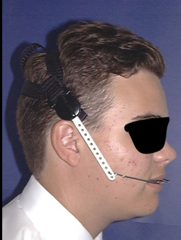
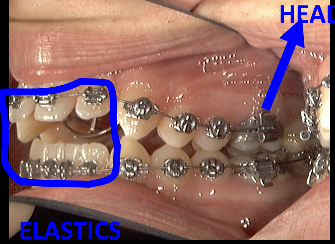
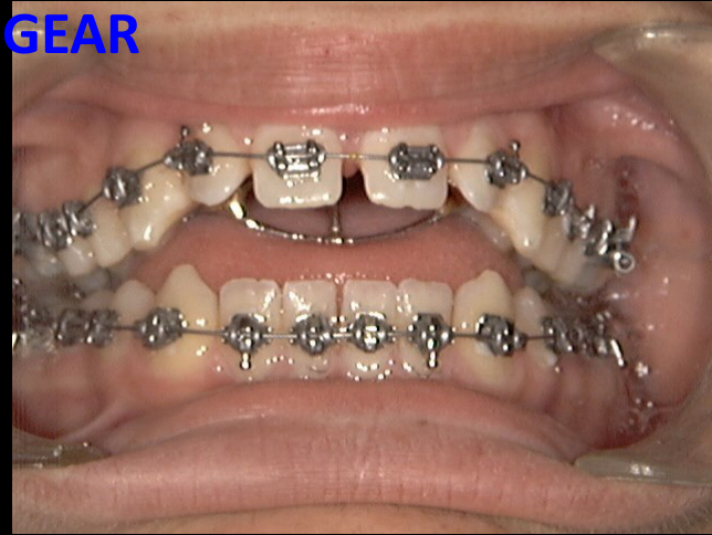
Headgear, elastics — older child
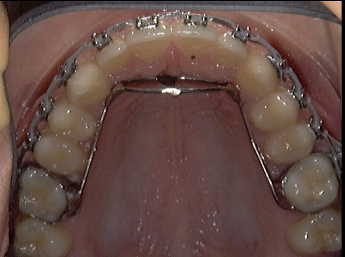
Headgear — add headgear to intrude posterior teeth
- Use a crib to control the tongue in an anterior open bite.
- Use high-pull headgear to control vertical maxillary development.
- Control posterior tooth eruption and reduce extrusion mechanics.
- Incorporate high-pull headgear into another appliance to prevent further vertical maxillary development and reduce worsening of the anterior open bite.
- For a deep bite, intrude the upper incisors, lower incisors, or both.
- Procline incisors that are upright or retroclined, including retroclined incisors in a Class II Division 2 pattern.
- Allow posterior teeth to erupt into functional space or use an anterior bite plane.
- Posterior eruption can produce clockwise mandibular rotation and reduce the anterior deep bite, with condylar growth helping stabilize the mandibular position.

Adjunctive Orthodontic Treatment Procedures66
- Describe indications for repositioning anterior teeth in adults.
- Discuss use of diagnostic set-ups in planning adjunctive incisor positioning.
- Describe indications and methods for extrusion of fractured teeth.
- Describe periodontal implications of orthodontic treatment in adults.
- Explain the indications for molar uprighting in adjunctive orthodontic treatment as part of general dental care.
- Describe the technique used to upright a tipped molar.
- Identify appropriate appliances for different clinical problems.
- Explain types of appliances, in terms of active and reactive units.
- Explain appliance placement, adjustments and timing.
- Identify potential side effects or sequela of treatment.
- Describe patient tolerance of the appliances.
- Identify expected treatment time.
- Describe retention procedures.
Repositioning Anterior Teeth in Adults67
- Describe indications for repositioning anterior teeth in adults.
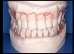
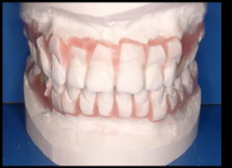
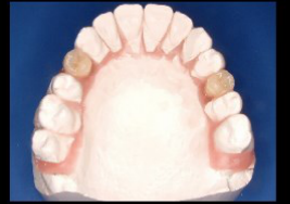
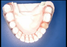
Diagnostic Set-Ups for Incisor Positioning68
- Discuss use of diagnostic set-ups in planning adjunctive incisor positioning.
Lecturer — Diagnostic Set-Ups
A diagnostic set-up simulates the final tooth positions before treatment.
- Teeth are removed from the study model and repositioned to represent the planned result.
- It assesses whether adequate engagement and occlusion can be achieved after treatment.
- It is primarily a diagnostic tool for the clinician, rather than a presentation for the patient.
 |  |  |
 |  |  |
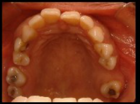 | 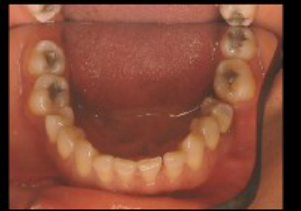 | 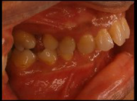 |
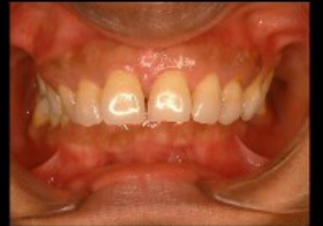 | 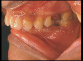 |
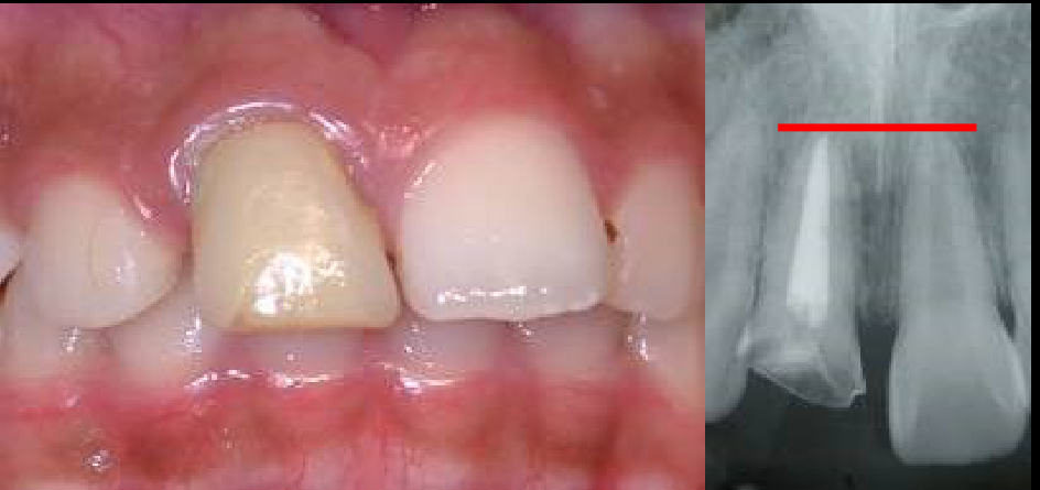
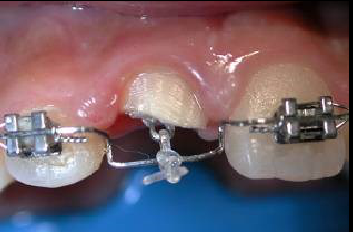
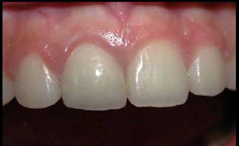
Extrusion of Fractured Teeth6970
- Describe indications and methods for extrusion of fractured teeth.
Occasionally just extrude and restore
 |  |
 |
Periodontal Implications of Orthodontic Treatment in Adults71
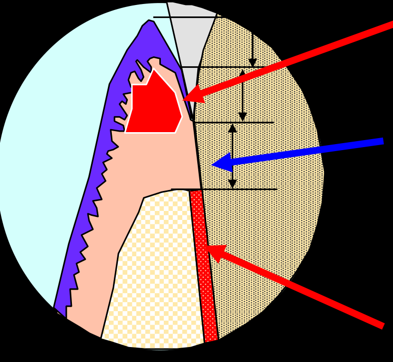
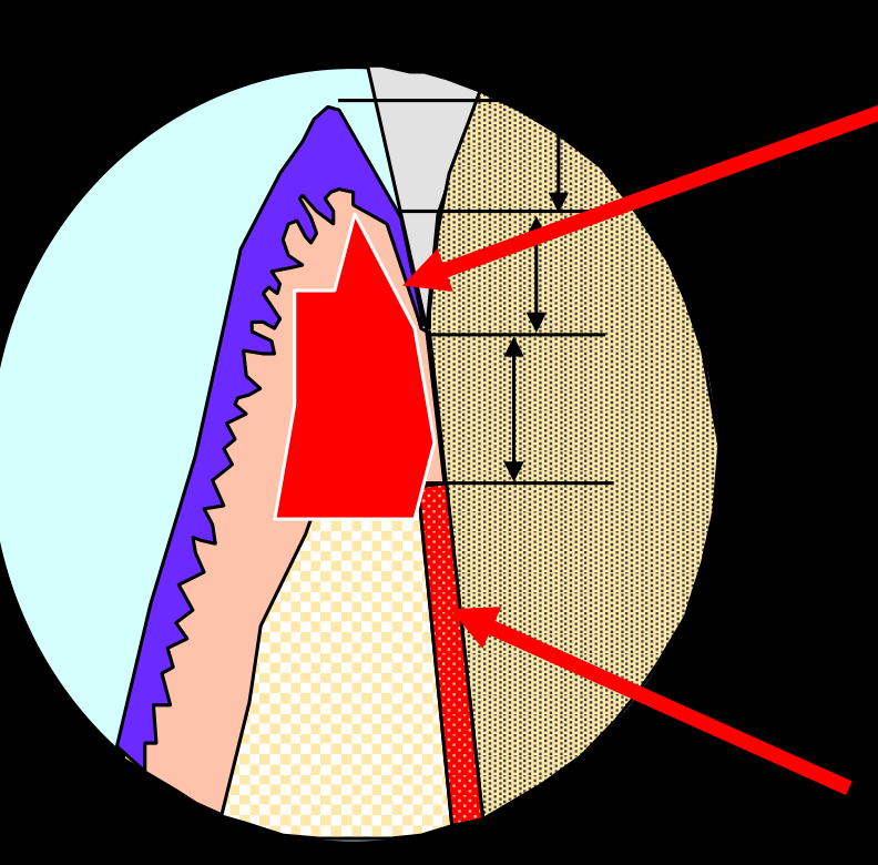
- Describe periodontal implications of orthodontic treatment in adults.
Gingivitis72
limited by buffer zone
- Bacterially induced inflammatory lesion
- Buffer Zone
- Traumatically induced inflammatory lesion
Goonewardene
Periodontitis (inflammation & trauma)73
Bacterially induced inflammatory lesion JOINED TO Traumatically induced inflammatory lesion
Goonewardene
Molar Uprighting Indications747576
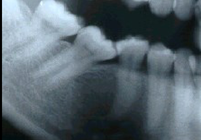
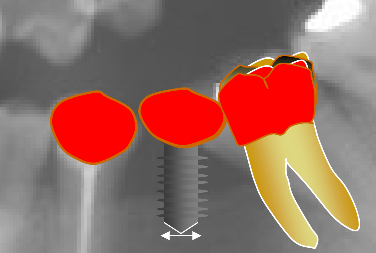
- Explain the indications for molar uprighting in adjunctive orthodontic treatment as part of general dental care.
Lecturer — Molar Uprighting
A tipped molar may be uprighted after early tooth loss for more than simply recovering space for a blocked premolar.
- Reopen space for an unerupted premolar
- Improve the health of the periodontal ligament.
- Create space for a bridge, pontic, or dental implant.
- Uprighting may be required before restorative treatment so the molar position permits appropriate placement of the restoration.
 |  |
Why do we upright?77
- Periodontal issues with tipped teeth
- Abutments — mechanically sound
- Loading through long axis
- Ease of preparation of abutments
Wise, R.J. and Kramer, G. M. (1983). Predetermination of osseous changes associated with uprighting tipped molars by probing. International Journal of Periodontics and Restorative Dentistry, 3, 68-81.
Upright molar — easier prep for bridge, or implant.
Technique for Uprighting a Tipped Molar78798081
- Describe the technique used to upright a tipped molar.
Lecturer — Uprighting Mechanics
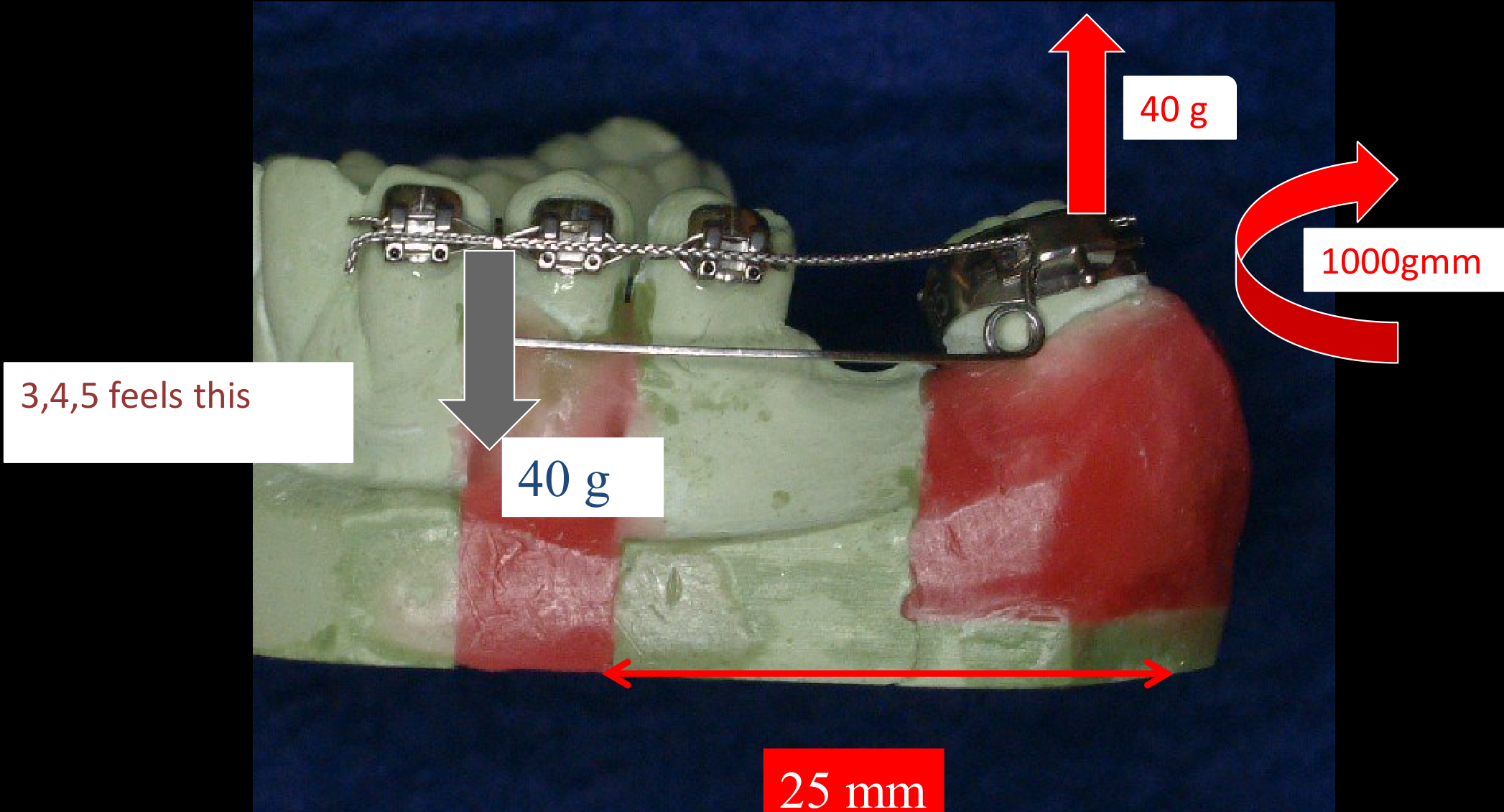
The anchorage unit consists of a canine and both premolars, while a spring with an arm is activated and engaged with the tipped molar.
- The spring is unbent during activation, and the arm engages the molar to produce rotation.
- Approximately 40 grams may be applied to each anchorage tooth.
- The moment is force multiplied by distance: 40 grams multiplied by 25 mm produces approximately 1,000 gram-millimetres.
- Because of the distance, the system produces a substantial uprighting moment with minimal intrusive force on the anchorage segment.
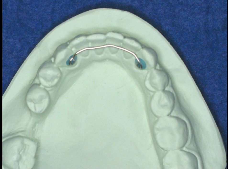
Canine to canine stabilizing arch
Helical uprighting spring “hooked” over segmental arch wire
- Teeth 3, 4, 5 feel this
- 40 g
- 40 g
- 1000 gmm
- 25 mm
From the frontal view the force is buccal to the centre of resistance of the teeth, so they tip to the buccal.
- 40 g
- CR
That’s why we have the 3-3 lingual retainer.
 |  |
Appliance Selection for Clinical Problems82
- Identify appropriate appliances for different clinical problems.
- Headgear
- Fixed appliances
- Removable appliances
- Adjunctive devices — numerous
Active and Reactive Appliance Units83
- Explain types of appliances, in terms of active and reactive units.
Appliance Placement, Adjustments, and Timing84
- Explain appliance placement, adjustments and timing.
Side Effects and Sequelae of Treatment85
- Identify potential side effects or sequela of treatment.
Patient Tolerance of Appliances86
- Describe patient tolerance of the appliances.
Expected Treatment Time87
- Identify expected treatment time.
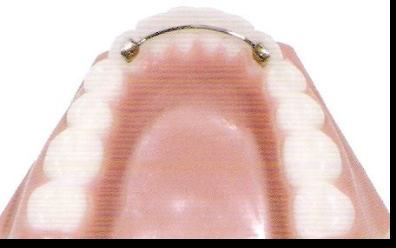
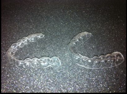
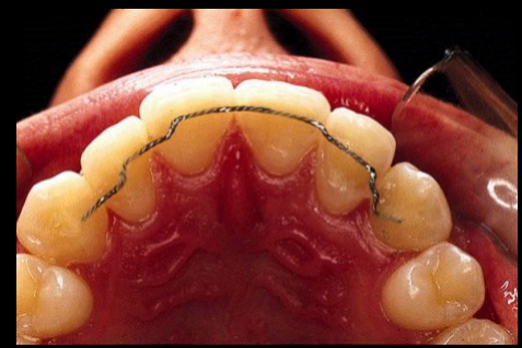
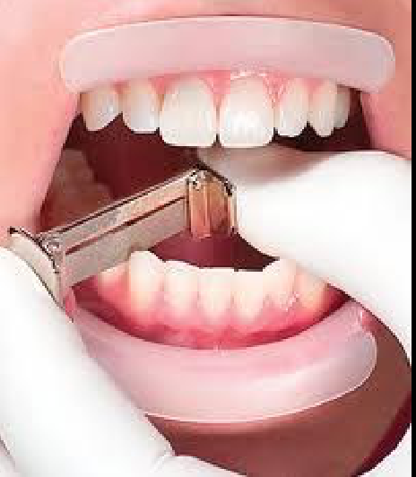
Retention Procedures88
- Describe retention procedures.
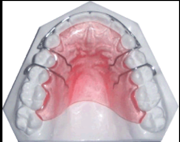
- Removable
- Fixed
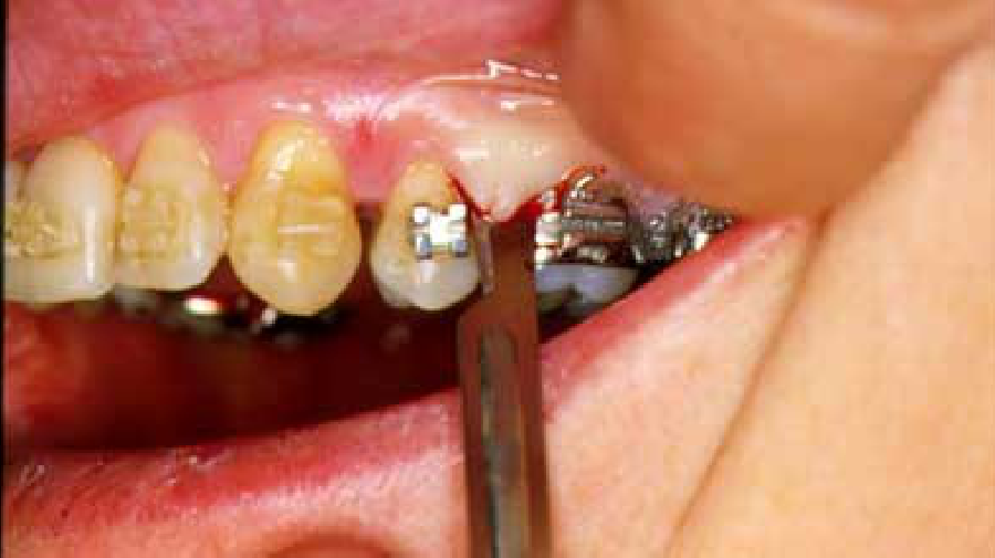
- Pericision
- IPR
 |  |  |
 |  |  |
Carried-over notes
Slide not transcribed — see PDF p.44
Crossbite — narrow dentition: expand the teeth
Clinical Cases
Case: Lower anterior crowding managed with a lingual arch
Question
Scenario: A patient presents with mild anterior crowding in the lower arch and large primary second molars, with no history of early tooth loss.
What’s shown: Clinical intraoral photographs of the mandibular arch showing anterior crowding and large primary second molars, along with images of a fixed space maintainer in place.
Consider: How to manage the lower anterior crowding and utilize the available space in the lower arch.
Answer
Observations:
- Mild anterior crowding in the lower arch.
- Large primary second molars providing approximately 3 mm of leeway space on each side of the lower arch.
- No early loss of primary teeth.
Reasoning: Placing a lingual arch prevents the physiological mesial drift of the permanent first molars. This allows the leeway space to be transferred to the anterior segment of the arch to resolve the crowding. However, preventing the mesial drift means the lower molars will not naturally settle into a Class I relationship.
Outcome: The lower anterior crowding is resolved using the lingual arch to transfer the leeway space. To compensate for the lower molars not engaging in Class I, the upper molars are slightly distalized to achieve a Class I molar relationship.
Takeaway: A lingual arch can be used to prevent mesial drift of the lower first molars and transfer leeway space to resolve mild anterior crowding, though this requires subsequent distalization of the upper molars to establish a Class I molar relationship.
Case: Anterior crossbite, lateral cephalometric radiograph and intraoral photographs
Question
Scenario: A female patient presents with an anterior crossbite.
What’s shown: A lateral cephalometric radiograph and a series of intraoral photographs showing the dentition before and during orthodontic treatment.
Consider: Evaluate the skeletal and dental relationships, determine the etiology of the crossbite, and decide on the appropriate management.
Answer
Observations:
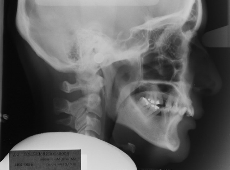
- The lateral cephalometric radiograph shows Point A and Point B in a normal relationship, indicating a Class 1 skeletal relationship rather than a true Class 3.
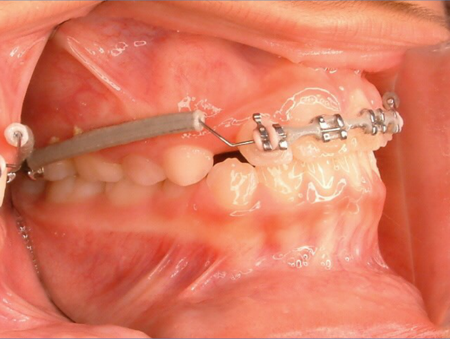
- The patient has a Class 1 molar relationship.
- The anterior crossbite is the result of an anterior shift of the mandible from the retruded contact position (RCP) to the intercuspal position (ICP).
- The patient exhibits a vertical growth pattern.
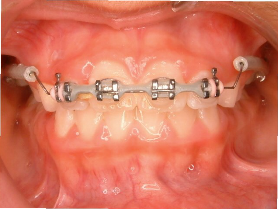
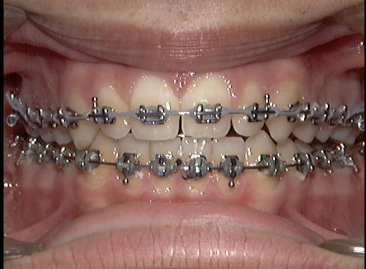
- The intraoral photographs show retroclined upper incisors and the placement of partial fixed orthodontic appliances bonded to the incisors and a molar.
Reasoning: The normal A and B points on the cephalogram rule out a true skeletal Class 3 relationship. The anterior shift from RCP to ICP explains the anterior crossbite in the presence of a Class 1 molar and skeletal relationship, diagnosing a pseudo Class III or dental anterior crossbite. The vertical growth pattern is noted as a factor in her future development. The treatment plan involves using partial braces to activate an archwire and procline the retroclined incisors, correcting the dental anterior crossbite while maintaining the Class 1 molar relationship.
Outcome: Partial braces were bonded to the incisors and molar, and the archwire was activated to protrude and procline the incisors. The anterior crossbite was corrected, and the patient maintained a Class 1 molar relationship.
Takeaway: An anterior crossbite can be caused by a functional anterior shift from RCP to ICP (pseudo Class III) rather than a true skeletal discrepancy, and can be effectively treated with localized orthodontic intervention to correct the dental inclination.
Case: Posterior crossbite with functional shift, intraoral and extraoral photographs
Question
Scenario: A patient presents with a posterior crossbite and apparent facial asymmetry.
What’s shown: Intraoral frontal views of the dentition and a frontal extraoral view of the face.
Consider: Identify the cause of the apparent asymmetry and the underlying occlusal relationship.
Answer
Observations:
- Unilateral posterior crossbite on the left side.
- Apparent facial asymmetry with the chin shifted to the right.
- Lower midline deviation to the right.
Reasoning: The posterior teeth have an unstable edge-to-edge relationship. To achieve a stable intercuspation, the patient functionally shifts their mandible to the right. This shift results in a unilateral posterior crossbite on the left, apparent chin asymmetry to the right, and a lower midline deviation, rather than a true skeletal asymmetry.
Outcome: The unstable condition is treated by expanding the upper arch with an expander to achieve a better, stable intercuspidation.
Takeaway: A functional mandibular shift to achieve intercuspation in the presence of an unstable edge-to-edge posterior relationship can mimic true facial asymmetry and cause midline deviation, which is resolved by maxillary expansion.
Case: Patient with posterior, dental, and anterior crossbite
Question
Scenario: A patient presents with a posterior crossbite, a dental crossbite, and an anterior crossbite of a lateral incisor.
What’s shown: The clinical challenge of addressing both the posterior/dental crossbite and the anterior crossbite of the lateral incisor.
Consider: How to design an appliance to correct both the posterior and anterior crossbites efficiently using a single device.
Answer
Observations:
- The patient has a posterior crossbite and a dental crossbite.
- The patient also has an anterior crossbite of a lateral incisor.
Reasoning:
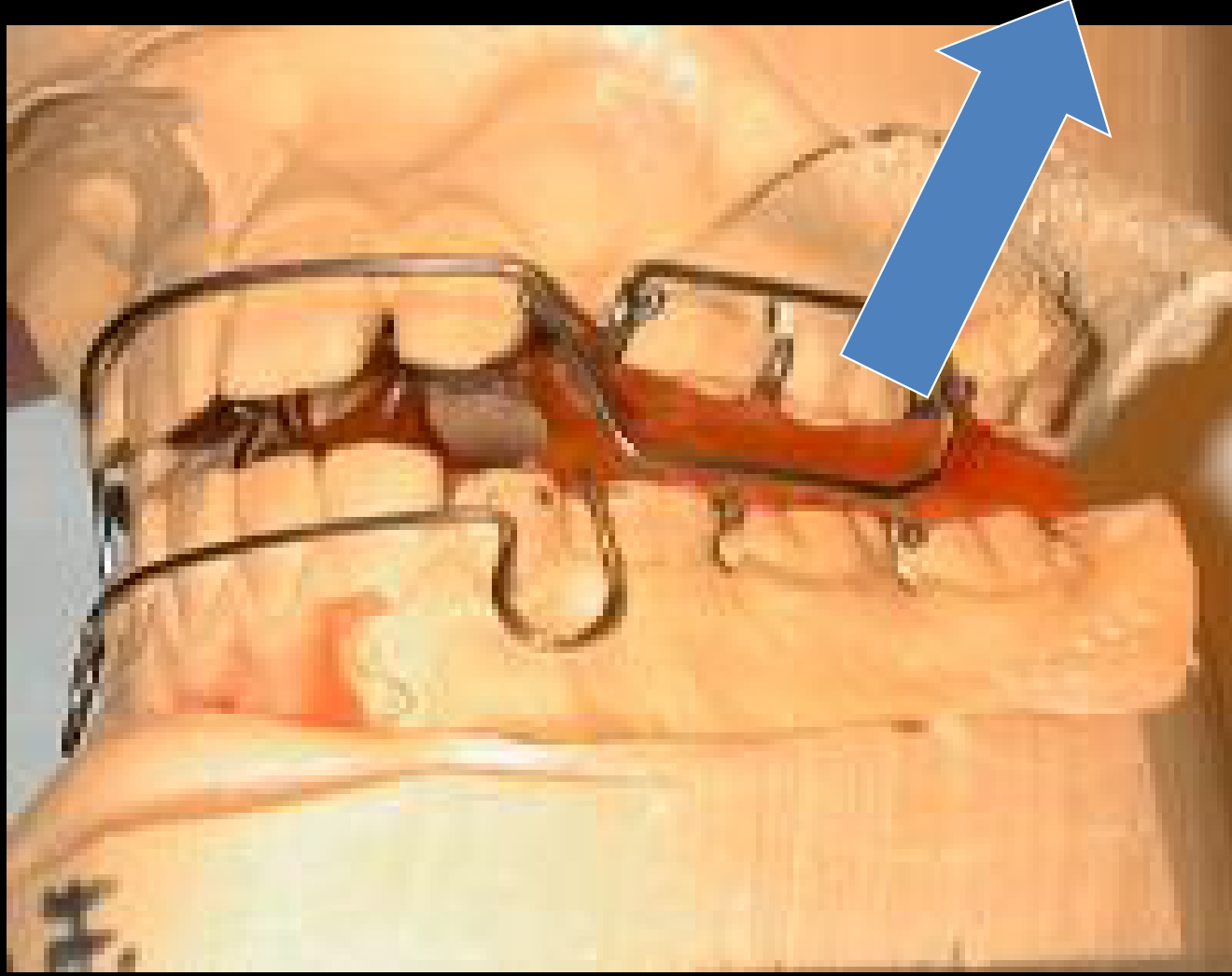
- A standard quad helix or W-arch can be used to expand the arch and correct the posterior and dental crossbites.
- By extending one of the arms of the appliance and soldering a spring to it, the same device can also apply the necessary force to correct the anterior crossbite of the lateral incisor.
Outcome:
- The combined anterior and posterior crossbites were corrected in two months.
- This resolved the anterior shift caused by the lateral incisor interference and the posterior shift resulting from the edge-to-edge relationship of the first molars.
Takeaway:
- A modified expansion appliance (such as a quad helix or W-arch with an extended arm and soldered spring) can efficiently treat concurrent posterior/dental and anterior crossbites with a single device, saving treatment time.
Footnotes
-
Original PDF page 1: L3.1 3BLevel III Unit B, p.1 ↩
-
Original PDF page 2: L3.1 3BLevel III Unit B, p.2 ↩
-
Original PDF page 3: L3.1 3BLevel III Unit B, p.3 ↩
-
Original PDF page 4: L3.1 3BLevel III Unit B, p.4 ↩
-
Original PDF page 5: L3.1 3BLevel III Unit B, p.5 ↩
-
Original PDF page 7: L3.1 3BLevel III Unit B, p.7 ↩
-
Original PDF page 8: L3.1 3BLevel III Unit B, p.8 ↩
-
Original PDF page 9: L3.1 3BLevel III Unit B, p.9 ↩
-
Original PDF page 10: L3.1 3BLevel III Unit B, p.10 ↩
-
Original PDF page 11: L3.1 3BLevel III Unit B, p.11 ↩
-
Original PDF page 12: L3.1 3BLevel III Unit B, p.12 ↩
-
Original PDF page 13: L3.1 3BLevel III Unit B, p.13 ↩
-
Original PDF page 14: L3.1 3BLevel III Unit B, p.14 ↩
-
Original PDF page 15: L3.1 3BLevel III Unit B, p.15 ↩
-
Original PDF page 16: L3.1 3BLevel III Unit B, p.16 ↩
-
Original PDF page 17: L3.1 3BLevel III Unit B, p.17 ↩
-
Original PDF page 18: L3.1 3BLevel III Unit B, p.18 ↩
-
Original PDF page 19: L3.1 3BLevel III Unit B, p.19 ↩
-
Original PDF page 20: L3.1 3BLevel III Unit B, p.20 ↩
-
Original PDF page 21: L3.1 3BLevel III Unit B, p.21 ↩
-
Original PDF page 22: L3.1 3BLevel III Unit B, p.22 ↩
-
Original PDF page 23: L3.1 3BLevel III Unit B, p.23 ↩
-
Original PDF page 24: L3.1 3BLevel III Unit B, p.24 ↩
-
Original PDF page 25: L3.1 3BLevel III Unit B, p.25 ↩
-
Original PDF page 26: L3.1 3BLevel III Unit B, p.26 ↩
-
Original PDF page 27: L3.1 3BLevel III Unit B, p.27 ↩
-
Original PDF page 28: L3.1 3BLevel III Unit B, p.28 ↩
-
Original PDF page 29: L3.1 3BLevel III Unit B, p.29 ↩
-
Original PDF page 30: L3.1 3BLevel III Unit B, p.30 ↩
-
Original PDF page 31: L3.1 3BLevel III Unit B, p.31 ↩
-
Original PDF page 32: L3.1 3BLevel III Unit B, p.32 ↩
-
Original PDF page 33: L3.1 3BLevel III Unit B, p.33 ↩
-
Original PDF page 34: L3.1 3BLevel III Unit B, p.34 ↩
-
Original PDF page 35: L3.1 3BLevel III Unit B, p.35 ↩
-
Original PDF page 37: L3.1 3BLevel III Unit B, p.37 ↩
-
Original PDF page 36: L3.1 3BLevel III Unit B, p.36 ↩
-
Original PDF page 38: L3.1 3BLevel III Unit B, p.38 ↩
-
Original PDF page 39: L3.1 3BLevel III Unit B, p.39 ↩
-
Original PDF page 40: L3.1 3BLevel III Unit B, p.40 ↩
-
Original PDF page 41: L3.1 3BLevel III Unit B, p.41 ↩
-
Original PDF page 42: L3.1 3BLevel III Unit B, p.42 ↩
-
Original PDF page 43: L3.1 3BLevel III Unit B, p.43 ↩
-
Original PDF page 44: L3.1 3BLevel III Unit B, p.44 ↩
-
Original PDF page 45: L3.1 3BLevel III Unit B, p.45 ↩
-
Original PDF page 46: L3.1 3BLevel III Unit B, p.46 ↩
-
Original PDF page 47: L3.1 3BLevel III Unit B, p.47 ↩
-
Original PDF page 48: L3.1 3BLevel III Unit B, p.48 ↩
-
Original PDF page 49: L3.1 3BLevel III Unit B, p.49 ↩
-
Original PDF page 50: L3.1 3BLevel III Unit B, p.50 ↩
-
Original PDF page 51: L3.1 3BLevel III Unit B, p.51 ↩
-
Original PDF page 52: L3.1 3BLevel III Unit B, p.52 ↩
-
Original PDF page 53: L3.1 3BLevel III Unit B, p.53 ↩
-
Original PDF page 54: L3.1 3BLevel III Unit B, p.54 ↩
-
Original PDF page 55: L3.1 3BLevel III Unit B, p.55 ↩
-
Original PDF page 56: L3.1 3BLevel III Unit B, p.56 ↩
-
Original PDF page 57: L3.1 3BLevel III Unit B, p.57 ↩
-
Original PDF page 58: L3.1 3BLevel III Unit B, p.58 ↩
-
Original PDF page 59: L3.1 3BLevel III Unit B, p.59 ↩
-
Original PDF page 60: L3.1 3BLevel III Unit B, p.60 ↩
-
Original PDF page 61: L3.1 3BLevel III Unit B, p.61 ↩
-
Original PDF page 62: L3.1 3BLevel III Unit B, p.62 ↩
-
Original PDF page 63: L3.1 3BLevel III Unit B, p.63 ↩
-
Original PDF page 64: L3.1 3BLevel III Unit B, p.64 ↩
-
Original PDF page 65: L3.1 3BLevel III Unit B, p.65 ↩
-
Original PDF page 66: L3.1 3BLevel III Unit B, p.66 ↩
-
Original PDF page 67: L3.1 3BLevel III Unit B, p.67 ↩
-
Original PDF page 68: L3.1 3BLevel III Unit B, p.68 ↩
-
Original PDF page 69: L3.1 3BLevel III Unit B, p.69 ↩
-
Original PDF page 70: L3.1 3BLevel III Unit B, p.70 ↩
-
Original PDF page 71: L3.1 3BLevel III Unit B, p.71 ↩
-
Original PDF page 72: L3.1 3BLevel III Unit B, p.72 ↩
-
Original PDF page 73: L3.1 3BLevel III Unit B, p.73 ↩
-
Original PDF page 74: L3.1 3BLevel III Unit B, p.74 ↩
-
Original PDF page 75: L3.1 3BLevel III Unit B, p.75 ↩
-
Original PDF page 77: L3.1 3BLevel III Unit B, p.77 ↩
-
Original PDF page 78: L3.1 3BLevel III Unit B, p.78 ↩
-
Original PDF page 76: L3.1 3BLevel III Unit B, p.76 ↩
-
Original PDF page 79: L3.1 3BLevel III Unit B, p.79 ↩
-
Original PDF page 80: L3.1 3BLevel III Unit B, p.80 ↩
-
Original PDF page 81: L3.1 3BLevel III Unit B, p.81 ↩
-
Original PDF page 82: L3.1 3BLevel III Unit B, p.82 ↩
-
Original PDF page 83: L3.1 3BLevel III Unit B, p.83 ↩
-
Original PDF page 84: L3.1 3BLevel III Unit B, p.84 ↩
-
Original PDF page 85: L3.1 3BLevel III Unit B, p.85 ↩
-
Original PDF page 86: L3.1 3BLevel III Unit B, p.86 ↩
-
Original PDF page 87: L3.1 3BLevel III Unit B, p.87 ↩
-
Original PDF page 88: L3.1 3BLevel III Unit B, p.88 ↩
-
Original PDF page 89: L3.1 3BLevel III Unit B, p.89 ↩