Retention1
J. Mike Razza, BS, DDS, Cert Orthod
Associate Professor - Orthodontics
University of Western Australia
School of Dentistry
Contemporary Orthodontics – Chapter 18
Level IV Advanced Orthodontics
Unit B Complex Orthodontics Retention
Historical Perspective2
- The Occlusion School
- The Apical Base School
- The Mandibular Incisor School
- The Musculature School
Lecturer — Retention Stability
Retention is the final stage of orthodontic treatment, and patient follow-up generally continues for approximately two to three years.
- The Occlusion School considers how the quality of intercuspation at the end of treatment influences instability.
- The Apical Base School emphasizes mandibular incisor inclination, with excessive proclination being less stable.
- The Mandibular Incisor School examines how to prevent the return of lower-arch crowding.
- The Musculature School evaluates function and the influence of muscles and joints on occlusal stability.
- Stability is also affected by aging: the alveolar region and surrounding tissues change over time, so teeth cannot be expected to remain completely unchanged for decades and must accommodate to achieve a stable position.
Riedel’s Theorems on Retention
A Review of the Literature Richard A. Riedel
- Teeth which have been moved in or through bone by orthodontic appliances often have a tendency to return to their former positions.
Theorems on Relapse and Stability345
-
The elimination of the causes of a malocclusion will prevent recurrence.
-
Overcorrection of a malocclusion is a safety factor in retention.
-
Occlusion is an important factor in retention; therefore, an orthodontist should attempt to produce the best possible occlusion of the teeth.
Lecturer — Occlusal Stability
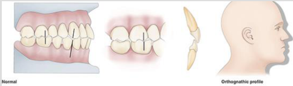
Good intercuspation can help stabilize the result and may function almost like a retainer.
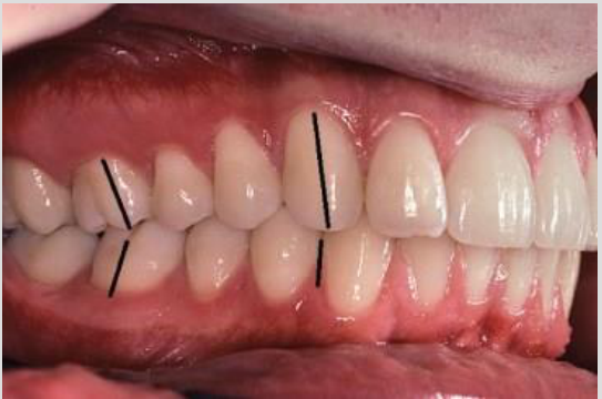
- Stability assessment includes checking protrusive movement, canine guidance during lateral movements, and group function.
- After correction of an anterior crossbite, sufficient overbite can help maintain the incisor position.
-
Bone and adjacent tissues must be allowed to reorganize around the newly positioned teeth for some length of time.
-
Bone and adjacent tissues must be allowed to reorganize around the newly positioned teeth for some length of time.
Lecturer — Fiber Reorganization
The final position of the teeth takes at least approximately one year to become stable.
- Collagen-containing fibers can behave like elastic material and continue exerting forces similar to orthodontic elastics or power chains until they adapt.
- Retainers provide time for collagen fibers, gingival and transeptal fibers, bone, and other adjacent tissues to reorganize.
- Periodontal ligament reorganization was described as taking approximately four to six months; transeptal fiber reorganization may require approximately one year, while supracrestal fiber stabilization was estimated at approximately 230 days.
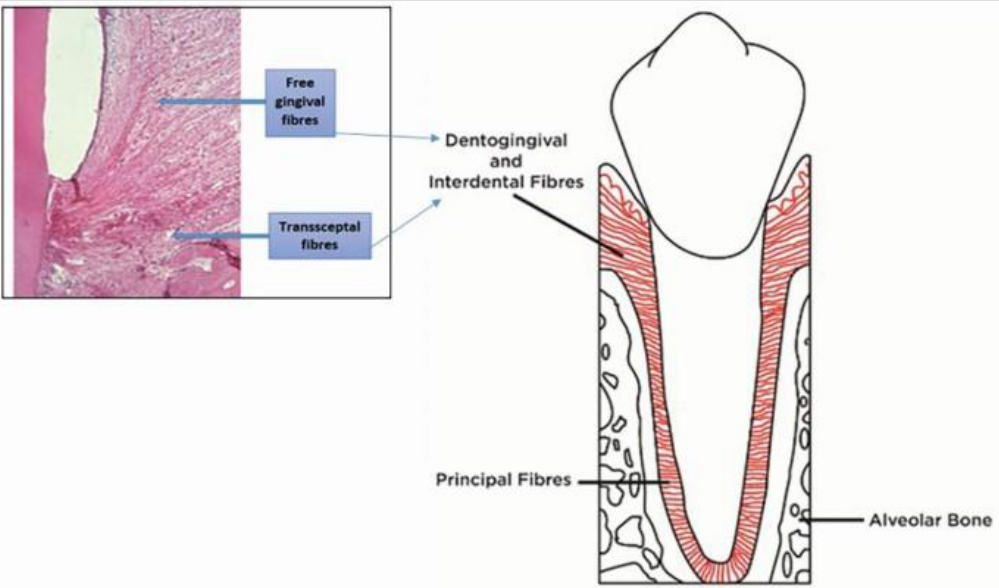
Principal Fiber Groups67891011
- Free gingival fibres
- Transseptal fibres
- Dentogingival and interdental fibres
- Principal fibres
- Alveolar bone
Principal fiber groups include:
- Interradicular group
- Apical group
- Oblique group
- Horizontal group
- Alveolar crest group
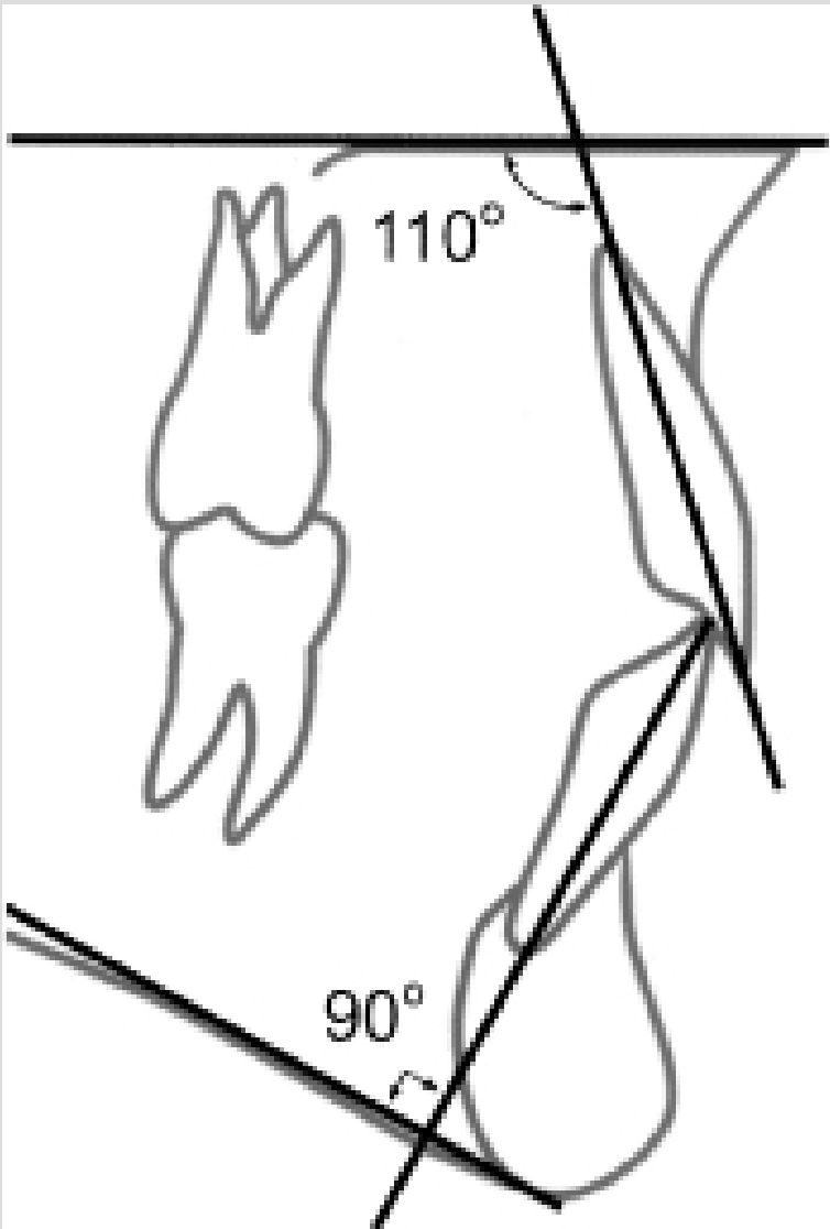
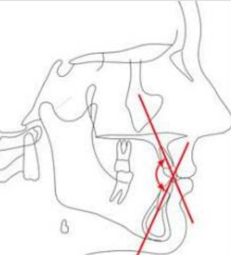
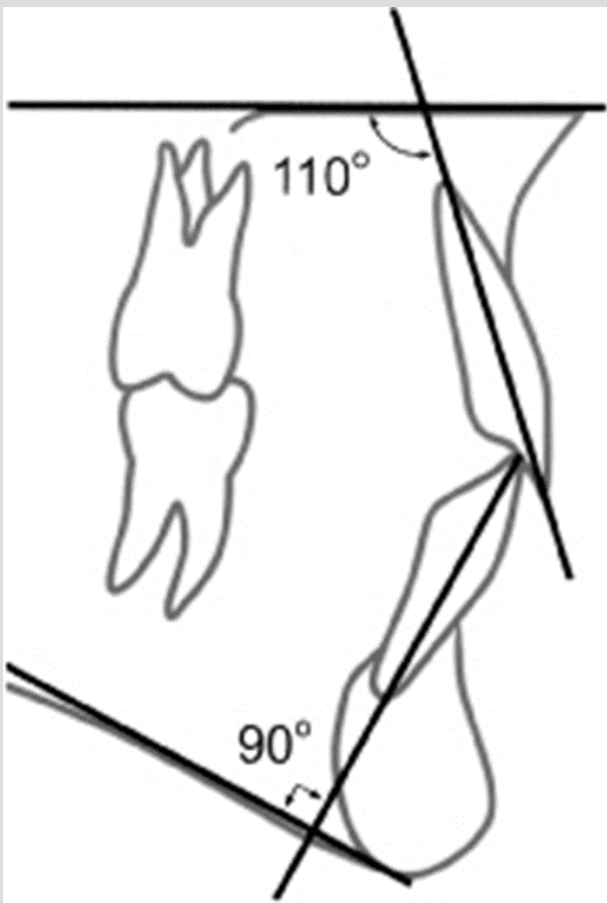
- Placing the lower incisors upright (plus or minus 5 degrees from perpendicular to the mandibular plane) over basal bone will result in a more stable correction of a malocclusion, and they are more likely to remain in good alignment.
Angle labels shown: 110°, 90°
-
Corrections carried out during periods when the patients are growing are less likely to relapse.
-
The farther teeth have been moved, the less the likelihood of relapse — similar to overcorrection.
-
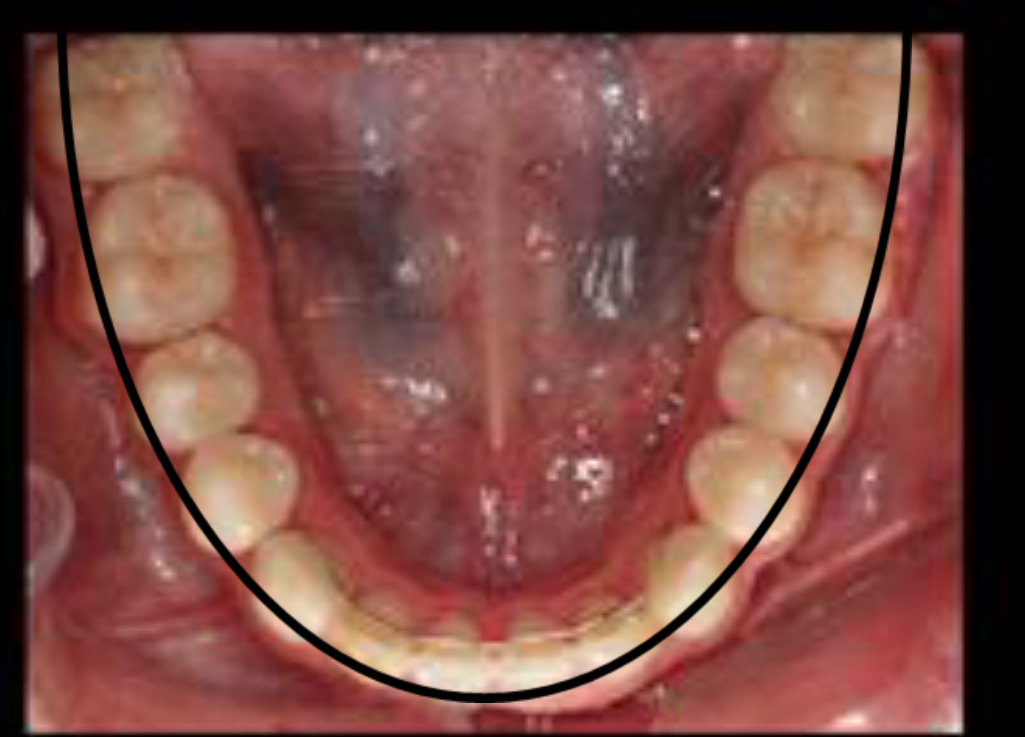
The arch form, particularly in the mandibular arch, cannot be altered permanently by appliance therapy. Treatment should be directed toward maintaining the arch form presented by the original malocclusion.
- Theorem 9: arch form
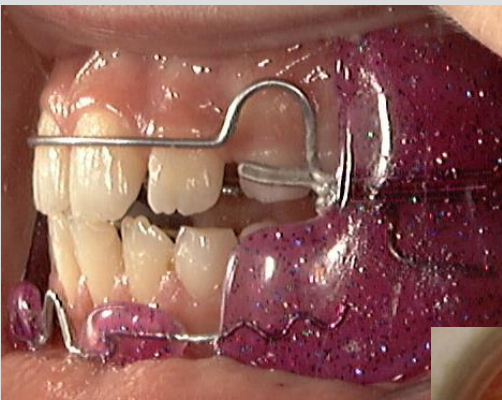
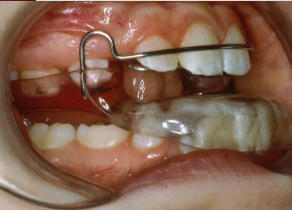
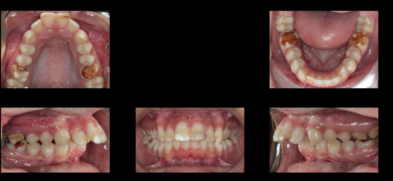
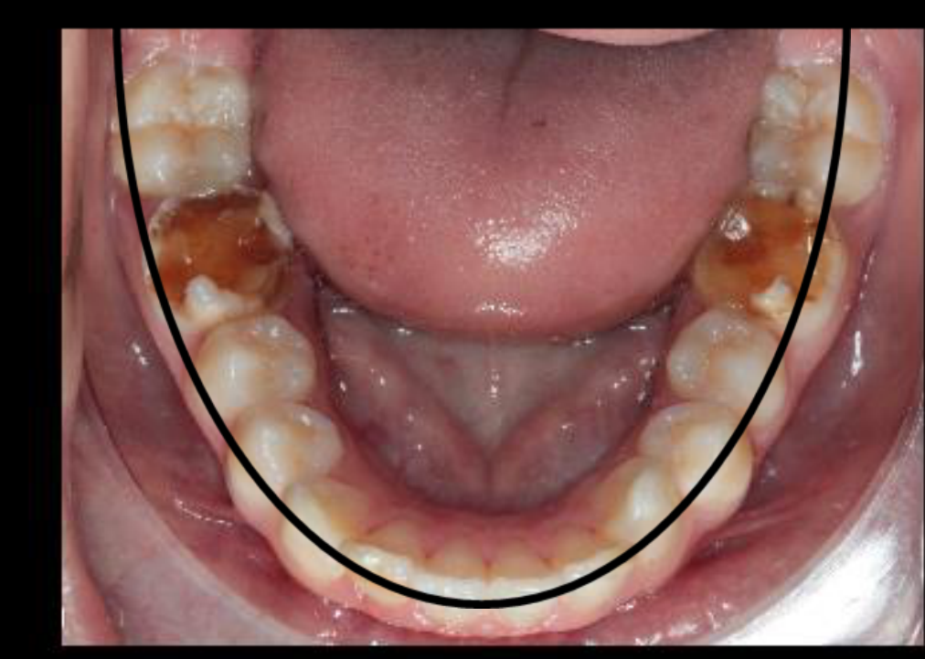
Riedel’s Three Most Important Rules12
Riedel considers the following three rules to be the most important:
- Teeth tend to move back toward their former position.
- The arch form of the mandibular arch cannot, in the majority of cases, be permanently altered by appliance therapy.
- Bone and adjacent tissues should be allowed time to reorganize around newly positioned teeth.
Other Factors Influencing Retention13
Other factors influencing the retention of treated malocclusions include:
- Tooth-size discrepancies
- Axial inclinations
- Growth
- Root paralleling in extraction cases
- Equilibration
- Musculature
- Duration of retention
Tooth-Size Discrepancies14
If the maxillary anterior teeth are too large for the mandibular, the maxillary teeth must be placed in one of several positions: deep overbite, greater overjet, combination of greater overbite and overjet, or the maxillary posterior teeth fitting into a more or less distal relationship to the mandible.
Lecturer — Tooth-Size Discrepancy
A tooth-size discrepancy can make the final occlusion and its stability less favorable.
- Wider upper teeth relative to the lower teeth may produce increased overjet or deep overbite.
- These discrepancies should be considered when planning the final tooth positions and retention.
Review “Bolton Discrepancy”
Axial Inclinations15
Axial inclination of maxillary and mandibular incisors influences the retention of correction of a deep overbite. Tipping incisors into too upright of a relationship usually results in a deep anterior overbite.
Lecturer — Incisor Inclination
The mandibular incisors are generally targeted near 90 degrees to the mandibular plane, with an approximate range of 85 to 95 degrees.
- When crowding and the facial profile make extraction undesirable, proclination may be used, but the result is more likely to relapse.
- Bonded canine-to-canine retention is frequently considered in these cases.
Growth and Sex Differences16
Growth is an aid in the correction of many types of orthodontic problems, and it may also be of such character as to cause relapse of treated orthodontic cases. There is a marked difference between the maturation of skeletal and dental patterns of male and female.
Lecturer — Growth Monitoring
During growth, bone compensation can help anchor moved teeth, particularly when extrusion or vertical correction is required.
- Mandibular growth in males may continue until approximately 21 or 22 years of age.
- Continued mandibular growth can favor Class II correction but create substantial difficulty for Class III cases.
- When excessive mandibular growth is possible, treatment may be delayed while lateral cephalometric radiographs monitor growth for two or three years; beginning Class III treatment too early can be followed by a new Class III relationship.
Root Paralleling in Extraction Cases17
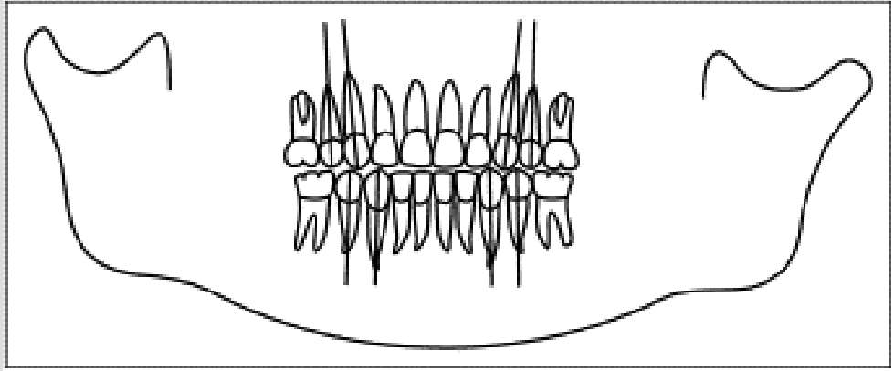
Unless the roots of teeth on either side of the extraction sites are made parallel, spaces may open or teeth rotate due to load distribution on the teeth.
Lecturer — Root Assessment
An OPG may be requested before finishing treatment to assess root position.
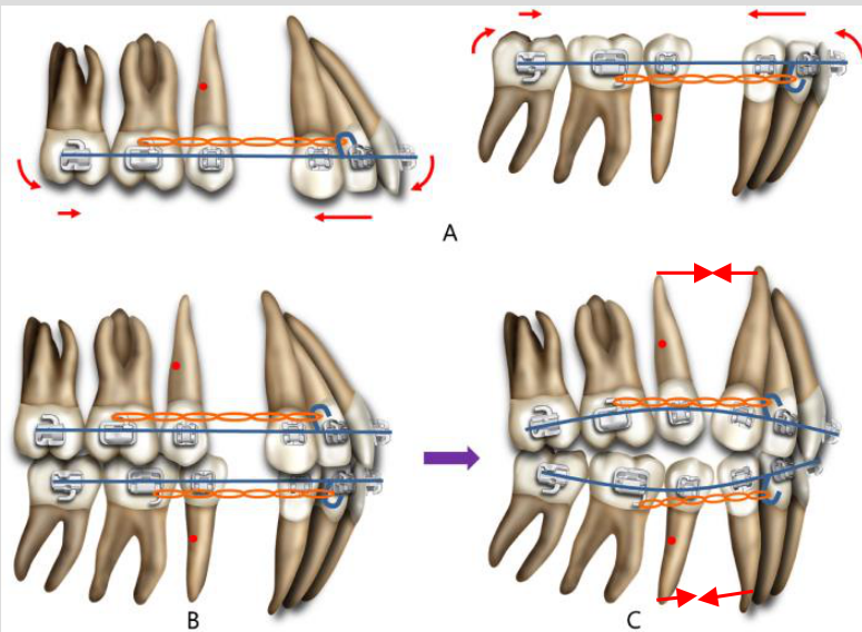
- Divergent roots can allow occlusal forces to encourage movement along the long axes of the teeth, leading to reopening of the extraction space.
- Roots should therefore be made as parallel as possible before treatment is completed.
 |  |
Equilibration18
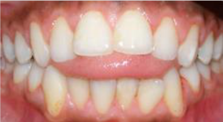
Riedel is of the opinion that functional interferences as a cause of relapse in treated orthodontic cases has been over-emphasized.
Musculature19
We cannot predict accurately a stable position of the teeth with respect to equilibrium of muscle forces.
Lecturer — Muscular Balance
Functional forces from the tongue and other oral muscles can strongly influence stability, especially after correction of an anterior open bite.
- If tongue interposition remains, the tongue may continue occupying a residual space or open bite and contribute to reopening.
- After closure, the tongue may gradually adapt if adequate functional sealing is established.
- Bonded retainers may be placed in both anterior segments, but patients with substantial muscular imbalance require realistic expectations about long-term stability.
Duration of Retention20
The debate concerning the length of use of retention devices has simply not been resolved. It is not known whether prolonged retention provides for greater stability or even if prolonged mechanical restraint is biologically desirable.
Lecturer — Retention Follow-Up
Retention duration should be monitored rather than determined by a universal schedule.
- One clinical protocol uses full-time retainer wear for the first three months, nighttime wear afterward, and follow-up for at least two years.
- Annual retention reviews can identify changes and help maintain the result.
- A lower bonded retainer may remain for ten years, whereas removing one after only three months may be followed by relapse; approximately six months may be considered for some removable treatments, while fixed treatment was suggested for at least one year.
Riedel’s Three Retention Categories21
Riedel divided retention requirements into 3 categories:
- Group I: no retention
- Group II: permanent or semi-permanent retention in one or both arches
- Group III: cases requiring varying lengths of retention
Group I: No Retention22
Group I: no retention (limited treatment)
- Anterior crossbite ✓
- Posterior crossbite after good interdigitation ✓
- High cuspid extraction cases (extraction only) ✓
- Cases in which maxillary or mandibular molars have been tipped distally or premolars tipped mesially to provide space for the eruption of second premolars ✓
- Class II cases treated with headgear, once growth period has passed ✓
Group II: Permanent or Semi-Permanent Retention23
Group II: continue permanent or semi-permanent retention in one or both arches
- Expansion cases ✓
- Class II or Class III relationship, e.g. strong Class II elastic therapy ✓
- Severe rotations ✓
- Spacing ✓
Lecturer — Group II Retention
Group II was described as containing many of the lecturer’s cases.
- Treatment commonly includes a removable thermoplastic retainer and, when significant rotations or other instability risks are present, a bonded canine-to-canine retainer.
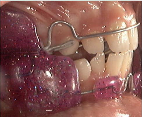
- Expansion may require a removable acrylic plate with an opening in the upper portion to stabilize the transverse relationship.
- Significant space closure and long-term Class II or Class III elastic use may also require permanent or semi-permanent retention.
Group III: Varying Lengths of Retention24
Group III: cases requiring varying lengths of retention or greater lengths of retention
- Class II cases, extraction or non-extraction: if good muscle balance has been achieved and no severe rotations, long-term retention is not necessary
- Deep overbite: retention is directly dependent on growth
- Class II division 2 cases
- Class III corrections with surgery require varying lengths of retention
- Cases involving the ectopic eruption of teeth or supernumeraries that have been present require varying lengths of retention period
Lecturer — Group III Retention
Retention length in Group III depends on the original malocclusion and the quality of the final result.
- Inadequate muscle balance, special-needs patients with substantial muscular imbalance, significant rotations, and substantial space closure may require longer or individualized retention.
- Class II Division 2 cases are particularly prone to return of the upper incisors toward retroclination after treatment and require particular attention during retention.
Perspectives in Orthodontic Stability25
Charles J. Burstone
- What is a stable dentition? Teeth after orthodontic treatment are not ankylosed to the bone, and the supporting tissue allows teeth to migrate under changing conditions.
- A relationship exists between functional occlusion and the pattern of tooth migration.
Normal Growth, Orthopedic Changes, and Relapse26
- The mandible grows and displaces forward at a faster rate than the maxilla.
- The typical growth helps treatment in Class II, but not in Class III malocclusions.
- The mandible may continue growing for a substantial period in male patients, making treatment timing important; Class III cases may require prolonged observation because continued mandibular growth can alter the corrected relationship.
- The typical growth helps treatment in Class II, but not in Class III malocclusions.
Stability and Mandibular Rotation During Treatment27
- High incidence of relapse in patients with deep overbite.
- In growing patients, the mandible grows vertically more than the maxilla to have enough room for the eruption of posterior teeth.
Lecturer — Vertical Growth
Excessive vertical mandibular growth and clockwise rotation may contribute to development of an anterior open bite.
- Vertical growth must be considered when correcting deep overbite, open bite, or Class II malocclusion with vertical involvement.
Arch Width and Stability28
-
Depends on equilibrium of many factors.
-
The original arch form, particularly the mandibular arch form, tends to reassert itself after treatment, so the original shape should be preserved as closely as possible.
-
An ovoid arch should generally remain ovoid after correction, even when crowding is corrected.
-
The archwire shape may be selected with reference to the patient’s original study model.
-
Expansion cases may require a removable acrylic plate to stabilize the corrected transverse relationship.
-
A posterior crossbite corrected by expansion may therefore require retention of the transverse correction.
Incisor Position and Stability: Three Dogmas293031
-
The most stable position for the lower incisors is a cephalometric mean: the lower incisor to mandibular plane is about 90° with a standard deviation of 5°.
- The best position for the lower incisors is its original position.
- There is only one stable position of the lower incisor. In fact, the original malocclusion may be the most stable position.
-
The most stable position for the lower incisors is a cephalometric mean: the lower incisor to mandibular plane is about 90° with a standard deviation of 5°.
-
The best position for the lower incisors is its original position.
- There is only one stable position of the lower incisor. In fact, the original malocclusion may be the most stable position.
-
The lecturer described approximately 85 to 95 degrees as a stable range for the mandibular incisors relative to the mandibular plane.
-
Excessive proclination increases the likelihood of relapse.
-
When crowding is present but sufficient space exists to preserve the original arch form, maintaining that form may provide greater stability.
-
Bonded canine-to-canine retention is frequently used when the lower incisor position is particularly vulnerable.
Intra-Arch Factors and Functional Occlusion32
- Intra-arch factors and stability:
- Reorganization of periodontal fibers.
- Transeptal and supracrestal periodontal fibers require substantial time to accommodate after tooth movement, with more than 280 days allowed for some transeptal fiber adaptation and approximately 230 days for supracrestal fiber stabilization.
- Reorganization of periodontal fibers.
- Functional occlusion and stability:
- A long centric of 0.5–1.0 mm from the CR is acceptable.
- Multidirectional chewing had minimal migration of teeth.
The Maintenance System and Occlusal Dynamics
Ronald H. Roth
Functional Versus Anatomical Occlusion33
One of the aims of orthodontic therapy is to establish a good functional occlusion that is in harmony with the TMJs and mandibular musculature, along with an efficient masticatory apparatus and healthy periodontium.
Lecturer — Occlusion Dynamics
Occlusion is dynamic rather than permanently fixed, and stability depends on the relationship among the teeth, periodontal tissues, bone, muscles, and other soft tissues.
- Aging changes the position and function of these structures.
- Orthodontic treatment therefore cannot control every change that occurs after treatment.
Criteria for an Ideal Functional Occlusion
Ronald H. Roth
Maximal Intercuspation34
- Stress should be directed down along the long axis
- The posterior teeth should contact equally and evenly
- Minimal OJ and OB, but sufficient overbite
- Minimal interference
Signs and Symptoms of Occlusal Disharmony3536
Ronald H. Roth
Occlusal disharmonies can result in the following symptoms:
- TMD
- Occlusal wear and bruxism
- Excessive tooth mobility and/or periodontal disease (from lateral stress)
- Movement or relapse of tooth positions
Lecturer — Tooth Mobility
Mobility caused by trauma may be reversible when a more stable occlusal position is achieved. Mobility associated with periodontal disease represents a different condition.
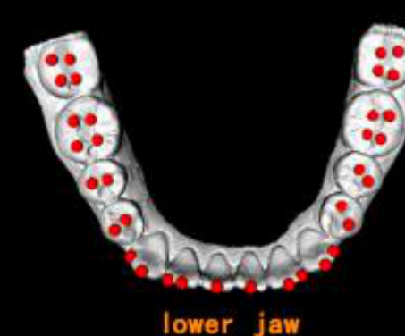
Normal
Orthognathic profile
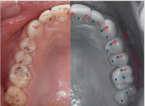
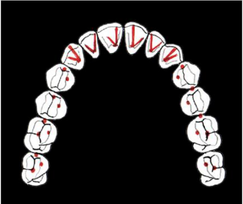
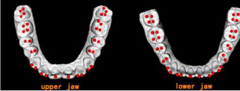
Upper jaw
Lower jaw
 |  |  |
 |  |  |
 |
Occlusal Relationships and Post-Treatment Tooth Movement37
Ronald H. Roth
Post-treatment occlusion should have maximum intercuspation. There are 4 types of centric discrepancy:
- The tooth or inclined plane interference deflects the mandible off the terminal hinge arc of closure
- Due to failure to correct the jaw relationship anteroposteriorly
- Due to insufficient ramus height or short posterior face height / skeletal openbite pattern
- Asymmetry of the mandible
Lecturer — Skeletal Imbalances
Mandibular asymmetry and insufficient ramus height can make the result difficult to maintain, particularly during growth. These skeletal and occlusal imbalances may contribute to post-treatment tooth movement.
Articulation of Teeth and Mandibular Movement38
Ronald H. Roth
- Straight protrusive movement
- Canine guidance when lateral movement
- Group function should also be evaluated where present.
Post-Treatment Relapse and Equilibrium
Post-treatment relapse results from a lack of equilibrium among the following factors:
Periodontal and Gingival Tissue39
- Reorganization: PDL — 3–4 months; collagen fiber — 4–6 months
- Elastic supracrestal fiber — 232 days
Lecturer — Tissue Reorganization
Gingival and transeptal fiber tension can contribute to relapse, and full-time use of removable retainers during the first three months was linked to the need for tissue reorganization.
- Periodontal ligament reorganization was described as taking approximately four to six months.
- Elastic supracrestal fibers may require approximately 230 days to stabilize.
Orofacial Soft Tissue (Muscle Activity)
- Lower labial segment
- Arch width
- Arch length
Lecturer — Muscular Balance
Muscle activity must remain balanced for the corrected position to be maintained.
- The tongue can contribute to relapse when it continues to interpose in an anterior open bite or space.
- Special needs patients may have substantial difficulty maintaining muscular balance.
- A considerable amount of the correction may be lost within a few months when the functional environment cannot be maintained.
Growth and Development
- Response, dentoalveolar adaptation
Lecturer — Growth Effects
Growth can improve stability when it allows the bone to adapt to tooth movement, but continued growth and rotation can also destabilize corrections.
- Continued mandibular growth can destabilize Class III corrections.
- Vertical growth and mandibular rotation can affect deep overbite and open bite corrections.
- The effects of aging and development cannot be completely controlled by the orthodontist.
Occlusal Factors and Forces
-
A well-interdigitating occlusion
-
Root divergence in extraction cases can contribute to reopening of spaces under occlusal load. Toward a perspective on orthodontic retention, AJODO, May 1998.
Planning the Retention Phase40
Six factors to consider when planning the retention phase:
- Obtaining informed consent: no guarantee
- Original malocclusion and patient’s growth pattern: retention device, long-term stability (Cl II, mild Cl III, openbite, rotated teeth)
- Type of treatment performed: removable (6 months), or fixed (1 year)
- Soft and hard tissue adjunctive procedures to enhance stability: fiberotomy, surgical gingivoplasty, frenectomy, interproximal stripping
- Type of retention: removable, fixed, passive or active
- Duration of retention: no clear indication
Toward a perspective on orthodontic retention, AJODO, May 1998
Lecturer — Retention Planning
Retention planning should begin with realistic expectations, because retention is a long-term commitment and not every post-treatment change can be controlled.
- Teeth have a natural tendency to move, while fibers and bone require time to reorganize.
- Growth and aging continue after treatment, occlusion remains dynamic, and patient cooperation affects the result.
- The plan also depends on the amount of rotation corrected, expansion or transverse correction, extraction and space-closure requirements, quality of intercuspation, and muscle balance or other functional conditions.
- Retention may combine removable and fixed appliances, but retainers should generally be passive rather than active.
- A surgical frenectomy may improve stability in selected diastema cases; approximately 2 mm of closure may be possible without additional consideration, but a low frenum insertion should prompt consideration of frenectomy before fixed retainers are placed.
- Follow-up should continue for at least two years, with annual reviews useful for detecting relapse.
Retainers
Fixed Retainers41
Advantages:
- Retain corrections of incisor irregularity
- Compliance free
Disadvantages:
- Less effective with inherently unstable
- Patients are often dismissed
- Plaque accumulation
- Technique sensitive (bond failure, stress of wire)
Lecturer — Fixed Retainer Design
Fixed retainers are commonly used for canine-to-canine retention of the lower incisors, anterior segment retention, corrected rotations, and spacing corrections. Designs may include thick plain wire, spiral wire, multistrand wire, round steel wire bonded to the canines, and zigzag designs that facilitate flossing in periodontal patients.
- Upper fixed retainers may be vulnerable to occlusal contact from the lower canine or lower incisors.
- Aging and minor relapse can alter the occlusion, causing breakage or occlusal interference.
- The bonding technique is controllable; the greater difficulty is patient cleaning and the perception that the retainer may need to remain indefinitely.
Removable Retainers
Advantages:
- Cleanness — flossing
- Inherently unstable procedure
- The capability of correcting minor tooth discrepancies
- Less time-consuming
- Patient can easily be weaned from the appliance
Disadvantage:
- Essix retainers — anterior openbite
Lecturer — Retainer Protocol
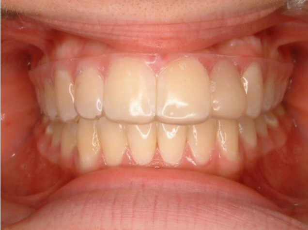
The routine approach uses a thermoplastic polymeric retainer that functions similarly to a static aligner. It is generally recommended for all cases, with 24-hour use for the first three months followed by nighttime use.
- A bonded canine-to-canine bar may be added based on rotation severity, lower incisor proclination, deep overbite, or the need to maintain the anterior segment.
- Long-term retention is challenging because consistent patient use is difficult to ensure.
Lecturer — Thermoplastic Replacement
Thermoplastic retainers may need replacement approximately once a year because they can develop cracks, show crack propagation, and become yellowish. Replacement is an expected part of treatment, and the cost of the thermoplastic material is low.
- Bite plates can be incorporated when vertical control is required.
- The posterior teeth can remain in acceptable intercuspation while a passive bite plate prevents extrusion of the incisors.
- Consistent patient use is required for the appliance to be effective.
A rationale for removable retainers, JCO 1998, Vol XXXII, No 2
Comparison of Essix and Hawley retainers, JCO 1998, Vol XXXII, No 2
A 3 year follow-up study of 3-3 reatiners, EJO, 1997
Fixed Versus Removable Retainers
Three-Year Follow-Up Study of 3-3 Retainers42
A 3-year follow-up study of various types of orthodontic 3-3 retainers:
- No difference in the stability of incisor alignment of 4 types of retainers:
- Thick plain wire bonded
- Thick spiral wire bonded
- Flexible spiral wire bonded
- Removable retainer
- 20% failure rate: technique sensitive
- Tendency for plaque and calculus build-up along the wire — no significant difference between baseline and follow-up examination
Lecturer — Retainer Duration
The important issue was not which retainer was selected, but how long it was used. Approximately 20% failure was reported, and bonding technique was identified as a factor the clinician can control.
Characteristics of the Ideal Removable Retainer43
The ideal removable retainer should be:
- Able to allow for functional occlusion
- Sturdy enough to withstand long-term use
- Flexible enough to allow normal physiological movement of teeth
- Convenient for orthodontist to provide and maintain
- Patient-friendly in both comfort and wear routine
A rationale for removable retainers, JCO 1998, Vol XXXII, No 2
Lecturer — Retainer Replacement
Thermoplastic retainers may be aesthetically preferred, but they require periodic replacement. Annual replacement of clear retainers is a practical approach, and replacement should be included in the practice system with patients called back for review and replacement when needed.
Clinical Applications44
Three Categories of Retention45
Retention can be divided into three categories:
- No retention required
- Limited retention
- Permanent or semi-permanent retention
No Retention Required46
No retention is required in the following situations:
-
Corrected anterior crossbites when adequate overbite has been established
-
Posterior crossbites if axial inclination is acceptable
-
Malocclusions treated by serial extraction only
-
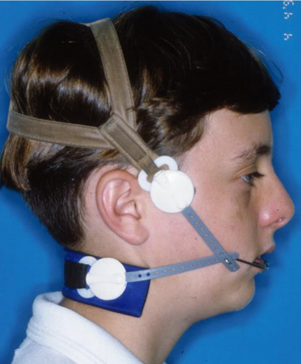
Corrections achieved by retardation of maxillary or acceleration of mandibular growth once the patient has passed the growth period
-
No retention may be considered when adequate intercuspation is achieved.
Limited Retention47
Most typical orthodontic cases fall into this category. The time required allows muscular adaptation and tissue reorganization. However, ideally retention is life-long in treated orthodontic cases.
Lecturer — Limited Retention
Limited retention depends on the specific case.
- Some corrected removable cases may require approximately six months of retention.
- Even when retention is initially described as limited, follow-up remains important because the occlusion and surrounding tissues continue to change.
Permanent or Semi-Permanent Retention48
Permanent or semi-permanent retention is indicated in:
-
Cases of considerable or generalized spacing
-
Instances of severe rotation or severe labio-lingual malposition
-
Spacing between maxillary central incisors
-
Severe overbite in Class II Div. 2
-
Cleft palate lateral expansion
-
Labiolingual malpositions of the lower incisors
-
Significant space closure
-
Cases with a continuing muscular imbalance
-
Retention may combine a bonded canine-to-canine bar with a removable clear retainer.
Retention Mechanics: Three Classes49
Retention mechanics can be divided into three classes:
- Fixed retention
- Removable retention
- Functional elastic retainers
Fixed Retention5051
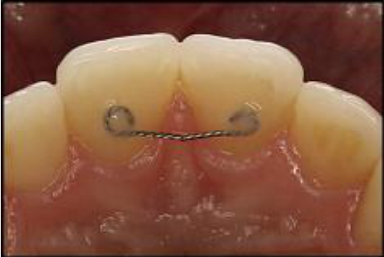
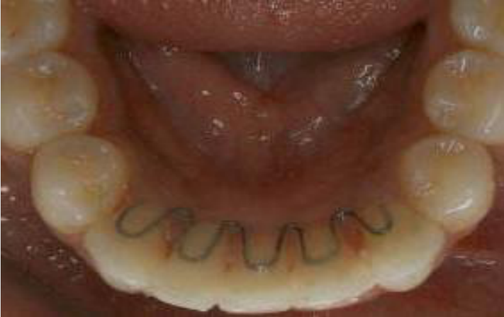
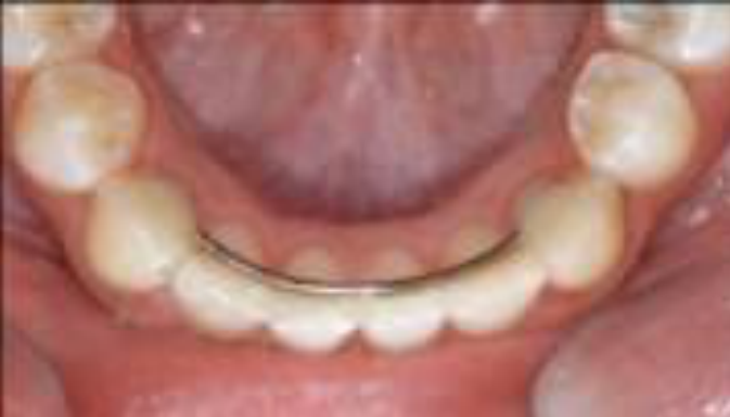
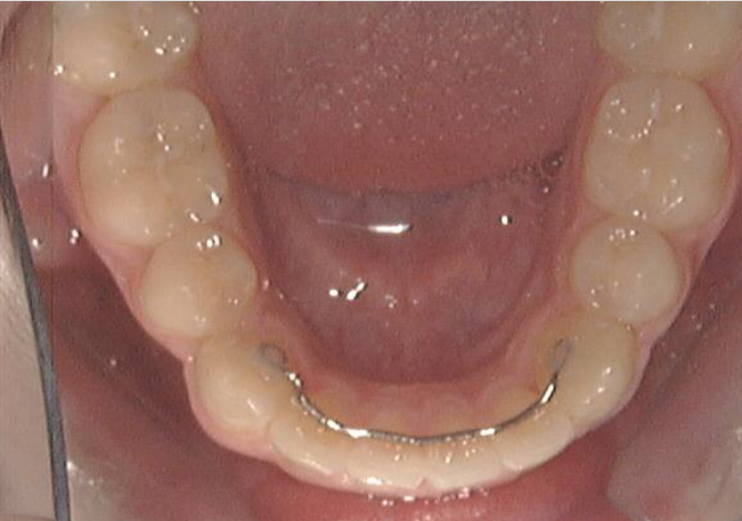
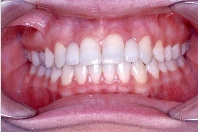
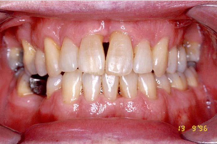
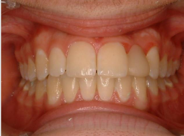
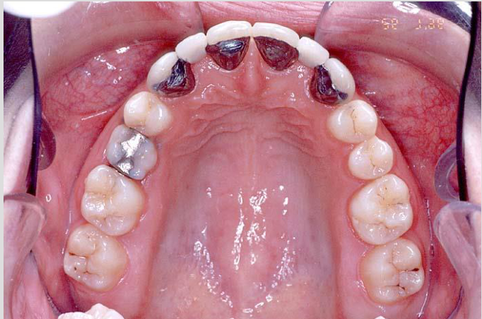
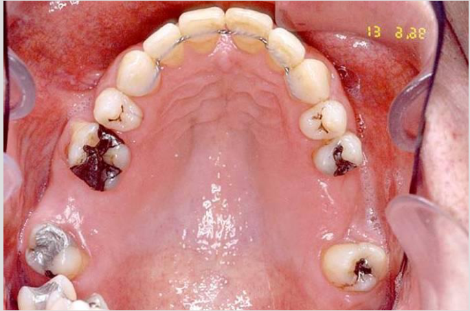
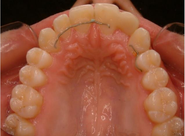
- Bonded canine-to-canine retainers are used frequently in the lower arch.
- Upper retainers may be extended or modified according to the case.
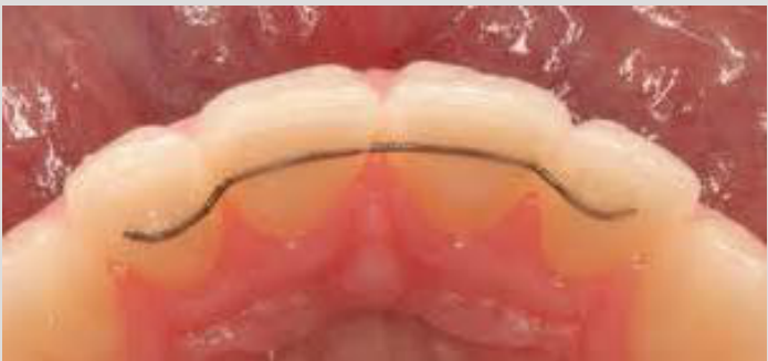
- For vertical problems such as overbite or open bite, a bonded bar may be placed from lateral incisor to lateral incisor to treat the incisors as a single unit.
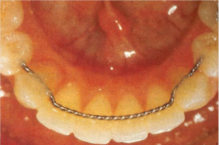
- In diastema cases, multistrand wire may be used to maintain the incisors after space closure.
- A frenectomy may precede fixed retention when a low-inserting frenum contributes to the diastema.
- Fixed retainers are useful when patient compliance with removable appliances is uncertain.
 |  |  |
 |  |  |
 |  |  |
 |  |  |
 |
Removable Retention52
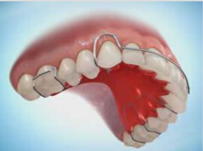
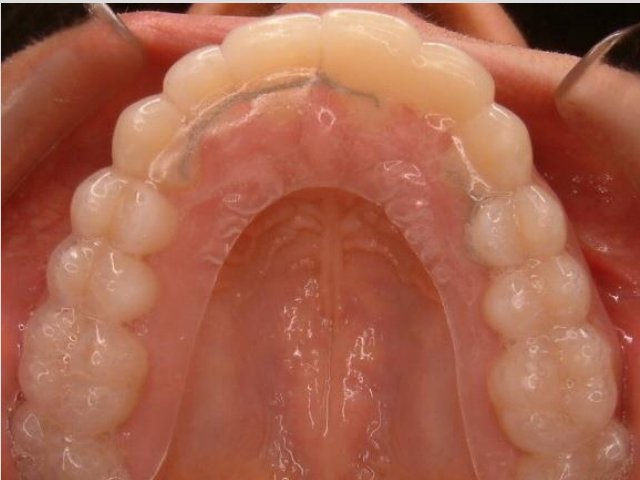
- Clear thermoplastic retainers are routinely used.
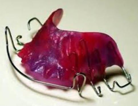
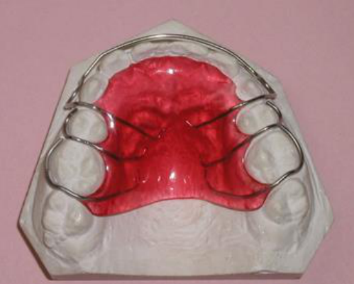
- Expansion cases may require an upper acrylic plate to maintain the corrected transverse relationship.
- A removable bite plate may help control deep overbite by maintaining posterior intercuspation, preventing incisor extrusion, and keeping the anterior teeth passive.
- The primary limitation is that the patient must use the appliance consistently.
- Patients may not always disclose that they have failed to use the retainer.
 |  |  |
 |  |  |
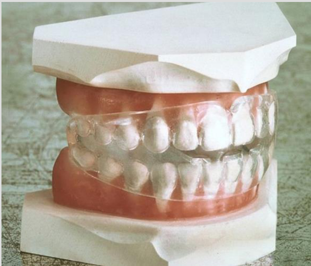
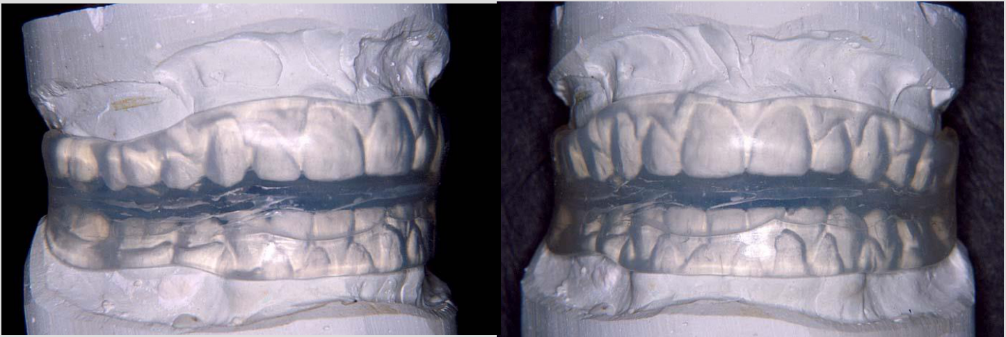
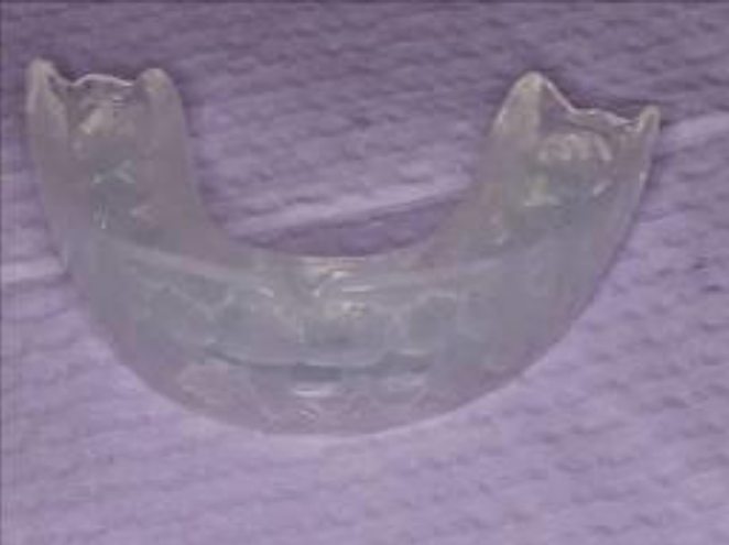
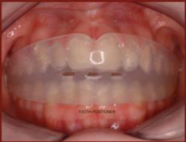
Functional Elastic Retainers53
Lecturer — Functional Stability
Functional effects from the tongue, muscles, and occlusion influence whether the result remains stable.
- The tongue may adapt after an anterior open bite is corrected, but persistent interposition can produce relapse.
- Retention must therefore be planned together with functional correction rather than relying only on an appliance.
 |  |
 |  |
Conclusion54
- An interesting and informative method of reviewing the retention problem is to look at some quotes by eminent orthodontists.
Quotes by Eminent Orthodontists5556
-
“We are in almost complete ignorance of the specific factors causing relapse and failures.” Hellman
-
”… That for girls they wear it (retainer) until they get married.” Ottolengui
-
“Retention is not a separate problem in orthodontia, but a continuance of what we are doing in treatment.” Hellman
-
“If anyone would take my cases when they are finished, retain them and be responsible for them afterwards, I would gladly give them half the fee.” Hawley
Lecturer — Retention Duration
The quotations illustrate how difficult and prolonged the retention phase can be.
- Female patients were suggested to use their retainers until marriage or until menopause.
Retention as Part of Overall Treatment57
- It is now generally recognized that retention must be appreciated as one aspect of the overall orthodontic treatment. Relapse of orthodontically treated teeth should be viewed not as a pathological or abnormal phenomenon, but as an unwanted or undesirable symptom of normal oral physiology.
Lecturer — Treatment Commitment
Retention is an integral part of orthodontic treatment, and orthodontic treatment is a long-term commitment.
- The final result depends on patient compliance, tissue reorganization, muscle balance, growth, aging, occlusal relationships, and the duration and type of retention.
- The clinician should explain that some changes cannot be controlled completely.
- If relapse occurs, the patient is generally expected to return to the orthodontist who completed the treatment.
- The practical approach is to monitor patients regularly, recognize that occlusion is dynamic, and use retention for as long as the individual case requires.
Direct to Consumer Orthodontics and Scope of Practice5859
Direct to Consumer Orthodontics
-
It is important to make it very clear to your patients that you as a practitioner do not accept any responsibility for retention for their direct to consumer orthodontic treatment.
-
It is recommended, appropriate and expected in most circumstances to refer a patient back to their orthodontist if there are retention issues.
At the end of the Orthodontic course, you have neither the didactic or clinical knowledge to include comprehensive orthodontic treatment in your Scope of Clinical Practice.
Clinical Cases
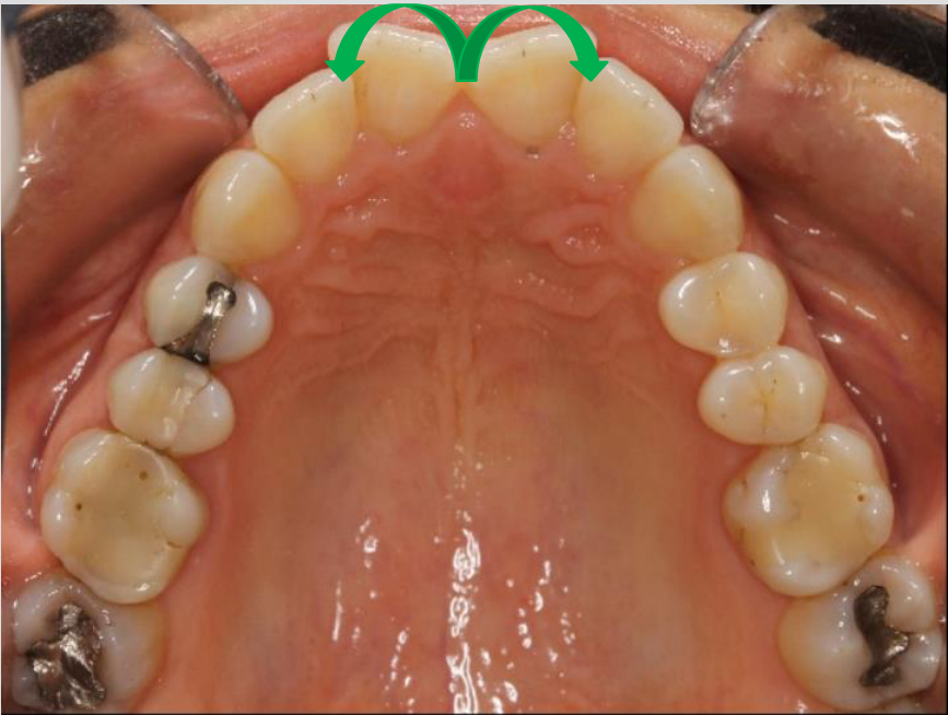
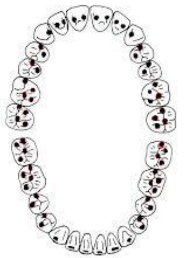
Case: Intraoral view of corrected misalignment
Question
Scenario: An intraoral photograph is presented showing a dental arch.
What’s shown: An occlusal view of a misalignment that has been corrected.
Consider: How to manage the tendency for the teeth to relapse after correcting this misalignment.
Answer
Observations:
- The image shows a corrected misalignment.
- There is a strong trend for the teeth to return to their original position (relapse).
Reasoning: Teeth tend to move back to their former position because of the elastic nature of collagen fibers. Correcting the misalignment alone is not enough to prevent this relapse, as the teeth may shift back up to 50% of the way.
Outcome: A bonded bar from canine to canine is placed to maintain the correction. It can be revised or removed after two to three years.
Takeaway: Placing a bonded bar from canine to canine is an important initial retention strategy to counteract the tendency for corrected misalignments to relapse.
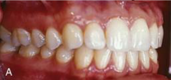
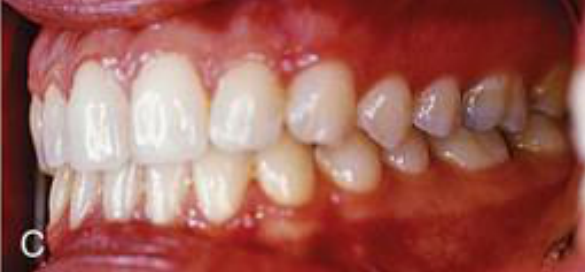
Case: Intraoral views demonstrating occlusion and retention
Question
Scenario: Two intraoral photographs are presented showing the occlusal relationship of a patient’s teeth.
What’s shown: Close-up and overall intraoral views of the teeth in occlusion, demonstrating specific functional and positional characteristics.
Consider: How the achieved occlusion contributes to retention and stability after orthodontic treatment.
Answer
Observations:
- The images show a case with very good intercuspidation.
- The canine guides the lateral movements.
- An anterior crossbite has been corrected, resulting in a normal overbite.
Reasoning: Achieving a good intercuspidation and proper functional guidance provides stability. The normal overbite helps keep the incisors in position.
Outcome: The achieved occlusion works better as a retainer than any removable retainer appliance.
Takeaway: A well-finished occlusion with proper intercuspidation, canine guidance, and corrected crossbites is a critical factor in retention, as it naturally maintains tooth position better than retainers alone.
Case: Extraction site with divergent roots
Question
Scenario: A patient who has undergone extractions during orthodontic treatment, with a radiograph taken before the finishing stage to assess root positions.
What’s shown: No image in this entry; the slide illustrates root paralleling with a line drawing of the mandible and a labelled diagram (the lecturer requests an OPG before finishing).
Consider: The effect of occlusal loading on the roots and the potential consequences for the extraction space if the roots are not parallel.
Answer
Observations:
- The roots of the teeth on either side of the extraction site are divergent rather than parallel.
Reasoning:
- During occlusal loading, forces affect the long axis of the teeth.
- There is a natural tendency for non-parallel roots to become parallel under these loads.
- This movement causes the extraction space to reopen.
Takeaway: Roots must be made parallel before finishing orthodontic treatment in extraction cases to prevent space reopening due to occlusal loads.
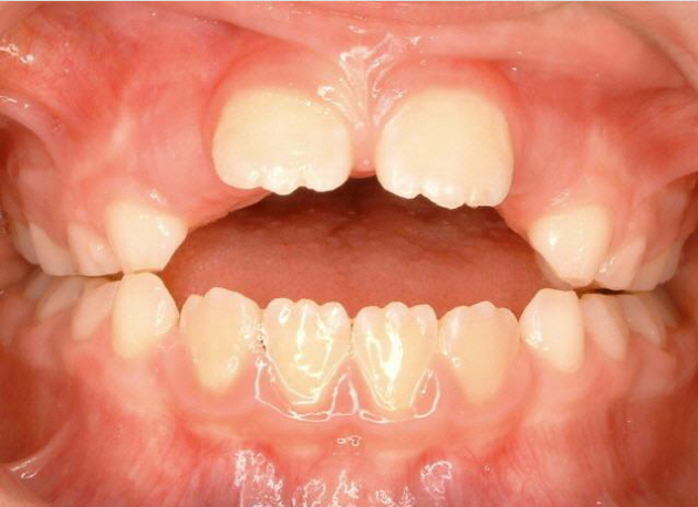
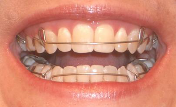
Case: Anterior open bite with tongue interposition
Question
Scenario: A patient presenting with an anterior open bite associated with tongue interposition.
What’s shown: A clinical photograph showing a frontal view of the patient’s dentition, illustrating the open bite.
Consider: The management of an anterior open bite caused by tongue interposition and how to stabilize the treatment result against relapse.
Answer
Observations:
- An anterior open bite is present.
- The condition is driven by tongue interposition.
Reasoning:
- If any space or slight open bite remains, the tongue will find a way to fit into it, causing relapse even after orthodontic treatment.
- The tongue accommodates and changes its behavior only when the space is completely closed and dysfunctional sealing is no longer demanded.
Outcome: The open bite is closed, and bonded bars are placed on the upper and lower anterior segments to stabilize the teeth while the tongue adapts to its new position.
Takeaway: In cases of tongue interposition, the anterior open bite must be completely closed and the anterior segment stabilized (e.g., with bonded bars) to prevent the tongue from causing relapse.
Case: Intraoral photos of fixed retainer designs and complications
Question
Scenario: The lecturer presents a series of intraoral clinical photographs showing various fixed orthodontic retainers in patients’ mouths to illustrate different designs and clinical challenges.
What’s shown: Clinical photos of mandibular and maxillary fixed retainers, including a canine-to-canine bar, a multi-strand wire following a surgical frenectomy, and an upper retainer that has been broken by the opposing lower canine.
Consider: What designs are used for different clinical situations, and what complications can arise with maxillary fixed retainers?
Answer
Observations:
- A canine-to-canine bar can be customized and extended depending on the case.
- A top bar from lateral to lateral is used for overbite or open bite to consider the incisors as a single unit and prevent vertical relapse.
- A multi-strand wire is used for diastema, as seen in a photo following a surgical frenectomy.
- Maxillary retainers often break because the lower incisors or canines occlude in that area, leading to relapse over time.
- Other designs include stranded wires for functional loading of the PDL, round steel wire bonded on canines, and zigzag wires or loops for periodontally compromised patients to allow flossing.
Reasoning: The lecturer walks through the different clinical photos to illustrate how retainer designs are customized based on the specific malocclusion (e.g., vertical plane issues, diastema, periodontal compromise). The broken upper retainer demonstrates a common complication where opposing teeth interfere with the retainer, which is why lower arch retainers are often more straightforward and preferred.
Outcome: Lower fixed retainers from canine to canine are straightforward and commonly used, while maxillary retainers require careful design to avoid occlusal interference and breakage.
Takeaway: Fixed retainer designs must be customized to the specific clinical situation, and maxillary retainers are prone to breakage from opposing occlusion, making mandibular retainers generally more predictable.
Footnotes
-
Original PDF page 1: L5 Retention, p.1 ↩
-
Original PDF page 2: L5 Retention, p.2 ↩
-
Original PDF page 3: L5 Retention, p.3 ↩
-
Original PDF page 4: L5 Retention, p.4 ↩
-
Original PDF page 5: L5 Retention, p.5 ↩
-
Original PDF page 6: L5 Retention, p.6 ↩
-
Original PDF page 7: L5 Retention, p.7 ↩
-
Original PDF page 8: L5 Retention, p.8 ↩
-
Original PDF page 9: L5 Retention, p.9 ↩
-
Original PDF page 10: L5 Retention, p.10 ↩
-
Original PDF page 11: L5 Retention, p.11 ↩
-
Original PDF page 12: L5 Retention, p.12 ↩
-
Original PDF page 13: L5 Retention, p.13 ↩
-
Original PDF page 14: L5 Retention, p.14 ↩
-
Original PDF page 15: L5 Retention, p.15 ↩
-
Original PDF page 16: L5 Retention, p.16 ↩
-
Original PDF page 17: L5 Retention, p.17 ↩
-
Original PDF page 18: L5 Retention, p.18 ↩
-
Original PDF page 19: L5 Retention, p.19 ↩
-
Original PDF page 20: L5 Retention, p.20 ↩
-
Original PDF page 21: L5 Retention, p.21 ↩
-
Original PDF page 22: L5 Retention, p.22 ↩
-
Original PDF page 23: L5 Retention, p.23 ↩
-
Original PDF page 24: L5 Retention, p.24 ↩
-
Original PDF page 25: L5 Retention, p.25 ↩
-
Original PDF page 26: L5 Retention, p.26 ↩
-
Original PDF page 27: L5 Retention, p.27 ↩
-
Original PDF page 28: L5 Retention, p.28 ↩
-
Original PDF page 29: L5 Retention, p.29 ↩
-
Original PDF page 30: L5 Retention, p.30 ↩
-
Original PDF page 31: L5 Retention, p.31 ↩
-
Original PDF page 32: L5 Retention, p.32 ↩
-
Original PDF page 33: L5 Retention, p.33 ↩
-
Original PDF page 34: L5 Retention, p.34 ↩
-
Original PDF page 35: L5 Retention, p.35 ↩
-
Original PDF page 36: L5 Retention, p.36 ↩
-
Original PDF page 37: L5 Retention, p.37 ↩
-
Original PDF page 38: L5 Retention, p.38 ↩
-
Original PDF page 39: L5 Retention, p.39 ↩
-
Original PDF page 40: L5 Retention, p.40 ↩
-
Original PDF page 41: L5 Retention, p.41 ↩
-
Original PDF page 42: L5 Retention, p.42 ↩
-
Original PDF page 43: L5 Retention, p.43 ↩
-
Original PDF page 44: L5 Retention, p.44 ↩
-
Original PDF page 45: L5 Retention, p.45 ↩
-
Original PDF page 46: L5 Retention, p.46 ↩
-
Original PDF page 47: L5 Retention, p.47 ↩
-
Original PDF page 48: L5 Retention, p.48 ↩
-
Original PDF page 49: L5 Retention, p.49 ↩
-
Original PDF page 50: L5 Retention, p.50 ↩
-
Original PDF page 51: L5 Retention, p.51 ↩
-
Original PDF page 52: L5 Retention, p.52 ↩
-
Original PDF page 53: L5 Retention, p.53 ↩
-
Original PDF page 54: L5 Retention, p.54 ↩
-
Original PDF page 55: L5 Retention, p.55 ↩
-
Original PDF page 56: L5 Retention, p.56 ↩
-
Original PDF page 57: L5 Retention, p.57 ↩
-
Original PDF page 59: L5 Retention, p.59 ↩
-
Original PDF page 60: L5 Retention, p.60 ↩