Orthodontic Diagnosis1
Growth and Development — Level IV, Unit A
The University of Western Australia
Why We Do Orthodontics
Psychosocial Benefits2
Why do we do orthodontics?
- Help patient overcome psychosocial handicaps
- Improve function
- Improve oral hygiene
Lecturer — Patient Motivations
Patients who independently seek orthodontic treatment usually do so because of appearance rather than function.
- Common concerns include facial appearance, crowding or lack of space, protruding teeth, and a parent’s concern that development is not proceeding correctly.
- Facial aesthetics can affect self-perception and perceived social fit, particularly during adolescence, while in adulthood appearance may influence perceptions related to employment and promotion.
- The appearance of the incisors, lips, chin, and facial profile can significantly influence quality of life.
- In profile analysis, the relationship of the glabella, lips, and chin to a vertical reference line helps assess facial harmony.
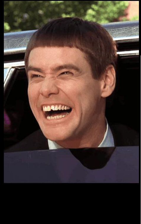
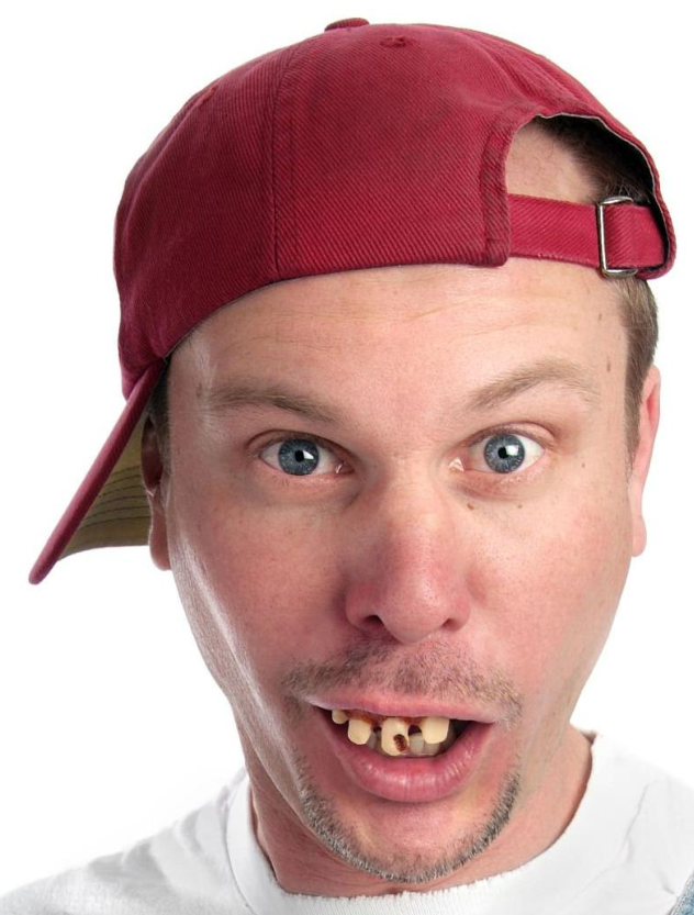
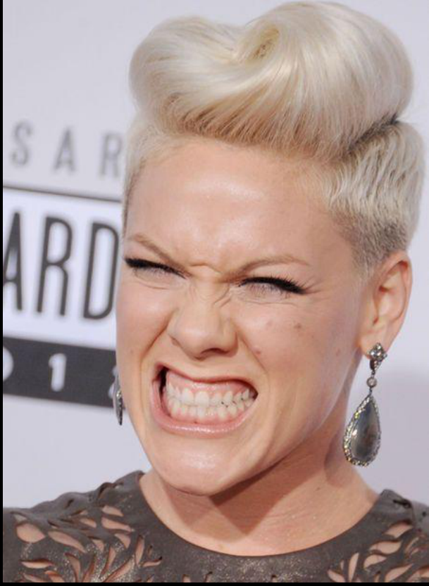
Stereotypes Associated With Malocclusion345
What are some stereotypes associated with malocclusion?
- Protrusive upper incisors suggest lack of intelligence
- 40-50% of children with Class II protrusive incisors will be teased
What are some stereotypes associated with malocclusion?
- Bad teeth suggest a lower economic status
What are some stereotypes associated with malocclusion?
- Chin prominence is associated with strength for males and anger for females
Malocclusions Associated With TMD6
What types of malocclusions are associated with TMD?
- Crossbite with a lateral shift
- Only 1:6 chance of developing TMD
- Class III
- Small chance of developing TMD if there is anterior shift
- Class II deep bite
- Associated with shift?
- Openbite — weak correlation
Lecturer — Functional Shifts
Functional shifts may make several malocclusions less stable, although patients can adapt over time and the correlation with joint problems is not strong.
- A crossbite with a shift should be treated as soon as possible to remove the shift and attempt to prevent joint problems; crossbites account for approximately 15–20% of problems.
- In Class III relationships, an edge-to-edge relationship is unstable and the patient may shift forward to obtain more comfortable and stable interdigitation.
- In Class II deep bites with a large overjet, an impinging overbite may cause the patient to shift forward to achieve a more stable relationship.
- Correcting the malocclusion may nevertheless make the condition less problematic for the patient.
- Panoramic radiographs should be used to compare the right and left condyles rather than expecting them to be identical.
Improved Oral Hygiene7
There is some evidence that patients with severe malocclusion are more likely to develop periodontal problems:
- Crooked teeth are harder to keep clean
- Improved occlusion can decrease the force felt by vulnerable teeth during bruxism
Lecturer — Hygiene Commitment
Improved oral hygiene depends heavily on the patient’s skills and commitment, and braces may be present for at least two years.
- A patient with good cleaning skills may maintain hygiene despite crowding.
- White spot lesions are a significant concern when patients do not brush three times daily, spend additional time cleaning, or follow oral-hygiene instructions at every appointment.
- Periodontal health must be under control before orthodontic treatment begins, with the periodontist remaining involved when appropriate.
The Diagnostic Process8
Level IV Unit A
Building The Database910
Three components feed into the database:
- Interview — history, reason for treatment
- Clinical Exam — health of tissues, jaw function, facial proportions
- Analysis of Diagnostic Records — photos / video, dental casts, radiographs
The database then leads, through classification, to the Problem List = Diagnosis.
Lecturer — Building the Database
Diagnosis should be approached systematically before beginning to collect patient information. In clinic, students use the D40 form and clinical examination to build the database.
- The database supports decisions about the type of malocclusion, treatment approach, patient problem list, and possible solutions.
- The problem list should be concise, using short bullet points or “red dots” rather than transcribing the entire form.
- Diagnosis and treatment possibilities are established by connecting information from the different clinical views.
Medical And Dental History11
Why is this patient seeking treatment, now?
- Chief complaint, motivation
How did things get to be the way they are?
- Medical / dental history, etiology
What is likely to change in the near future?
- Medical condition, growth status
What does he or she expect as a result?
- Internal / external motivation, expectation
Pertinent History Information12
What information from the medical/ dental history is pertinent to orthodontic treatment?
- Nickel Allergy — why is it important and what are possible options for these patients?
- Pre-medication — which orthodontic procedure requires it?
- Rheumatoid and Juvenile arthritis — what might you see with these patients?
- What drugs interfere with orthodontic movements?
Lecturer — Medication Effects
Nickel allergy should be investigated carefully because stainless steel orthodontic materials are stabilized with nickel to prevent corrosion. A patient who cannot wear most earrings but can wear gold earrings may need ceramic brackets and non-stainless-steel or titanium archwires, and nickel exposure should be avoided or reduced because the problem may be cumulative.
- Nonsteroidal anti-inflammatory drugs used for arthritis may interfere with tooth movement through prolonged blocking of the prostaglandin pathway, so the patient should be discussed with the involved physician.
- Bisphosphonates block osteoclast activity and reduce bone turnover, which can make tooth movement more difficult.
- When a patient is taking an antiresorptive medication, treatment may need to be delayed; the lecture stated that it may take approximately three months for the medication to become effective.
- Physiologic tooth movement, such as supereruption after tooth loss, may still occur but in a reduced and slower manner.
Medical Conditions And Growth Status13
- Why is growth status important in orthodontic treatment?
- How do we assess it?
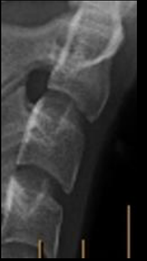
Cervical vertebrae stages: CS1 CS2 CS3 CS4 CS5 CS6
Lecturer — Growth Timing
The second and third cervical vertebrae change from a trapezoidal form toward a more square shape as the patient approaches the peak growth spurt. The peak is located between the third and fourth stages, which is important when growth modification is planned.
- In a Class 2 patient, treatment may attempt to restrict forward movement of the maxilla or promote mandibular advancement.
- Working near the growth peak may improve mechanics, reduce the duration of appliance use, and improve the likelihood of patient cooperation.
- Even when an appliance is prescribed only at night, patient cooperation is necessary.
Clinical Examination14
Facial proportions / tooth-lip relationships
- Facial form analysis
Health of intra-oral hard and soft tissues
- Caries, periodontal disease, other lesions
Jaw function
- Chewing ability, TM joint function
What diagnostic records are needed?
The diagnostic database is built from three inputs:
- Interview — history, reason for treatment
- Clinical Exam — health of tissues, jaw function, facial proportions
- Analysis of Diagnostic Records — photos / video, dental casts, radiographs
Through classification, the database leads to the Problem List = Diagnosis.
Lecturer — Clinical Records
Facial analysis should follow a consistent sequence. Extraoral examination assesses facial shape and symmetry, the relationship of the nose, lips, and chin to a vertical line, lower facial height, facial fifths, and the facial midline.
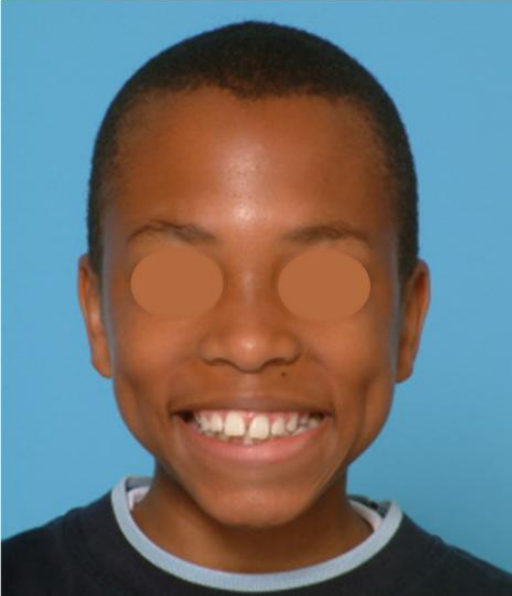
- The smiling frontal view also assesses dental midlines, smile arc, buccal corridors, occlusal cant, gingival zeniths, gingival margins, incisor position, and the relationship of the arch to the lower lip.
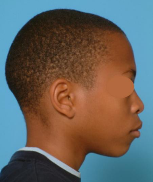
- Profile examination assesses the profile, chin, nose, nasolabial angle, lip position, vertical facial pattern, and midface prominence or deficiency.
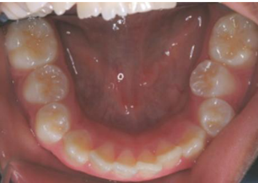
- Intraoral photographs assess midlines, overbite, edge-to-edge relationships, molar and canine relationships, overjet, crossbite tendency, arch shape, crowding, rotations, and available space.
- An edge-to-edge relationship is not an open bite: the teeth contact at the cusps or incisal edges without complete interdigitation, whereas an open bite involves a gap between the teeth.
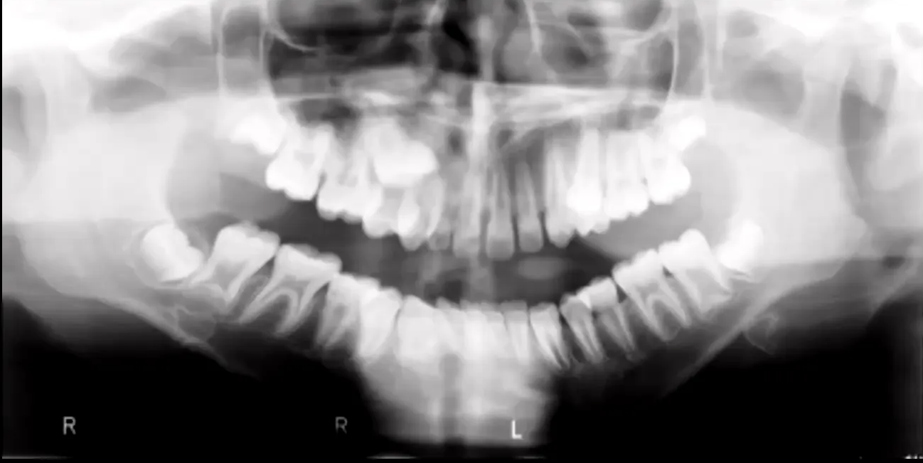
- Panoramic radiographs help count teeth, identify pathology, evaluate the condyles and dental development, determine whether teeth are impacted, and evaluate space and eruption conditions.
Problem List And Ackerman-Proffit Classification15
Ackerman-Proffit Classification
- Dentofacial esthetics — lip protrusion, anterior tooth display
- Alignment and symmetry — crowding/ spacing, arch form
- Transverse — posterior crossbite (skeletal vs dental) and midline deviations
- Anterior Posterior — skeletal and dental AP, Angle classification, overjet
- Vertical — skeletal and dental, overbite, face height, mandibular plane angle
Lecturer — Class 2 Diagnosis
Diagnosis should distinguish whether a Class 2 relationship is primarily skeletal or dental. In a Class 2 case, the clinician must determine whether the problem is a protrusive maxilla, a deficient mandible, or a combination of both.
- A headgear may be used to restrict forward maxillary movement, whereas a functional appliance may be used to advance the mandible.
- The lecture stated that approximately 60% of Class 2 cases are associated with mandibular deficiency.
- Class 2 correctors may be removable, such as twin blocks, or fixed, such as the Herbst appliance.
- These appliances take advantage of growth and aim to produce a more protruded mandibular position, including remodeling of the glenoid fossa and mandible.
Orthodontic Treatment Planning16
Controlling Pathology First
Problem List = Diagnosis
Pathology (Caries, Perio, Etc.)17
- Control before orthodontic treatment
Pathologic problems must be under control before treatment of developmental problems begins.
Lecturer — Pathology Planning
The clinician should continue evaluating pathology while planning orthodontic treatment.
- Panoramic radiographs help identify pathology and evaluate developing and impacted teeth.
- Medications that affect bone turnover may require additional planning and a delay before tooth movement is attempted.
For patients with existing periodontal disease, orthodontics should not start until the perio control phase is completed and can be maintained.
Why?
Prioritizing Problems And Solutions
Problem List = Diagnosis →
Orthodontic (Developmental) Problems in Priority Order18
- Problem A → Possible Solution A
- Problem B → Possible Solution B
- Problem C → Possible Solution C
- etc. → etc.
Lecturer — Space Loss and Overjet
Severe space loss from early loss of primary molars may require extraction.
- Space associated with impacted or out-of-arch premolars cannot always be counted toward overjet correction.
- The plan may require extraction and reopening or redistributing space, including moving a maxillary molar buccally, to obtain enough space to correct the overjet.
Lecturer — Class 3 Treatment
In a maxillary deficiency associated with a Class 3 relationship, treatment timing affects the achievable skeletal correction.
- A face mask is more appropriate at a younger age, around eight years old.
- Rapid expansion may be performed before pressure is applied to the circummaxillary sutures.
- At approximately 11–12 years old, the same skeletal effect may be difficult to obtain, so treatment may provide primarily dental improvement with possible later consideration of surgery.
- Excessive dental compensation should be avoided when a skeletal Class 3 relationship is treated dentally.
- Additional mandibular growth may worsen the relationship when the maxilla is retruded.
The interaction of problems and solutions is evaluated in terms of:
Lecturer — Space Preservation
Severe crowding may require space preservation before the primary molars exfoliate.
- A lower lingual arch placed in the late mixed dentition prevents mesial drift of the lower first permanent molars and preserves leeway space.
- The preserved space can be redirected toward anterior crowding.
- The ideal time to place the appliance is before exfoliation of the primary molars.
- A distal shoe serves a different purpose: it guides eruption of the first permanent molar after early loss of a primary molar and is not the preferred bilateral appliance for preventing mesial drift in significant crowding.
- Interaction
- Compromise
- Cost/Benefit
- Other factors
This leads to the Patient-Parent Consult step, involving:
Lecturer — Mixed Dentition Occlusion
In mixed dentition, a flush terminal relationship may be physiologic, but the treatment objective changes when crowding is present.
- Mesial drift of the lower first permanent molars using leeway space may produce Class 1 molar occlusion.
- When crowding is present, that space may instead be used to address the crowding, leaving the molars short of Class 1.
- The molars can later be distalized to establish a Class 1 relationship.
- Mild crowding of approximately three to four millimeters may be managed without extractions in the described mixed-dentition situation.
- Expanded archwires may be considered when a constricted arch produces a tendency toward crossbite.
- Correcting a rotated molar may improve a Class 2 relationship because molar rotation can worsen it.
- Alternative plans
- Patient input
Patient-Parent Consultation And Informed Consent19
Patient-Parent Consult (alternative plans, patient input) → Informed Consent → Tx Plan Concept → evaluated for Effectiveness and Efficiency → Tx Plan Details
- Clinical examples
Clinical Cases20
Case 121222324
Case 225262728
Case moved — see Case: Case 2, clinical and radiographic orthodontic assessment
Case 329303132
Case 43334353637
Course Disclaimer38
At the end of this course you DO NOT have the didactic knowledge or clinical skills to treat comprehensive orthodontic cases.
Clinical Cases
Case: Young female patient, orthodontic clinical examination and diagnosis
Question
Scenario: A young female patient presents for an orthodontic evaluation. The clinician reviews her extraoral and intraoral clinical photographs to establish a problem list and diagnosis.
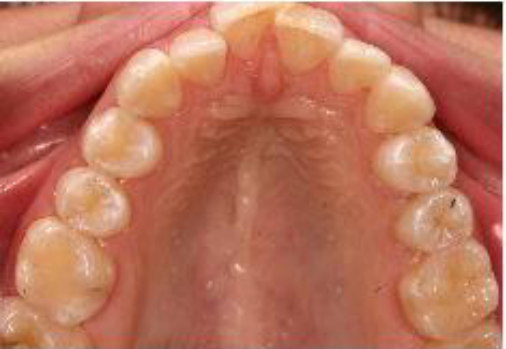
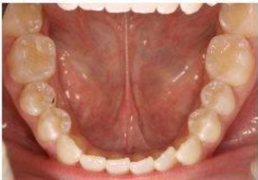
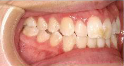
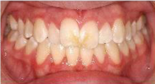
What’s shown: Extraoral frontal and profile photographs, a smile view, and a composite of intraoral photographs (frontal, lateral, and occlusal views).
Consider: What are the key facial, smile, and intraoral findings, and what is the proposed orthodontic management based on these observations?
Answer
Observations:
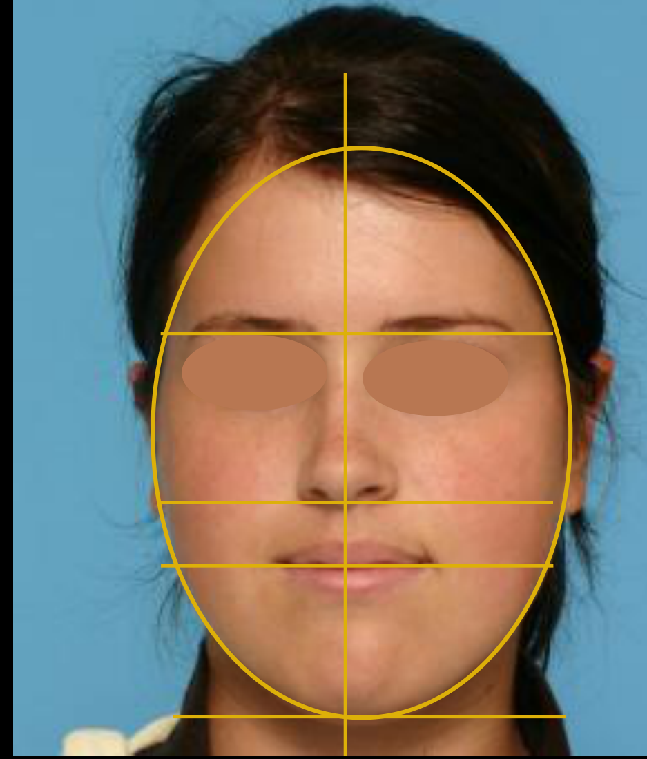
- Facial analysis: Ovoid face shape with the chin slightly shifted to the left. Increased lower facial height indicating a hyperdivergent (vertical) growth pattern.
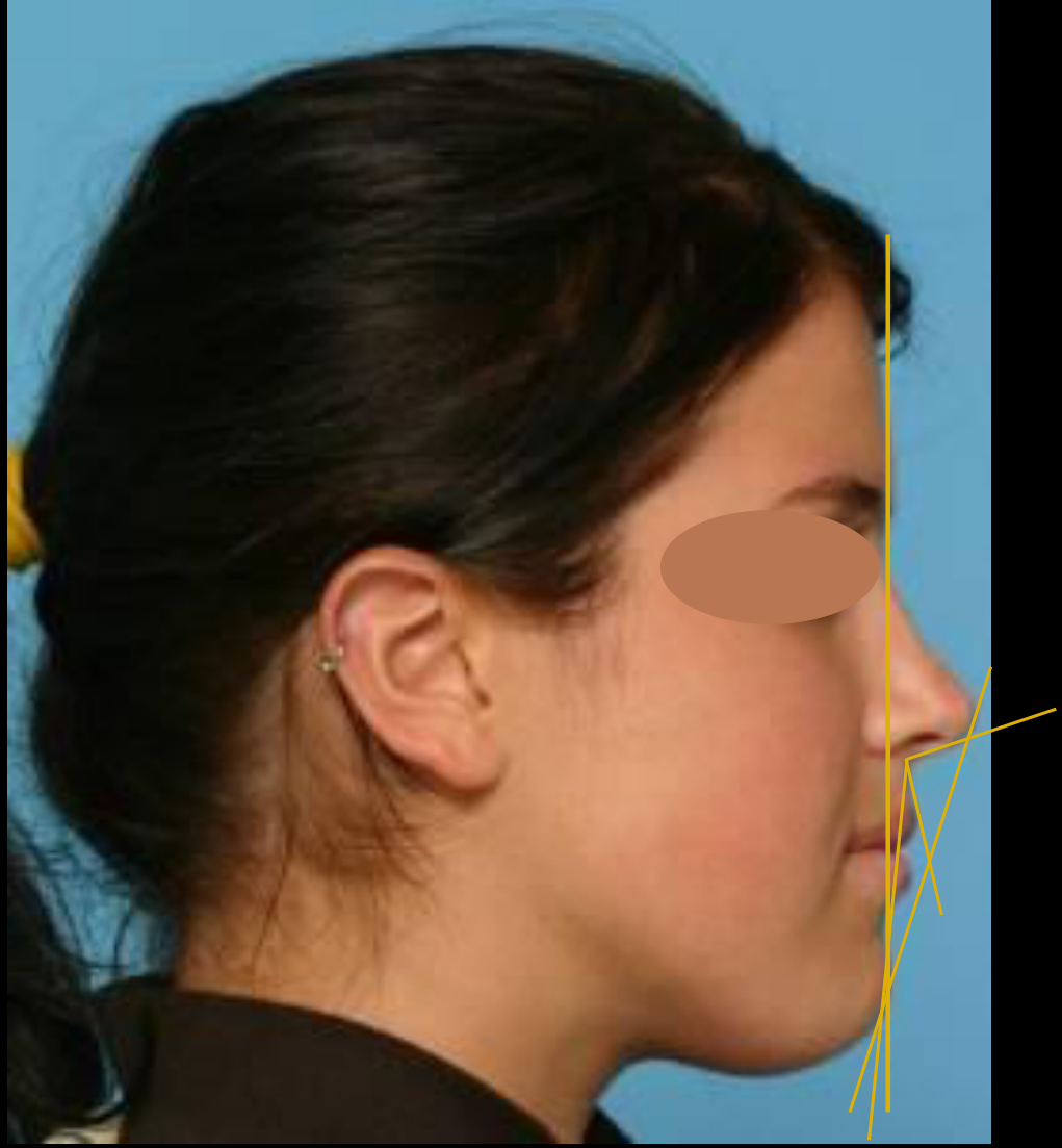
- Profile: Harmonious, slightly convex profile. The chin is in a good position relative to the E-line, while the lips are slightly ahead of the line. The nasolabial angle is almost straight.
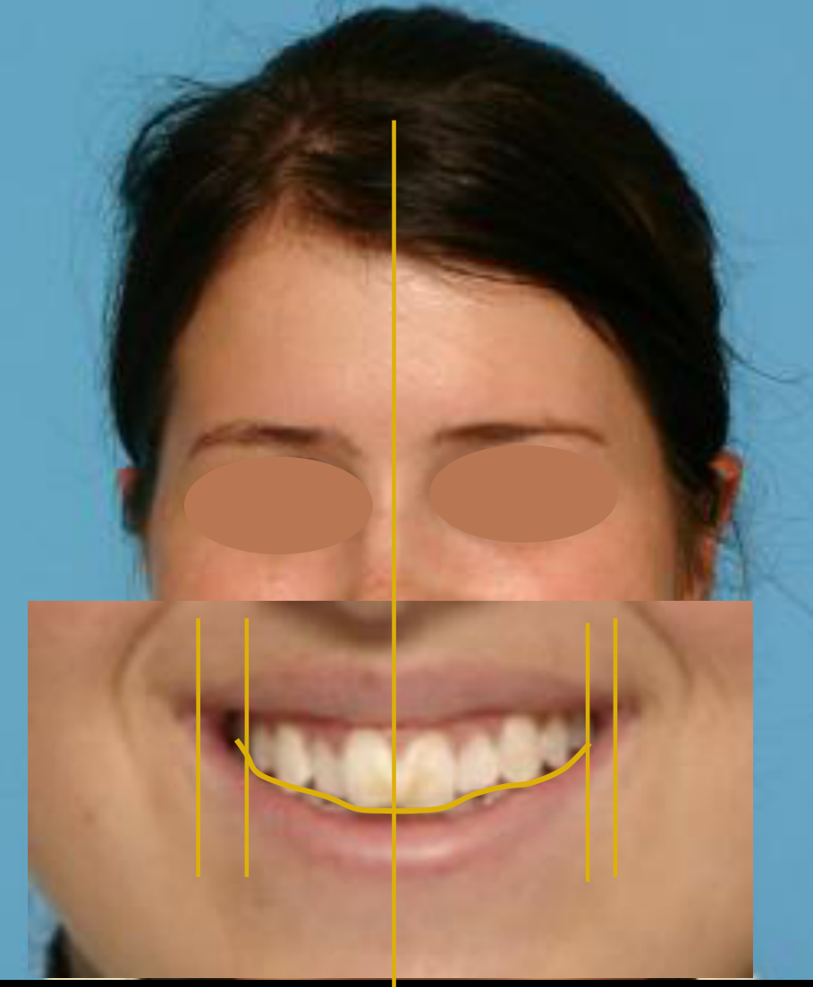
- Smile: The upper midline is 1 mm off to the left. There is a slight occlusal cant on the right side and some buccal corridor on the right.
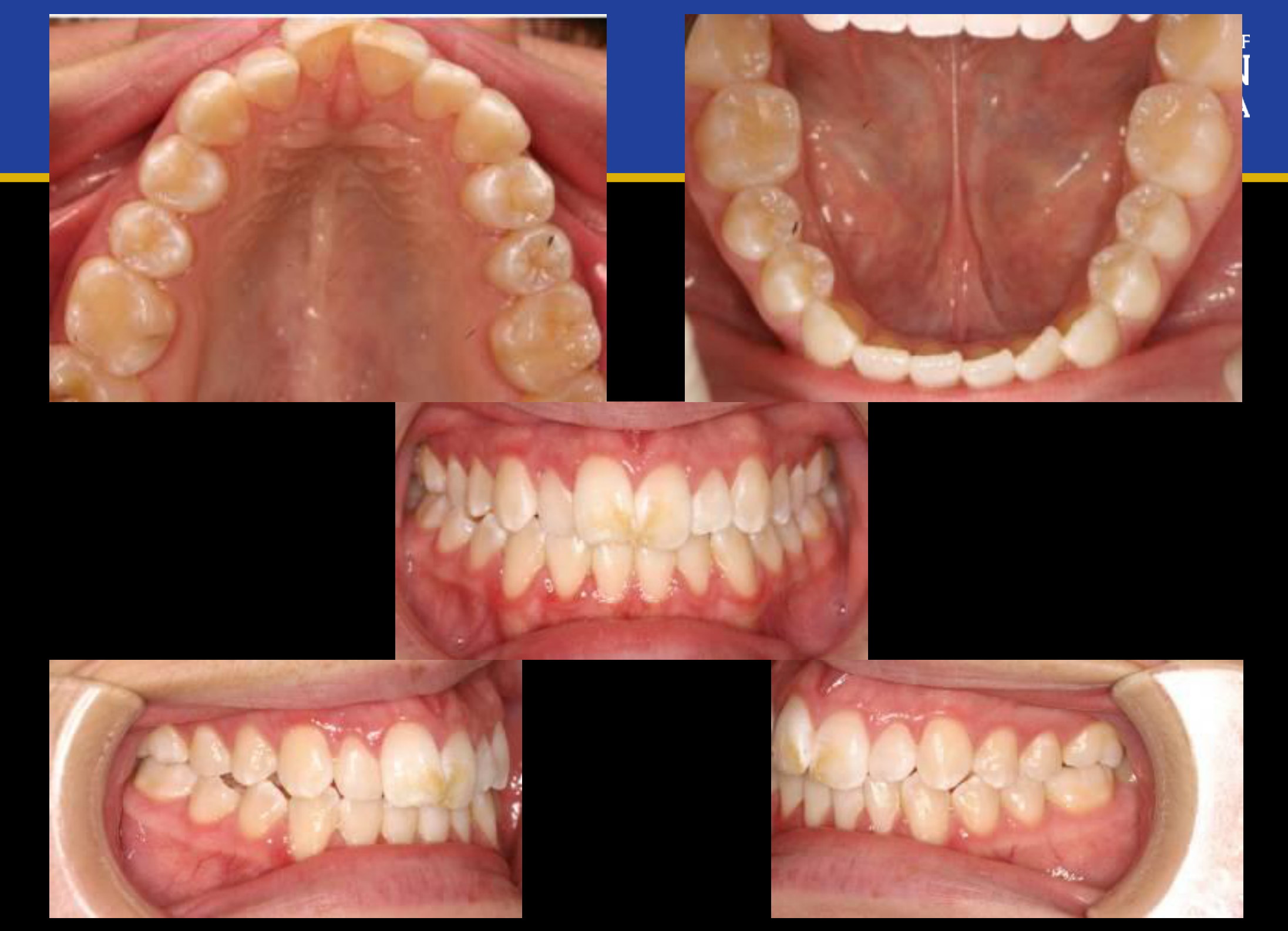
- Intraoral: The upper and lower midlines are coincident (both 1 mm to the left). The overbite is around 20% (reduced, consistent with a vertical patient). The molar relationship is Class I bilaterally. The premolars on one side show an edge-to-edge relationship (touching at the cusp tips, which is not an open bite). There is mild crowding (around 3-4 mm) with some rotated incisors in both arches. The upper arch is narrow, particularly squished on the right side, contributing to a crossbite tendency.
Reasoning: The systematic evaluation connects the extraoral vertical pattern (increased lower face height, reduced overbite) with the intraoral findings. The narrow, squished upper arch on the right explains the edge-to-edge premolar relationship and the crossbite tendency. Because the crowding is mild (3-4 mm) and space can be regained, extractions are not indicated. The edge-to-edge contact is distinguished from an open bite because the teeth are actively touching at the cusp tips, lacking interdigitation but maintaining contact.
Outcome: The treatment plan involves using expanded arch wires to correct the narrow upper arch and resolve the right-sided crossbite tendency.
Takeaway: A systematic clinical examination—progressing from facial proportions and profile to smile analysis, and finally to intraoral relationships and arch form—allows the clinician to accurately identify specific discrepancies, such as distinguishing an edge-to-edge crossbite tendency from an open bite, and to formulate a targeted, non-extraction expansion plan.
From the lecture slides
16 y/o female.
Case 1 Problem List39
- Dentofacial esthetic and protrusion
- Alignment and symmetry
- Transverse
- AP
- Vertical
Lecturer — Case 1 Findings
The patient has an ovoid, slightly vertically oriented face with a small left chin shift, increased lower facial height, and a possible hyperdivergent pattern. The profile is slightly convex but harmonious, with lips slightly anterior to the reference line and a nearly straight nasolabial angle.
- Intraorally, both midlines are approximately one millimeter left but remain reasonably aligned, with about 20% overbite, bilateral Class 1 molars, no increased overjet, and approximately three to four millimeters of crowding.
- The upper arch is constricted, especially on the right, with a slight crossbite tendency and premolars in a cusp-to-cusp relationship rather than an open bite.
- Extractions were not considered necessary; expanded archwires could address the constriction and crossbite tendency.
 |  |  |
 |  |  |
 |  |  |
 |
Case: Case 2, clinical and radiographic orthodontic assessment
Question
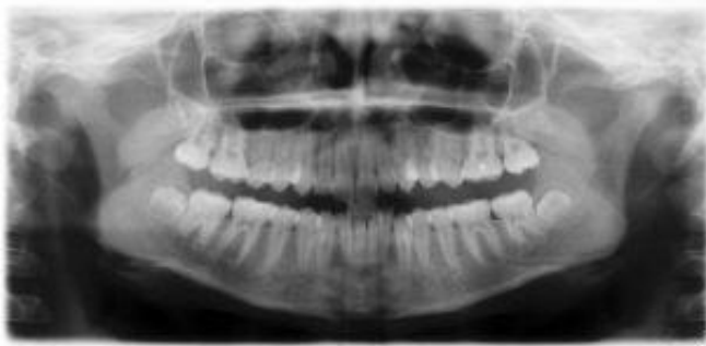
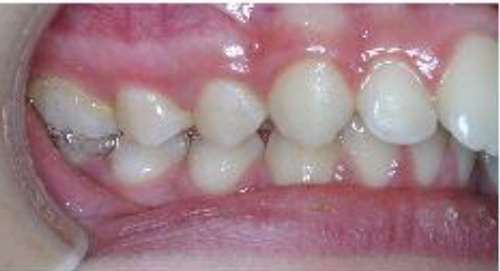
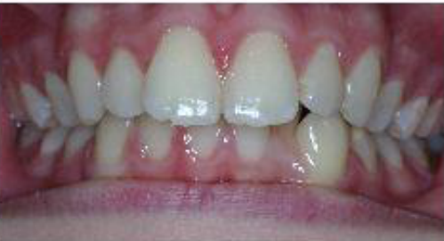
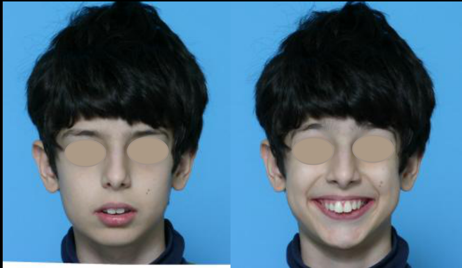
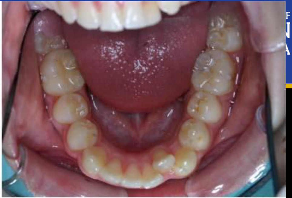
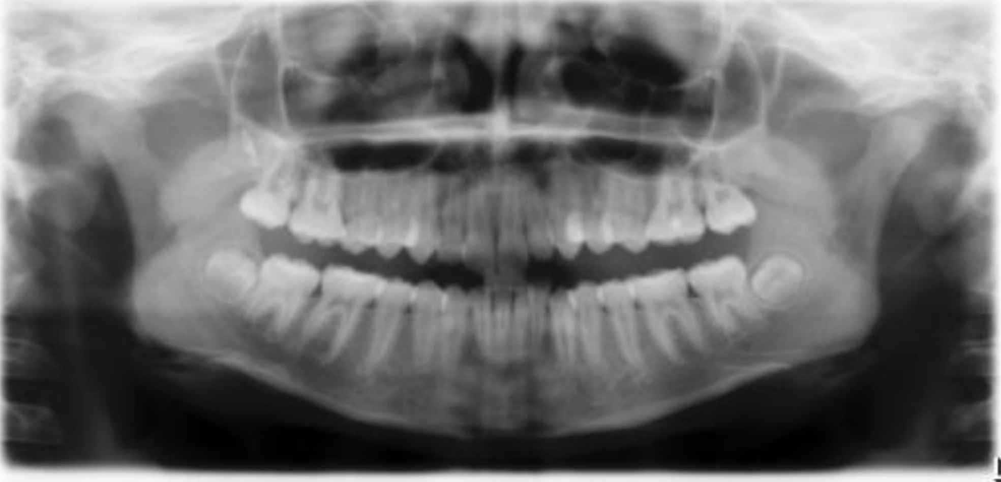
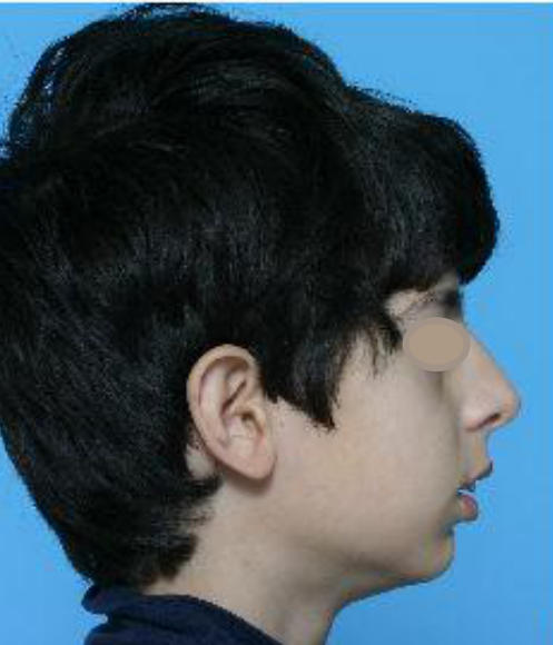
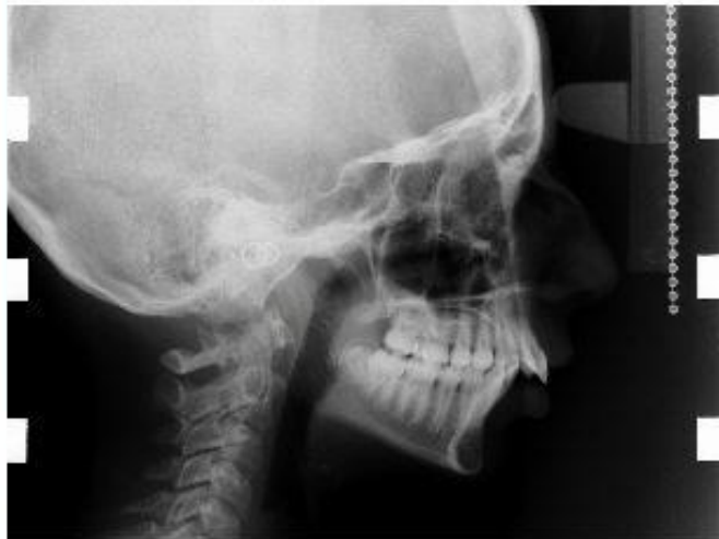
Scenario: A patient is presented for orthodontic evaluation using a comprehensive set of extraoral and intraoral photographs, along with a panoramic radiograph.
What’s shown: Extraoral frontal and lateral profile photographs, intraoral frontal, lateral, and occlusal views, and a panoramic radiograph.
Consider: Based on the systematic clinical and radiographic examination, what are the findings and the resulting problem list for this patient?
Answer
Observations:
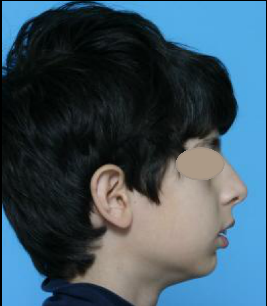
- Extraoral frontal: Incompetent lips, reduced lower face height, midline deviated to the left, smile overcovering the lower lip, no buccal corridor, and a slight cant on the right and left.
- Extraoral lateral: Convex profile with a deficient mandible and retrusive chin, acute angle, large nose, reduced nasolabial angle, and incompetent, protrusive lips.
- Intraoral frontal: Increased overbite (50-60%) and lower midline deviated to the left by approximately 1 mm.
- Intraoral lateral: Class 2 molar relationship on the right and Class 2 half-unit on the left, increased overjet, and no posterior crossbite.
- Intraoral occlusal: Severe lower arch space deficiency (no space for the lateral incisor) and rotations of premolars and molars, with one molar completely rotated clockwise.
- Panoramic radiograph: All teeth present with no pathology; condyles are not fully seated in the glenoid fossa.
Reasoning:
- The facial features indicate a Class 2 profile with a deficient mandible and forward-positioned maxilla, leading to protrusive incisors (likely >22 degrees) and incompetent lips.
- The intraoral findings confirm a Class 2 malocclusion with increased overjet and overbite.
- The clockwise rotation of the molar exacerbates the Class 2 relationship; correcting this rotation would rotate the major buccal cusp and help amend the Class 2 relationship.
- The unseated condylar position on the panoramic radiograph is an artifact of the patient’s large overjet, which required a protrusive shift to bite the positioning block.
Outcome:
- A comprehensive problem list is formulated, encompassing frontal static issues (lips, smile, deficient chin, reduced lower facial height, cant), profile issues (retrusive chin, large nose, reduced nasolabial angle, protruded incisors), and intraoral issues (Class 2 molar relationship, increased overjet/overbite, lower arch crowding, molar rotation).
Takeaway:
- A systematic top-to-bottom and extraoral-to-intraoral approach is essential for compiling a comprehensive orthodontic problem list, and recognizing how rotational discrepancies can exacerbate sagittal relationships is key for treatment planning.
From the lecture slides
Lecturer — Case 2 Findings
The patient has incompetent, protrusive lips, reduced lower facial height, a convex profile, and a retrusive or deficient chin, suggesting a Class 2 relationship. The overbite is approximately 50–60%, the overjet is increased, and the molar relationship is Class 2 on both sides, approximately half-unit on the left.
- The lower arch has significant crowding, including no space for the lateral incisor and rotations of the premolars and molars.
- The condyles appeared slightly unseated because the patient likely protruded while biting on the bite block in response to the large overjet.
- The principal diagnostic question is whether the Class 2 relationship results from maxillary protrusion or mandibular deficiency; approximately 60% of Class 2 cases were described as involving mandibular deficiency.
- Twin blocks or a fixed Herbst may be considered for mandibular deficiency, whereas headgear may be considered when restricting forward maxillary movement is the objective. Growth modification is most effective near the growth peak.
Case 2. 13 y/o male.
How would you characterize his AP skeletal relationship? Lip posture? Plane angle? Vertical proportions?
13 y/o male.
Case 2 Problem List40
- Dentofacial esthetic and protrusion
- Alignment and symmetry
- Transverse
- AP
- Vertical
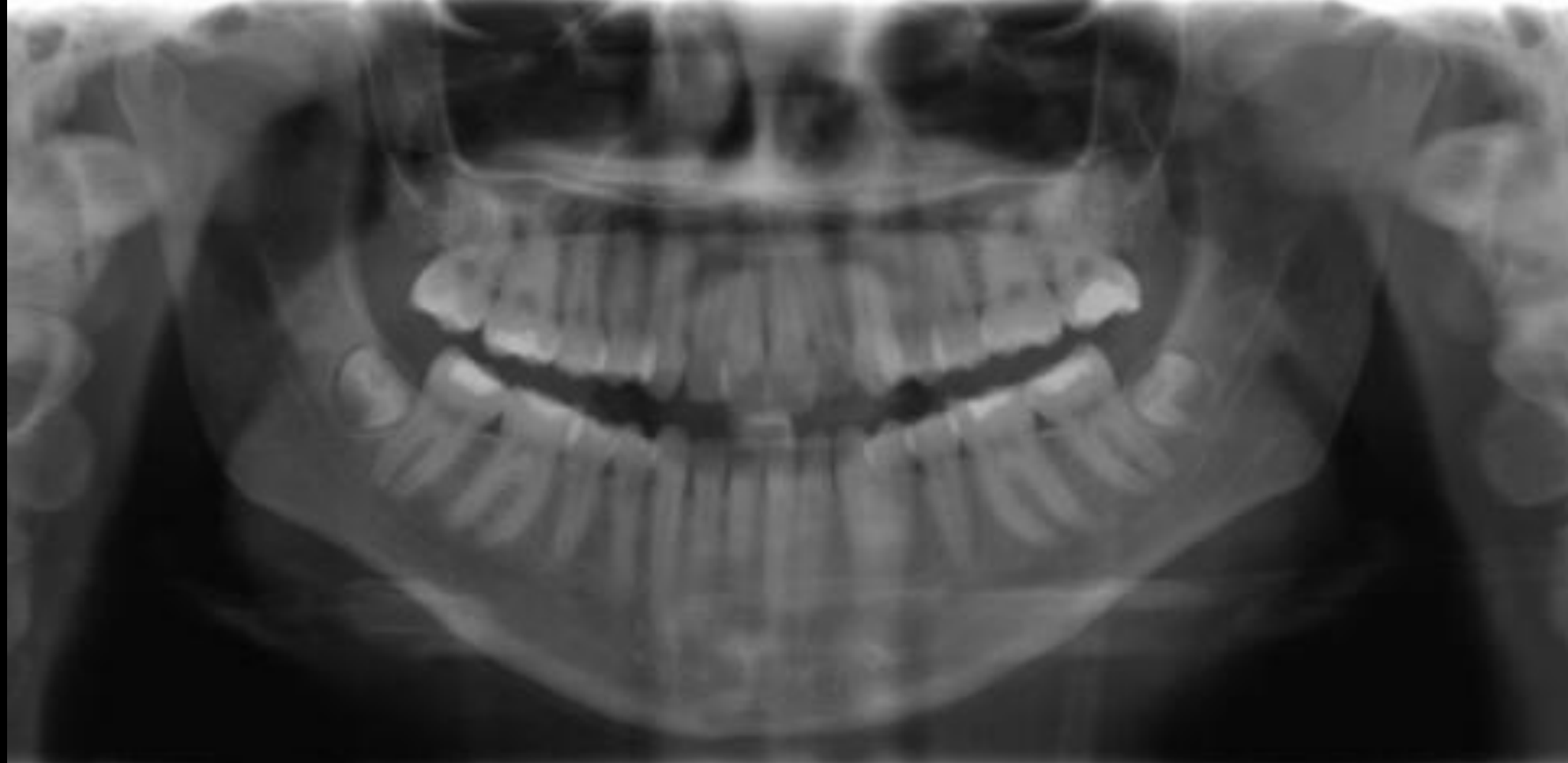
 |  | 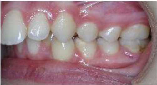 |
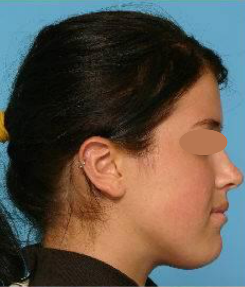
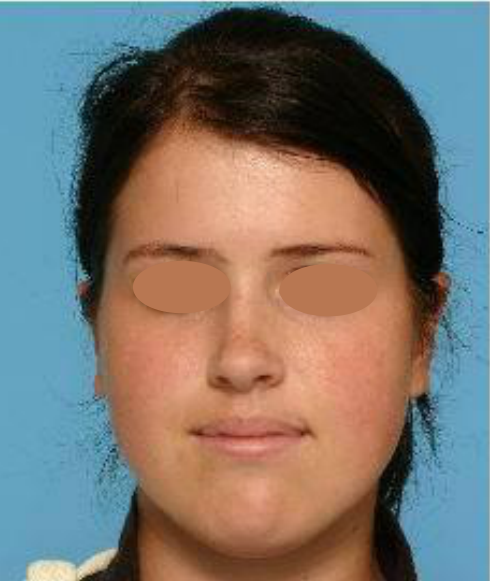
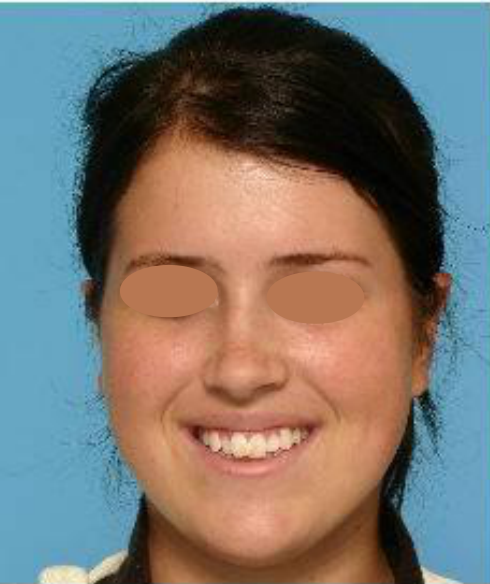
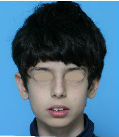
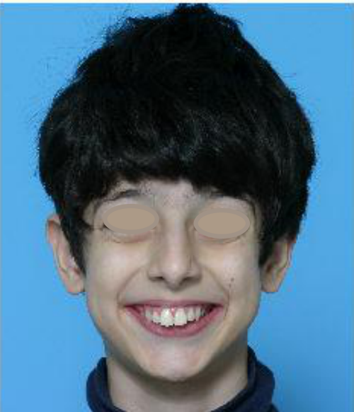
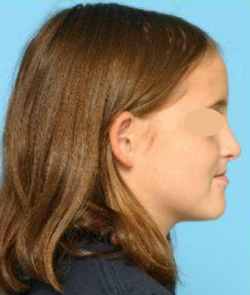
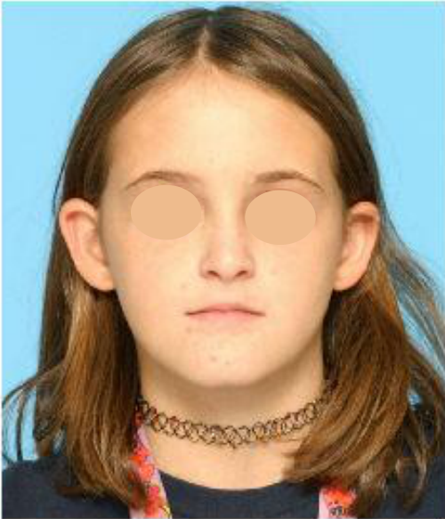
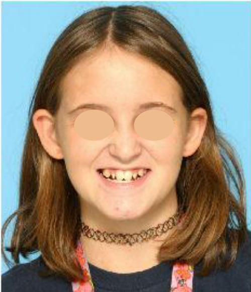
Case: Orthodontic evaluation of a young patient with a concave profile
Question
Scenario: A young patient presents for an orthodontic assessment with various clinical photographs and a panoramic radiograph.
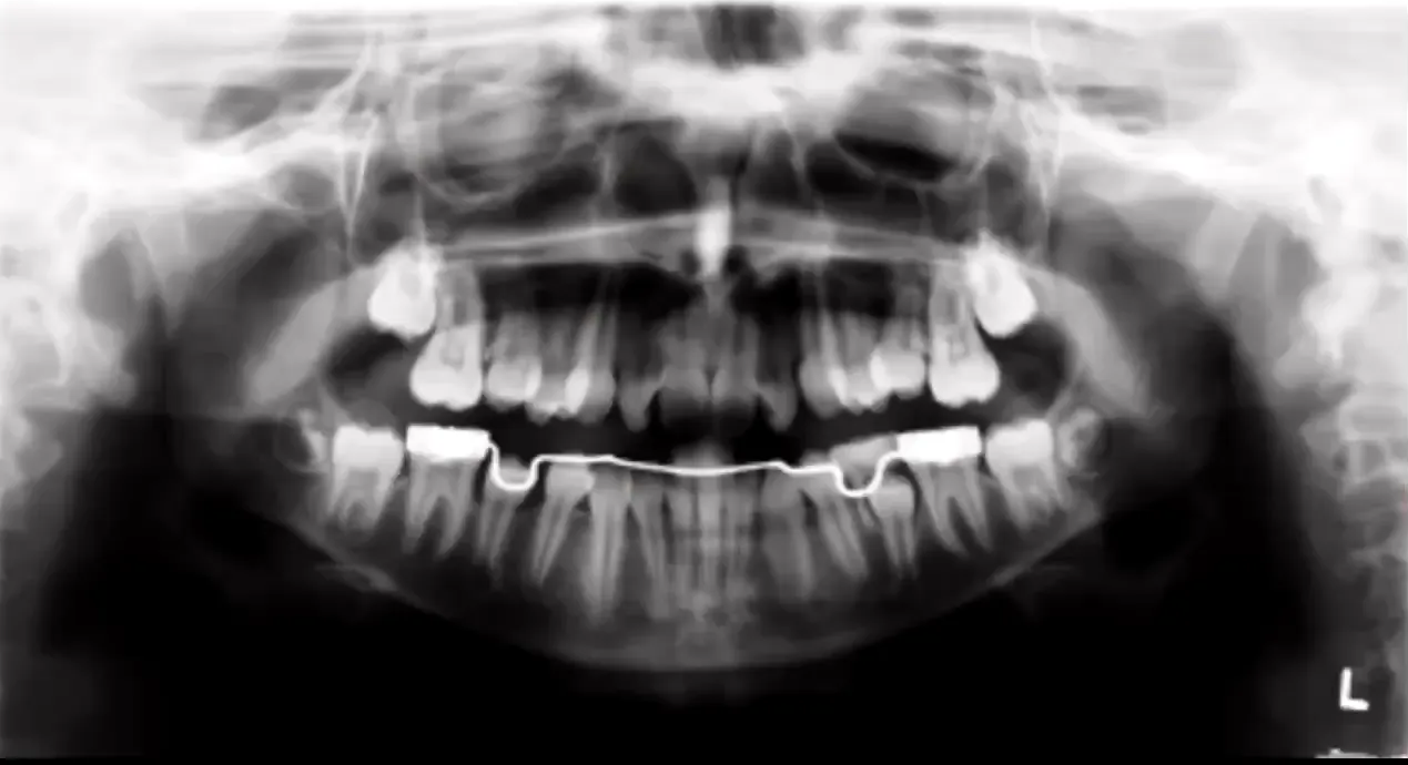
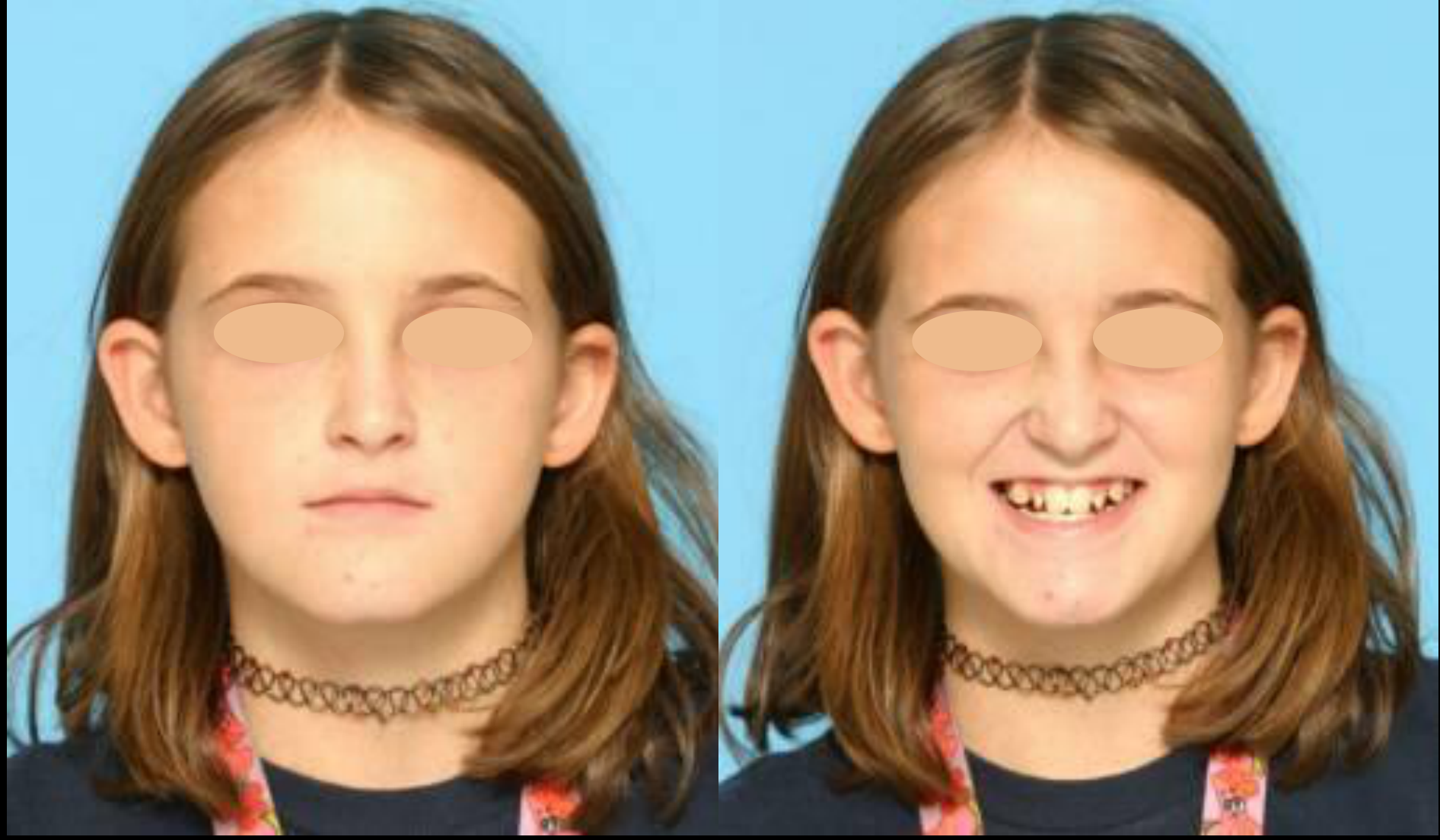
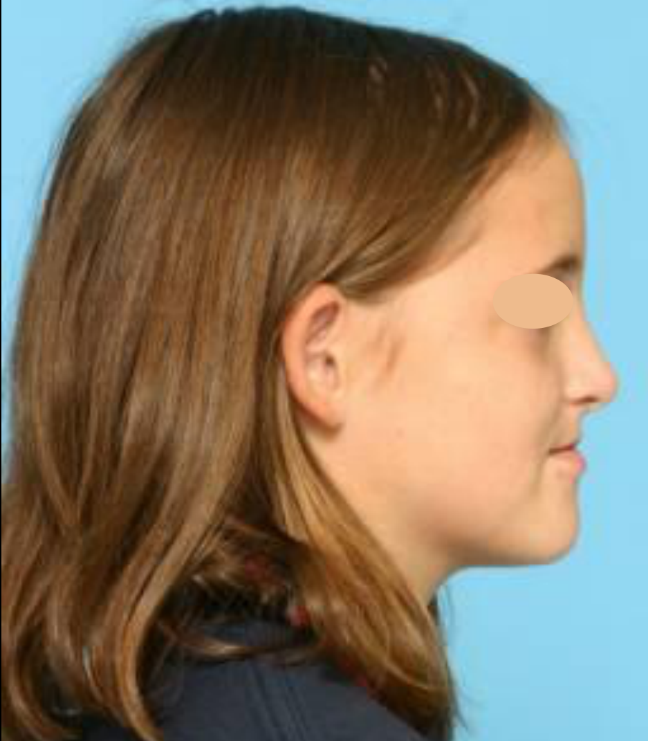
What’s shown: Frontal (at rest and smiling) and profile photographs of the face, alongside a panoramic radiograph.
Consider: Analyze the facial and dental features to determine the underlying skeletal discrepancy, evaluate the impact of the patient’s age on treatment options, and formulate a management plan for the crowding and skeletal issues.
Answer
Observations:
- Frontal view: Long face, deficient paranasal area, edge-to-edge incisor relationship, midline shift, and lack of space for the upper lateral incisors.
- Profile view: Slightly concave profile.
- Lateral view: Chin is in a good orthogonal position, nasolabial angle is open (>90 degrees), lips are retruded, and the paranasal area is deficient. The molar relationship appears to be Class 3, but the teeth are touching with no actual occlusion (not an open bite).
- Panoramic radiograph: Shows a lower lingual arch in place, preserving leeway space on the left side that would otherwise be lost to mesial drift of the lower first molars.
Reasoning:
- The concave profile, deficient midface, and open nasolabial angle point to a deficient (retrusive) maxilla rather than a mandibular excess. The chin’s alignment with the true vertical line confirms the mandible is in a normal position, making this a skeletal Class 3 due to maxillary deficiency.
- The patient’s dental age is estimated at 11 to 12 years based on the lower second premolars. At this age, it is too late for a face mask (typically used around age 8 with rapid expansion) to achieve significant skeletal protraction of the maxilla; treatment would only yield dental improvements.
- The severe crowding in both arches combined with the retrusive maxilla indicates that extractions or orthognathic surgery will likely be required later.
- A lower lingual arch is utilized to prevent the physiological mesial drift of the lower first molars, thereby preserving and redirecting the leeway space to help manage the anterior crowding.
Outcome:
- Skeletal correction with a face mask is no longer viable due to the patient’s age.
- A lower lingual arch is placed to manage space, but the long-term treatment plan will likely involve extractions or surgery.
Takeaway:
- Differentiating between maxillary deficiency and mandibular excess is critical in Class 3 malocclusions to select the appropriate appliance (e.g., face mask), but the timing of intervention is equally important, as maxillary protraction is most effective around age 8 and yields only dental changes if delayed until age 11-12.
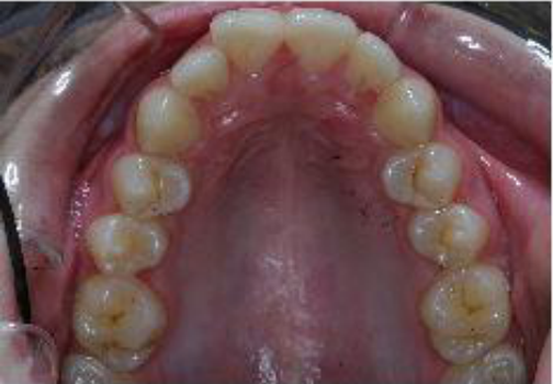
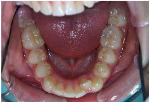
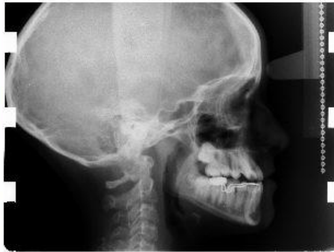
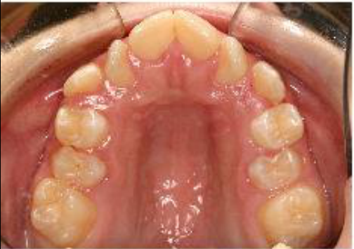
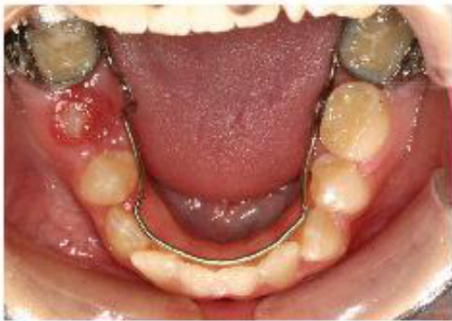
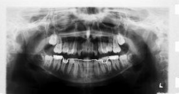
Case: 12-year-old female with maxillary deficiency and severe crowding
Question
Scenario: A 12-year-old female patient presents for orthodontic evaluation with a concave profile and severe crowding.
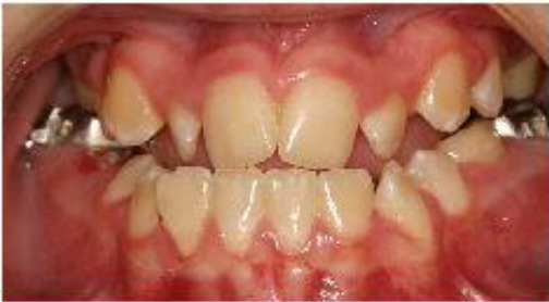
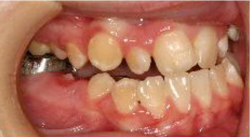
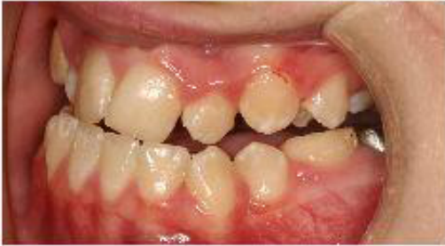
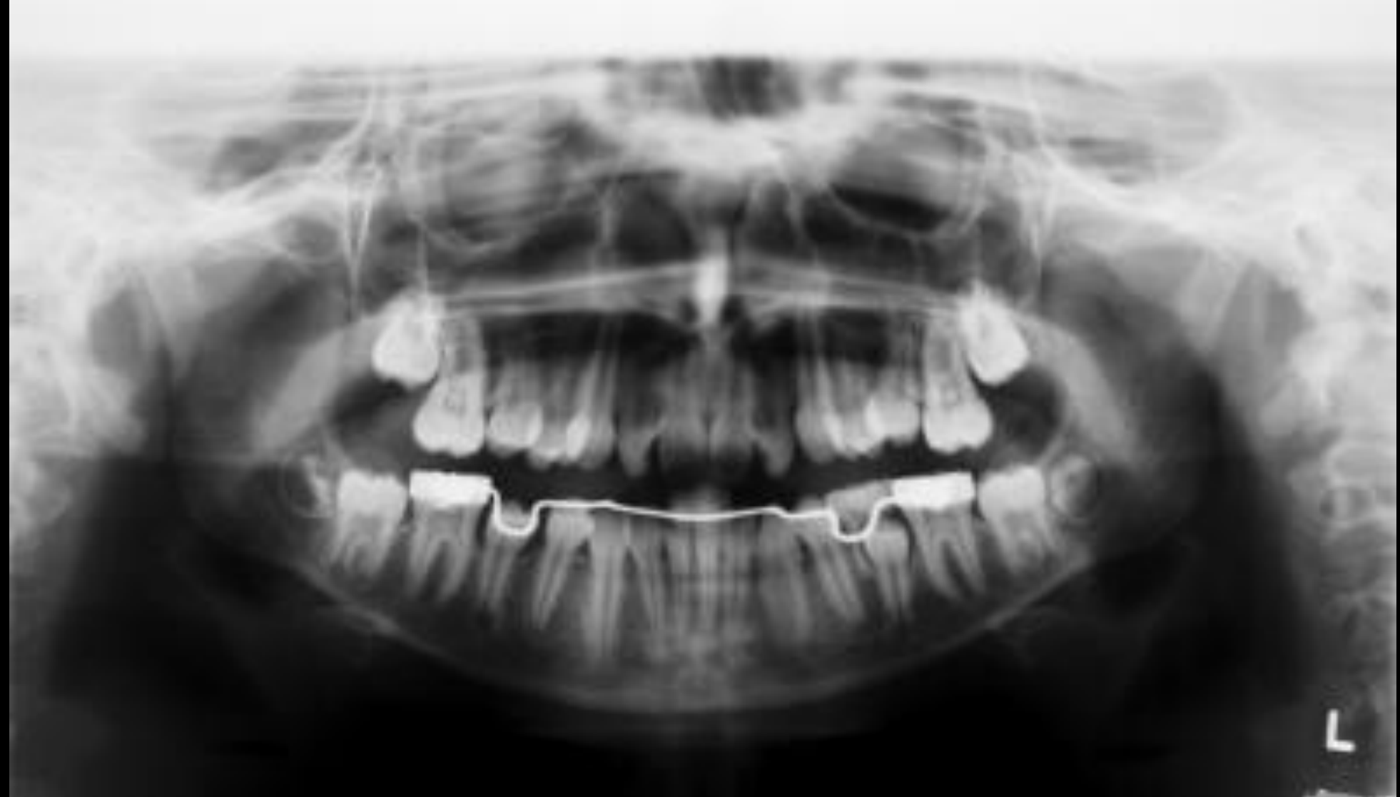
What’s shown: A panoramic radiograph showing a lower lingual arch in place with preserved leeway space, alongside clinical photographs revealing an edge-to-edge incisor relationship and intraoral views of the dentition.
Consider: Identify the skeletal and dental findings, the rationale for the appliance used, and the proposed treatment plan given the patient’s age and growth status.
Answer
Observations:
- Panoramic radiograph shows a lower lingual arch in place, preserving leeway space.
- Concave facial profile due to a deficient/retrusive maxilla.
- Edge-to-edge incisor relationship with a tendency for anterior crossbite.
- Lower incisors are retroclined, while the upper incisors are proclined as a dental compensation.
- Severe crowding is present.
Reasoning:
- The lower lingual arch is used as a bilateral device to block the physiological mesial drift of the lower first molars, thereby preserving the leeway space to resolve the severe anterior crowding. A distal shoe is not indicated here because bilateral space maintenance is required.
- The patient’s dental age is too late for orthopedic protraction of the maxilla using a face mask.
- The compensatory inclination of the incisors is an attempt to mask the underlying skeletal discrepancy.
Outcome:
- The immediate plan is to dentally correct the anterior crossbite.
- The patient will be followed up to monitor growth; orthognathic surgery will be considered later depending on the amount of remaining growth, as further mandibular growth would exacerbate the maxillary deficiency.
Takeaway:
- In cases of severe crowding with a deficient maxilla, a lower lingual arch is indicated to block mesial drift and preserve leeway space. When a patient is past the age for maxillary protraction, treatment shifts to dental compensation and planning for potential future orthognathic surgery.
From the lecture slides
What molar relationship would you expect to see in this patient? How will her jaws grow during puberty?
Lecturer — Case 3 Growth and Treatment
The principal problem is a retruded or deficient maxilla, with a slightly concave profile, deficient midface, edge-to-edge incisors, and zero overbite. If treated at approximately eight years old, the patient could have been a candidate for rapid expansion and a face mask; at approximately 11–12 years old, the same treatment is unlikely to produce the desired skeletal effect.
- At the current dental age, treatment may provide mainly dental correction of the anterior crossbite through expansion and advancement of the upper teeth.
- Complete correction cannot be achieved dentally without excessive incisor compensation, and future surgical treatment may need to be considered.
- Further mandibular growth could worsen the skeletal relationship.
- Why do we have a LLHA?
- When is the best time to place a LLHA?
What is her dental age?
Lecturer — Lingual Arch Timing
A lower lingual arch should be placed before exfoliation of the primary molars in this severely crowded patient. It prevents mesial drift of the lower first permanent molars and preserves leeway space; without it, the molars would physiologically drift forward and occupy the available space.
- A distal shoe guides eruption of a first permanent molar after early loss of a primary molar, but it is not the preferred appliance for bilateral prevention of mesial drift in severe crowding.
- The lingual arch was visible on the panoramic radiograph, and the patient’s dental age was too advanced for a face mask to produce the desired maxillary advancement.
Case 3 Problem List41
- Dentofacial esthetic and protrusion
- Alignment and symmetry
- Transverse
- AP
- Vertical
 |  |  |
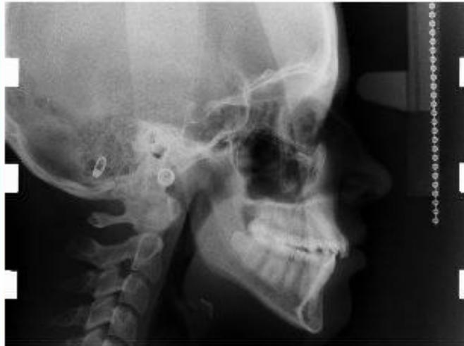
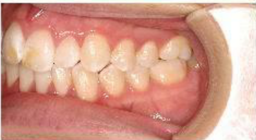
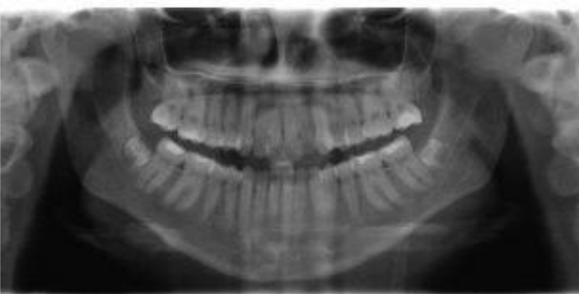
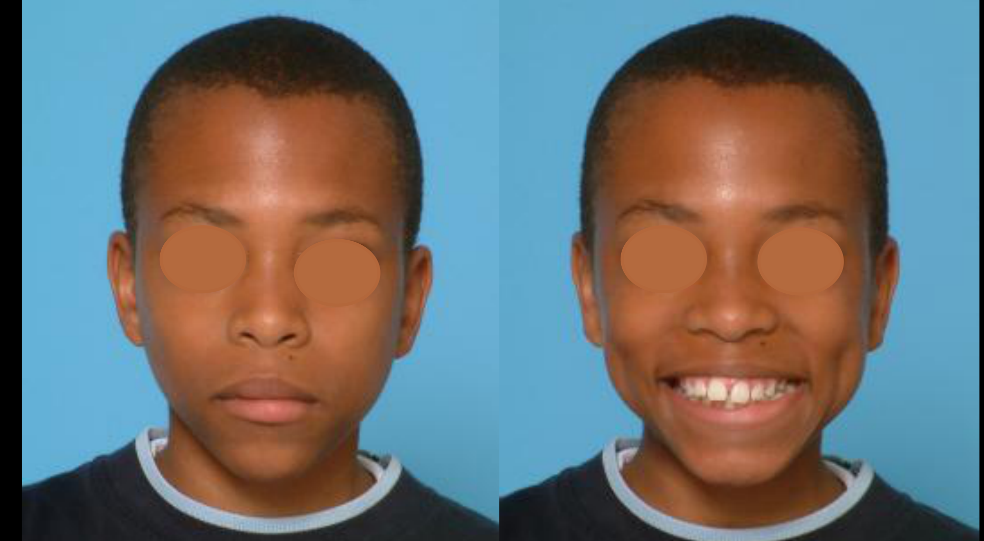
Case: Orthodontic assessment of a patient with bimaxillary protrusion and Class II malocclusion
Question
Scenario: A patient presents for orthodontic evaluation. The lecturer walks through the patient’s extraoral and intraoral photographs, as well as radiographs, to assess the facial profile, occlusion, and skeletal relationships.
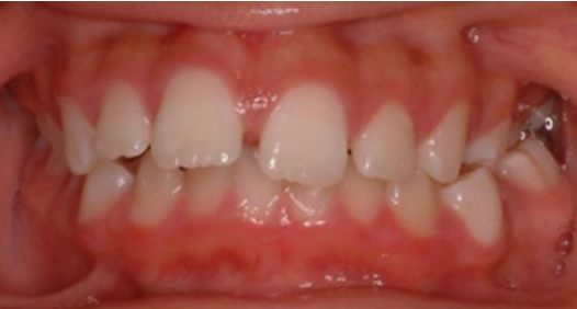
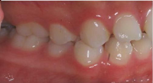
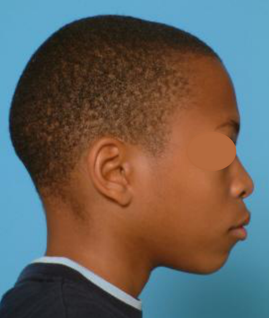
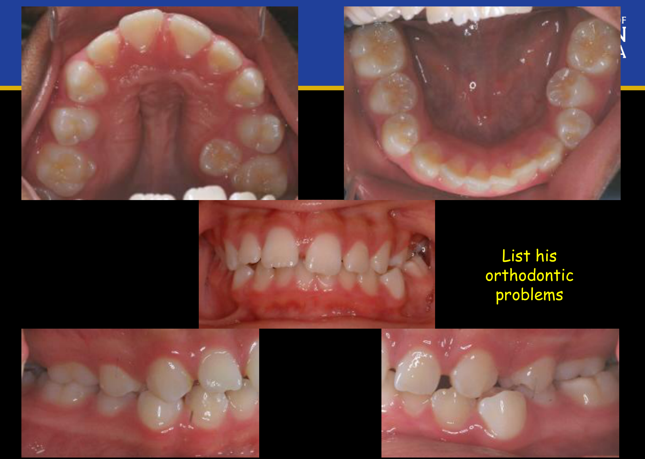
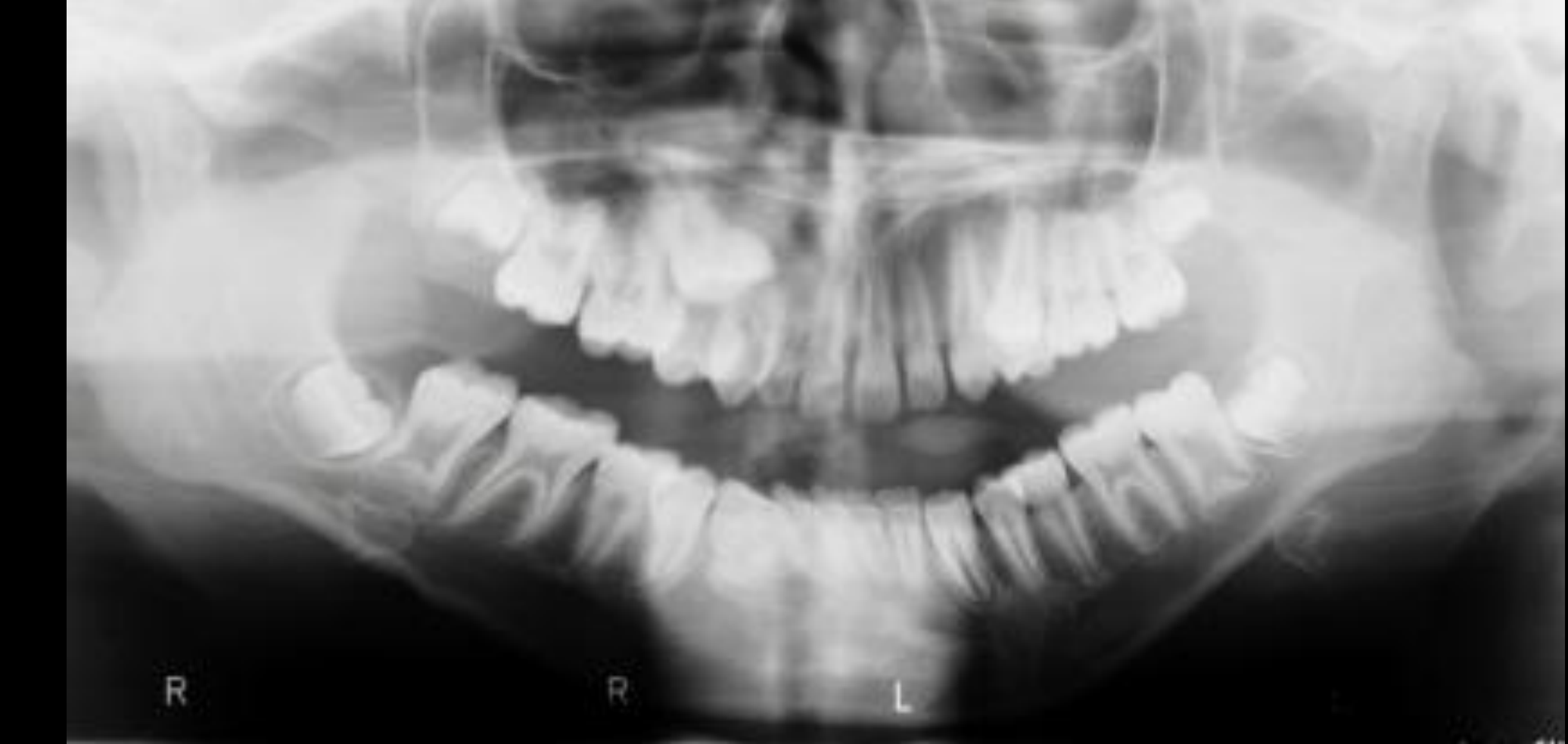
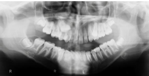
What’s shown: Extraoral photos showing a chin deviated slightly to the left, increased overjet, occlusal cant, buccal corridors, and a convex, bimaxillary protrusive profile with competent lips. Intraoral photos show midlines off by one incisor, left-sided crossbite tendency, Class II molar relationship, increased overbite and overjet, and lack of space for the upper second premolars with squished lower second premolars and misaligned lower first premolars. The panoramic radiograph reveals an impacted upper right second premolar and mesial drift of the first molars.
Consider: What are the clinical and radiographic findings, and what is the proposed management plan for this patient?
Answer
Observations:
- Extraoral: Chin slightly off to the left, increased overjet, occlusal cant, buccal corridors, lack of harmony with the lower lip, and a significantly convex, bimaxillary protrusive profile with competent lips.
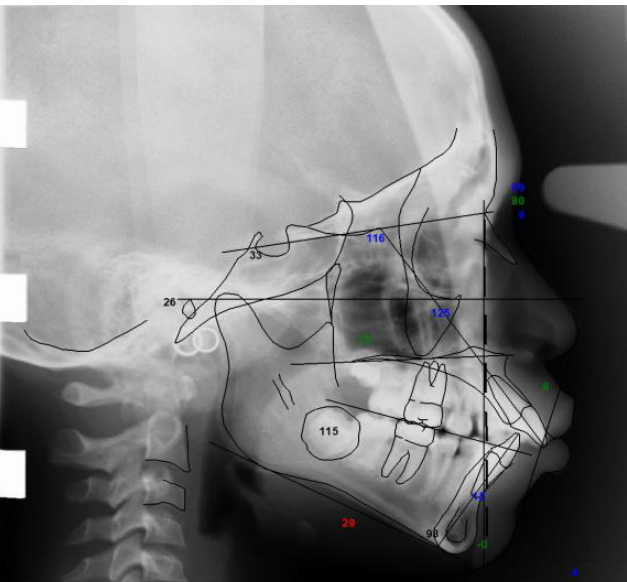
- Intraoral: Midlines off by one incisor, tendency of crossbite on the left, Class II molar relationship (half to full unit), increased overbite (approx. 60%) and overjet, no space for upper second premolars, squished lower second premolars, and misaligned lower first premolars.
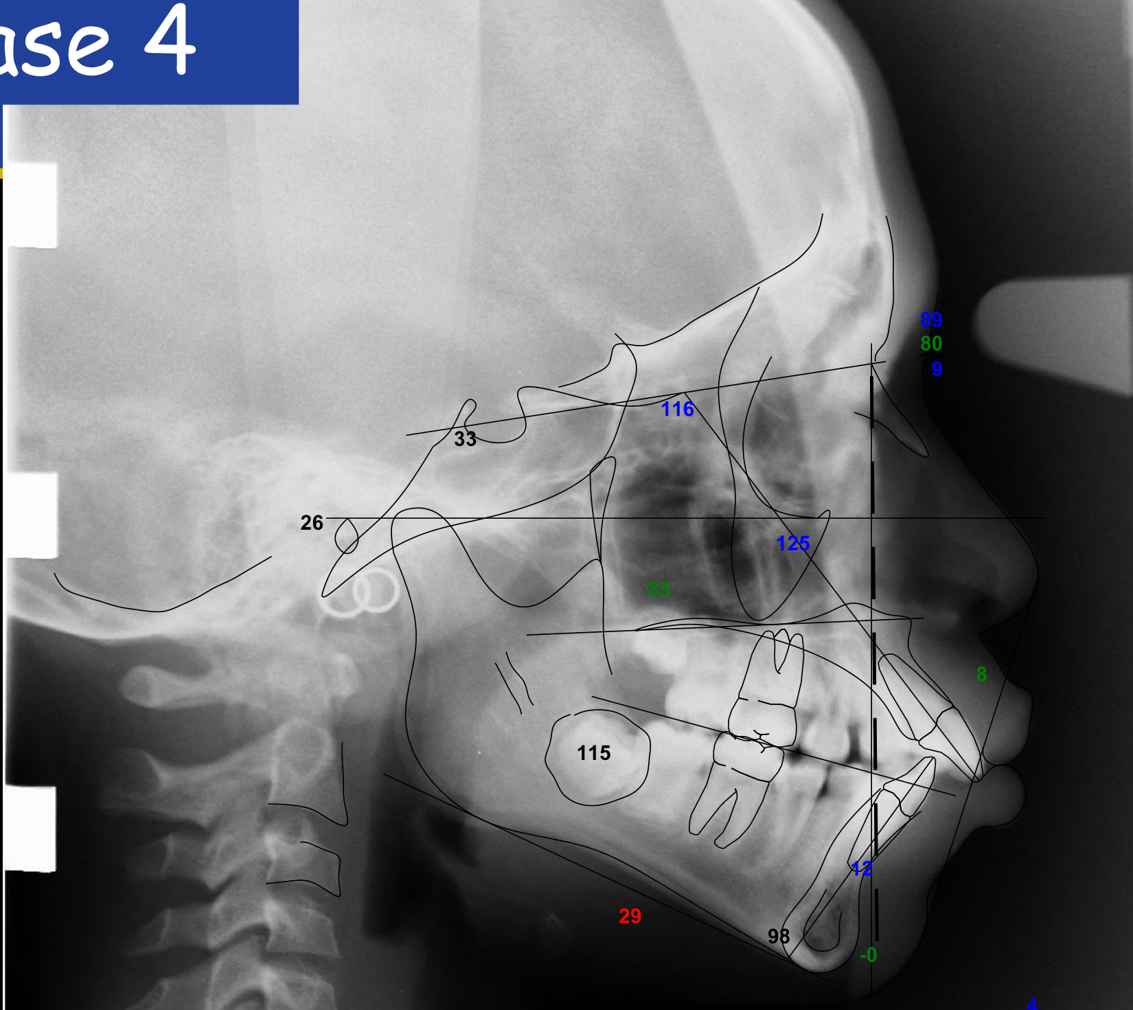
- Radiographic: Impacted upper right second premolar, mesial drift of first molars, SNA 89, SNB 80, ANB 9 (full skeletal Class II), protrusive and proclined incisors, and CVM stage showing trapezoidal vertebral bodies (achieving CS3) indicating significant remaining growth with peak in about one year.
Reasoning:
- The facial profile and cephalometric values (SNA 89, SNB 80, ANB 9) confirm a full skeletal Class II pattern with bimaxillary protrusion.
- The lack of space for the second premolars and their impaction or misalignment is attributed to the early loss of primary second molars and subsequent mesial drift of the first molars, without the use of a space-maintaining device.
- The convex profile and proclined incisors contribute to the increased overjet and overbite.
Outcome:
- The case requires extractions, likely of four premolars.
- Because the second premolars are out of the arch and space is consumed, space must be reopened by moving the upper first molars buccally to correct the overjet.
- The patient’s growth must be followed closely, as he is still growing and approaching his peak growth spurt in about one year.
Takeaway:
- Early loss of primary second molars without space maintenance leads to mesial drift of permanent first molars, resulting in space loss and impaction of second premolars, which complicates the orthodontic management of Class II and bimaxillary protrusion cases.
From the lecture slides
Lecturer — Case 4 Findings
The patient has a full skeletal Class 2 relationship with approximately 89 SNA, 80 SNB, and an A-B difference of approximately nine degrees. The molar relationships are full-unit Class 2, with protrusive and proclined incisors, increased overjet and overbite, severe premolar space deficiency, and an impacted right premolar.
- Early loss of the primary molars likely allowed mesial drift of the first permanent molars, consuming the space required for premolar eruption.
- The case was likely an extraction case, potentially requiring removal of four premolars and reopening of spaces that had already been consumed.
- The upper first molar might need to be moved buccally or otherwise repositioned to create enough space to correct the overjet.
- The patient was approximately at CS3 and approaching the growth peak, estimated to be about one year away, so growth should be followed closely.
List his orthodontic problems.
What is the likely cause of the impaction?
Case 4 Problem List42
- Dentofacial esthetic and protrusion
- Alignment and symmetry
- Transverse
- AP
- Vertical
 |  |  |
Footnotes
-
Original PDF page 1: L9 Level IV UnitA, p.1 ↩
-
Original PDF page 2: L9 Level IV UnitA, p.2 ↩
-
Original PDF page 3: L9 Level IV UnitA, p.3 ↩
-
Original PDF page 4: L9 Level IV UnitA, p.4 ↩
-
Original PDF page 5: L9 Level IV UnitA, p.5 ↩
-
Original PDF page 6: L9 Level IV UnitA, p.6 ↩
-
Original PDF page 7: L9 Level IV UnitA, p.7 ↩
-
Original PDF page 8: L9 Level IV UnitA, p.8 ↩
-
Original PDF page 9: L9 Level IV UnitA, p.9 ↩
-
Original PDF page 14: L9 Level IV UnitA, p.14 ↩
-
Original PDF page 10: L9 Level IV UnitA, p.10 ↩
-
Original PDF page 11: L9 Level IV UnitA, p.11 ↩
-
Original PDF page 12: L9 Level IV UnitA, p.12 ↩
-
Original PDF page 13: L9 Level IV UnitA, p.13 ↩
-
Original PDF page 15: L9 Level IV UnitA, p.15 ↩
-
Original PDF page 16: L9 Level IV UnitA, p.16 ↩
-
Original PDF page 17: L9 Level IV UnitA, p.17 ↩
-
Original PDF page 18: L9 Level IV UnitA, p.18 ↩
-
Original PDF page 19: L9 Level IV UnitA, p.19 ↩
-
Original PDF page 20: L9 Level IV UnitA, p.20 ↩
-
Original PDF page 21: L9 Level IV UnitA, p.21 ↩
-
Original PDF page 22: L9 Level IV UnitA, p.22 ↩
-
Original PDF page 23: L9 Level IV UnitA, p.23 ↩
-
Original PDF page 24: L9 Level IV UnitA, p.24 ↩
-
Original PDF page 26: L9 Level IV UnitA, p.26 ↩
-
Original PDF page 27: L9 Level IV UnitA, p.27 ↩
-
Original PDF page 28: L9 Level IV UnitA, p.28 ↩
-
Original PDF page 29: L9 Level IV UnitA, p.29 ↩
-
Original PDF page 31: L9 Level IV UnitA, p.31 ↩
-
Original PDF page 32: L9 Level IV UnitA, p.32 ↩
-
Original PDF page 33: L9 Level IV UnitA, p.33 ↩
-
Original PDF page 34: L9 Level IV UnitA, p.34 ↩
-
Original PDF page 36: L9 Level IV UnitA, p.36 ↩
-
Original PDF page 37: L9 Level IV UnitA, p.37 ↩
-
Original PDF page 38: L9 Level IV UnitA, p.38 ↩
-
Original PDF page 39: L9 Level IV UnitA, p.39 ↩
-
Original PDF page 40: L9 Level IV UnitA, p.40 ↩
-
Original PDF page 42: L9 Level IV UnitA, p.42 ↩
-
Original PDF page 25: L9 Level IV UnitA, p.25 ↩
-
Original PDF page 30: L9 Level IV UnitA, p.30 ↩
-
Original PDF page 35: L9 Level IV UnitA, p.35 ↩
-
Original PDF page 41: L9 Level IV UnitA, p.41 ↩