Natal and Neonatal Teeth1
Introduction and Definitions2
- Definitions and nomenclature
- History
- Prevalence
- Etiology
- Clinical presentation and classification
- Histology and ultrastructure
- Complications
- Diagnosis and differential diagnosis
- Management
- Conclusion
Terminology and Nomenclature34
Definitions
- Natal Teeth are teeth present in the oral cavity of the infant at the time of birth
- Massler and Savara, 1950
- Neonatal teeth are teeth which erupt during the neonatal period (from birth to 30 days)
- Massler and Savara, 1950
- Early infancy teeth are teeth which erupt within one to three and a half months of life
- Spouge and Feasby, 1966
- Precocious Teeth are teeth which erupt during the third to fifth month of life
- Bodenhoff and Gorlin, 1963
Nomenclature
- Congenital teeth
- Fetal teeth
- Infancy teeth
- Pre-deciduous teeth
- Dentitia praecox
- Dens connatalis
- Precocious dentition
(Zhu and King, 1995; Mayhall, 1967; Kates et al. 1984)
Historical Perspectives and Superstitions5
History
- Natal and neonatal teeth have been a subject of curiosity and study since the time it was first documented by Titus Livius, in 59 BC
- Gaius Plinius Secundus (the Elder), in 23 BC, believed that a splendid future awaited male infants with natal teeth. In females it was considered a bad sign and that they bring disaster to their community
Diverse superstition6
- Africa, India, Poland: monstrous and bearer of misfortune
- In parts of East Africa, such as Nigeria, natal teeth are a major social issue where children may be associated with evil spirits, hidden by parents, or bullied.
- In some extreme instances, cultural beliefs have led to parents being told to dispose of the child.
- China: bad omen for girls
- England: grow up into famous soldiers
- France & Italy: future conquerors of the world
(Allwright, 1958; Bodenhoff & Gorlin, 1963; Zhu & King, 1995)
History
SUPERSTITION
- <mark>If these primary teeth are lost or extracted, no other tooth will appear in that space until the permanent teeth erupt.</mark>
Prevalence and Incidence7
- Natal teeth are three times more common than neonatal teeth
- Rao and Mathad, 2009
- 95%: true primary teeth; 5%: supernumerary teeth
- Kates et al. 1984
- The incidence of natal and neonatal teeth has been investigated in multiple studies:
- 1:716 to 1:30,000 (Zhu and King, 1995)
- 1:2,000 to 1:3,500 (Chow, 1980)
- Incidence rates vary depending on the health of the child; oral anomalies like natal teeth are more likely in children with complex medical histories, such as those with cleft lip and palate or specific syndromes.
Demographics and Anatomical Sites
Gender predilection8
- F > M
- F : M = 66% : 31%
- F = M
(Zhu & King, 1995; Goncalves et al. 1998; Anegundi et al. 2002; Basavanthappa et al. 2011)
Gender Discrepancy
There is no significant gender discrepancy; reported differences are often just the result of specific cohort studies and sample sizes.
Site
- Mandibular incisor region (85%)
- This is the most common site as these are typically the first teeth to erupt.
- Maxillary incisors (11%)
- Mandibular canine (3%)
- Maxillary canine and molar (<1%)
- Multiple neonatal and natal teeth are rare
(Gardina, 1961; Bodenhoff & Gorlin, 1963; King and Lee, 1989; Ooshima et al. 1986)
Developmental Red Flag
Eruption of molars (such as a “D”) without incisors is considered a “red flag” for abnormal development.
Aetiology9
- Unknown
- Superficial position of tooth germ in alveolar bone during development (Cunha et al. 2001
- The most accepted theory is that the tooth germs are positioned superficially, causing them to erupt earlier than usual.)
- Trauma or infection
- Osteoclastic activity within tooth germ area (bone remodelling) (Goncalves, 1998)
- Hereditary transmission of an autosomal dominant gene, with a familial trait in 8–62% of cases (Hyatt, 1965; Zhu & King, 1995)
- Certain syndromes and newborns with orofacial clefts (Motoyama et al. 1967)
Maternal and Environmental Factors
Maternal-related factors10
- dietary deficiencies (malnutrition)
- poor maternal health
- endocrine disturbances: pyelitis and hypovitaminosis during pregnancy
- excessive secretion of pituitary, thyroid or gonads (Khatib et al. 2005; Vucicevic et al. 2007)
- While some studies suggest associations with maternal malnutrition or other risk factors, these are not proven causes.
Febrile state
Increased body temperature can affect the normal eruption of teeth, e.g. fever, exanthemata (Massler & Savara, 1959; Vucicevic et al. 2007)
Environmental factors
- polychlorinated biphenoyls & bibenzofurans? (Yamaguchi et al. 1971; Gladen et al. 1990)
Clinical Presentation and Classification1112
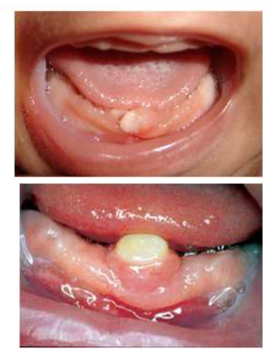
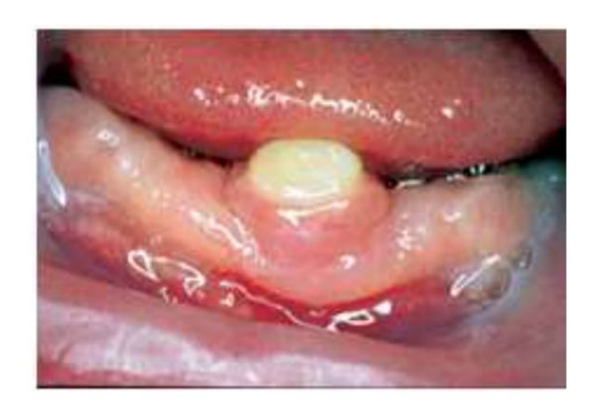
- Usually small and conical
- May resemble normal primary teeth in size & shape
- Usually have an immature appearance with poorly-developed, hypoplastic enamel and dentine
- Usually occurs in pair
(Zhu & King, 1995; Galassi et al. 2004; Ziai et al. 2005)
Clinical Presentation
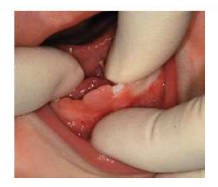
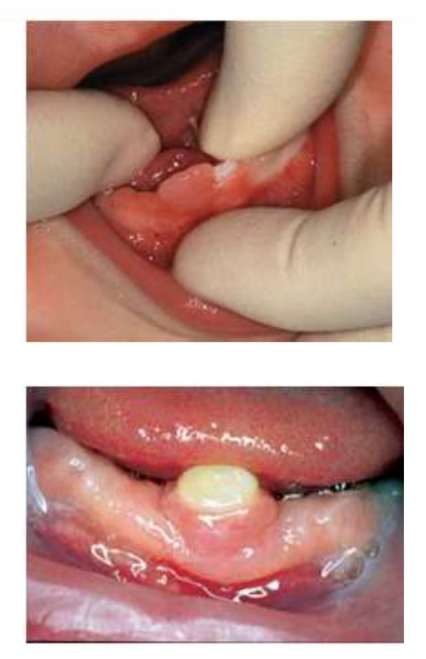
- May be attached to the mucosa and are occasionally covered by a pad of soft tissue
- Absent or poor root development
- Mostly mobile
(Zhu & King, 1995; Galassi et al. 2004; Ziai et al. 2005)
 |  |
 |  |
Classification Systems
Classification - Spouge & Feasby 196613
A mature natal or neonatal tooth
- a tooth which is nearly or fully developed
- relatively good prognosis for maintenance
The immature natal or neonatal tooth
-
a tooth with incomplete or substandard structure
-
implies a **poor prognosis
-
The lecturer notes it is often difficult to clinically distinguish the level of maturity in a neonate.
Classification - Hebling et al. 199714
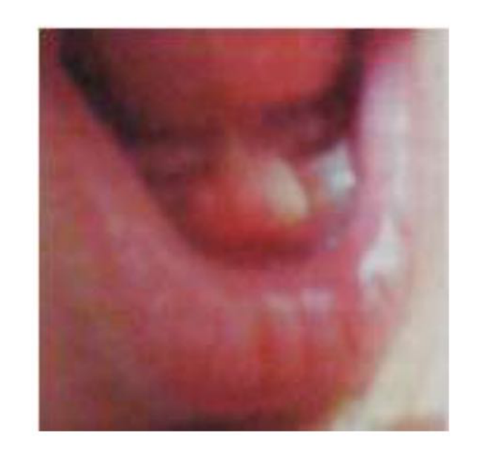
The appearance of each natal tooth in the oral cavity can be classified into four categories given as follows, as the teeth emerge in the oral cavity:
1. Shell-shaped crown
- poorly fixed to the alveolus by the gingival tissue
- absence of a root
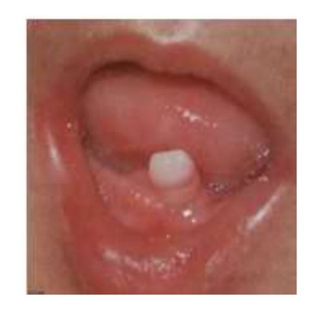
2. Solid crown
- poorly fixed to the alveolus by the gingival tissue
- little or no root
3. Eruption of the incisal margin of the crown through the gingival tissue15
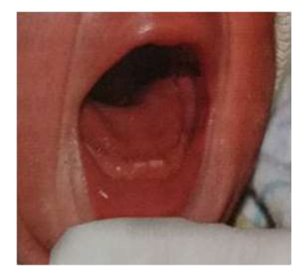 |
4. Edema of the gingival tissue with an unerupted palpable tooth
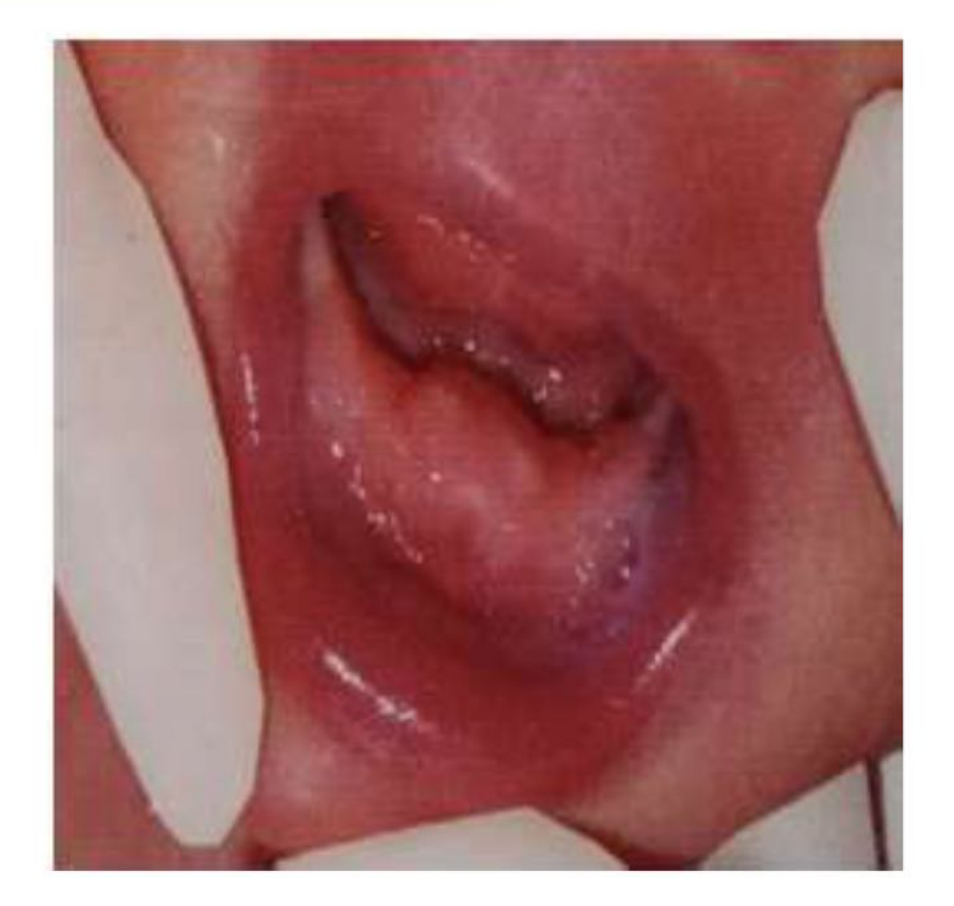
Histology and Structural Characteristics161718
Histology
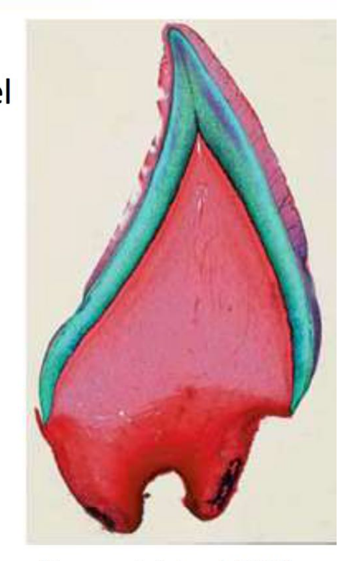
Enamel (Ndiokwelu et al. 2004, To 1992)
- thin layer of enamel or absence of the enamel
- enamel hypoplasia/hypomineralisation
- Enamel and dentine often show defects, are hyper-mineralized, and lack normal histological structure.
Dentin (Massler & Savara, 1950; Zhu & King, 1996; Uzamis et al. 1999; Leung & Robson, 2006)
- interglobular dentin in the coronal region
- irregular dentin and cell inclusions in the cervical region
- atypical dentin (due to the response to irritant stimulus from oral cavity)
(Baumgart & Lussi, 2006)
Histology
- Dentino-enamel junction: not scalloped
Cementum
- lack of cementum and Hertwig’s sheath
- acellular cementum in some cases (**thinner than normal
- Cementum is difficult to assess because the roots are usually not properly developed.**)
Pulp canal and pulp chamber (Howkins 1932, Massler & Savara 1950, Hals, 1957, Clergeau-Guerithault, 1991, Friend et al. 1991)
- larger than normal
- vascularised pulps along with few inflammatory cells
- Increased vascularization and size make the teeth more susceptible to abscesses and infection if the enamel breaks down.
Roots (Massler & Savara, 1950, Allwright 1958, Leung & Robson 2006)
- lack of root formation (despite the tooth being erupted in the oral cavity)
Structural Characteristics
- Enamel of the teeth exhibited hypoplastic, depressed areas, and the incisal edge of natal tooth lacked enamel (Jasmin & Clergeau-Guerithault, 1991)
- Thickness of enamel was around 280 microns compared to up to 1200 microns in normal teeth (Uzamis et al. 1999)
- Enamel has normal prism structure and mineralization (prism structure can be absent in the cervical part of the enamel)
- Cervical and apical dentin is tubular
- In developing teeth, the dentin in these regions changed to an irregularly formed hard tissue of osteodentin character (in which enclosed cells could be observed) (Masatomi et al. 1991)
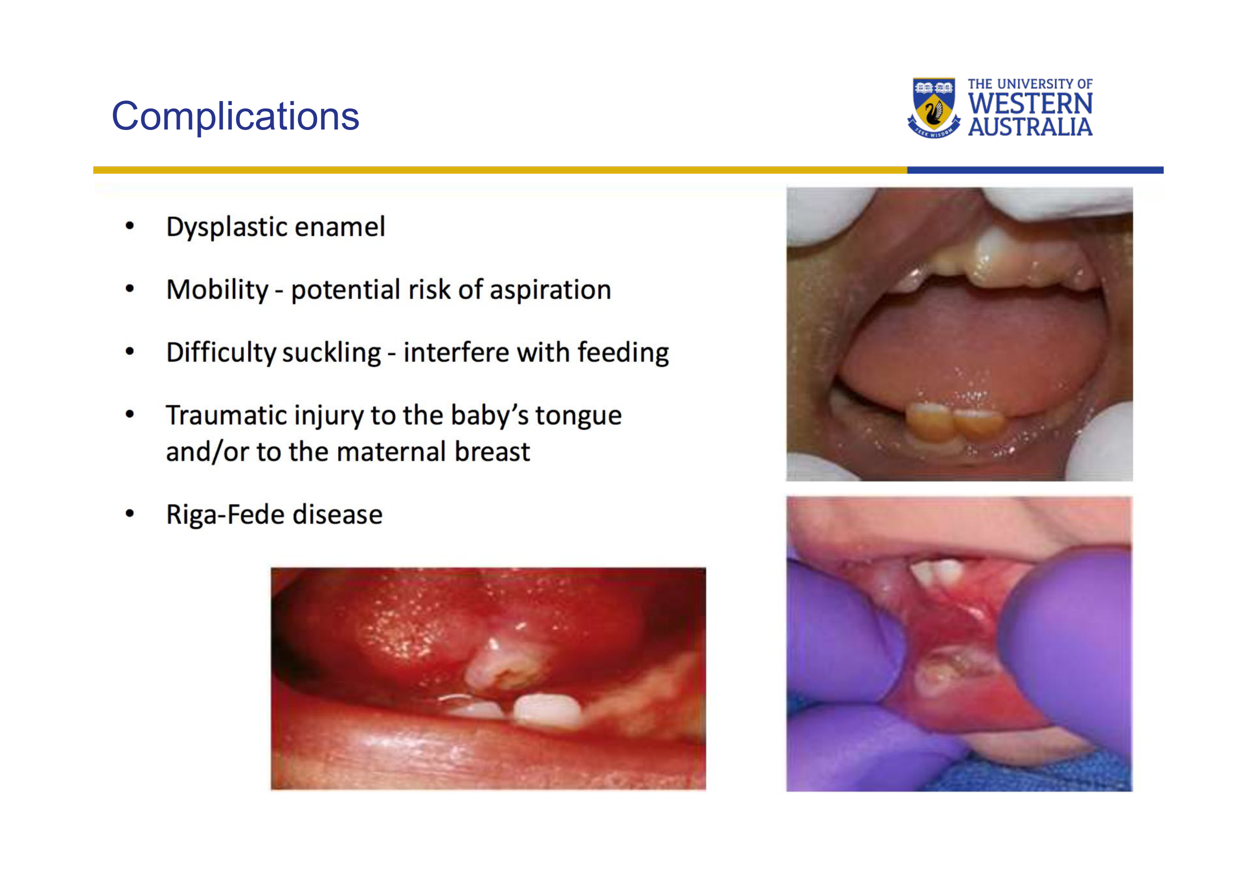
Complications19
- Dysplastic enamel
- Mobility - potential risk of aspiration
- Difficulty suckling - interfere with feeding
- Traumatic injury to the baby’s tongue and/or to the maternal breast
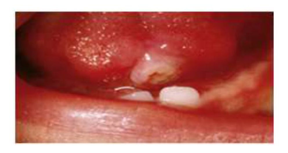
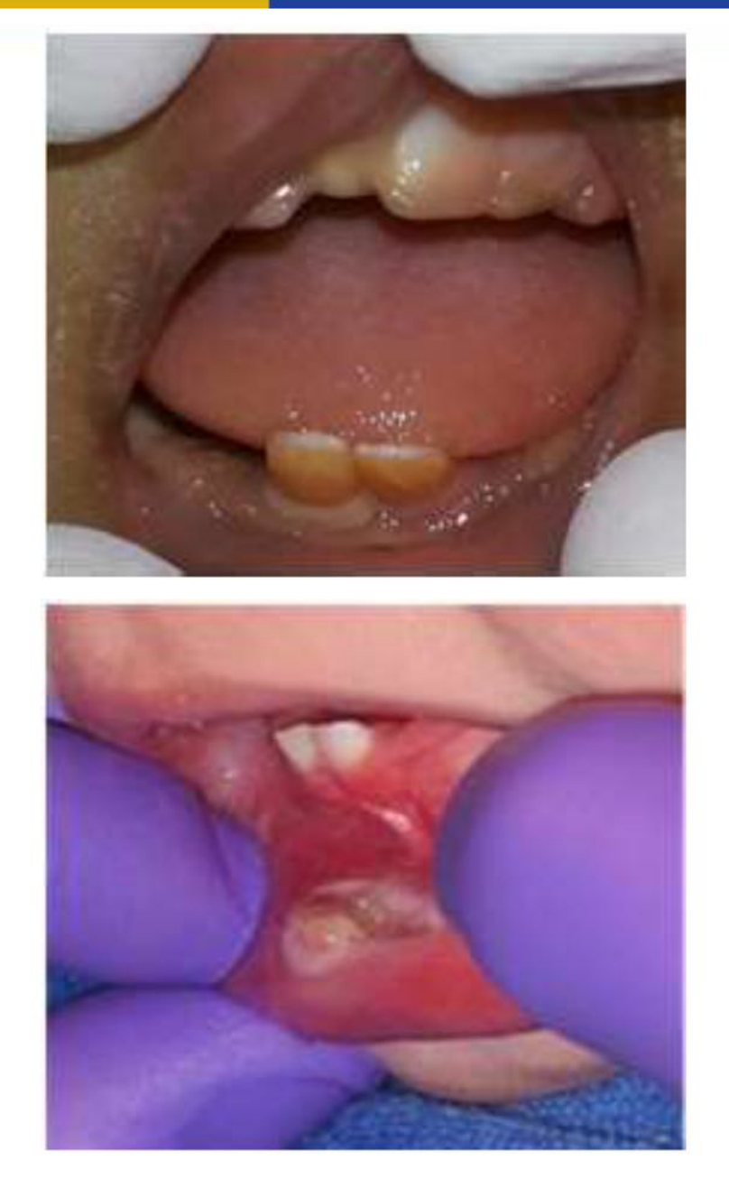
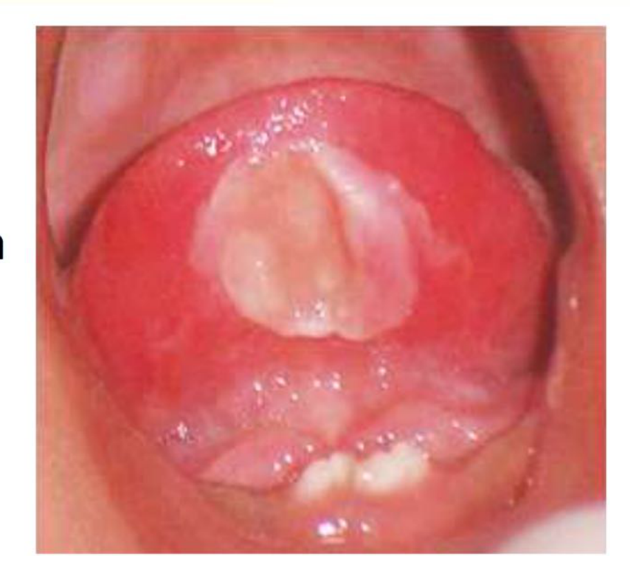
- Riga-Fede disease: Natal teeth causing ulceration of the tongue
- Aspiration is not a complication, there isn’t a single case of natal tooth aspiration!
 |  |
Poor quality enamel20
- increased risk of caries
- chipping and attrition
- periapical abscesses (Allwright, 1958)
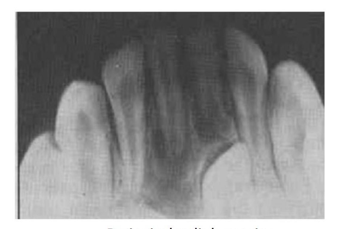
Periapical radiolucencies (Kates et al. 1984)
Mobility and Aspiration Risk21
Mobility
- No reports of inhalation of an exfoliated natal tooth (Gardiner 1961)
- “Inhalation of one of these teeth has never been reported in the literature and the danger is probably more imaginary than real” (Spouge & Feasby 1966)
- The teeth are usually more firmly attached to the gingival tissue than their mobility suggests.
Feeding Difficulties22
Difficulty Feeding
- Presence of natal teeth may cause discomfort for the mother when the infant is suckling
- interfere with feeding
- ulceration
- laceration
(Bodenhoff & Gorlin 1963, Massler & Savara 1950)
- Children with normal primary teeth have often been nursed up to 2-3 years of age without complication
(Massler & Savara 1950)
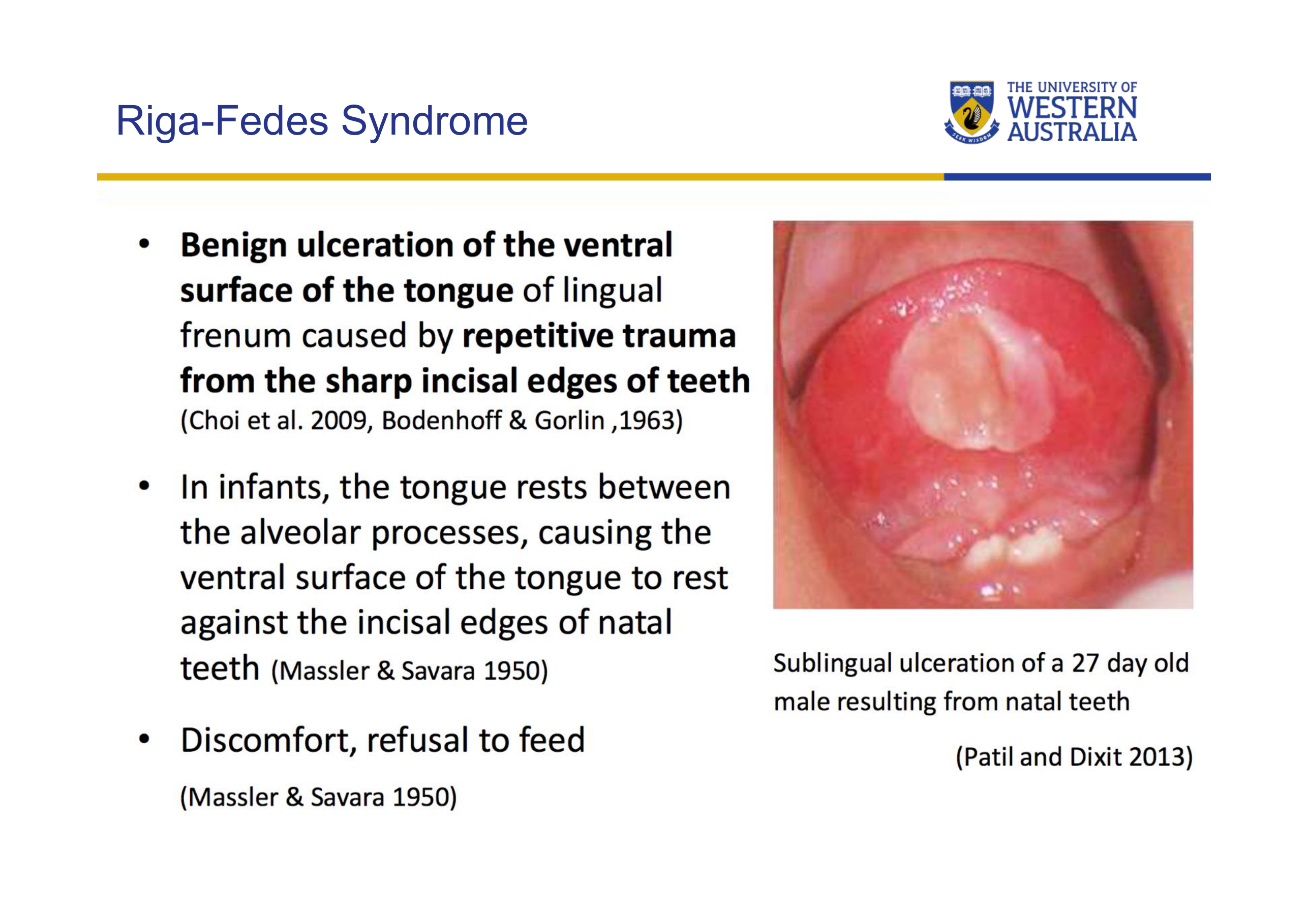
Riga-Fede Syndrome23
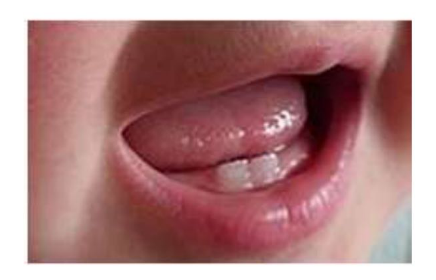
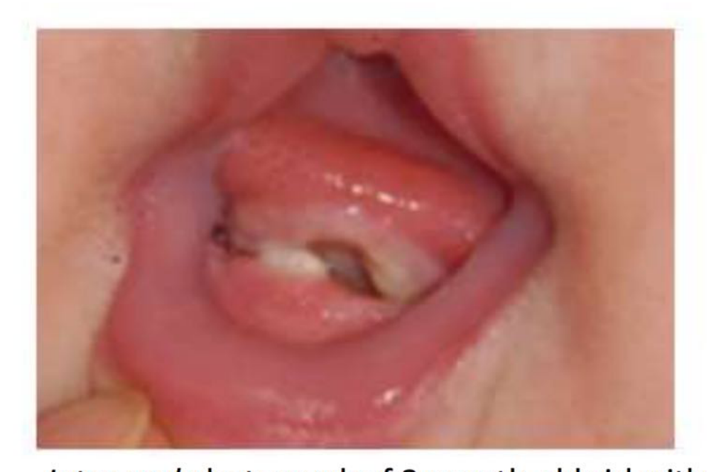
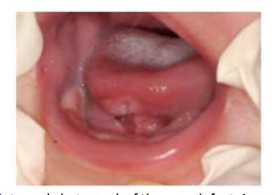
- Benign ulceration of the ventral surface of the tongue or lingual frenum caused by repetitive trauma from the sharp incisal edges of teeth (Choi et al. 2009, Bodenhoff & Gorlin, 1963)
- In infants, the tongue rests between the alveolar processes, causing the ventral surface of the tongue to rest against the incisal edges of natal teeth (Massler & Savara 1950)
- Discomfort, refusal to feed (Massler & Savara 1950)
Clarification on Riga-Fede
This is not a “disease” but a term for the ulceration of the ventral surface of the tongue caused by the sharp edges of natal teeth. It can be quite painful and hinder feeding.
Associated Systemic Conditions24
Systemic Conditions
-
Cleft lip, Palate and cyclopia (Boyd & Miles 1951, de Almeida & Gomide 1996)
-
Ellis-Van Creveld (Chondroectodermal Dysplasia) (Weiss, 1955)
-
Pachyonychia Congenital (Jadassohn-Lewandowsky) (Bodenhoff & Gorlin 1963, Leung 1986)
-
Hallermann-Streif (Oculomandibulodyscephaly with Hypotrichosis) (Robotta & Schafer, 2011)
-
Steatocystoma Multiplex (McDonald & Reed 1976, King & Lee 1987)
-
Pierre-Robin
-
Pallister-Hall
-
Short Rib-Polydactyly (type II)
-
Wiedemann-Rautenstrauch (Neonatal Progeria)
-
Pfeiffer, Ectodermal Dysplasia, Craniofacial Dysostosis
-
Rubinstein-Taybi
-
Sotos
-
Adrenogenital
-
Epidermolysis-Bullosa Simplex including Van der Woude (Ndiokwelu et al. 2004)
-
Down’s Syndrome
-
Walker-Warburg Syndromes (Venkatesh and Adhisivam, 2011)
-
The lecturer clarifies that conditions like caries are environmental (bacteria, diet, time) and not a “feature” of the syndrome itself.
Diagnosis and Differential Diagnosis2526
Diagnosis
- History
- Clinical examination
- Radiograph ?
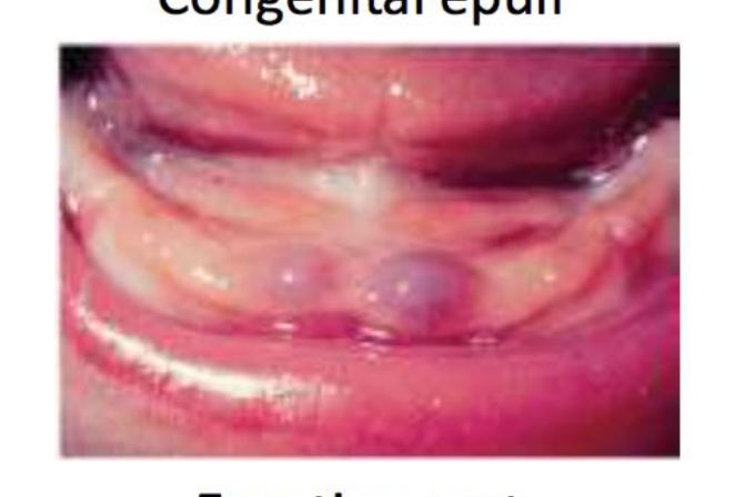
Differential Diagnosis
- Supernumerary teeth (Zhu & King, 1995)
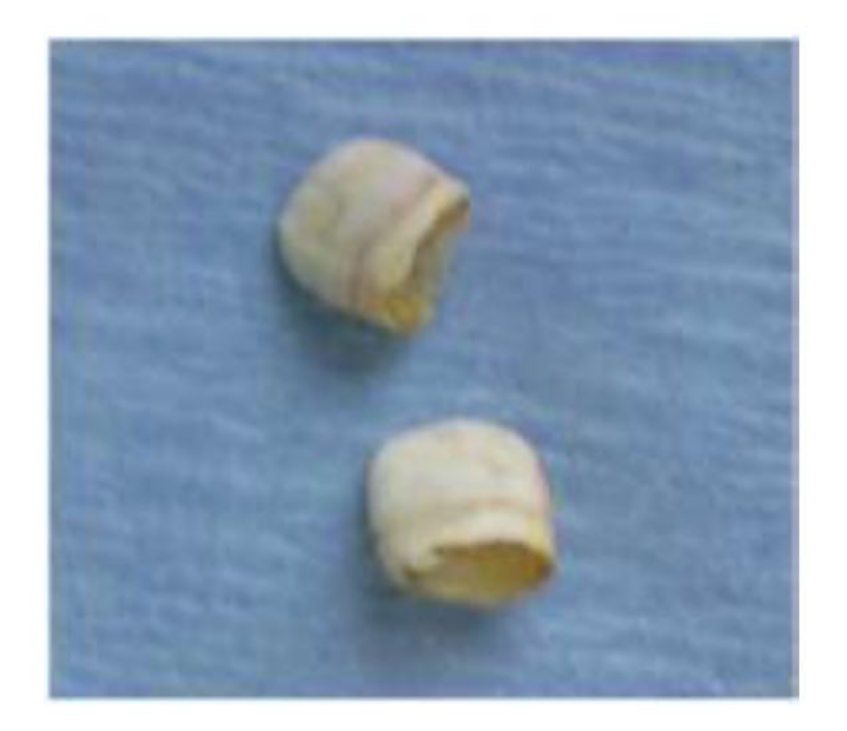
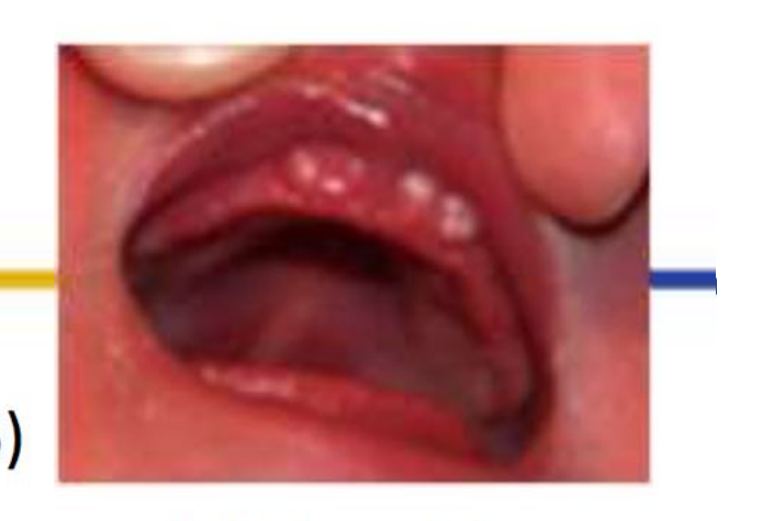
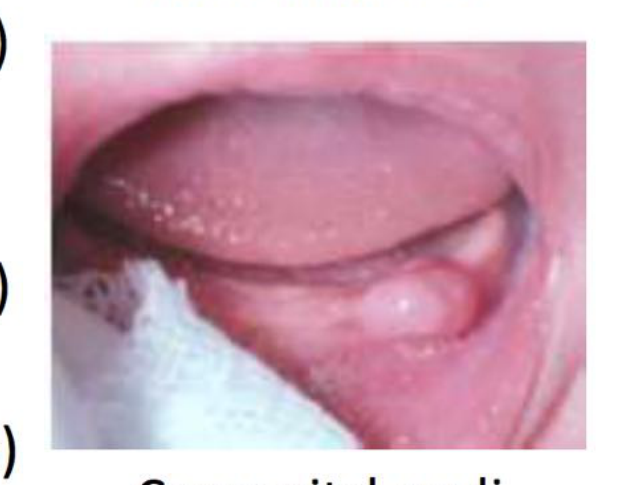
- Bohn’s nodules (Leung and Robson, 2006)
- Bohn’s Nodules: White keratin-filled cysts on the alveolar ridges (prevalence ~60%). They exfoliate naturally.
- Epstein Pearls: Similar to Bohn’s nodules but found along the palatal suture line.
- Dental lamina cysts (gingival cyst of newborn) (Leung and Robson, 2006)
- Inclusion cysts (Kumar et al. 2008)
- Congenital epulis
- Odontogenic hamartomas
- Lymphangioma of the alveolus (Leung, 1986; Leung and Robson, 2006)
- Neuroectodermal tumors of infancy
- Eruption cysts
 |  |
 |  |
Radiographic Evaluation272829
Radiographs
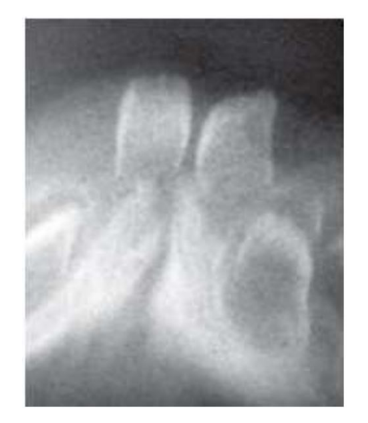
What can you see?
When does calcification of primary incisors begin?
When does calcification of permanent incisors begin?
Clinical Note
At birth, permanent incisors have not yet calcified, so they may not appear on a film.
Radiographs
- A radiographic verification of the relationship between a natal and/or neonatal tooth and adjacent structures, nearby teeth, and the presence or absence of a germ in the primary tooth area
- Differentiate the premature eruption of a primary tooth from a supernumerary tooth
- Provides information about root development
- Shows tooth’s relationship to underlying structures
(Massler & Savara 1950; Leung, 1986; Almeida et al. 1997; Cunha et al. 2001)
Radiographs
- ALARA principle…
- Diagnostic quality of radiograph
- Risk / benefit
- The lecturer expresses skepticism regarding the necessity of radiographs for neonates, as the risks of radiation to a two-day-old often outweigh the benefits and findings rarely change the management plan.
 |  |
Management Strategies30
Management
The treatment planning of these natal or neonatal teeth depends on various factors including:
- degree of mobility
- if the tooth is supernumerary or is part of the normal dentition
- inconvenience during suckling
- problems associated with breastfeeding
If the erupted tooth is diagnosed as a tooth of the normal dentition - maintenance of these teeth in the mouth is the first treatment option (Chow, 1980; Roberts, 1992)
Reassurance
Reassurance is key. If the child is feeding well and the mother is happy, the preferred approach is no treatment.
Conservative Treatment and Monitoring31
Management
- No treatment
- if the tooth is asymptomatic
- breastfeeding is not impaired (Boyd and Miles, 1951)
- Mobile teeth often “firm up” as the root continues to develop.
- Application of a corticosteroid in orabase (such as kenalog) can aid in relieving the symptoms of the ulcer (Slayton, 2000; Choi et al. 2009)
 |  |
Grinding and Smoothing
Grinding32
Smoothing of the tooth by grinding should be carried out if tooth is causing:
- Mild subgingival ulceration or mild discomfort to the mother
- Trauma to the infant’s tongue
Finishing burs, sandpaper disc, soflex disc can be used for smoothing (Allwright 1958, Choi et al. 2009, Slayton 2000)
Risk of Injury
Smoothing sharp edges can help with ulceration, but it is “dodgy” to perform on a neonate due to the risk of injury to the tongue or lip.
 |  |
Protective Coverage and Splints
Coverage33
- Placed something over the tooth in order to reduce the trauma to the tongue and nipple
- Options are:
- a smooth rounded composite dome restoration (Goho, 1996; Choi et al. 2009)
- a breast feeding splint (Bjuggren, 1973)
- stomahesive wafers (Buchanan & Jenkins, 1997)
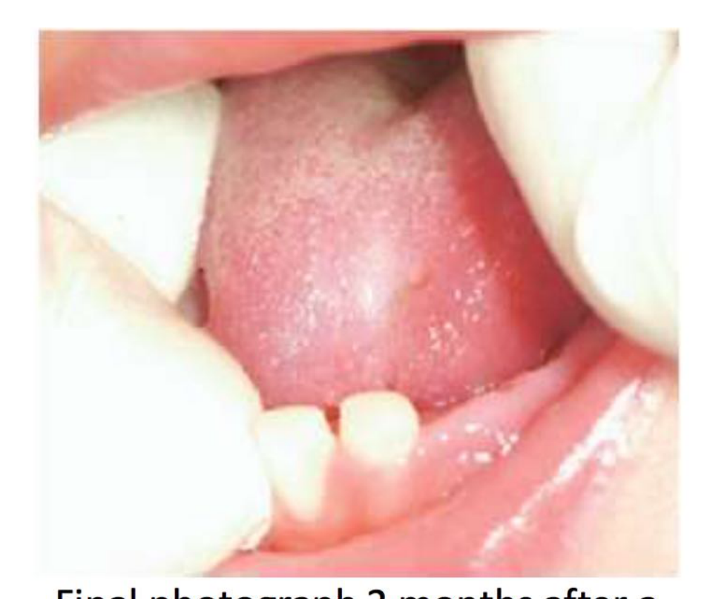
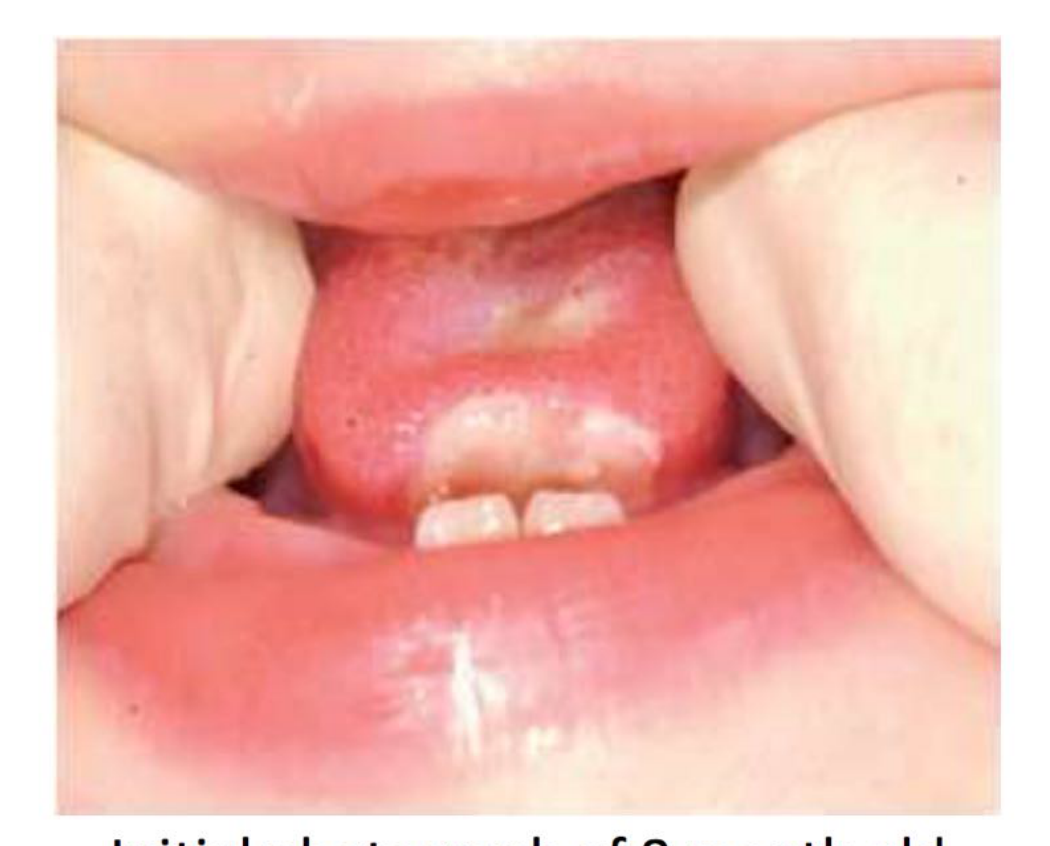
Coverage - Composite build-up34
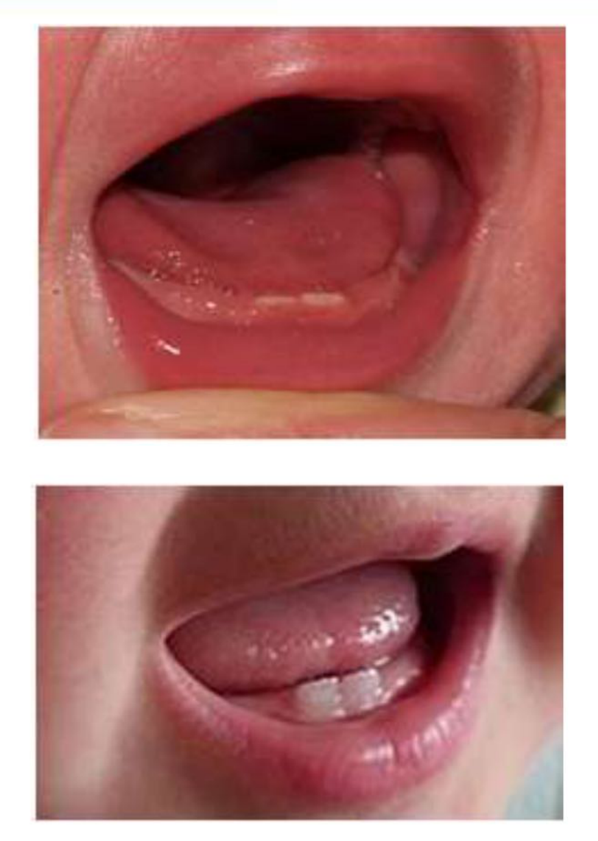
Initial photograph of 8 month old boy with a sublingual ulcer caused by primary lower anterior teeth
Final photograph 2 months after a resin composite build up was placed on the teeth
- Applying resin composite is considered nearly impossible in a neonate due to moisture control issues; GIC (Glass Ionomer Cement) may be a better alternative.
(Choi et al. 2009)
 |  |
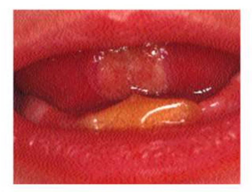
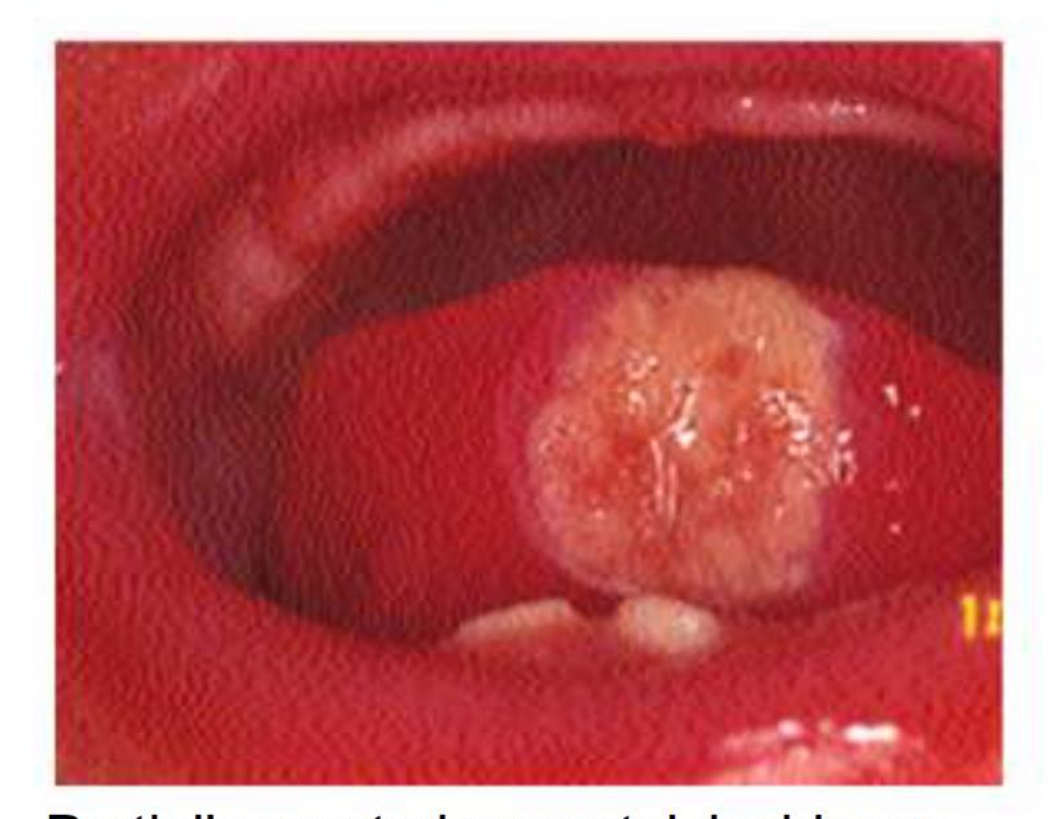
Coverage - Stomahesive Wafer35
Stomahesive Wafer placed over neonatal deciduous lower central incisors protecting tongue from further trauma
(Buchanan and Jenkins, 1997)
- A thin, biocompatible adhesive material that molds to the tissues. It protects the tongue, allows ulcers to heal, and improves feeding.
 |  |
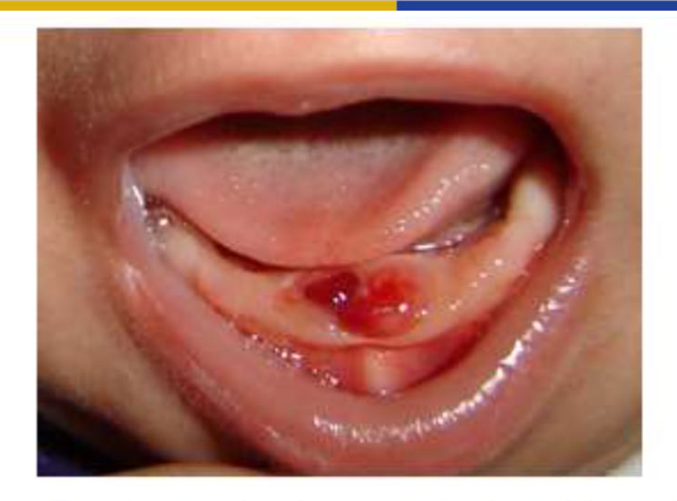
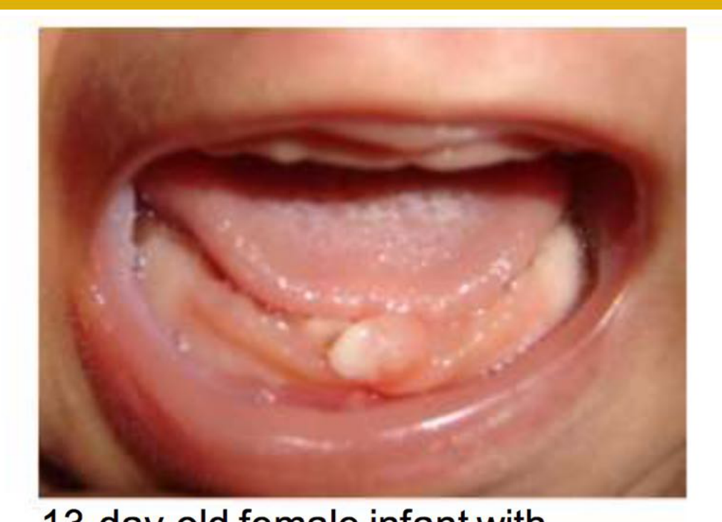
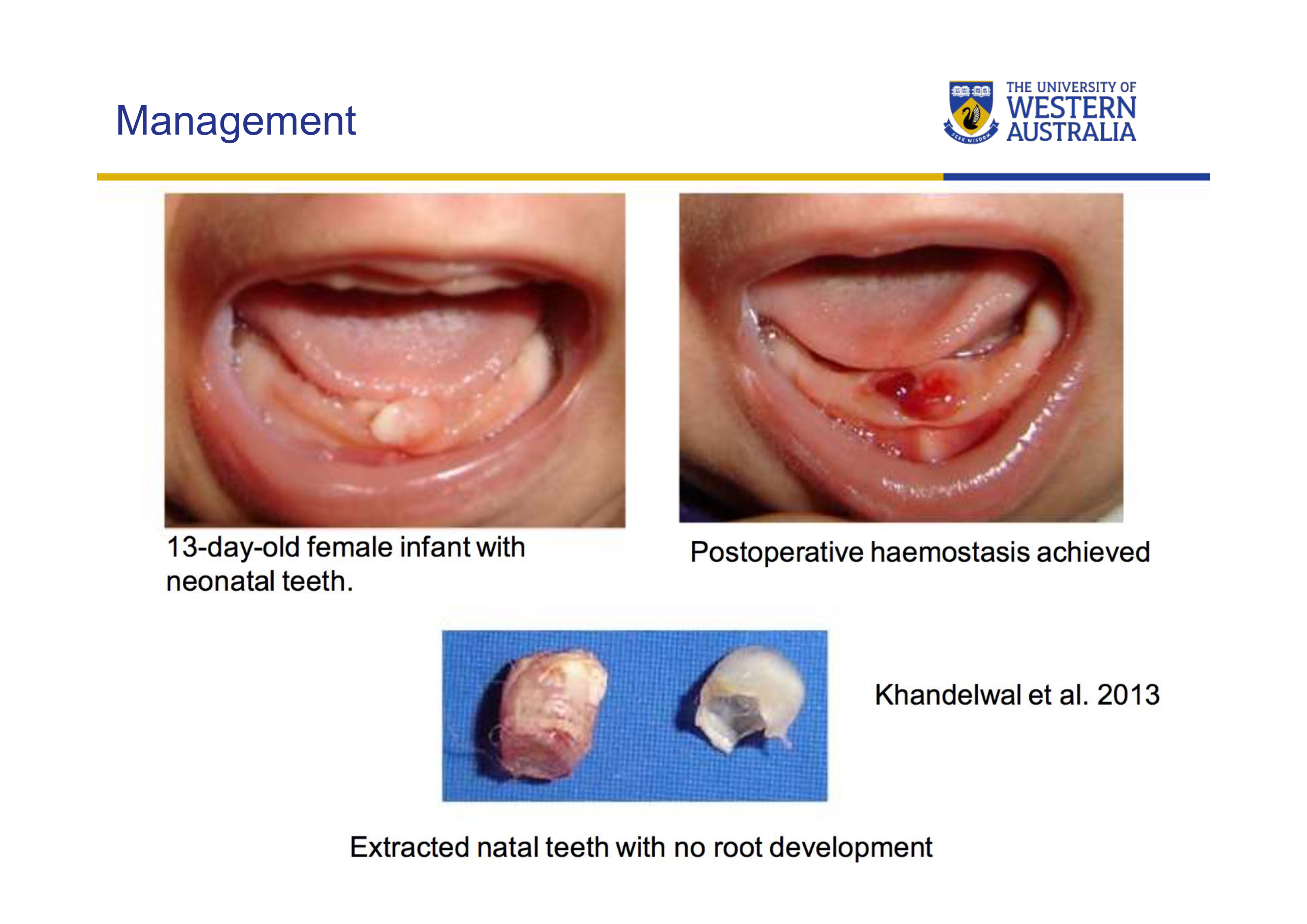
Surgical Extraction36
 |  |
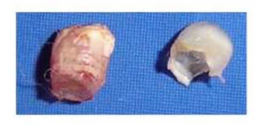 |
Extraction - Indications37
- Extraction is not contraindicated for any other clinical reasons (Mhaske et al. 2013)
- If the tooth is confirmed supernumerary through radiographs (Mhaske et al. 2013)
- Excessive mobility: risk of spontaneous exfoliation and consequent aspiration (Allwright, 1958; Kates et al. 1984)
- The tooth is unsuitable to retain due to severe dysplasia (Coho, 1996
- Significant enamel defects leading to breakdown or abscess.)
- The tooth developed an abscess or pulpal hyperplasia (Coho, 1996)
- Tooth interfering with the pre-surgical orthopaedic or surgical phase of oral cleft management (Ziai et al. 2005)
- Severe sublingual ulceration which is seriously interfering with feeding and the child is severely malnourished or dehydrated (Allwright, 1958; Slayton, 2000; Choi et al. 2009)
Management
Extraction Precautions and Complications
Extraction – Precautions
- If the treatment option is extraction only, certain precautions should be taken:
- Considering the general health condition of the infant prior to extraction
- Avoiding extraction up to the 10th day of life to prevent hemorrhage?
- Assessing the need to administer vitamin K before extraction (0.5-1.0 mg IM)
- Avoiding unnecessary injury to the gingiva
- Socket should be curetted following extraction to prevent continued development of the cells of the dental papilla (Leung & Robson 2006, Zhu & King 1996
- It is vital to remove Hertwig’s epithelial root sheath and the follicle; otherwise, a root fragment may continue to form, requiring a second extraction later.)
- Being alert to the risk of aspiration during removal
- General practitioners are advised to refer these cases to pediatric dentists or hospital settings.
- Risk of lignocaine toxicity in low-weight neonates (2-3 kg).
- The tooth is tiny and easy to drop during the procedure.
Extraction – Complications
- Potential increased risk of space loss following extraction
- Gardiner, 1961 reported 9 cases with space loss following extraction of natal teeth, however the space was regained when the permanent teeth erupted
- Extracting anterior natal teeth rarely leads to significant space loss; space is usually regained when permanent teeth erupt.
Conclusions
- Majority of natal teeth are true primary teeth rather than supernumeraries
- Management ranges from no treatment, conservative options including grinding or coverage, to extraction
- Extraction should only be performed where indicated, rather than performed as a routine procedure for natal teeth
Audio Appendix
Additional Audio Content
The following sections from the lecture audio did not correspond to any heading in the main document.
Case Study: Preterm Newborn
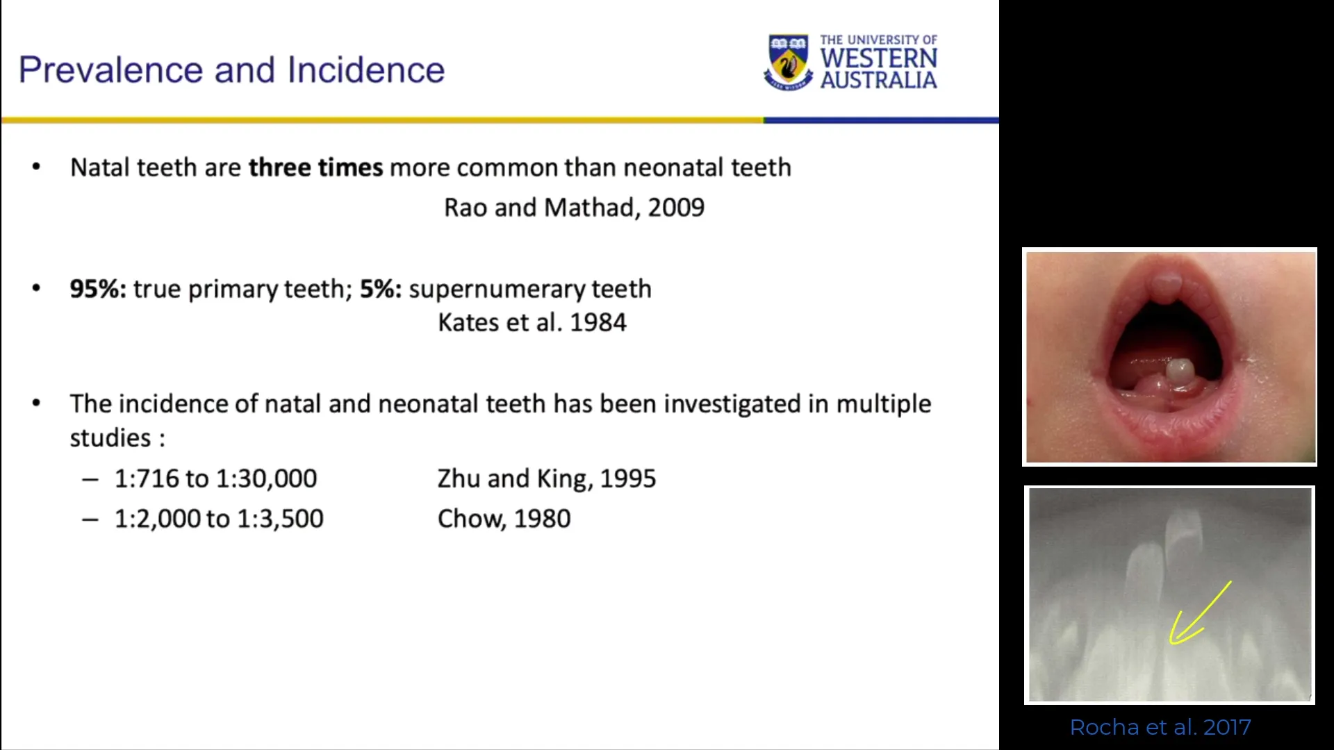
- A case report showed a natal tooth (7.1) in a preterm newborn. Radiographs confirmed it was a primary tooth, not a supernumerary, with underlying permanent teeth (3.1, 4.1) and an 8.1 tooth close to eruption.
Case Study: Cleft Lip and Palate
- A child with a unilateral cleft lip and palate presented with a natal tooth and a feeding ulcer on the tongue. Local anesthetic was delivered via a “ligna jet” (intraligamentary) to precisely control the dose (0.2 mil per click) and avoid toxicity. The tooth and follicle were extracted to prevent further ulceration and interference with cleft management.
Case Study: Abscess and Enamel Defect
- A case where a yellow-colored natal tooth appeared to have a minor defect. Upon extraction, a significant groove was discovered on the lingual surface that was not visible during the initial exam. An abscess was also identified only after the lip was fully retracted.
Clinical Cases
Case: Natal Tooth in a Preterm Newborn
Question
Have a look at these images from a case report showing a natal tooth in a preterm newborn. You can see a 71 (mandibular left primary central incisor) that is erupted intraorally. What can be observed on the radiograph regarding this tooth and the surrounding structures?
Findings and Reasoning
- Identification: The radiograph confirms that this is in fact the 71; it is not a supernumerary tooth.
- Developmental Status: You can see the presence of the underlying permanent teeth (31 and 41).
- Adjacent Teeth: There is also an 81 (mandibular right primary central incisor) which is close to erupting in the same area.
- Context: Most natal teeth (95%) are actual primary teeth rather than supernumerary. If they are lost or extracted, there will be no tooth in that space until the permanent teeth erupt years later.
Case: Riga-Fede Disease/Syndrome
Question
Consider a case where a newborn presents with natal teeth and a significant ulceration on the ventral surface of the tongue. What is this clinical presentation called and what are the implications for the patient?
Findings and Reasoning
- Diagnosis: This is known as Riga-Fede disease or syndrome. It is not a systemic “disease” but rather a traumatic ulceration caused by the sharp incisal edges of natal teeth rubbing against the tongue.
- Clinical Presentation: The ventral surface of the tongue shows a benign but painful ulcer.
- Complications: Because the ulcer is painful, it often leads to interference with feeding. The child may be uncomfortable and refuse to feed.
- Management: Treatment aims to allow the ulcer to heal. Options include smoothing the sharp edges (though difficult in neonates), covering the tooth with a material like a GIC or a stoma adhesive wafer, or extraction if feeding issues are significant.
Case: Natal Tooth with Unilateral Cleft Lip and Palate
Question
Look at this case of a child with a unilateral cleft lip and palate. You can see the natal tooth, the cleft, the nose, and the vomer. There is also a feeding ulcer on the tongue. How should this be managed if extraction is required?
Findings and Reasoning
- Anatomical Risks: In a cleft patient, extraction is high-risk because the tooth could easily slip and be displaced into the nasal cavity.
- Anesthesia: Local anesthetic must be administered with extreme caution regarding dosage. The lecturer uses a “ligna jet” (intraligamentary syringe) to deliver precise 0.2 ml increments to avoid lidocaine toxicity in a small infant (2-3 kg).
- Surgical Technique: After extracting the crown, it is critical to curette the socket. This is because Hertwig’s epithelial root sheath and dental follicle cells may remain; if not removed, a root fragment may continue to form, requiring a second surgery later.
- Hemostasis: One must verify the child has received vitamin K injections (standard for newborns) to ensure proper clotting before proceeding.
Case: Mobile Natal Tooth “Floating” on Tissue
Question
Look at this case of a healthy child (no cleft) where a natal tooth is present in the mandibular alveolus. The tooth appears to be “flapping around” or floating. What is the clinical situation here?
Findings and Reasoning
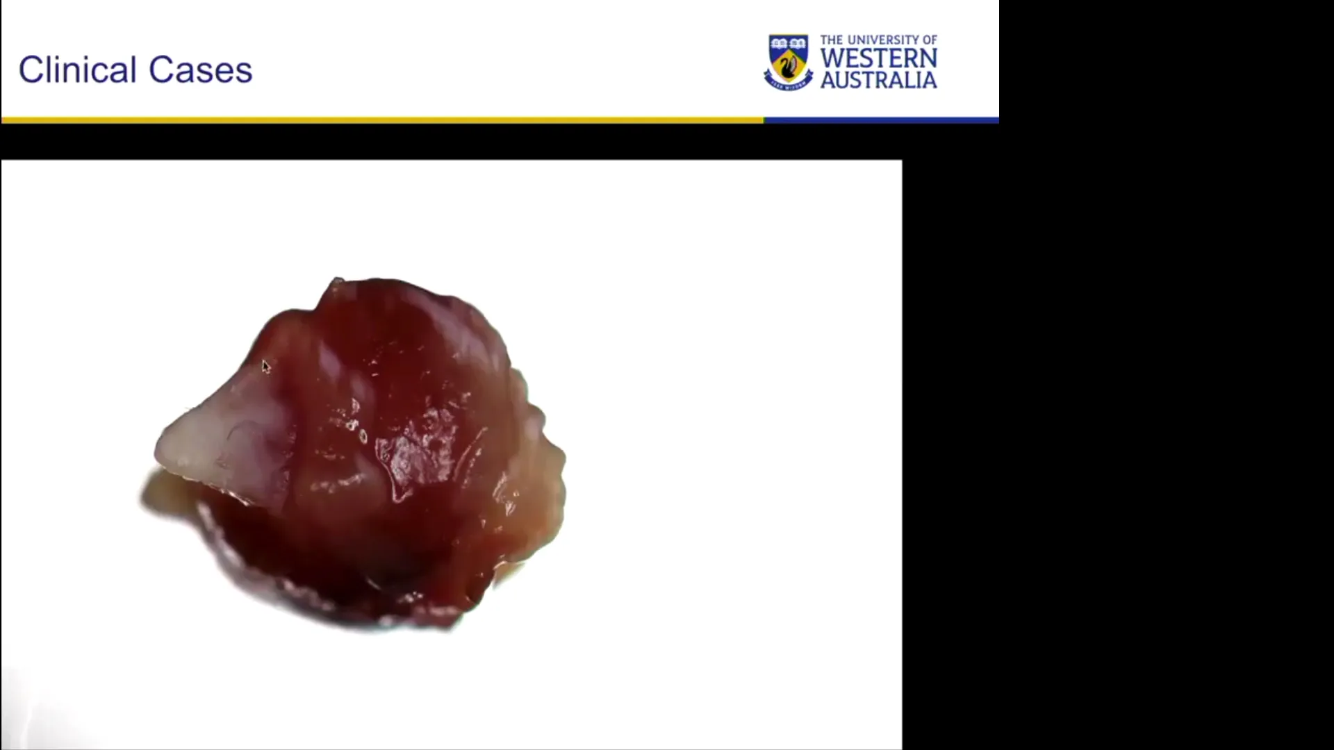
- Observation: The tooth is extremely mobile, essentially floating on a follicle of tissue. It moves every time the child moves their tongue.
- Etiology: This occurs because the tooth bud was positioned very superficially, leading to extremely early eruption before any root development occurred.
- Management: While many mobile natal teeth can be left to “firm up” as the root develops, in cases where the tooth is only attached by a thin stalk of soft tissue and interferes with function, extraction is indicated.
- Post-Extraction Findings: Upon inspection of the extracted tooth, it often shows dysplastic enamel (yellowish discoloration) and structural defects, such as a deep groove running through the tooth that might not have been visible while intraoral.
Case: Infected Natal Tooth with Abscess
Question
Look at this clinical photo of a natal tooth. If you only looked at the tooth itself, you might miss the primary complication. What must you do during the examination to ensure a correct diagnosis?
Findings and Reasoning
- Examination Technique: You must physically lift the lip all the way out of the way. Because a neonate’s head is so small, an abscess can be easily missed if the tissue is not fully retracted.
- Findings: Lifting the lip reveals a localized swelling/abscess associated with the natal tooth.
- Reasoning: Natal teeth often have thin, hypoplastic enamel and large, vascularized pulps. This makes them highly susceptible to rapid breakdown, caries, and subsequent pulp infection/abscess formation.
- Management: A tooth with an abscess and significant structural breakdown is a clear indication for extraction.
Footnotes
-
Original PDF page 1: L1 Natal Teeth 2020, p.1 ↩
-
Original PDF page 2: L1 Natal Teeth 2020, p.2 ↩
-
Original PDF page 3: L1 Natal Teeth 2020, p.3 ↩
-
Original PDF page 4: L1 Natal Teeth 2020, p.4 ↩
-
Original PDF page 6: L1 Natal Teeth 2020, p.6 ↩
-
Original PDF page 5: L1 Natal Teeth 2020, p.5 ↩
-
Original PDF page 7: L1 Natal Teeth 2020, p.7 ↩
-
Original PDF page 8: L1 Natal Teeth 2020, p.8 ↩
-
Original PDF page 9: L1 Natal Teeth 2020, p.9 ↩
-
Original PDF page 10: L1 Natal Teeth 2020, p.10 ↩
-
Original PDF page 11: L1 Natal Teeth 2020, p.11 ↩
-
Original PDF page 12: L1 Natal Teeth 2020, p.12 ↩
-
Original PDF page 13: L1 Natal Teeth 2020, p.13 ↩
-
Original PDF page 14: L1 Natal Teeth 2020, p.14 ↩
-
Original PDF page 15: L1 Natal Teeth 2020, p.15 ↩
-
Original PDF page 16: L1 Natal Teeth 2020, p.16 ↩
-
Original PDF page 17: L1 Natal Teeth 2020, p.17 ↩
-
Original PDF page 18: L1 Natal Teeth 2020, p.18 ↩
-
Original PDF page 19: L1 Natal Teeth 2020, p.19 ↩
-
Original PDF page 20: L1 Natal Teeth 2020, p.20 ↩
-
Original PDF page 21: L1 Natal Teeth 2020, p.21 ↩
-
Original PDF page 22: L1 Natal Teeth 2020, p.22 ↩
-
Original PDF page 23: L1 Natal Teeth 2020, p.23 ↩
-
Original PDF page 24: L1 Natal Teeth 2020, p.24 ↩
-
Original PDF page 25: L1 Natal Teeth 2020, p.25 ↩
-
Original PDF page 26: L1 Natal Teeth 2020, p.26 ↩
-
Original PDF page 27: L1 Natal Teeth 2020, p.27 ↩
-
Original PDF page 28: L1 Natal Teeth 2020, p.28 ↩
-
Original PDF page 29: L1 Natal Teeth 2020, p.29 ↩
-
Original PDF page 30: L1 Natal Teeth 2020, p.30 ↩
-
Original PDF page 31: L1 Natal Teeth 2020, p.31 ↩
-
Original PDF page 32: L1 Natal Teeth 2020, p.32 ↩
-
Original PDF page 33: L1 Natal Teeth 2020, p.33 ↩
-
Original PDF page 34: L1 Natal Teeth 2020, p.34 ↩
-
Original PDF page 35: L1 Natal Teeth 2020, p.35 ↩
-
Original PDF page 37: L1 Natal Teeth 2020, p.37 ↩
-
Original PDF page 36: L1 Natal Teeth 2020, p.36 ↩