Oral Medicine of Soft and Hard Tissues in Paediatric Dentistry12
The University of Western Australia
2025
-
Dr Mo Halane
-
Dr Joti Lidder
-
Dr Sarah Zeitoun
-
Diagnosis
- Sieve
-
Oral Pathology in paediatric patients
- Soft tissue lesions
- Bony lesions and hard tissue pathology
-
Treatment options
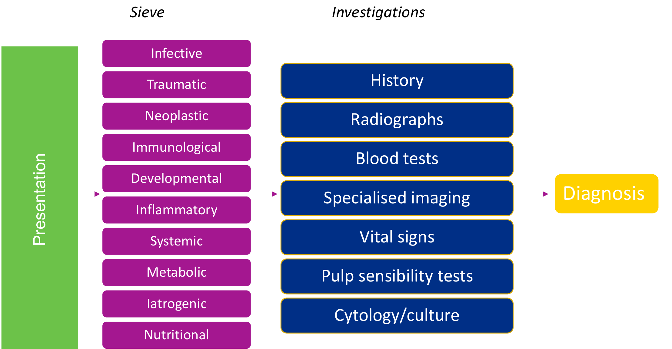
Diagnosis Framework
Sieve3
Presentation
-
Infective
-
Traumatic
-
Neoplastic
-
Immunological
-
Developmental
-
Inflammatory
-
Systemic
-
Metabolic
-
Iatrogenic
-
Nutritional
-
Distinguish normal variation from pathology and decide whether the lesion can be reviewed or requires intervention. Investigations
-
History
-
Radiographs
-
Blood tests
-
Specialised imaging
-
Vital signs
-
Pulp sensibility tests
-
Cytology/culture
-
Record when the lesion was first noticed, its duration, associated symptoms, progression, changes in appearance, previous appearance including parent photographs, relevant medical history, medications and possible trauma.
-
Describe whether the lesion is flat or raised, its size and extent, colour, surface characteristics, mobility and consistency, and its relationship to adjacent teeth and tissues.
-
Consider biopsy and histopathology, two-dimensional and three-dimensional radiographs, sensibility testing, and swabs where infection is suspected. → Diagnosis
Lecturer — Systematic Diagnosis
A systematic diagnostic approach should be used for every paediatric oral or dental problem.
- The same diagnostic sieve applies to soft-tissue lesions, hard-tissue lesions and general dental problems.
- A systematic approach helps avoid misdiagnosis, such as diagnosing sinusitis when the underlying issue is an extensively necrotic dental quadrant.
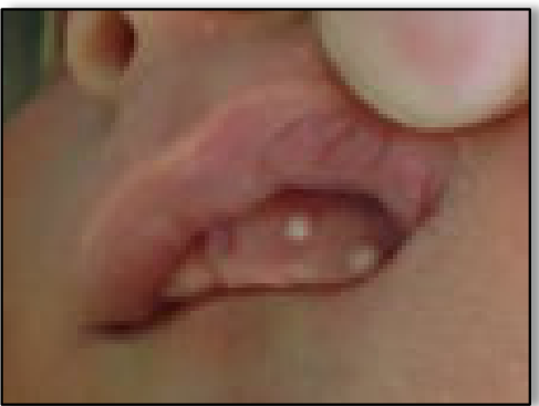
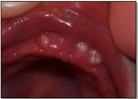
Newborns and Infants
- Bohn nodules
- Appearing on the buccal and lingual aspects of the alveolar ridges
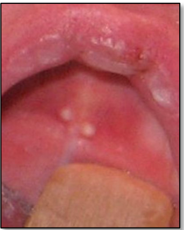
- Epstein pearls
- Appearing at the junction of fusion of embryological processes, appearing at the midline of the hard palate
- Dental Lamina Cysts
- Appearing on the crest of the alveolar ridges
Bohn Nodules, Epstein Pearls and Dental Lamina Cysts4
- A rare benign lesions of unknown origin found only in neonates
- No predilection for arch
- More common in girls
- Arises from the gingival crest but not odontogenic in origin
Lecturer — Gingival Cysts
These lesions are commonly referred to collectively as gingival cysts of the newborn, although terminology varies according to location.
- They appear as firm white or yellow nodules, approximately 1–3 mm in size, occurring singly or in clusters and sometimes being mistaken for natal teeth.
- The terms may overlap; broader terminology includes gingival inclusion cysts and palatal cysts.
- They arise from entrapped residual epithelial tissue or dental lamina remnants. Palatal lesions may result from entrapment between developing palatal shelves during the tenth week of intrauterine life, and histologically they are keratin-filled cystic spaces.
- Management consists of reassuring parents and reviewing rather than intervening. They are generally self-limiting and usually resolve within weeks to months, presumably through degeneration or rupture.
 |  |
 |
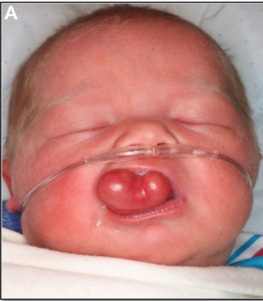
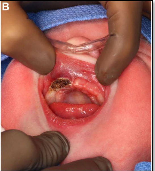
Congenital Epulis5
Manlove et al Oral Maxillofacial Surg Clin N Am 2024;36:265-282
Lecturer — Congenital Epulis
Congenital epulis is also known as a congenital granular cell tumour and may occasionally be diagnosed antenatally.
- It is usually painless, with a smooth and sometimes multilobular appearance.
- Histologically, it resembles a granular cell tumour, although granular cell tumours are more commonly found on the tongue; it develops through proliferation of mesenchymal cells with granular cytoplasm.
- Management depends on lesion size and whether feeding is impaired. Some lesions regress with time, while larger lesions or those interfering with feeding may require surgical excision.
- Eruption of the teeth is usually unaffected by either conservative management or excision.
 |  |
Pigmented, Vascular and Red Lesions6
- Physiological pigmentation
- Eruption cyst (overlaps with hard tissue eruption pathology)
- Haemangioma, lymphangioma, AV malformations
- Petechiae – systemic and local causes
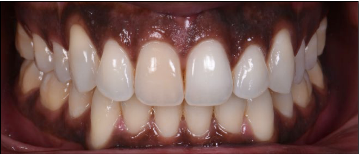
Physiological Pigmentation7
- Diffuse light to dark brown pigmentation of the oral mucosa
- Often on the attached gingiva
- Not to be confused with heavy metal toxicity which is confined to the marginal gingiva
Lecturer — Normal Variation
Normal gingival pigmentation varies considerably between individuals.
- Some patients have small areas of brown pigmentation, while others have generalized dark brown pigmentation with little or no pink gingiva.
- Recognizing normal variation is important before diagnosing pathology.
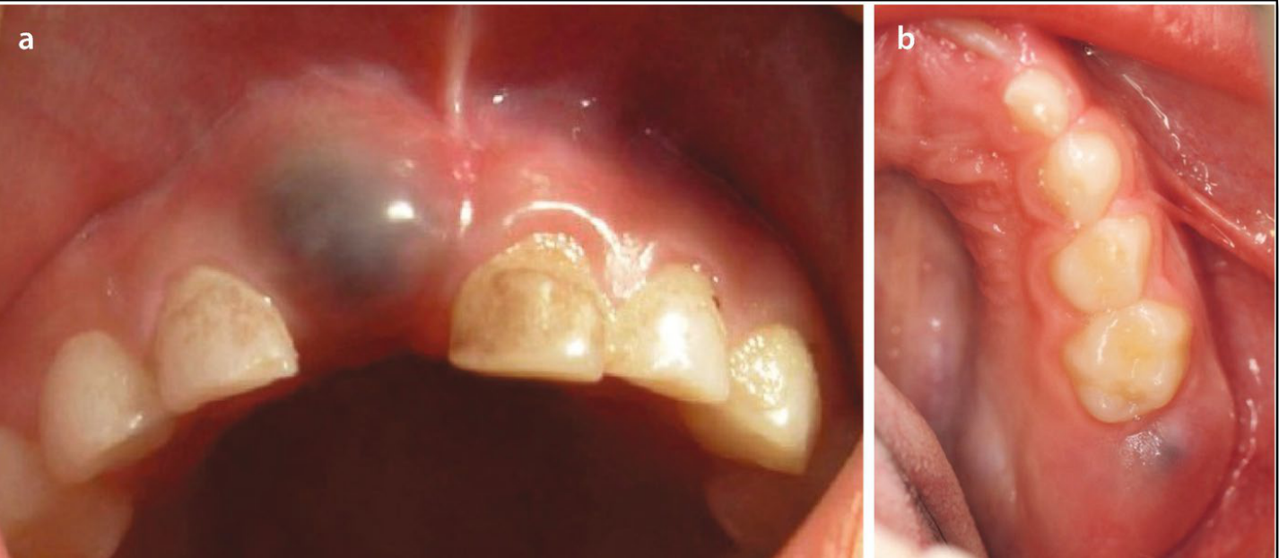
Eruption Cysts8
- Presenting as follicular enlargement just before eruption of the tooth
- Lesions tend to be blue/black as they contain blood
- Usually require no treatment
- Reassurance
- Review
Lecturer — Eruption Cysts
An eruption cyst, also called an eruption haematoma, is a developmental odontogenic cyst occurring before the eruption of a primary or permanent tooth.
- It presents as a soft, well-demarcated swelling over the crown of an erupting tooth and may contain fluid or blood.
- It is thought to develop when epithelium separates from the crown during eruption, allowing fluid or blood to accumulate.
- Spontaneous rupture commonly resolves the lesion and allows the tooth to erupt; surgical opening may be required if it becomes infected.
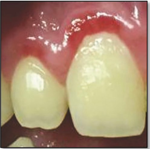
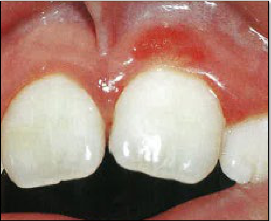
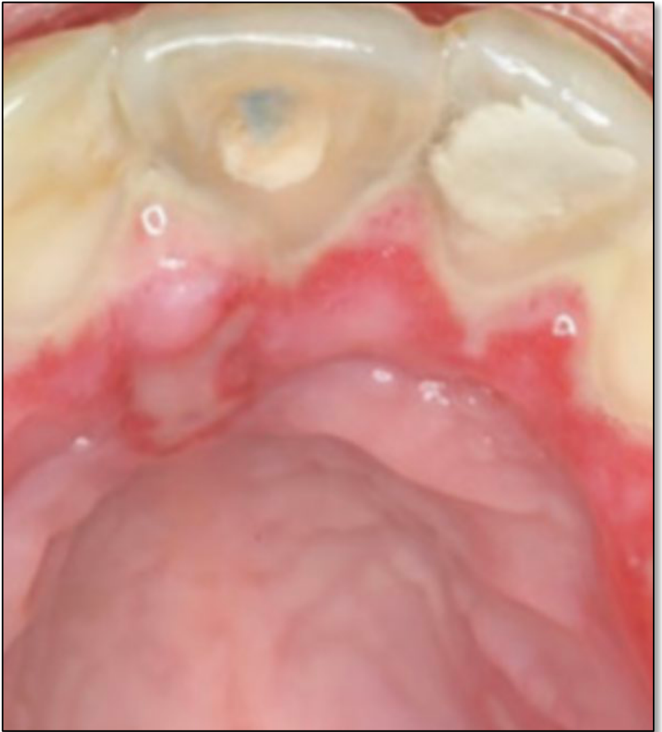
Localised Juvenile Spongiotic Gingival Hyperplasia9
- Bright red, sessile lesion
- Attached gingiva
- Anterior maxilla
- Girls > Boys
- Do not respond to oral hygiene
Illustration from Pediatric Dentistry A Clinical Approach 3rd Ed.
Lecturer — Juvenile Hyperplasia
This lesion is common but frequently unrecognised and can be mistaken for gingivitis.
- It occurs most commonly in children and young adults, is usually painless, and may bleed during tooth brushing or probing.
- It has a subtle papillary surface architecture and is more accurately considered a gingival hyperplasia rather than an inflammatory process.
- The cause is not fully understood, and management may involve review, monitoring, or surgical excision depending on the effect of the lesion and the clinical circumstances.
 |  |
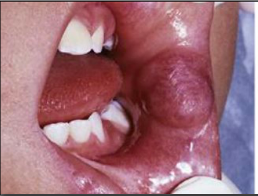
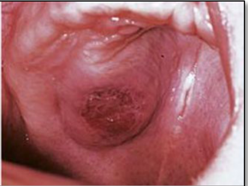
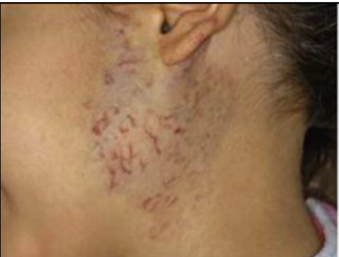
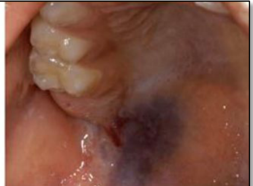
Vascular Lesions and Malformations10
-
Haemangioma
- Superficial, deep, compound
-
Syndromes
- Maffucci syndrome, Peutz-Jeghers syndrome
-
Prolonged bleeding from gingivae
-
Warm and or pulse sensation
-
Enlargement of periodontal space
-
Abnormal trabeculae
-
Mobile teeth
-
Facial asymmetry
Lecturer — Vascular Malformations
Blanching on pressure is an important clinical sign, and vascular lesions may result in a regional space filled with blood.
- Superficial haemangiomas originate from the papillary dermis, present as bright red macules or papillary masses, may grow with the infant and then regress into adolescence, and often require observation only.
- Vascular malformations may involve blood vessels, lymphatic vessels, or capillary, venous, or arterial components; low-flow lesions involve capillary, venous, or lymphatic components, while high-flow lesions involve an arterial component.
- Some vascular malformations can cause life-threatening haemorrhage, and suspected lesions require referral to oral medicine or maxillofacial services because outpatient dental treatment may carry a high risk.
 |  |
 |  |
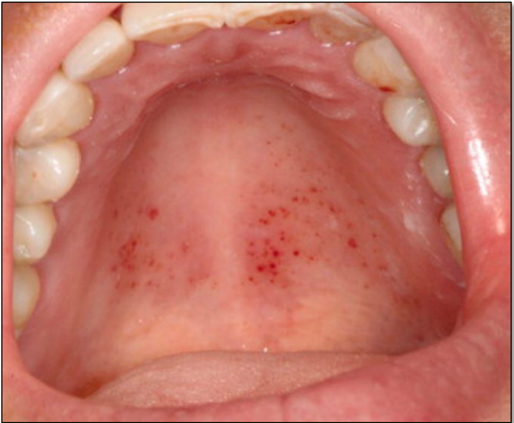
Petechiae and Purpura11
- Petechiae
- Pinpoint spots, often <2mm
- Purpura
- Larger, measuring between 2mm-1cm
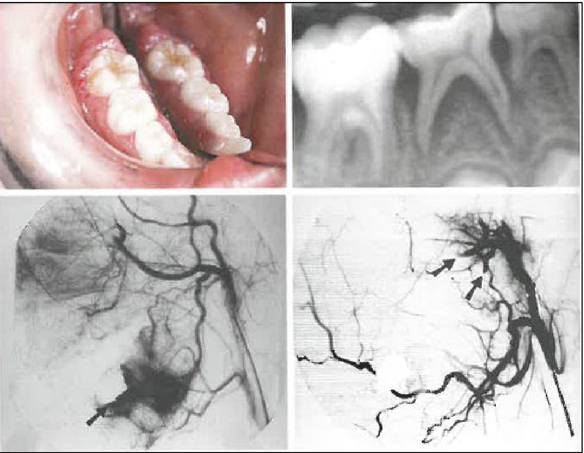 | 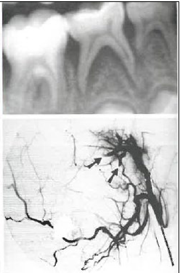 |
Diagnosis12
- History
- Medications
- Full blood counts
Dioguardi et al 2015
Lecturer — Petechiae and Purpura
Petechiae and purpura may occur with severe bleeding disorders, coagulopathies, low platelet counts, or medications and conditions affecting platelet production, including leukaemia.
- Lesions may begin bright red and become blue-brown as the blood is metabolised.
- Minor trauma during chewing or swallowing can produce these lesions in patients with low platelet counts.
- Petechiae or purpura without an obvious cause may indicate significant chronic illness and require further investigation.
- Medications should be considered, particularly in older patients; in young children, blood-thinning medication would generally be identified through the medical history.
Infections13
- Oral candidiasis (thrush)
- Primary herpetic gingivostomatitis
- Varicella, Hand-Foot-Mouth disease
- Syphilis (oral manifestations)
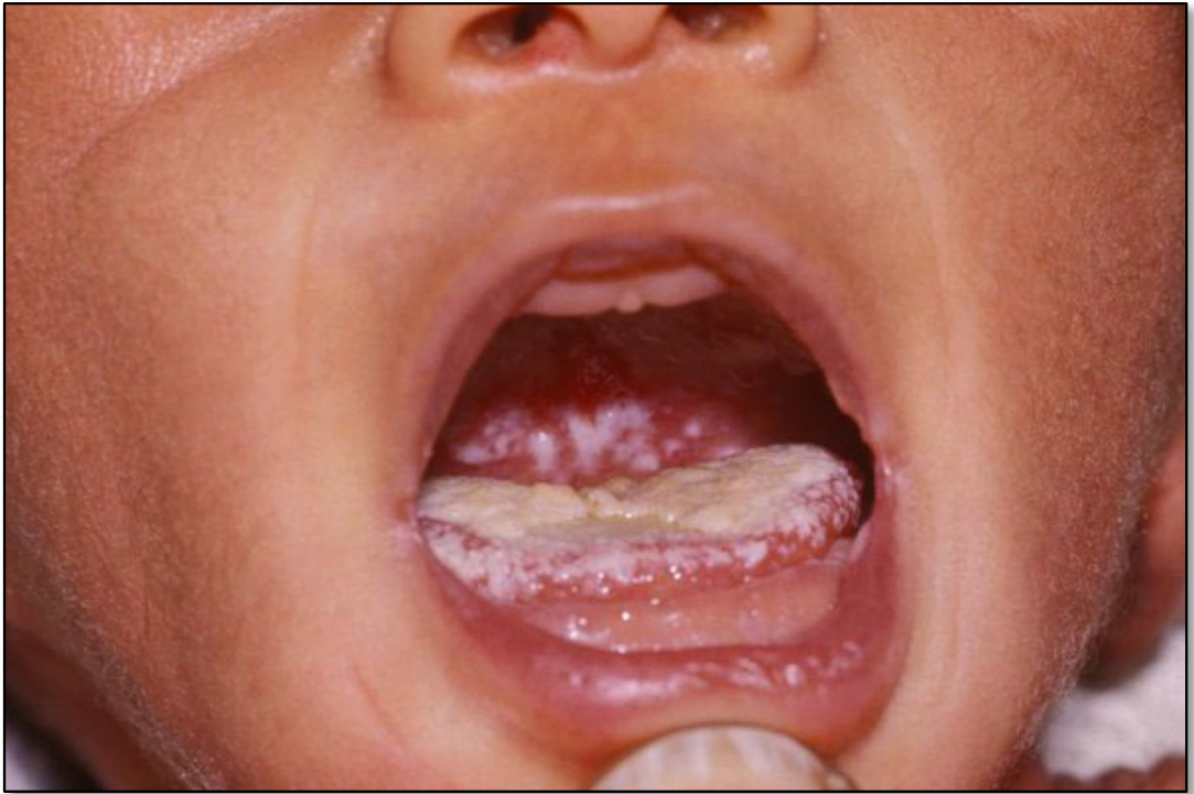
Oral Candidiasis14
- Acute pseudomembranous candidiasis
- Most common presentation in infants
- Caused by Candida Albicans
- Clinical
- Raised pearly-white patches overlying oral mucosa, most often on the buccal and labial mucosa, soft palate, and tongue
- The plaques are easily removed with a dry gauze and the underlying mucosa is erythematous
Lecturer — Candidosis Development
Candida albicans is normally present in the oral flora, but disease occurs when the oral balance changes and allows overgrowth.
- Predisposing circumstances include viral illness, diabetes, immune compromise, medication use and other therapies.
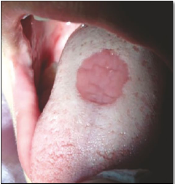
- Clinical forms also include erythematous candidiasis, which causes generalized erythema, and median rhomboid glossitis, which presents as an erythematous, depapillated, well-demarcated area in the midline of the dorsal tongue.
Neonates and children <2y15
- Address predisposing factors
- Nystatin
- 100 000 units/mL suspension 1mL topically 4xdaily after feeding
- 7-14 days
- Continue 3 days after relief
- Miconazole 2%
- 1.25 mL topically 4xdaily after feeding
- 7-14 days
- Continue 7 days after relief
Hygiene and Transmission Prevention (Therapeutic Guidelines: Oral and Dental)
- Apply the dose in the anterior part of the mouth
- Do not share dummies, cups or eating utensils
- Regularly wash toys and other objects that children put in their mouths
Lecturer — Infant Thrush Management
Thrush is relatively common in infants younger than six months and often clears as the child’s immune system becomes established.
- Management begins with identifying and addressing the cause; for example, a spacer may be required when thrush is associated with a steroid inhaler.
- Compliance can be poor because of taste, gagging and spitting out the medication, so successful treatment requires adequate contact time with the affected mucosa.
- Depending on associated medical conditions, treatment may also be initiated by the medical practitioner.
Philipone, Yoon & Koutlas 2023
Primary Herpetic Gingivostomatitis16
- The symptomatic presentation following initial exposure to herpes simplex virus 1
- Rare cases from HSV-2
- Appearance
- Numerous small vesicles which erupt into ulcers
- Stomatitis
Lecturer — Herpetic Gingivostomatitis
Primary infection has a different presentation from recurrent cold sores and peaks at approximately six to eighteen months of age.
- A prodromal phase of approximately three to four days may occur, and infection may be acquired from a parent.
- Clinical features include painful shallow ulcers, inflamed gingiva, lymphadenitis, fever, malaise, headache, irritability and reduced oral intake.
- Management is supportive, including oral fluids, hydration, pain relief and ice chips; dehydration is the most common complication.
- The disease is self-limiting, with ulcers healing spontaneously without scarring. Antiviral therapy may be considered in immunocompromised patients, but evidence of benefit after the first 72 hours is limited.
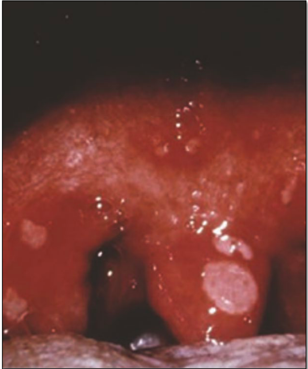
Hand Foot and Mouth Disease and Herpangina17
- Caused by the Coxsackie group of viruses
- Highly contagious, symptomatic and self limiting
- Appearance
- Herpangina
- Cluster of four to five vesicles on the palate/fauces/pharynx
- Hand foot and mouth
- Up to 10 vesicles around the mouth + hands and feet
- Herpangina
Lecturer — Coxsackie Infections
These infections spread readily through day-care centres and schools, and the vesicles subsequently ulcerate and may resemble oral aphthae.
- Management is supportive: maintain fluid intake, provide pain relief and monitor for dehydration.
- Parents should be advised not to send an affected child to school while infectious.
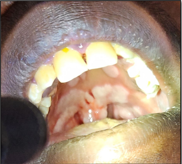
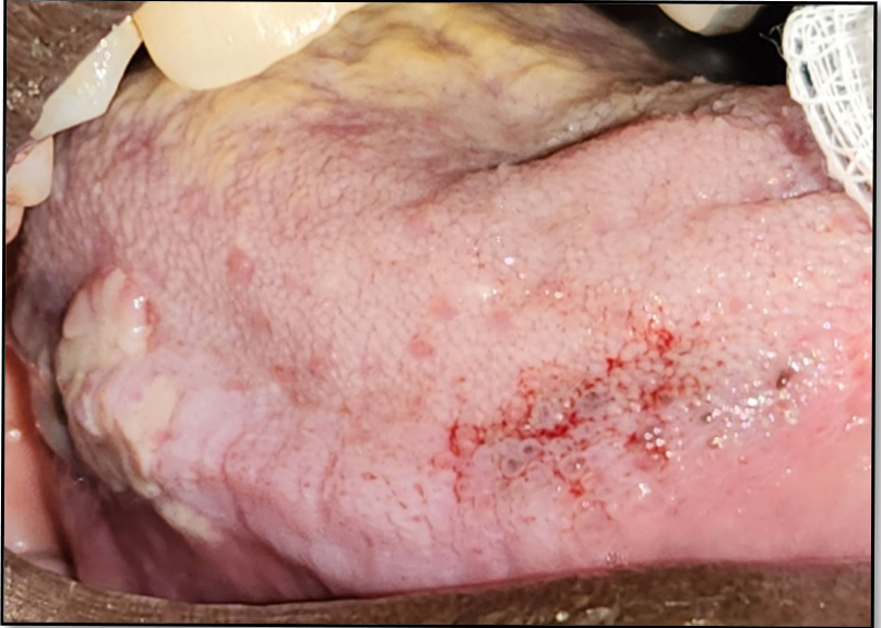
Syphilis18
- Initial presentation can be variable due to the different stages the patient can be in
- Primary
- Secondary
- Latent
- Tertiary
- Congenital
Lecturer — Syphilis Stages
Syphilis was described as increasing in Australia, with notifications in 2022 reported as double the recorded rate in 2015; an ongoing outbreak in Western Australia was also noted.
- Primary disease begins as a papule 10–90 days after infection, ulcerates and may cause regional lymphadenopathy before usually healing over the following weeks.
- Secondary disease occurs approximately four to ten weeks after the primary lesion and may include headache, fatigue, lymphadenopathy, low-grade fever, sore throat and mucocutaneous lesions.
- Latent disease leaves the patient infected but asymptomatic for many years. Oral lesions may include ulceration of the palate and lateral border of the tongue.
- Management requires a multidisciplinary approach involving the appropriate medical teams.
 |  |
Epulides and Exophytic Lesions19
- Pyogenic granuloma
- Fibrous epulis
- Squamous papilloma
- Verruca vulgaris
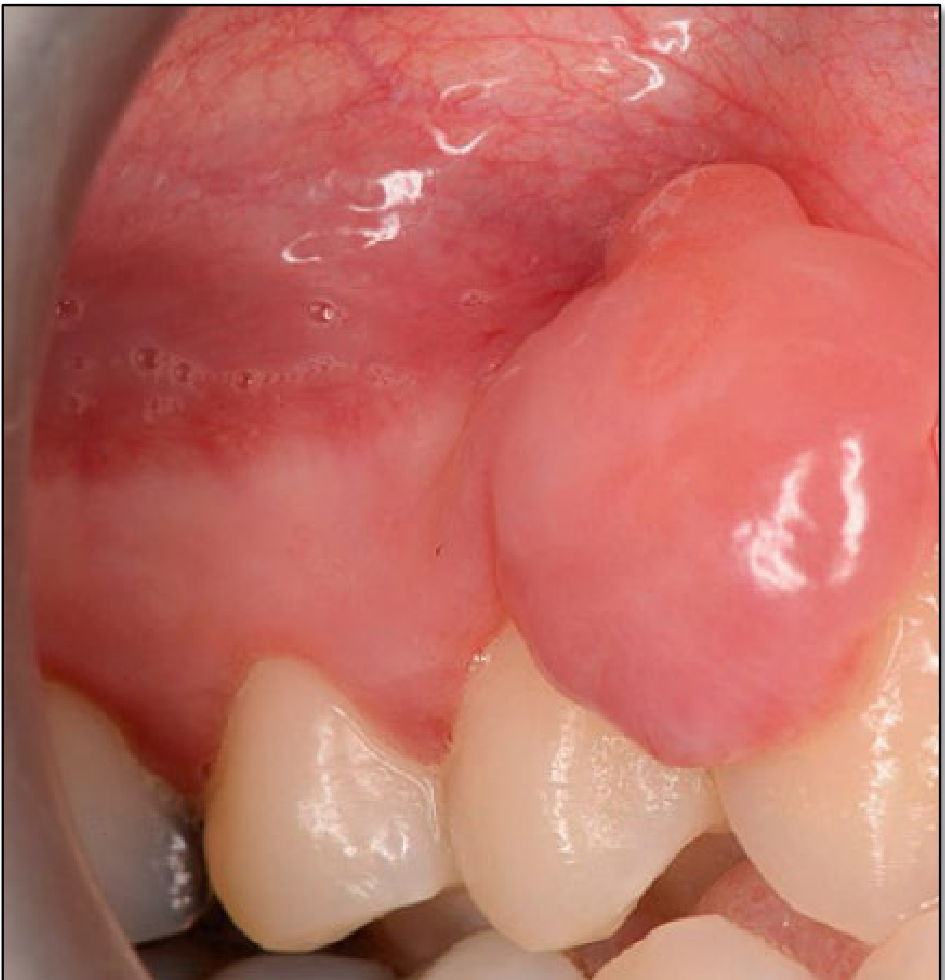
Fibrous Epulis20
-
Fibroepithelial reaction
- Response to local trauma or irritation
-
Appearance
- Smooth
- Pink
- Swelling of fibrous tissue
- Sessile
-
Treatment
- Oral hygiene instruction
- Surgical excision
Photo from Simms & Lewis, Dental Update 2024 48:4, 707-709.
Lecturer — Fibrous Epulis Details
Fibrous epulis is generally slow growing and may vary considerably in size.
- It may appear white because of surface keratinisation or may be ulcerated.
- Size can range from a few millimetres to several centimetres.
- It may occur anywhere, particularly on the buccal mucosa, lower lip, or around a denture.
- Management involves removal of the irritant and surgical removal if required.
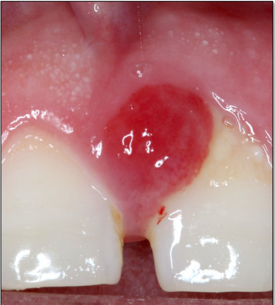
Pyogenic Granuloma21
-
Misnamed lesion
- No pus
- No granulomas
-
An ulcerated lobulated capillary haemangioma
-
Appearance
- Smooth surfaced tissue mass that has a red appearance
- Bleeds when manipulated
- Often pedunculated
-
Aetiology
- Often resulting from local irritation (plaque) or trauma
Lecturer — Pyogenic Granuloma
A pyogenic granuloma is histologically called a lobular capillary haemangioma and may represent a vascular counterpart to a fibrous epulis.
- It is an inflamed, vascular red lump.
- Management includes removing the local irritant and surgically removing the lesion if required.
- Considerable bleeding should be anticipated because of its vascular nature.
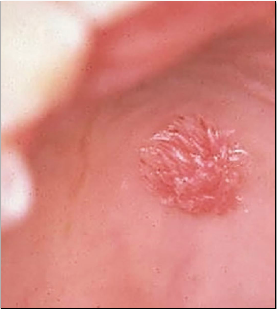
Squamous Papilloma22
-
Benign neoplasm caused by human papilloma virus
-
Appearance
- Cauliflower like growth
- Papillary
- Exophytic
- Colour depending upon keratinization amount
Philipone, Yoon & Koutlas 2023
Lecturer — Papilloma Management
Most papillomatous lesions regress spontaneously within two years through a cell-mediated immune response. When excision is required, the specimen should be submitted for oral pathology review.
- Include the stalk and a border of normal tissue during excision.
- Oral HPV can occur through several pathways, including sexual transmission.
- When a papilloma is identified in a child, investigate the possibility of genital origin using specific questions.
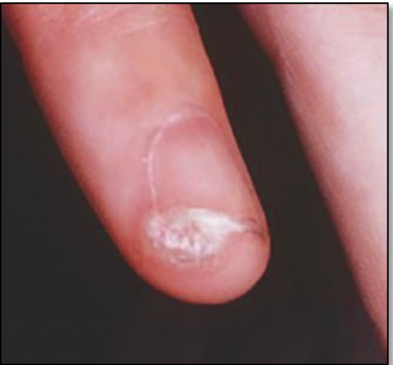
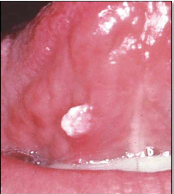
Verruca Vulgaris23
-
Also known as the common skin wart
-
Uncommon in the mouth; more frequently on the hands
-
Caused by HPV
-
Appearance
- Papillary
- Exophytic
- White growth
- May be single or multiple
Lecturer — Verruca Treatment
Treatment is similar to that of other papillomatous lesions, although the cause differs.
 |  |
Gingival Enlargement24
- Drug induced
- Syndromes with gingival enlargement
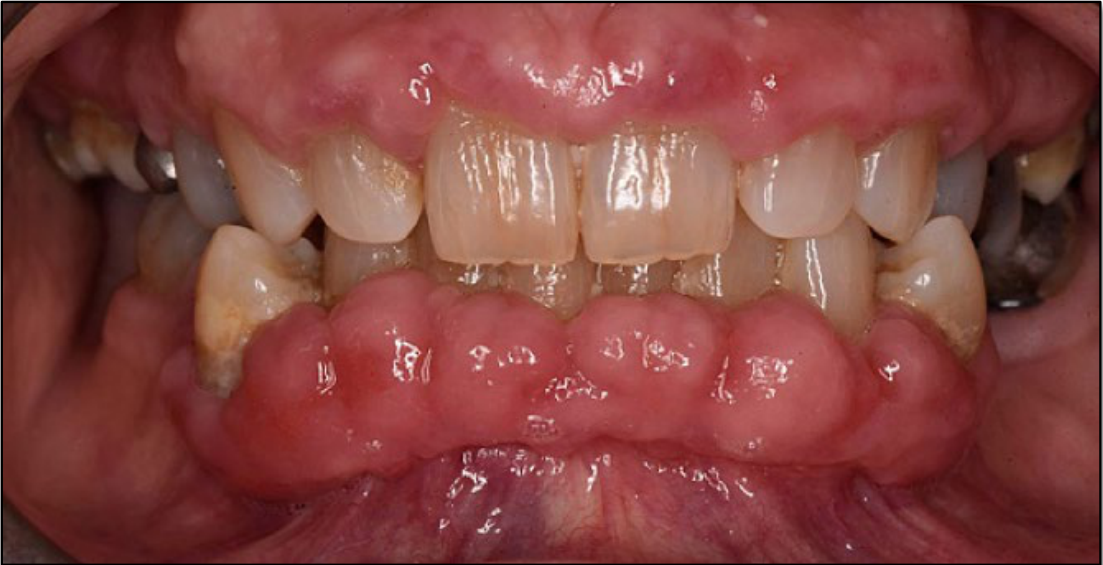
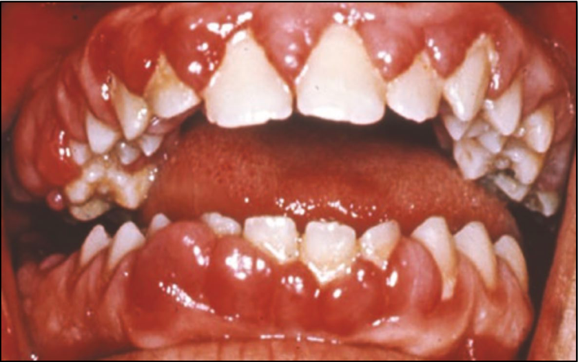
Drug Induced Gingival Enlargement25
Causes:
- Phenytoin
- Cyclosporin A
- Nifedipine
- Verapamil
Not all patients taking these drugs experience this.
Photo from Simms & Lewis, Dental Update 2024 48:4, 707-709.
Lecturer — Drug Effects
Drug-induced gingival enlargement has several additional clinical features and management considerations.
- It can be mistaken for a primary abscess, foreign-body swelling, or food impaction.
- Plaque-induced inflammation may occur secondarily and worsen the enlargement.
- Phenytoin is used to treat epilepsy; withdrawal resolves most cases, except severe cases.
- Cyclosporine A is an immunosuppressant used after organ transplantation, and gingival overgrowth occurs in approximately 30–70% of patients taking it.
- Nifedipine and verapamil may be used in children to control cyclosporine-induced hypertension after transplantation, producing a combined effect with cyclosporine.
- Proposed mechanisms include decreased collagen degradation, increased collagen synthesis, and changes in the extracellular compartment.
- Management includes emphasising oral hygiene and performing gingivectomy if required.
- In children, gingival overgrowth may impede exfoliation or eruption and cause ectopic tooth eruption.
 |  |
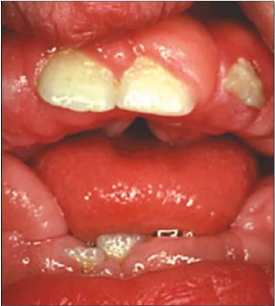
Hereditary Gingival Fibromatosis26
- The most common syndromic cause of gingival enlargement
- Autosomal dominant condition usually appearing at the time of eruption of the permanent dentition
- Other hereditary conditions include Zimmerman-Laband syndrome
Lecturer — Hereditary Fibromatosis
Management of hereditary gingival fibromatosis is generally beyond routine care and may require referral to a tertiary centre such as Perth Children’s Hospital.
Systemic Disease Manifestations27
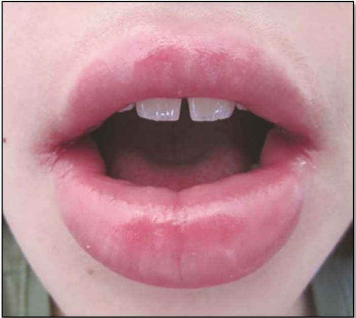
- Crohn’s disease/Orofacial granulomatosis, Melkersson–Rosenthal syndrome
Crohn’s Disease
- Chronic relapsing inflammatory bowel disease
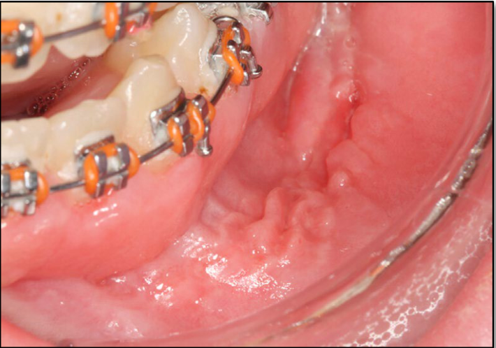
Appearance28
-
Diffuse or localised swelling of the lips and buccal mucosa (orofacial granulomatotis)
-
Recurrent aphthous like ulcerations
-
Pyostomatitis vegetans
-
Mucosal tags or folds
-
Cobblestoning
-
Mucogingivitis
-
Swollen lips or orofacial granulomatosis may be an initial presentation.
-
Recurrent ulcers should prompt questions about gastrointestinal symptoms, including discomfort, bloating, constipation and diarrhoea.
-
Pyostomatitis vegetans may present with erythematous, thickened oral mucosa, multiple pustules and superficial erosions.
-
Mucosal tags or folds may occur in the labial vestibules, buccal vestibules and retromolar mucosa.
-
Posterior buccal folds may become swollen, fissured and corrugated, producing a cobblestone appearance.
-
Cobblestone mucogingivitis may include oedematous, granular or hyperplastic gingiva.
-
Recurrent aphthae may also occur with other malabsorption syndromes, including coeliac disease.
 |  |
Management
-
Medical management of the condition involving steroids/DMARDS
-
Management focuses on the underlying condition, with referral to the appropriate medical teams.
-
Dental clinicians may provide symptomatic relief.
-
Recurrent aphthous ulceration should prompt consideration of vitamin B12 deficiency, iron deficiency and abnormal white cell counts.
-
A full blood count and relevant investigations may be recommended.
Salivary Gland Lesions29
- Mucocele
- Ranula
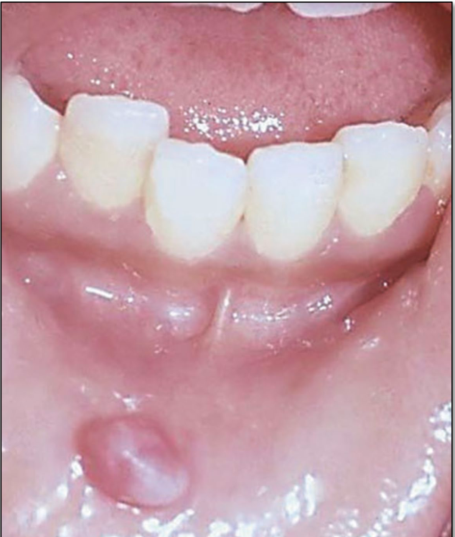
Mucocele
- Also known as a mucous extravasation cyst
- Arises from damage to the duct of one of the minor salivary glands in the lower lip or cheek
- The lower lip is the most common site.
- A history of biting or other recent trauma may support the diagnosis.
- Lip or cheek biting
- Build up of mucous in the connective tissue that is then surrounded by fibrous tissue
Appearance30
-
Well circumscribed swellings
-
Can appear blue
-
Can have a keratinized surface following trauma
-
From a few mm to a cm
-
The lesion appears as a mucus-filled swelling.
-
Diagnosis often depends on the clinical history and examination.
Treatment
-
Some regress spontaneously
-
Surgical excision
-
Persistent lesions may require referral.
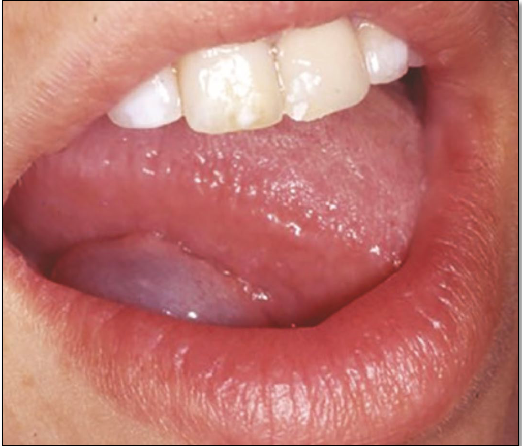
Ranula31
-
Is a mucocele of the floor of the mouth involving the sublingual or possibly the submandibular gland
-
A soft, bluish swelling on one side of the floor of the mouth
-
A plunging ranula is when the lesion herniates through the mylohyoid muscle
-
Occur lateral to the midline along the ducts
- Midline indicating a different type of cyst
-
A ranula may be caused by trauma or obstruction of one or more ducts.
-
If obstruction is due to a sialolith, removal of the sialolith may resolve the lesion.
-
Surgical excision or deroofing may otherwise be required.
Ulcerative and Vesiculobullous Lesions32
- Traumatic ulcers
- Riga–Fede
- Immune-mediated:
- RAU
- Erythema multiforme
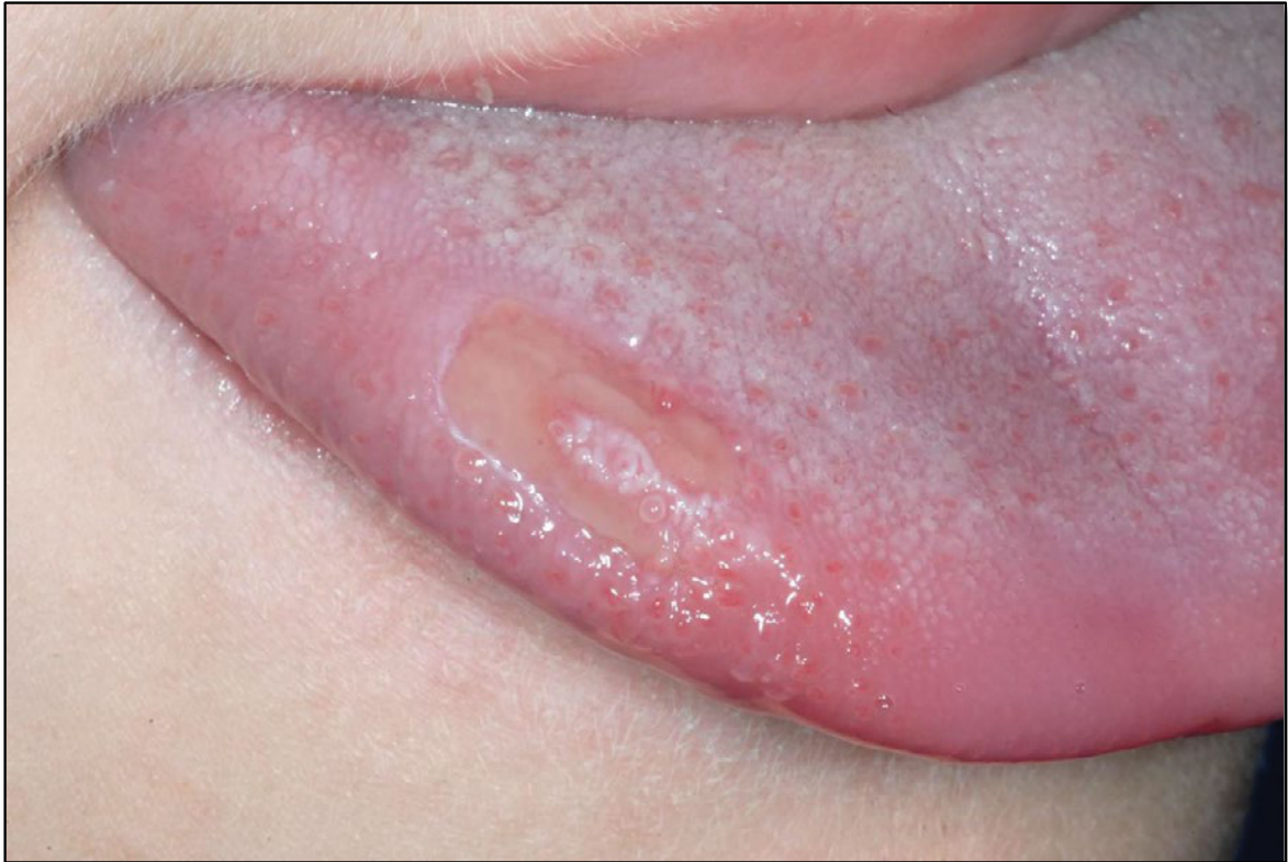
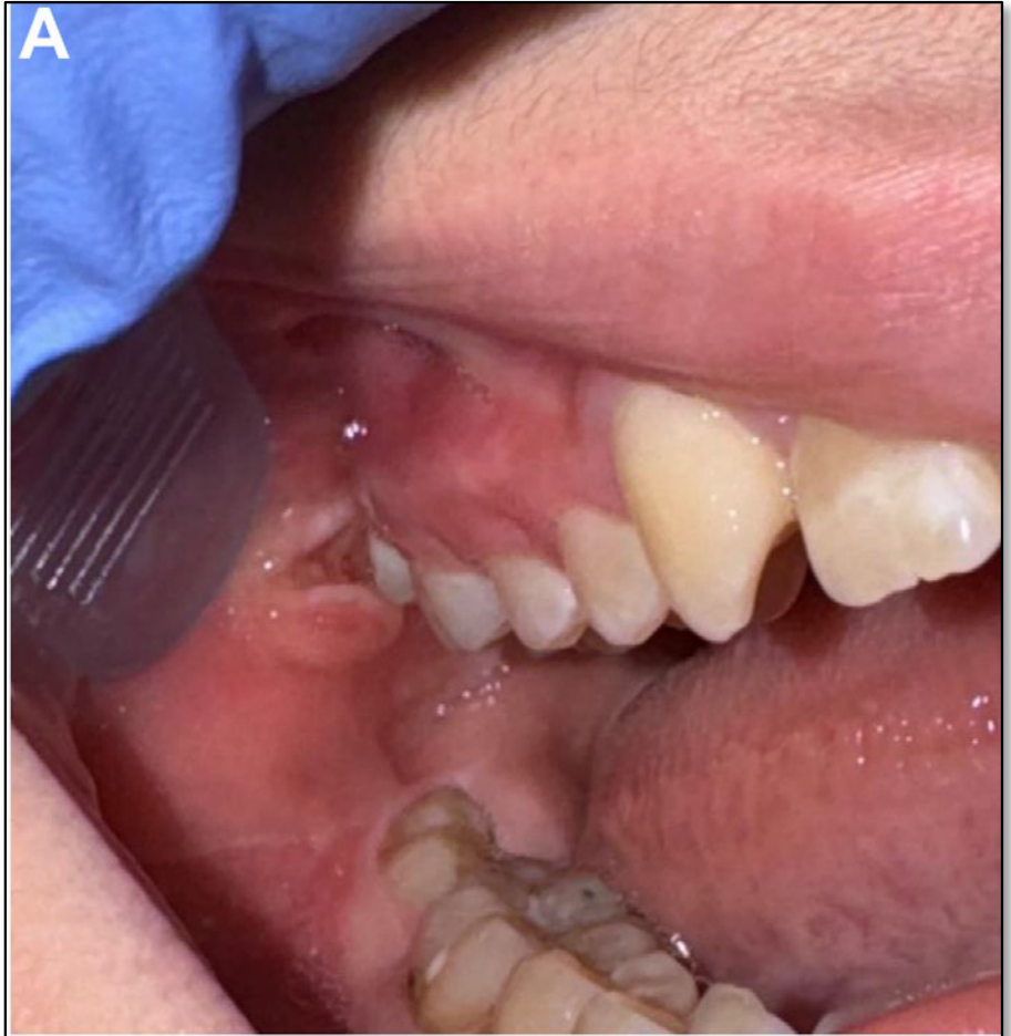
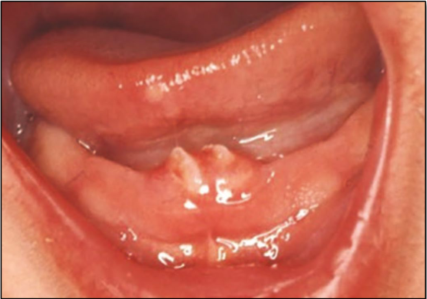
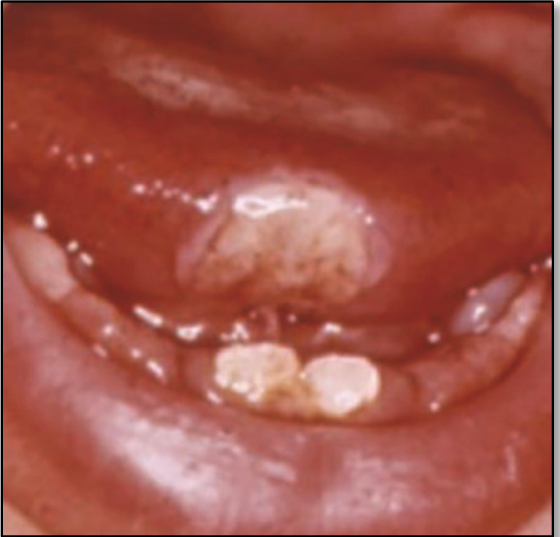
Traumatic Ulcers and Riga-Fede3334
McKernon et al Br Dent J 2023:235(4);255-261
Manlove et al Oral Maxillofacial Surg Clin N Am 2024;36:265-282
- A type of trauma induced ulcer on the ventral surface of the tongue caused by trauma from the continual protrusive and retrusive movements of the tongue over the lower incisors
- Often noted alongside the presence of natal teeth or sharp incisors
- Treatment can involve conservative review, smoothing sharp edges of teeth or rarely extraction of the natal teeth
Lecturer — Traumatic Ulcers
Traumatic ulcers in children are common and may result from mechanical, thermal, or chemical injury.
- The cause may be an obvious remembered bite or impact, but restorations, orthodontic brackets and wires, oral habits, and biting during stress or sleep may also contribute.
- The relationship between the ulcer and adjacent teeth should be assessed.
- Management is usually symptomatic unless the injury is severe enough to require suturing; options include warm salt-water rinses, chlorhexidine gel, and analgesia.
- Glass ionomer cement can be placed over a sharp tooth edge, and extraction may be needed if the ulcer is severe.
 |  |
 |  |
Recurrent Aphthous Ulcers
- Classified according to size, duration and severity
- Minor
- Major
- Herpetiform
- Aetiology
- Not well understood
- Nutritional deficiencies
- Immune mediated
- Trauma
Lecturer — Aphthous Stomatitis
Recurrent aphthous stomatitis is one of the most common oral mucosal lesions and affects approximately one-third of children.
- It may occur after colds or infections, with immunosuppressive medication, or alongside Crohn’s disease and other malabsorption syndromes.
- A full blood count and investigations for deficiencies may be appropriate, including vitamin B12, iron studies, and white cell counts.
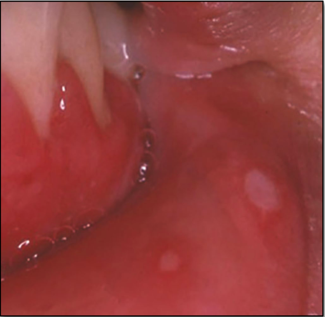
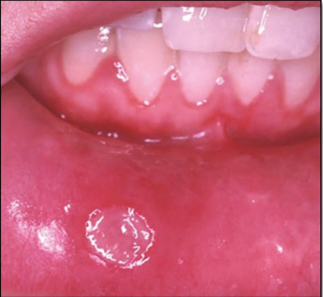
Appearance: Minor35
-
Non keratinized mucosa
-
<1cm
-
Lasting 7-14 days
-
Heal without scarring
-
1-5 in number
-
Sites include the labial mucosa, buccal mucosa, dorsum of the tongue, and lateral borders of the tongue.
-
Symptomatic management may include chlorhexidine mouthwash or topical steroids.
 |  |
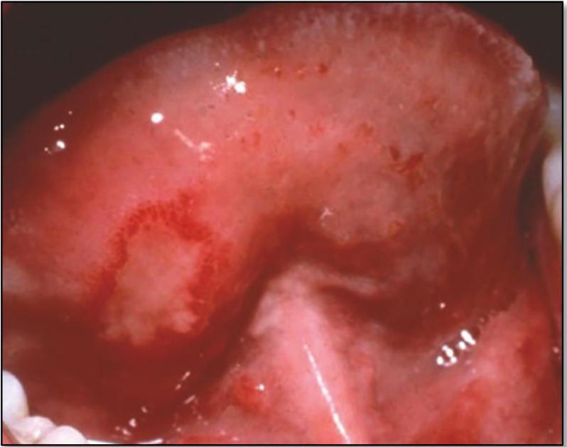
Appearance: Major36
- Smaller in number; 1-3
- Larger in size hence the name; >1cm
- Heal with scarring
- Healing taking longer; up to 6 weeks
Lecturer — Major Aphthous Ulcers
Major ulcers are larger and deeper than minor ulcers and may have irregular borders.
- They can occur on both non-keratinised and keratinised tissue.
- Because they may be very painful and associated with systemic deficiencies, further investigation may be required.
 | 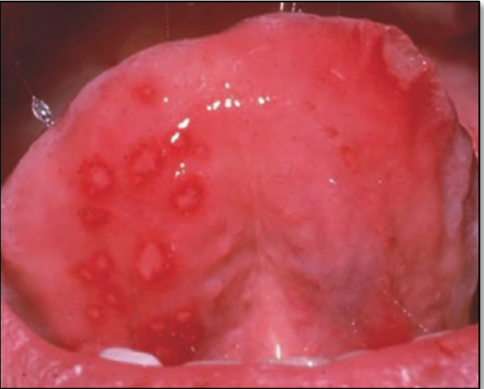 |
Appearance: Herpetiform
-
Resemble a primary herpetic infection
-
Smaller than minor and occur in clusters
-
Healing within 7-10 days
-
Keratinized and non keratinized surfaces
-
Can coalesce to form large ulcers
-
Multiple herpetiform ulcers may occur, and management depends on their severity and clinical presentation.
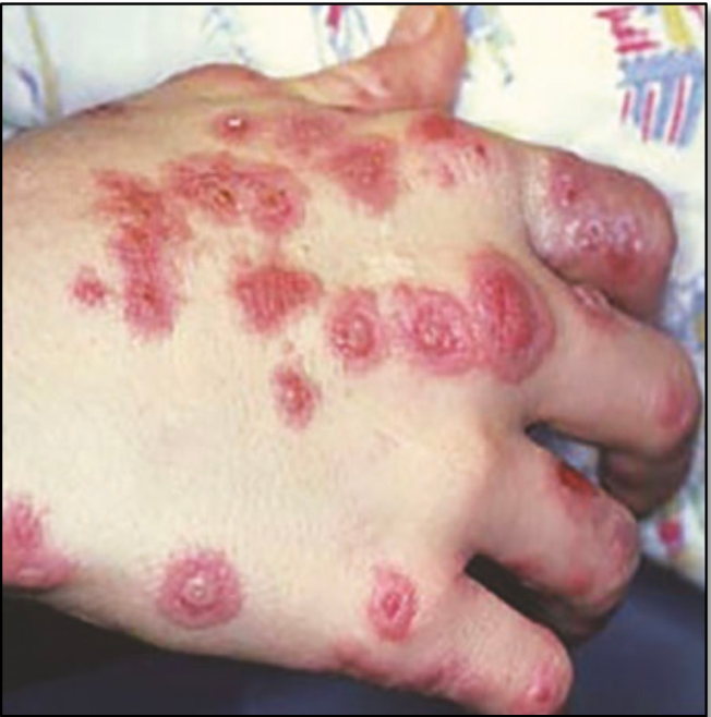
Erythema Multiforme3738
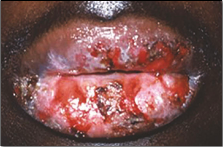
- Original description is that of a self limiting but recurrent seasonal skin disease with mucosa involvement in the oral cavity
- Aetiology
- Considered a type IV hypersensitivity reaction
- Appearance
- Target shape lesions on the skin
- Ulceration of lips with blood staining
- Oral lesions starting as erythematous patches that undergo necrosis and become ulcers
- Treatment
- EM is self limiting but being aware of progression
Lecturer — Erythema Multiforme
Erythema multiforme, Stevens-Johnson syndrome, and toxic epidermal necrolysis were described as different levels of severity of a basic disorder, ranging from mild to life-threatening.
- Recurrence of herpes simplex infection may be a cause.
- Management requires liaison with the medical team and may include antiviral therapy where a cause is identified, a short course of systemic steroids, fluid maintenance, hydration, pain control, and monitoring of severity.
- Toxic epidermal necrolysis may be life-threatening and require hospitalisation.
 |  |
Hard Tissue Lesions39
- Cystic lesions
- Tumors of the jaw
- Other bony lesions
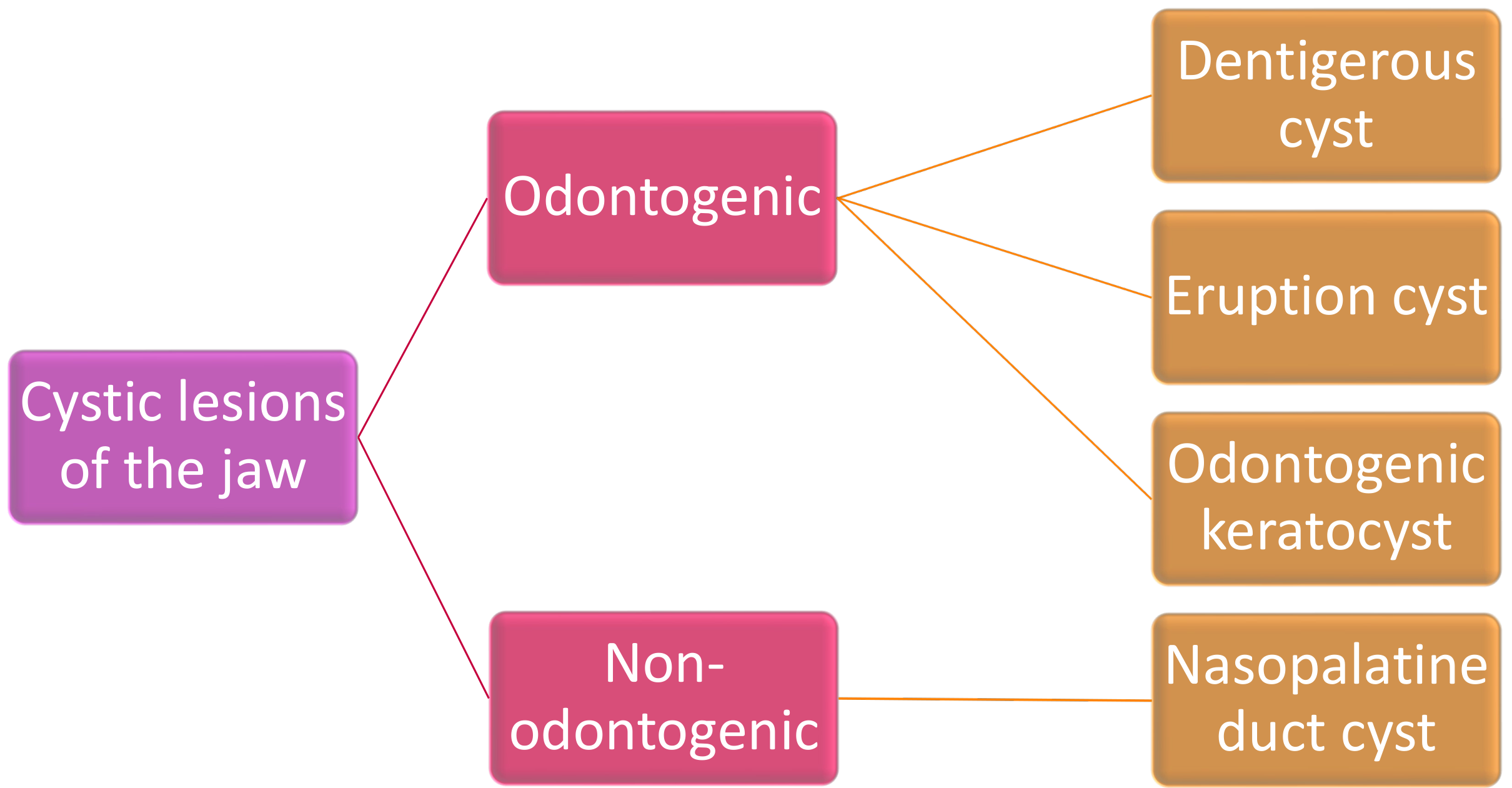
Cystic Lesions of the Jaws40
Cystic lesions of the jaw are classified as follows:
- Odontogenic
- Dentigerous cyst
- Eruption cyst
- Odontogenic keratocyst
- Non-odontogenic
- Nasopalatine duct cyst
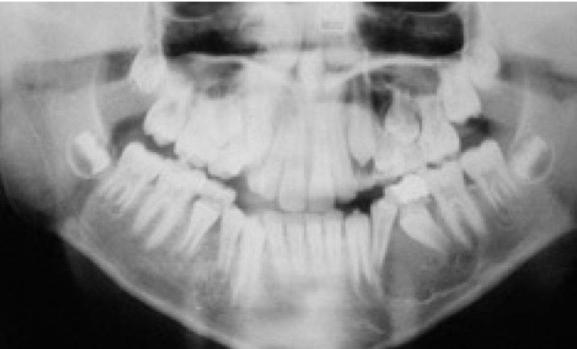
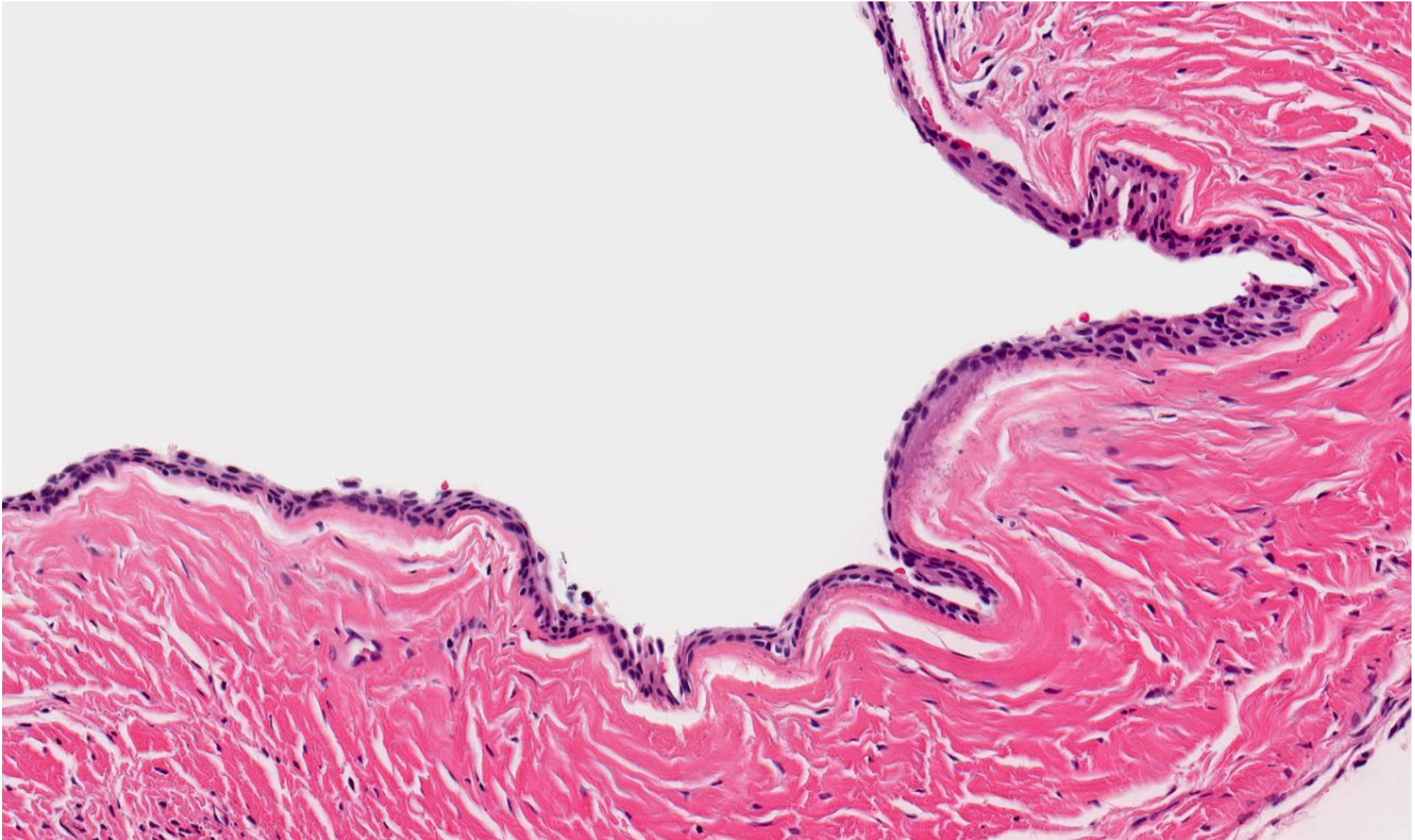
Dentigerous Cyst41424344
-
Benign, odontogenic cyst that forms around the crown of an unerupted or developing tooth, attached to the CEJ
-
Aetiology: accumulation of fluid between the reduced enamel epithelium and crown of the unerupted tooth
-
Site: most commonly in the mandible (75%); impacted third molars and canines
-
Clinical: usually asymptomatic, may be discovered incidentally
- Swelling, delayed tooth eruption, displacement of adjacent teeth
-
Radiographic: well-defined, radiolucent lesion that surrounds the crown of an unerupted tooth and attaches at the CEJ
-
Treatment: enucleation or marsupialisation
- Good prognosis, low risk of recurrence if removed completely
Lecturer — Dentigerous Cyst Details
Dentigerous cysts may also develop in response to chronic periapical inflammation from an infected primary tooth, affecting the underlying permanent tooth.
- Aspiration may produce straw-coloured fluid containing cholesterol crystals.
- Histology may show a thin, flattened, non-keratinised stratified squamous epithelial lining, with inflammatory cells if infected.
- A missing tooth or altered eruption path should always be investigated further.
SEEK WISDOM
Eruption Cyst45
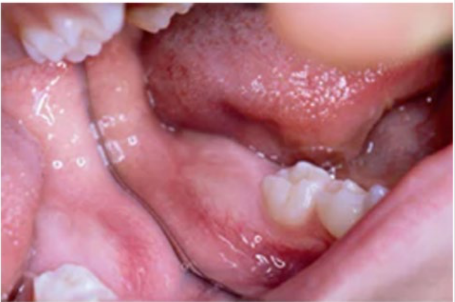
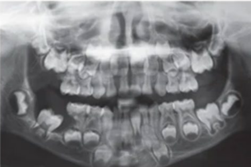
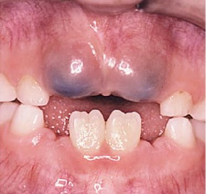
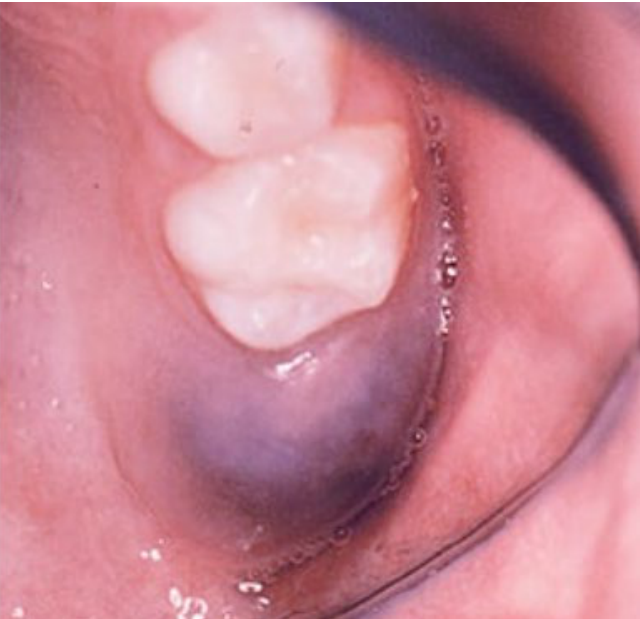
- Dentigerous cyst whose cyst lining becomes continuous with the oral epithelium
- Clinical: fluctuant bluish swelling overlying an unerupted tooth
- Usually self-resolving; may require marsupialization
- Reassure parents because the lesion usually resolves as the tooth erupts; consider marsupialisation if it is very large or eruption is significantly delayed.
 |  |
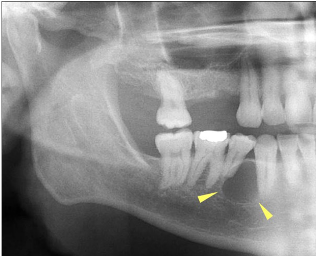
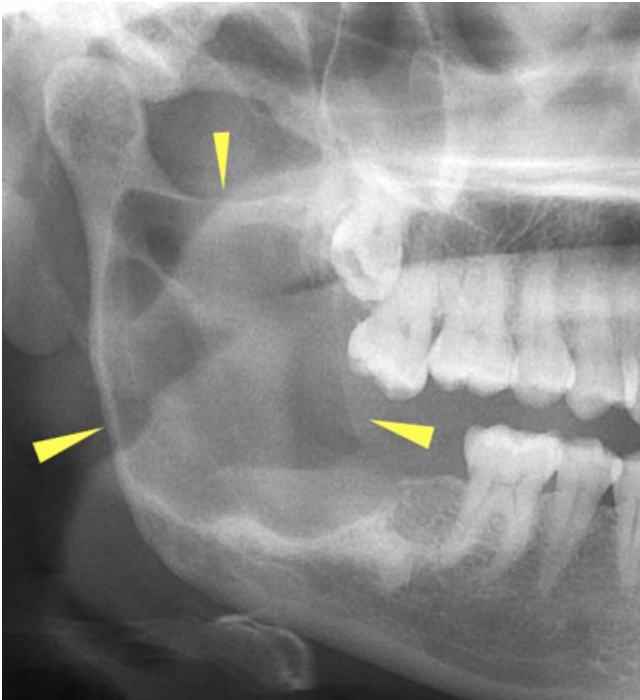
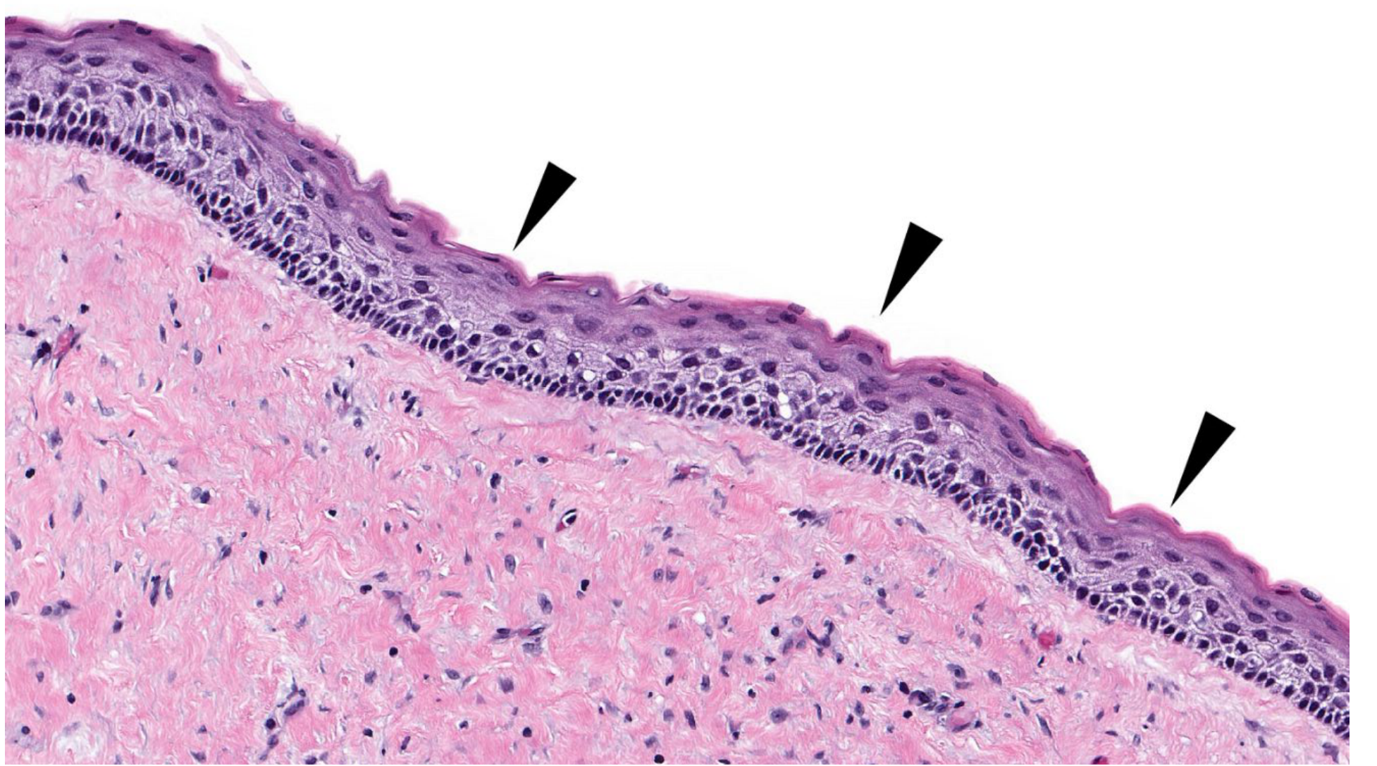
Keratocystic Odontogenic Tumour464748
- Reclassified by WHO in 2005 as a true neoplasm
- Previously known as odontogenic keratocyst
- Locally aggressive, developmental odontogenic origin
- Aetiology: dental lamina; involvement of sonic hedgehog signaling pathways in many cases; syndromic cases (Nevoid basal cell carcinoma syndrome)
Gorlin Syndrome
-
Multiple, early-onset BCCs
-
Calcification of the faux cerebri
-
Hyper-telorism & broad nasal root
-
Odontogenic keratocysts
-
Palmar and plantar pits
-
Bifid ribs
-
Site: 70% occur in mandible, 50% in angle/ramus
-
Clinical: may be small and asymptomatic, incidentally detected on radiographs
- Large lesions produce a painless bony expansion which can displace teeth
- Risk of pathological fracture
-
Radiographic:
- Unilocular or multilocular radiolucency with smooth or scalloped margins
- Cancellous enlargement anterio-posteriorly
-
Treatment: surgical enucleation ± curettage
- Budding of satellite cysts from the basal layer = high risk of reoccurrence
Lecturer — Keratocystic Tumour
Recurrence rates may be as high as 60% because satellite cysts can bud from the primary cyst.
- Large lesions may cause cortical perforation.
- Histologically, the lesion has a parakeratinised stratified squamous epithelial lining with a relatively flat epithelial–mesenchymal junction.
 |  |
 |
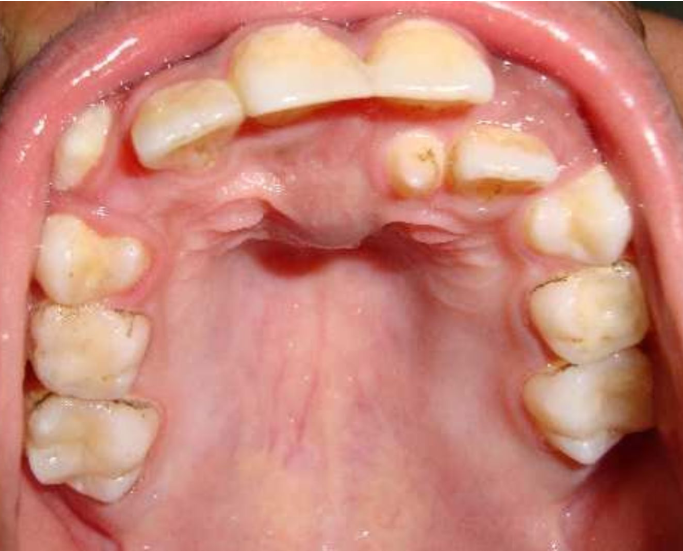
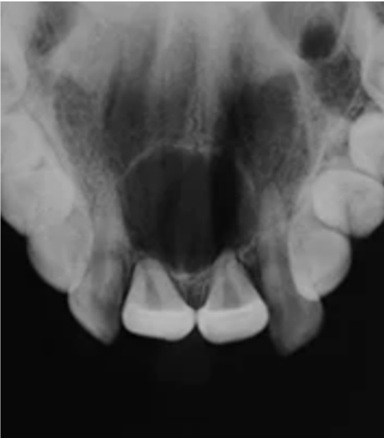
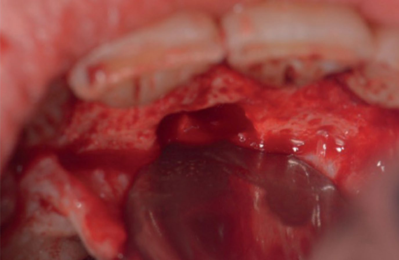
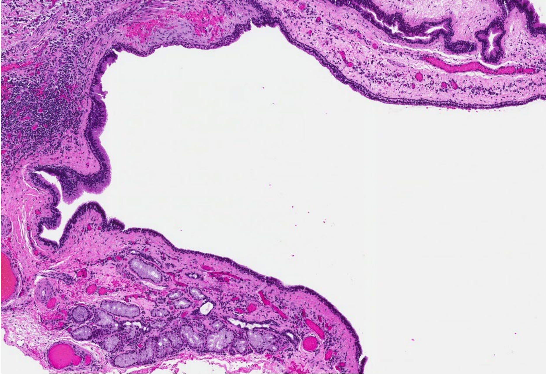
Nasopalatine Cyst49505152
-
Also called incisive cyst
-
Benign, non-odontogenic cyst located in the midline of the anterior maxilla
-
Aetiology: spontaneous proliferation of remnants of nasopalatine duct within the incisive canal
-
Clinical: often asymptomatic, incidental findings on radiograph
- Midline swelling in the hard palate
- May be painful with exudate
- Incisors are vital
-
Radiographic: well circumscribed, rounded or heart-shaped radiolucency of the anterior maxilla
-
Treatment: surgical enucleation
- Recurrence risk is low
-
Prognosis: excellent
Lecturer — Nasopalatine Cyst Details
The radiolucency is centred on the nasopalatine canal and may lie between the roots of the central incisors; root resorption can occur.
- The cyst wall contains fibrous tissue with nerves, arteries and veins.
- Histology may show stratified squamous, pseudostratified columnar, simple columnar or simple cuboidal epithelium.
- Follow-up is recommended to confirm complete resolution.
Tumors of the Jaws53
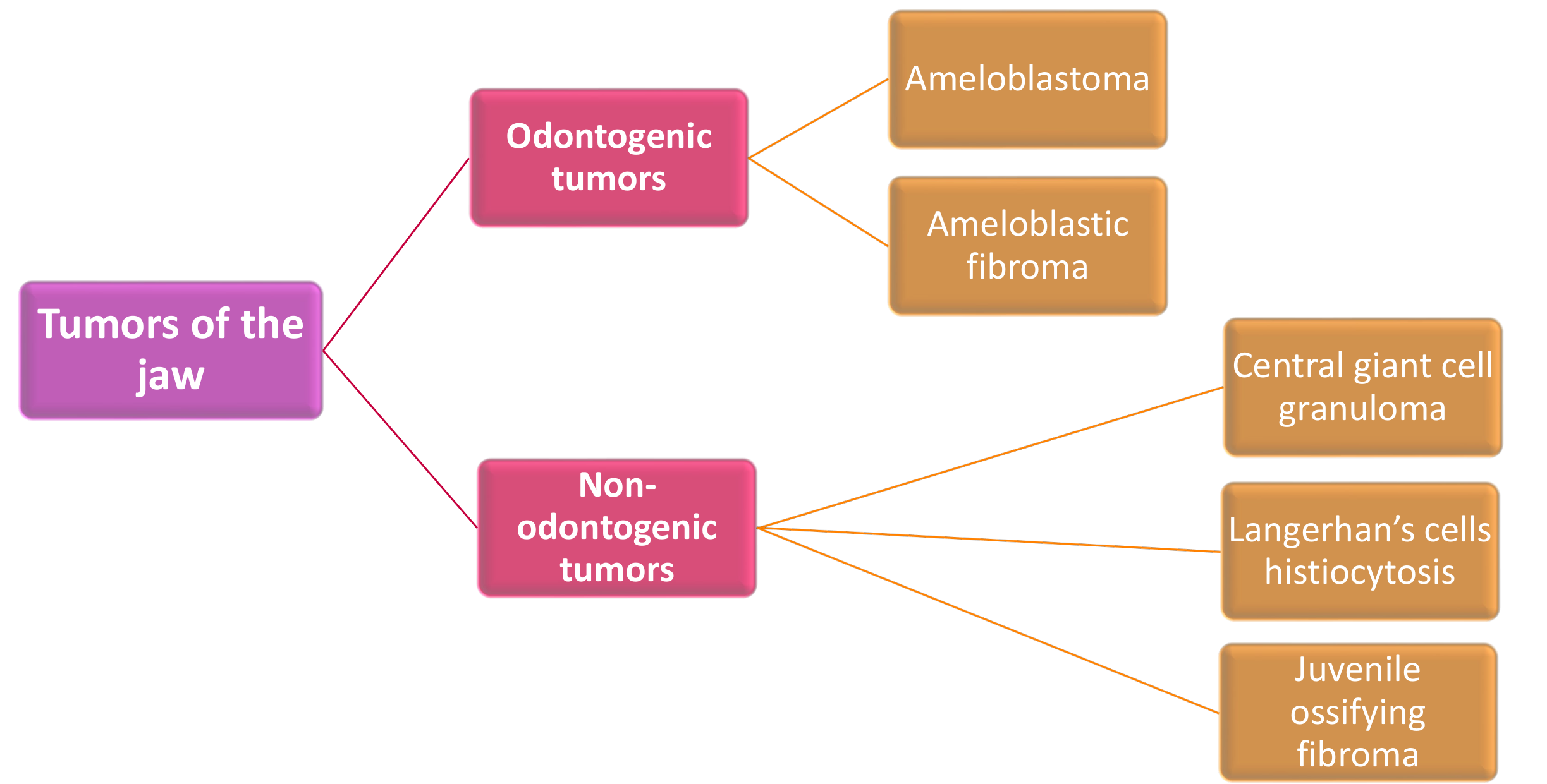
Tumors of the jaw
- Odontogenic tumors
- Ameloblastoma
- Ameloblastic fibroma
- Non-odontogenic tumors
- Central giant cell granuloma
- Langerhan’s cells histiocytosis
- Juvenile ossifying fibroma
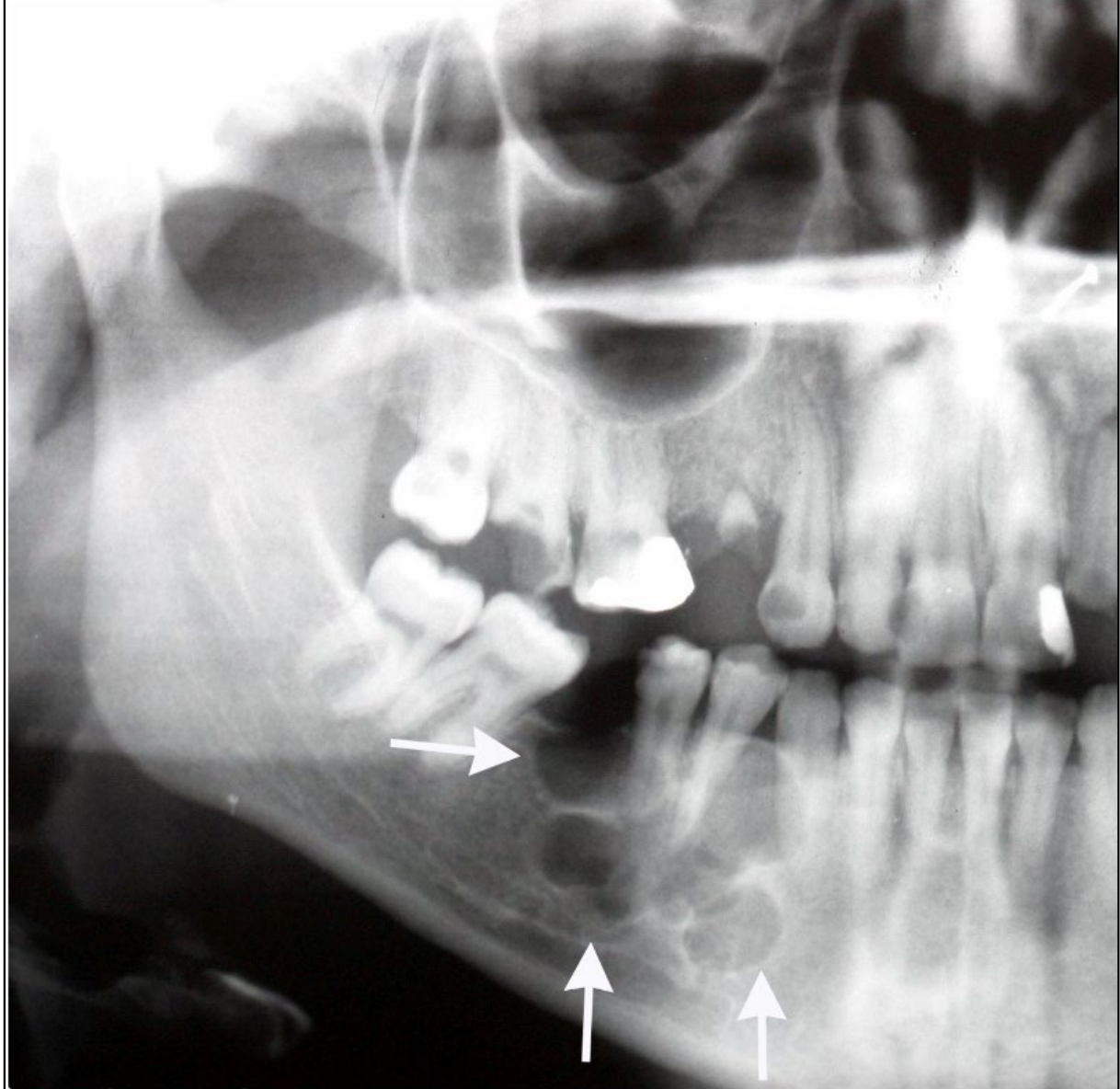
Ameloblastoma545556
-
Benign but locally aggressive tumor
-
More common after age 40
-
Aetiology: Epithelial remnants of the dental lamina or enamel organ
-
Clinical: Painless, slow-growing swelling of the jaw
- Displacement of teeth, loose teeth, malocclusion
- Large tumours — facial deformity, restricted mouth opening, airway obstruction
-
Radiology:
- Expansile, multi-locular radiolucency with well-defined corticated border
- May see classic soap bubble appearance
- Resorption or displacement of tooth roots
-
Treatment: resection with uninvolved margins
- Loss of bone and teeth
- High rate of recurrence
Lecturer — Ameloblastoma Management
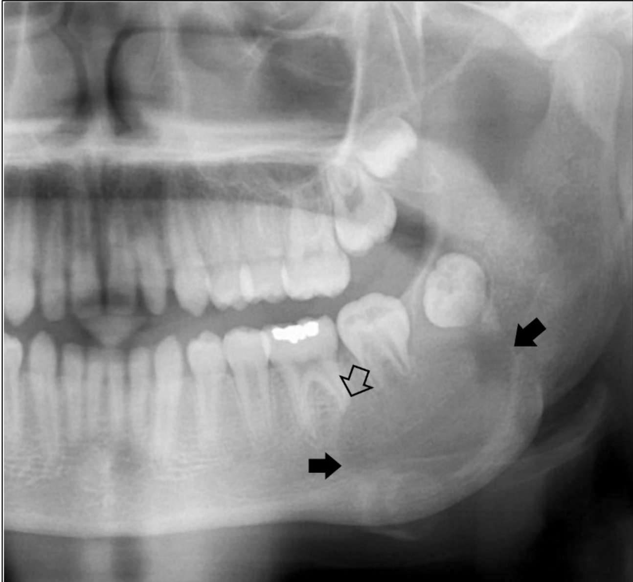
Unicystic lesions may appear unilocular on radiographs.
- Involved teeth may need to be extracted.
- Long-term follow-up is required because recurrence is common.
 |  |
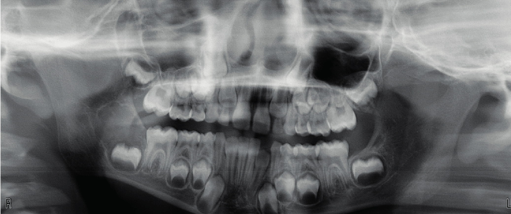
Ameloblastic Fibroma575859
-
True mixed odontogenic neoplasm containing both epithelial and mesenchymal tissues
- Dentine in the specimen = ameloblastic fibro-dentinoma (tends to occur in young children)
-
Slow growing, benign
- Less aggressive than ameloblastoma
-
Usually <20 years
-
Majority of cases occur in mandible
-
Radiographic: well-defined, corticated unilocular or multilocular radiolucency
-
Treatment: complete removal but more conservatively than with ameloblastoma
Lecturer — Ameloblastic Fibroma Details
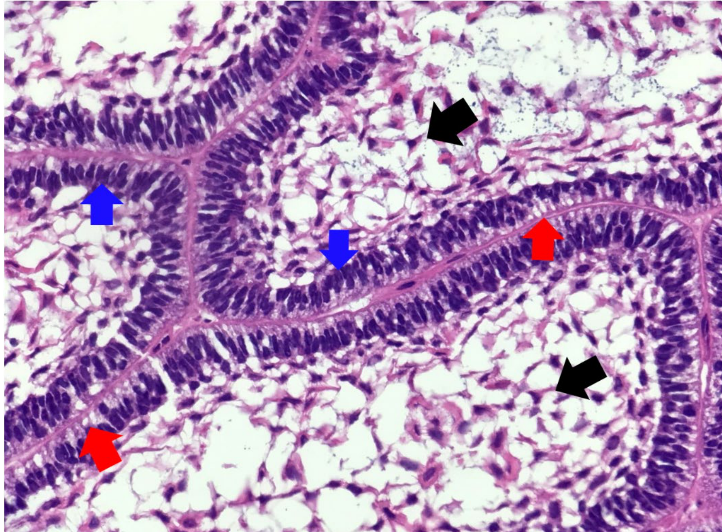
More recent reports suggest that ameloblastic fibroma may have potential for recurrence and malignant transformation.
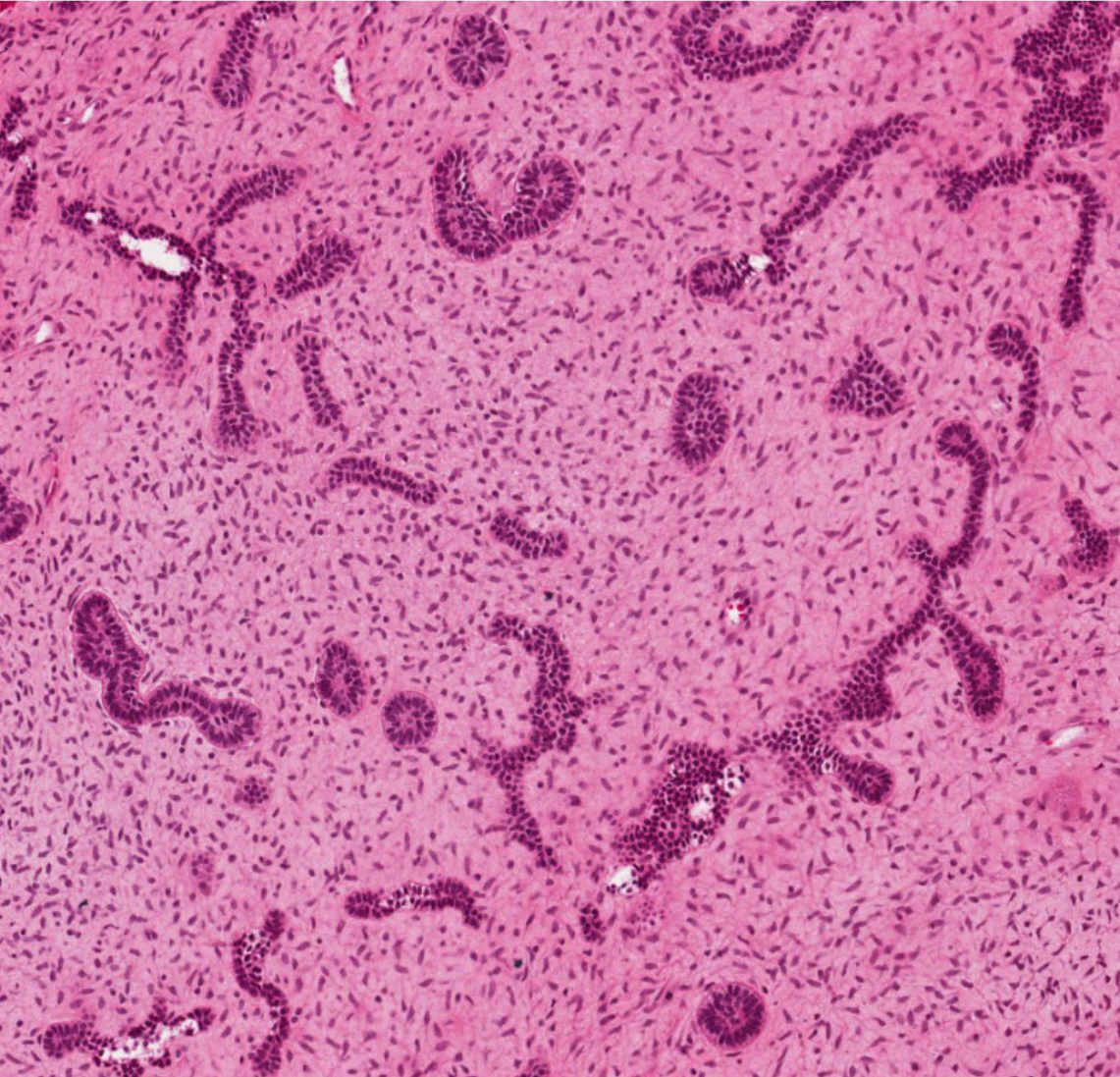
- Histology may show islands and cords of ameloblastic epithelium in immature or dense collagenous stroma.
- Other features include hyperchromatic columnar basal cells, peripheral palisading, reverse polarisation, subnuclear vacuolisation and stellate-reticulum-like suprabasal cells.
 |  |
 |
Central Giant Cell Granuloma606162
-
Langerhans’ cell histiocytosis
-
Juvenile ossifying fibroma
-
Non-neoplastic non-odontogenic reactionary lesion
-
Site: 70% anterior mandible
-
F>M, majority <30 years of age
-
Clinical:
- Slowly growing asymptomatic swelling
- Rapid growth, pain, resorption of teeth, cortical bone perforation
-
Aetiology: local trauma, repair processes, inflammatory lesions
-
Treatment:
- Surgical curettage, intra-lesional steroids, calcitonin, INFα2a, resection
- High recurrence rate in paediatric patients — 41%
Diab et al 2022
Lecturer — Giant Cell Granuloma Details
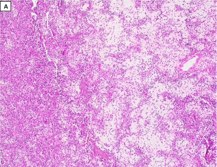
The lesion consists of cellular fibrous tissue containing haemorrhage, aggregates of multinucleated giant cells and occasionally woven bone.
- Proposed associations include developmental disorders, cherubism, Noonan syndrome and neurofibromatosis type 1.
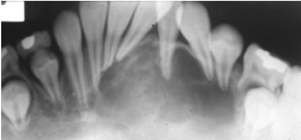
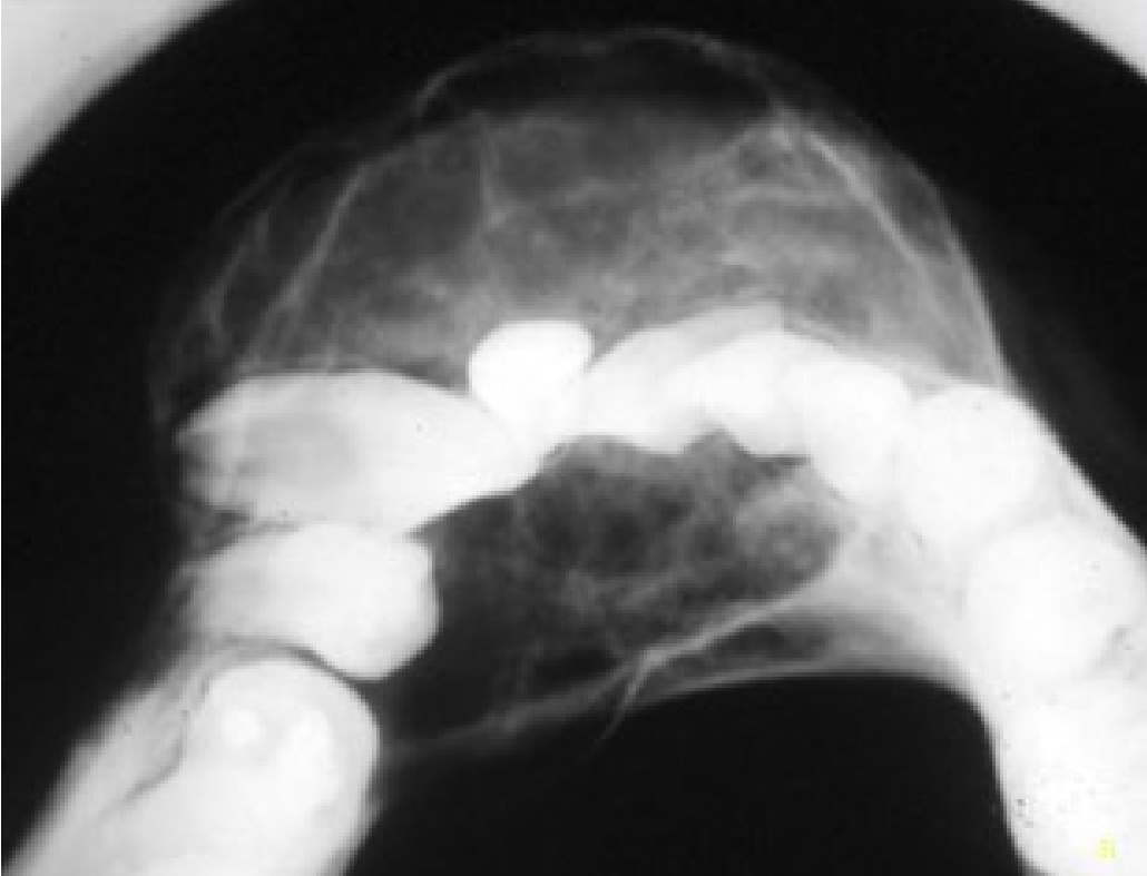
- Radiographs may show a well-defined, variably corticated unilocular or multilocular radiolucency with tooth displacement or occasional root resorption.
- Histology shows multinucleated giant cells within spindle-cell fibroblastic tissue, with haemorrhage and haemosiderin deposits.
 |  |
 |  |
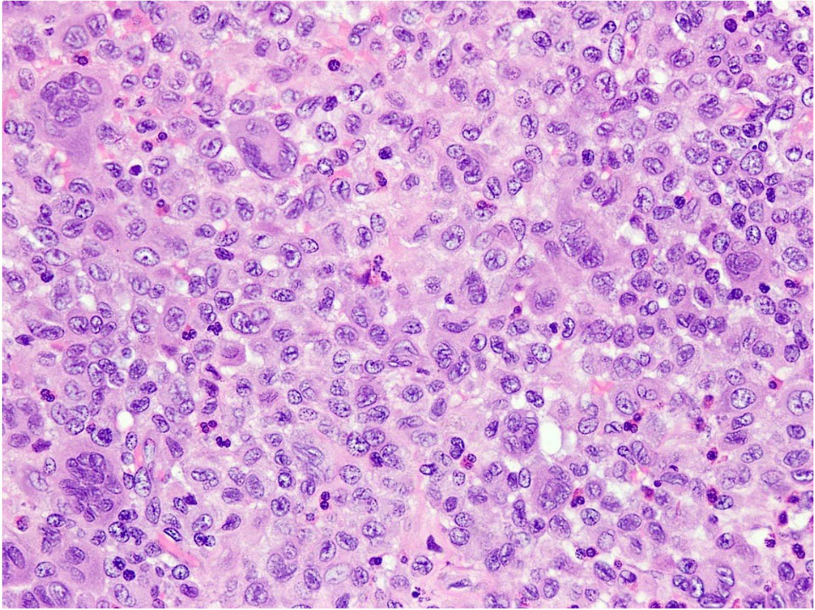
Langerhans Cell Histiocytosis636465666768
-
Reactive condition involving the proliferation of histiocytes involving almost any organ
-
Children and adolescents > adults
-
Presentation:
- Solitary or multiple painful, well-defined and often ulcerated lesions in the mouth — gingiva, buccal mucosa, tongue
- Typically, all four quadrants are affected
- Lesions typically extend forward to the canines, but rarely involve the incisors
- Early mobility and loss of primary teeth
- Malaise
- Skin manifestations — anogenital and postauricular rash, cradle cap-like rash
- Diabetes insipidus, growth hormone deficiency, thyroid dysfunction (pituitary gland involvement)
- Solitary or multiple painful, well-defined and often ulcerated lesions in the mouth — gingiva, buccal mucosa, tongue
-
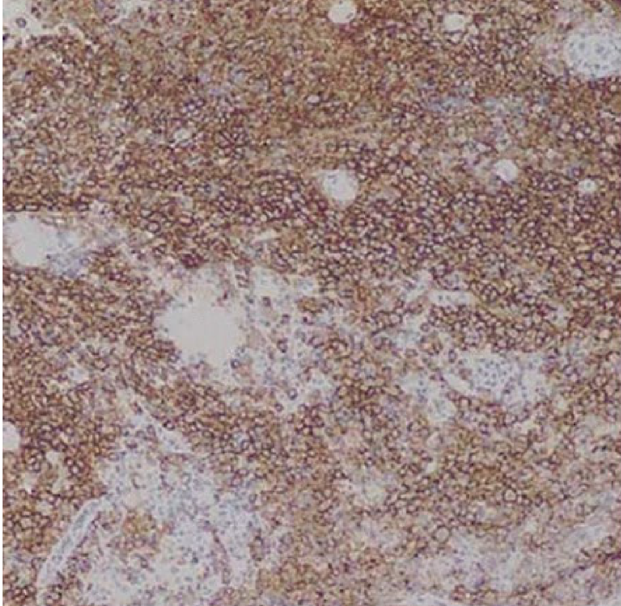
Diagnosis — biopsy of lesions — cells positive for CD1a
-
Treatment: excision and curettage of oral lesions and extraction of involved teeth
- Multiagent chemotherapy is required for disseminated disease
Lecturer — Langerhans Cell Findings
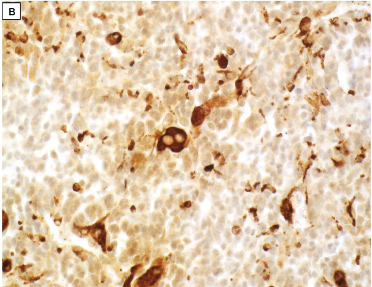
Langerhans cells are dendritic cells derived from bone marrow cells and have coffee-bean-shaped or folded nuclei with abundant cytoplasm.
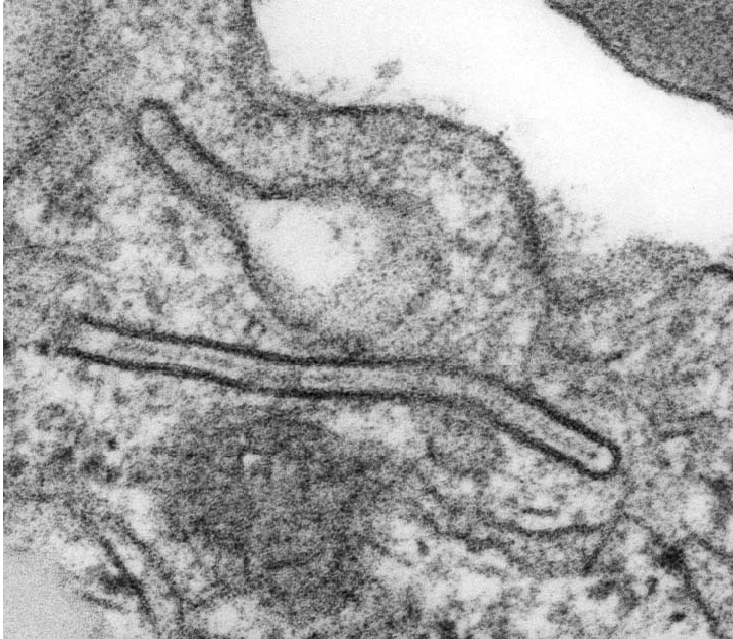
- The cells stain positively for CD1a and S100 protein, and may contain Birbeck granules with a tennis-racket appearance on electron microscopy.
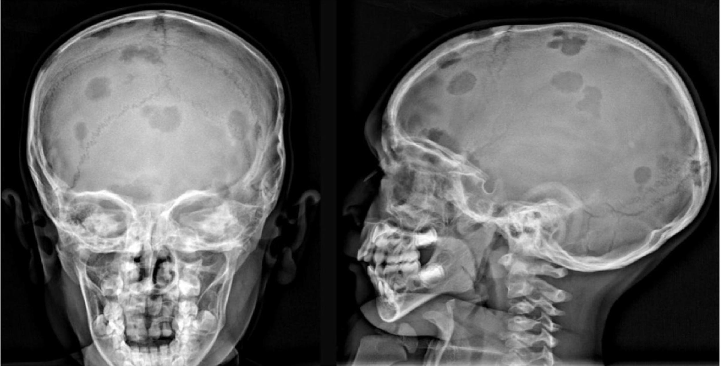
- Radiographs may show punched-out lytic lesions, loss of alveolar bone and teeth that appear to float in the air.
- Involvement of the hypothalamic–pituitary axis may cause diabetes insipidus, while other possible features include increased intracranial pressure, visual disturbances, cranial nerve palsies and seizures.
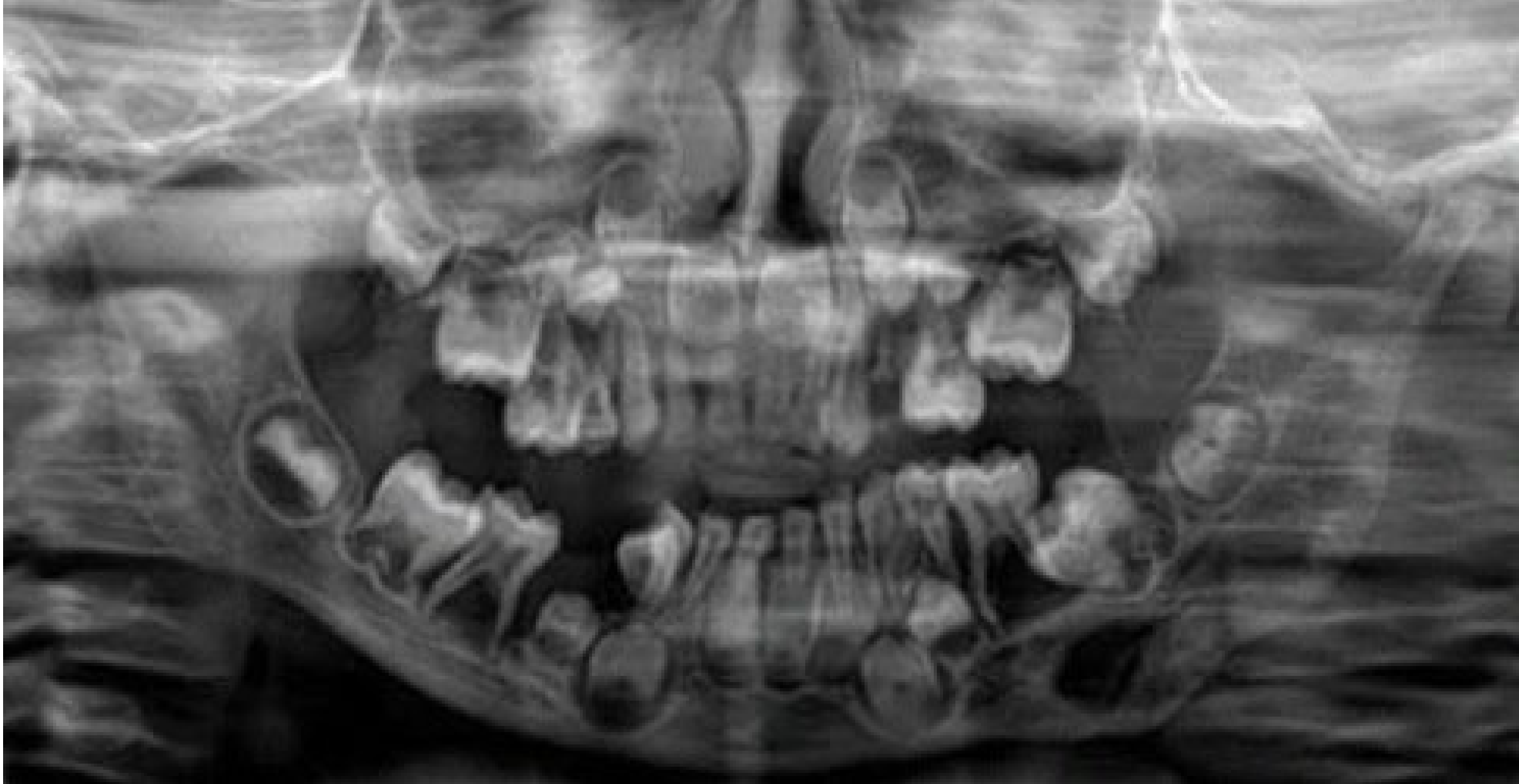
Juvenile Ossifying Fibroma6970717273
-
Rare variant of ossifying fibroma, a benign fibro-osseous lesion
-
Clinical: slow-growing, painless swelling or mass in the jaw
- Facial asymmetry or displacement of teeth
- Functional issues such as difficulty in chewing or speaking
-
Locally aggressive behaviour
-
Primarily affects children and adolescents
-
Trabecular variant — maxilla and mandible
-
Psammomatoid variant — paranasal sinuses
-
Radiographic: well-defined, radiolucent area with varying degrees of radiopacity due to the mixed bone and fibrous tissue composition
- May have corticated border and show expansion of the cortical bone
-
Treatment: Surgical excision
- High recurrence rate
Juvenile Ossifying Fibroma Case
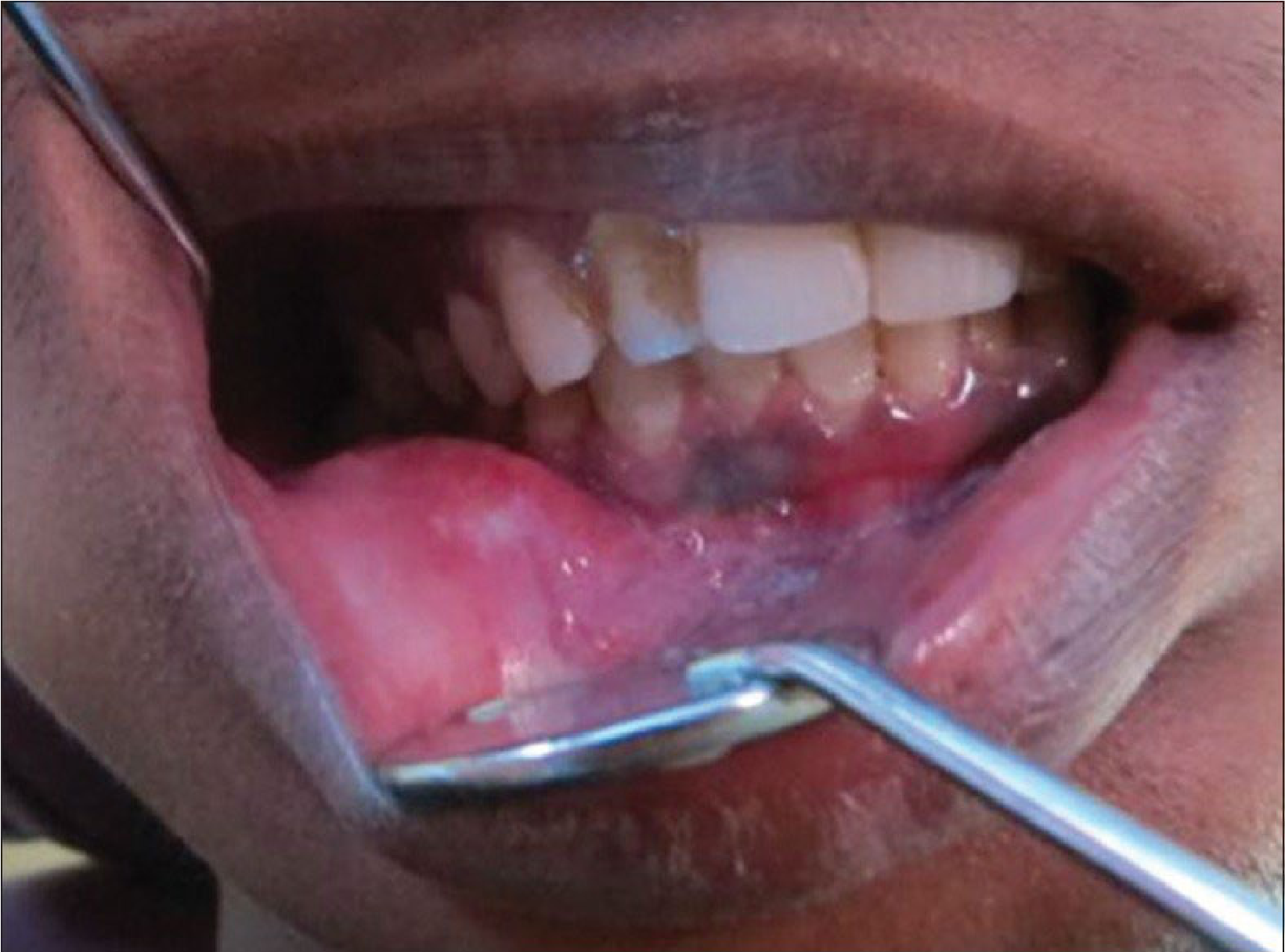
A 20-year-old male presented with a one-year history of swelling in the right lower jaw and loose, mobile teeth. There was diffuse, hard, non-tender swelling of the right lower face without increased local temperature or clinical lymphadenopathy, while intraoral examination showed bony alveolar expansion and obliteration of the gingivobuccal sulcus.
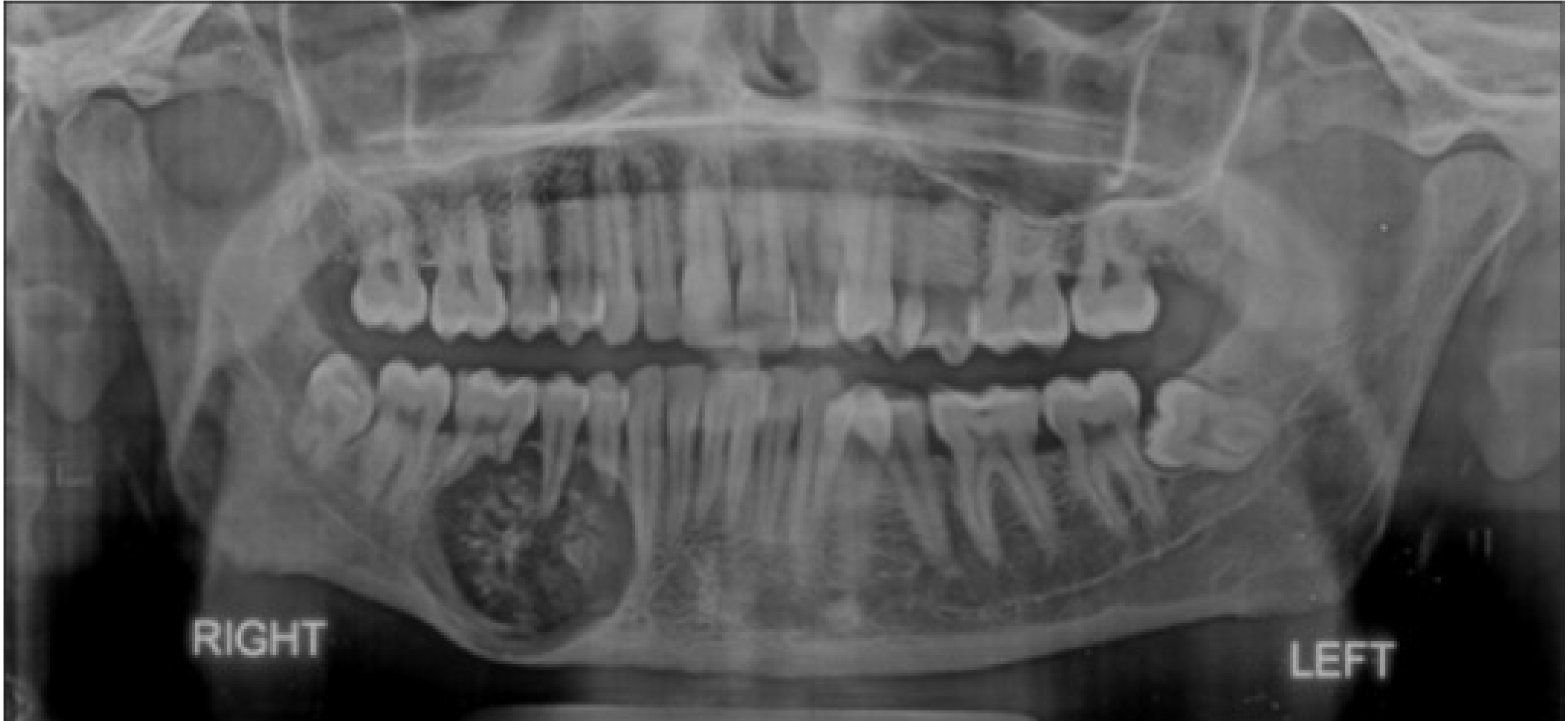
- An OPG three months earlier showed a mixed radiopaque-radiolucent lesion with root resorption involving the premolars and first molar.
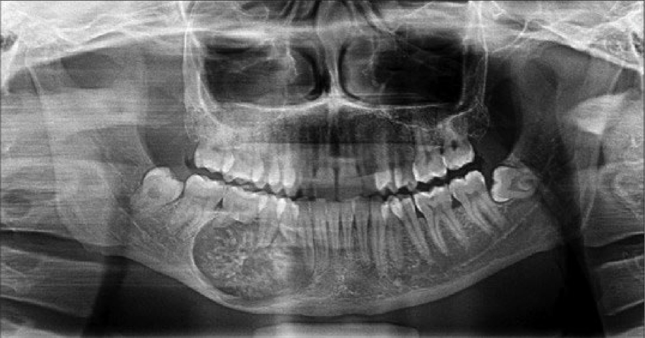
- A second OPG showed enlargement extending to the canine and second molar, with root resorption in the associated teeth.
- The increase in size over three months demonstrated the aggressive nature of the lesion.
RIGHT LEFT
Fibro-Osseous Lesions74
Fibro-osseous lesions comprise two main entities:
- Cherubism
- Fibrous dysplasia
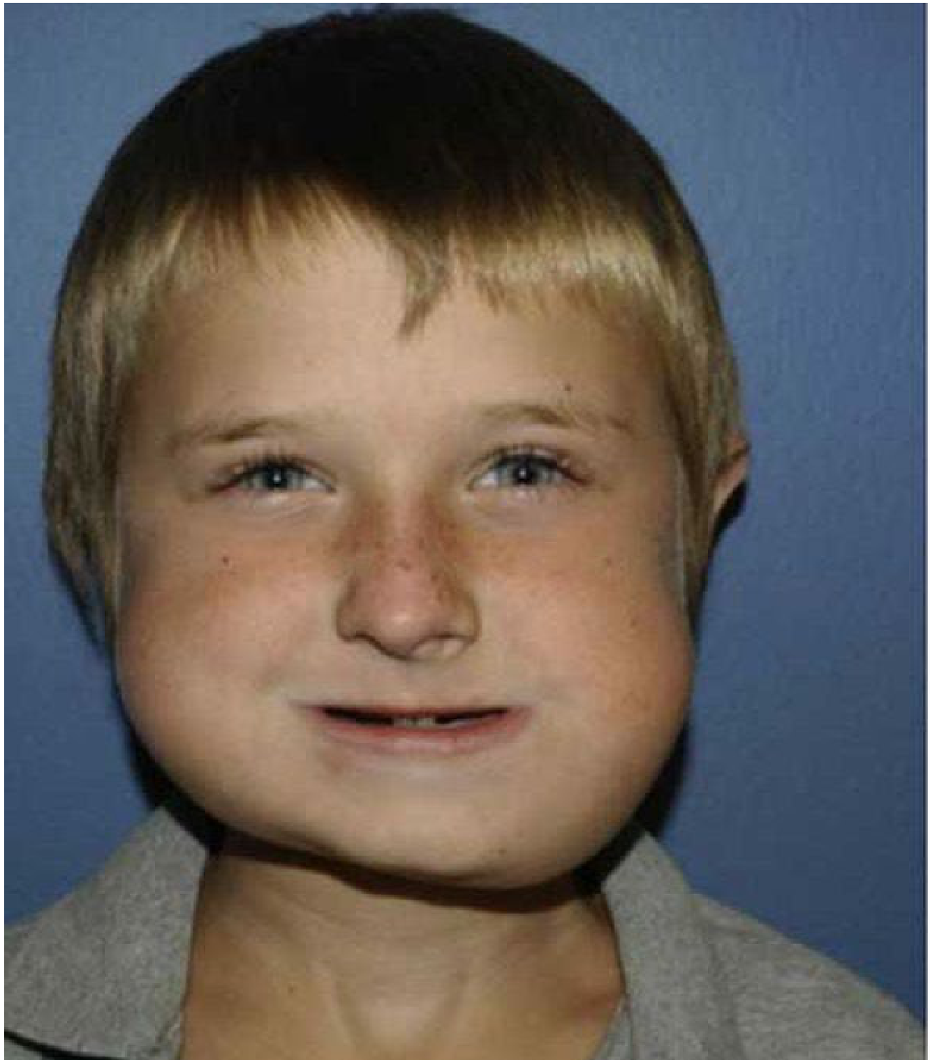
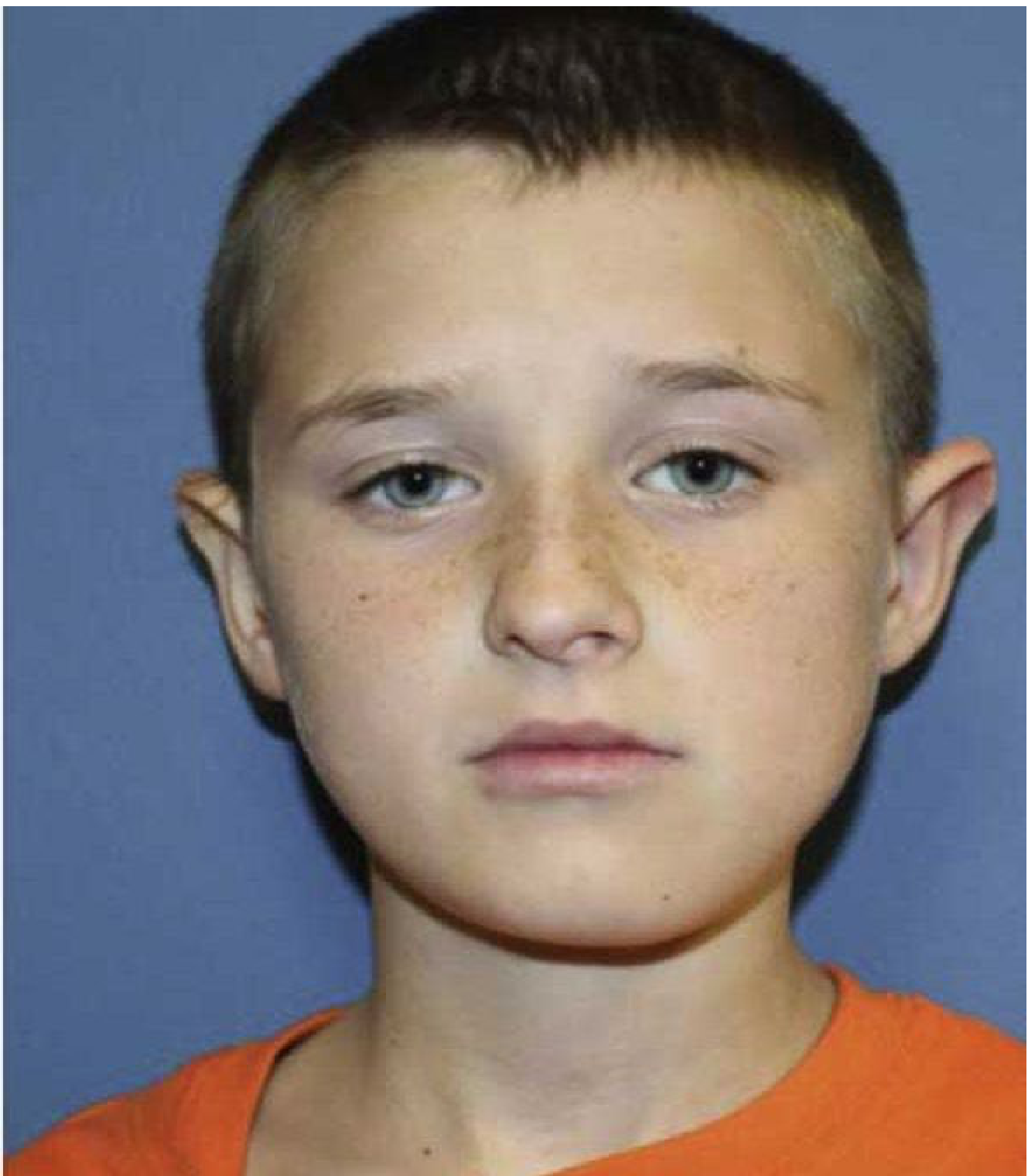
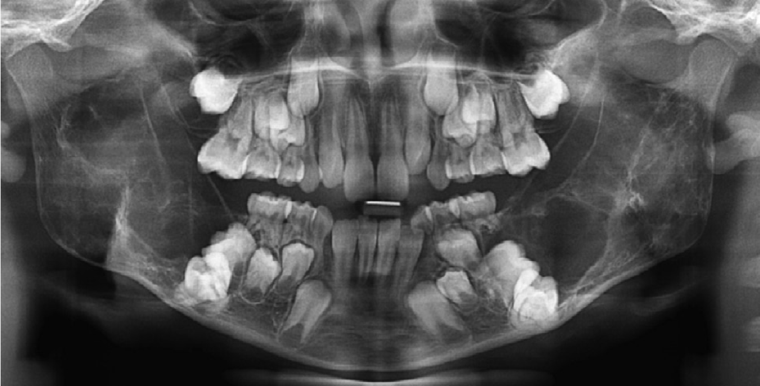
Cherubism757677
- Hereditary and intraosseous fibrous swellings of the jaws
- Autosomal dominant condition
 |  |
 |
Clinical Features78
-
Present in childhood
-
Bilateral swelling of the mandible ± maxilla and ribs
-
Failed eruption and displacement of teeth (typically mandibular molars)
-
Radiographic: multilocular radiolucencies, displaced teeth
-
Histology: multinucleate giant cells in a fibrous tissue stroma
-
Prognosis: progresses into adolescence, then tends to resolve so that by the 3rd and 4th decade may no longer see radiographic changes
-
Treatment: regular review (mild) or surgical reduction (more severe)
-
Radiolucencies typically involve the angles of the mandible.
-
The lower sclera may be exposed, creating a cherubic or upward gaze.
-
Mild cases may only be detected on routine radiographs and may not be clinically evident. SEEK WISDOM
Fibrous Dysplasia798081828384
-
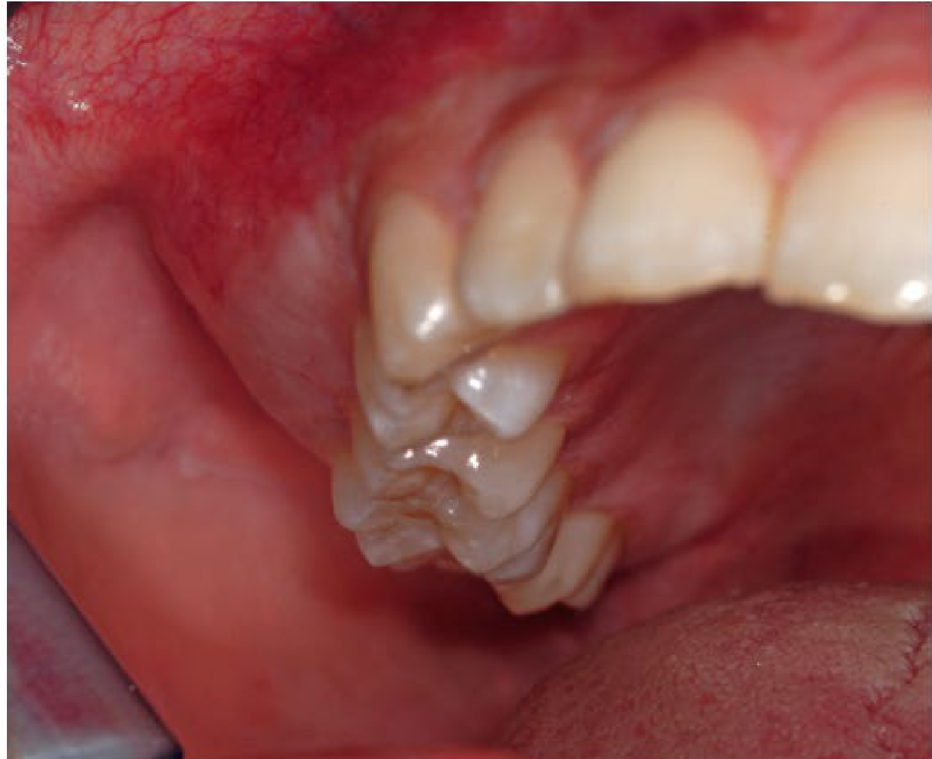
Benign monostotic or polyostotic fibro-osseous lesion
- Polyostotic form more common in childhood
-
Developmental disorder resulting in the failure in bone maturation
- Failure to form mature lamellar bone, arrest as woven bone, fibrous proliferation
-
Mass forming developmental defect composed of woven bone and fibroblast-like spindle cells
-
Clinical: painless slow growing expansion of bone
- Maxilla > mandible
- Facial asymmetry, malocclusion due to displaced teeth
- Headache, hearing loss
- Laboratory test – elevated serum alkaline phosphatase
- May have pain in affected limbs
-
Replacement of medullary bone by fibro-osseous tissue results in structurally weak bone.
-
Radiographically, fibrous dysplasia may appear as a diffuse, poorly delineated lesion with a characteristic ground-glass appearance.
-
Additional radiographic features include cortical expansion, narrow periodontal ligament spaces and an obscured lamina dura.
-
No treatment since growth is self limited and responsive to pubertal hormonal changes
- Surgery in cases with fracture or bony deformity
-
McCune-Albright Syndrome: endocrine anomalies, café au lait spots, fibrous dysplasia
Take Home Messages8586
-
Systematic approach to diagnosis is essential
-
Recognise overlap between soft tissue and hard tissue pathology
-
Early referral and intervention improves prognosis
-
Always consider systemic associations
-
Special care in paediatric patients due to growth and development
-
Use a systematic diagnostic approach for every paediatric oral and dental presentation, distinguishing normal variation from pathology.
-
Consider the history, lesion characteristics, associated symptoms and progression.
-
A missing tooth, altered eruption pathway or unexplained radiolucency requires further investigation.
-
Consider medication effects and bleeding disorders when assessing paediatric oral and dental presentations.
-
Vascular malformations must be considered before extracting a tooth associated with an unexplained radiolucency. Thank you
Clinical Cases
Case: Child with lesions on hands, mouth, and feet
Question
Scenario: A child presents to the dental clinic with a contagious viral illness.
What’s shown: Lesions covering the child’s hands, mouth, and feet.
Consider: What is the diagnosis and what is the appropriate management?
Answer
Observations:
- Lesions covering the hands, mouth, and feet.
Reasoning: The coxsackie virus causes Hand, Foot, and Mouth disease, which presents with vesicles that subsequently ulcerate and resemble oral aphthae. It is highly contagious and spreads easily in daycares and schools.
Outcome: The child was sent home. Management is symptomatic, focusing on maintaining fluid intake and providing pain relief. Parents are advised not to send the child to school, as the condition is self-limiting.
Takeaway: Hand, Foot, and Mouth disease is highly contagious and managed with symptomatic care and infection control advice.
Case: Mandibular conventional ameloblastoma
Question
Scenario: A radiograph of the mandible is presented for evaluation.
What’s shown: A multilocular radiolucency with a specific appearance in the mandible.
Consider: Identify the radiographic features and the type of ameloblastoma.
Answer
Observations:
- Multilocular radiographic pattern
- Classic soap bubble appearance
- Well-defined corticated border
Reasoning: The expansile multilocular radiolucency with a well-defined corticated border and classic soap bubble appearance is characteristic of a conventional ameloblastoma.
Outcome: Treatment involves resection with uninvolved margins, which may cause loss of bone and teeth. Follow-up is needed due to a high rate of recurrence.
Takeaway: Conventional ameloblastoma typically presents as a multilocular radiolucency with a soap bubble appearance and requires aggressive surgical resection.
Case: Mandibular unicystic ameloblastoma
Question
Scenario: A panoramic radiograph of the mandible is presented for evaluation.
What’s shown: A unilocular radiolucency with effects on the adjacent tooth roots.
Consider: Identify the radiographic features and the type of ameloblastoma.
Answer
Observations:
- Unilocular appearance
- Root resorption
Reasoning: The unilocular radiolucency with root resorption in the mandible is indicative of a unicystic type of ameloblastoma.
Takeaway: Unicystic ameloblastoma can present as a unilocular radiolucency with root resorption, distinguishing it from the multilocular conventional type.
Case: Ameloblastic fibroma, panoramic radiograph
Question
Scenario: A panoramic dental X-ray is presented for evaluation.
What’s shown: A well-defined radiolucency in the jaw.
Consider: Identify the radiographic features and the likely diagnosis.
Answer
Observations:
- Well-defined, corticated radiolucency
- Unilocular or multilocular appearance
Reasoning: The well-defined, corticated unilocular or multilocular radiolucency is characteristic of an ameloblastic fibroma.
Outcome: Treated by complete removal, but more conservatively than an ameloblastoma.
Takeaway: Ameloblastic fibroma presents as a well-defined corticated radiolucency and is treated more conservatively than ameloblastoma.
Case: Ameloblastic fibroma and ameloblastoma, histology
Question
Scenario: Histological sections of odontogenic tumors are presented.
What’s shown: Micrographs showing epithelial islands and cords in a primitive stroma, and columnar cells with peripheral palisading.
Consider: Identify the histological features of ameloblastic fibroma and ameloblastoma.
Answer
Observations:
- Small islands and cords of markedly attenuated ameloblastic epithelium in a dense, immature collagenous stroma
- Epithelial islands and cords of conventional ameloblastoma and cystic lining of unicystic ameloblastoma
- Columnar cells with hyperchromatic nuclei, peripheral palisading, reverse polarization, and subnuclear vacuolization
- Suprabasal cells with a loose network-like arrangement (stellate reticulum)
Reasoning: The biphasic tissue pattern with odontogenic epithelium in a primitive stroma indicates ameloblastic fibroma. The columnar cells with reverse polarization and stellate reticulum-like arrangements are characteristic of ameloblastoma.
Takeaway: Ameloblastic fibroma shows a biphasic pattern of odontogenic epithelium in a primitive mesenchymal stroma, while ameloblastoma shows peripheral palisading and reverse polarization.
Case: Central giant cell granuloma, radiograph and histology
Question
Scenario: Radiographs and histological slides of a jaw lesion are presented.
What’s shown: A well-defined radiolucent lesion and micrographs showing multinucleated giant cells in a cellular stroma.
Consider: Identify the radiographic and histological features of the lesion.
Answer
Observations:
- Well-defined radiolucent lesion with varying degrees of cortication, unilocular or multilocular
- Cellular stroma with numerous multinucleated giant cells, hemosiderin deposits, and spindle-shaped fibroblasts
- CD68 immunostain highlighting multinucleated giant cells
Reasoning: The well-defined radiolucency combined with a cellular stroma containing multinucleated giant cells and hemosiderin deposits is diagnostic of a central giant cell granuloma.
Outcome: Aggressive lesions are treated with wider resection, while non-aggressive forms may use conservative surgery or non-surgical treatments like corticosteroids, interferon, or calcitonin. Recurrence rate is 15-20%, higher in pediatric patients.
Takeaway: Central giant cell granuloma presents as a well-defined radiolucency and histologically shows multinucleated giant cells in a cellular stroma with hemosiderin.
Case: Langerhans cell histiocytosis, clinical presentation
Question
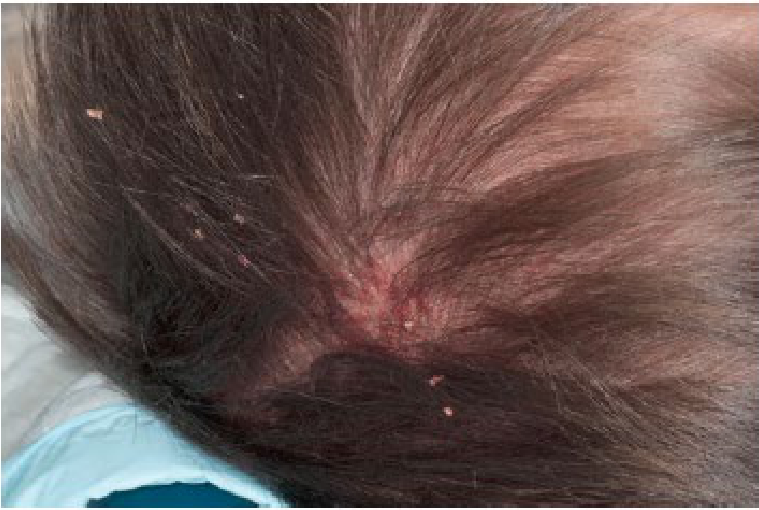
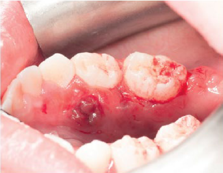
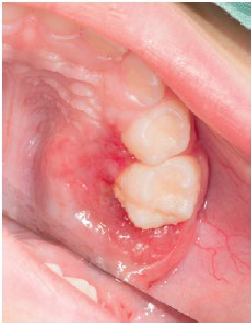
Scenario: Clinical photographs of a patient with scalp and intraoral lesions are presented.
What’s shown: A cradle cap-like rash on the scalp and intraoral lesions in all four quadrants with mobile teeth and lingual alveolar perforation.
Consider: Identify the clinical features and the diagnosis.
Answer
Observations:
- Cradle cap-like rash on the head
- Intraoral lesions in all four quadrants
- Excessively mobile teeth retained only by soft tissue
- Perforation of the lesion through the lingual alveoli
Reasoning: The combination of a cradle cap-like rash and intraoral lesions with floating teeth and alveolar perforation is characteristic of Langerhans cell histiocytosis.
Outcome: Diagnosed by biopsy (CD1A positive). Treatment includes excision and curettage of oral lesions and extraction of involved teeth. Multi-agent chemotherapy is required for disseminated disease.
Takeaway: Langerhans cell histiocytosis can present with a cradle cap-like rash and intraoral lesions causing tooth mobility and alveolar bone loss.
Case: Langerhans cell histiocytosis, histology and immunohistochemistry
Question
Scenario: Histopathology and immunohistochemistry micrographs are presented.
What’s shown: Large cells with abundant pale cytoplasm and folded nuclei, showing diffuse membranous positivity for a specific antigen.
Consider: Identify the histological and immunohistochemical features.
Answer
Observations:
- Large cells with abundant pale to pink cytoplasm
- Folded nuclei with coffee bean-shaped nuclear groups
- Diffuse membranous positivity for CD1A antigen on immunohistochemistry
Reasoning: The presence of cells with coffee bean-shaped nuclei and CD1A positivity is diagnostic of Langerhans cell histiocytosis.
Takeaway: Langerhans cell histiocytosis is characterized histologically by cells with coffee bean-shaped nuclei and positive staining for CD1A.
Case: Langerhans cell histiocytosis, electron microscopy
Question
Scenario: An electron micrograph is presented.
What’s shown: Characteristic intracytoplasmic granules with a specific shape.
Consider: Identify the ultrastructural feature.
Answer
Observations:
- Tennis racket-shaped granules (Birbeck granules)
Reasoning: The presence of Birbeck granules on transmission electron microscopy is a characteristic ultrastructural feature of Langerhans cell histiocytosis.
Takeaway: Birbeck granules, which are tennis racket-shaped, are a characteristic ultrastructural finding in Langerhans cell histiocytosis.
Case: Langerhans cell histiocytosis, skull and sternum radiograph
Question
Scenario: A composite radiographic image is presented.
What’s shown: Punched-out lytic lesions involving flat bones.
Consider: Identify the radiographic features.
Answer
Observations:
- Punched-out lytic lesions
- Involvement of flat bones such as the skull and sternum
Reasoning: Punched-out lytic lesions in flat bones are a classic radiographic presentation of Langerhans cell histiocytosis.
Takeaway: Langerhans cell histiocytosis typically presents radiographically as punched-out lytic lesions in flat bones like the skull and sternum.
Case: Langerhans cell histiocytosis, jaw radiograph
Question
Scenario: A panoramic dental X-ray is presented.
What’s shown: A radiolucent lesion with cortical thinning and teeth appearing to float in air.
Consider: Identify the radiographic features in the jaw.
Answer
Observations:
- Radiolucent lesion with thinning of the cortex
- Loss of alveolar bone
- Teeth appearing to float in the air
Reasoning: The loss of alveolar bone causing teeth to appear as if they are floating in air is a classic radiographic sign of Langerhans cell histiocytosis in the jaw.
Takeaway: In the jaw, Langerhans cell histiocytosis causes alveolar bone loss resulting in teeth appearing to float in air on a radiograph.
Case: Right lower jaw swelling in a 20-year-old male
Question
Scenario: A 20-year-old male presents with a one-year history of swelling in the right lower jaw and loose, mobile teeth in the same region.
What’s shown: Clinical photographs and panoramic radiographs (OPGs) taken three months apart.
Consider: Evaluate the clinical and radiographic findings to determine the nature and behavior of the lesion.
Answer
Observations:
- Extraoral examination reveals a diffuse, hard, non-tender swelling over the right lower third of the face with no localized rise in temperature and no clinical lymphadenopathy.
- Intraorally, there is bony expansion of the alveolus with obliteration of the gingival buccal sulcus.
- The OPG taken three months prior shows a mixed radiopaque-radiolucent lesion involving the premolars and first molars, with resorption of their roots.
- The OPG at presentation shows an increase in the size of the lesion, now involving the canine and second molar, with root resorption of the associated teeth.
Reasoning: The rapid increase in size over three months, mixed radiographic appearance, and aggressive root resorption of multiple teeth are characteristic of a locally aggressive benign fibro-osseous lesion, specifically the juvenile variant of ossifying fibroma.
Outcome: The lesion demonstrates aggressive behavior requiring recognition of its high recurrence rate and need for surgical excision.
Takeaway: Juvenile ossifying fibroma is a locally aggressive lesion that can cause rapid expansion and significant root resorption, distinguishing it from the conventional type.
Case: Bilateral mandibular radiolucencies in an 8-year-old boy
Question
Scenario: An 8-year-old boy is evaluated for a condition affecting his jaws.
What’s shown: A panoramic dental radiograph (OPG) showing the maxilla and mandible.
Consider: Identify the radiographic features and their clinical implications for this patient.
Answer
Observations:
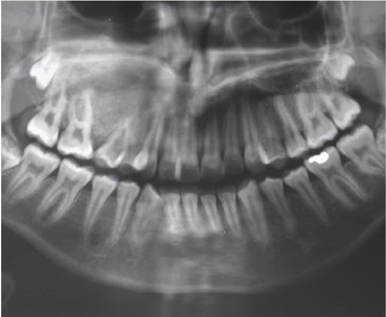
- The OPG shows almost symmetrical multilocular radiolucencies in the angles of the mandible.
- There is displacement of the developing molars and delayed eruption of teeth.
Reasoning: The symmetrical multilocular radiolucencies in the mandibular angles, combined with displaced and delayed tooth eruption, are classic radiographic signs of cherubism, a hereditary intraosseous fibrous swelling.
Outcome: Treatment involves regular review for mild cases or surgical reduction for more severe cases. The condition typically progresses into adolescence and then tends to resolve by the 3rd or 4th decade.
Takeaway: Cherubism presents with bilateral symmetrical multilocular radiolucencies in the jaw angles and displaced teeth, and is generally self-resolving after puberty.
Case: Painless maxillary swelling in a 16-year-old boy
Question
Scenario: A 16-year-old boy presents because he is unable to fit his mouthguard due to a painless swelling in the maxilla.
What’s shown: An intraoral clinical photograph and a panoramic dental radiograph.
Consider: Identify the radiographic appearance and key features of the lesion.
Answer
Observations:
- The radiograph shows a characteristic ground glass appearance.
- The lesions are diffuse and poorly delineated.
- There is cortical expansion, narrow periodontal ligament (PDL) spaces, and obscure lamina dura.
Reasoning: The painless maxillary swelling with a diffuse, poorly delineated ground glass appearance, narrow PDL spaces, and obscure lamina dura is the classic presentation of fibrous dysplasia.
Outcome: No treatment is typically needed as the growth is self-limiting and responsive to pubertal hormonal changes, though surgery may be required if fractures or bony deformities occur.
Takeaway: Fibrous dysplasia presents with a ground glass appearance and narrow PDL spaces, and is a self-limiting developmental defect that stabilizes after puberty.
Case: Uncontrolled hemorrhage after tooth extraction in an 11-year-old girl
Question
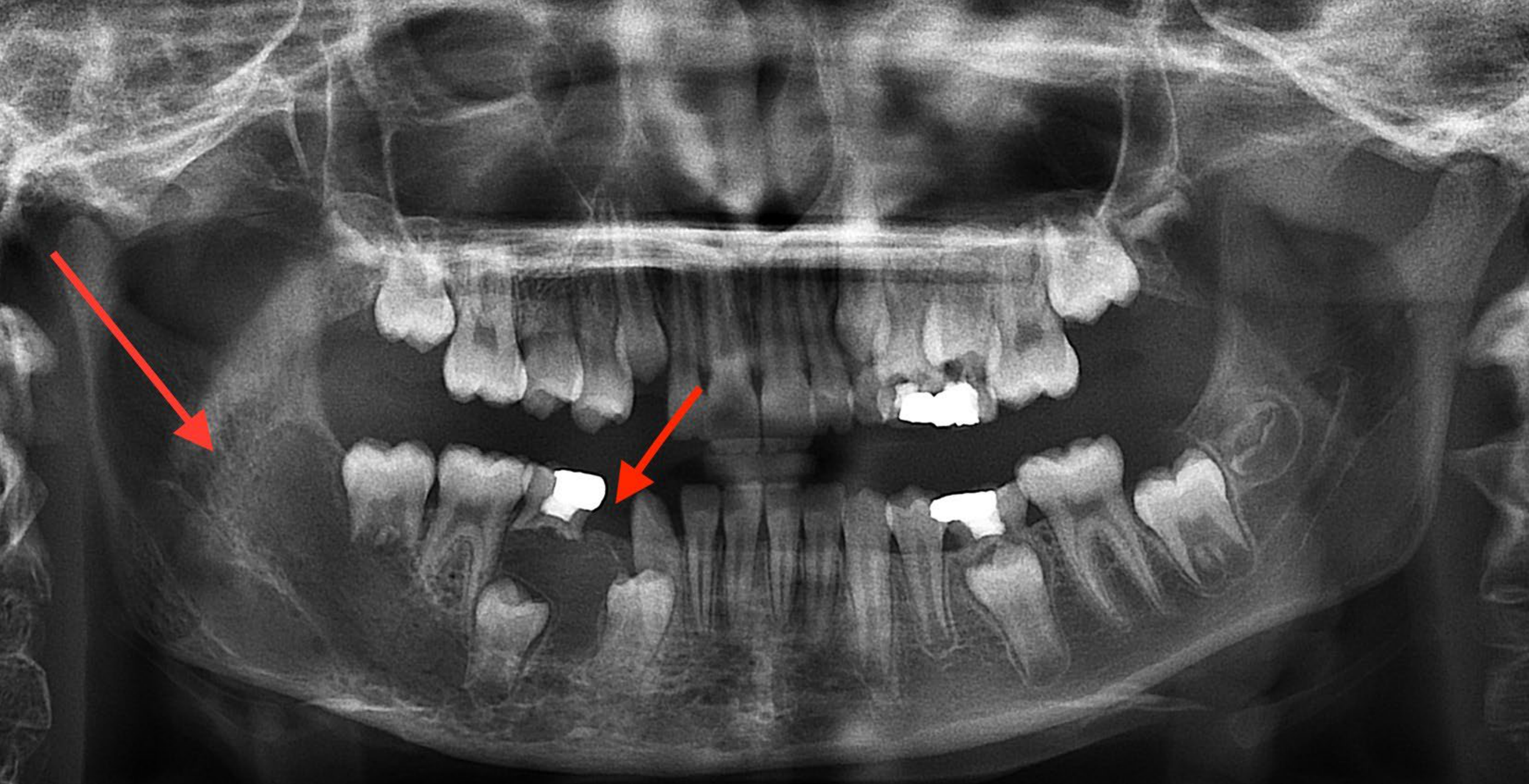
Scenario: An 11-year-old girl with no past medical or family history of bleeding is brought to the emergency room for uncontrolled hemorrhage and recurrent oral bleeding following the extraction of tooth 84, which was performed without prior radiographs.
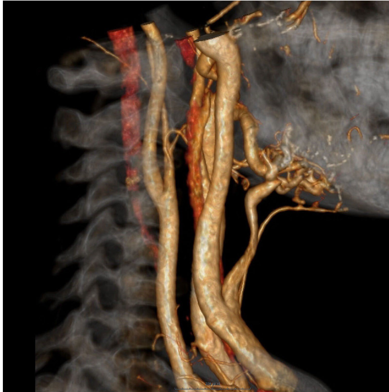
What’s shown: A 3D volume-rendered CT angiogram of the cervical region.
Consider: Identify the cause of the hemorrhage, the management steps taken, and the lesson for future practice.
Answer
Observations:
- The patient presented with low blood pressure, tachycardia, and pulsatile blood flow from the extraction socket that did not stop with continuous pressure or tranexamic acid.
- Intraoral bleeding was temporarily controlled with a silicone compression tray, and the child was stabilized with a blood transfusion.
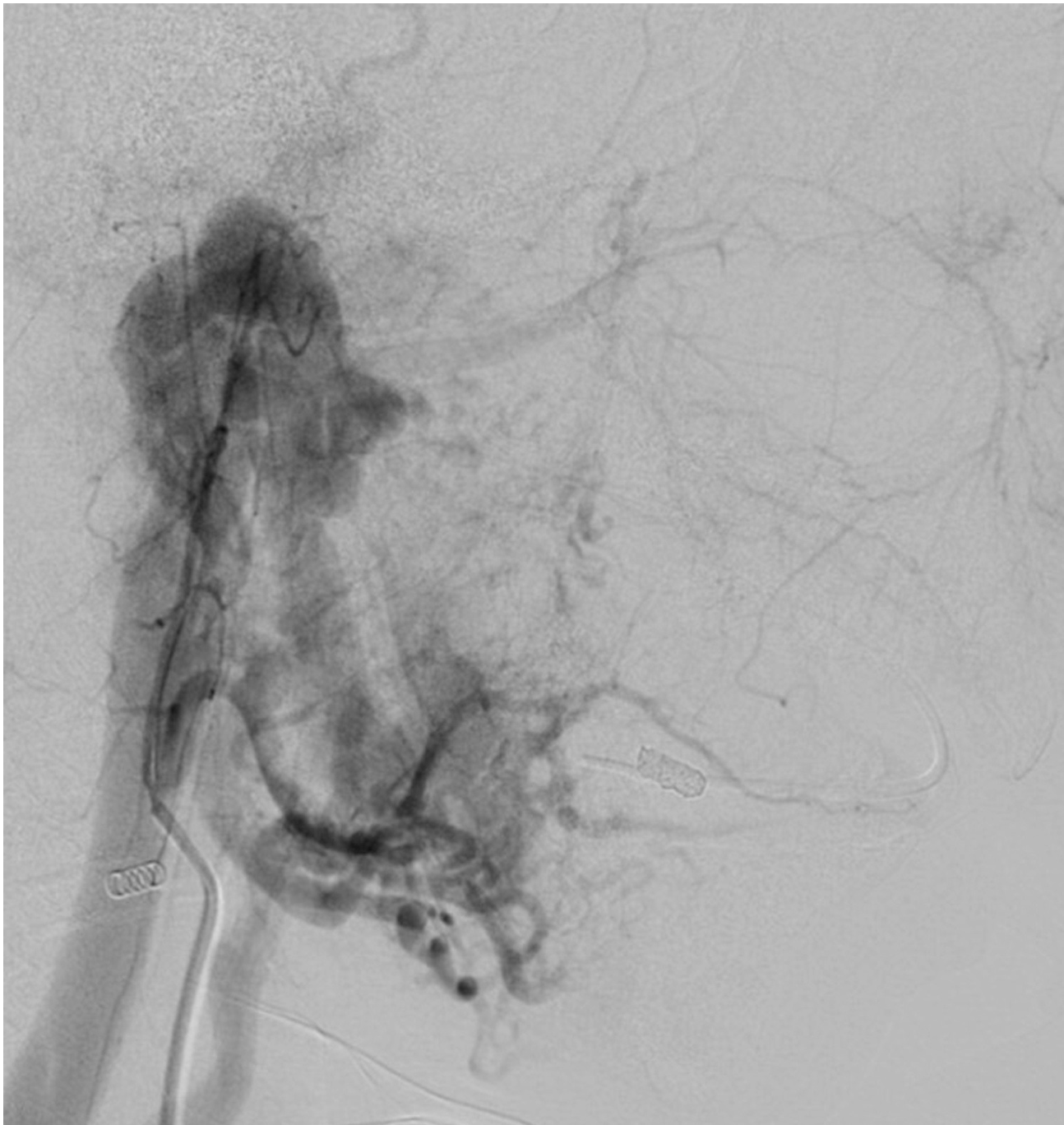
- The CT angiogram reveals a large arteriovenous malformation fed by a branch of the right carotid artery.
Reasoning: The pulsatile, uncontrolled bleeding from an extraction socket in a patient with no bleeding diathesis strongly suggests a vascular anomaly. Imaging confirms a high-flow arteriovenous malformation, a rare anomaly resulting from errors in vascular morphogenesis.
Outcome: Management of the malformation involved artery and venous immobilization to reduce the lesion size.
Takeaway: Always obtain and review radiographs before extractions; if a lucency is observed and there is any doubt about it being a non-odontogenic lesion, further investigation is essential to avoid catastrophic hemorrhage.
Footnotes
-
Original PDF page 1: L10 Oral Med Lesions in Children (Soft and Hard Tissues) 2025 2, p.1 ↩
-
Original PDF page 2: L10 Oral Med Lesions in Children (Soft and Hard Tissues) 2025 2, p.2 ↩
-
Original PDF page 3: L10 Oral Med Lesions in Children (Soft and Hard Tissues) 2025 2, p.3 ↩
-
Original PDF page 4: L10 Oral Med Lesions in Children (Soft and Hard Tissues) 2025 2, p.4 ↩
-
Original PDF page 5: L10 Oral Med Lesions in Children (Soft and Hard Tissues) 2025 2, p.5 ↩
-
Original PDF page 6: L10 Oral Med Lesions in Children (Soft and Hard Tissues) 2025 2, p.6 ↩
-
Original PDF page 7: L10 Oral Med Lesions in Children (Soft and Hard Tissues) 2025 2, p.7 ↩
-
Original PDF page 8: L10 Oral Med Lesions in Children (Soft and Hard Tissues) 2025 2, p.8 ↩
-
Original PDF page 9: L10 Oral Med Lesions in Children (Soft and Hard Tissues) 2025 2, p.9 ↩
-
Original PDF page 10: L10 Oral Med Lesions in Children (Soft and Hard Tissues) 2025 2, p.10 ↩
-
Original PDF page 11: L10 Oral Med Lesions in Children (Soft and Hard Tissues) 2025 2, p.11 ↩
-
Original PDF page 12: L10 Oral Med Lesions in Children (Soft and Hard Tissues) 2025 2, p.12 ↩
-
Original PDF page 13: L10 Oral Med Lesions in Children (Soft and Hard Tissues) 2025 2, p.13 ↩
-
Original PDF page 14: L10 Oral Med Lesions in Children (Soft and Hard Tissues) 2025 2, p.14 ↩
-
Original PDF page 15: L10 Oral Med Lesions in Children (Soft and Hard Tissues) 2025 2, p.15 ↩
-
Original PDF page 16: L10 Oral Med Lesions in Children (Soft and Hard Tissues) 2025 2, p.16 ↩
-
Original PDF page 17: L10 Oral Med Lesions in Children (Soft and Hard Tissues) 2025 2, p.17 ↩
-
Original PDF page 18: L10 Oral Med Lesions in Children (Soft and Hard Tissues) 2025 2, p.18 ↩
-
Original PDF page 19: L10 Oral Med Lesions in Children (Soft and Hard Tissues) 2025 2, p.19 ↩
-
Original PDF page 20: L10 Oral Med Lesions in Children (Soft and Hard Tissues) 2025 2, p.20 ↩
-
Original PDF page 21: L10 Oral Med Lesions in Children (Soft and Hard Tissues) 2025 2, p.21 ↩
-
Original PDF page 22: L10 Oral Med Lesions in Children (Soft and Hard Tissues) 2025 2, p.22 ↩
-
Original PDF page 23: L10 Oral Med Lesions in Children (Soft and Hard Tissues) 2025 2, p.23 ↩
-
Original PDF page 24: L10 Oral Med Lesions in Children (Soft and Hard Tissues) 2025 2, p.24 ↩
-
Original PDF page 25: L10 Oral Med Lesions in Children (Soft and Hard Tissues) 2025 2, p.25 ↩
-
Original PDF page 26: L10 Oral Med Lesions in Children (Soft and Hard Tissues) 2025 2, p.26 ↩
-
Original PDF page 27: L10 Oral Med Lesions in Children (Soft and Hard Tissues) 2025 2, p.27 ↩
-
Original PDF page 28: L10 Oral Med Lesions in Children (Soft and Hard Tissues) 2025 2, p.28 ↩
-
Original PDF page 29: L10 Oral Med Lesions in Children (Soft and Hard Tissues) 2025 2, p.29 ↩
-
Original PDF page 30: L10 Oral Med Lesions in Children (Soft and Hard Tissues) 2025 2, p.30 ↩
-
Original PDF page 31: L10 Oral Med Lesions in Children (Soft and Hard Tissues) 2025 2, p.31 ↩
-
Original PDF page 32: L10 Oral Med Lesions in Children (Soft and Hard Tissues) 2025 2, p.32 ↩
-
Original PDF page 33: L10 Oral Med Lesions in Children (Soft and Hard Tissues) 2025 2, p.33 ↩
-
Original PDF page 34: L10 Oral Med Lesions in Children (Soft and Hard Tissues) 2025 2, p.34 ↩
-
Original PDF page 35: L10 Oral Med Lesions in Children (Soft and Hard Tissues) 2025 2, p.35 ↩
-
Original PDF page 36: L10 Oral Med Lesions in Children (Soft and Hard Tissues) 2025 2, p.36 ↩
-
Original PDF page 37: L10 Oral Med Lesions in Children (Soft and Hard Tissues) 2025 2, p.37 ↩
-
Original PDF page 38: L10 Oral Med Lesions in Children (Soft and Hard Tissues) 2025 2, p.38 ↩
-
Original PDF page 39: L10 Oral Med Lesions in Children (Soft and Hard Tissues) 2025 2, p.39 ↩
-
Original PDF page 40: L10 Oral Med Lesions in Children (Soft and Hard Tissues) 2025 2, p.40 ↩
-
Original PDF page 41: L10 Oral Med Lesions in Children (Soft and Hard Tissues) 2025 2, p.41 ↩
-
Original PDF page 42: L10 Oral Med Lesions in Children (Soft and Hard Tissues) 2025 2, p.42 ↩
-
Original PDF page 43: L10 Oral Med Lesions in Children (Soft and Hard Tissues) 2025 2, p.43 ↩
-
Original PDF page 44: L10 Oral Med Lesions in Children (Soft and Hard Tissues) 2025 2, p.44 ↩
-
Original PDF page 45: L10 Oral Med Lesions in Children (Soft and Hard Tissues) 2025 2, p.45 ↩
-
Original PDF page 47: L10 Oral Med Lesions in Children (Soft and Hard Tissues) 2025 2, p.47 ↩
-
Original PDF page 48: L10 Oral Med Lesions in Children (Soft and Hard Tissues) 2025 2, p.48 ↩
-
Original PDF page 49: L10 Oral Med Lesions in Children (Soft and Hard Tissues) 2025 2, p.49 ↩
-
Original PDF page 50: L10 Oral Med Lesions in Children (Soft and Hard Tissues) 2025 2, p.50 ↩
-
Original PDF page 51: L10 Oral Med Lesions in Children (Soft and Hard Tissues) 2025 2, p.51 ↩
-
Original PDF page 52: L10 Oral Med Lesions in Children (Soft and Hard Tissues) 2025 2, p.52 ↩
-
Original PDF page 53: L10 Oral Med Lesions in Children (Soft and Hard Tissues) 2025 2, p.53 ↩
-
Original PDF page 54: L10 Oral Med Lesions in Children (Soft and Hard Tissues) 2025 2, p.54 ↩
-
Original PDF page 55: L10 Oral Med Lesions in Children (Soft and Hard Tissues) 2025 2, p.55 ↩
-
Original PDF page 56: L10 Oral Med Lesions in Children (Soft and Hard Tissues) 2025 2, p.56 ↩
-
Original PDF page 57: L10 Oral Med Lesions in Children (Soft and Hard Tissues) 2025 2, p.57 ↩
-
Original PDF page 58: L10 Oral Med Lesions in Children (Soft and Hard Tissues) 2025 2, p.58 ↩
-
Original PDF page 59: L10 Oral Med Lesions in Children (Soft and Hard Tissues) 2025 2, p.59 ↩
-
Original PDF page 60: L10 Oral Med Lesions in Children (Soft and Hard Tissues) 2025 2, p.60 ↩
-
Original PDF page 61: L10 Oral Med Lesions in Children (Soft and Hard Tissues) 2025 2, p.61 ↩
-
Original PDF page 62: L10 Oral Med Lesions in Children (Soft and Hard Tissues) 2025 2, p.62 ↩
-
Original PDF page 63: L10 Oral Med Lesions in Children (Soft and Hard Tissues) 2025 2, p.63 ↩
-
Original PDF page 64: L10 Oral Med Lesions in Children (Soft and Hard Tissues) 2025 2, p.64 ↩
-
Original PDF page 65: L10 Oral Med Lesions in Children (Soft and Hard Tissues) 2025 2, p.65 ↩
-
Original PDF page 66: L10 Oral Med Lesions in Children (Soft and Hard Tissues) 2025 2, p.66 ↩
-
Original PDF page 67: L10 Oral Med Lesions in Children (Soft and Hard Tissues) 2025 2, p.67 ↩
-
Original PDF page 68: L10 Oral Med Lesions in Children (Soft and Hard Tissues) 2025 2, p.68 ↩
-
Original PDF page 69: L10 Oral Med Lesions in Children (Soft and Hard Tissues) 2025 2, p.69 ↩
-
Original PDF page 70: L10 Oral Med Lesions in Children (Soft and Hard Tissues) 2025 2, p.70 ↩
-
Original PDF page 71: L10 Oral Med Lesions in Children (Soft and Hard Tissues) 2025 2, p.71 ↩
-
Original PDF page 72: L10 Oral Med Lesions in Children (Soft and Hard Tissues) 2025 2, p.72 ↩
-
Original PDF page 73: L10 Oral Med Lesions in Children (Soft and Hard Tissues) 2025 2, p.73 ↩
-
Original PDF page 74: L10 Oral Med Lesions in Children (Soft and Hard Tissues) 2025 2, p.74 ↩
-
Original PDF page 75: L10 Oral Med Lesions in Children (Soft and Hard Tissues) 2025 2, p.75 ↩
-
Original PDF page 77: L10 Oral Med Lesions in Children (Soft and Hard Tissues) 2025 2, p.77 ↩
-
Original PDF page 78: L10 Oral Med Lesions in Children (Soft and Hard Tissues) 2025 2, p.78 ↩
-
Original PDF page 79: L10 Oral Med Lesions in Children (Soft and Hard Tissues) 2025 2, p.79 ↩
-
Original PDF page 76: L10 Oral Med Lesions in Children (Soft and Hard Tissues) 2025 2, p.76 ↩
-
Original PDF page 80: L10 Oral Med Lesions in Children (Soft and Hard Tissues) 2025 2, p.80 ↩
-
Original PDF page 81: L10 Oral Med Lesions in Children (Soft and Hard Tissues) 2025 2, p.81 ↩
-
Original PDF page 82: L10 Oral Med Lesions in Children (Soft and Hard Tissues) 2025 2, p.82 ↩
-
Original PDF page 83: L10 Oral Med Lesions in Children (Soft and Hard Tissues) 2025 2, p.83 ↩
-
Original PDF page 84: L10 Oral Med Lesions in Children (Soft and Hard Tissues) 2025 2, p.84 ↩
-
Original PDF page 85: L10 Oral Med Lesions in Children (Soft and Hard Tissues) 2025 2, p.85 ↩
-
Original PDF page 86: L10 Oral Med Lesions in Children (Soft and Hard Tissues) 2025 2, p.86 ↩
-
Original PDF page 87: L10 Oral Med Lesions in Children (Soft and Hard Tissues) 2025 2, p.87 ↩