Atraumatic Restorative Treatment In Paediatric Dentistry1
Dr Jilen Patel — BDSc (Hons) WA, DClinDent, MRACDS, FADI, FICD
Specialist Paediatric Dentist
Senior Lecturer, Clinical Dentistry
UWA Dental School
Overview2
- Restorative management of dental caries
- What is ART
- Rationale for ART
- Indications and Contraindications
- ART in the primary dentition: clinical considerations
- ART vs ITR
Lecturer — Caries Management
The central clinical message was that practitioners must manage the caries disease process rather than simply restore individual lesions.
- Restorative treatment alone may place a “band aid” over the problem without addressing the factors that caused the lesion.
- Contemporary practice should combine caries-risk management, prevention, and appropriate restoration of existing lesions.
- Practitioners were cautioned against becoming focused only on drilling, filling, and reproducing tooth anatomy and contours.
- Anatomical differences between primary and permanent teeth were also discussed.
Dental Caries Background3
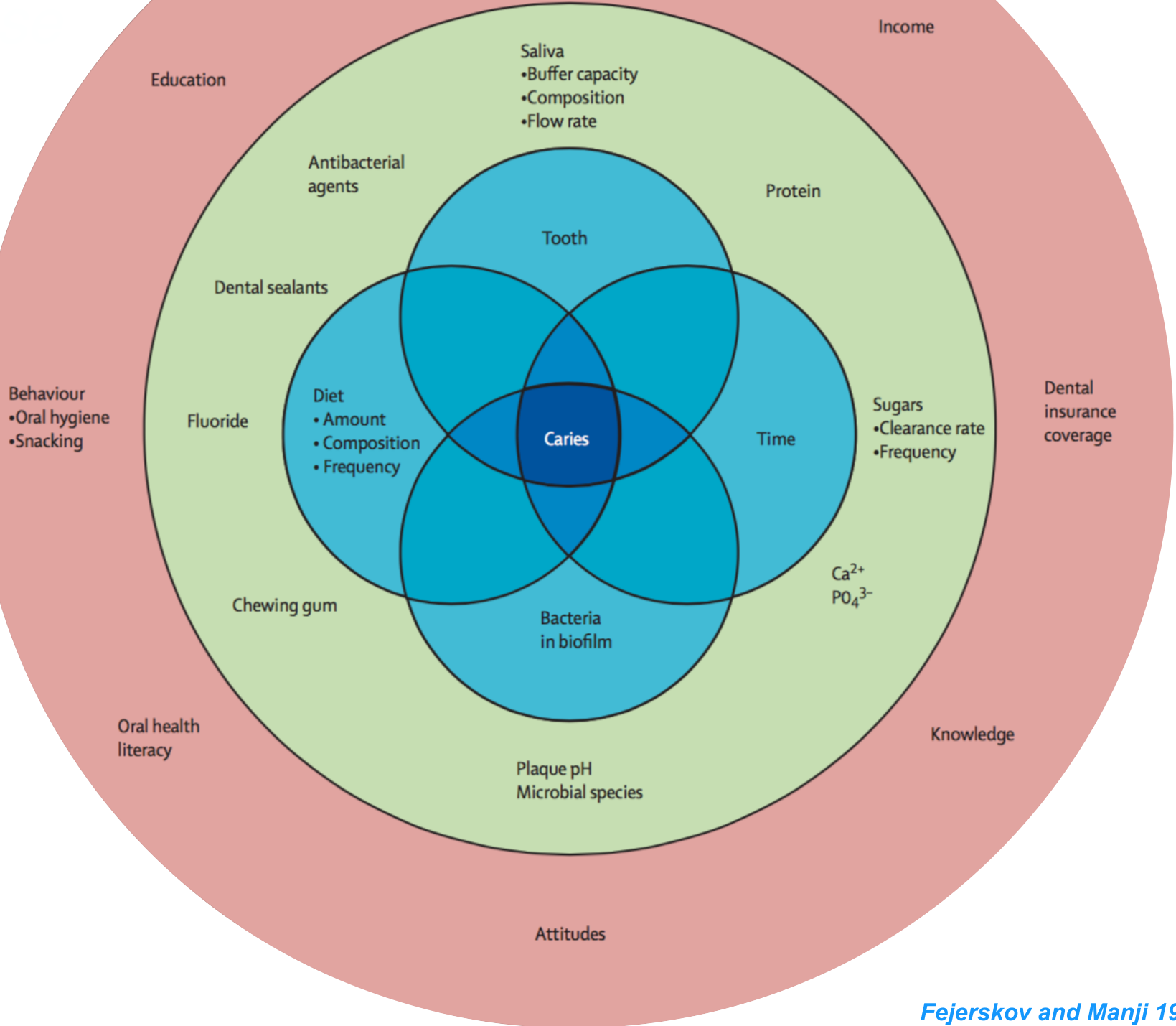
The caries process is shaped by interacting factors across the host, environment, and time:
- Tooth — protein, Ca²⁺, PO₄³⁻
- Saliva — buffer capacity, composition, flow rate
- Bacteria in biofilm — plaque pH, microbial species
- Diet — amount, composition, frequency; sugars (clearance rate, frequency)
- Behaviour — oral hygiene, snacking
- Sociodemographic status — income, education, dental insurance coverage
- Knowledge and attitudes — oral health literacy
- Preventive measures — fluoride, dental sealants, antibacterial agents, chewing gum
- Time
All of these factors converge to produce caries.
Fejerskov and Manji 1990
Lecturer — Caries Model
The traditional Keyes triad described caries as an interaction between bacteria, dietary carbohydrates, and acids on the tooth surface.
- Time was later added to emphasize the frequency of sugar exposure, how long sugars remain in contact with teeth, and the overall dietary exposure pattern.
- Contemporary cariology recognizes influences ranging from education and access to dental services to social circumstances and calcium and phosphate buffering at the molecular level.
- Dental caries is therefore also described as a socially mediated disease, rather than a condition caused only by bacteria and diet.
Key Definitions
What is dental caries?4
Dental caries is a continuum of disease states of increasing severity and tooth destruction that ranges from sub-clinical sub-surface changes at the molecular level to lesions with dentinal involvement, either with an intact surface or obvious cavitation.
Selwitz et al. 2007
Caries Res. 2020;54(1):7-14. doi: 10.1159/000503309. Epub 2019 Oct 7.
Terminology of Dental Caries and Dental Caries Management: Consensus Report of a Workshop Organized by ORCA and Cariology Research Group of IADR.
Machiulskiene V, Campus G, Carvalho JC, Dige I, Ekstrand KR, Jablonski-Momeni A, Maltz M, Manton DJ, Martignon S, Martinez-Mier EA, Pitts NB, Schulte AG, Splieth CH, Tenuta LMA, Ferreira Zandona A, Nyvad B.
Lecturer — Caries Continuum
The continuum emphasizes that caries begins before a clinically obvious cavity develops.
- Clinicians should recognize subsurface and molecular-level changes so progression can be prevented before a patient develops a lifelong restorative burden.
- A carious lesion is the clinical sign of caries and may be categorized by anatomical location, severity, depth of penetration, and activity status.
- A validated diagnostic tool such as ICDAS should be used for caries detection and diagnosis; it was described as internationally accepted and updated for use on the lecture’s base charting system.
Dental Caries (100%)5
Dental caries is a biofilm-mediated, diet modulated, multifactorial, non-communicable, dynamic disease resulting in net mineral loss of dental hard tissues [Fejerskov 1997; Pitts et al., 2017]. It is determined by biological, behavioral, psychosocial, and environmental factors. As a consequence of this process, a caries lesion develops.
Lecturer — ART Development and Technique
ART was developed in Tanzania for communities where sugar had become widely available but access to definitive dental care remained limited.
- The technique was intended for areas affected by poverty and disparities in oral health, including parts of Africa and Southeast Asia.
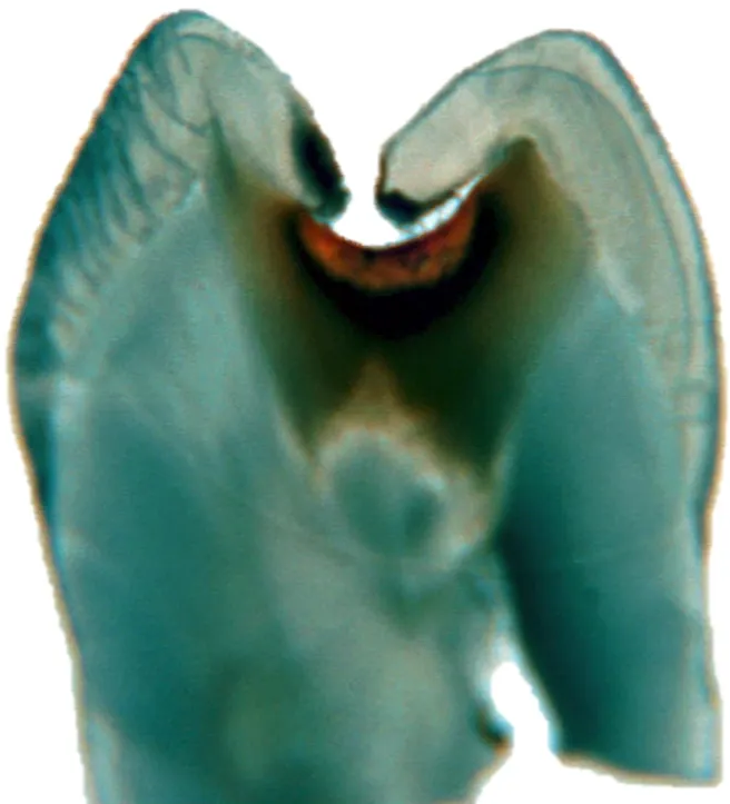
- ART uses a specific enamel hatchet and other instruments to excavate caries by hand, remove unsupported enamel, and shape the cavity so that it is self-cleansable and well supported while maintaining sufficient distance from the pulp.
- In field settings, hand mixing the glass ionomer creates challenges with powder-to-liquid proportions, temperature, and moisture control.
- Adequate isolation is essential because contamination can compromise the restoration’s seal.
- After placement, the glass ionomer should be covered with cocoa butter, Vaseline, or Ketac glaze to prevent desiccation and moisture uptake during setting.
- Children should avoid biting directly on the restoration during its initial set; a cotton wool roll between the incisors can keep the molars out of occlusion for approximately 10–15 minutes.
ICDAS 3 Therapeutic Sealing
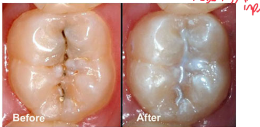
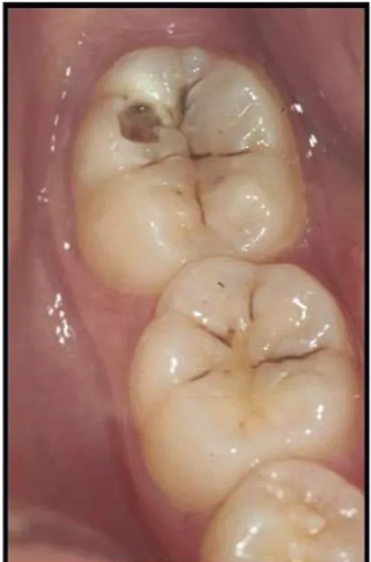
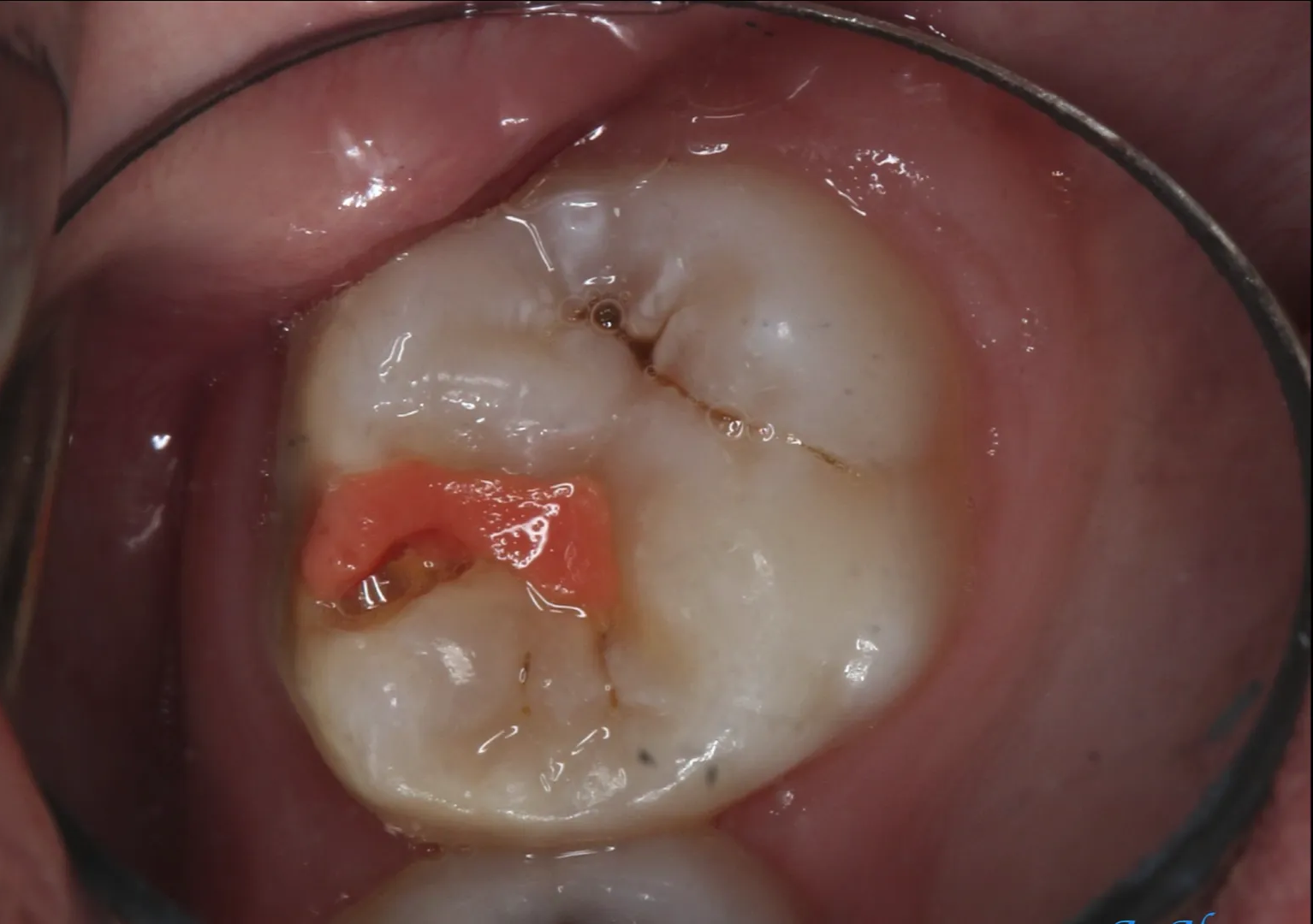
An ICDAS 3 lesion on the occlusal surface of a first permanent molar showed initial enamel cavitation. The surface was cleaned with a bristle brush, the cavitated area was not removed, and sealant was placed directly over the lesion.
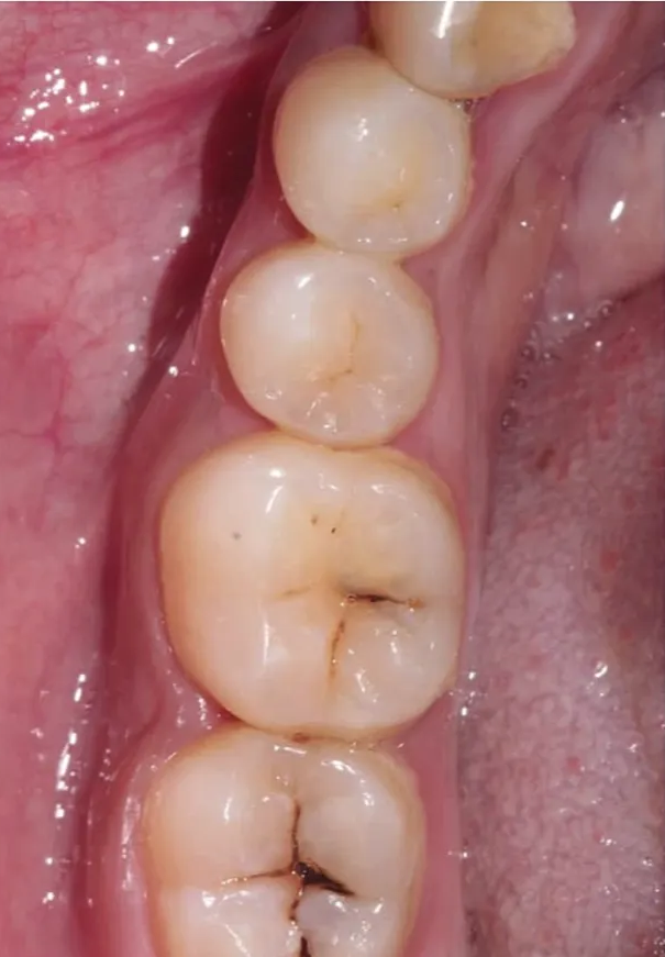
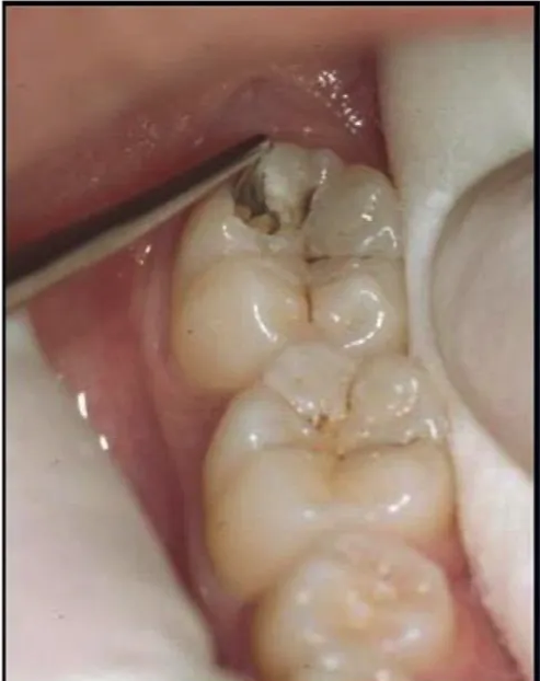
- Another lesion classified as ICDAS 3, possibly ICDAS 4, showed grey shadowing of the tooth structure and was also sealed directly.
- Therapeutic sealing requires careful documentation because stained material may remain beneath the sealant and could later be mistaken for recurrent caries.
- The seal must be as complete as possible: salivary contamination, bacteria, marginal discrepancies, or marginal deficiencies can create bacterial accumulation sites, microscopic anaerobic niches, and marginal overhangs that encourage recurrent caries.
- Perform therapeutic sealing only with careful isolation, accurate placement, and appropriate follow-up.
What Is ART
- Minimally invasive care approach in preventing dental caries and stopping its further progression
Frencken et al. 2012
Main concepts:
- Sealing caries prone pits and fissures
- Restoring cavitated lesions with sealant-restorations
Frencken and Holmgren 1999
Atraumatic Restorative Treatment6
Atraumatic restorative treatment is the tissue-saving caries management approach that uses hand instruments for opening caries cavities and removing decomposed carious dentine, followed by restoration with a high-viscosity glass ionomer. The technique does not require access to electrically driven equipment and running water.
Frencken et al., 1996.
- Minimally invasive care approach in preventing dental caries and stopping its further progression
Frencken et al. 2012
- Sealing caries prone pits and fissures
- Restoring cavitated lesions with sealant-restorations
Frencken and Holmgren 1999
Preventive Sealant And Sealing7
Preventive sealant/sealing is the application of a thin physical barrier over a clinically sound caries predilection site, in order to prevent the initiation of a caries lesion. These can be applied to pits, fissures, and fossae using resin composite or glass ionomer cement.
- ART-type procedures and fissure sealants are particularly useful in remote or resource-limited field settings without access to electricity.
- Minimally invasive care approach in preventing dental caries and stopping its further progression
Frencken et al. 2012
- Sealing caries prone pits and fissures
- Restoring cavitated lesions with sealant-restorations
Frencken and Holmgren 1999
Therapeutic Sealant And Sealing8
Therapeutic sealant/sealing is the application of a thin physical barrier over a caries lesion in order to prevent its progression. These can be applied to pits, fissures, fossae, and smooth surfaces using resin composite or glass ionomer cement.
Martignon et al., 2006; Alkizy et al., 2009.
Lecturer — ART and Anxiety
Dental anxiety and fear often originate from childhood dental experiences, so treatment provided to a child may have a lifelong impact on willingness to attend dental care.
- ART has been associated with reduced pain and anxiety, but the evidence is not uniform.
- Some studies report less pain and discomfort because local anaesthetic and a dental drill may not be required.
- Other studies, particularly when treatment is provided by a specialist, found no difference in pain or anxiety between ART and conventional rotary treatment.
- Anxiety reduction depends substantially on appropriate training, clinical skill, and the way treatment is delivered; ART alone will not necessarily eliminate anxiety.
Rationale For ART9
Case 5: 1350 → enamel hatchet
No difference between ART and rotary instruments.
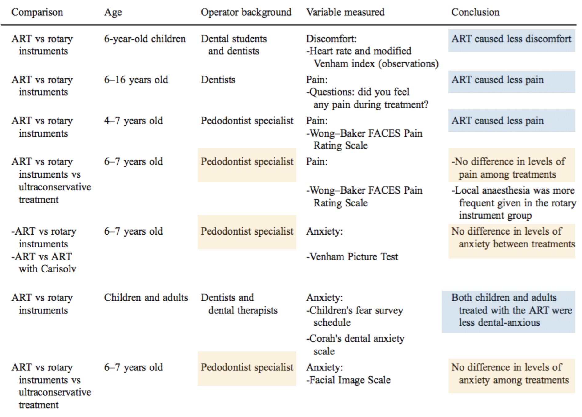
| Comparison | Age | Operator background | Variable measured | Conclusion |
|---|---|---|---|---|
| ART vs rotary instruments | 6-year-old children | Dental students and dentists | Discomfort: heart rate and modified Venham index (observations) | ART caused less discomfort |
| ART vs rotary instruments | 6–16 years old | Dentists | Pain: questions — did you feel any pain during treatment? | ART caused less pain |
| ART vs rotary instruments | 4–7 years old | Pedodontist specialist | Pain: Wong–Baker FACES Pain Rating Scale | ART caused less pain |
| ART vs rotary instruments vs ultraconservative treatment | 6–7 years old | Pedodontist specialist | Pain: Wong–Baker FACES Pain Rating Scale | No difference in levels of pain among treatments; local anaesthesia was more frequently given in the rotary instrument group |
| ART vs rotary instruments; ART vs ART with Carisolv | 6–7 years old | Pedodontist specialist | Anxiety: Venham Picture Test | No difference in levels of anxiety between treatments |
| ART vs rotary instruments | Children and adults | Dentists and dental therapists | Anxiety: Children’s Fear Survey Schedule; Corah’s Dental Anxiety Scale | Both children and adults treated with the ART were less dental-anxious |
| ART vs rotary instruments vs ultraconservative treatment | 6–7 years old | Pedodontist specialist | Anxiety: Facial Image Scale | No difference in levels of anxiety among treatments |
Frencken et al. 2012
ART Advantages10
- Relatively inexpensive → GIC
- Simple
- Minimal need for specialist equipment
- Conservative approach
- Fluoride leaching: potential to reduce secondary caries
- Chemical adhesion of glass-ionomer cement
- No need for local anaesthetic
- Reduced anxiety — useful for young children
Frencken et al. 1996
Lecturer — ART Limitations
The absence of a drill and local anaesthetic may be useful in selected children and resource-limited settings.
- These advantages must not be interpreted as evidence that ART is appropriate for every lesion.
- ART is not a replacement for all other restorative procedures.
ART Abuse11
Inadequate ART and GIC Complications
The simplicity and apparent advantages of ART can lead to clinical abuse, and GIC should not be treated as a universal restorative material.
- GIC should not be used to create restorations such as GIC bridges.
- Chemical adhesion and fluoride release do not prevent all caries and coronal breakdown. Treatment must still provide an adequate seal, appropriate cavity design, sufficient strength, and a restoration suitable for the lesion and tooth.
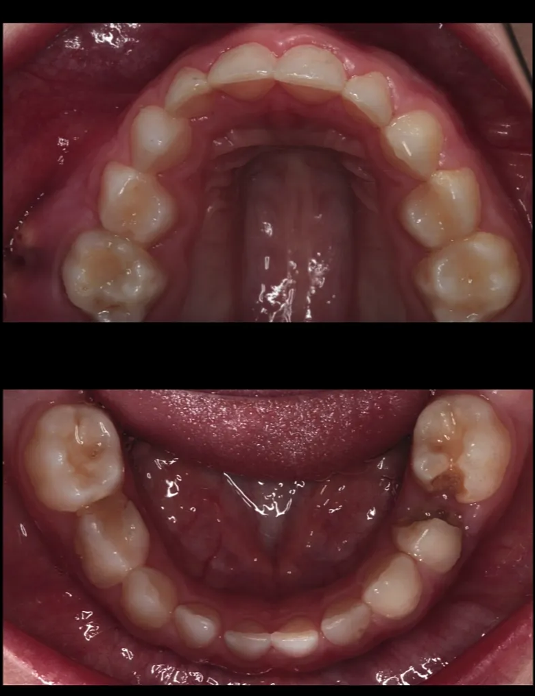
- A child presented with facial cellulitis after a dentist had been attempting to slow tooth decay. The child required hospital admission, overnight intravenous antibiotics, general anaesthesia, and extraction of the affected teeth. No matrix band had been used, and adjacent teeth had effectively been joined together by unsatisfactory restorative material.
- Another child under six presented with facial cellulitis and multiple abscesses. One primary molar had an occlusal restoration and radiographic evidence suggesting a direct pulp cap; direct pulp caps are not performed on primary teeth. Another primary molar had an excessively wide restoration with marginal ridge breakdown and a buccal abscess. The child required intravenous antibiotics, an overnight hospital stay, general anaesthesia, and extraction of at least five teeth.
- Excessively large multisurface restorations in primary teeth cannot reliably withstand their functional burden.
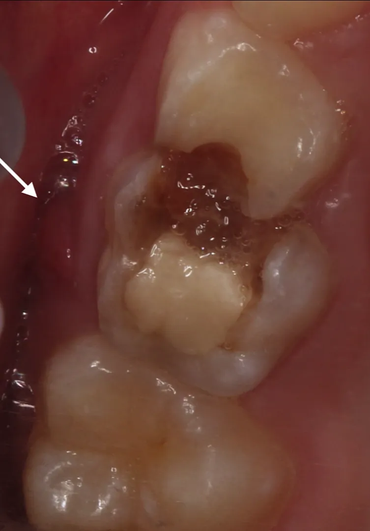
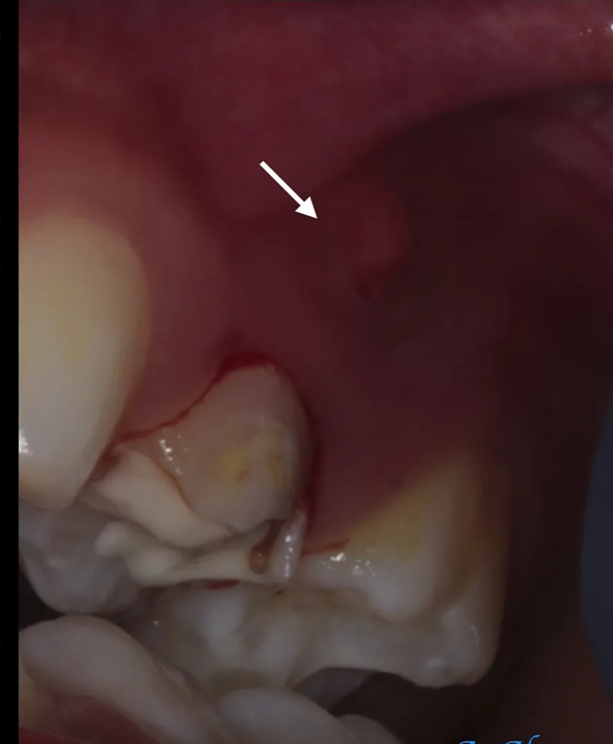
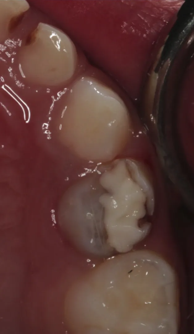
- Further cases showed recurrent caries and abscesses associated with GIC, including a necrotic tooth with intrinsic grey discoloration and a Fuji 7 sealant with recurrent caries beneath it because part of the distal pit and fissure system had not been sealed.
- A child with an unsatisfactory large disto-occlusal restoration presented in significant pain, had been unable to eat properly for several days, tolerated only a liquid diet, and had pus involving the palatal and buccal tissues.
- Poorly selected or poorly executed GIC and ART procedures can therefore produce significant harm.
Lecturer — ART Failure Patterns
The reduction in survival from single-surface to multisurface restorations is substantial.
- A 12% survival rate means treatment fails in approximately 90% of cases in the worst scenario over only two years.
- Many unsuccessful examples involved disto-occlusal, mesio-occlusal, and large multisurface restorations, particularly proximal lesions in primary teeth.
- The quality of the seal is critical when placing ART sealants.

ART Longevity121314
Two year survival rates: single surface ART restorations
- Primary teeth: 93%
- Permanent teeth: 85%
De Amorim et al. 2012
Two year survival rates: single surface ART restorations
- Primary teeth: 93%
- Permanent teeth: 85%
De Amorim et al. 2012
Two year survival rates: multi surface ART restorations
- Primary teeth: 12% - 60%
- Permanent teeth: 30%
Luo et al. 1999, van Gemert-Schriks et al. 2007
Conclusions from a 25-year review of ART
-
High viscosity GIC can be used for single surface cavities in primary and permanent posterior teeth
-
ART sealants have a high caries preventive effect
-
ART using high viscosity GIC cannot be used for multi-surface cavities in primary teeth
Frencken et al. 2012
-
GIC cannot be recommended for Class II cavities in primary molars
Chadwick and Evans 2007
Class II Cavity Preparations15
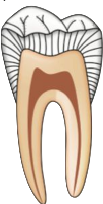
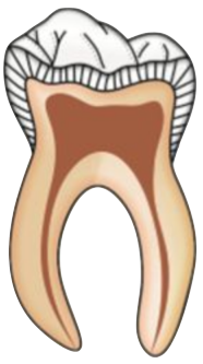
Primary tooth anatomy: coronal morphology16
-
Thinner enamel and dentine
- Thickness approximately half that of permanent teeth
-
Direction of the enamel rods in the cervical area is angled towards the occlusal surface
-
In the permanent teeth they are towards and apical direction
-
This orientation contributes to a greater amount of unsupported enamel in primary teeth and must be considered when planning Class II preparations and restorations.
 |  |
Primary tooth anatomy: pulp and root canal system17
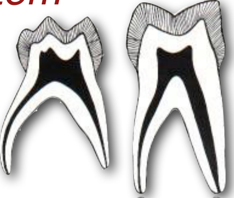
- Size of the pulp relative to the crown is larger
- Pulp horns are higher and closer to the DEJ and to the outer surface of the crown
- Mesial pulp horns are higher than the distal pulp horns
Int J Paediatr Dent. 2009 Jan;19(1):26-33. doi: 10.1111/j.1365-263X.2008.00962.x.
Lecturer — Pulpal Risk
Class II cavity preparation and restoration in a primary tooth can easily encroach on the pulp.
- Even if the pulp is not exposed during preparation, bacteria from a proximal carious lesion may already have invaded the pulp.
Histological comparison of pulpal inflammation in primary teeth with occlusal or proximal caries18
Kassa D, Day P, High A, Duggal M.
Author information: Department of Paediatric Dentistry, Leeds Dental Institute, Leeds, UK.
Deep Proximal Lesion in a Primary Molar
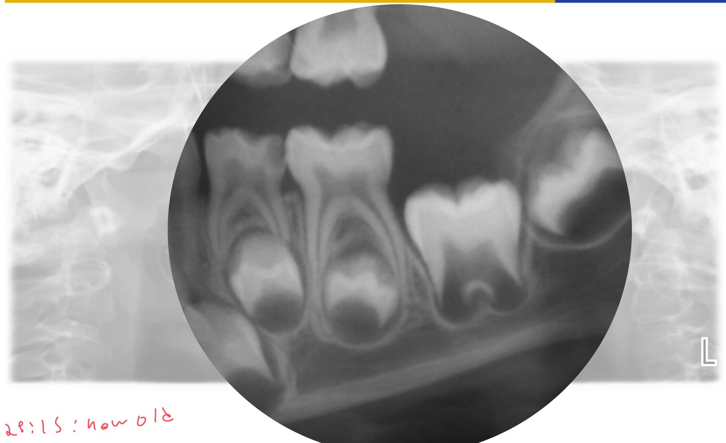
The radiograph suggested a child approximately four to five years old dentally: the first permanent molar had not erupted, the second primary molar was erupted and in occlusion, and the relevant primary molar roots were fully formed.
- The 74 had a disto-occlusal lesion extending into the inner third of dentine.
- Possible furcal changes were difficult to interpret because of angulation, differences in trabecular pattern, and superimposition of developing premolar crypts. Similar furcal lucency was present around the 75 despite no significant lesion.
- The primary molar had much thinner enamel than the developing permanent first molar, and its pulp horns were much closer to the proximal surface.
- The proposed treatment was a pulpotomy and stainless steel crown if the tooth was restorable.
- Local anaesthetic could be an infiltration of 4% articaine with 1:100,000 adrenaline; an inferior alveolar block was avoided where possible because postoperative lip and tongue numbness may increase biting risk in a child under six.
- The 75 was clamped using a slit technique extending toward the 73, its occlusal surface was sealed, and the 74 occlusal surface was reduced. The mesial surface of the 75 was then inspected for an ICDAS lesion.
- Caries was removed to obtain a peripheral caries-free margin, the distal aspect was begun with Fuji 2, and wedges were placed in the gingival proximal areas to prevent bleeding contamination.
- The pulp was opened with a clean round bur by cutting upwards rather than downwards to reduce the risk of furcal perforation. A clean cotton pellet was placed in the chamber to assess bleeding and pulp colour.
- Bleeding that stopped after approximately one minute was considered more consistent with reversible inflammation; profuse bleeding or a dark red pulp suggested irreversible inflammation, while no bleeding suggested a pulpless or necrotic pulp for which extraction might be required.
- Depending on pulp status, medicaments included ferric sulfate, MTA, or Biodentine. The area was gently rinsed to avoid trapping a blood clot.
- The proposed restoration sequence was approximately 2–3 mm of Cavit, a Fuji 2 base, and stainless steel crown placement.
Abstract
OBJECTIVE: A number of clinical and histological studies have investigated caries-related changes in the primary tooth pulp, but the effect of caries site, as a clinical variable, has not been previously considered. This study sought to compare inflammatory changes within the pulp of primary molars according to the location of the caries lesion (occlusal or proximal).
METHODS: Eighty-three primary molars were extracted under general anaesthesia for caries and/or orthodontic reasons, and were split immediately after removal and fixed in 10% formalin. Teeth were then decalcified, sectioned, and stained with haematoxylin and eosin for histological examination using light microscopy. Caries depth was measured using a graticule, and the site of the caries lesion noted as occlusal or proximal. Samples were further classified into one of five subgroups according to the observed degree of pulpal inflammation.
RESULTS: Key findings were that where caries depth was less than 50% of the total dentine thickness, there were no significant differences in inflammatory status according to caries site. In contrast, marked inflammatory changes were significantly more likely throughout the coronal pulp of teeth with proximal caries compared to teeth with occlusal caries where caries depth was equal to, or greater than, 50% of the total dentine thickness (P = 0.017, Fisher’s exact test).
CONCLUSION: Primary teeth with proximal carious lesions extending more than 50% through the dentine thickness appear to have more extensive inflammatory pulpal changes than teeth with occlusal caries of a similar depth. This finding has clinical implications and may help inform treatment decisions in the management of primary teeth with deep carious lesions.
Lecturer — Deep Proximal Lesions
A proximal lesion extending into the inner third of dentine on a bitewing may require consideration of pulpotomy rather than an intracoronal restoration without pulp treatment.
- A lesion that appears clinically small at the enamel surface may extend extensively through dentine and reach the pulp.
- The extent of the lesion may therefore be considerably greater histologically than it appears clinically.
(I do pulpotomy instead)
28:15: how old
Glass Ionomer Cement Properties
GICs And Fluoride19
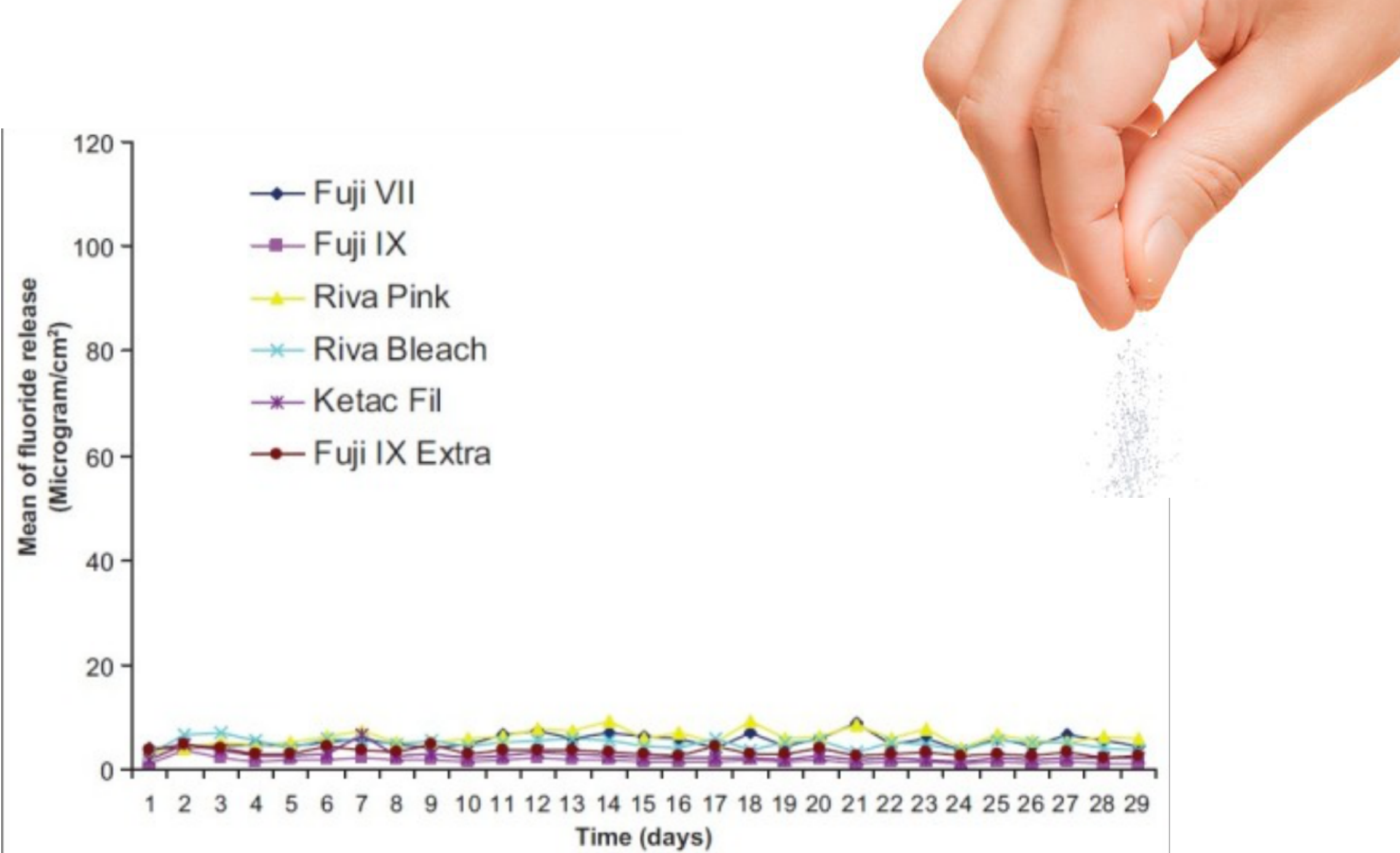
Mean fluoride release (microgram/cm²) over 29 days, comparing six materials:
-
Fuji VII
-
Fuji IX
-
Riva Pink
-
Riva Bleach
-
Ketac Fil
-
Fuji IX Extra
-
Y-axis: Mean of fluoride release (Microgram/cm²), scale 0–120
-
X-axis: Time (days), 1–29
Lecturer — Fluoride Limitations
Fluoride release is often cited as an advantage of GIC and ART, but the exposure remains relatively low.
- GIC produces an initial burst of fluoride release that decreases substantially after approximately 7–10 days.
- Fluoride release may recharge later if materials such as varnish are applied.
- Fluoride release alone is unlikely to counteract all factors that caused the original lesion; placing GIC does not necessarily change patient behaviour, restore overall tooth integrity, or resolve the underlying caries disease.
- GIC may reduce plaque stagnation and alter the local environment, but its long-term caries-preventive effect should not be overstated, particularly when enamel has already cavitated.
Cariostatic Potential Of GIC
Quintessence Int. 1996 Mar;27(3):171-4.
Glass-ionomer cement restorations and secondary caries: a preliminary report20
Mjör IA¹.
Abstract
This preliminary report is based on a survey of the reasons for inserting 790 glass-ionomer cement, 2,609 resin composite, and 898 amalgam restorations in adults in general practice. Glass-ionomer materials were used about equally for the treatment of primary caries and for replacement of failed restorations, unlike resin composite and amalgam, which were most often used to replace failed restorations. Almost half of the 412 replaced glass-ionomer cement restorations were removed because of the clinical diagnosis of secondary (recurrent) caries, which was by far the most common reason for replacement of all three materials.
Lecturer — Cariostatic Effect
There is a strong perception that GIC has a powerful cariostatic effect, but this perception should be treated cautiously.
- GIC is not immune to secondary or recurrent caries, and fluoride release does not guarantee long-term prevention around the restoration.
- Treatment success still depends on appropriate case selection, adequate cavity design, a satisfactory seal, management of the patient’s overall caries risk, and follow-up and review.
- Fluoride release and presumed caries prevention should not be the primary justification for placing GIC when the restoration is otherwise unsuitable.
So.. GIC fails due to recurrent caries
Modified ART Is Not ART21
- Opening the cavity with rotating instruments, followed by cleaning it with hand instruments and restoring it with an adhesive restorative material, is not considered ART nor can calling it modified ART be justified
Lecturer — ART Technique
Using a slow-speed bur to remove peripheral caries, placing GIC, and calling the procedure “modified ART” is not considered ART.
- The use of hand instruments versus rotary instruments changes the pulpal response, the extent and shape of cavity opening, and the resulting margins.
- Preliminary studies have identified significant differences in pulpal changes between hand-instrument and rotary preparation, so the technique must be identified correctly rather than labelled ART simply because GIC is used.
Frencken and Leal 2010
Interim Therapeutic Restoration22
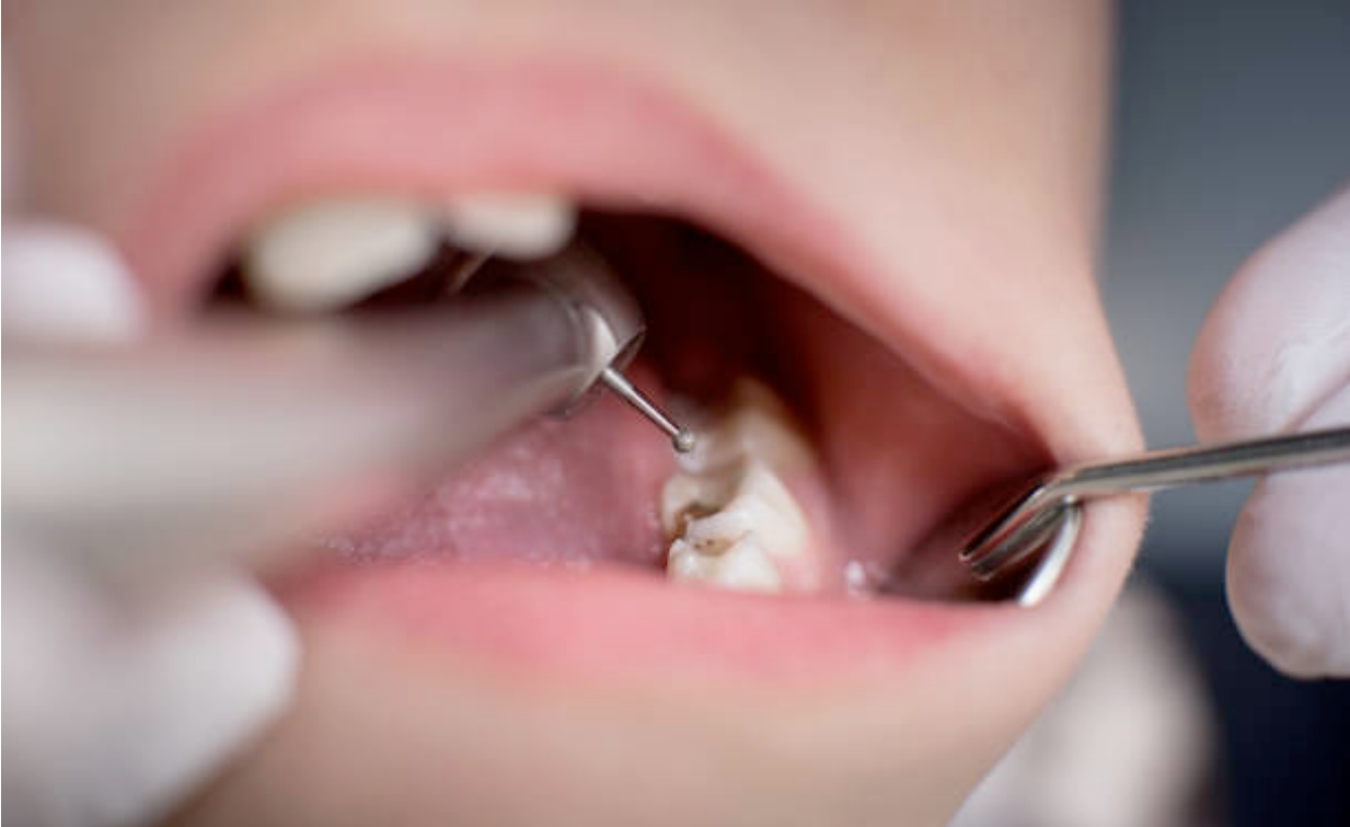
- ITR utilises similar techniques but has different therapeutic goals to ART
- ITR may be used to restore, arrest or prevent the progression of carious lesions in young patients, uncooperative patients, or patients with special health care needs or when traditional cavity preparation and/or placement of traditional dental restorations are not feasible and need to be postponed
AAPD 2017
ITR Technique23
-
Removal of caries using hand or rotary instruments with caution not to expose the pulp
-
Leakage of the restoration can be minimised with maximum caries removal from the periphery of the lesion
-
Following preparation, the tooth is restored with an adhesive restorative material such as glass ionomer or resin-modified glass ionomer cement
-
Selective caries removal may be used, allowing carious material to remain at the depth of the lesion.
-
Resin composite may also be used as the restorative material. AAPD 2017
ITR Limitations24
- High risk of failures in multi-surface cavities
- Increased need for follow-up and review
- INTERIM… vs DEFINITIVE
- The use of ITR has been shown to reduce the levels of cariogenic oral bacteria immediately following placement BUT levels are likely to return to pretreatment counts after six months if no other treatment is provided
Lecturer — Definitive Treatment
ITR is a temporary measure, so patients and parents must understand that the restoration is not final and may fail.
- Close follow-up is required until the child becomes more cooperative or confidence has been established.
- The temporary restoration should then be replaced with definitive treatment.
- Definitive treatment may involve local anaesthetic, rubber dam isolation, definitive caries removal, and placement of an appropriate final restoration.
AAPD 2017
Conclusions25
- Contemporary practice necessitates the need to differentiate between management of a carious lesion and caries the disease.
- ART and ITR are both valuable tools in paediatric dentistry when used in the correct contexts.
- Their simplicity, however, predisposes them to clinical abuse.
- Multi-surface restorations in the primary dentition should be placed with caution and carry a high risk of failure.
- Caries-risk assessment and prevention must form part of the treatment plan for both children and adults.
- ART and ITR are valuable tools in paediatric and adult dentistry when used in the correct context.
- Inappropriate treatment can result in restoration failure, hospital admission, intravenous antibiotics, general anaesthesia, extraction, and harm to the patient.
- Stainless steel crowns were presented as the gold standard for multisurface cavities in primary teeth.
- The overall objective is to manage the disease, preserve tooth structure where appropriate, provide suitable definitive care, and avoid causing preventable harm.
Clinical Cases
Case: Therapeutic sealant on an occlusal lesion
Question
Scenario: A clinical image of a posterior tooth (tooth 36) is presented to demonstrate the application of a therapeutic sealant over an occlusal lesion.
What’s shown: An intraoral close-up view of an occlusal surface showing initial cavitation within the enamel and gray shadowing of the tooth structure, before and after the application of a sealant.
Consider: What are the clinical considerations and potential pitfalls when applying a therapeutic sealant over an unremoved carious lesion?
Answer
Observations:
- The occlusal surface of tooth 36 shows initial cavitation within the enamel and gray shadowing of the tooth structure.
- The surface was cleaned with a bristle brush, and a sealant was placed directly on top without removing the stained material.
Reasoning: Leaving stained material can lead to misinterpretation as recurrent caries by another clinician in the future. It is critical to ensure a perfect seal; contamination by saliva or bacteria, marginal discrepancies, or deficiencies will create anaerobic niches and overhangs, providing an ideal site for bacterial accumulation and recurrent caries.
Outcome: The technique requires a high degree of caution and thorough documentation in the patient’s notes to prevent future clinicians from mistakenly drilling out the restoration and occlusal enamel under the assumption of recurrent caries.
Takeaway: Therapeutic sealants can be placed over early carious lesions to arrest progression, but they must be meticulously sealed and well-documented to avoid misdiagnosis of recurrent caries and iatrogenic damage from unnecessary drilling.
Case: Pit and fissure lesion with contraindicated over-preparation
Question
Scenario: A clinical exhibit showing a posterior tooth with pit and fissure staining and a subsequent restoration is presented to discuss contemporary cariology practices.
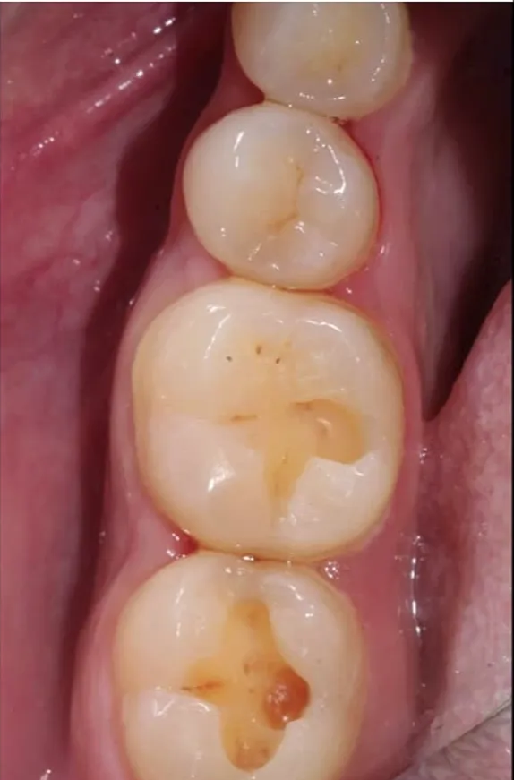
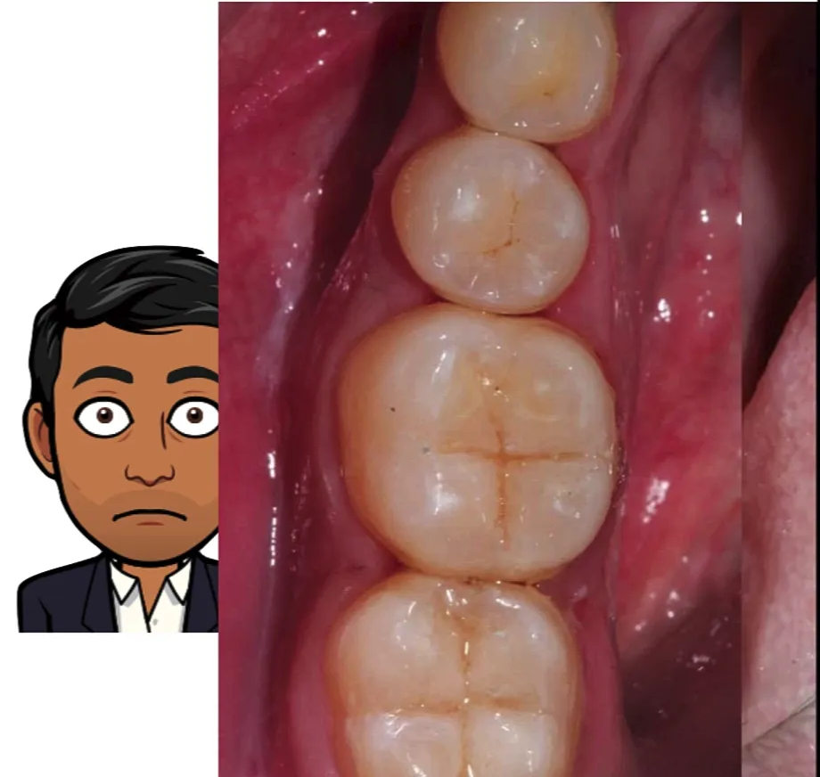
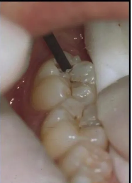
What’s shown: An intraoral view of a lesion with initial pit and fissure staining, alongside an image of a preparation where the staining has been removed.
Consider: What is the assessment of the lesion, and what is the evaluation of the preparation shown?

Answer
Observations:
- The lesion presents with initial pit and fissure staining, assessed as ICDAS 3 or potentially 4 at maximum on the molars.
- The preparation has removed all the staining but significantly reduced the occlusal integrity of the tooth.
- An excess amount of tooth structure has been removed.
Reasoning: Over-preparing an ICDAS 3 or 4 lesion to remove staining is absolutely contraindicated in contemporary cariology. It removes an excess amount of inherent tooth structure and compromises the occlusal integrity of the tooth, which should be avoided in modern practice.
Outcome: The preparation is deemed contraindicated.
Takeaway: Over-preparation of initial pit and fissure lesions (ICDAS 3/4) to remove staining is contraindicated as it unnecessarily sacrifices tooth structure and occlusal integrity.
Case: Histology of an enamel and dentine lesion for ART cavity design
Question
Scenario: A histological exhibit of a carious lesion is presented to illustrate the extent of tissue breakdown and its implications for cavity design.
What’s shown: A histology slide showing cavitation and breakdown within the enamel, spreading down through the dentine.
Consider: How does the histological extent of the lesion inform the cavity design when using hand instruments?
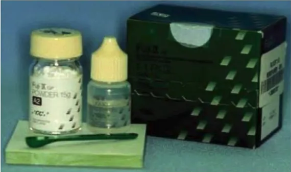
Answer
Observations:
- The histology shows cavitation and breakdown within the enamel.
- The lesion is spreading down through the dentine.
Reasoning: When using hand instruments for ART, the cavity created must be self-cleansable and well-supported. The preparation removes any unsupported enamel while maintaining a sufficient distance away from the pulp, which differs from the cavity design when using rotary instruments.
Outcome: The resulting cavity is self-cleansable, well-supported, with unsupported enamel removed and a safe distance from the pulp maintained.
Takeaway: ART cavity design using hand instruments aims to create a self-cleansable, well-supported preparation that removes unsupported enamel while safely avoiding the pulp, guided by the histological extent of the lesion.
Case: Child with facial cellulitis and joined restorations
Question
Scenario: A child presents to the emergency department with facial cellulitis, accompanied by a distressed mother.
What’s shown: Extracted primary teeth with existing restorations.
Consider: What is evident about the restorative technique used on these teeth, and what was the clinical outcome for this patient?
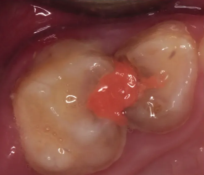
Answer
Observations:
- The extracted teeth show restorations where no matrix band was used.
- The teeth have been basically joined together.
Reasoning: The lack of a matrix band and the joining of the teeth indicate poor restorative technique, which the lecturer describes as “ART abuse” and bordering on neglect, rather than satisfactory restorations.
Outcome: The child was admitted to the hospital for an overnight stay, received IV antibiotics, and underwent a general anesthetic for the extraction of the teeth.
Takeaway: Poorly executed restorations, such as joining teeth without a matrix band, can lead to severe infections like facial cellulitis, requiring hospital admission and extractions.
Case: Child with facial cellulitis, direct pulp cap, and failed GIC restorations
Question
Scenario: Another child under six presents to the emergency department with facial cellulitis.
What’s shown: Radiographs and clinical images of primary molars (55 and 84).
Consider: What restorative procedures were attempted on these teeth, what are the radiographic and clinical findings, and what was the outcome?
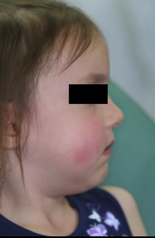
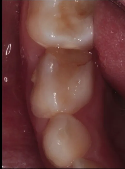
Answer
Observations:
- Tooth 55 has an occlusal restoration that radiographically appears to be a direct pulp cap, with a clearly visible buccal abscess.
- Tooth 84 has a very ambitious GIC restoration (wide and large multi-surface) showing significant marginal ridge breakdown and an associated buccal abscess.
- The child has multiple abscesses and grossly broken down posterior dentition.
Reasoning: Direct pulp caps are not indicated for primary teeth. The ambitious GIC restoration on 84 failed because primary teeth are not capable of withstanding the burden of excessively large multi-surface restorations, leading to marginal breakdown and abscess formation.
Outcome: The child required IV antibiotics, an overnight hospital stay, a general anesthetic, and the extraction of at least five teeth.
Takeaway: Inappropriate use of GIC for large multi-surface restorations and direct pulp caps in primary teeth can lead to severe complications, including multiple abscesses and the need for extractions under general anesthesia.
Case: Upper primary teeth with recurrent caries and necrosis
Question
Scenario: Clinical images of upper primary teeth demonstrating restorative failures.
What’s shown: Teeth with existing restorations and associated pathology.
Consider: What are the clinical findings in these teeth, and what do they indicate about the restorative material used?
Answer
Observations:
- Upper E and D show recurrent caries and a buccal abscess.
- Another tooth has a GIC restoration with a buccal abscess, intrinsic gray discoloration, and necrosis underneath.
Reasoning: The presence of recurrent caries, abscesses, and necrosis under GIC restorations demonstrates that the chemical bonding and fluoride release of GIC do not magically prevent caries and coronal breakdown, highlighting the abuse of the ART technique.
Outcome: Not explicitly stated for this specific exhibit, but it serves as an example of restorative failure and tissue necrosis.
Takeaway: GIC restorations do not inherently prevent recurrent caries or necrosis if the restoration fails or is improperly indicated, emphasizing the need for caution and proper case selection in ART.
Case: Child in severe pain with an abscessed primary molar
Question
Scenario: A young female child presents to the emergency department in significant pain, having not eaten for days and only tolerating a liquid diet.
What’s shown: Clinical images of an upper first primary molar (D) and surrounding tissues.
Consider: What is the clinical presentation of the tooth and surrounding tissues, and what is the source of the pathology?
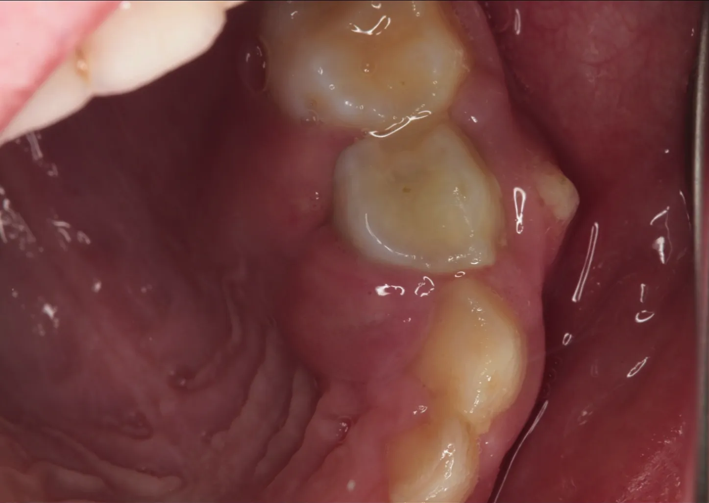
Answer
Observations:
- The upper D (first primary molar) has a fairly large disto-occlusal restoration.
- The palatal and buccal tissues are filled with pus.
Reasoning: The pus-filled tissues indicate a severe abscess stemming from the unsatisfactory large disto-occlusal restoration placed on the primary molar, which failed and led to infection.
Outcome: The child presented in severe pain and distress, requiring emergency care for the abscess resulting from the failed restoration.
Takeaway: Large, unsatisfactory multi-surface restorations in primary teeth can fail and lead to severe, painful abscesses that significantly impact a child’s ability to eat and their overall well-being.
Case: Radiograph of quadrant 7 in a young child
Question
Scenario: A clinical case is presented for discussion, focusing on a child’s radiograph of the lower left quadrant.
What’s shown: A cropped radiographic image (from an OPG) showing the lower left quadrant, including the unerupted first permanent molar, the erupted second primary molar, and the first primary molar.
Consider: How to estimate the child’s dental age, what radiographic findings are present on the first primary molar, and what the ideal treatment option and procedural steps would be.
Answer
Observations:
- The first permanent molar (36) has not yet erupted.
- The second primary molar is erupted and in occlusion.
- The roots of the first (74) and second (75) primary molars are fully formed.
- There is an obvious radiolucency on the disto-occlusal aspect of the 74 extending into the inner third of the dentine.
- There is a potential radiolucency in the furcal area of the 74, though it may be superimposed by premolar crypts.
- The mesial enamel thickness on the 36 is significantly thicker than on the 75, and the pulp horns on the primary teeth are much closer to the proximal surface compared to the developing permanent molar.
Reasoning:
- The child’s dental age is estimated to be between 4 and 5 years based on the eruption and occlusion of the second primary molar, and the fully formed roots of the primary molars.
- The deep proximal lesion on the 74 indicates a high risk of pulpal inflammation, as it extends past 50% into the dentine.
- Because sufficient coronal tooth structure remains, the tooth is restorable. However, multi-surface intra-coronal restorations in primary teeth have a high failure rate.
- Therefore, a pulpotomy followed by a stainless steel crown is the ideal treatment option.
- The true pulp status (reversible vs. irreversible inflammation, or necrosis) cannot be definitively diagnosed radiographically and must be assessed clinically upon opening the pulp chamber by observing bleeding and tissue color.
Outcome:
- The proposed treatment is a pulpotomy and stainless steel crown for the 74.
- The parent is informed that if the pulp is found to be irreversibly inflamed (profuse bleeding, dark red color) or necrotic (no bleeding) during the procedure, the tooth may need to be extracted.
- The procedure involves local anesthesia (infiltration with 4% articaine to avoid soft tissue biting in a child under six), rubber dam isolation, occlusal reduction, caries removal, wedging, pulpotomy (cutting upwards with a round bur), hemostasis assessment, application of a medicament (e.g., ferric sulfate, MTA, biodentine), a Cavit seal, a Fuji 2 base, and finally the stainless steel crown.
Takeaway:
- In primary teeth with deep proximal caries extending into the inner third of dentine, a pulpotomy and stainless steel crown is the ideal treatment, but clinicians must be prepared for potential extraction if the pulp is found to be irreversibly inflamed or necrotic upon access.
Footnotes
-
Original PDF page 1: L3 Atraumatic Restorative Treatment DMD, p.1 ↩
-
Original PDF page 2: L3 Atraumatic Restorative Treatment DMD, p.2 ↩
-
Original PDF page 3: L3 Atraumatic Restorative Treatment DMD, p.3 ↩
-
Original PDF page 4: L3 Atraumatic Restorative Treatment DMD, p.4 ↩
-
Original PDF page 5: L3 Atraumatic Restorative Treatment DMD, p.5 ↩
-
Original PDF page 7: L3 Atraumatic Restorative Treatment DMD, p.7 ↩
-
Original PDF page 8: L3 Atraumatic Restorative Treatment DMD, p.8 ↩
-
Original PDF page 9: L3 Atraumatic Restorative Treatment DMD, p.9 ↩
-
Original PDF page 11: L3 Atraumatic Restorative Treatment DMD, p.11 ↩
-
Original PDF page 12: L3 Atraumatic Restorative Treatment DMD, p.12 ↩
-
Original PDF page 13: L3 Atraumatic Restorative Treatment DMD, p.13 ↩
-
Original PDF page 14: L3 Atraumatic Restorative Treatment DMD, p.14 ↩
-
Original PDF page 15: L3 Atraumatic Restorative Treatment DMD, p.15 ↩
-
Original PDF page 16: L3 Atraumatic Restorative Treatment DMD, p.16 ↩
-
Original PDF page 20: L3 Atraumatic Restorative Treatment DMD, p.20 ↩
-
Original PDF page 17: L3 Atraumatic Restorative Treatment DMD, p.17 ↩
-
Original PDF page 18: L3 Atraumatic Restorative Treatment DMD, p.18 ↩
-
Original PDF page 19: L3 Atraumatic Restorative Treatment DMD, p.19 ↩
-
Original PDF page 21: L3 Atraumatic Restorative Treatment DMD, p.21 ↩
-
Original PDF page 22: L3 Atraumatic Restorative Treatment DMD, p.22 ↩
-
Original PDF page 23: L3 Atraumatic Restorative Treatment DMD, p.23 ↩
-
Original PDF page 24: L3 Atraumatic Restorative Treatment DMD, p.24 ↩
-
Original PDF page 25: L3 Atraumatic Restorative Treatment DMD, p.25 ↩
-
Original PDF page 26: L3 Atraumatic Restorative Treatment DMD, p.26 ↩
-
Original PDF page 27: L3 Atraumatic Restorative Treatment DMD, p.27 ↩