Non-Restorative Caries Control1
A Gross Distortion of Science and Medicine..?
Dr Jilen Patel BDSc(Hons), DClinDent(Paed Dent), MRACDS (Paed), FADI, FICD, FPFA
Senior Lecturer, UWA Dental School
Consultant, Perth Children’s Hospital
Lecture Outcomes2
-
Justify the use of non-restorative caries control methods
-
Understand the different techniques and materials used in non-restorative caries control
-
Explain the mechanism of action of different non-restorative caries control methods
-
Determine the most appropriate method for different carious lesions in primary teeth
-
Recognize that caries management requires consideration of the disease process, lesion characteristics, patient factors, compliance, and clinical context. Fuji 7 distal pit missed at 2:30
Mummy case at 4:25
Aetiology and Historical Perspectives
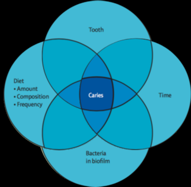
A four-factor model relating tooth, diet, bacteria, and time to caries:
- Tooth
- Diet
- Amount
- Composition
- Frequency
- Bacteria in biofilm
- Time
All four factors converge to produce Caries.
Newbrun 1982
Lecturer — Historical Perspectives
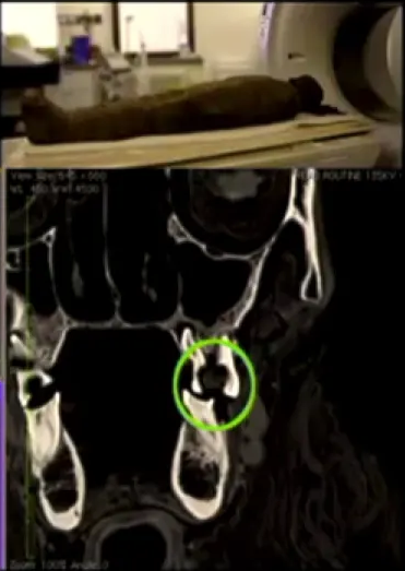
Dental caries is not a new disease. Evidence from a CT scan of a 2,000-year-old mummy showed extensive carious lesions and periapical areas of resorption, possibly extending into the sinus and potentially contributing to death.
- Traditional dental education has focused heavily on operative dentistry, including cutting Class I and Class II cavities and developing hand-eye skills.
- This emphasis can result in clinicians spending more time learning to prepare cavities than understanding how to treat and manage the underlying disease.
- The lecturer described a shift toward a biological approach, a medical model of caries management, caries risk assessment, and management of the disease process rather than only its clinical outcome.
Keynes Four Factors Model3
Lecturer — Caries Risk Factors
The historical Keyes triad described bacteria, diet, and tooth structure, with time later added as a fourth factor. Caries is more complex than sugar consumption alone.
- Relevant considerations include food type, consumption frequency, bacteria, tooth susceptibility, and duration of exposure.
- Other influences include buffering capacity, fluoride, socioeconomic circumstances, education, health literacy, and attitudes toward health.
- Clinicians often focus on factors that are easiest to modify, such as the tooth and the lesion, while giving less attention to health literacy, attitudes, and broader social influences.
Sugar and Dental Caries Evidence
Review > Science. 1982 Jul 30;217(4558):418-23. doi: 10.1126/science.7046052.
Sugar and dental caries: a review of human studies
E Newbrun
PMID: 7046052 DOI: 10.1126/science.7046052
Abstract4
Studies of special population groups, epidemiological surveys, controlled longitudinal studies of humans, and longitudinal studies on the effect of sugar substitutes indicate that frequent or high intake of sugary foods predisposes to dental decay. The relation is not always clear-cut, and most studies have important methodological problems and limitations. Longitudinal measurements of caries increments combined with multiple dietary histories are needed to clarify the association between caries and eating habits. The relative cariogenicity of specific foods can be assessed by a combination of in vitro tests, human in vivo tests, and experimental caries in animals. Human diets, however, vary in food items eaten and the frequency and sequence of eating, and these factors can affect the cariogenicity of a food. Therefore, reported correlations must be interpreted with caution.
Lecturer — Broader Caries Management
Caries cannot be explained only by statements such as “the patient eats sugar” or “the patient is not brushing twice a day.” Diet-related assessment needs to include the type and frequency of food and drink consumption, together with the broader biological and social context.
Newbrun 1982
The Stephan Curve5
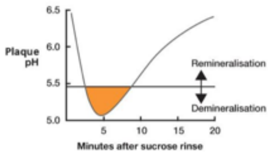
Plaque pH response after a sucrose rinse:
- Plaque pH scale shown from 5.0 to 6.5
- Time axis: 5, 10, 15, 20 minutes after sucrose rinse
- pH falls below the threshold → Demineralisation
- pH recovers above the threshold → Remineralisation
Stephan and Miller 1943
Odontology (2013) 101:2–8. DOI 10.1007/s10366-012-0092-z. REVIEW ARTICLE.
The Stephan Curve revisited — William H. Bowen
Received: 6 August 2012 / Accepted: 6 November 2012 / Published online: 6 December 2012. © The Society of The Nippon Dental University 2012.
Abstract6
The Stephan Curve has played a dominant role in caries research over the past several decades. What is so remarkable about the Stephan Curve is the plethora of interactions it illustrates and yet acid production remains the dominant focus. Using sophisticated technology, it is possible to measure pH changes in plaque; however, these observations may carry a false sense of accuracy. Recent observations have shown that there may be multiple pH values within the plaque matrix, thus emphasizing the importance of the milieu within which acid is formed. Although acid production is indeed the immediate proximate cause of tooth dissolution, the influence of alkali production within plaque has received relative scant attention. Excessive reliance on Stephan Curve leads to describing foods as “safe” if they do not lower the pH below the so-called “critical pH” at which point it is postulated enamel dissolves. Acid production is just one of many biological processes that occur within plaque when exposed to sugar. Exploration of methods to enhance alkali production could produce rich research dividends.
Lecturer — Critical Plaque pH
The lecturer discussed a critical plaque pH of approximately 5.5, but clarified that this value is useful rather than absolute. Foods and drinks with a pH below 5.5 should not automatically be classified as cariogenic, because pH varies substantially within the same tooth and biofilm.
Keywords: Stephan Curve · Acid tolerance · Alkali generation · Polysaccharide · Plaque matrix
Introduction
For more than a century, oral microbiologists have focused on acid production by the oral flora as the primary etiological cause of dental caries. This line of research developed from the chemo-parasitic theory for the pathogenesis of dental caries proposed by Miller [1] and has been the core of much research into the etiology and pathogenesis of dental caries since.
Stephan [2, 3] became a leading proponent of the importance of acid production in the pathogenesis of dental caries. His early work investigated acid production by plaque removed from the tooth surfaces. These early observations, although an advance at the time, contributed little to the understanding of the processes occurring on the tooth surfaces and to the dynamics of acid production and other processes occurring at the plaque tooth interfaces. It is noteworthy that Stephan observed, “Nevertheless, the findings do not exclude of other factors other than pH which might produce or modify the decalcification of teeth in caries.” Following the development of microelectrodes, it was possible to measure pH in dental plaques in situ, from multiple sites over time (for review see [4]). Data gleaned from such investigations lead to the construction of the Stephan Curve. The original published curve is shown in Fig. 1 (reproduced from J. dent Res with permission from Sage Publishing).
It is important to note that the Stephan Curve is a composite resulting from an average of numerous readings collected from many teeth.
Measurement of pH in Plaque
A myriad of different approaches have been developed to determine pH values in dental plaque in situ. This review will examine just a few and illustrate the difficulties some of them present.
The most commonly used electrodes in early investigations were constructed from antimony or glass, iridium
Lecturer — Plaque pH Variation
Plaque pH is not uniform throughout the mouth or even within a single biofilm. Therefore, caries assessment should not be based solely on whether pH falls below 5.5.
W. H. Bowen — Center for Oral Biology, University of Rochester, 601 Elmwood Avenue, Box 611, Rochester, NY 14642, USA. e-mail: William_Bowen@umc.rochester.edu
Bowen 2013
Ecological Plaque Hypothesis7
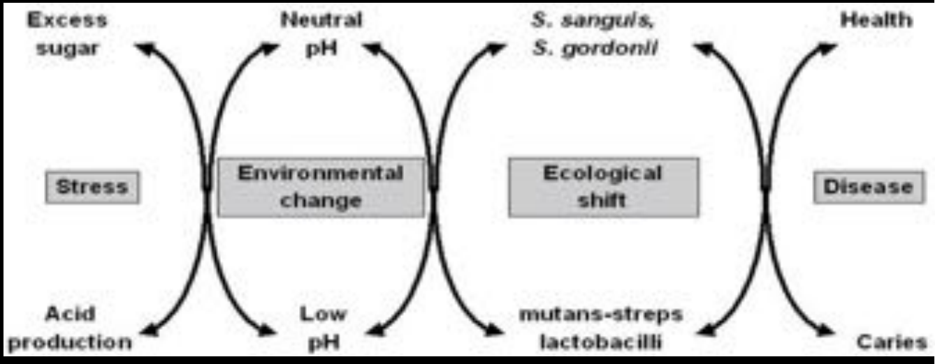
The ecological shift in plaque following environmental change:
-
At neutral pH: health — S. sanguis, S. gordonii predominate
-
Trigger: excess sugar, stress
-
Result: acid production → low pH (environmental change)
-
Ecological shift: mutans streptococci and lactobacilli predominate
-
Outcome: disease — caries
-
More recent models include changes in the biofilm, buffering capacity, fluoride, and interactions among multiple biological and environmental factors. Marsh 1994
Lessons from History89
If we do not learn from history, we shall be compelled to relieve it. True. But if we do not change the future, we shall be compelled to endure it. And that could be worse…
— Alvin Toffler
Historical statements reflecting traditional restorative thinking:
- “the pits and fissures should be probed with a sharp explorer to feel for ‘soft sticky’ substrate”
- “dental caries is a bacterial infection that requires surgical treatment to be cured”
- “one has to strive for as complete caries removal as possible prior to restoration”
- “restorative treatment in children is futile…”
Lecturer — Historical Clinical Beliefs
Several historical or anecdotal beliefs continue to influence clinical practice, including “I do this because it works in my hands.” Feeling pits and fissures to determine whether they are soft or sticky may persist because it has been taught or used for many years, but historical duration does not make an approach evidence-based.
- The traditional focus on treating the lesion has overshadowed management of the caries disease process.
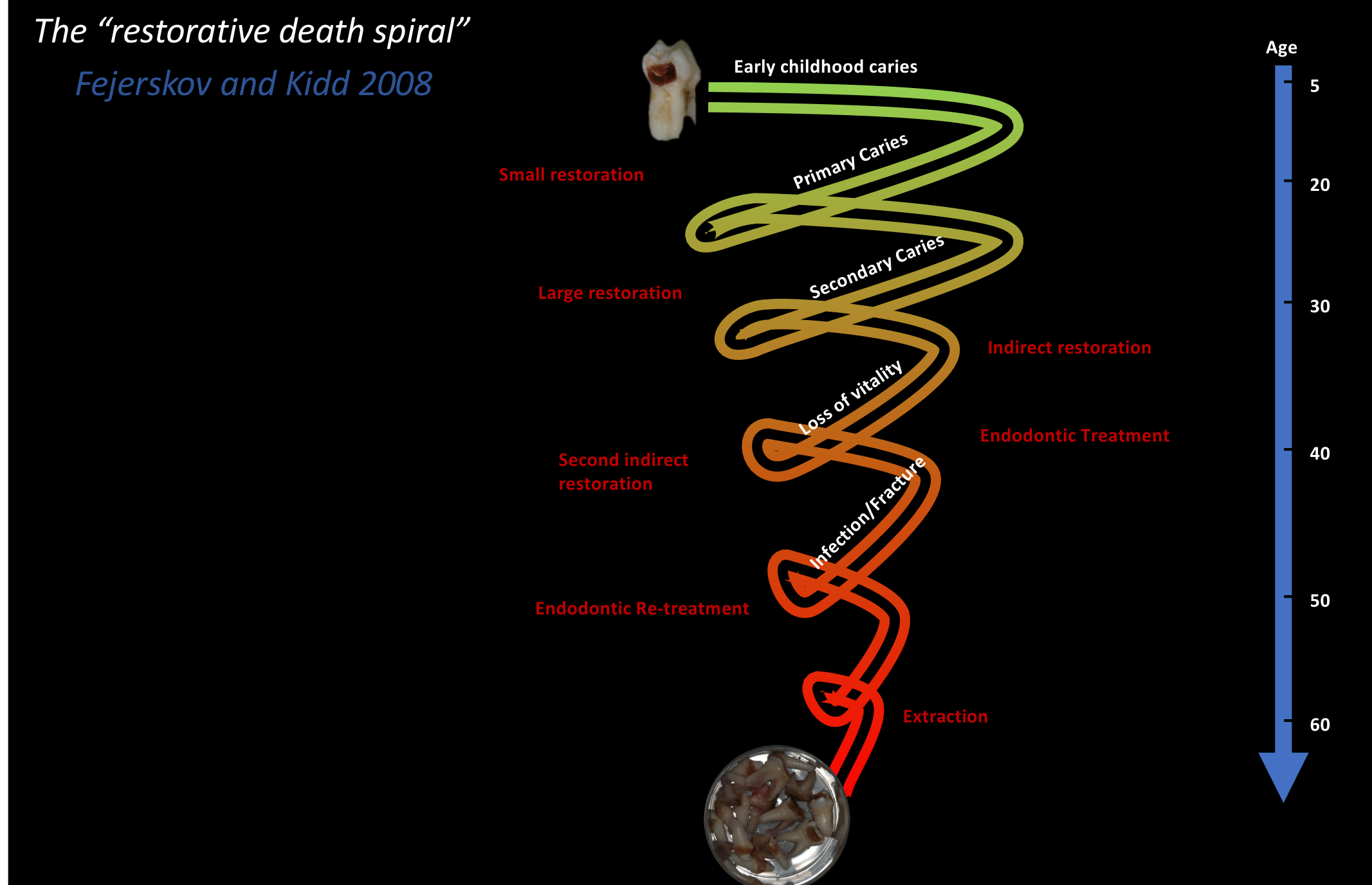
Lecturer — Restorative Cycle
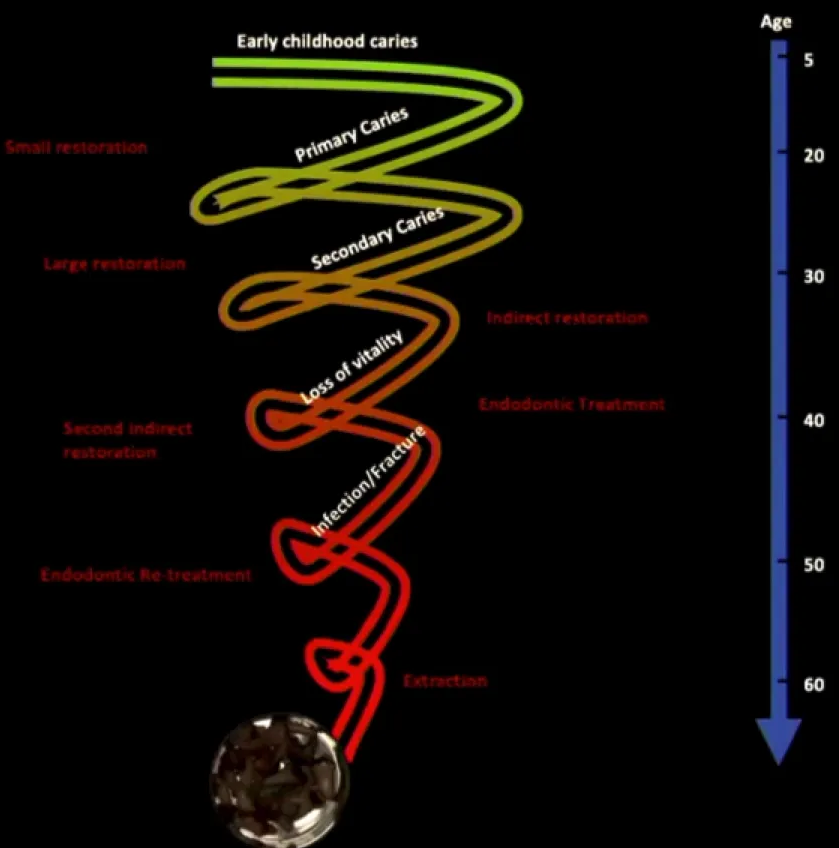
The first restoration placed in a tooth may initiate a long-term cycle of restorative treatment.
- A restoration may later develop marginal staining or breakdown, requiring a larger restoration and further treatment.
- The lecturer cautioned against placing a handpiece on a permanent tooth in a child without careful consideration, because this may commit the tooth to decades of restorative treatment.
- Clinicians should consider the long-term consequences several times before preparing a permanent tooth in a child.
Dental Caries Terminology and Management
Caries Res. 2020;54(1):7-14. doi: 10.1159/000503309. Epub 2019 Oct 7.
Terminology of Dental Caries and Dental Caries Management: Consensus Report of a Workshop Organized by ORCA and Cariology Research Group of IADR
Vita Machiulskiene, Guglielmo Campus, Joana Christina Carvalho, Irene Dige, Kim Rud Ekstrand, Anahita Jablonski-Momeni, Marisa Maltz, David J Manton, Stefania Martignon, E Angeles Martinez-Mier, Nigel B Pitts, Andreas G Schulte, Christian H Splieth, Livia Maria Andaló Tenuta, Andrea Ferreira Zandona, Bente Nyvad
PMID: 31590168 — DOI: 10.1159/000503309 — Free article
Consensus Terminology of Dental Caries
Abstract10
A 2-day workshop of ORCA and the IADR Cariology Research Group was organized to discuss and reach consensus on definitions of the most commonly used terms in cariology. The aims were to identify and to select the most commonly used terms of dental caries and dental caries management and to define them based on current concepts. Terms related to definition, diagnosis, risk assessment, and monitoring of dental caries were included.
The Delphi process was used to establish terms to be considered using the nominal group method favored by consensus. Of 222 terms originally suggested by six cariologists from different countries, a total of 59 terms were reviewed after removing duplicates and unnecessary words. Sixteen experts in cariology took part in the process of reaching consensus about the definitions of the selected caries terms. Decisions were made following thorough “round table” discussions of each term and confirmed by secret electronic voting.
Full agreement (100%) was reached on 17 terms, while the definitions of 6 terms were below the agreed 80% threshold of consensus. The suggested terminology is recommended for use in research, in public health, as well as in clinical practice.
Lecturer — Caries Terminology
The global Delphi process addressed conflicting terminology, including biofilm, sealant, and restoration.
- The definition of dental caries was developed through extensive consultation, with attention to the inclusion and placement of each word.
- In 2020, a distinction was made between the carious lesion and the caries disease: the lesion is the clinical result of a broader disease process.
Lecturer — Restorative Limitations
Restorative treatment is expensive, time-consuming, and potentially traumatic, and it does not necessarily address the poor dental behaviours or biological conditions that caused the need for treatment.
- Poor restorative treatment may provide little benefit compared with no treatment.
- Fluoride released from glass ionomer cement should not be assumed to cure the caries process.
- A primary tooth still requires appropriate restorative principles; omitting a matrix band when needed or joining teeth together as a temporary solution has no appropriate justification simply because the tooth is primary.
- When caries progresses after treatment, the failure may reflect poor treatment quality, continued disease activity, caries progression, or secondary caries.
- Restorative treatment can place a “band-aid” over the problem without controlling the underlying disease.
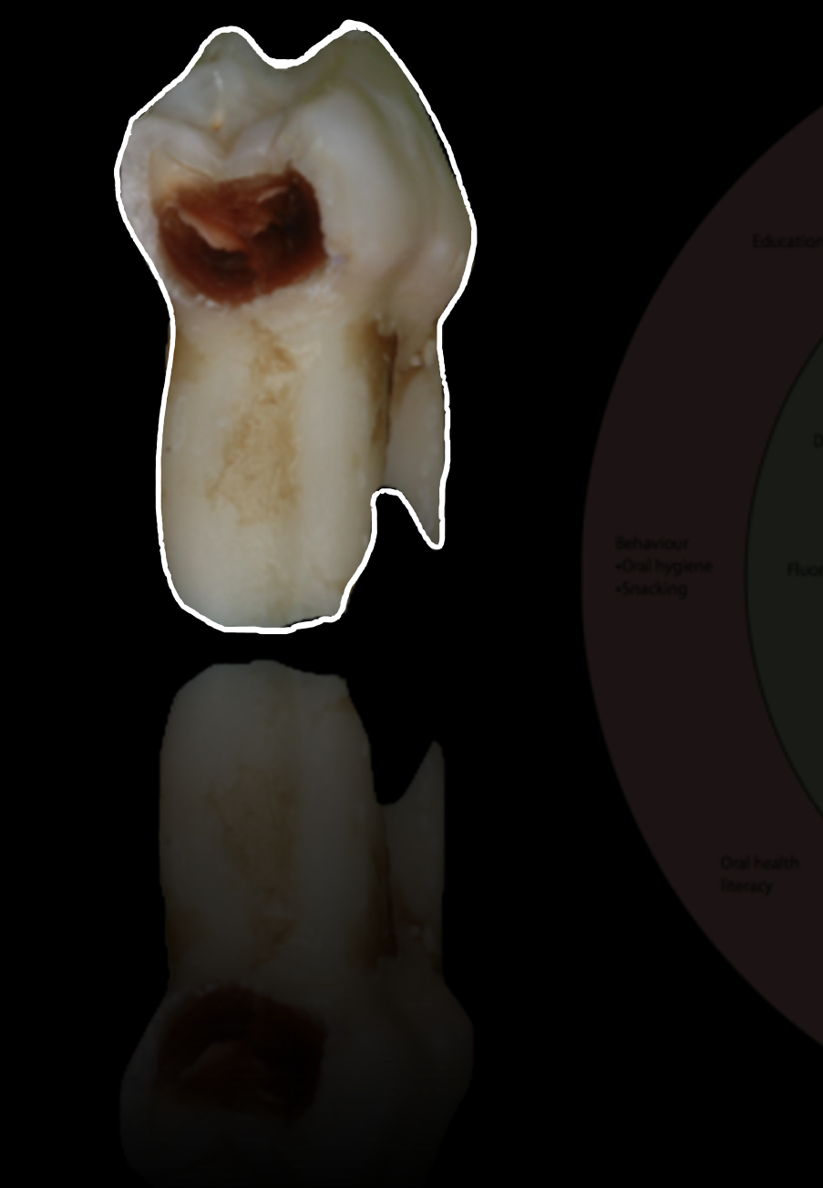
Fate of Carious Primary Teeth111213
Br Dent J. 2002 Feb 23;192(4):219-23. doi: 10.1038/sj.bdj.4801338.
The fate of the carious primary teeth of children who regularly attend the general dental service — M Tickle, K Milsom, D King, P Kearney-Mitchell, A Blinkhorn
Lecturer — Primary Tooth Outcomes
Tickle examined more than 4,000 carious primary teeth, and approximately 80% exfoliated without causing pain, whether or not they had been restored.
- A child’s expression of pain differs from an adult’s, and a child may report no pain at age 10 despite significant lesions.
- Children may previously have experienced pain at age two or three, including difficulty sleeping, eating, or concentrating, but may normalize ongoing pain and assume it is simply part of life.
- Cross-sectional assessments may therefore miss previous pain and its impact on quality of life.
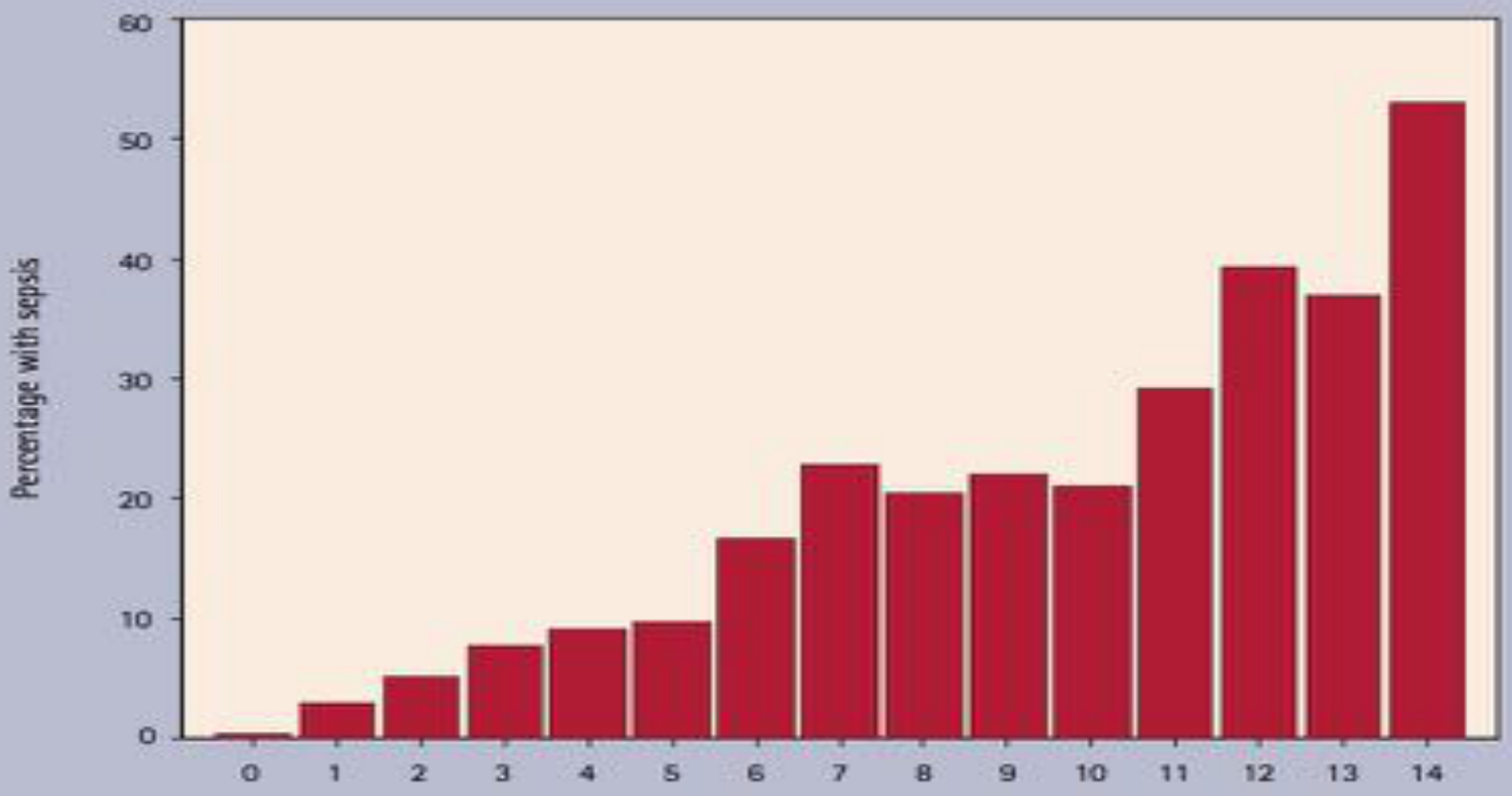
- Pine demonstrated a relationship between the number of untreated carious teeth and sepsis, so leaving carious primary teeth untreated simply because they will exfoliate is not appropriate.
- Children’s perception of pain
- ‘Normalisation’ of dental pain
 |  |
The Restorative Death Spiral14
The “restorative death spiral” — Fejerskov and Kidd 2008
Progression over age (5 → 60 years):
- Early childhood caries
- Primary caries
- Small restoration
- Secondary caries
- Large restoration
- Infection/fracture
- Loss of vitality
- Endodontic treatment
- Endodontic re-treatment
- Indirect restoration
- Second indirect restoration
- Extraction
Limitations of Restorative Care15
“Is restorative care useful while the poor dental behaviour that caused the need for the fillings persists?”
Kidd 2012
Introduction to Non-Restorative Caries Control16
ALL OF A SUDDEN — EVERYBODY HAS BECOME SHELDON
“Alright, does everyone have sound?”
When you find out your normal daily lifestyle is called “quarantine”
Planning your next wine tour made easy
When you normally spend all your time at home anyway but now the government says you have to — well now I am not doing it
Lecturer — NRCC Approach
Non-restorative caries control represents a shift from thoughtless operative care toward biological caries management. Interest in these methods increased during the global pandemic because of the demand for non-aerosol-generating procedures.
- The appropriate treatment depends on the tooth surface, whether the lesion is cavitated or non-cavitated, the lesion severity, the patient, the patient’s compliance, and the clinical context.
- Caries is a spectrum, so a cavitated proximal lesion cannot be managed in exactly the same way as a non-cavitated occlusal lesion.

Agents and Materials for NRCC17
Agents and materials used in NRCC:
-
Sodium Bicarbonate
-
Sodium Fluoride
-
Stannous Fluoride
-
Acidulated Phosphate Fluoride
-
SDF
-
Difluorsilane
-
Polyols
-
Chlorhexidine
-
Amorphous Calcium Phosphate
-
Casein Phosphopeptide-ACP
-
Nanohydroxy-apatite
-
Tricalcium Phosphate
-
Pre and probiotics
-
Lasers
-
Resin Infiltration
-
Fluoride foam
-
Fluoride varnish
-
Xylitol
-
Sorbitol
-
Sealants
-
The Hall technique
-
Non-restorative caries treatment involving opening a lesion so it becomes cleansable
Evidence for Specific Agents
Pre and Probiotics18
Trifa Hedayati-Hajikand, Ulrika Lundberg, Catarina Eldh, Svante Twetman. Effect of probiotic chewing tablets on early childhood caries — a randomized controlled trial. BMC Oral Health. 2015 Sep 24;15(1):112. doi: 10.1186/s12903-015-0096-5. PMID: 26403472. PMCID: PMC4583157. Free PMC article.
Background: To evaluate the effect of probiotic chewing tablets on early childhood caries development in preschool children living in a low socioeconomic multicultural area.
Methods: The investigation employed a randomized double-blind placebo-controlled design. The study group consisted of 138 healthy 2-3-year-old children that were consecutively recruited after informed parental consent. After enrollment, they were randomized to a test or a placebo group. The parents of the test group were instructed to give their child one chewing tablet per day containing three strains of live probiotic bacteria (ProBiora3) and the placebo group got identical tablets without bacteria. The duration was one year and the prevalence and increment of initial and manifest caries lesions was examined at baseline and follow-up. All parents were thoroughly instructed to brush the teeth of their off-springs twice daily with fluoride toothpaste.
Results: The groups were balanced at baseline and the attrition rate was 20%. Around 2/3 of the children in both groups reported an acceptable compliance. The caries increment (Δds) was significantly lower in the test group when compared with the placebo group, 0.2 vs. 0.8 (p < 0.05). The risk reduction was 0.47 (95% CI 0.24–0.98) and the number needed to treat close to five. No differences were displayed between the groups concerning presence of visible plaque or bleeding-on-brushing. No side effects were reported.
Conclusions: The results suggested that early childhood caries development could be reduced through administration of these probiotic chewing tablets as adjunct to daily use of fluoride toothpaste in preschool children. Further studies on a possible dose-response relationship seem justified.
Polyols19
K K Mäkinen, P L Mäkinen, H R Pape Jr, P Allen, C A Bennett, P J Isokangas, K P Isotupa. Stabilisation of rampant caries: polyol gums and arrest of dentine caries in two long-term cohort studies in young subjects. Int Dent J. 1995 Feb;45(1 Suppl 1):93-107. PMID: 7607749.
The purpose of this paper is to report clinical observations on the stabilisation of dentine caries in two chewing gum studies carried out in young subjects. One study focused on the permanent dentition of 1,277 initially 10-year old subjects, while the other study was carried out on 510 initially 6-year old subjects and focused on the primary dentition.
In both trials, several chewing gum formulas containing dietary polyols (xylitol and sorbitol, or their combinations) were used for up to 40 months (10-year olds) or up to 24 months (6-year olds). The daily consumption level of both polyols was up to 10.7 g per subject, used normally in 5-minute chewing episodes 3 to 5 times per day. Supervised chewing in schools lasted 5 min per episode, and for variable times during non-school days.
Rehardening of dentine caries lesions was observed clinically by experienced, blinded examiners using sharp dental explorers and fibre optic light. After 40 months (permanent dentition) or after 18 months (primary dentition), rehardening (caries arrest) generally occurred more frequently in subjects who used polyol gums than in subjects who did not receive gum as part of the programmes, or who received sucrose gum.
Arrest or non-progression of dentine caries was most frequently observed in subjects who used 100 per cent xylitol gums or mixtures of xylitol and sorbitol, but the differences between gums were not consistently significant. However, the usage of 100 per cent xylitol pellet-shaped gum was more frequently associated with arrest of dentine caries than other treatments.
These results and previous studies suggest that high-xylitol chewing gum usage can retard or arrest even rampant dentine caries in conditions where effective restoration and prevention programmes have not been instituted, and can also provide additional protection against further caries development during full implementation of restorative procedures by holding the lesion in a non-progressive condition.
Lecturer — Chewing Gum Effects
When interpreting chewing-gum studies, it is necessary to consider whether the benefit results from the xylitol itself or from the salivary effects of chewing gum. These possible influences are confounding variables.
Nanohydroxyapatite20
K Najibfard, K Ramalingam, I Chedjie, B T Amaechi. Remineralization of early caries by a nano-hydroxyapatite dentifrice. J Clin Dent. 2011;22(5):139-43. PMID: 22403978.
Objective: The purpose of this randomized, double-blind, crossover, in situ study was to evaluate the efficacy of nano-hydroxyapatite (nHAP) dentifrices on caries remineralization and demineralization inhibition.
Methods: Three demineralized enamel blocks (A, B, C) and one healthy block (D), cut from each of 30 molars, were exposed respectively to dentifrices of A) 5% nHAP, B) 10% nHAP, C) 1100 ppm fluoride, and D) 10% nHAP via an intra-oral appliance worn by 30 adults in this four-phase study lasting 28 days per phase. Baseline and post-test mineral loss (delta Z) and lesion depth (LD) were quantified using microradiography.
Results: Pair-wise comparison (baseline versus test) demonstrated significant (p < 0.001) reductions in delta Z and LD in A, B, and C. ANOVA showed no significant differences among the three products in percent mineral gain. No demineralization occurred in the sound enamel specimens exposed intra-orally while using 10% nHAP.
Conclusion: nHAP dentifrice caused remineralization comparable to a fluoride dentifrice, and inhibited caries development, thus suggesting that an nHAP dentifrice can be an effective alternative to fluoride toothpaste.
Lecturer — Nanohydroxyapatite Evidence
In vitro studies have reported a deeper depth of remineralization, a more coherent remineralized structure, and a potentially superior remineralization profile with nanohydroxyapatite. Evidence must distinguish between preventing carious lesions from occurring and arresting existing carious lesions. Arrest studies are more difficult to conduct and are central to the discussion of caries control.
Goals of NRCC21
- Preventing new carious lesions from occurring
- incidence studies, dmft
- effectiveness of NaF/sealants etc
- Arresting/reversing existing carious lesions
Lecturer — NRCC Goals
The goal is to manage the disease process and arrest existing lesions rather than automatically restore every lesion. Treatment should change the local environment so that plaque stagnation sites are reduced, food can be removed, lesions become cleansable, and caries progression is reduced.
- Non-restorative management should not be interpreted as a universal replacement for restorative treatment.
- The clinician must select an appropriate technique for the lesion and monitor the patient regularly.
Managing Non-Cavitated Lesions
Occlusal Surfaces22
-
0.2% NaF mouthwash + supervised toothbrushing
-
1.23% APF gel
-
5% NaF varnish
-
Resin infiltration + 5% NaF varnish
-
Sealants + 5% NaF varnish → most effective
-
Sealants alone
-
2–3x greater chance of caries arrest than no treatment
- 1,575 lesions in primary and permanent teeth
- 8 to 12 months
- Evidence came from seven clinical trials, with moderate certainty.
Agrawal et al. 2011; Autio-Gold et al. 2001; Bakhshandeh et al. 2015; Borges et al. 2010; da Silveira et al. 2012; Florio et al. 2001; Honkala et al. 2015.
Proximal Surfaces23
- Radiolucencies (lesions in enamel to outer third of the dentin)
- Resin infiltration
- Sealants after tooth separation
- 5% NaF varnish alone
- 2x greater chance of caries arrest (low certainty)
- Resin infiltration + 5% NaF varnish → most effective
- 5x greater chance of caries arrest (low certainty)
Lecturer — Proximal Evidence
The certainty of evidence for non-cavitated proximal lesions was lower than for occlusal lesions.
- The studies were heterogeneous, making interpretation more difficult.
Martignon et al. 2006; Ekstrand et al. 2010; Martignon et al. 2010; Paris et al. 2010; Martignon et al. 2012; Meyer-Lueckel et al. 2012; Gomez et al. 2015.
Buccal and Lingual Surfaces24
- 5% NaF varnish alone → most effective
- 1.23% APF + Oral Health Education
- 2–3x greater chance of caries arrest (low-moderate certainty)
- 10% CPP-ACP + toothbrushing with 1000ppm toothpaste
- 2x greater chance of caries arrest (low certainty)
Lecturer — Tooth Mousse
Tooth mousse has shown strong in vitro results, but clinical trials have produced conflicting outcomes.
- Patient compliance is a major influence on its success.
- Fluoride varnish provides more predictable results in this context.
Autio-Gold et al. 2001; Bailey et al. 2009; Agrawal et al. 2011; Bonow et al. 2013; Turska-Szybka et al. 2016.
Lecturer — SDF Case Selection
Careful diagnosis of the lesion and tooth is essential before applying silver diamine fluoride. Deep lesions are particularly concerning because of the risk of treatment failure and pulpal involvement, including possible microscopic pulp exposure.
Managing Cavitated Lesions
Coronal Surfaces25
- 30% SDF solution annually is superior to 5% NaF applied 1/wk for 3 wks
- high certainty
- 38% SDF solution biannually is superior to 12% biannually or 38% annually
- high certainty
Lecturer — Coronal Lesions
Silver diamine fluoride forms a substantial part of the evidence for managing cavitated carious lesions. Twice-yearly application was identified as the most effective approach among the methods discussed, but lesions still require careful diagnosis and monitoring.
Duangthip et al. 2016; Fung, et al. 2018; Duangthip, et al. 2018; Fung et al. 2016; Llodra et al. 2005; Yee et al. 2009.
Lecturer — Cleansable Lesions
Non-restorative treatment of cavitated lesions may involve opening a lesion so that it becomes cleansable. This improves access for cleaning, allows food to be removed, eliminates plaque stagnation sites, and promotes caries arrest.
Silver Diamine Fluoride on Coronal Surfaces
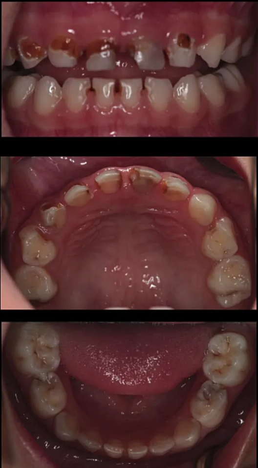
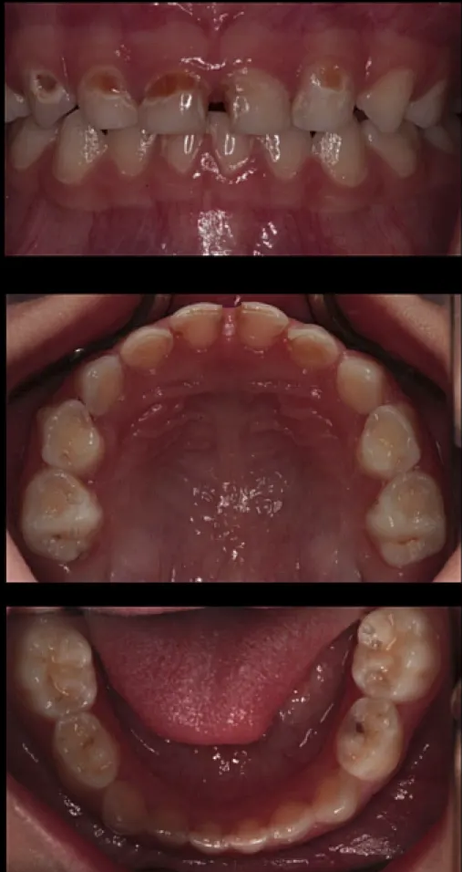
Four-Year-Old With Caries
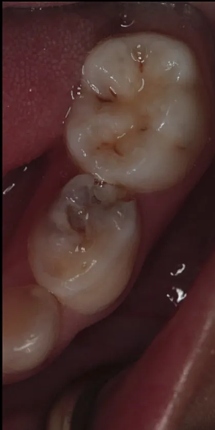
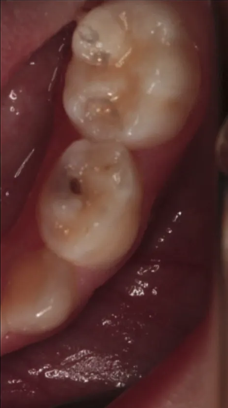
In a four-year-old child with early childhood carious lesions, silver diamine fluoride was applied, but significant staining was present at 12 months and almost every lesion had progressed. In a mandibular primary molar, an ICDAS4 lesion progressed to an ICDAS5 lesion with marginal ridge breakdown after 12 months despite twice-yearly application, demonstrating that research success does not guarantee arrest in every clinical case.
Mechanisms of Action of SDF
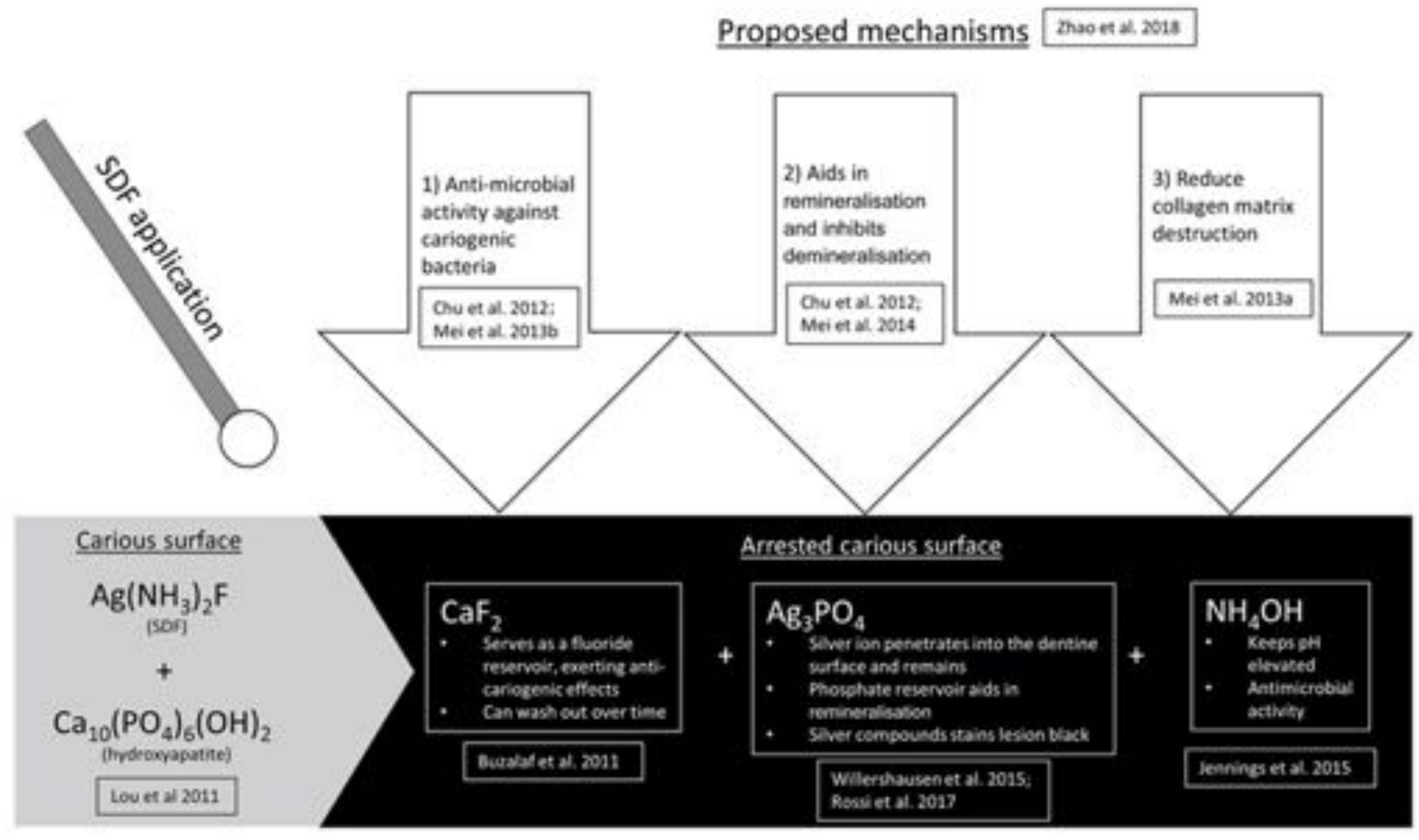
Proposed Mechanisms (Zhao et al. 2018)26
SDF application:
- Anti-microbial activity against cariogenic bacteria (Chu et al. 2012; Mei et al. 2013b)
- Aids in remineralisation and inhibits demineralisation (Chu et al. 2012; Mei et al. 2014)
- Reduce collagen matrix destruction (Mei et al. 2013a)
Lecturer — SDF Effects
Silver diamine fluoride inhibits multiple bacterial species and produces a highly remineralized zone when applied to dentine lesions. Its effects reflect the combined actions of silver on bacteria and fluoride in remineralization.
Reaction Products
Carious surface: Ag(NH₃)₂F (SDF) + Ca₁₀(PO₄)₆(OH)₂ (hydroxyapatite) → arrested carious surface (Lou et al 2011)
CaF₂ (Buzalaf et al. 2011)
- Serves as a fluoride reservoir, exerting anti-cariogenic effects
- Can wash out over time
Ag₃PO₄ (Willershausen et al. 2015; Rossi et al. 2017)
- Silver ion penetrates into the dentine surface and remains
- Phosphate reservoir aids in remineralisation
- Silver compounds stain lesion black
NH₄OH (Jennings et al. 2015)
- Keeps pH elevated
- Antimicrobial activity
Zhao et al; Hu et al. 2018.
Regulatory History of SDF27
- 2014: US FDA approves SDF as a treatment for dentinal sensitivity
- prior to this SDF had been used off-label for caries arrest
- 2016: FDA awarded ‘Breakthrough Therapy status’ for approval of SDF as a drug to treat severe early childhood caries
- allows for SDF to be evaluated in clinical trials in the treatment of dental caries
- 2016: “Interim Caries Arresting Medicament Application”
Lecturer — SDF Regulation
Silver diamine fluoride has been used in Japan since the 1970s. In Australia, it is not approved by the TGA for caries management, so its use for caries control is off-label; later in 2016, the American Dental Association added a separate code for its application.
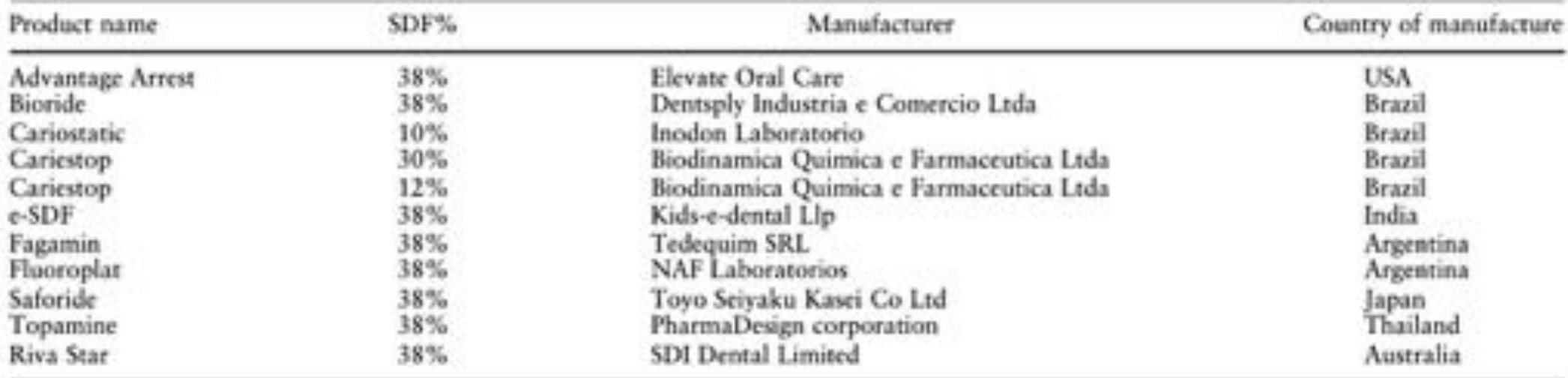
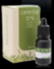
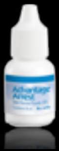
Commercial SDF Products28
| Product name | SDF% | Manufacturer | Country of manufacture |
|---|---|---|---|
| Advantage Arrest | 38% | Elevate Oral Care | USA |
| Bioride | 38% | Dentsply Industria e Comercio Ltda | Brazil |
| Cariostatic | 10% | Inodon Laboratorio | Brazil |
| Cariestop | 30% | Biodinamica Quimica e Farmaceutica Ltda | Brazil |
| Cariestop | 12% | Biodinamica Quimica e Farmaceutica Ltda | Brazil |
| e-SDF | 38% | Kids-e-dental Llp | India |
| Fagamin | 38% | Tedequim SRL | Argentina |
| Fluoroplast | 38% | NAF Laboratorios | Argentina |
| Saforide | 38% | Toyo Seiyaku Kasei Co Ltd | Japan |
| Topamine | 38% | PharmaDesign corporation | Thailand |
| Riva Star | 38% | SDI Dental Limited | Australia |
Patel et al. 2021.
 |  |  |
 |  |  |
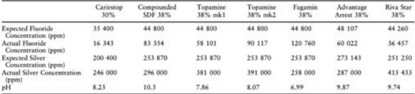
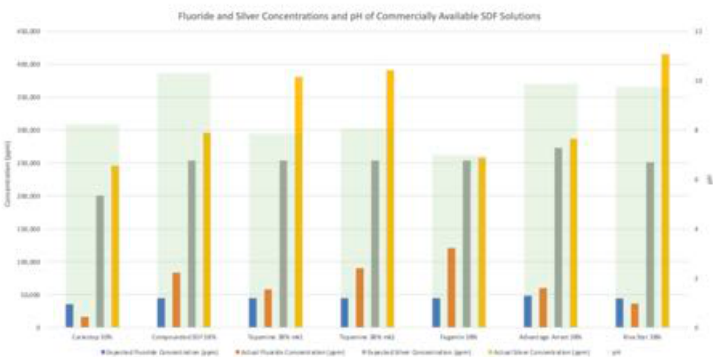
Fluoride and Silver Concentrations of SDF Products29
| Cariestop 30% | Compounded SDF 38% | Topamine 38% mk1 | Topamine 38% mk2 | Fagamin 38% | Advantage Arrest 38% | Riva Star 38% | |
|---|---|---|---|---|---|---|---|
| Expected Fluoride Concentration (ppm) | 35 400 | 44 800 | 44 800 | 44 800 | 44 800 | 48 107 | 44 260 |
| Actual Fluoride Concentration (ppm) | 16 343 | 83 354 | 58 101 | 90 117 | 120 760 | 60 022 | 36 457 |
| Expected Silver Concentration (ppm) | 200 400 | 253 870 | 253 870 | 253 870 | 253 870 | 273 143 | 251 250 |
| Actual Silver Concentration (ppm) | 246 000 | 296 000 | 381 000 | 391 000 | 258 000 | 287 000 | 415 433 |
| pH | 8.23 | 10.3 | 7.86 | 8.07 | 6.99 | 9.87 | 9.74 |
Patel et al. 2021.
Lecturer — Product Differences
Rivastar was identified as the only Australian-made compound, while Topamine is marketed in Australia but was described as not being widely commercially available. Products described as silver diamine fluoride are not necessarily identical in composition.
 |  |
Contraindications for SDF30
- teeth with evidence of pulpitis
- deep dentine lesions encroaching the pulp
- silver allergy
- aesthetic demand
Patel et al. 2019.
Alternative Caries Management Options Trial31
Randomized Controlled Trial — Caries Res. 2018 Jan;51(6):605-614. doi: 10.1159/000477855. Epub 2017 Dec 20.
Alternative Caries Management Options for Primary Molars: 2.5-Year Outcomes of a Randomised Clinical Trial
Ruth M Santamaria, N P T Innes, Vita Machiulskiene, Julian Schmoeckel, Mohammad Alkilzy, Christian H Splieth
- Hall Technique (HT)
- Non-restorative caries treatment (NRCT)
- Conventional restorations (CR)
Santamaria et al. 2018.
Treatment Protocols Compared
Hall Technique (HT)
- No caries removal or tooth preparation
- No local anaesthesia
- Ortho separators
- Cementation of stainless steel crown
Non-restorative Caries Treatment (NRCT)
- Lesions were opened with a high-speed bur
- Cavity made cleansable
- Application of 5% NaF
Conventional Restorations (CR)
- Complete caries removal under local anaesthesia
- Matrix band
- Restored using a compomer
Santamaria et al. 2018.
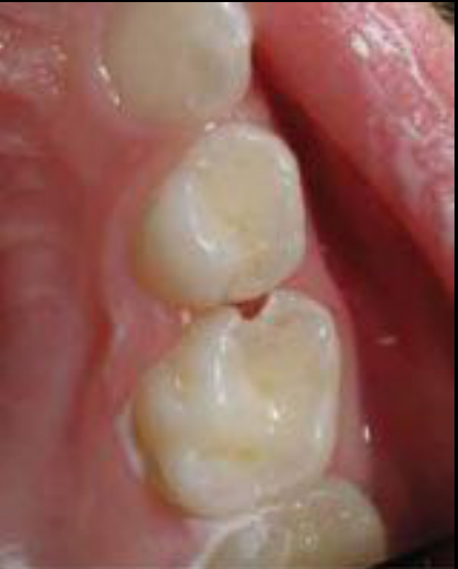
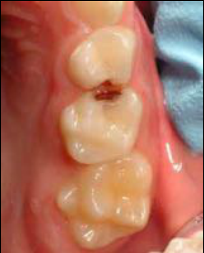
Non-Restorative Caries Treatment Rationale32
Kidd et al. 2012.
Lecturer — Non-Restorative Rationale
The non-restorative approach is biological rather than focused on removing all caries. Opening the lesion makes it accessible for cleaning, allows food to be removed, and removes plaque stagnation sites, but informed consent and regular monitoring are important because parents and other professionals may find the approach difficult to understand.
 |  |
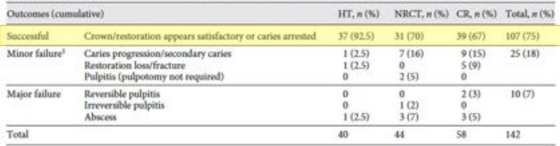
Trial Outcomes33
| Outcomes (cumulative) | HT, n (%) | NRCT, n (%) | CR, n (%) | Total, n (%) |
|---|---|---|---|---|
| Successful — crown/restoration appears satisfactory or caries arrested | 37 (92.5) | 31 (70) | 39 (67) | 107 (75) |
| Minor failure — caries progression/secondary caries | 1 (2.5) | 7 (16) | 9 (15) | 25 (18) |
| — Restoration loss/fracture | 1 (2.5) | 0 | 5 (9) | |
| — Pulpitis (pulpotomy not required) | 0 | 2 (5) | 0 | |
| Major failure — reversible pulpitis | 0 | 0 | 2 (3) | 10 (7) |
| — Irreversible pulpitis | 0 | 1 (2) | 0 | |
| — Abscess | 1 (2.5) | 3 (7) | 3 (5) | |
| Total | 40 | 44 | 58 | 142 |
- No statistically significant difference between NRCT and CR
- Hall technique has a better seal — need to ensure dentine between lesion and pulp
Santamaria et al. 2018.
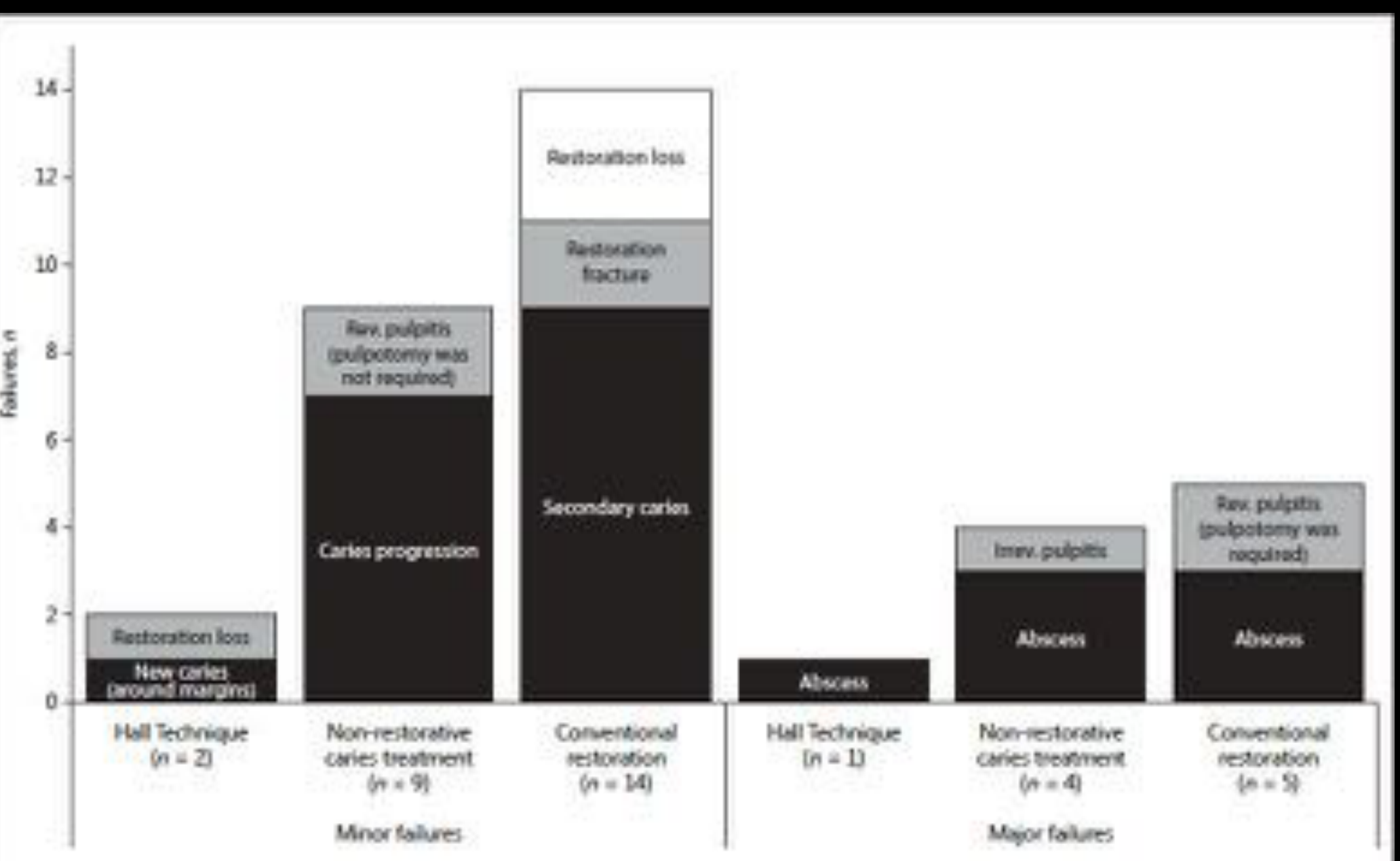
Failure Analysis34
Failure counts (y-axis 0–14):
Minor failures
- Hall Technique (n = 1): restoration loss
- Non-restorative caries treatment (n = 4): caries progression; restoration loss; restoration fracture; secondary caries
- Conventional restoration (n = 5): rev. pulpitis (pulpotomy was not required); restoration loss; new caries (around margins)
Major failures
- Hall Technique (n = 2): abscess
- Non-restorative caries treatment (n = 9): rev. pulpitis (pulpotomy was required); irreversible pulpitis; abscess
- Conventional restoration (n = 14): abscess; restoration loss
Santamaria et al. 2018.
Lecturer — Trial Interpretation
Non-restorative caries treatment and conventional restorative treatment each had approximately a 70% success rate, whereas the Hall technique achieved approximately a 90% success rate. The success of the Hall technique was attributed to the quality of the seal rather than simply to the absence of caries removal.
Lecturer — Hall Technique Selection
A bitewing radiograph should show a clear band of dentine between the carious lesion and the pulp before using the Hall technique. If this band is absent, failure is more likely, and neither fluoride release nor a high reported success rate removes the need for diagnosis and case selection.
Managing Root Surfaces35
- 5000 ppm F toothpaste
- 3x greater chance of caries arrest (low certainty)
- 1% Chx + 1% thymol varnish
- 38% SDF annually
- 38% SDF + KI annually
- 5% NaF varnish
- 2–3x greater chance of caries arrest (v low – low certainty)
Best outcome.
Schaeken et al. 1991; Wallace et al. 1993; Lynch et al. 2000; Baysan et al. 2001; Brailsford et al. 2002; Wyatt et al. 2004; Ekstrand et al. 2008; Baca et al. 2009; Ekstrand et al. 2013; Zhang et al. 2013; Li et al. 2016.
Lecturer — Root Caries Context
Root caries is not generally encountered in the pediatric population but was included for completeness. Silver diamine fluoride is also being used for patients in residential aged care who may have complex medical histories, be unable to maintain root-surface restorations, or have limited access to dental care.
Summary and Clinical Guidelines
Summary of Systematic Reviews36
- Non-cavitated proximal surfaces — Sealants and resin infiltration are more effective than other non-invasive treatments. Dorri et al. 2015
- Non-cavitated buccal/facial surfaces — 5% NaF is more effective than 1.23% APF or no treatment. Lenzi et al. 2016
- Non-cavitated occlusal surfaces — Sealants are more effective than other non-invasive treatments. Wright et al. 2016
- Cavitated dentine caries — 38% SDF solution is more effective than other non-invasive treatments. Contreras et al. 2017
Lecturer — Evidence and Consent
Systematic reviews reflect the emerging evidence for caries arrest, and the literature is relatively recent because caries arrest is only now being examined more extensively.
- Traditional operative treatments are increasingly being challenged by biological and non-restorative approaches.
- Treatment selection should depend on the lesion, tooth surface, patient, and clinical context.
- The American Dental Association has developed clinical practice guidelines for non-restorative caries control in primary and permanent teeth, drawing substantially on a 2019 network meta-analysis by Urquhart.
- Care should include informed consent, discussion of treatment options, explanation of their advantages and limitations, and regular monitoring.
Clinical Recommendations for Primary Teeth
Recommendations by coronal surface and lesion status:
Occlusal Surfaces37
- Noncavitated*: Sealants plus 5% NaF varnish‡ or sealants alone
- Cavitated†:
- 5% NaF varnish†,§ or
- Resin infiltration alone or
- Resin infiltration plus 5% NaF varnish# or
- Sealants alone
Lecturer — Sealant Technique
Sealants must be placed carefully because a poorly placed sealant can create a plaque-retentive niche and cause more harm than benefit.
- Moisture control is essential, and all pits and fissures must be adequately sealed.
- Pay particular attention to distal pits on upper molars and buccal pits on lower molars.
- Remove excess material, floss the contacts, and check for excess under an operculum, especially around partially erupted teeth.
- Less material is preferable to excess material.
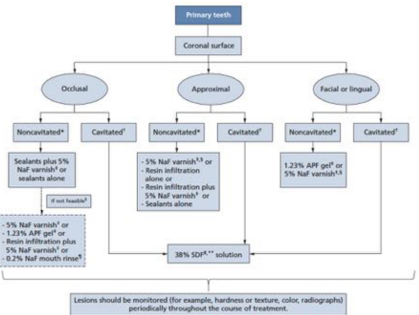
Approximal Surfaces
- Noncavitated*:
- Sealants plus 5% NaF varnish‡ or sealants alone
- If not feasible§:
- 5% NaF varnish† or
- 1.23% APF gel# or
- Resin infiltration plus 5% NaF varnish† or
- 0.2% NaF mouth rinse¶
- Cavitated†: 1.23% APF gel# or 5% NaF varnish†,§
Lecturer — Proximal Lesions
Cavitated proximal lesions require a different approach from non-cavitated lesions and must be assessed for cleansability and severity. For non-cavitated proximal lesions, resin infiltration and sodium fluoride varnish were likely to provide the greatest chance of arrest, although the evidence was heterogeneous.
Facial or Lingual Surfaces
- Noncavitated*: Sealants plus 5% NaF varnish‡ or sealants alone
- Cavitated†: 38% SDF** solution
Lecturer — Tooth Mousse
Tooth mousse has inconsistent clinical evidence despite favorable in vitro findings. Its success is strongly affected by patient compliance.
Lesions should be monitored (for example, hardness or texture, color, radiographs) periodically throughout the course of treatment.
Slayton et al. 2018
Clinical Recommendations for Permanent Teeth
Recommendations by coronal and root surface, and lesion status:
Occlusal Surfaces38
- Noncavitated*: Sealants plus 5% NaF varnish‡ or sealants alone
- Cavitated†:
- 5% NaF varnish‡ or
- 1.23% APF gel¶ or
- 0.2% NaF mouth rinse#
- If not feasible§, use the alternatives above
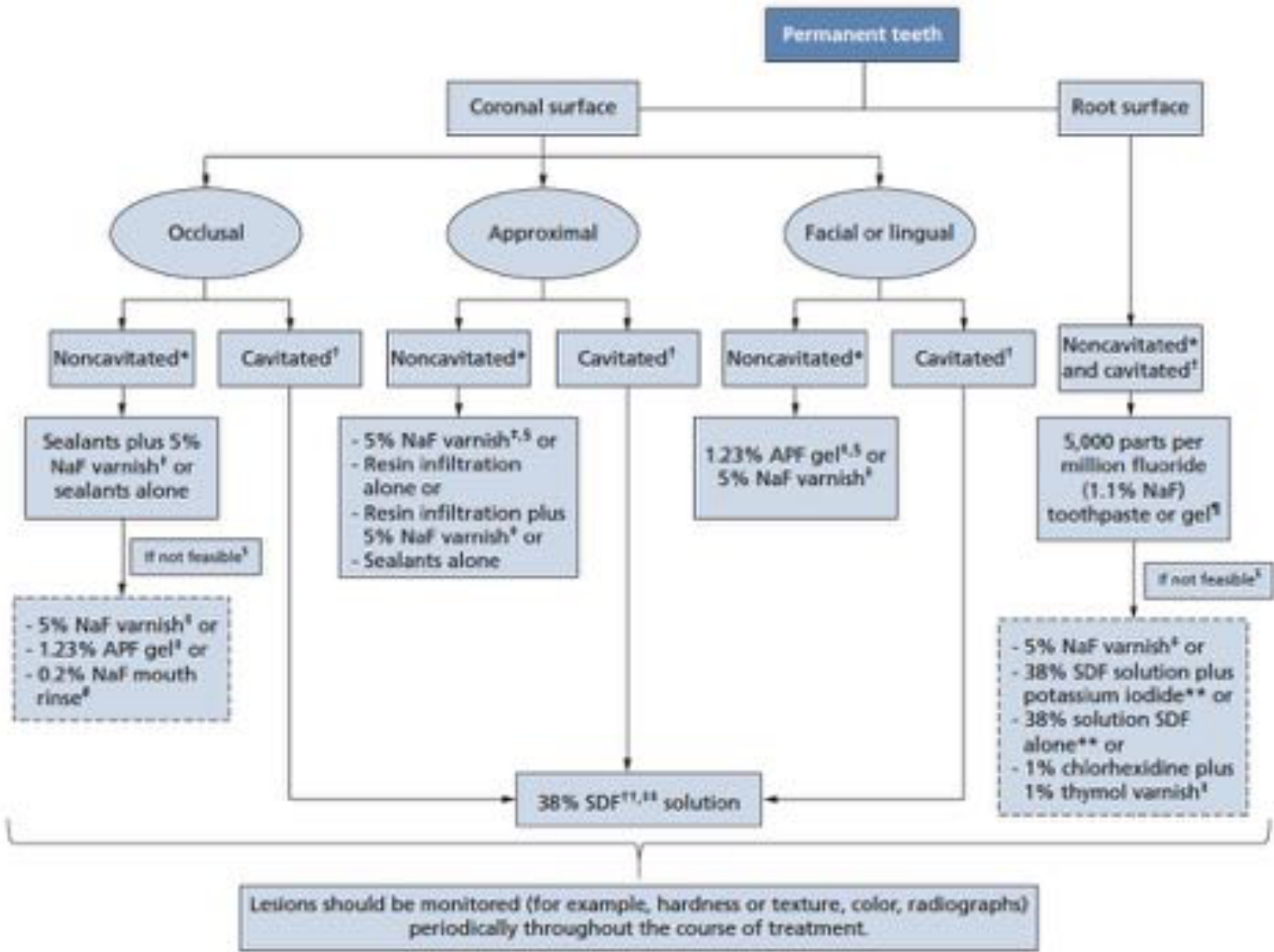
Approximal Surfaces
- Noncavitated*:
- 5% NaF varnish‡,§ or
- Resin infiltration alone or
- Resin infiltration plus 5% NaF varnish‡ or
- Sealants alone
- Cavitated†: 1.23% APF gel¶,§ or 5% NaF varnish‡
Facial or Lingual Surfaces
- Noncavitated*: Sealants plus 5% NaF varnish‡ or sealants alone
- Cavitated†: 38% SDF††,## solution
Root Surfaces
- Noncavitated* and cavitated†:
- 5,000 parts per million fluoride (1.1% NaF) toothpaste or gel#
- If not feasible§:
- 5% NaF varnish‡ or
- 38% SDF solution plus potassium iodide** or
- 38% solution SDF alone** or
- 1% chlorhexidine plus 1% thymol varnish††
Lecturer — Root Surface Care
Root-surface restorations have poor longevity and success rates. Simple, low-cost, and less technique-sensitive approaches may be particularly appropriate for patients with complex medical conditions, limited access to care, or difficulty maintaining restorations.
Lesions should be monitored (for example, hardness or texture, color, radiographs) periodically throughout the course of treatment.
Slayton et al. 2018
Clinical Cases
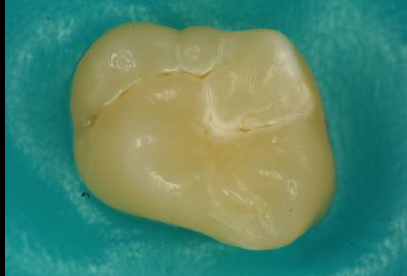
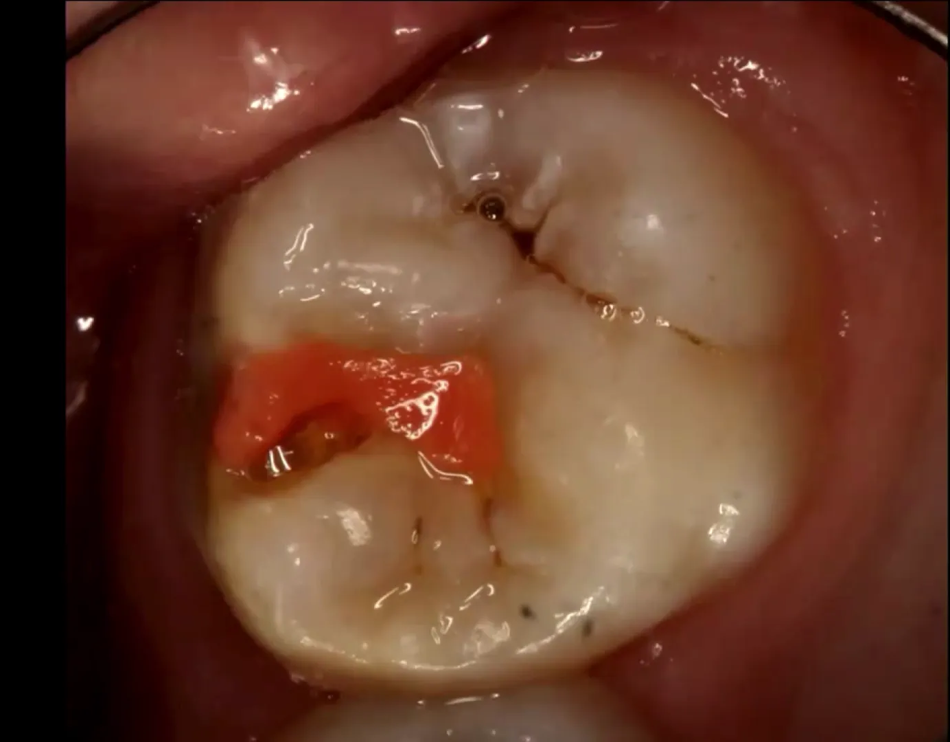
Case: Poorly placed fissure sealant on a molar
Question
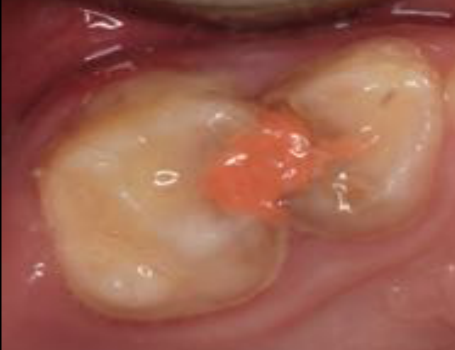
Scenario: A clinical photo of a molar with a fissure sealant placed using glass ionomer cement (Fuji 7).
What’s shown: A close-up view of a tooth showing a fissure sealant with incomplete coverage and poor isolation.
Consider: What technical errors are evident in this sealant placement, and what are the potential consequences?
Answer
Observations:
- The sealant was placed without adequate moisture control.
- Cotton roll placement was unsatisfactory.
- The distal pit was not sealed.
Reasoning: The most commonly missed areas in sealant placement are the distal pits on upper molars and buccal pits on lower molars. Placing a sealant without proper moisture control or leaving excess material under an operculum creates a niche for plaque. Since the goal of a fissure sealant is to prevent caries, creating a niche does more harm than good.
Takeaway: Fissure sealant placement with glass ionomer cement is highly technique-sensitive; inadequate moisture control and incomplete coverage can create carious niches and cause more harm than good.
Case: CT scan of a 2000-year-old mummy
Question
Scenario: A CT scan of a 2000-year-old mummy, presented to illustrate the historical presence and severity of dental disease.
What’s shown: A CT scan revealing extensive dental and periapical pathology.
Consider: What specific dental findings are visible on this scan, and what do they suggest about the individual’s fate?
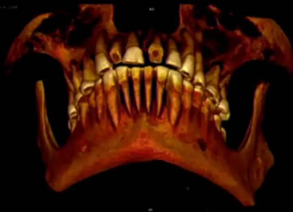
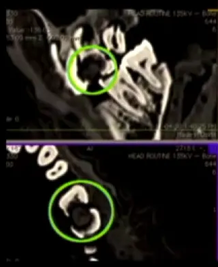
Answer
Observations:
- Fairly large carious lesions.
- Periapical areas of resorption.
- Probable extension of the infection into the sinus.
Reasoning: The extensive carious lesions and periapical resorption extending into the sinus indicate a severe, unchecked infection. Given the extent of the disease, it was likely the cause of the individual’s death.
Outcome: The individual likely died as a result of these severe dental infections.
Takeaway: Dental caries is an ancient disease, and severe, untreated carious lesions with periapical extension can be fatal.
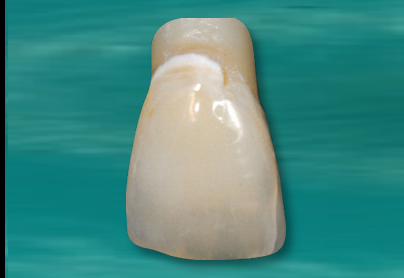
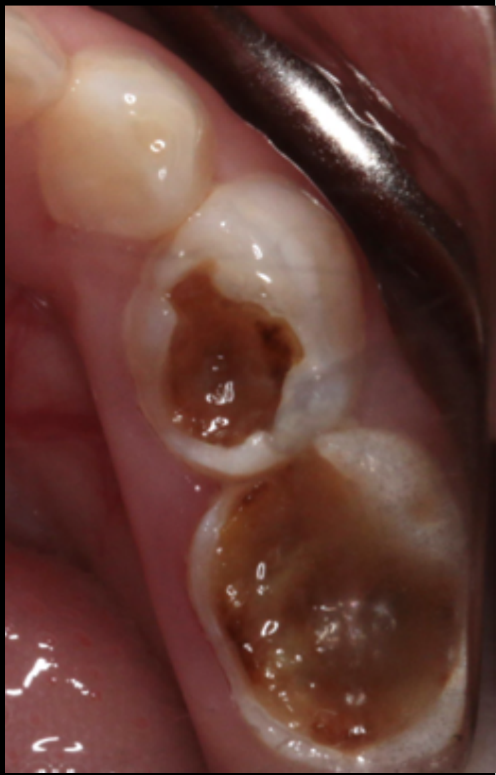
Case: Poor posterior restorations, clinical photo
Question
Scenario: A clinical photograph of a posterior restoration is presented to illustrate poor restorative technique in primary teeth.
What’s shown: An intraoral view of two posterior teeth that have been restored together as a single unit.
Consider: What specific technical flaws are evident in this restoration, and is there any justification for this approach?
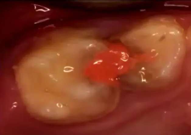
Answer
Observations:
- The two posterior teeth are joined together in a single restoration.
- The procedure was apparently performed without local anesthesia.
- The procedure was performed without a matrix band.
- A material such as Fuji 7 was used, based on the assumption that its fluoride release would cure the caries.
Reasoning: The lecturer identifies these choices as an abuse of materials and “stupid ideas.” The assumption that primary teeth do not require local anesthesia or a matrix band, or that joining them together is acceptable, is completely unfounded. The lecturer emphasizes that a dentist would have very little justification to support doing this treatment.
Takeaway: Dentists must not invent their own techniques or cut corners when restoring primary teeth; standard restorative principles must be followed, as there is no justification for such shortcuts in practice.
Case: Four-year-old child with early childhood caries treated with SDF
Question
Scenario: A four-year-old child presents with early childhood carious lesions and is treated with silver diamine fluoride (SDF).
What’s shown: An intraoral clinical photograph showing the child’s teeth, including anterior teeth and a mandibular primary molar, evaluated at baseline and at a 12-month follow-up.
Consider: Evaluate the clinical outcome of the SDF application at the 12-month follow-up and what this demonstrates about the limitations of SDF in clinical practice.
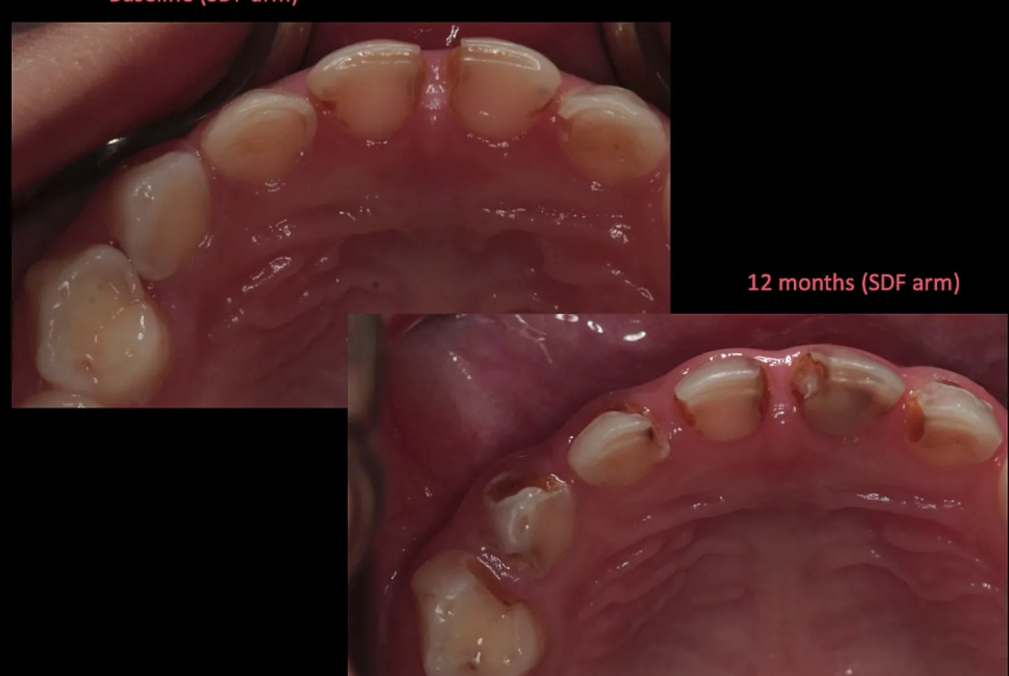
Answer
Observations:
- Significant black staining of the carious lesions following SDF application.
- Progression of almost every lesion in size and severity at 12 months.
- The mandibular primary molar progressed from an ICDAS 4 lesion at baseline to an ICDAS 5 lesion with marginal ridge breakdown.
- Anterior lesions progressed despite biannual SDF application.
Reasoning: While SDF is highly effective in research settings for arresting caries, it is not a “silver bullet” or panacea in pragmatic clinical practice. Lesions that are not open and cleansable may continue to progress despite treatment, and clinicians must avoid being lulled into a false sense of security by its proven efficacy in controlled studies.
Outcome: At the 12-month follow-up, the lesions showed significant progression and staining, demonstrating treatment failure in this pragmatic scenario.
Takeaway: SDF is not a magic cure for dental caries; for the most favorable results, it should be used to treat lesions that are open and cleansable, and clinicians must carefully diagnose and understand the limitations of the carious lesion before application.
Case: Non-restorative caries treatment example
Question
Scenario: An example of the non-restorative caries treatment (NRCT) approach is presented.
What’s shown: Clinical photographs of posterior teeth showing the treated lesion.
Consider: How does this approach manage the carious lesion?
Answer
Observations:
- The lesion has been opened up to make it cleansable.
- Food can be easily removed from the area.
- Sites of plaque stagnation have been removed.
Reasoning: This approach represents a biological way of managing a lesion. By changing the local environment to make the area cleansable and removing plaque stagnation, it acts to promote caries arrest.
Takeaway: Non-restorative caries treatment is a biological approach that manages lesions by changing the local environment to make them cleansable and removing plaque stagnation, thereby promoting caries arrest.
Footnotes
-
Original PDF page 1: L4 Non-restorative caries control DMD, p.1 ↩
-
Original PDF page 2: L4 Non-restorative caries control DMD, p.2 ↩
-
Original PDF page 3: L4 Non-restorative caries control DMD, p.3 ↩
-
Original PDF page 4: L4 Non-restorative caries control DMD, p.4 ↩
-
Original PDF page 5: L4 Non-restorative caries control DMD, p.5 ↩
-
Original PDF page 6: L4 Non-restorative caries control DMD, p.6 ↩
-
Original PDF page 7: L4 Non-restorative caries control DMD, p.7 ↩
-
Original PDF page 8: L4 Non-restorative caries control DMD, p.8 ↩
-
Original PDF page 9: L4 Non-restorative caries control DMD, p.9 ↩
-
Original PDF page 10: L4 Non-restorative caries control DMD, p.10 ↩
-
Original PDF page 11: L4 Non-restorative caries control DMD, p.11 ↩
-
Original PDF page 12: L4 Non-restorative caries control DMD, p.12 ↩
-
Original PDF page 13: L4 Non-restorative caries control DMD, p.13 ↩
-
Original PDF page 14: L4 Non-restorative caries control DMD, p.14 ↩
-
Original PDF page 15: L4 Non-restorative caries control DMD, p.15 ↩
-
Original PDF page 16: L4 Non-restorative caries control DMD, p.16 ↩
-
Original PDF page 17: L4 Non-restorative caries control DMD, p.17 ↩
-
Original PDF page 18: L4 Non-restorative caries control DMD, p.18 ↩
-
Original PDF page 19: L4 Non-restorative caries control DMD, p.19 ↩
-
Original PDF page 20: L4 Non-restorative caries control DMD, p.20 ↩
-
Original PDF page 21: L4 Non-restorative caries control DMD, p.21 ↩
-
Original PDF page 22: L4 Non-restorative caries control DMD, p.22 ↩
-
Original PDF page 23: L4 Non-restorative caries control DMD, p.23 ↩
-
Original PDF page 24: L4 Non-restorative caries control DMD, p.24 ↩
-
Original PDF page 25: L4 Non-restorative caries control DMD, p.25 ↩
-
Original PDF page 26: L4 Non-restorative caries control DMD, p.26 ↩
-
Original PDF page 27: L4 Non-restorative caries control DMD, p.27 ↩
-
Original PDF page 28: L4 Non-restorative caries control DMD, p.28 ↩
-
Original PDF page 29: L4 Non-restorative caries control DMD, p.29 ↩
-
Original PDF page 30: L4 Non-restorative caries control DMD, p.30 ↩
-
Original PDF page 31: L4 Non-restorative caries control DMD, p.31 ↩
-
Original PDF page 33: L4 Non-restorative caries control DMD, p.33 ↩
-
Original PDF page 34: L4 Non-restorative caries control DMD, p.34 ↩
-
Original PDF page 35: L4 Non-restorative caries control DMD, p.35 ↩
-
Original PDF page 36: L4 Non-restorative caries control DMD, p.36 ↩
-
Original PDF page 37: L4 Non-restorative caries control DMD, p.37 ↩
-
Original PDF page 38: L4 Non-restorative caries control DMD, p.38 ↩
-
Original PDF page 39: L4 Non-restorative caries control DMD, p.39 ↩