Cleft Lip And Palate1
Lecturer — Syndromic Associations
Cleft lip and palate alone is not usually caused by a syndrome or systemic disorder.
- Approximately 70% of children with cleft lip and palate have no other significant condition apart from the oral cleft.
- Cleft palate only is less common and has a somewhat different aetiology.
- Cleft palate only has a higher proportion of associations with syndromes.
- Historical stigma has resulted from assumptions that children with oral clefts are necessarily syndromic, have systemic disorders, or have intellectual delay. These assumptions are not generally correct.
Dr Jilen Patel — BDSc (Hons) WA, DClinDent, MRACDS, FADI, FICD
Specialist Paediatric Dentist
Senior Lecturer, Clinical Dentistry
UWA Dental School
Background And Prevalence
Prevalence2
- Racial variation
- 1:600 live births in Caucasians
- 1:1000 live births in African Americans
- 1:500 live births in Inuit
- Western Australia
- 1:830 live births (cleft lip and palate)
- 1:1000 live births (cleft palate only)
- Slightly higher prevalence in Aboriginal Australians
- Bell et al. 2013
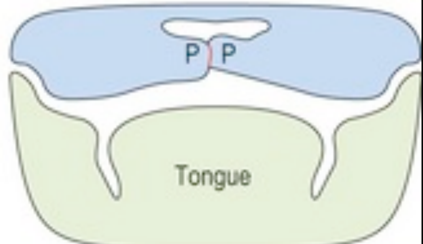
Embryology34
Lecturer — Embryologic Development
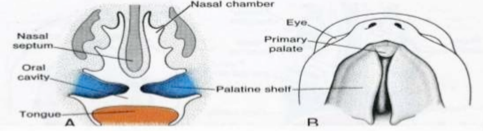
Embryology helps explain cleft anatomy, potential causative factors, effects on growth, and the timing of procedures.
- Around week four, the pharyngeal arches form, with the first arch contributing to the mandibular and maxillary components.
- During weeks six and seven, the primary palate forms through contributions from the maxillary processes and frontonasal prominence.
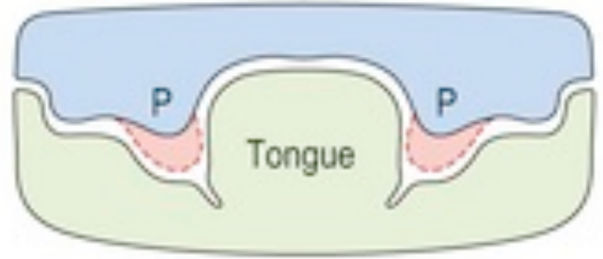
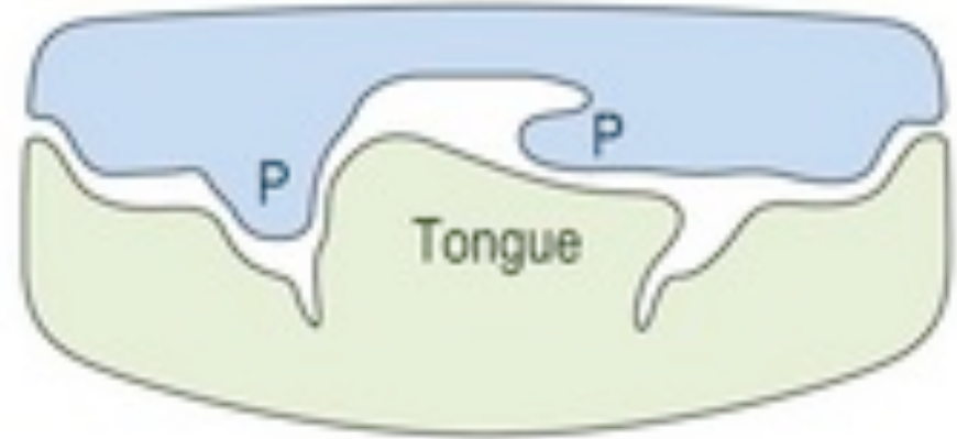
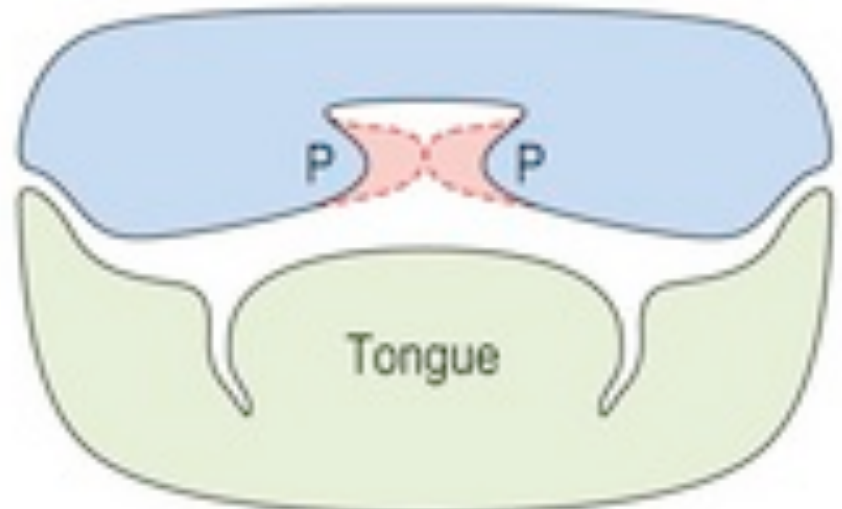
- By approximately week nine, the secondary palatal shelves fuse and separate the oral and nasal cavities; palate formation is generally complete by week ten.
- Proposed mechanisms for clefting include failure of palatal shelf fusion, elevation, or growth.
 |  |  |
 |  |
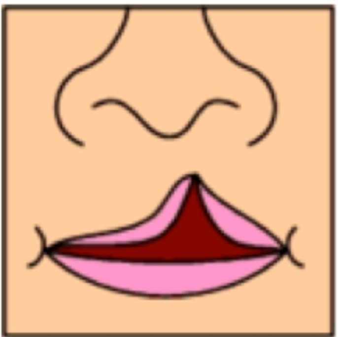
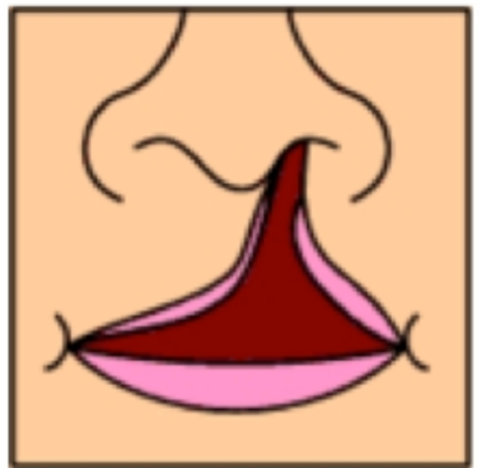
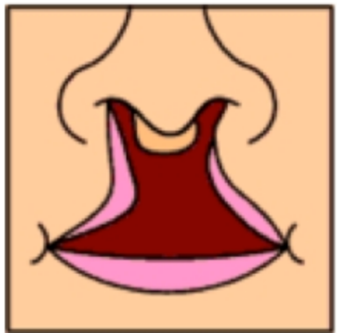
Classification56
 |  |  |
 |  |  |
 |  |  |
 |
Classification Systems
Lecturer — Classification Terminology
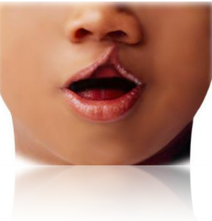
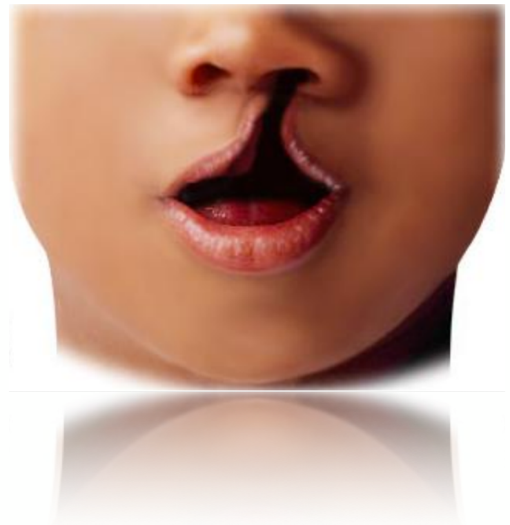
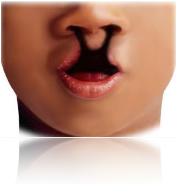
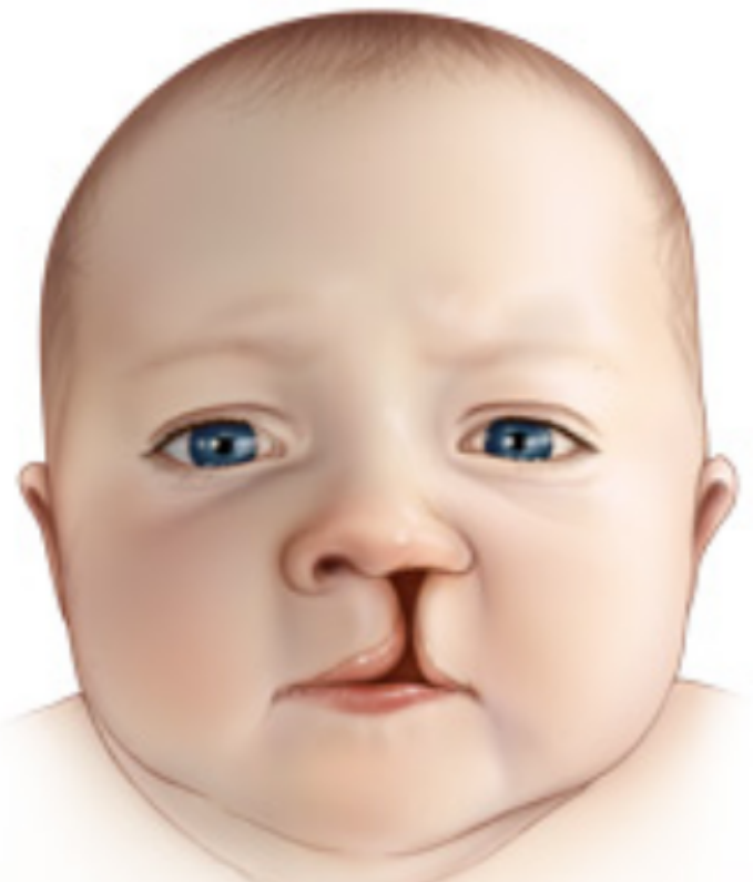
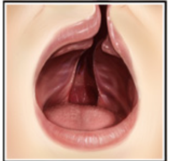
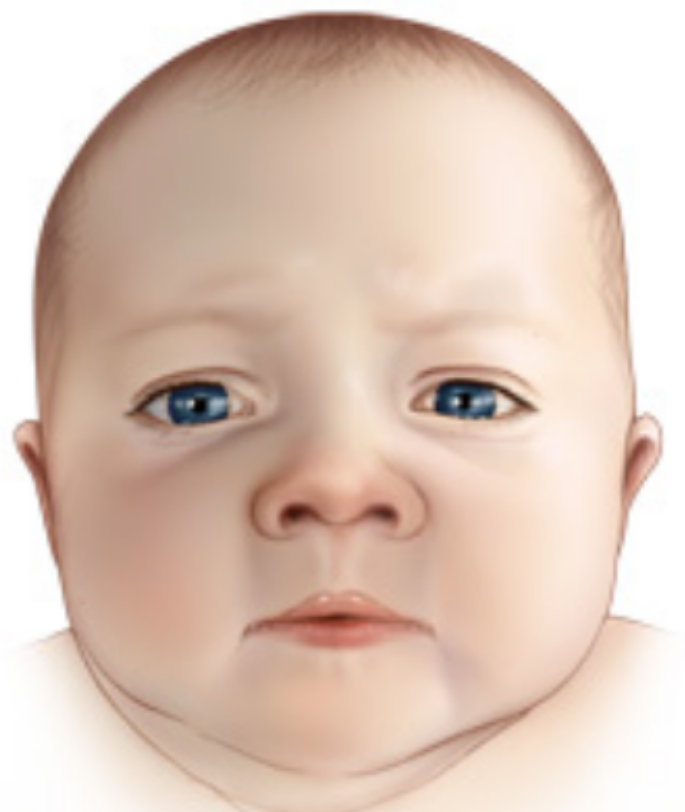
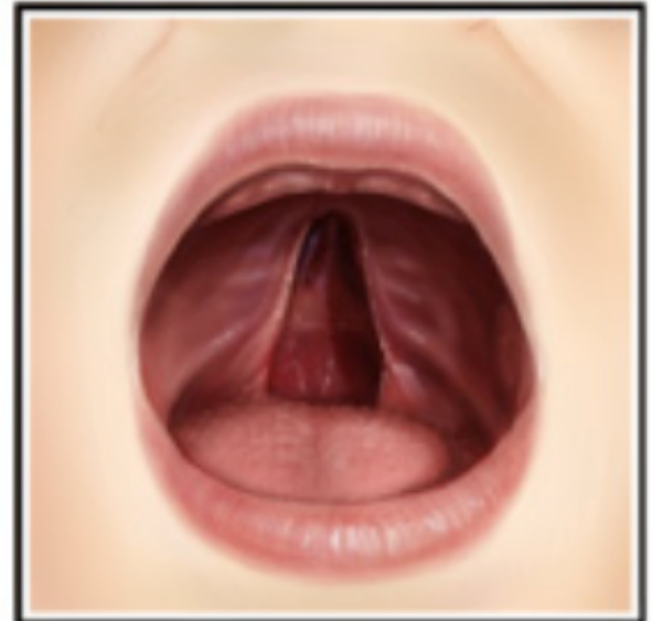
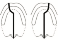
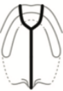
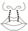
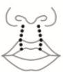
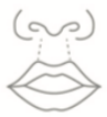
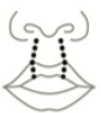
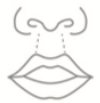
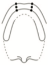
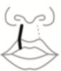
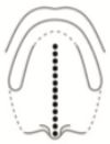
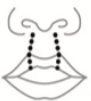
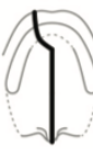
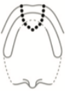
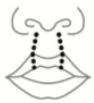
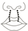
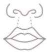
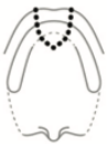
Clefts range from a microform cleft involving only the lip to complete unilateral or bilateral clefts involving the lip, alveolus, and palate.
- Clefts may involve the lip only; the lip and alveolus; the lip, alveolus, and palate; the hard and soft palate; or the palate alone.
- Classification systems commonly distinguish between unilateral and bilateral clefts.
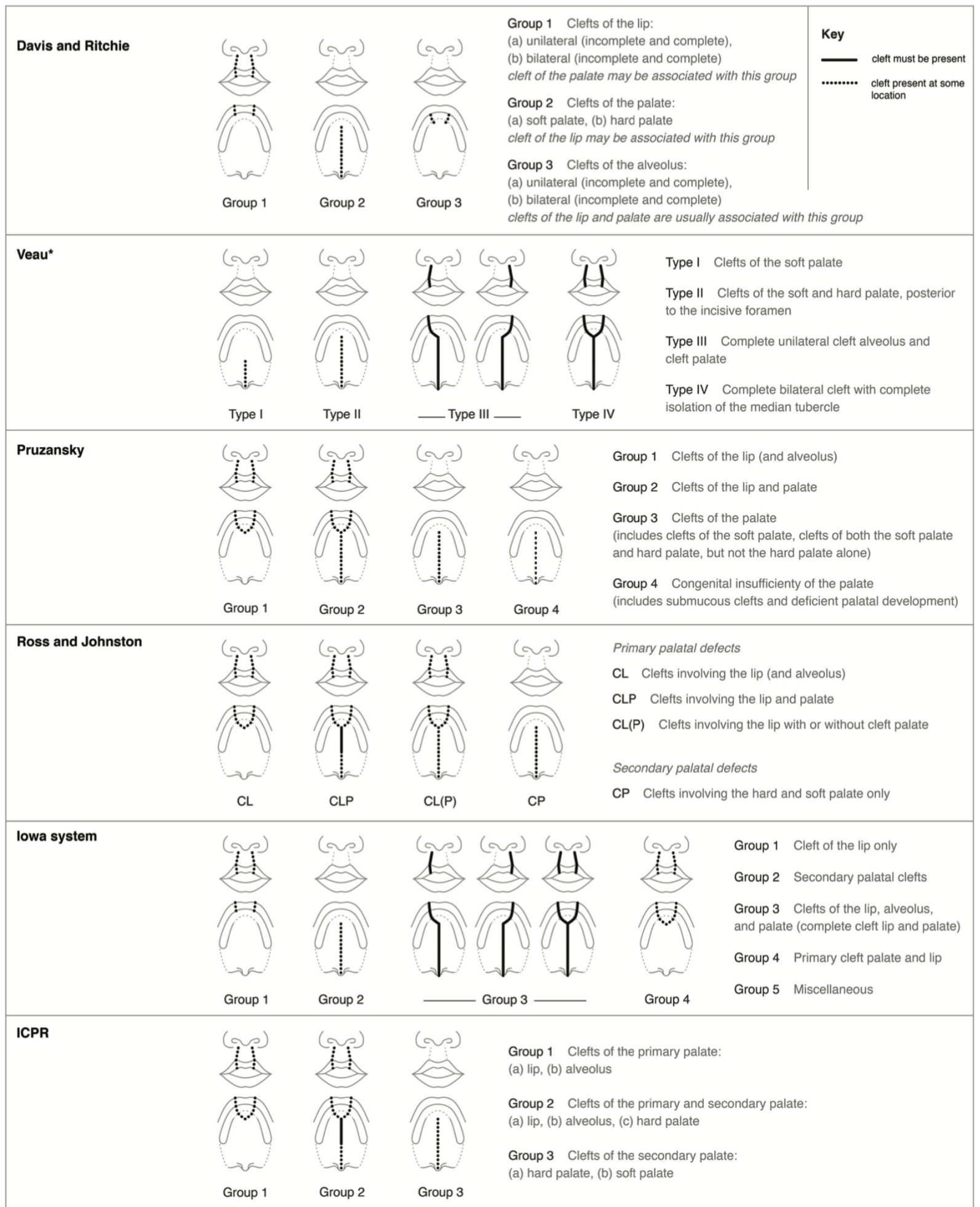
- Because classification systems vary substantially, clear descriptive terminology is usually preferred to a classification number that may not be understood outside its particular system.
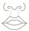
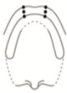
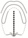
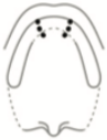
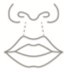
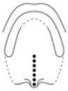
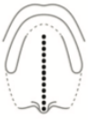
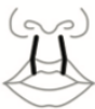
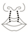
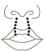
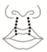
Davis and Ritchie7
Group 1 | Group 2 | Group 3
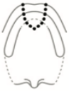
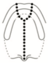
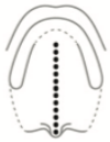
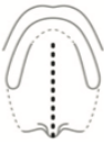
Key: a cleft must be present; cleft present at some location.
- Group 1 — Clefts of the lip:
- (a) unilateral (incomplete and complete)
- (b) bilateral (incomplete and complete)
- cleft of the palate may be associated with this group
- Group 2 — Clefts of the palate:
- (a) soft palate
- (b) hard palate
- cleft of the lip may be associated with this group
- Group 3 — Clefts of the alveolus:
- (a) unilateral (incomplete and complete)
- (b) bilateral (incomplete and complete)
- clefts of the lip and palate are usually associated with this group
 |  |  |
 |  |  |
 |  |  |
 |  |  |
 |  |  |
 |  |  |
 |  |  |
 |  |  |
 |  |  |
 |  |  |
 |  |  |
 |  |  |
 |  |  |
 |  |  |
 | 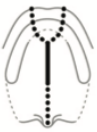 | 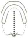 |
Veau
Type I | Type II | Type III | Type IV
- Type I — Clefts of the soft palate
- Type II — Clefts of the soft and hard palate, posterior to the incisive foramen
- Type III — Complete unilateral cleft alveolus and cleft palate
- Type IV — Complete bilateral cleft with complete isolation of the median tubercle
Pruzansky
Group 1 | Group 2 | Group 3 | Group 4
- Group 1 — Clefts of the lip (and alveolus)
- Group 2 — Clefts of the lip and palate
- Group 3 — Clefts of the palate
- (includes clefts of the soft palate, clefts of both the soft palate and hard palate, but not the hard palate alone)
- Group 4 — Congenital insufficiency of the palate
- (includes submucous clefts and deficient palatal development)
Ross and Johnston
CL | CLP | CL(P) | CP
Primary palatal defects
- CL — Clefts involving the lip (and alveolus)
- CLP — Clefts involving the lip and palate
- CL(P) — Clefts involving the lip with or without cleft palate
Secondary palatal defects
- CP — Clefts involving the hard and soft palate only
Iowa System
Group 1 | Group 2 | Group 3 | Group 4 | Group 5
- Group 1 — Cleft of the lip only
- Group 2 — Secondary palatal clefts
- Group 3 — Clefts of the lip, alveolus, and palate (complete cleft lip and palate)
- Group 4 — Primary cleft palate and lip
- Group 5 — Miscellaneous
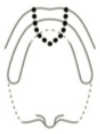
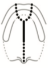
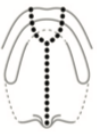
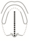
ICPR
Group 1 | Group 2 | Group 3
- Group 1 — Clefts of the primary palate:
- (a) lip
- (b) alveolus
- Group 2 — Clefts of the primary and secondary palate:
- (a) lip
- (b) alveolus
- (c) hard palate
- Group 3 — Clefts of the secondary palate:
- (a) hard palate
- (b) soft palate
Wang et al. 2014
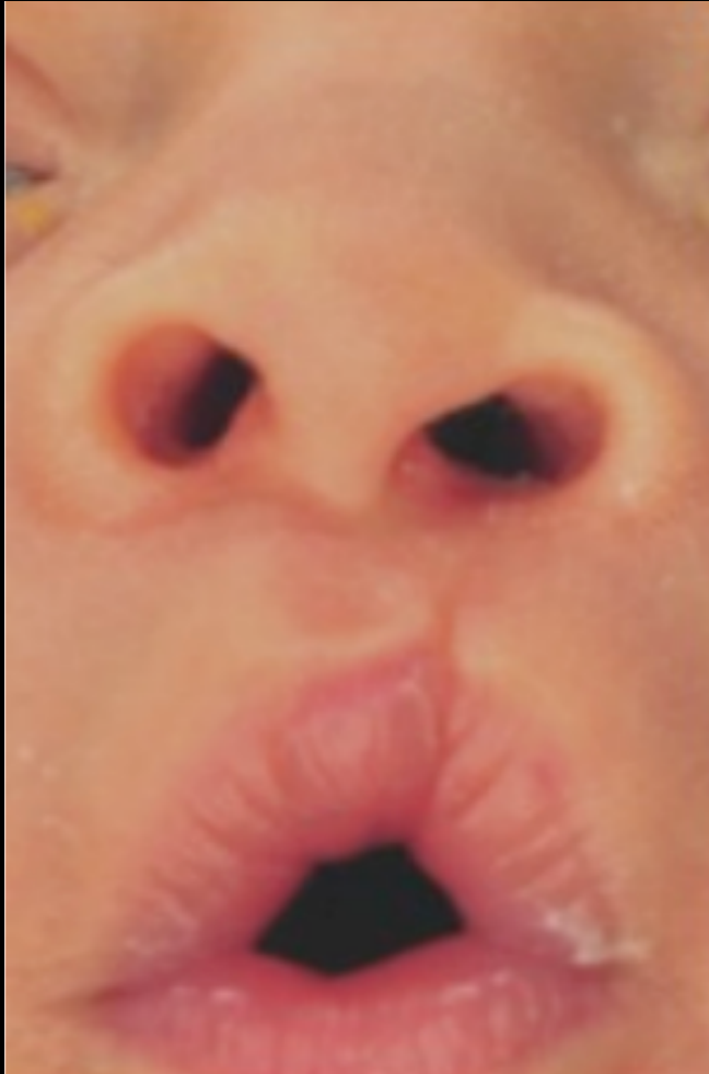
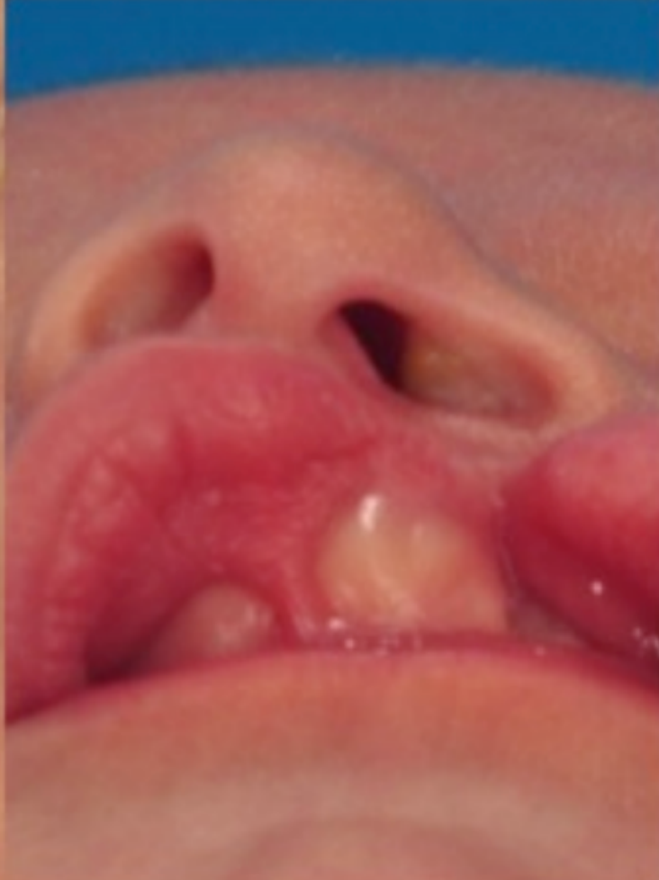
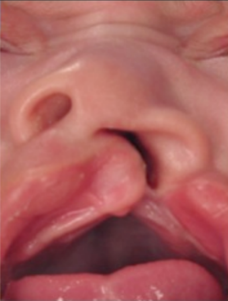
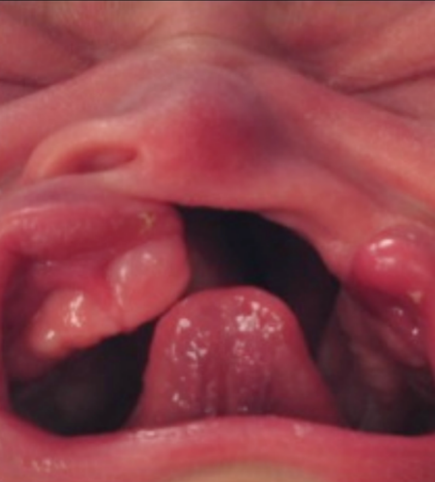
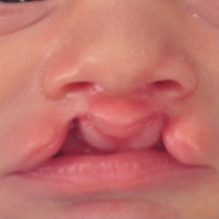
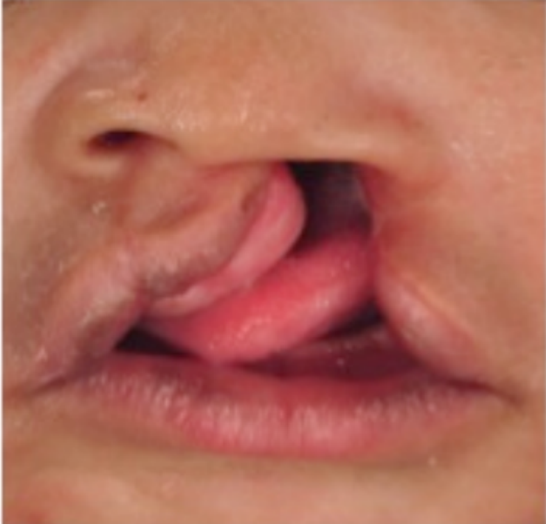
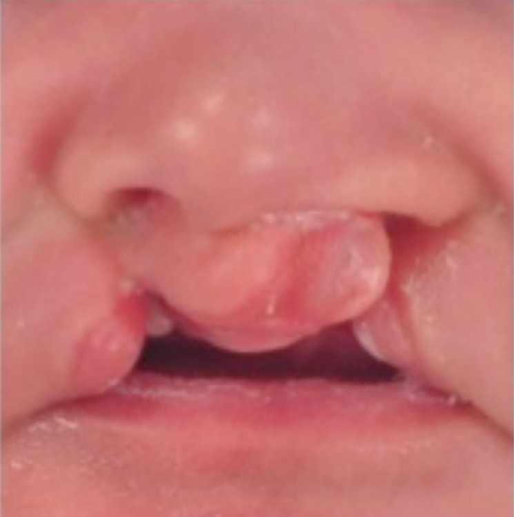
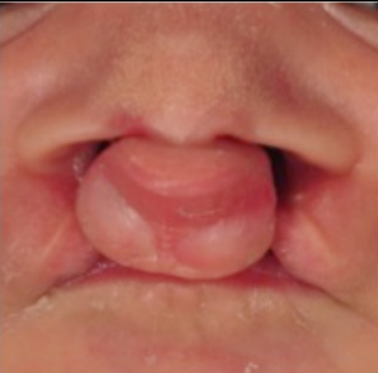
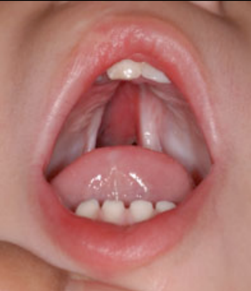
Clinical Examples8910
-
isolation
-
cleft palate
-
Unilateral examples range from a microform cleft lip to a cleft lip and alveolus or a complete cleft lip and palate.
-
Bilateral clefts affect both sides and are generally rarer and more difficult to manage.
-
Bilateral complete cleft lip and palate may leave the premaxillary segment unsupported.
-
These cases may require substantial treatment, including bone grafting, to obtain adequate arch continuity.
-
An isolated cleft palate is distinct from cleft lip and palate and has a different pattern of associations.
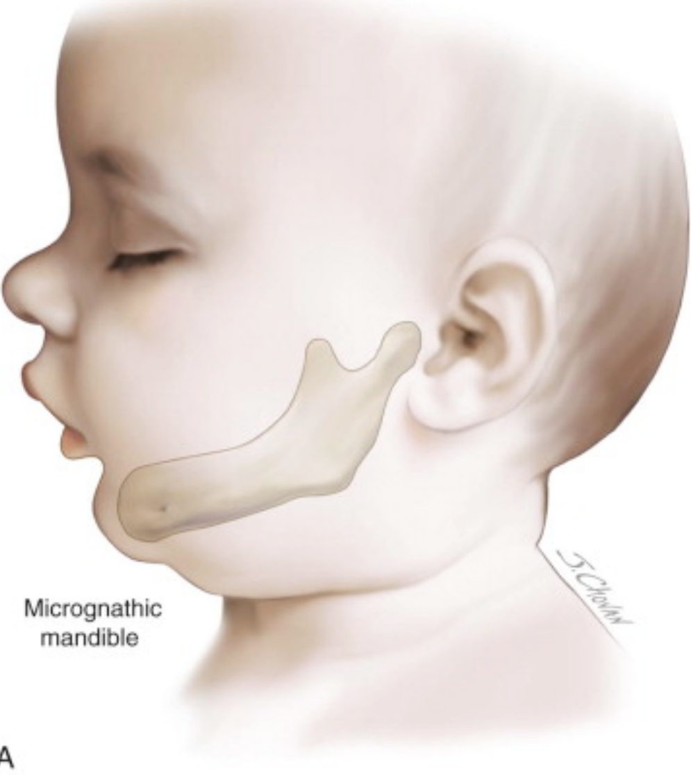
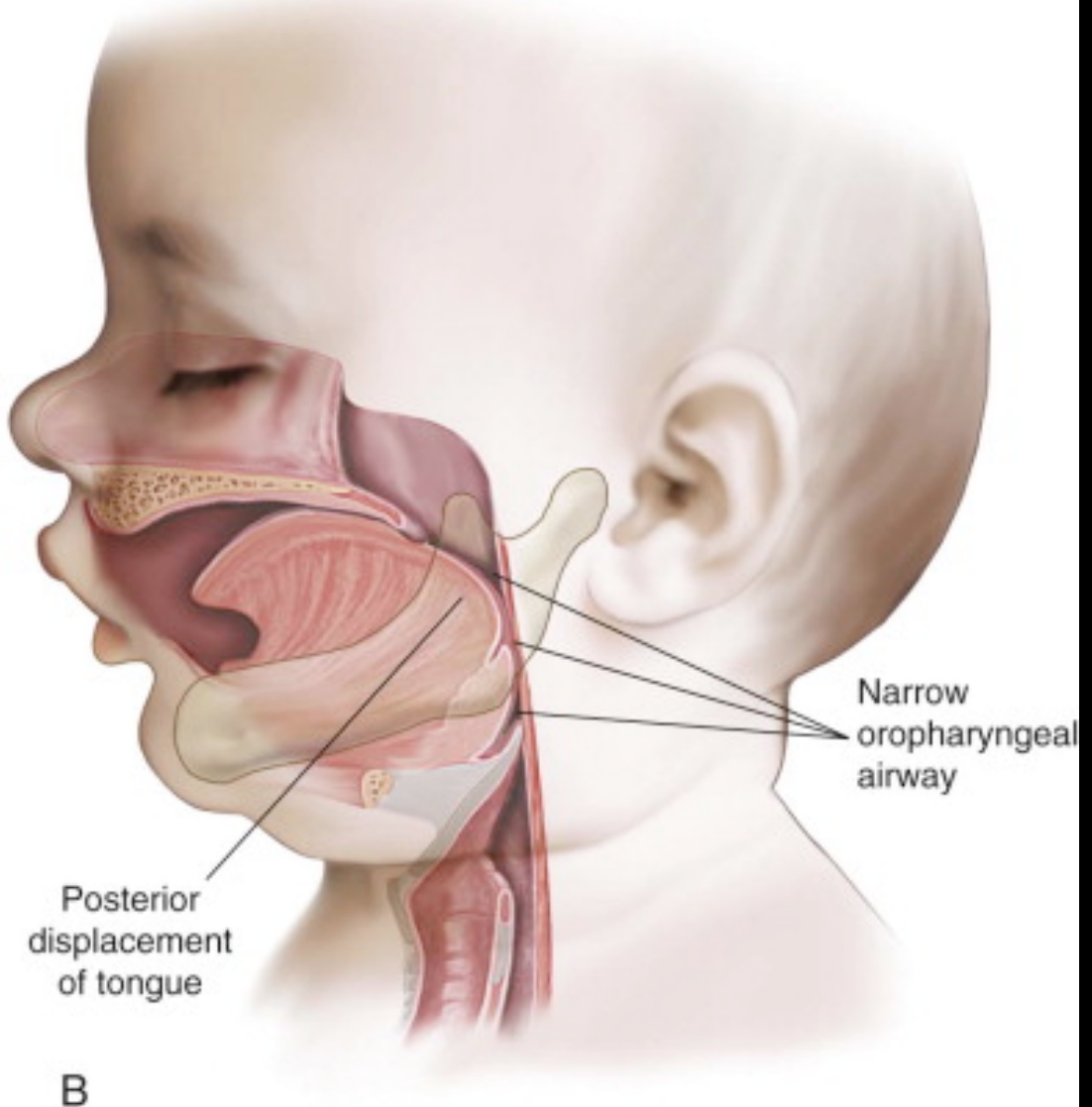
Pierre Robin Sequence11
Lecturer — Pierre Robin Sequence
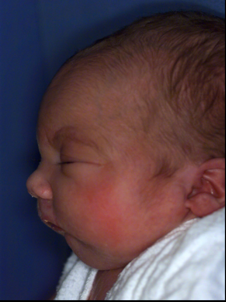
Pierre Robin sequence was previously called Pierre Robin syndrome, but sequence is preferred because the findings occur as a sequence of events.
- A micrognathic mandible develops.
- The tongue is displaced posteriorly, producing glossoptosis.
- The posteriorly positioned tongue occupies space needed for palatal shelf elevation, resulting in a cleft palate.
- The sequence therefore consists of micrognathia, glossoptosis, and cleft palate, with each feature contributing to the next.
 |  |
 |
Aetiology12
- Genetic
- MSX1, IRF6, FGFR1
- Approximately 70% of all cases of CL/P and 50% of cases of cleft palate only are considered to be non-syndromic
- MSX1 is a transcription factor involved in epithelial and mesenchymal interactions
- IRF6 is associated with connective tissue formation, particularly in the palate
- Mutations in IRF6 can cause van der Woude syndrome, in which lower lip pits are often a sign
- Fibroblast growth factor receptors and other cellular signalling pathways are involved in cell differentiation, proliferation and migration
- Environmental
- Maternal smoking (1.3 x higher odds)
- Maternal alcohol consumption, nutritional deficiencies (folic acid), stress, maternal obesity, ionising radiation
Antenatal Diagnosis13
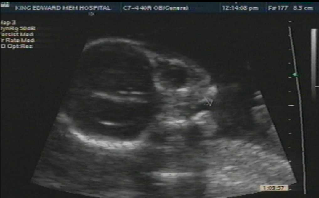
- Ultrasound detection rates
- Significant increase in antenatal diagnosis of CLP in WA
- 40% detection of CLP from 1996–2003 (Hanikeri et al. 2006)
- 71% detection of CLP from 2003–2012 (Nicholls et al. 2017)
- Impact of early detection and need for a supportive environment
- Detection has increased to approximately 70% and into the high 70% range at the 20-week ultrasound
- Early detection allows supportive counselling, family preparation, early access to social work, genetics consultation, and contact with surgical and cleft teams
- 14 out of 15 pregnancies ended in termination following early diagnosis of cleft lip (Bronshtein et al 1996)
Lecturer — Postdiagnosis Support
An international study from the Middle East reported that 14 of 15 pregnancies were terminated after early diagnosis of cleft lip.
- This was presented as an outcome that should not be encouraged.
- Families should instead receive appropriate information and access to the relevant clinical and social support teams.
- Cleft coordinators
Lecturer — Family Coordination
Once a child is diagnosed, whether antenatally or after birth, the relevant teams are notified and coordinate care. The child and family require both clinical management and sensitive emotional support.
- <mark>Implant planning and orthognathic surgery planning</mark>
Lecturer — Treatment Stages
Treatment was described chronologically.
- Birth to approximately three months involves feeding support and pre-surgical management.
- Early childhood includes speech, audiology, preventive dental care, and monitoring.
- Mixed dentition involves pre-surgical orthodontics and alveolar bone grafting.
- Later childhood and adolescence may involve definitive orthodontic, orthognathic, or implant-related treatment.
Treatment Overview
- Treatment is complex and there remains extensive disagreement on the timings of interventions, the interventions themselves and the various techniques involved in surgical correction of the cleft (Nguyen et al 1993)
- The ultimate aim of cleft surgery is that the patient look well, feed well, and speak well (Wilhelmsen et al 1966)
Lecturer — Treatment Trade-Offs
No single treatment approach was described as definitively superior.
- Choices depend substantially on the preferences of the cleft team and individual departments.
- Surgical treatment may improve aesthetics and function but can also restrict maxillary growth.
- Treatment therefore requires balancing immediate needs against the long-term effects of procedures.
Treatment Aims14
Multidisciplinary Management15
Multidisciplinary care of patients with craniofacial anomalies involves:
- Surgical
- Plastics
- OMFS
- Dental
- ENT
- Nursing
- Genetics
- Social Support
- Ophthalmology
- Psychology
- Paediatrics
- Speech therapy
- Obstetrics
- ENT
- Audiology
- Dentistry
- Paediatrics
- Orthodontics
- Prosthodontics
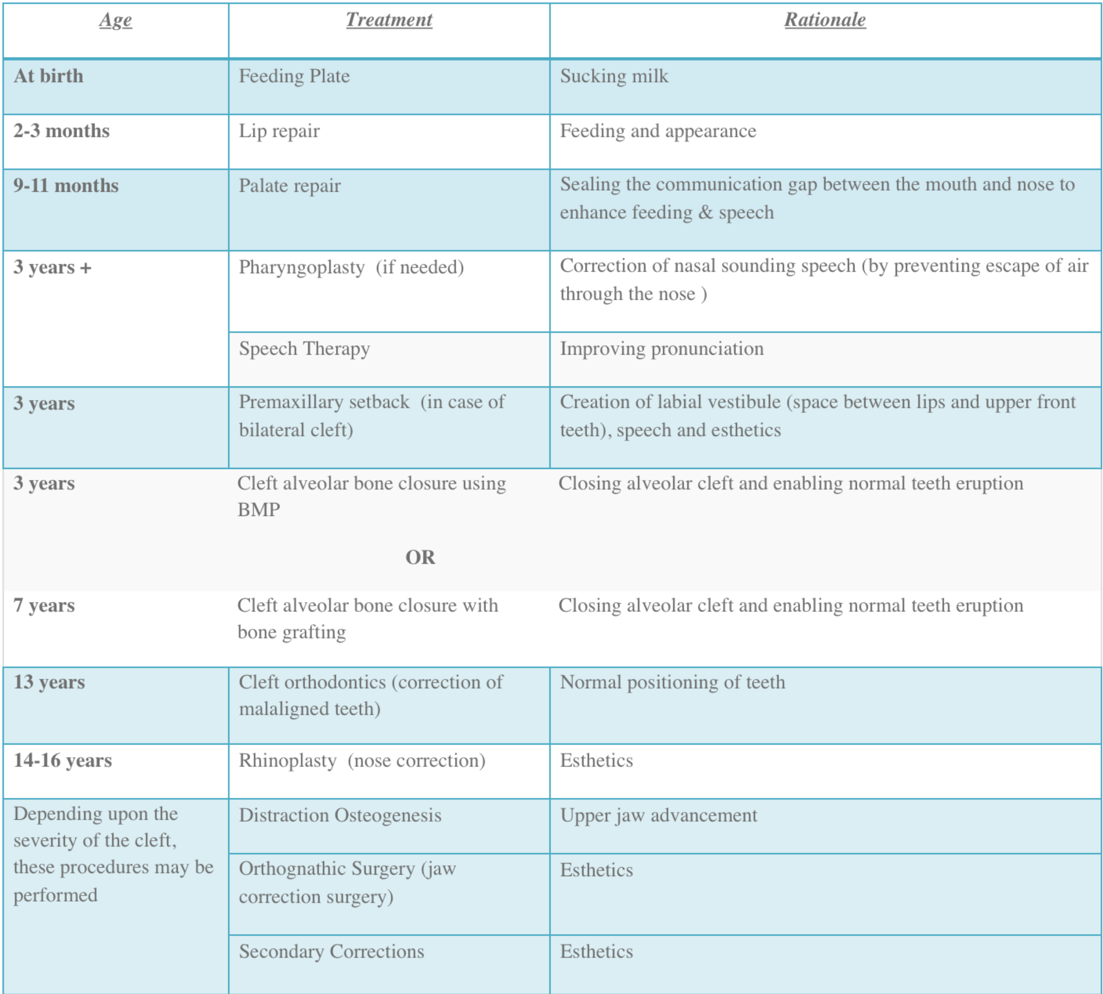
Treatment Timeline1617181920
- Short-Term: Presurgical, Surgical (lip & palate repair)
- Medium-Term: Dental Care, Speech therapy, Surgical Revisions
- Long-Term: Alveolar bone grafting, Orthognathic surgery
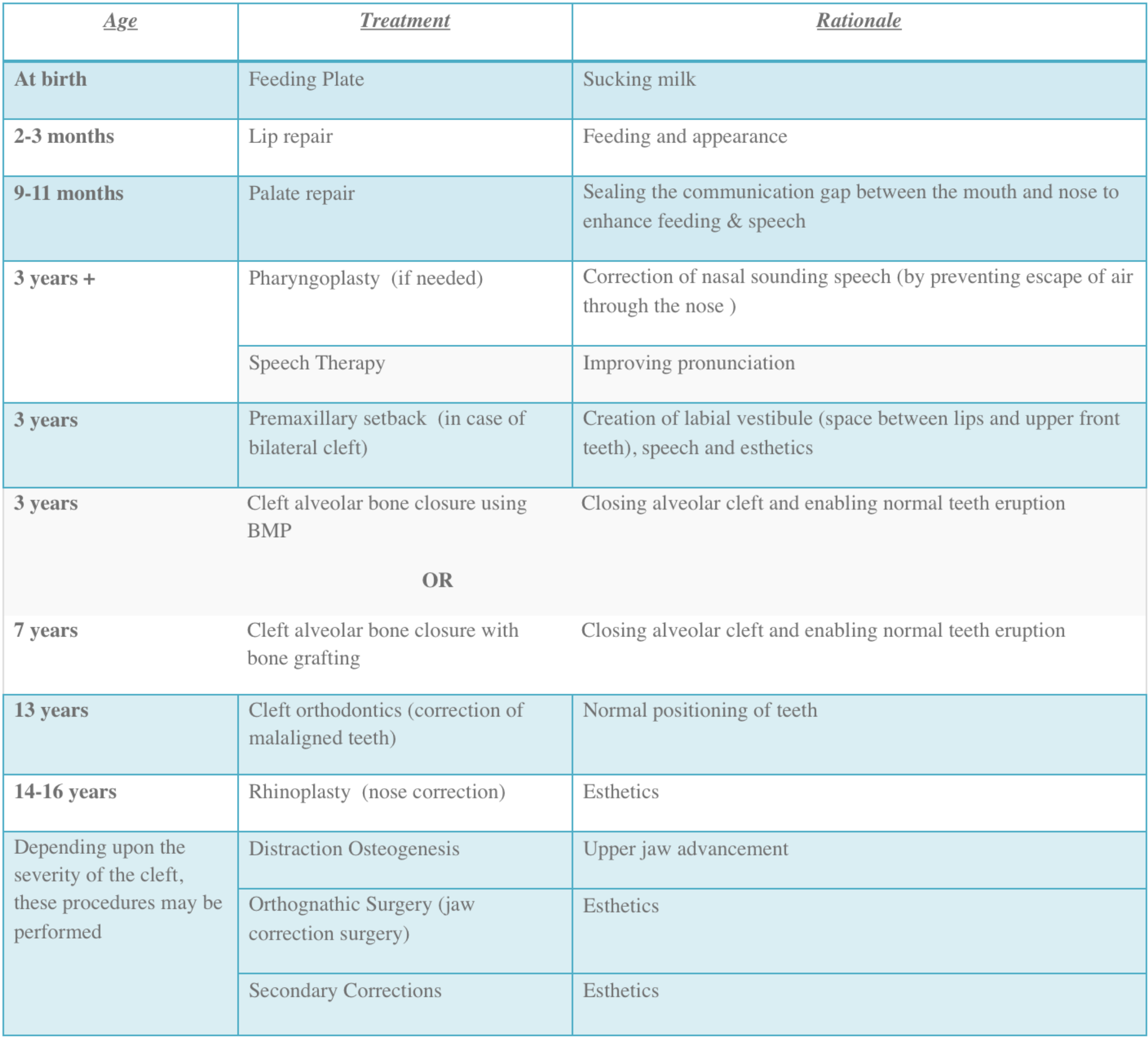
| Age | Treatment | Rationale |
|---|---|---|
| At birth | Feeding Plate | Sucking milk |
| 2-3 months | Lip repair | Feeding and appearance |
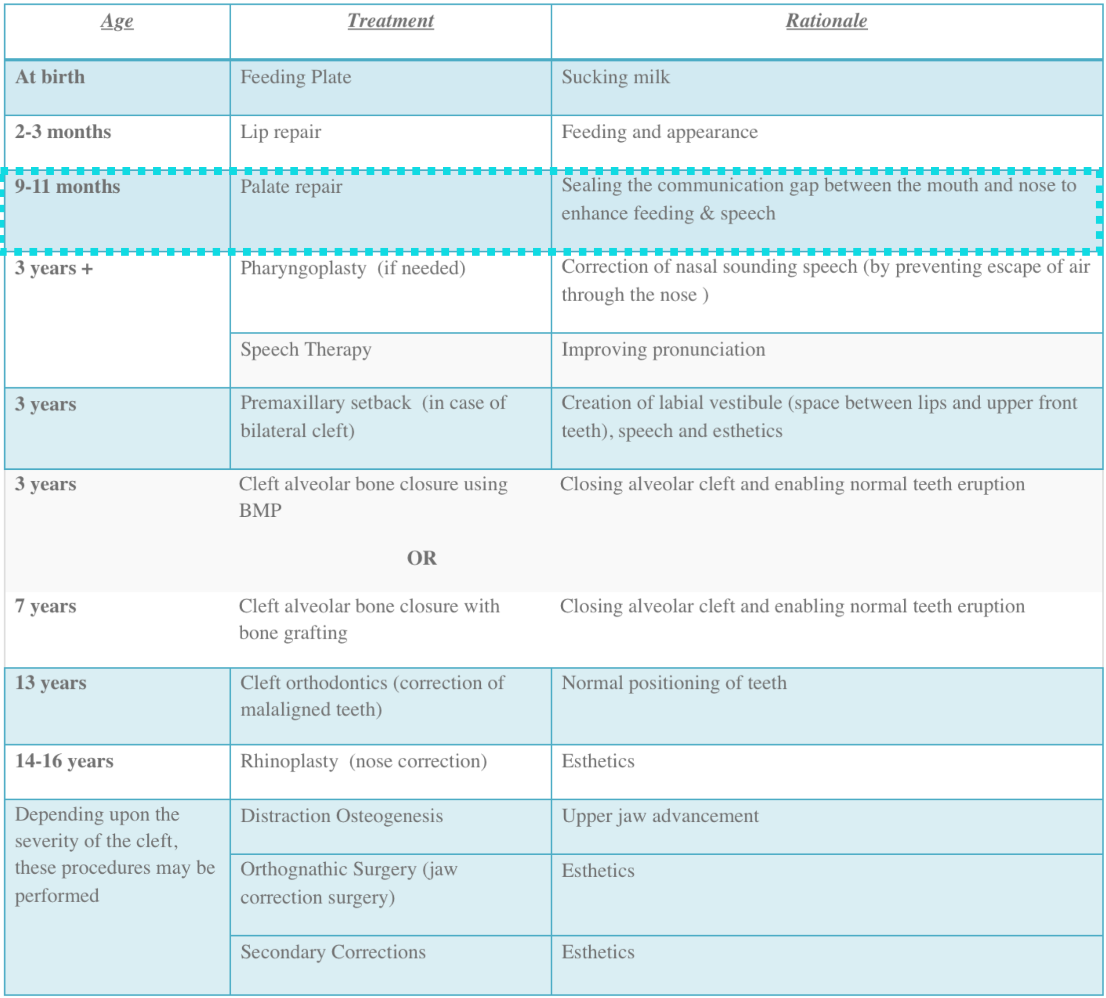
| 9-11 months | Palate repair | Sealing the communication gap between the mouth and nose to enhance feeding & speech |
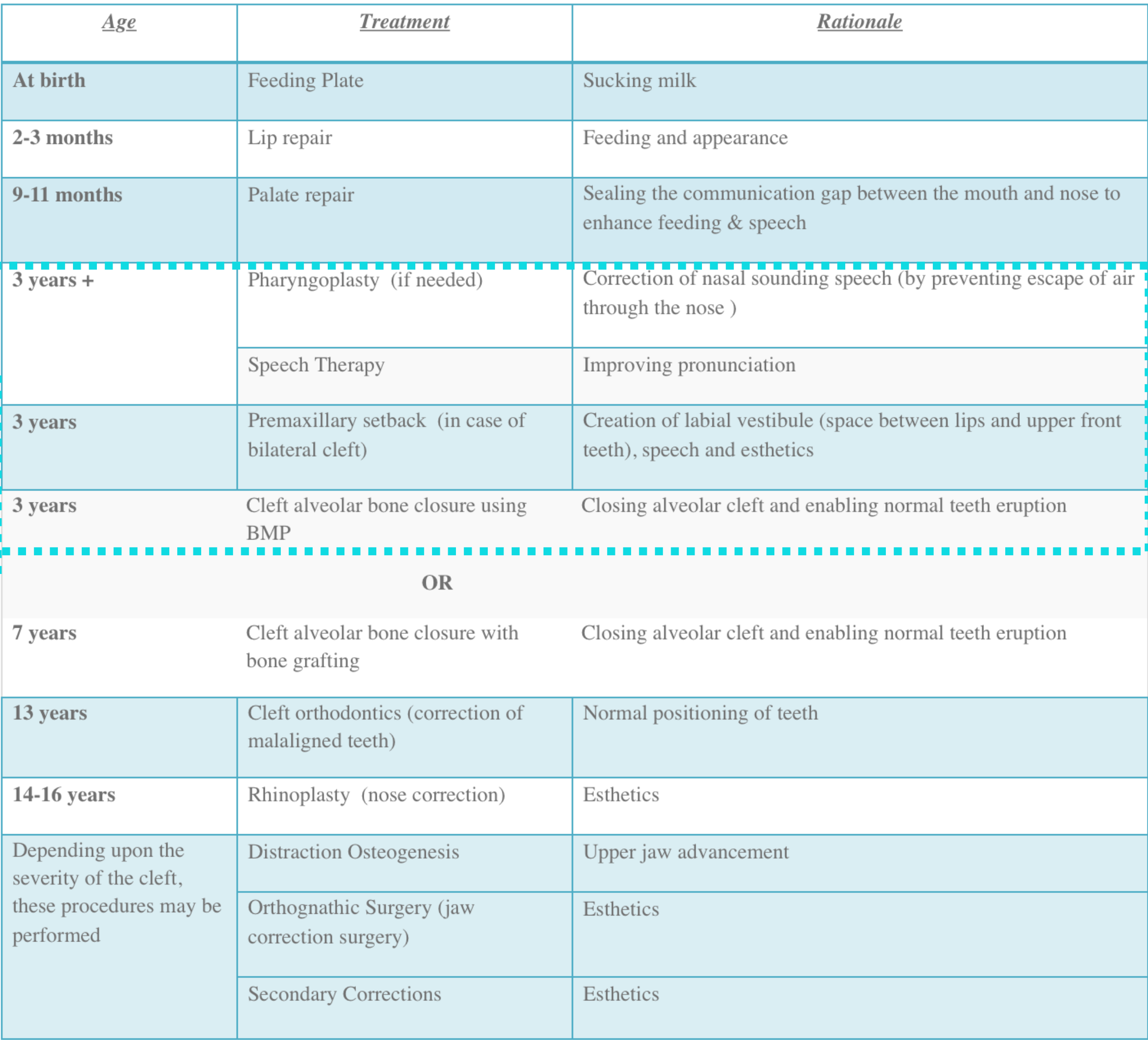
| 3 years + | Pharyngoplasty (if needed) | Correction of nasal sounding speech (by preventing escape of air through the nose) |
| Speech Therapy | Improving pronunciation | |
| 3 years | Premaxillary setback (in case of bilateral cleft) | Creation of labial vestibule (space between lips and upper front teeth), speech and esthetics |
| 3 years | Cleft alveolar bone closure using BMP OR | Closing alveolar cleft and enabling normal teeth eruption |
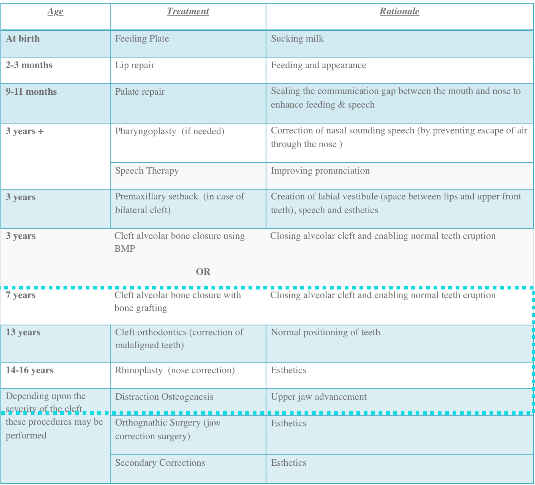
| 7 years | Cleft alveolar bone closure with bone grafting | Closing alveolar cleft and enabling normal teeth eruption |
| 13 years | Cleft orthodontics (correction of malaligned teeth) | Normal positioning of teeth |
| 14-16 years | Rhinoplasty (nose correction) | Esthetics |
| Depending upon the severity of the cleft, these procedures may be performed | Distraction Osteogenesis | Upper jaw advancement |
| Orthognathic Surgery (jaw correction surgery) | Esthetics | |
| Secondary Corrections | Esthetics |
Pre-Surgical Management21
- Feeding instruction
- Feeding appliances
- Methods of repositioning and aligning the bony cleft segments prior to surgical closure of the lip
- Pre-surgical orthopaedics
McKinstry 1998; Nahai et al 2005
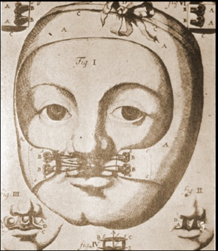
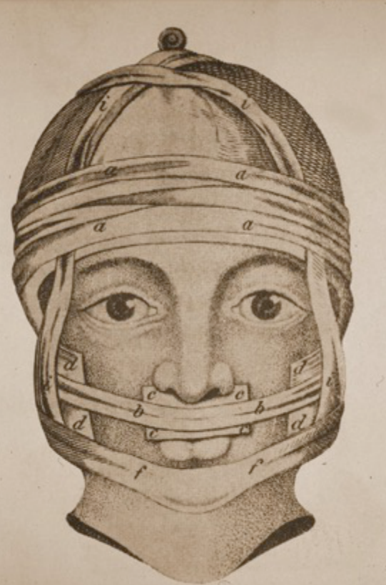
Pre-Surgical Orthopaedics222324252627282930
- Methods of repositioning and aligning the bony cleft segments prior to surgical closure of the lip
- Facilitate primary lip repair
Lecturer — Early Orthopaedics
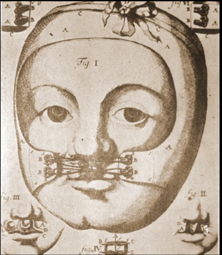
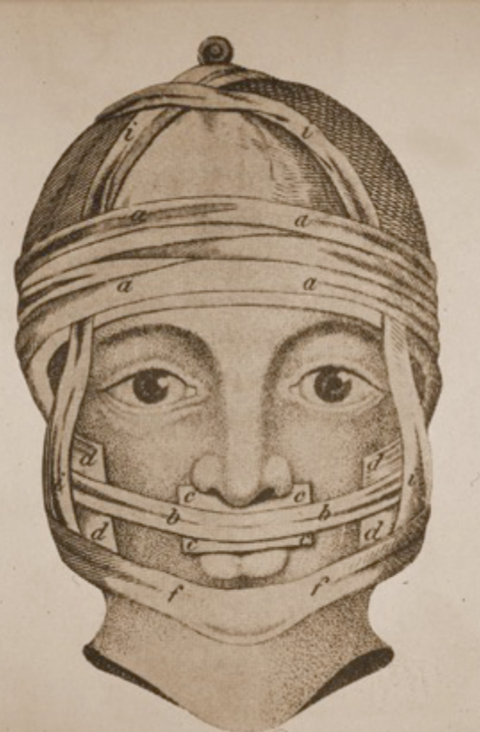
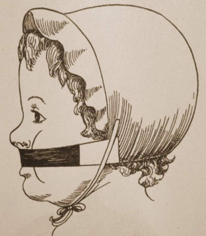
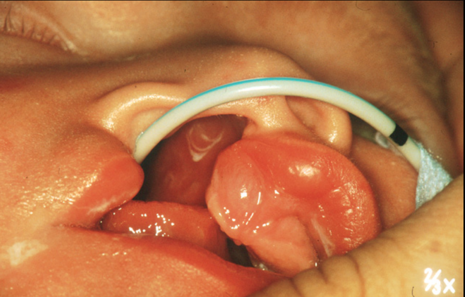
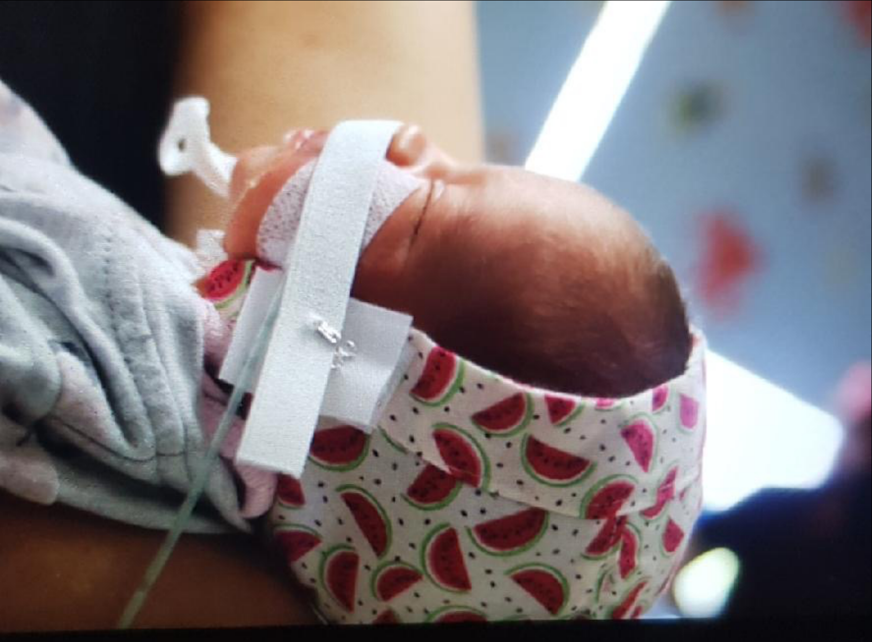
Pre-surgical orthopaedics usually occurs from birth to approximately three months. Early treatment is possible because maternal oestrogen is still circulating and the tissues are relatively malleable and pliable.
Lecturer — Clinical Context
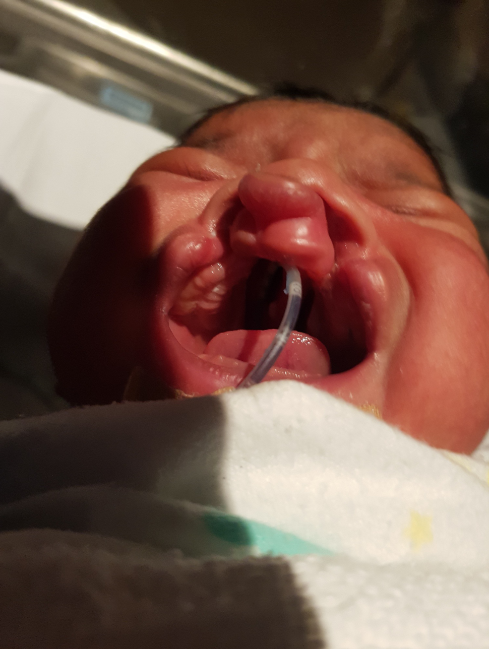
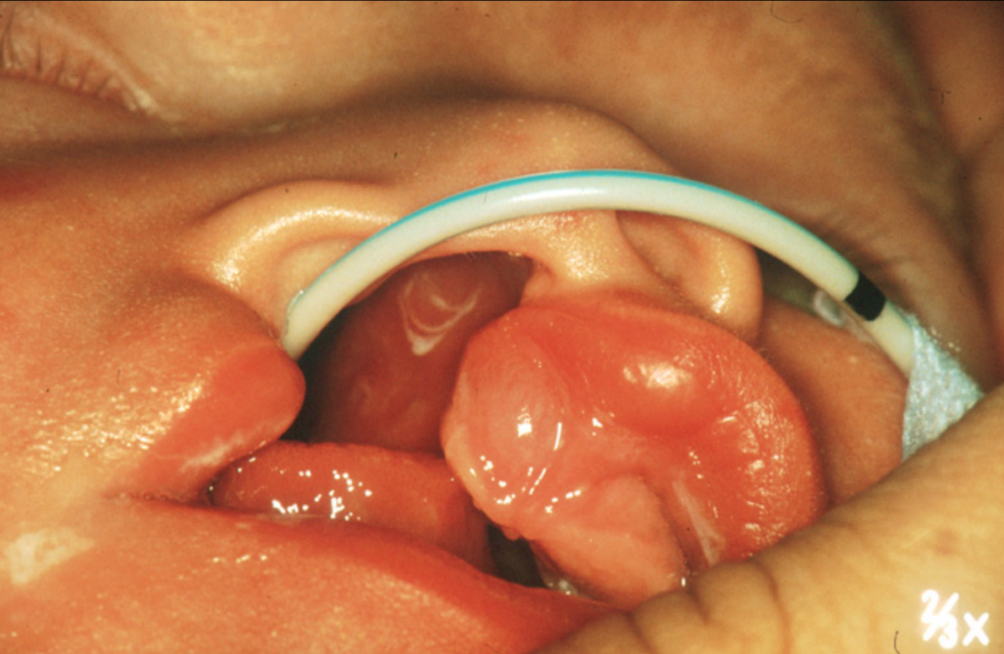
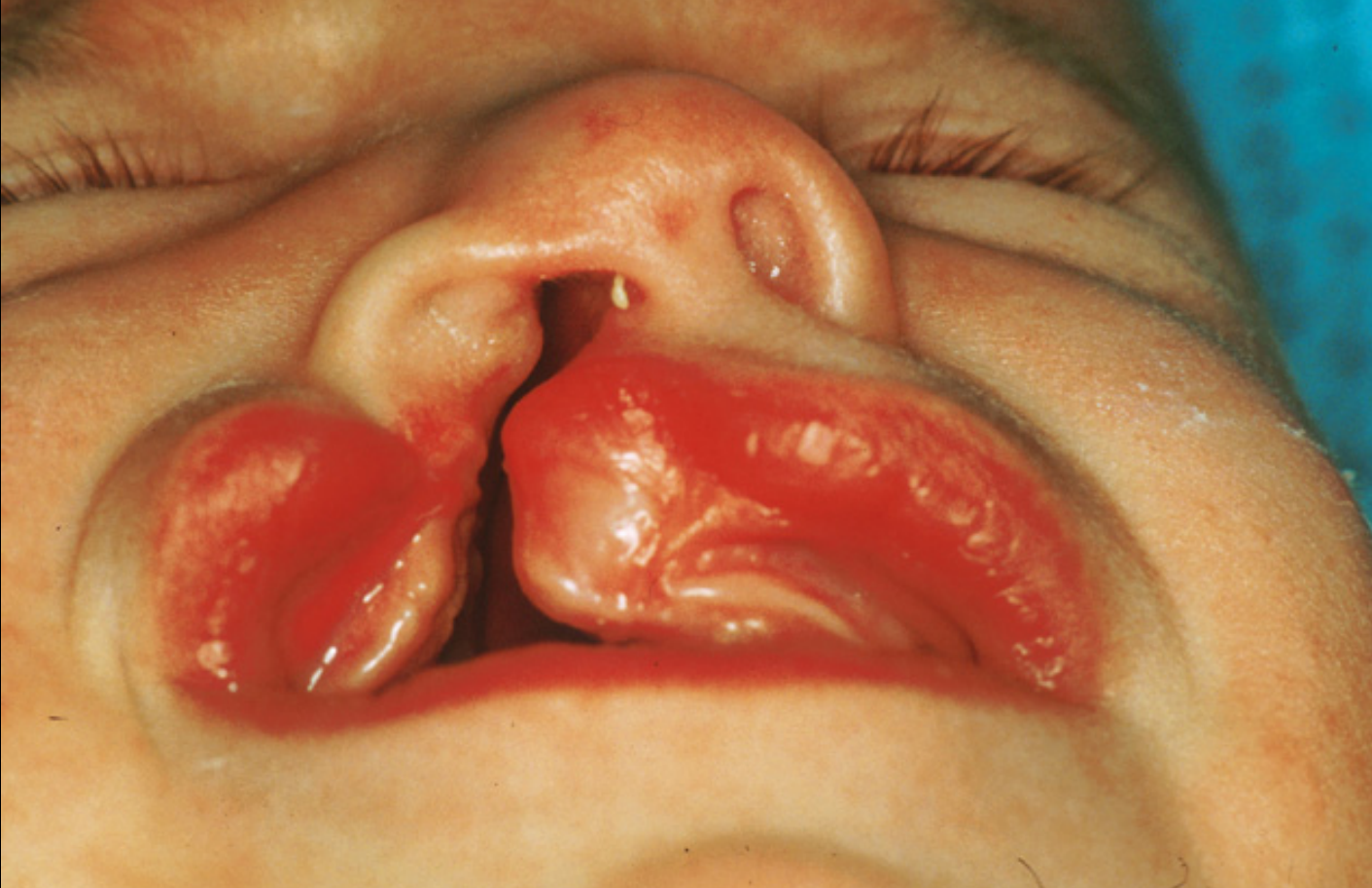
In a unilateral complete cleft lip, alveolus, and palate, treatment from birth to three months can substantially reduce cleft width and produce a more continuous arch form. The paediatric dental team may be among the first teams to diagnose the cleft definitively, often in the neonatal intensive care unit, and should provide empathetic, professional counselling to the parents.
Winters 1998
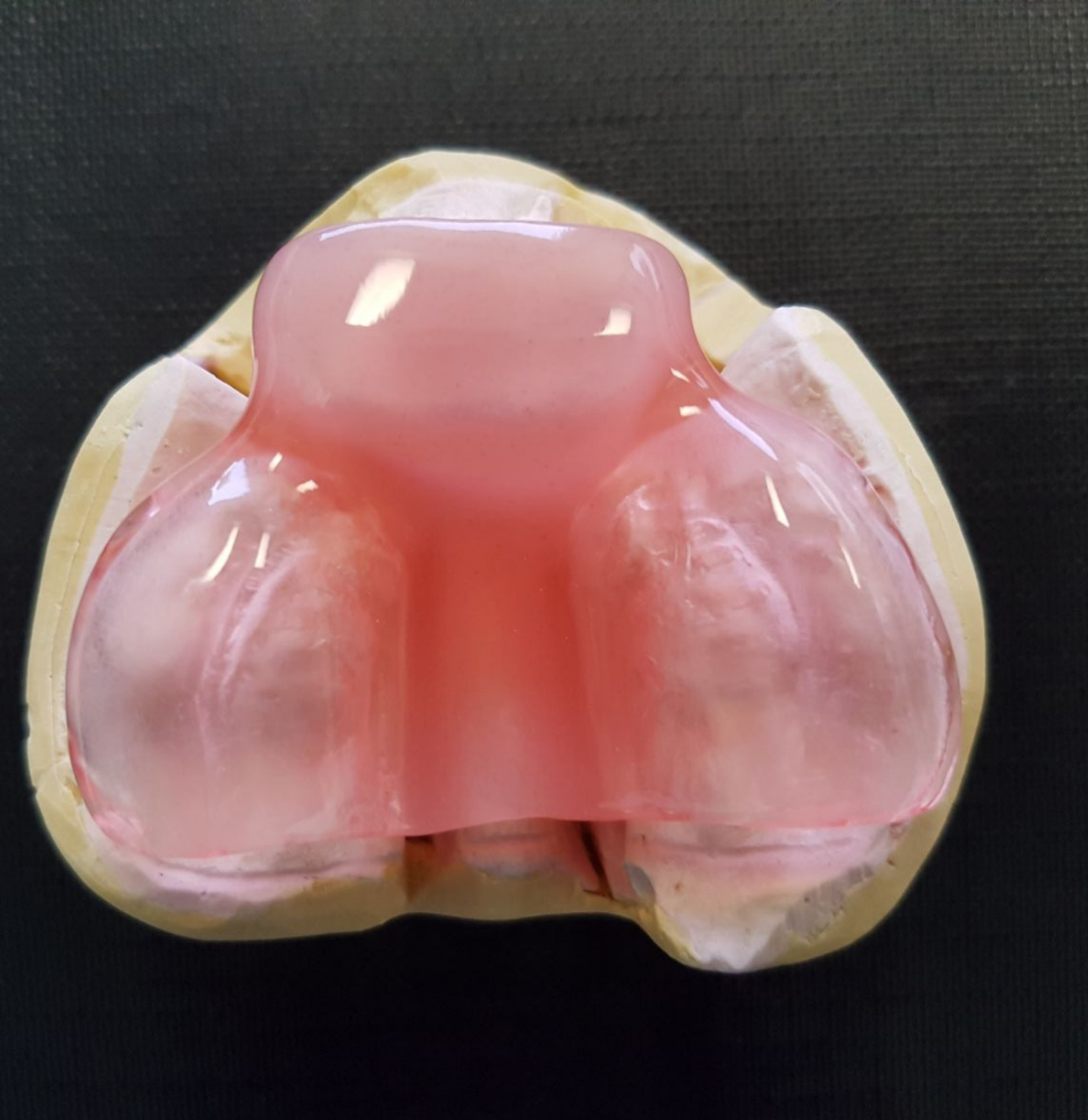
- Intraoral appliance
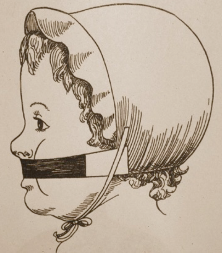
- Extraoral traction (bonnet/strapping)
Lecturer — Appliance Mechanics
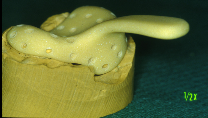
The appliance sits passively rather than as a tight-fitting denture, allowing the cleft segments to move. The bonnet and strapping provide the force, while the intraoral appliance guides the direction of movement.
Images Courtesy of Dr John Winters
-
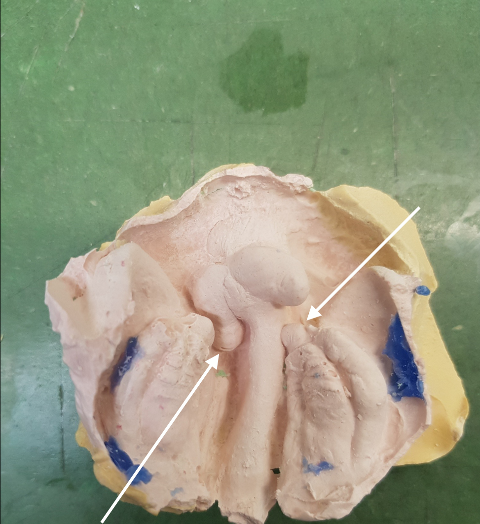
Neonatal tooth follicles
-
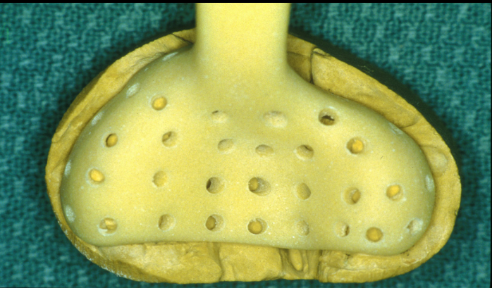
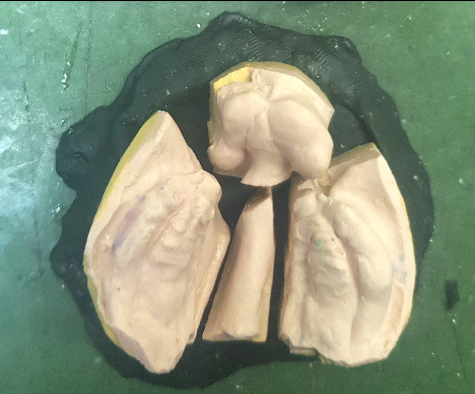
Working Cast
-
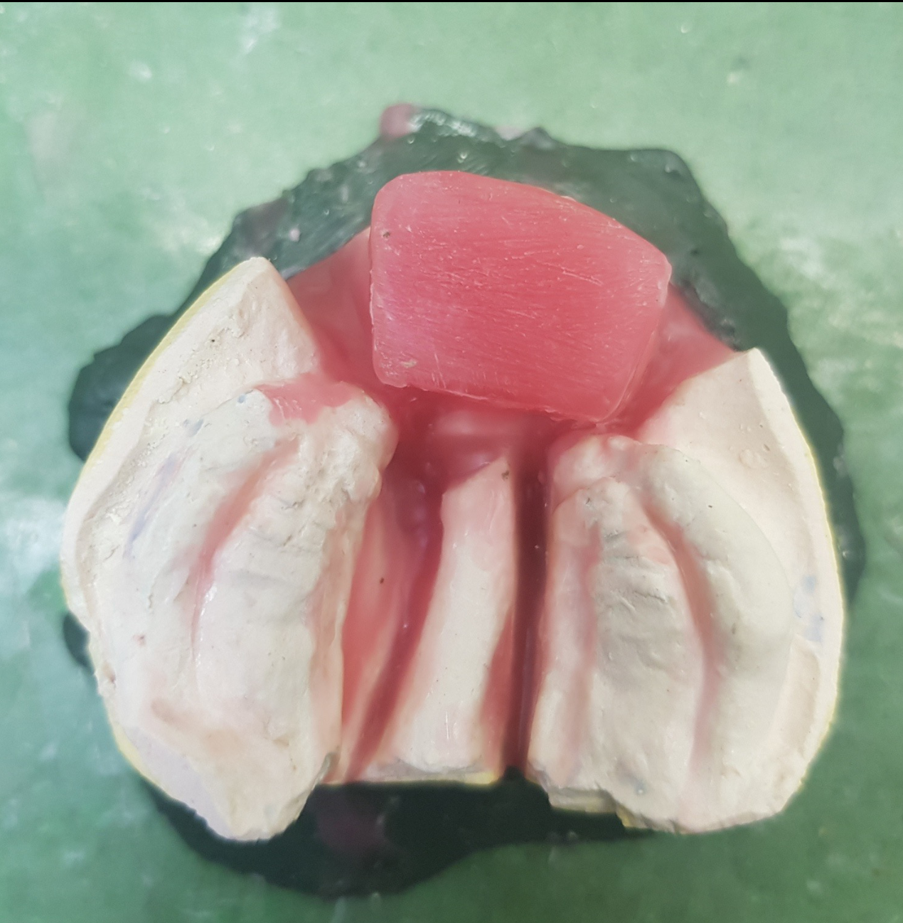
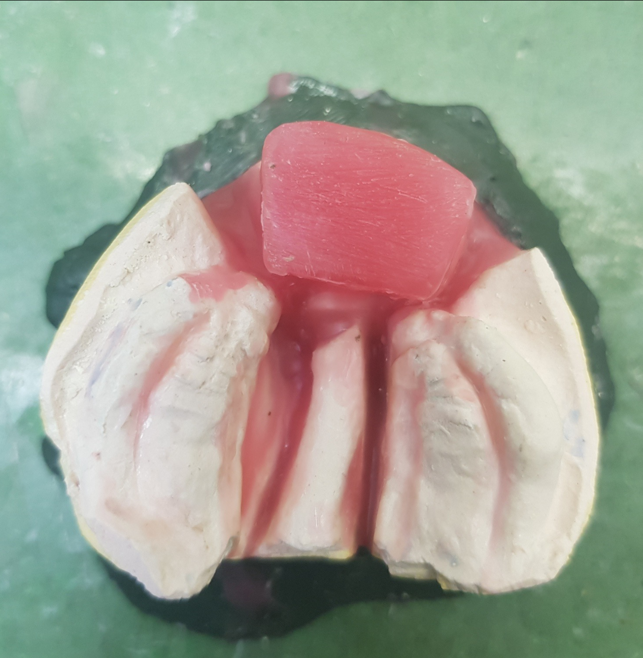
Model Surgery
-
Final wax-up
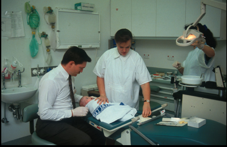
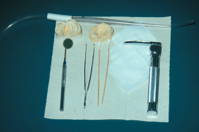
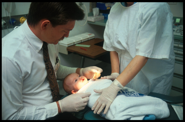
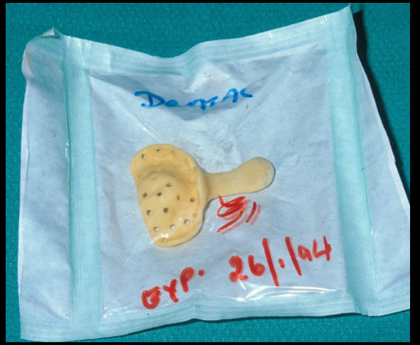
Lecturer — Impression Safety
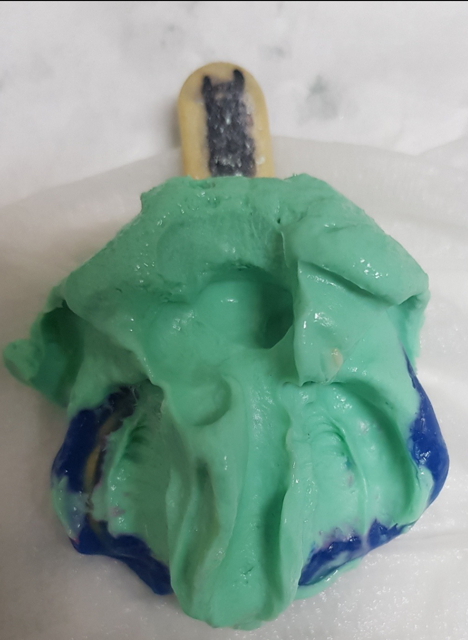
Impressions are taken soon after birth using small infant trays, but the procedure is not suitable for inexperienced operators. Full resuscitation equipment, laryngoscopes, and oxygen must be available because airway obstruction, choking, and aspiration are possible; the impression should ideally be completed within the first or second attempt.
Images Courtesy of Dr John Winters
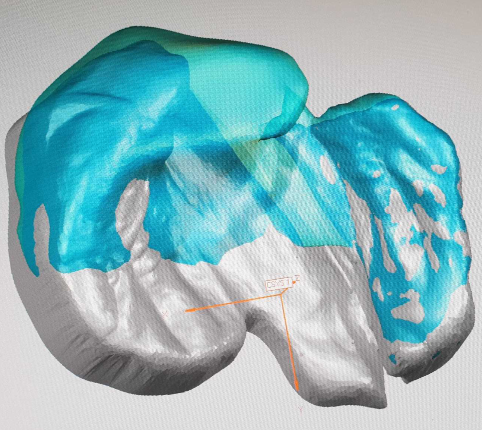
Digital Design And Workflow313233
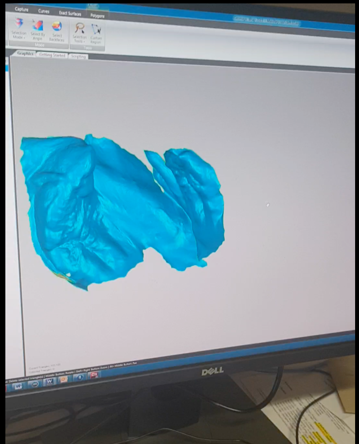
- Optical scanning and confocal microscopy
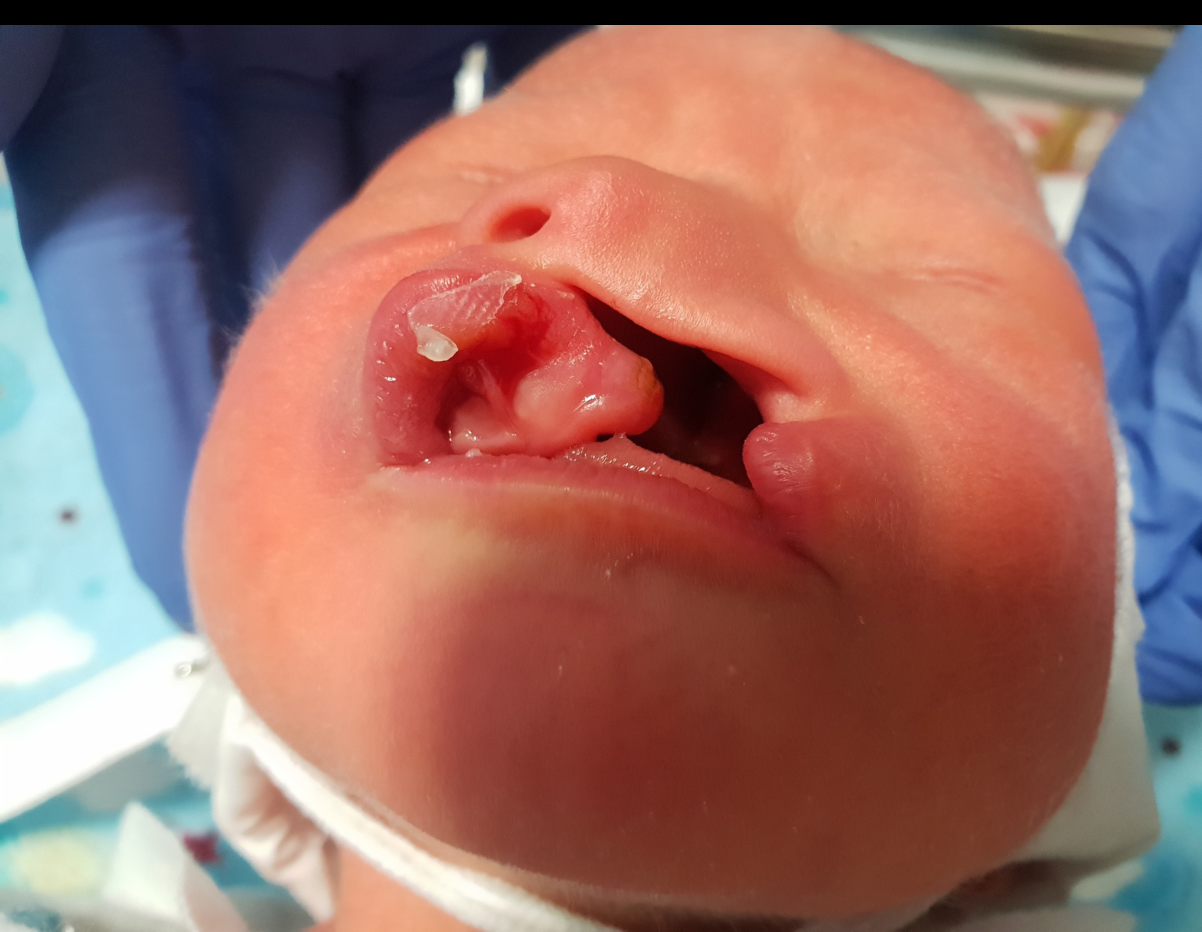
- Accuracy and technique for optical impression taking in a neonate
 |  |  |
 |  |  |
Published Abstract34
The Cleft Palate-Craniofacial Journal — Cleft Palate Craniofac J. 2019 Mar 11:1055665619835082. doi: 10.1177/1055665619835082. [Epub ahead of print]. Intraoral Digital Impression Technique for a Neonate With Bilateral Cleft Lip and Palate. Patel J, Winters J, Walters M.
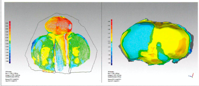
Objective: Dental casts are an important aspect in the treatment planning, documentation, and analysis of the dental arch forms of infants with cleft lip and palate (CLP). Impression taking in a neonate is a technique-sensitive procedure, which can result in foreign body dislodgement and carries a small risk of aspiration and airway obstruction. The advent of digital dental technologies and intraoral scanning may facilitate safer, more effective, and accurate impressions for CLP infants.
Design: A digital intraoral scan of a 3-month-old with bilateral CLP (BCLP) was compared with a conventional alginate impression taken prior to primary lip repair.
Setting: Princess Margaret Hospital for Children.
Main Outcome Measures: To test the applicability and accuracy of digital impression taking for a neonate with BCLP and palate using digital morphometrics.
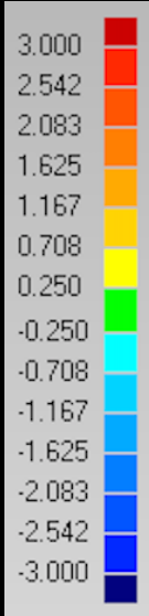
Results: The average deviation of points ranged above and below the plane of superimposition from +0.78 mm to -0.42 mm with a maximum range of +2.80 mm to -2.80 mm and standard deviation of 0.88 mm. The premaxillary segment showed the greatest degree of variation.
Conclusions: This is to our knowledge the first report that illustrates the use of a digital impression system to scan the oral structures of a neonate with BCLP. Digital scanning was found to be fast, accurate, and safe, when compared to a conventional alginate impression technique.
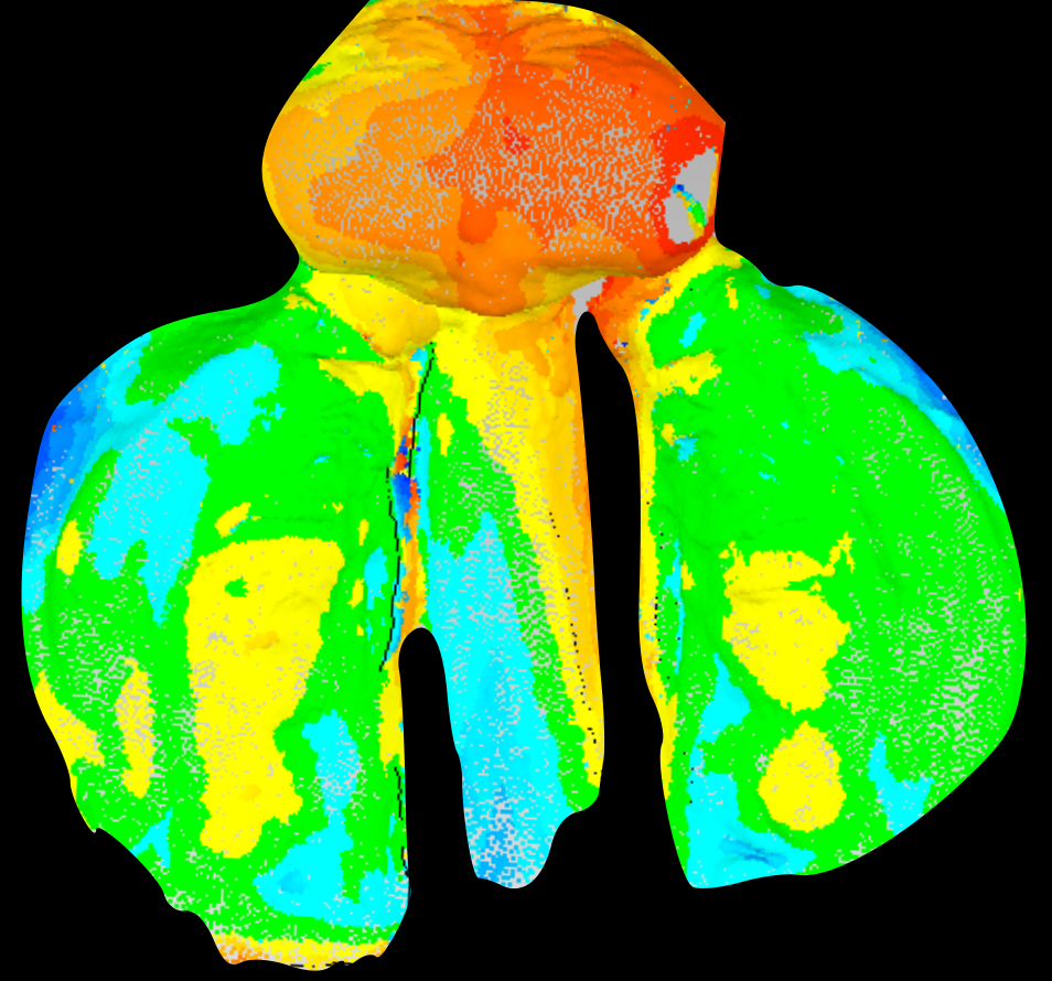
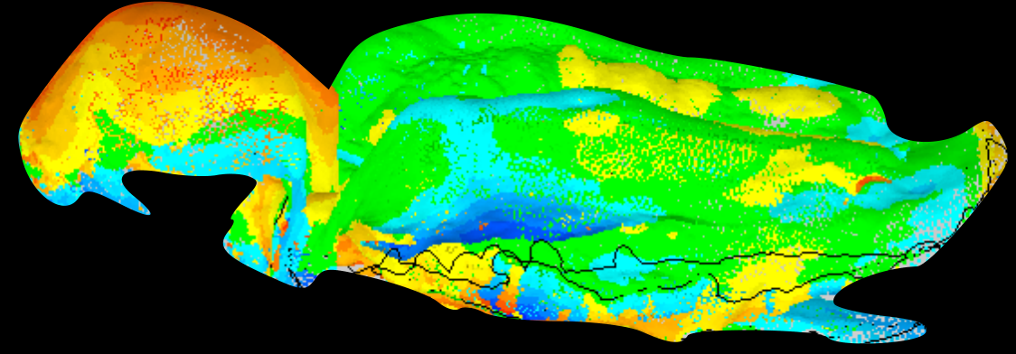
Lecturer — Digital Versus Functional
Alginate produces a functional impression because pressure can physically displace tissues, including the premaxilla, whereas optical scanning records tissues in a static environment. The differences between the methods can be represented using topographical maps.
The Cleft Palate-Craniofacial Journal, Volume 56 • Number 8 • September 2019. www.cpcjournal.org • Official Publication of the American Cleft Palate–Craniofacial Association. journals.sagepub.com/home/cpc • ISSN: 1055-6656
- Digital impression
- Safe, accurate (+0.78mm to -0.42mm), faster
- Compared to conventional alginate impressions
- Current limitations: both hardware and software issues
Lecturer — Digital Workflow
Digital workflow has extended beyond scanning to three-dimensional wax-ups, treatment planning, midline assessment, printed stents, and printed models. It can also allow oral appliances to be manufactured without conventional impressions; however, the technique requires an understanding of cleft anatomy, intraoral landmarks, and the limitations of static optical scanning.
- Digital workflow
- Translation to clinical practice?
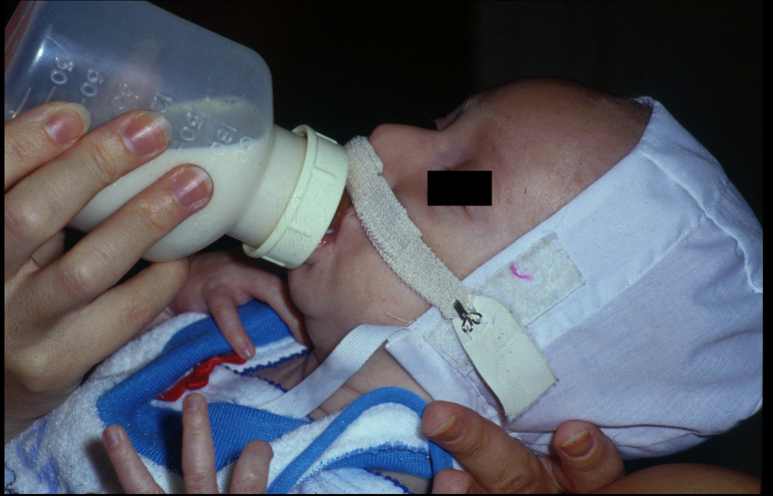
Feeding
-
Difficulty creating suction, poor seal (Reilly et al 2013)
-
Abnormally arranged oral and velopharyngeal musculature (McKinstry 1998; Nahai et al 2005)
-
Inefficient feeding
-
Increased feeding time
-
Weight loss / slow weight gain
-
Parental frustration
-
Feeds may require considerable effort, and families should be supported by specialist feeding teams and lactation consultants.
Feeding Concerns35
(Reilly et al 2013)
Feeding Strategies3637
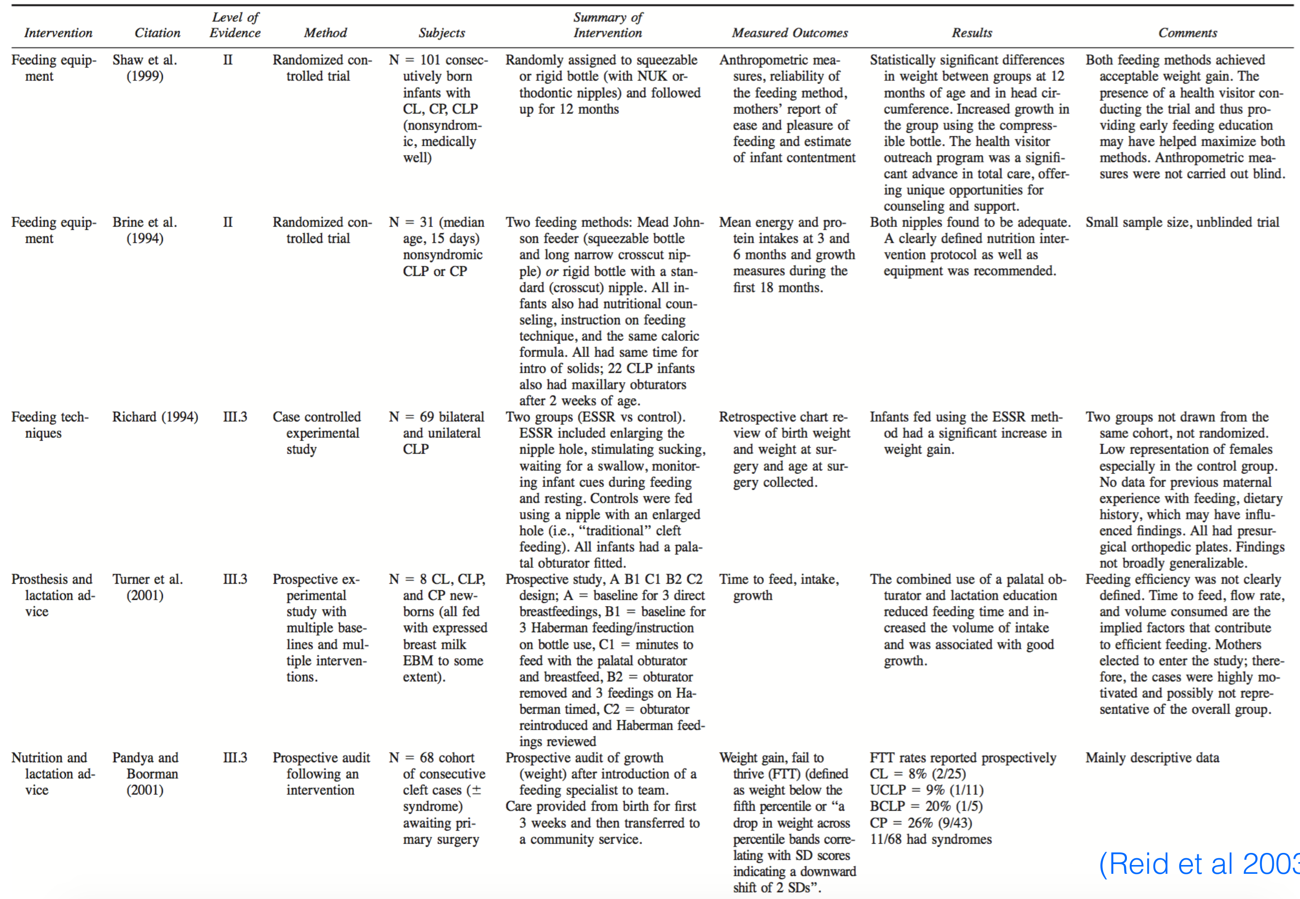
TABLE 4 Data-Driven Feeding Interventions Studies for Infants with Cleft Palate or Cleft Lip that Provide the Strongest Levels of Evidence for Practice*
| Intervention | Citation | Level of Evidence | Method | Subjects | Summary of Intervention | Measured Outcomes | Results | Comments |
|---|---|---|---|---|---|---|---|---|
| Feeding equipment | Shaw et al. (1999) | II | Randomized controlled trial | N = 101 consecutively born infants with CL, CP, CLP (nonsyndromic, medically well) | Randomly assigned to squeezable or rigid bottle (with NUK orthodontic nipples) and followed up for 12 months | Anthropometric measures, reliability of the feeding method, mothers’ report of ease and pleasure of feeding and estimate of infant contentment | Statistically significant differences in weight between groups at 12 months of age and in head circumference. Increased growth in the group using the compressible bottle. The health visitor outreach program was a significant advance in total care, offering unique opportunities for counseling and support. | Both feeding methods achieved acceptable weight gain. The presence of a health visitor conducting the trial and thus providing early feeding education may have helped maximize both methods. Anthropometric measures were not carried out blind. |
| Feeding equipment | Brine et al. (1994) | II | Randomized controlled trial | N = 31 (median age, 15 days) nonsyndromic CLP or CP | Two feeding methods: Mead Johnson feeder (squeezable bottle and long narrow crosscut nipple) or rigid bottle with a standard (crosscut) nipple. All infants also had nutritional counseling, instruction on feeding technique, and the same caloric formula. All had same time for intro of solids; 22 CLP infants also had maxillary obturators after 2 weeks of age. | Mean energy and protein intakes at 3 and 6 months and growth measures during the first 18 months. | Both nipples found to be adequate. A clearly defined nutrition intervention protocol as well as equipment was recommended. | Small sample size, unblinded trial |
| Feeding techniques | Richard (1994) | III.3 | Case controlled experimental study | N = 69 bilateral and unilateral CLP | Two groups (ESSR vs control). ESSR included enlarging the nipple |
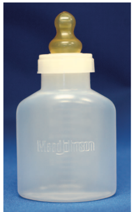
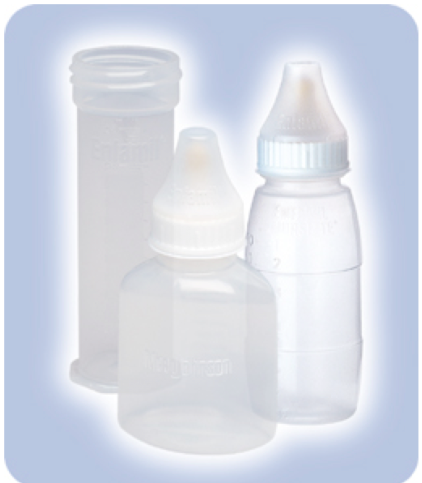
- Mead Johnson Cleft Nurser
- Mead Johnson Cleft Palate Nurser
Mead Johnson Bottle is a soft squeeze bottle with long cross cut nipple. Mead Johnson Bottle is designed to be “pulse” squeezed.
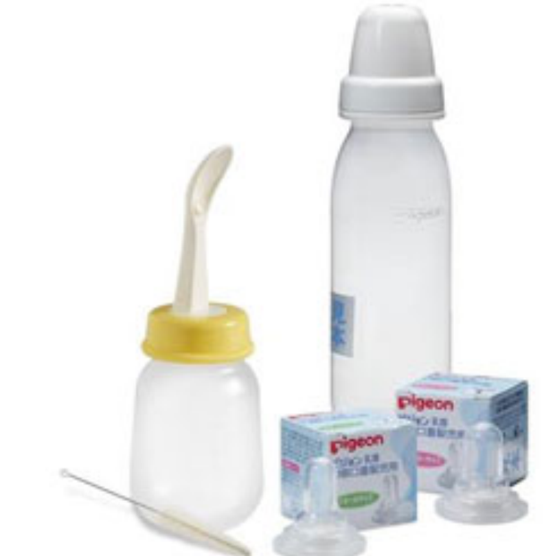
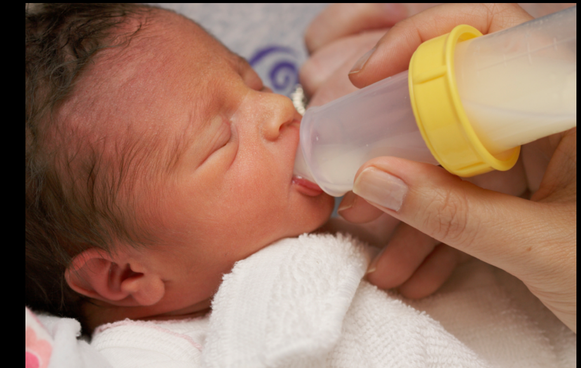
Lecturer — Feeding Equipment
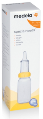
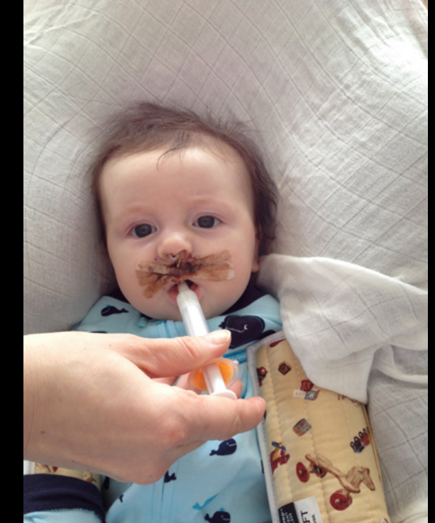
Most children with cleft lip and palate do not necessarily require an appliance for feeding.
- Specialist teams assess posture, bottle type, teat type, and the child’s individual feeding needs.
- Potential feeding aids include Haberman feeders, syringes, spoon feeds, Pigeon bottles, cross teats, and flask-flow nipples.
- The best approach depends on the child, the extent of the cleft, and the efficiency of feeding.
 |  |  |
 |  |  |
 |
Phases of Treatment38
- Short-Term: Presurgical; Surgical (lip & palate repair)
- Medium-Term: Dental care; Speech therapy; Surgical revisions
- Long-Term: Alveolar bone grafting; Orthognathic surgery
Lecturer — Short-Term Care
Short-term care extends from birth to approximately three months.
- During this phase, management focuses on feeding, weight gain, family support, pre-surgical orthopaedics where indicated, and preparation for lip repair.
Treatment Schedule by Age
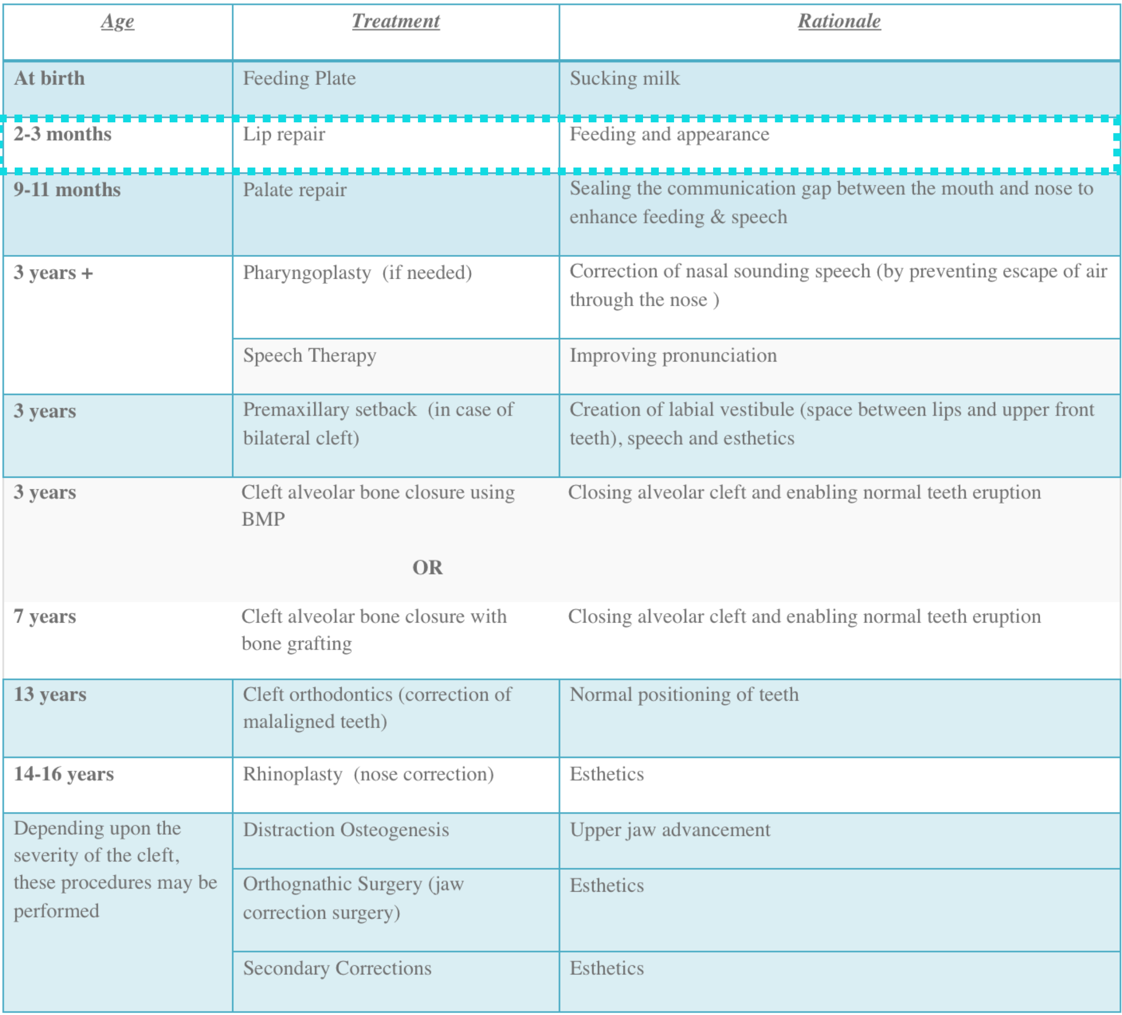
| Age | Treatment | Rationale |
|---|---|---|
| At birth | Feeding plate | Sucking milk |
| 2–3 months | Lip repair | Feeding and appearance |
| 9–11 months | Palate repair | Sealing the communication gap between the mouth and nose to enhance feeding & speech |
| 3 years + | Pharyngoplasty (if needed) | Correction of nasal sounding speech (by preventing escape of air through the nose) |
| Speech therapy | Improving pronunciation | |
| 3 years | Premaxillary setback (in case of bilateral cleft) | Creation of labial vestibule (space between lips and upper front teeth), speech and esthetics |
| 3 years | Cleft alveolar bone closure using BMP | Closing alveolar cleft and enabling normal teeth eruption |
| OR | ||
| 7 years | Cleft alveolar bone closure with bone grafting | Closing alveolar cleft and enabling normal teeth eruption |
| 13 years | Cleft orthodontics (correction of malaligned teeth) | Normal positioning of teeth |
| 14–16 years | Rhinoplasty (nose correction) | Esthetics |
| Depending upon the severity of the cleft, these procedures may be performed | Distraction osteogenesis | Upper jaw advancement |
| Orthognathic surgery (jaw correction surgery) | Esthetics | |
| Secondary corrections | Esthetics |
Lecturer — Later Treatment Schedule
The treatment schedule also includes several broader age-based stages.
- From birth to approximately three months, support may include feeding support and possible pre-surgical orthopaedics.
- From one to five years, care includes speech therapy, audiology, dental assessment, and preventive care.
- During mixed dentition, care includes pre-surgical orthodontics and alveolar bone grafting.
- Later childhood and adolescence may involve definitive orthodontics and possible future orthognathic surgery or implant planning.
Surgical Treatment
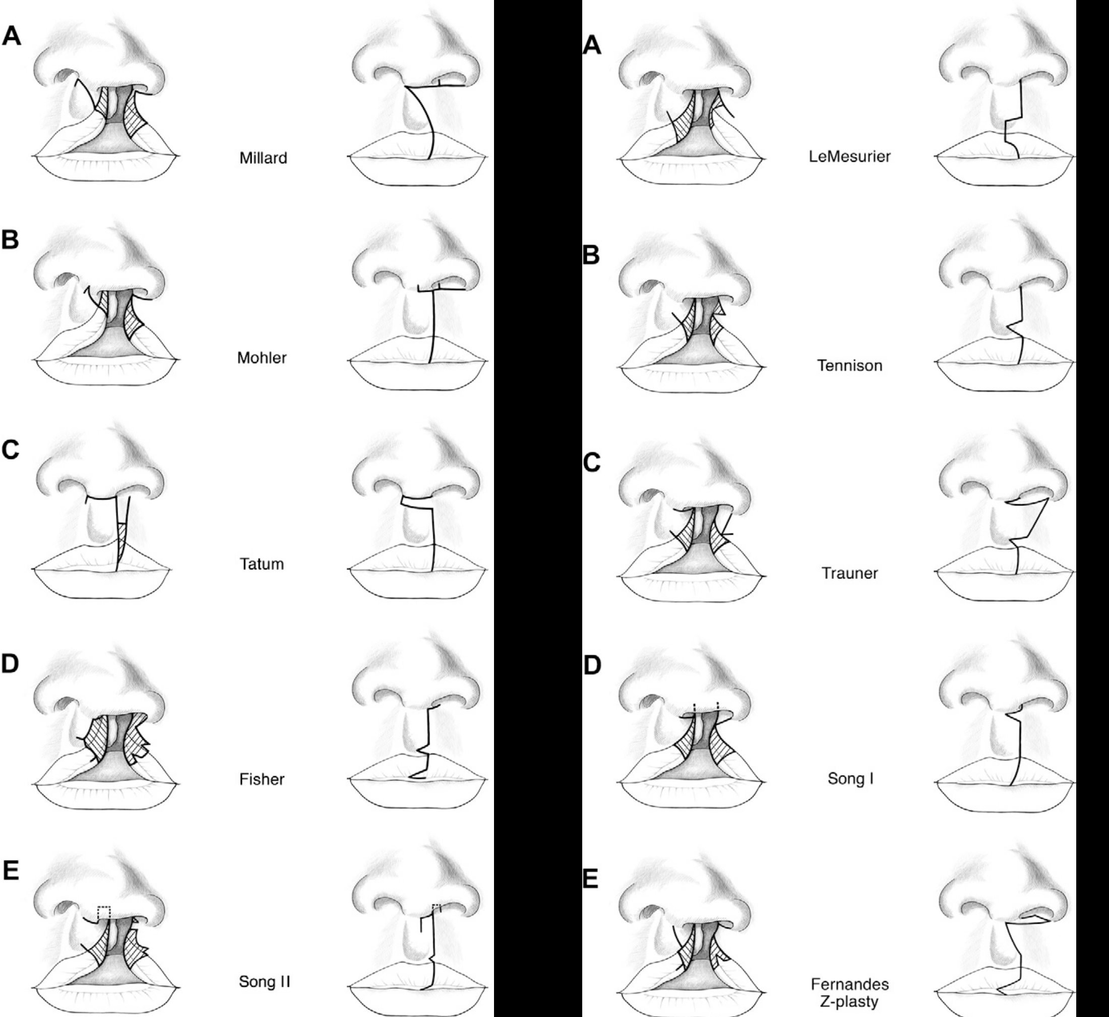
Primary Lip Repair39
Deshpande et al 2014
Surgical Techniques40
-
A — Millard
-
B — Mohler
-
C — Tatum
-
D — Fisher
-
E — Song II
-
A — LeMesurier
-
B — Tennison
-
C — Trauner
-
D — Song I
-
E — Fernandes Z-plasty
Lecturer — Lip Repair Timing
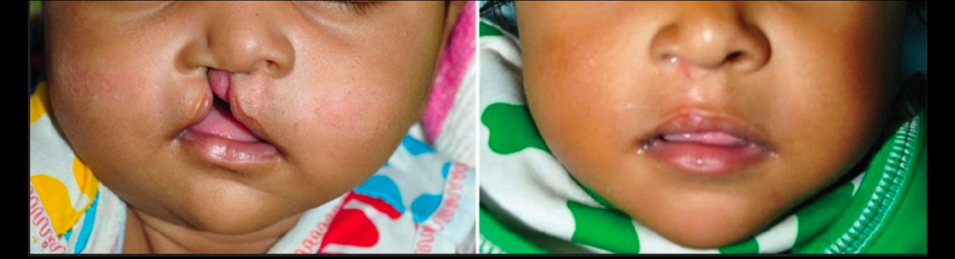
Primary lip repair generally occurs at approximately three months rather than at birth.
- The child must have adequate weight, haemoglobin levels, and general capacity to tolerate the procedure.
- Pre-surgical orthopaedics may reduce the cleft width and improve the conditions for repair.
- Numerous techniques and modified flaps have been described, but the technique depends primarily on the surgical team’s preference; no single technique is universally superior.
Demke et al 2011
| Age | Treatment | Rationale |
|---|---|---|
| At birth | Feeding plate | Sucking milk |
| 2–3 months | Lip repair | Feeding and appearance |
| 9–11 months | Palate repair | Sealing the communication gap between the mouth and nose to enhance feeding & speech |
| 3 years + | Pharyngoplasty (if needed) | Correction of nasal sounding speech (by preventing escape of air through the nose) |
| Speech therapy | Improving pronunciation | |
| 3 years | Premaxillary setback (in case of bilateral cleft) | Creation of labial vestibule (space between lips and upper front teeth), speech and esthetics |
| 3 years | Cleft alveolar bone closure using BMP | Closing alveolar cleft and enabling normal teeth eruption |
| OR | ||
| 7 years | Cleft alveolar bone closure with bone grafting | Closing alveolar cleft and enabling normal teeth eruption |
| 13 years | Cleft orthodontics (correction of malaligned teeth) | Normal positioning of teeth |
| 14–16 years | Rhinoplasty (nose correction) | Esthetics |
| Depending upon the severity of the cleft, these procedures may be performed | Distraction osteogenesis | Upper jaw advancement |
| Orthognathic surgery (jaw correction surgery) | Esthetics | |
| Secondary corrections | Esthetics |
- Short-Term: Presurgical; Surgical (lip & palate repair)
- Medium-Term: Dental care; Speech therapy; Surgical revisions
- Long-Term: Alveolar bone grafting; Orthognathic surgery
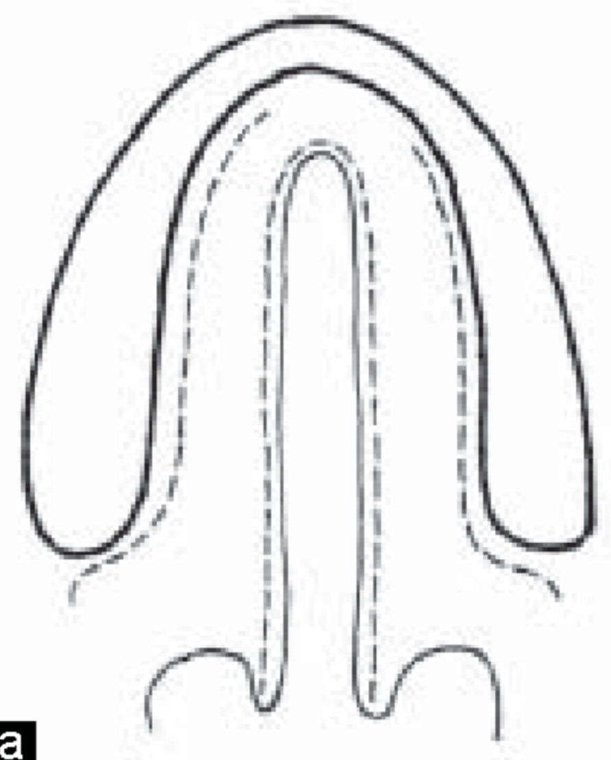
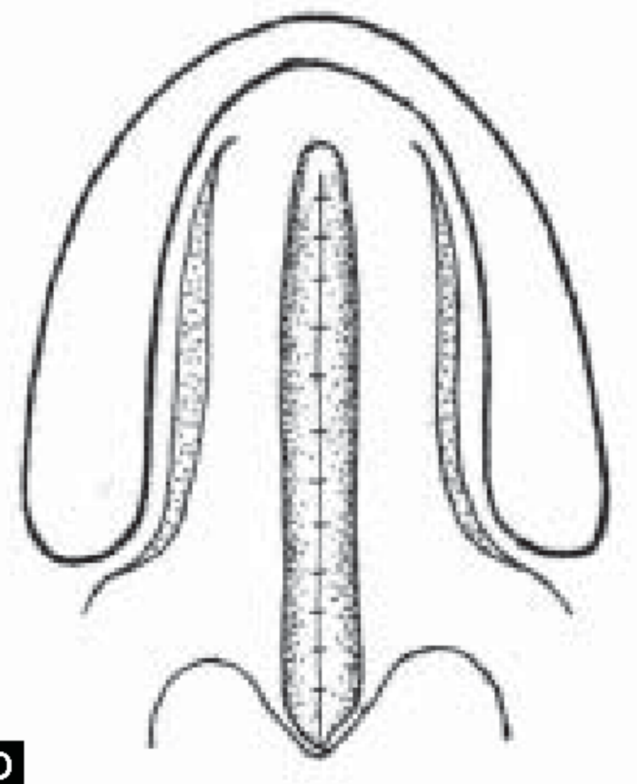
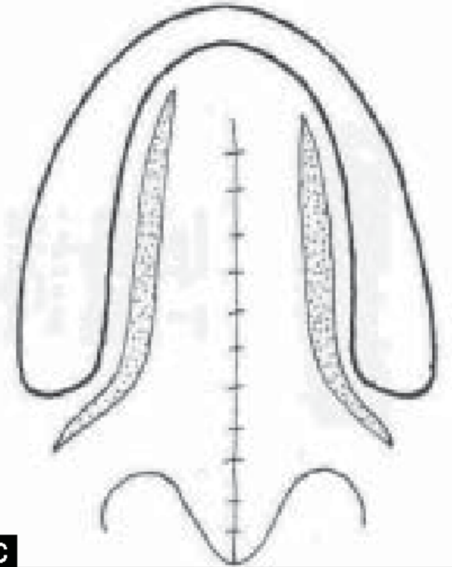
Palate Repair4142
 |  |  |
 |  |  |
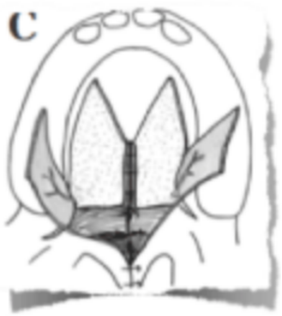 |
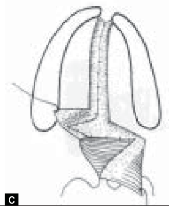
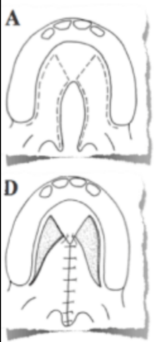
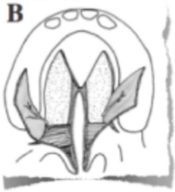
Surgical Techniques43
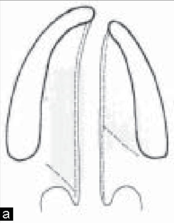
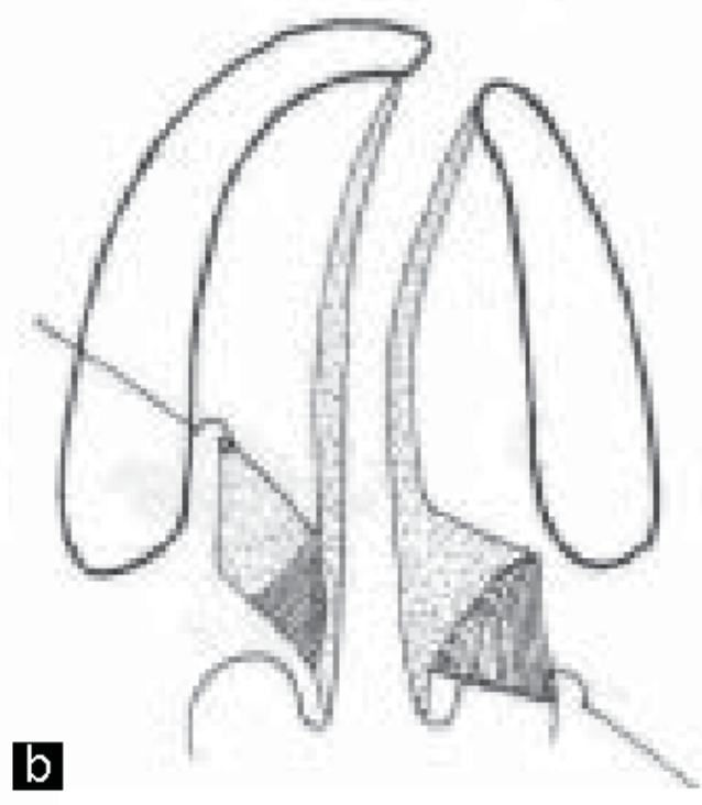
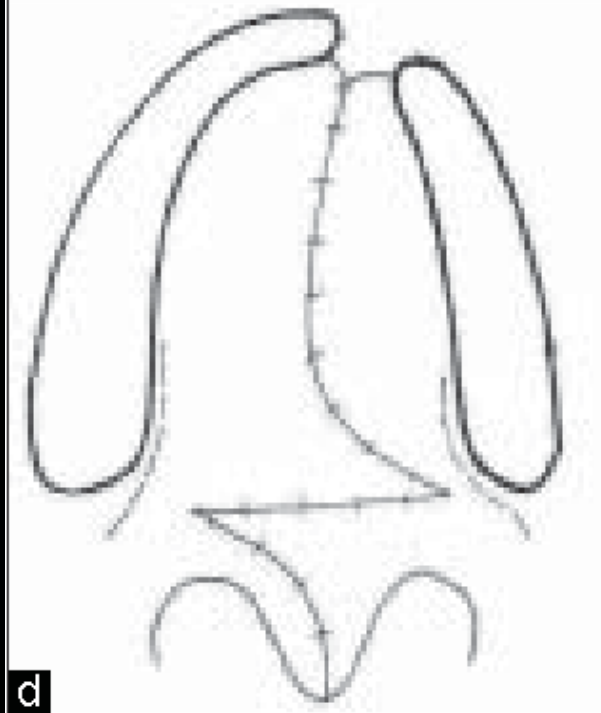
von Langenbeck palatoplasty
Agrawal et al 2009
Furlow Z-plasty
Agrawal et al 2009
V-Y pushback
Panels: A, B, C, D
Kilner et al 1937
Lecturer — Palate Repair Timing
There is usually a period between lip repair and palate repair in a child with a complete cleft lip and palate.
- After lip repair, the child continues to have a palatal cleft.
- Palate repair generally occurs at approximately nine to twelve months, closing the palatal cleft after the earlier lip procedure.
- Multiple techniques, including rotational flaps, may be used; the choice depends on the surgical team and the individual cleft.
- These techniques have existed for many years but have been modified over time.
- Short-Term: Presurgical; Surgical (lip & palate repair)
- Medium-Term: Dental care; Speech therapy; Surgical revisions
- Long-Term: Alveolar bone grafting; Orthognathic surgery
| Age | Treatment | Rationale |
|---|---|---|
| At birth | Feeding plate | Sucking milk |
| 2–3 months | Lip repair | Feeding and appearance |
| 9–11 months | Palate repair | Sealing the communication gap between the mouth and nose to enhance feeding & speech |
| 3 years + | Pharyngoplasty (if needed) | Correction of nasal sounding speech (by preventing escape of air through the nose) |
| Speech therapy | Improving pronunciation | |
| 3 years | Premaxillary setback (in case of bilateral cleft) | Creation of labial vestibule (space between lips and upper front teeth), speech and esthetics |
| 3 years | Cleft alveolar bone closure using BMP | Closing alveolar cleft and enabling normal teeth eruption |
| OR | ||
| 7 years | Cleft alveolar bone closure with bone grafting | Closing alveolar cleft and enabling normal teeth eruption |
| 13 years | Cleft orthodontics (correction of malaligned teeth) | Normal positioning of teeth |
| 14–16 years | Rhinoplasty (nose correction) | Esthetics |
| Depending upon the severity of the cleft, these procedures may be performed | Distraction osteogenesis | Upper jaw advancement |
| Orthognathic surgery (jaw correction surgery) | Esthetics | |
| Secondary corrections | Esthetics |
 |  |
 |
Dental Care
- ‘Well baby’ checks
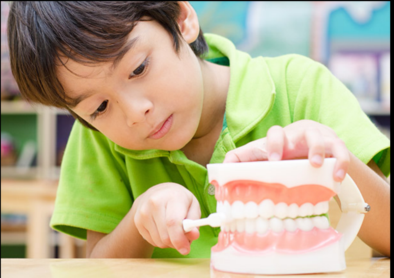
Preventive Dental Care4445
-
‘Well baby’ checks
-
Monitor and improve the effectiveness of plaque control
-
Professional cleaning and topical fluoride application
-
Dietary assessment and counselling
-
Fissure sealants to teeth at risk of pit and fissure caries
-
Early diagnosis and management of pathology
-
Occlusal development
-
Fabrication of a mouthguard
-
Dental care becomes particularly important once the teeth begin to erupt.
-
Toothpaste guidance.
-
Monitoring hypermineralised second primary molars.
-
Mouthguard provision for children participating in contact sports.
-
Children with cleft lip and palate may already undergo many procedures with several clinical teams.
-
Preventive care can reduce the need for further invasive treatment.
Restorative Dental Care46
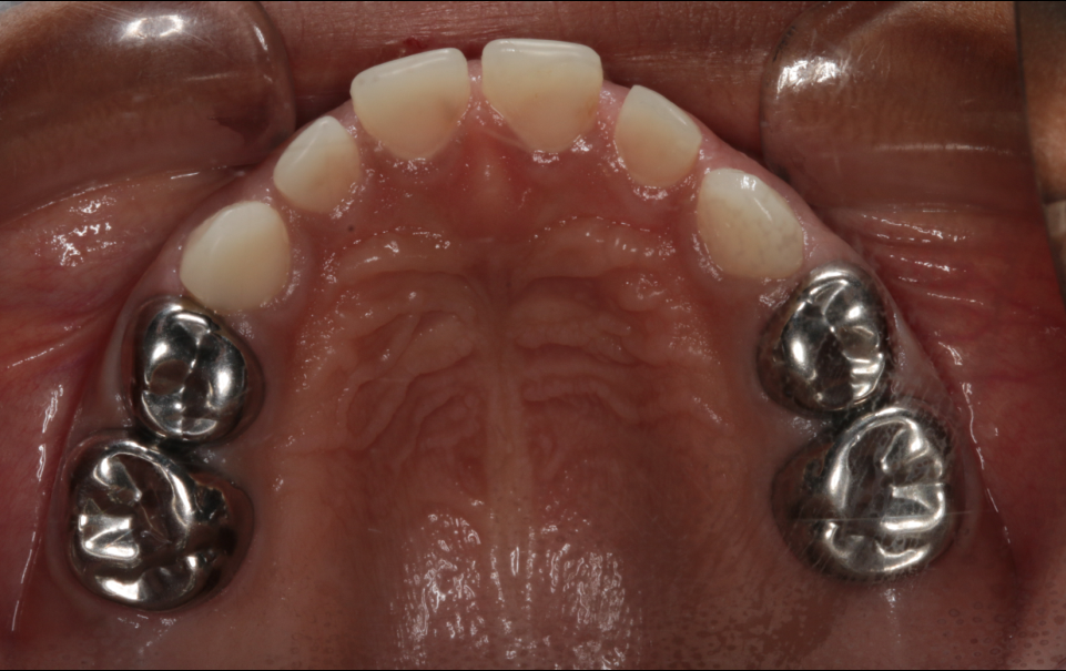
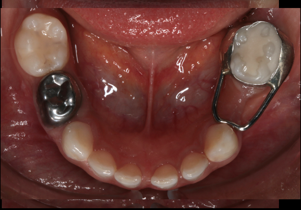
Lecturer — Restorative Treatment
Restorative treatment should be definitive and durable, because repeated failed treatments increase the risk of treatment fatigue or burnout.
- Treatment should be completed well the first time where possible.
- Patchwork composite or glass ionomer restorations may not be in the child’s best interest when a longer-lasting approach is needed.
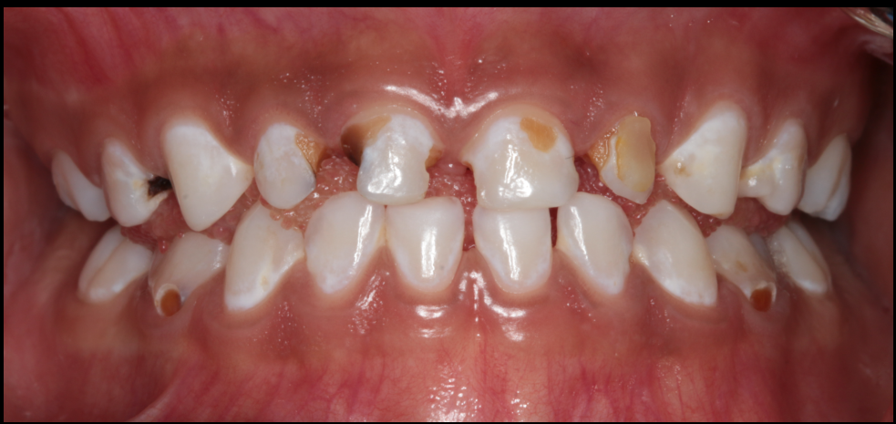
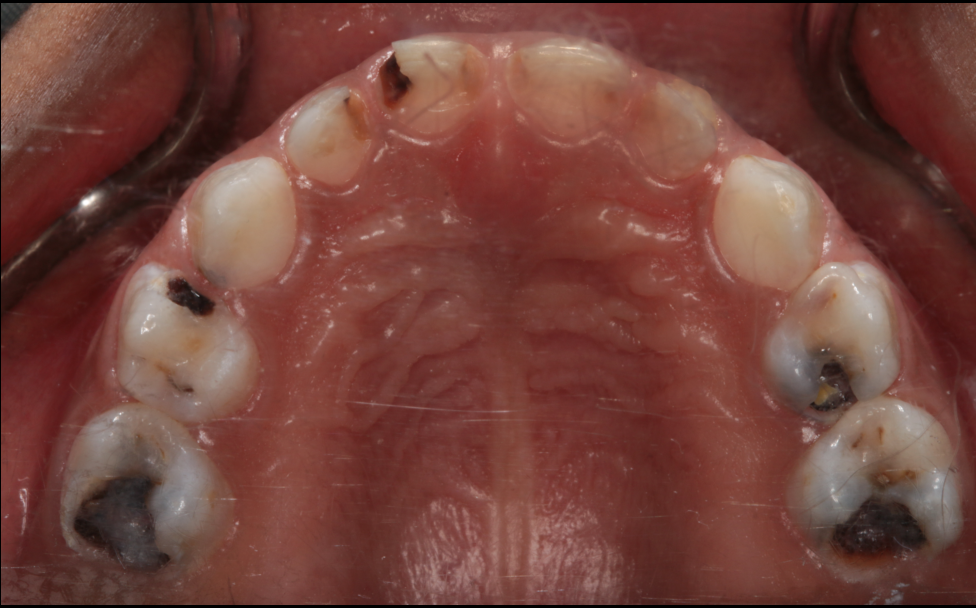
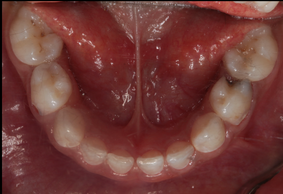
- Hypermineralised second primary molars may develop atypical caries patterns, including unusual distal palatal lesions associated with the enamel defects.
 |  |  |
 |  |  |
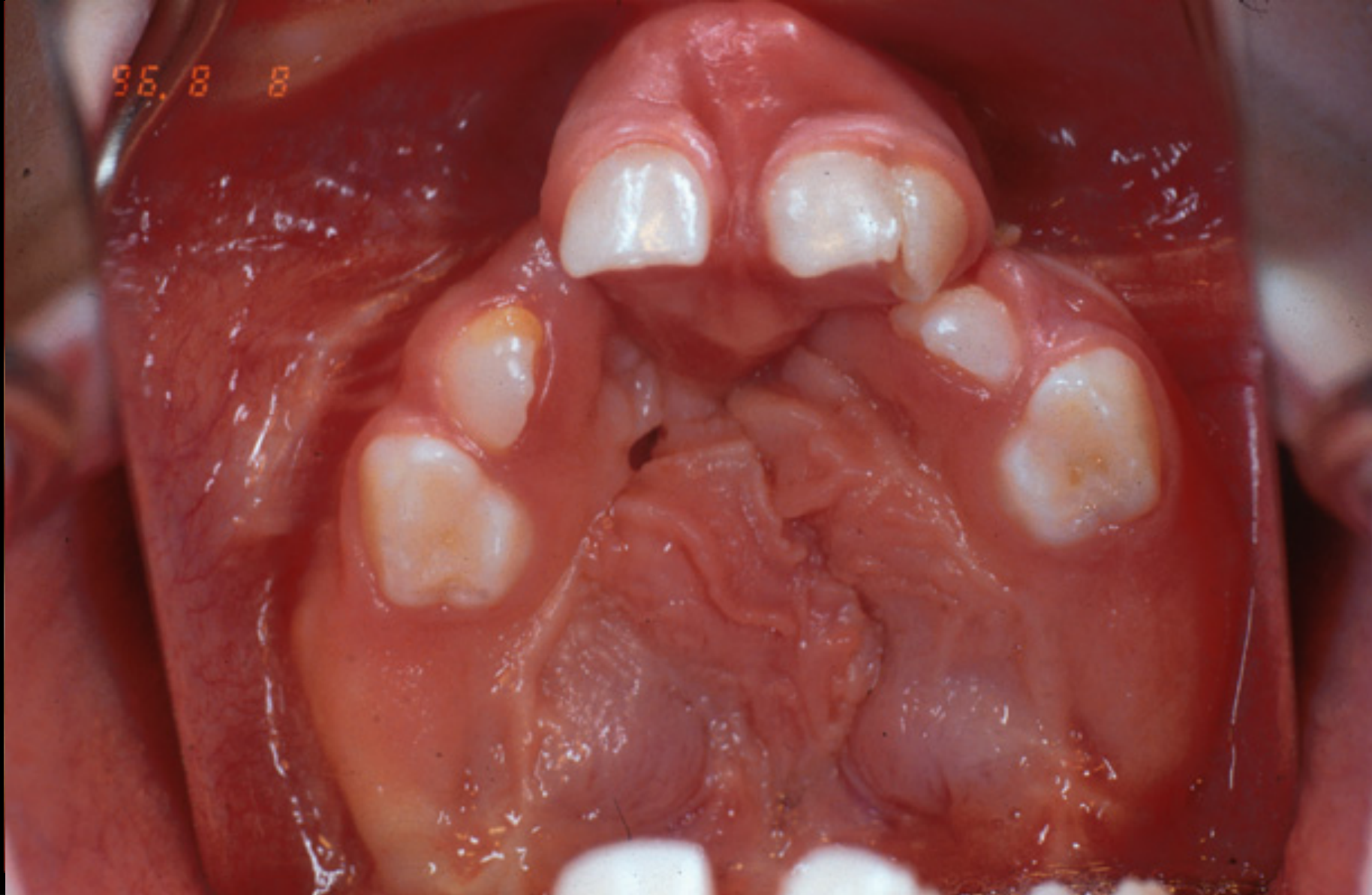
Dental Anomalies4748
-
Developmental defects of enamel
-
Missing teeth
-
Supernumerary teeth
-
Accessory cusps
-
Dilaceration
-
Microform teeth
-
Ectopic teeth
-
Microdont teeth.
-
Conical teeth.
-
Talon cusps.
-
Rotations.
-
Space loss.
-
Tooth migration.
-
Atypical resorption. Images courtesy of Dr John Winters.
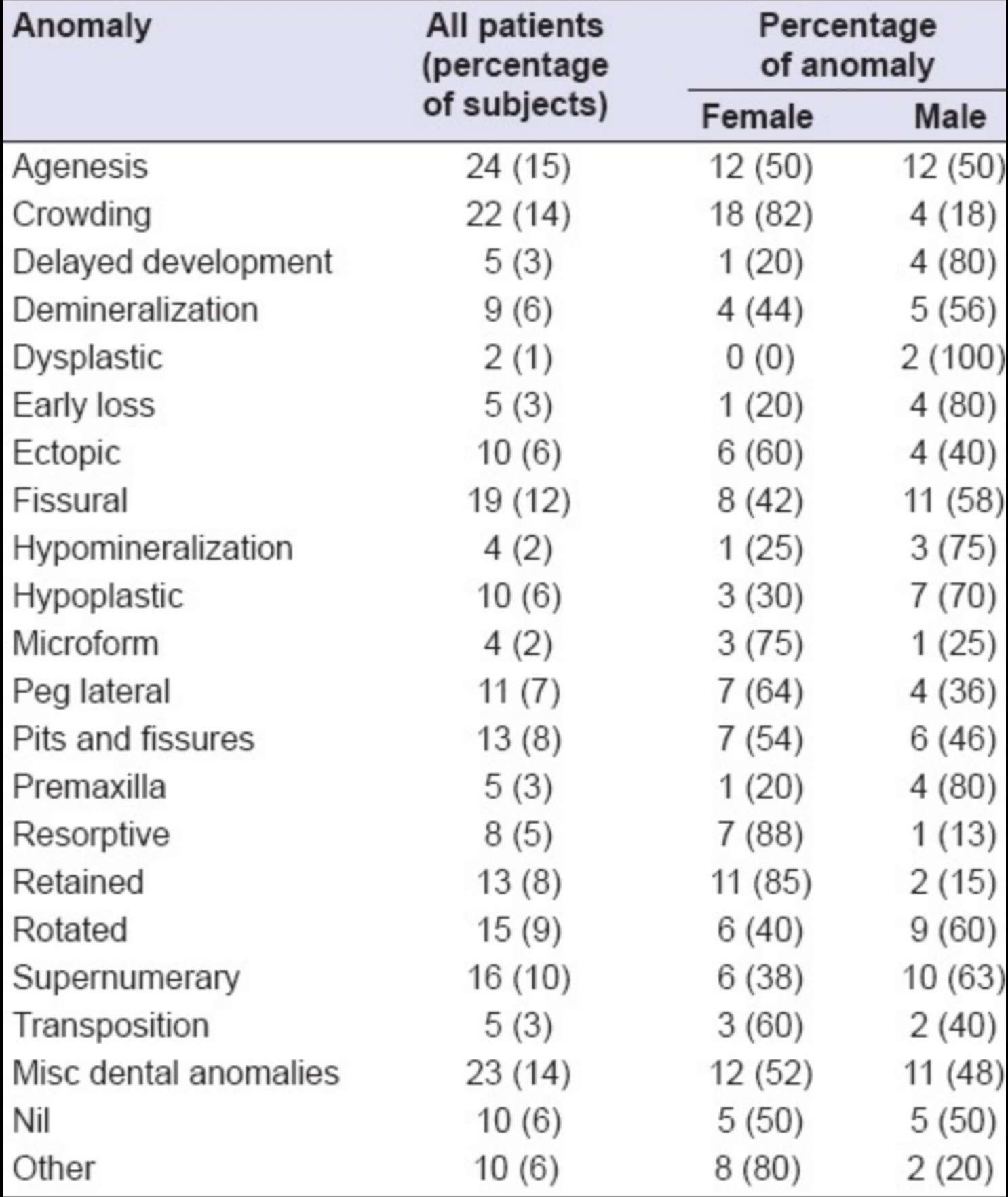
| Anomaly | All patients (percentage of subjects) | Percentage of anomaly Female | Percentage of anomaly Male |
|---|---|---|---|
| Agenesis | 24 (15) | 12 (50) | 12 (50) |
| Crowding | 22 (14) | 18 (82) | 4 (18) |
| Delayed development | 5 (3) | 1 (20) | 4 (80) |
| Demineralization | 9 (6) | 4 (44) | 5 (56) |
| Dysplastic | 2 (1) | 0 (0) | 2 (100) |
| Early loss | 5 (3) | 1 (20) | 4 (80) |
| Ectopic | 10 (6) | 6 (60) | 4 (40) |
| Fissural | 19 (12) | 8 (42) | 11 (58) |
| Hypomineralization | 4 (2) | 1 (25) | 3 (75) |
| Hypoplastic | 10 (6) | 3 (30) | 7 (70) |
| Microform | 4 (2) | 3 (75) | 1 (25) |
| Peg lateral | 11 (7) | 7 (64) | 4 (36) |
| Pits and fissures | 13 (8) | 7 (54) | 6 (46) |
| Premaxilla | 5 (3) | 1 (20) | 4 (80) |
| Resorptive | 8 (5) | 7 (88) | 1 (13) |
| Retained | 13 (8) | 11 (85) | 2 (15) |
| Rotated | 15 (9) | 6 (40) | 9 (60) |
| Supernumerary | 16 (10) | 6 (38) | 10 (63) |
| Transposition | 5 (3) | 3 (60) | 2 (40) |
| Misc dental anomalies | 23 (14) | 12 (52) | 11 (48) |
| Nil | 10 (6) | 5 (50) | 5 (50) |
| Other | 10 (6) | 8 (80) | 2 (20) |
(Nicholls et al 2016)
 |  |
Alveolar Bone Grafting49
Alveolar Bone Grafting (ABG)
 |  |
 |  |
ABG Objectives50
-
To obtain maxillary arch continuity
-
To maximize bone support for the dentition and facilitate eruption of teeth in the area of the alveolar cleft
-
To stabilize the maxillary segments after orthodontic treatment, especially the mobile primary palate of bilateral clefts
-
To eliminate oronasal fistulae
-
To provide nasal alar cartilage support
-
To establish ideal alveolar morphology; and
-
To provide available bone with attached soft tissue for future endosteal implant placement in cases where there is a residual dental space.
Lecturer — Residual Bony Support
After lip and palate repair, the cleft may be closed by soft tissue but still lack adequate bony support. The graft therefore restores structural support in addition to closing the cleft.
Nadjmi et al. 2018, Cho-Lee et al. 2013.
Nadjmi et al 2018.
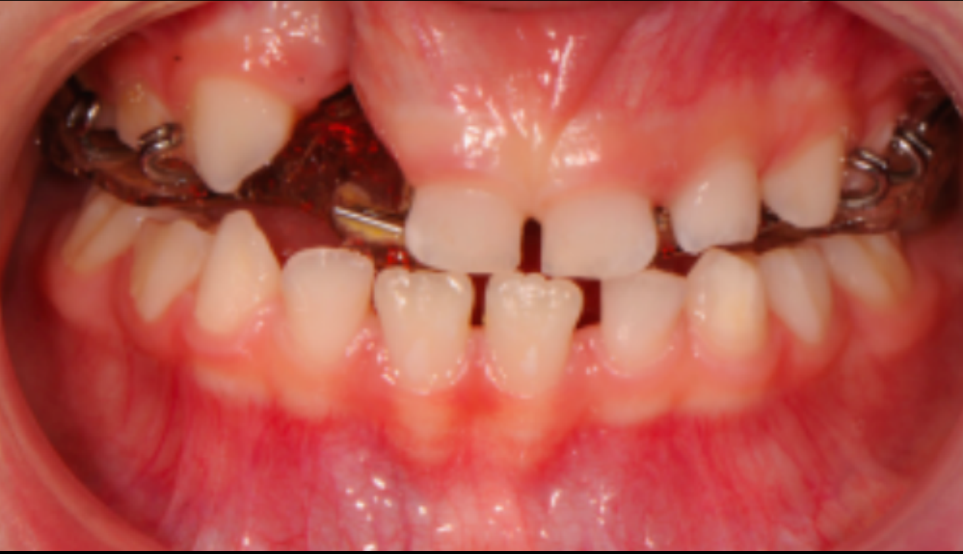
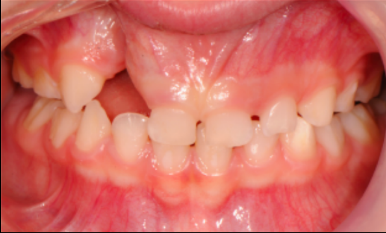
ABG Pre-Surgical Orthodontics51
- To provide optimal arch alignment prior to bone grafting
Nadjmi et al 2018.
Lecturer — Presurgical Orthodontics
Presurgical orthodontics aligns and expands the maxillary segments into a favourable position, but it is not definitive orthodontic treatment. Definitive orthodontics occurs after the bone graft has healed, and imaging with multidisciplinary review helps identify present, missing, supernumerary, retained primary, and extraction-required teeth.
 |  |
ABG Protocols52
- Secondary alveolar bone grafting (ABG) performed after dental alignment and maxillary arch expansion.
- The retained primary dentition adjacent to the cleft are extracted at least 4 weeks before bone grafting to allow for adequate healing of the keratinized mucosa.
- Periodontal review and work-up to ensure gingival tissues are in optimal condition leading up to ABG, this includes regular use of chlorhexidine oral gel and mouthwash.
- ABG surgery conducted by means of iliac crest autogenous bone grafts in the majority of the cases.
- Strict post-operative oral hygiene and periodontal maintenance.
- Postoperative clinical and radiological evaluation.
- Recommencement of orthodontic treatment approx six weeks after the surgery.
Nadjmi et al 2018.
Lecturer — Graft Timing and Health
The timing of grafting depends on the child’s development, the formation and root development of the permanent canine or lateral incisor, and which teeth are present in the cleft region. Periodontal health is important because poor oral hygiene and gingivitis are associated with more adverse events during bone grafting.
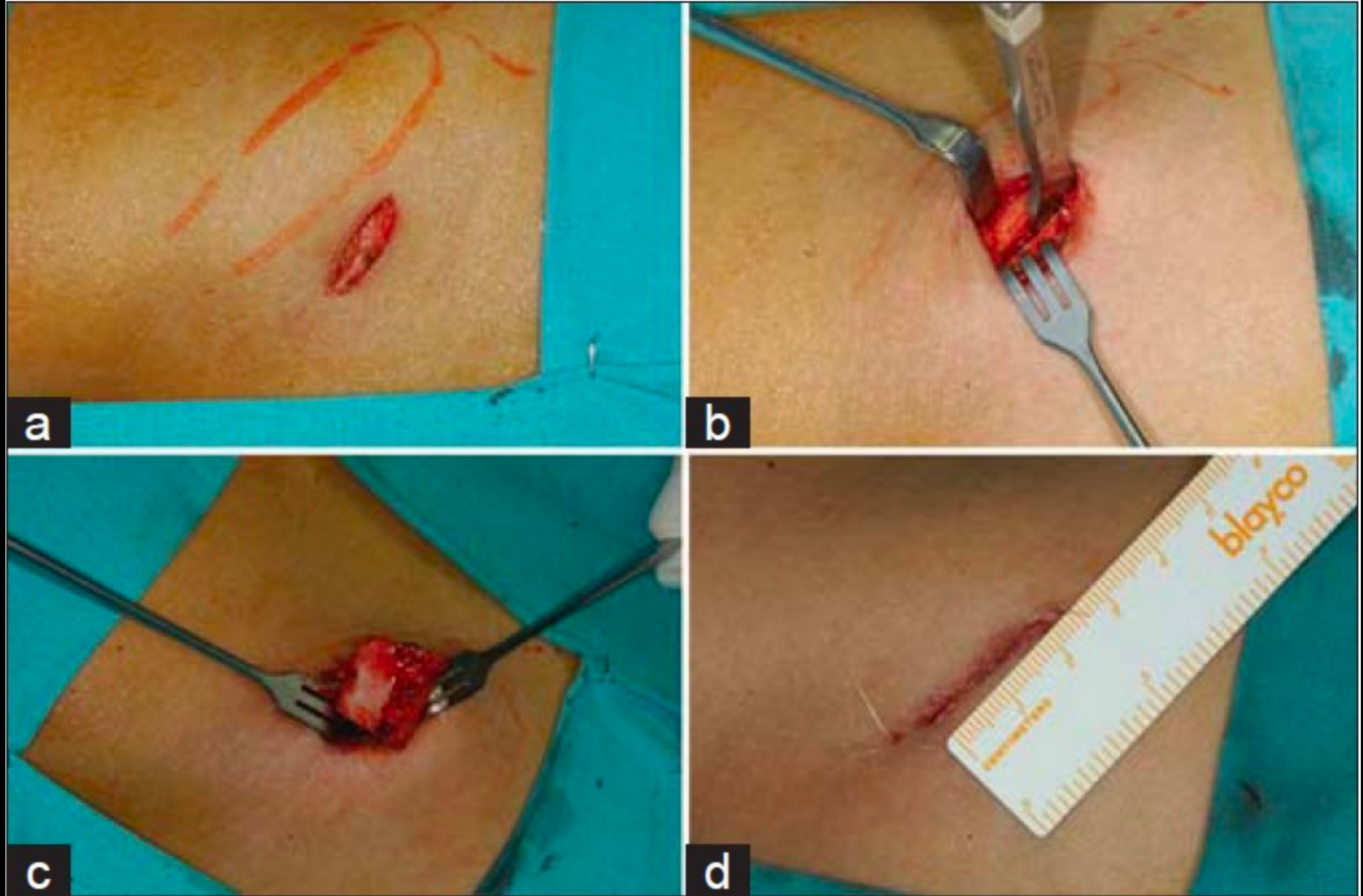
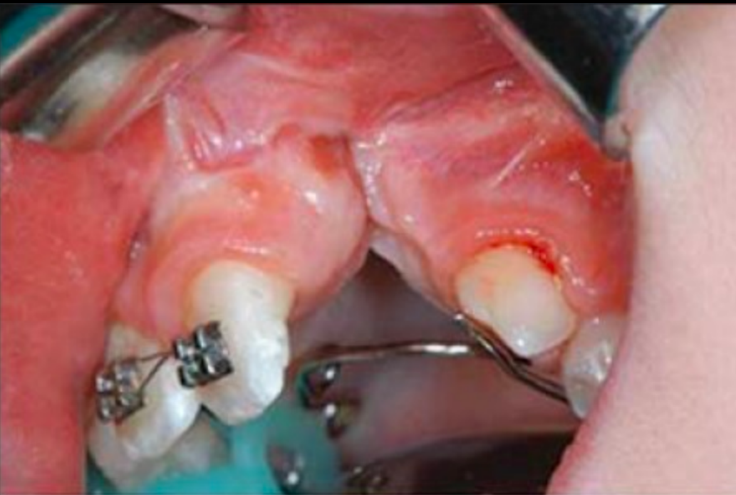
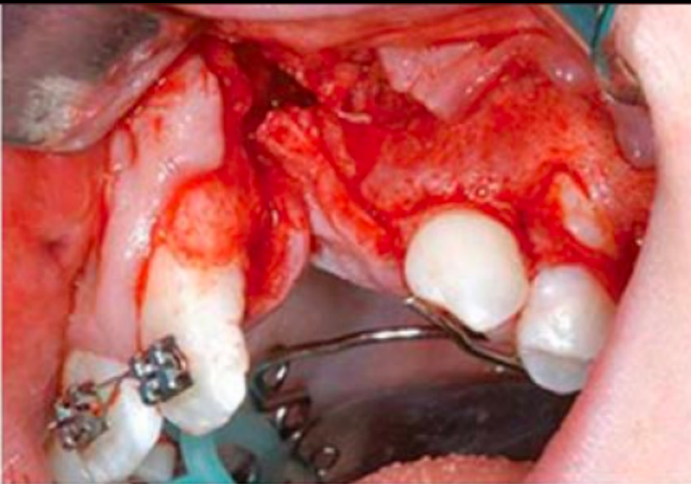
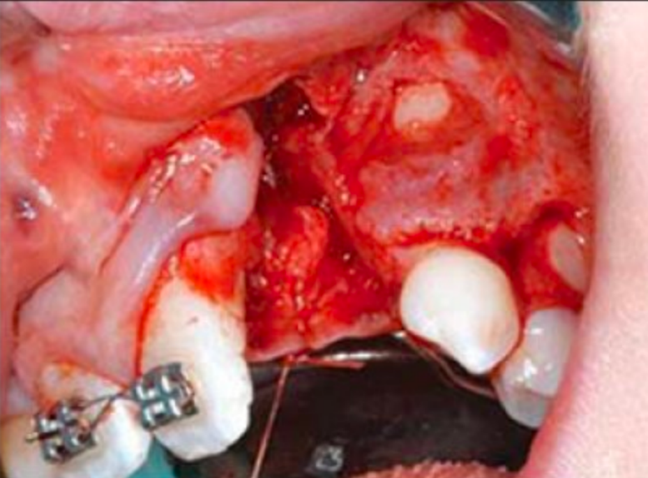
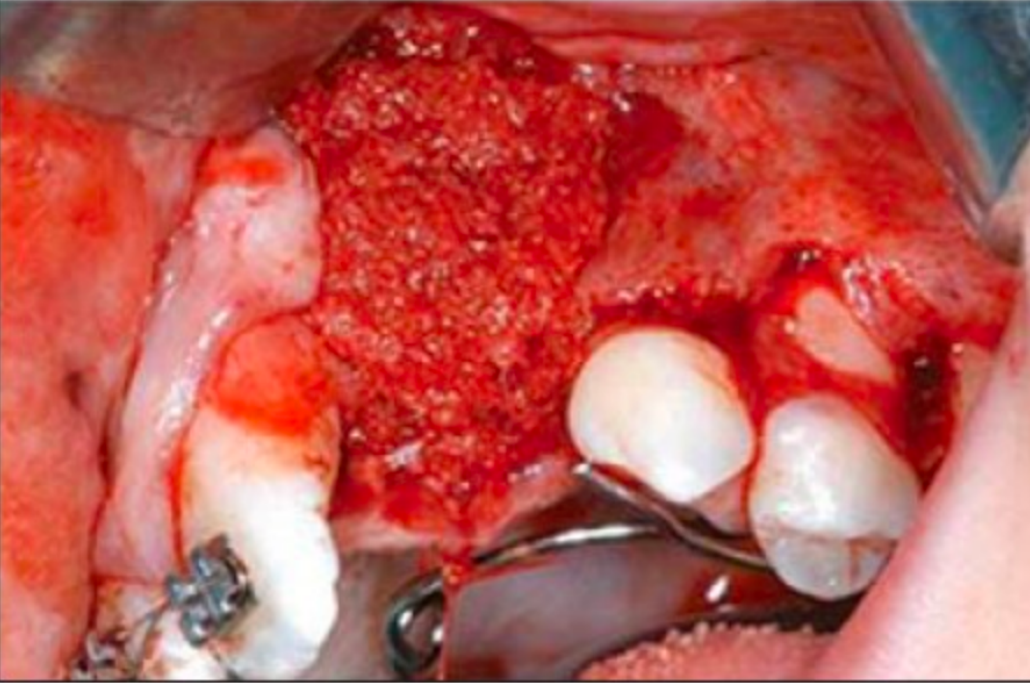
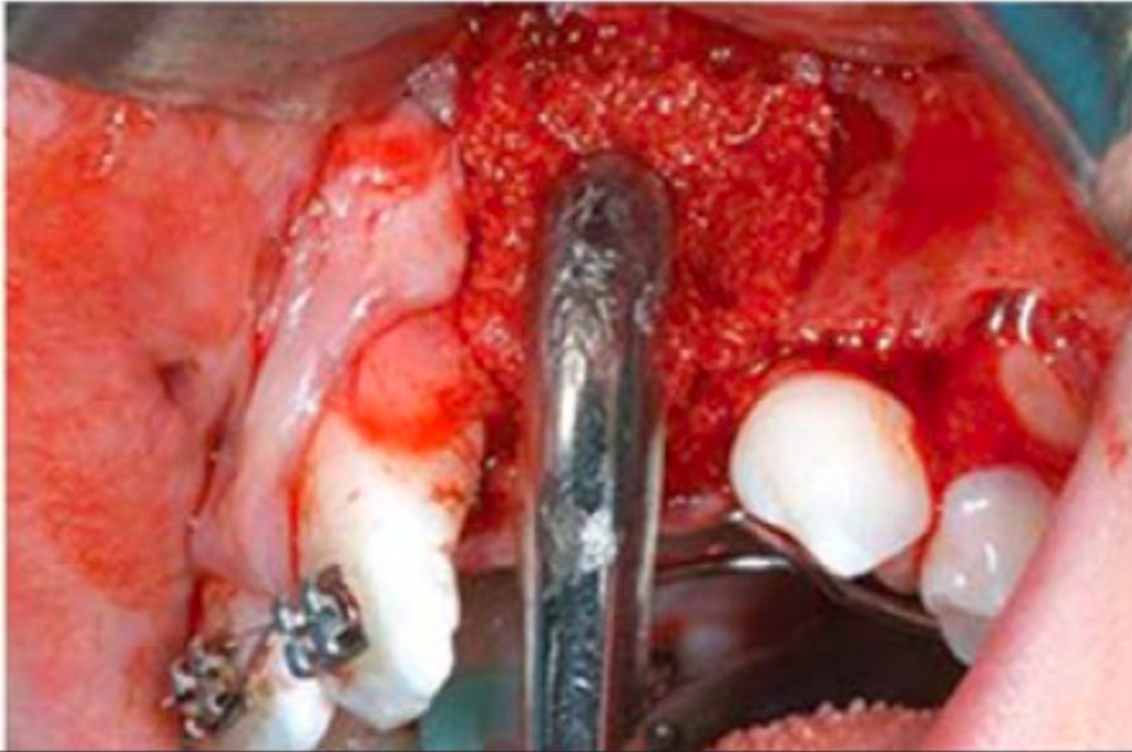
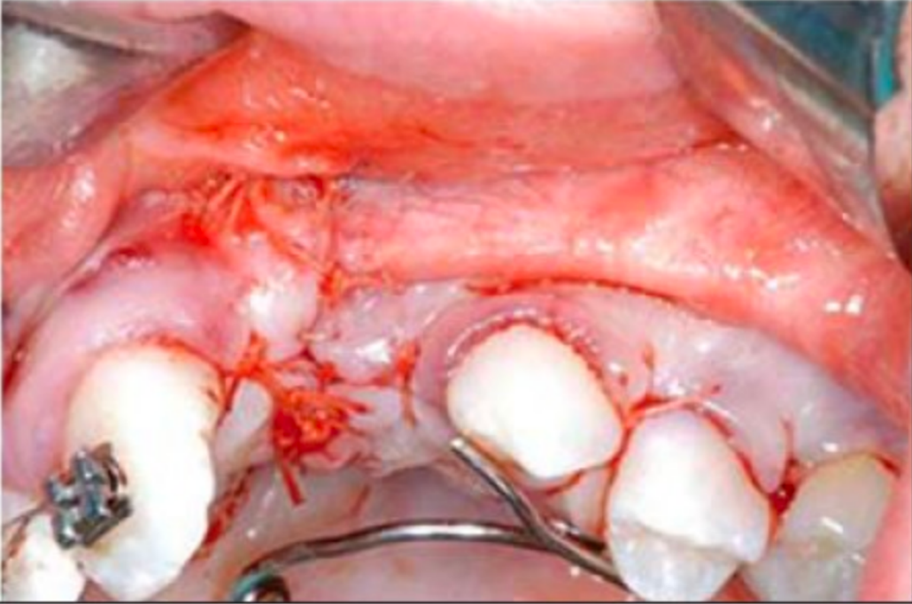
ABG Surgical Procedures5354
- Iliac bone grafting
Cho-Lee et al 2013.
- Alveolar cleft repair
Cho-Lee et al 2013.
Lecturer — Iliac Crest Harvesting
At Perth Children’s Hospital, bone is harvested from the iliac crest because it is relatively accessible, associated with lower morbidity, and suitable for simultaneous work by two surgical teams. One team can harvest the bone while another prepares the cleft site intraorally.
 |  |  |
 |  |  |
 |
Short-Term55
- Presurgical
- Surgical (lip & palate repair)
Lecturer — Intraoral Cleft Repair
Intraoral surgery uses gingival sulcus, labial, and palatal incisions to raise full-thickness mucoperiosteal flaps. The nasal mucosa is separated from the gingiva and elevated, the nasal floor is repaired, bone is compacted into the cleft, and the area is sutured closed.
Medium-Term
- Dental Care
- Speech therapy
- Surgical Revisions
Lecturer — Short-Term Management
Short-term treatment includes bone harvesting, preparation of the cleft site, closure of the nasal and oral tissues, placement of the bone graft, and maintenance of the maxillary segments in a favourable position. Expansion appliances may remain in place to maintain segment position.
Long-Term
- Alveolar bone grafting
- Orthognathic surgery
Lecturer — Postoperative Management
Medium-term management includes healing, periodontal maintenance, clinical and radiographic evaluation, and assessment of graft incorporation before orthodontic treatment resumes. Long-term planning may include assessment of maxillary growth, orthognathic surgery, and implants where suitable bone and soft-tissue support are available; earlier surgery may restrict maxillary growth.
| Age | Treatment | Rationale |
|---|---|---|
| At birth | Feeding Plate | Sucking milk |
| 2-3 months | Lip repair | Feeding and appearance |
| 9-11 months | Palate repair | Sealing the communication gap between the mouth and nose to enhance feeding & speech |
| 3 years + | Pharyngoplasty (if needed) | Correction of nasal sounding speech (by preventing escape of air through the nose) |
| Speech Therapy | Improving pronunciation | |
| 3 years | Premaxillary setback (in case of bilateral cleft) | Creation of labial vestibule (space between lips and upper front teeth), speech and esthetics |
| 3 years | Cleft alveolar bone closure using BMP OR | Closing alveolar cleft and enabling normal teeth eruption |
| 7 years | Cleft alveolar bone closure with bone grafting | Closing alveolar cleft and enabling normal teeth eruption |
| 13 years | Cleft orthodontics (correction of malaligned teeth) | Normal positioning of teeth |
| 14-16 years | Rhinoplasty (nose correction) | Esthetics |
| Depending upon the severity of the cleft, these procedures may be performed | Distraction Osteogenesis | Upper jaw advancement |
| Orthognathic Surgery (jaw correction surgery) | Esthetics | |
| Secondary Corrections | Esthetics |
Lecturer — Coordinated Cleft Care
Children with cleft lip and palate undergo a long chronological sequence of care, from feeding management and possible presurgical orthopaedics through repair, preventive dental care, dental-anomaly management, mixed-dentition orthodontics and grafting, and later definitive treatment when necessary. Cleft lip and palate affects the child as a whole, so coordinated multidisciplinary care must consider the experiences of the child and family and balance surgical benefits against restriction of maxillary growth and other long-term effects.
Closing And Clinical Cases565758
Thank You
Clinical Cases
Clinical Cases
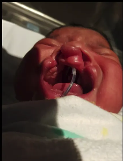
Case: Bilateral complete cleft lip and palate, pre-surgical orthopaedics workflow
Question
Scenario: A newborn with a bilateral cleft is presented to demonstrate the steps of pre-surgical orthopaedics, from impression taking to appliance placement.
What’s shown: Clinical photos and dental models showing the initial alginate impression, the poured cast, model surgery, wax-up, and the final intraoral appliance with extraoral bonnet and strapping.
Consider: Describe the findings on the initial model and the steps taken to fabricate and apply the pre-surgical orthopaedic appliance.
Answer
Observations:
- The initial alginate impression and poured model reveal a significantly deviated premaxillary segment and neonatal tooth follicles.
- Model surgery is performed using a manual hand saw to align the segments to an approximately well-aligned arch form.
- A wax-up is created, followed by the fabrication of a passive, loosely fitting intraoral appliance.
- An extraoral bonnet and strapping are applied to provide the force needed to move the segments.
Reasoning: In bilateral cases, the premaxilla is often significantly deviated. Capturing the anatomy with an impression soon after birth allows for model surgery to plan the desired arch form. A loosely fitting intraoral appliance guides the segments, while the extraoral bonnet and strapping apply the necessary force to move the segments into alignment, utilizing the malleability of tissues in the early postnatal period due to circulating maternal estrogen.
Outcome: The appliance and strapping are placed to align the bony segments and facilitate primary lip repair.
Takeaway: Pre-surgical orthopaedics for bilateral clefts involves impression taking, model surgery, and the use of combined intraoral and extraoral appliances to align deviated segments before surgery.
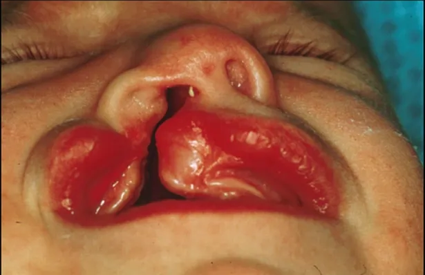
Case: Unilateral complete cleft lip, alveolus and palate, before and after pre-surgical orthopaedics
Question
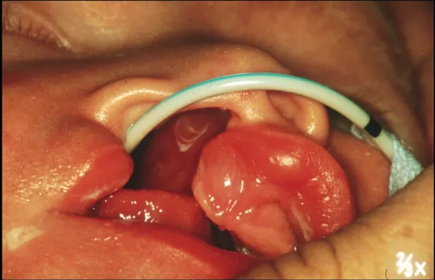
Scenario: A newborn infant with a unilateral cleft is presented at birth and then at three months of age to demonstrate a pre-surgical orthopaedic treatment process.
What’s shown: Clinical photos of the infant’s oral cavity at birth showing the cleft, and at three months showing the arch form after treatment.
Consider: Identify the initial presentation and the outcome of the pre-surgical orthopaedic treatment.
Answer
Observations:
- At birth: unilateral complete cleft lip, alveolus, and palate.
- At three months: significant decrease in cleft width and a much more continuous arch form.
Reasoning: Pre-surgical orthopaedics uses an intraoral appliance to guide the segments and an extraoral bonnet or strapping to apply pressure, taking advantage of tissue malleability in the early days post-birth to narrow the cleft and align the arch prior to surgical lip repair.
Outcome: After three months of pre-surgical orthopaedic treatment, the cleft width significantly decreased, resulting in a much more continuous arch form to facilitate surgical closure.
Takeaway: Pre-surgical orthopaedics can significantly narrow the cleft width and improve arch continuity before surgical repair.
Case: Topographical maps comparing digital and alginate impressions
Question
Scenario: A study comparing optical scanning with conventional alginate impressions in children with cleft lip and palate.
What’s shown: Color-coded 3D topographical maps showing differences between the two impression methods.
Consider: The differences in tissue displacement between digital and conventional impressions.
Answer
Observations:
- Color-coded 3D topographical maps.
- Red areas in the premaxillary segment indicating differences between the alginate and digital scan.
Reasoning: The red areas show wider variation because alginate impressions physically displace the premaxilla (functional impression), whereas optical scans are done in a static environment.
Outcome: Digital impressions were found to be more accurate, faster, and safer, though with some software and hardware limitations.
Takeaway: Alginate impressions record a functional impression that displaces tissues, whereas digital optical scans capture a static environment.
Case: Three-month-old infant, intraoral scan and 3D models
Question
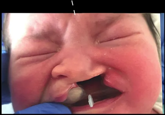
Scenario: A three-month-old baby with a cleft lip and palate undergoing digital assessment.
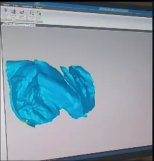
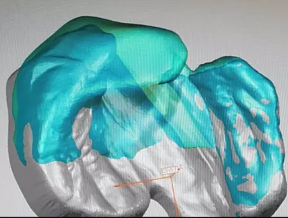
What’s shown: Intraoral scan, 3D wax-up, 3D printed stents, and models.
Consider: How the digital workflow is applied to assess anatomy and plan appliances.
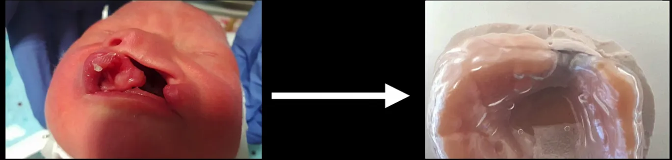
Answer
Observations:
- Live intraoral scan of a three-month-old baby.
- 3D wax-ups and three-dimensional planning.
- Assessment of midlines using 3D printed stents and models.
Reasoning: The digital workflow allows the team to understand landmarks and cleft anatomy, assess midlines, and create oral appliances without the child needing conventional impressions.
Outcome: Oral appliances are created without the need for traditional impressions.
Takeaway: Digital scanning and 3D planning can replace conventional impressions in pre-surgical orthopaedics for infants with cleft lip and palate.
Case: Unilateral cleft lip and palate, lower face photographs
Question
Scenario: An infant with a unilateral cleft lip and palate at around three months of age.
What’s shown: Clinical photographs of the lower face before and after a surgical procedure.
Consider: The timing and outcome of the surgical intervention.
Answer
Observations:
- Pre-operative appearance of a unilateral cleft lip and palate.
- Post-operative appearance following surgical repair.
Reasoning: The lip is typically repaired around the three-month mark, provided the child’s weight and hemoglobin levels are adequate to cope with the surgical procedure.
Outcome: Successful primary lip repair.
Takeaway: Primary lip repair is generally performed around three months of age once the child is medically stable.
Case: Child with failed previous restorations and developmental molar defects
Question
Scenario: A child between three and five years old presenting with previous dental treatment and restorative failures.
What’s shown: Intraoral photographs showing a discolored composite restoration with marginal overhangs and recurrent caries, as well as second primary molars with developmental defects and distal palatal lesions.
Consider: The appropriate restorative management for a child with extensive restorative failures and developmental defects.
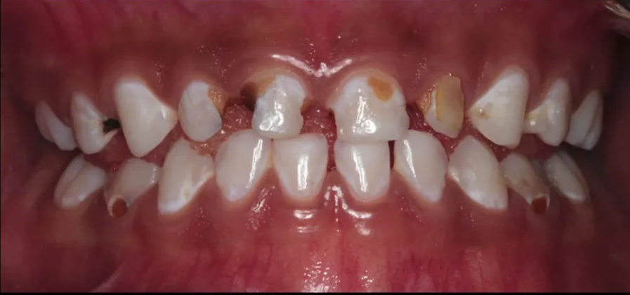
Answer
Observations:
- Unsatisfactory 6:2 composite restoration: discolored, marginal overhangs, poor adaptation, and recurrent mesial caries.
- Developmental defects of the second primary molars (55 and 65) with secondary caries and significant distal palatal lesions.
- Missing teeth distal to 5.5 and 6.5 (atypical presentation).
- Extraction of the 74.
Reasoning: The child is subject to multiple procedures and at risk of burnout, so definitive, long-lasting care is required rather than patchwork composite or GIC restorations. Multi-surface composites would not last the nine years before exfoliation. The aggressive treatment is justified by the atypical and severe distal palatal lesions secondary to the developmental defects.
Outcome: Comprehensive treatment including extraction of the 74 and placement of a space maintainer.
Takeaway: Children with extensive restorative needs and developmental defects require definitive, long-lasting treatment to avoid multiple failed visits and burnout, rather than temporary patchwork restorations.
Case: Child with developmental defects and caries in primary molars
Question
Scenario: A child between three and five years old presents with developmental defects and caries in the second primary molars.
What’s shown: Clinical photographs showing developmental defects (described as hypermineralized) in the second primary molars (55 and 65) with secondary caries, significant distal palatal lesions, and missing teeth distal to 55 and 65.
Consider: How to manage the atypical presentation and caries in a young child where restorations may not last until exfoliation.
Answer
Observations:
- Developmental defects (described as hypermineralized) in second primary molars (55 and 65) with secondary caries.
- Atypical presentation with significant distal palatal lesions and no teeth distal to 55 or 65.
Reasoning: Multi-surface composite restorations are not indicated because they will not last the remaining nine years before the teeth exfoliate. More aggressive treatment is required due to the atypical presentation and distal palatal lesions.
Outcome: The 74 was extracted, and a space maintainer was placed later. The child was comprehensively treated.
Takeaway: In young children with severe developmental defects and atypical caries patterns where restorations are unlikely to survive until exfoliation, aggressive treatment such as extraction and space maintenance is preferred over multi-surface restorations.
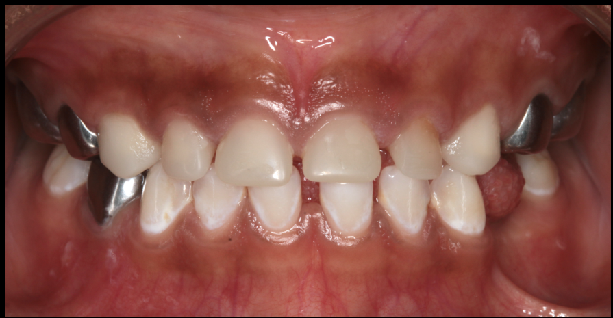
Case: Child with bilateral cleft lip and palate and associated dental anomalies
Question
Scenario: A child with bilateral cleft lip and palate presents with multiple dental anomalies.
What’s shown: Clinical photograph showing bilateral cleft lip and palate, talon cusps on a lateral incisor, developmental defects in tooth 21, and an over-retained (transcribed as overattained), necrotic tooth 51, with space loss and rotated teeth.
Consider: Identify the dental anomalies and the challenges they present.
Answer
Observations:
- Bilateral cleft lip and palate.
- Talon cusps (accessory cusps) on the lateral incisor.
- Developmental defects in tooth 21.
- Over-retained and necrotic tooth 51.
- Space loss and migration/rotation of teeth.
Reasoning: The presence of these anomalies, rotations, and space loss makes oral hygiene and cleaning around the teeth much more difficult. It also complicates orthodontic treatment.
Outcome: None stated.
Takeaway: Cleft lip and palate is associated with a range of dental anomalies, including talon cusps, developmental defects, and necrosis, which complicate oral hygiene and orthodontic management.
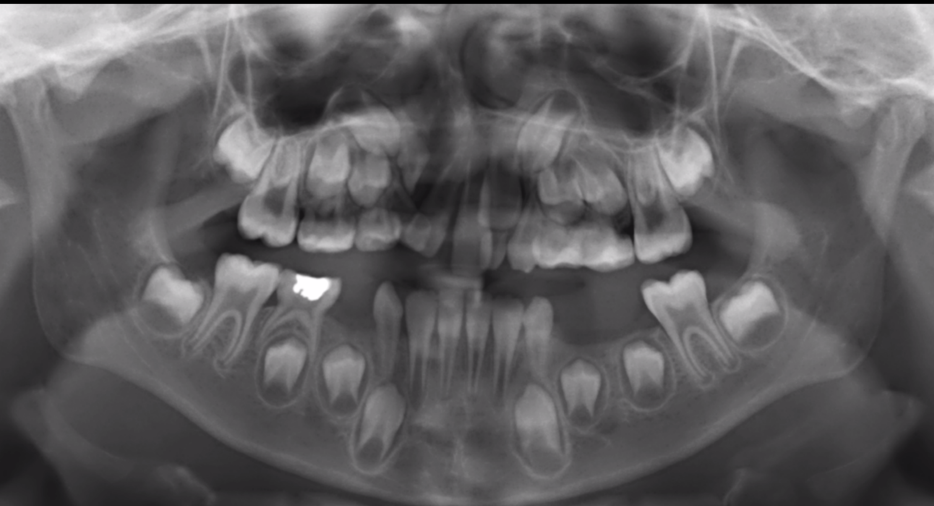
Case: Child with right unilateral cleft lip and palate, OPG and maxillary occlusal radiographs
Question
Scenario: A patient between 7 and 8 years of age is presented with radiographs to identify the cleft, hard tissue anomalies, and dental age.
What’s shown: OPG and maxillary occlusal radiographs showing a bony lucency in the upper right quadrant extending into the nasal cavity, a radiopaque nasal tube, and multiple dental anomalies including missing, microdont, supernumerary, and rotated teeth.
Consider: Identify the location of the cleft, list hard tissue anomalies, and estimate the patient’s dental age.
Answer
Observations:
- Bony lucency extending from the alveolus to the floor of the nasal cavity on the upper right, with altered septum and nasal morphology, consistent with a right unilateral cleft lip and palate.
- Radiopaque nasal tube indicating the radiograph was taken under general anesthesia.
- Altered coronal morphology of tooth 12 (fissural, conical, microdont tooth).
- Microdont tooth 62 (transcribed as distributed six, two).
- Step deformity associated with ectopic eruption and distal resorption of tooth 26.
- Metallic restoration in a primary tooth and occlusal restorations.
- Incisal irregularity and dilaceration/curvature in the root of tooth 52, with coronal radiolucency.
- Rotated tooth 11 and labial surface defect on tooth 21.
- Supernumerary tooth in the primary dentition series.
- Microdont permanent lateral incisor.
- Taurodontism (transcribed as tordontism) of upper first permanent molars.
Reasoning: The cleft is identified by the radiolucency through the alveolus into the nasal cavity. Hard tissue anomalies include fissural/microdont teeth, ectopic eruption with resorption, and supernumerary teeth. Dental age is estimated using the first permanent molar (erupts at 6, full occlusion at 7, roots complete at 9) and the eruption status of anterior teeth and canines, placing the patient between 7 and 8 years old.
Outcome: None stated (diagnostic workup).
Takeaway: Radiographic assessment of cleft lip and palate requires a holistic approach to identify the cleft extent, associated hard tissue anomalies (such as microdont, supernumerary, and ectopic teeth), and to accurately estimate dental age using stable markers like the first permanent molar.
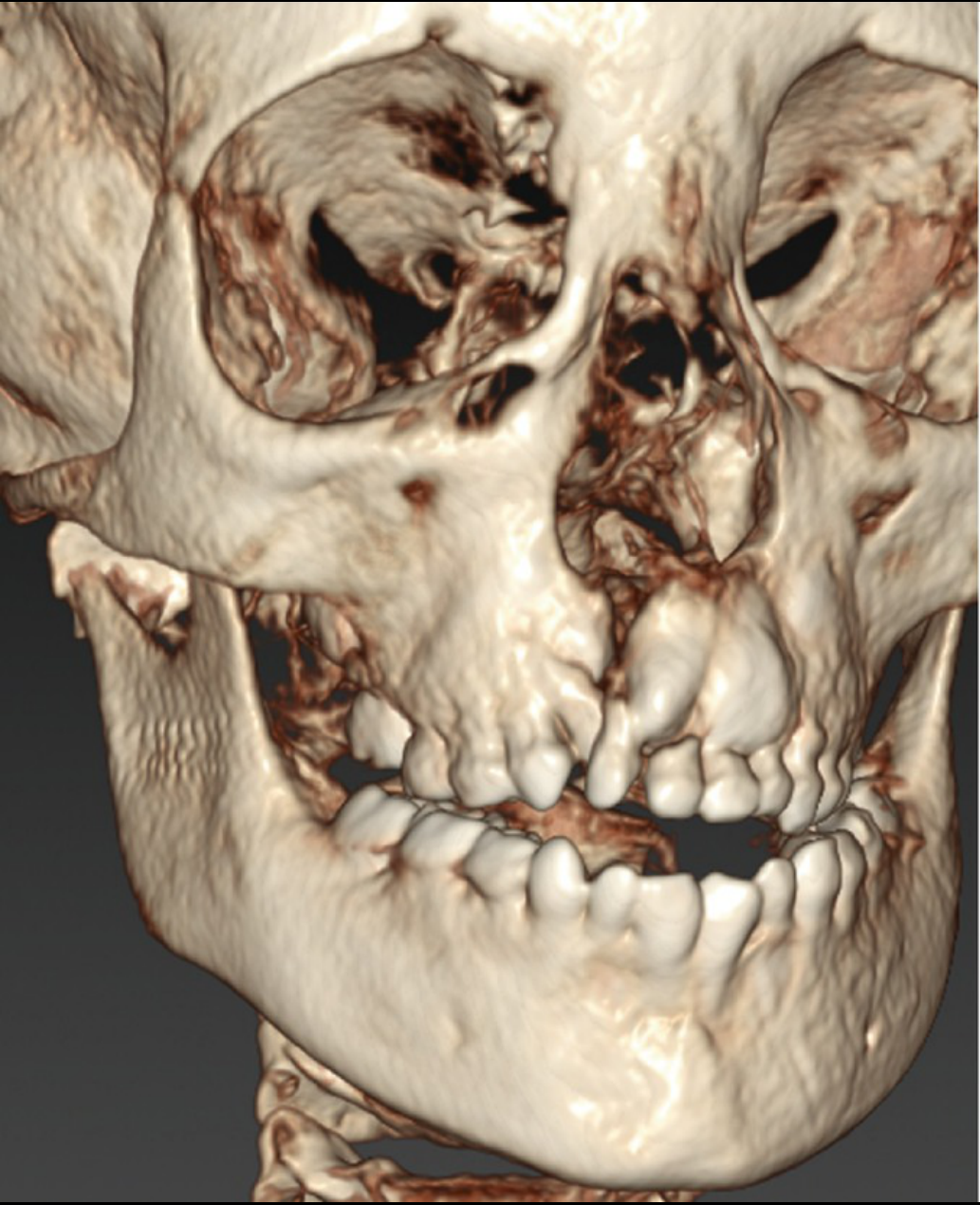
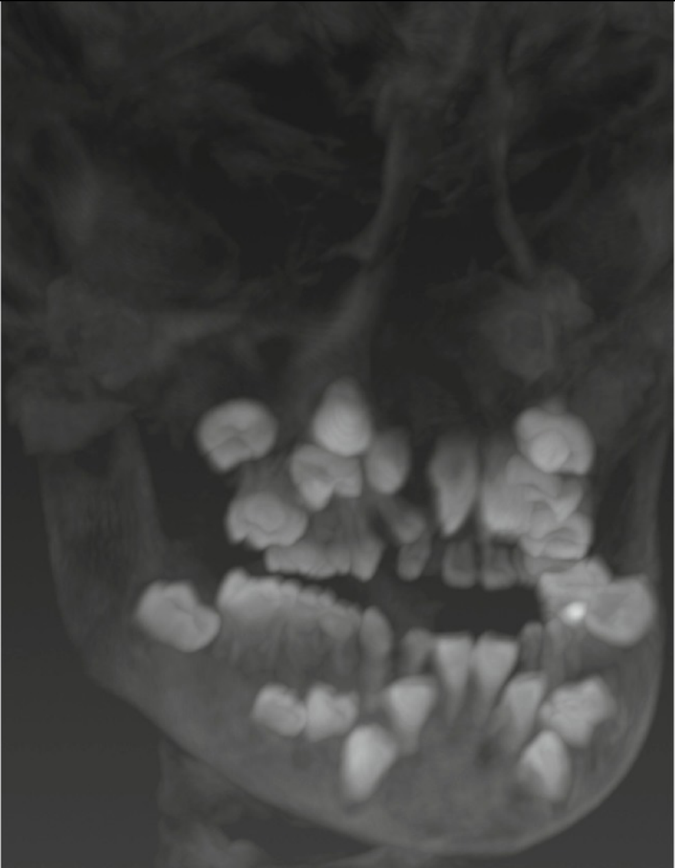
Case: Child undergoing workup prior to alveolar bone grafting
Question
Scenario: A child is undergoing a workup prior to alveolar bone grafting, requiring decisions about missing, extra, or over-retained teeth.
What’s shown: 3D CT reconstruction, 3D volume-rendered CT scan, and dental radiographs showing multiple teeth in the anterior maxillary region.
Consider: Identify which teeth are missing, extra, or over-retained to plan the bone grafting and multidisciplinary treatment.
Answer
Observations:
- Multiple teeth in the anterior maxillary region on 3D CT and radiographs.
Reasoning: The presence of many teeth in the anterior maxilla makes it difficult to decide which are missing, extra, or over-retained, necessitating multidisciplinary consults to arrange treatments well in advance.
Outcome: Multidisciplinary consults are required to arrange treatments in advance.
Takeaway: Workups for alveolar bone grafting in the mixed dentition often reveal complex dental anomalies in the anterior maxilla, requiring multidisciplinary team planning to determine which teeth to retain, extract, or manage.
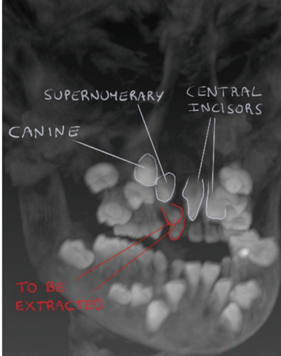
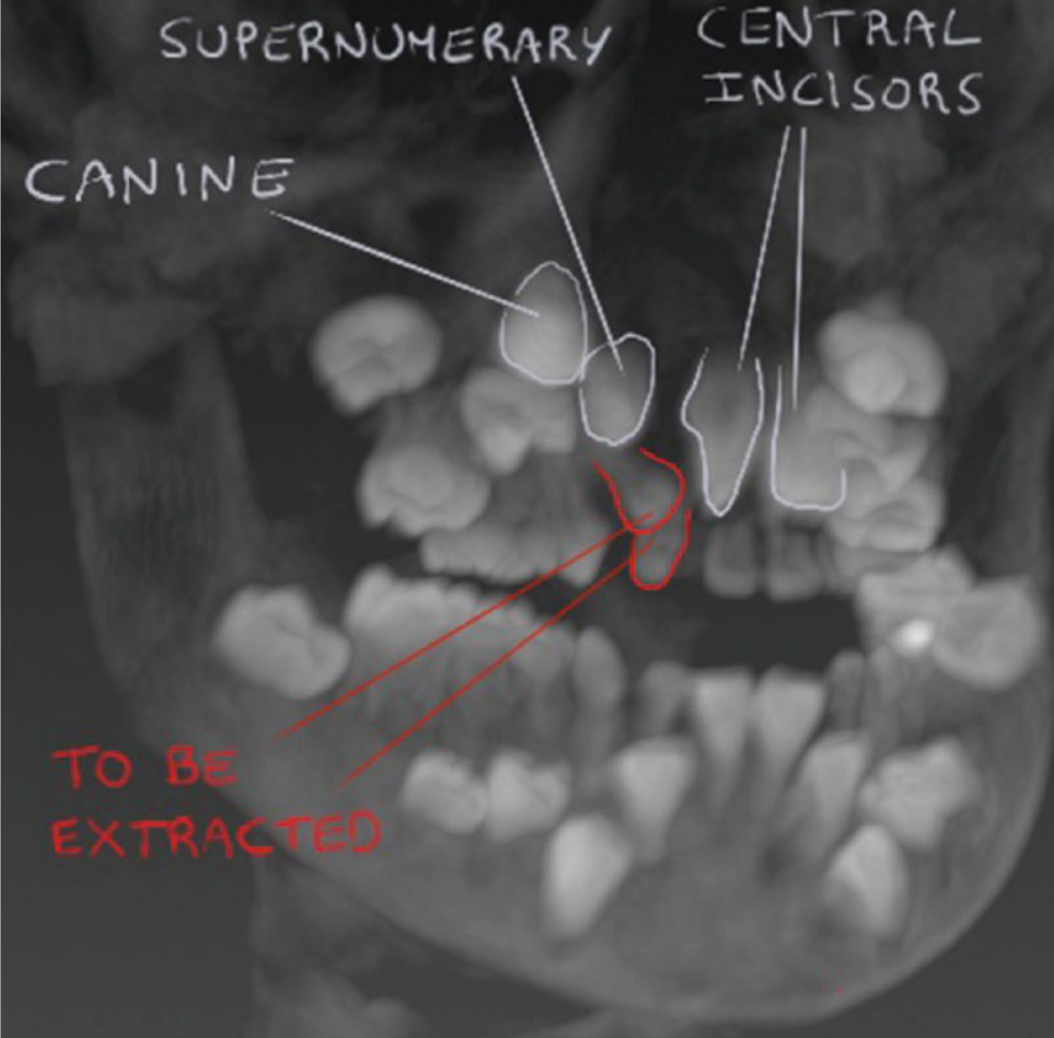
Case: Child undergoing workup prior to alveolar bone grafting, CT and radiograph
Question
Scenario: A child is undergoing a workup prior to alveolar bone grafting.
What’s shown: CT scan and radiographic images of the anterior maxillary region.
Consider: Identify the dental anomalies and present teeth in the cleft area, and determine the necessary preparatory steps.
Answer
Observations:
- A large number of teeth are present in the anterior maxillary region.
- A supernumerary tooth is visible.
- A canine and central incisors are present.
- The lateral incisor is missing.
- Retained primary teeth are present in the cleft area.
Reasoning: Determining which teeth are missing, extra, or over-retained is difficult and requires multidisciplinary consults so that treatments can be arranged well in advance. The retained primary teeth in the cleft area need to be extracted prior to the bone grafting procedure.
Outcome: The retained primary teeth in the cleft area are extracted beforehand, with treatments arranged via multidisciplinary team consults.
Takeaway: Pre-bone grafting workups for cleft patients often reveal complex dental anomalies that require multidisciplinary evaluation to identify missing, supernumerary, or retained teeth and plan extractions prior to grafting.
Footnotes
-
Original PDF page 1: L6 Cleft Lip and Palate Lecture DMD Handout, p.1 ↩
-
Original PDF page 2: L6 Cleft Lip and Palate Lecture DMD Handout, p.2 ↩
-
Original PDF page 3: L6 Cleft Lip and Palate Lecture DMD Handout, p.3 ↩
-
Original PDF page 4: L6 Cleft Lip and Palate Lecture DMD Handout, p.4 ↩
-
Original PDF page 5: L6 Cleft Lip and Palate Lecture DMD Handout, p.5 ↩
-
Original PDF page 6: L6 Cleft Lip and Palate Lecture DMD Handout, p.6 ↩
-
Original PDF page 7: L6 Cleft Lip and Palate Lecture DMD Handout, p.7 ↩
-
Original PDF page 8: L6 Cleft Lip and Palate Lecture DMD Handout, p.8 ↩
-
Original PDF page 9: L6 Cleft Lip and Palate Lecture DMD Handout, p.9 ↩
-
Original PDF page 10: L6 Cleft Lip and Palate Lecture DMD Handout, p.10 ↩
-
Original PDF page 11: L6 Cleft Lip and Palate Lecture DMD Handout, p.11 ↩
-
Original PDF page 12: L6 Cleft Lip and Palate Lecture DMD Handout, p.12 ↩
-
Original PDF page 13: L6 Cleft Lip and Palate Lecture DMD Handout, p.13 ↩
-
Original PDF page 14: L6 Cleft Lip and Palate Lecture DMD Handout, p.14 ↩
-
Original PDF page 15: L6 Cleft Lip and Palate Lecture DMD Handout, p.15 ↩
-
Original PDF page 16: L6 Cleft Lip and Palate Lecture DMD Handout, p.16 ↩
-
Original PDF page 17: L6 Cleft Lip and Palate Lecture DMD Handout, p.17 ↩
-
Original PDF page 38: L6 Cleft Lip and Palate Lecture DMD Handout, p.38 ↩
-
Original PDF page 42: L6 Cleft Lip and Palate Lecture DMD Handout, p.42 ↩
-
Original PDF page 48: L6 Cleft Lip and Palate Lecture DMD Handout, p.48 ↩
-
Original PDF page 18: L6 Cleft Lip and Palate Lecture DMD Handout, p.18 ↩
-
Original PDF page 19: L6 Cleft Lip and Palate Lecture DMD Handout, p.19 ↩
-
Original PDF page 20: L6 Cleft Lip and Palate Lecture DMD Handout, p.20 ↩
-
Original PDF page 21: L6 Cleft Lip and Palate Lecture DMD Handout, p.21 ↩
-
Original PDF page 22: L6 Cleft Lip and Palate Lecture DMD Handout, p.22 ↩
-
Original PDF page 23: L6 Cleft Lip and Palate Lecture DMD Handout, p.23 ↩
-
Original PDF page 24: L6 Cleft Lip and Palate Lecture DMD Handout, p.24 ↩
-
Original PDF page 25: L6 Cleft Lip and Palate Lecture DMD Handout, p.25 ↩
-
Original PDF page 26: L6 Cleft Lip and Palate Lecture DMD Handout, p.26 ↩
-
Original PDF page 27: L6 Cleft Lip and Palate Lecture DMD Handout, p.27 ↩
-
Original PDF page 29: L6 Cleft Lip and Palate Lecture DMD Handout, p.29 ↩
-
Original PDF page 30: L6 Cleft Lip and Palate Lecture DMD Handout, p.30 ↩
-
Original PDF page 31: L6 Cleft Lip and Palate Lecture DMD Handout, p.31 ↩
-
Original PDF page 28: L6 Cleft Lip and Palate Lecture DMD Handout, p.28 ↩
-
Original PDF page 32: L6 Cleft Lip and Palate Lecture DMD Handout, p.32 ↩
-
Original PDF page 33: L6 Cleft Lip and Palate Lecture DMD Handout, p.33 ↩
-
Original PDF page 34: L6 Cleft Lip and Palate Lecture DMD Handout, p.34 ↩
-
Original PDF page 35: L6 Cleft Lip and Palate Lecture DMD Handout, p.35 ↩
-
Original PDF page 36: L6 Cleft Lip and Palate Lecture DMD Handout, p.36 ↩
-
Original PDF page 37: L6 Cleft Lip and Palate Lecture DMD Handout, p.37 ↩
-
Original PDF page 40: L6 Cleft Lip and Palate Lecture DMD Handout, p.40 ↩
-
Original PDF page 41: L6 Cleft Lip and Palate Lecture DMD Handout, p.41 ↩
-
Original PDF page 39: L6 Cleft Lip and Palate Lecture DMD Handout, p.39 ↩
-
Original PDF page 43: L6 Cleft Lip and Palate Lecture DMD Handout, p.43 ↩
-
Original PDF page 44: L6 Cleft Lip and Palate Lecture DMD Handout, p.44 ↩
-
Original PDF page 45: L6 Cleft Lip and Palate Lecture DMD Handout, p.45 ↩
-
Original PDF page 46: L6 Cleft Lip and Palate Lecture DMD Handout, p.46 ↩
-
Original PDF page 47: L6 Cleft Lip and Palate Lecture DMD Handout, p.47 ↩
-
Original PDF page 50: L6 Cleft Lip and Palate Lecture DMD Handout, p.50 ↩
-
Original PDF page 49: L6 Cleft Lip and Palate Lecture DMD Handout, p.49 ↩
-
Original PDF page 51: L6 Cleft Lip and Palate Lecture DMD Handout, p.51 ↩
-
Original PDF page 52: L6 Cleft Lip and Palate Lecture DMD Handout, p.52 ↩
-
Original PDF page 53: L6 Cleft Lip and Palate Lecture DMD Handout, p.53 ↩
-
Original PDF page 54: L6 Cleft Lip and Palate Lecture DMD Handout, p.54 ↩
-
Original PDF page 55: L6 Cleft Lip and Palate Lecture DMD Handout, p.55 ↩
-
Original PDF page 56: L6 Cleft Lip and Palate Lecture DMD Handout, p.56 ↩
-
Original PDF page 57: L6 Cleft Lip and Palate Lecture DMD Handout, p.57 ↩
-
Original PDF page 58: L6 Cleft Lip and Palate Lecture DMD Handout, p.58 ↩