Amelogenesis Imperfecta: Classification, Characteristics and Management1
Classification, characteristics and management
Dr Jilen Patel
Definitions23
-
AI “implies a generalized fault of enamel structure affecting all the teeth of one or both dentitions. The faults do not seem to be related to any specific period of time during enamel development, nor to any intercurrent disease or dietary abnormality. It may be hereditary or may occur spontaneously in one or more members of a family” (Darling 1956)
-
“all the individuals in the kindred show essentially the same defect and which are unassociated with known morphologic or biochemical changes elsewhere in the body” (Rao et al 1971)
-
one gene, one condition?
-
“A group of conditions, genomic in origin, which affect the structure and clinical appearance of enamel of all or nearly all the teeth, and which may be associated with morphologic or biochemical changes elsewhere in the body” (Aldred et al 2003)
-
AI a feature of various syndromes including Morquio, trichodento-osseus, Kohlschütter and, oculo-dento-osseous dysplasia, tuberous sclerosis and vitamin D-dependent rickets (Pindborg 1982)
Lecturer — Definitions
Several clinical and historical points further clarify the definition of amelogenesis imperfecta.
- In clinical practice, investigate the family history when a young child presents with unusual, extensively broken-down teeth, as the condition may result from a spontaneous mutation.
- The generalized involvement helps distinguish amelogenesis imperfecta from localized defects such as molar-incisor hypomineralisation or a Turner tooth.
- The condition was first reported in 1890 and described as resembling “hereditary brown teeth,” reflecting the frequent discoloration and breakdown.
- A distinction was made between defects confined to enamel and those affecting dentine, such as dentinogenesis imperfecta.
- It is safer to initially describe an abnormality as a developmental defect, then specify whether it is qualitative or quantitative, and finally provide a more specific classification if justified.
Classification
Historical Development of Classification4
History
- First reported by Spokes in 1890, who described the condition as “hereditary brown teeth”
- Distinction between hereditary defects confined to enamel (i.e., AI) and those confined to dentine (Finn 1938)
- AI could be classified into two types: hypoplastic and hypocalcified (Weinmann et al 1945)
Phenotypic and Genotypic Basis5
Classification based on phenotype and mode of inheritance is useful for clinicians.
Problems?
- Variability in the phenotypic expression
- Genotypic information should be used to distinguish between forms of AI
- Patients want information about their condition that relates not only to themselves but also to their families
(Aldred et al 2003)
Lecturer — Classification Limits
Traditional classification relied mainly on phenotype, but the same broad type can have substantially different clinical appearances.
- More recent work emphasizes molecular and genetic information to distinguish forms more accurately.
- Genetic information may assist with family guidance and genetic counselling, although some families decline counselling because they may not wish to receive information revealed by genetic testing.
- Classification may also consider the mode of inheritance, and a single genetic condition can have variable phenotypic expression.
- X-linked conditions may produce different manifestations in females; alternating affected and unaffected enamel bands may occur because of the protective effect of the other chromosome, a phenomenon referred to as lionization.
- Genetic testing is therefore important because phenotype alone may not reliably identify the precise subtype.
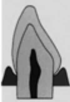
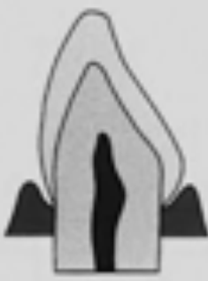
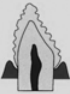
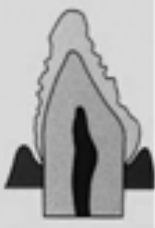
Basic Types of Enamel Defects6
Figure 1. Amelogenesis imperfecta. Diagram of enamel defects of basic types.
-
Hypocalcified — normal thickness, smooth surface, less hardness
-
Hypoplastic, pitted — normal thickness, pitted surface, normal hardness
-
Hypoplastic, generalized — reduced thickness, smooth surface, normal hardness
-
Hypomaturation — normal thickness, chipped surface, less hardness, opaque white coloration
-
Hypoplastic defects are primarily quantitative, with enamel that does not reach normal thickness.
-
Hypomaturation defects are primarily qualitative, with enamel that is generally of normal thickness but has abnormal appearance or maturation.
-
Hypocalcified defects involve very soft enamel that is rapidly lost after eruption.
-
Hypomaturation is generally less severe, hypoplastic disease may be severe, and hypocalcified disease may be the most severe because enamel can be lost shortly after eruption. This figure was published in Contemporary Oral and Maxillofacial Pathology, Sapp J, Eversole L, Wysocki G; Developmental Disturbances of the Oral Region, page 19; Mosby, Inc. 2004.¹⁸
 |  |  |
 |  |
Prevalence7
- Among 56,663 children from Northern Sweden, the prevalence of AI was found to be 1:714 (Bäckman 1986)
- Among 96,471 children from the state of Michigan, USA, a prevalence of 1:14,000 was reported (Witkop Jr 1958)
- Distribution by type: hypoplastic (60–73%), hypomaturation (20–40%), and hypocalcification (7%) (Chaudhary et al 2009; Kwok-Tung et al 2007)
Lecturer — Prevalence Estimates
Prevalence estimates vary depending on the age at which children are examined and which teeth have erupted.
- Localized conditions such as molar-incisor hypomineralisation may be incorrectly classified as amelogenesis imperfecta.
- A six- or seven-year-old with only incisors and first permanent molars erupted could be mistakenly diagnosed before the remaining teeth erupt.
- The lecturer considered the condition probably rarer than a reported estimate of 1 in 800.
Lecturer — Genetic Aetiology
Amelogenesis imperfecta is generally genetic in origin.
- It may be inherited or arise through a spontaneous mutation.
- A clear, single-gene Mendelian pattern is not present in every case.
- Family history is important but is not definitive because spontaneous mutations can occur.
- Environmental and medical history are also important when investigating generalized enamel defects, particularly when considering alternative diagnoses.
Aetiology8
Organic matrix molecules:
- Enamelin (ENAM; 4q21, OMIM *606585)
- Amelogenin (AMELX; Xp22.3-p22.1, OMIM *300391)
- Ameloblastin (AMBN; 4q21, OMIM *601259)
- Tuftelin (TUFT1; 1q21, OMIM *600087)
- Amelotin (AMELOTIN; 4q13) [14]
- Dentine sialophosphoprotein (DSPP; 4q21.3, OMIM *125485)
Enzymes:
- Kallikrein 4 (KLK4; 19q13.3–q13.4, OMIM *603767)
- Matrix metalloproteinase 20 (MMP20; 11q22.3–q23, OMIM *604629)
Lecturer — Maturation Abnormalities
Abnormalities during maturation can produce enamel of normal thickness but abnormal appearance and physical properties.
Amelogenesis Process
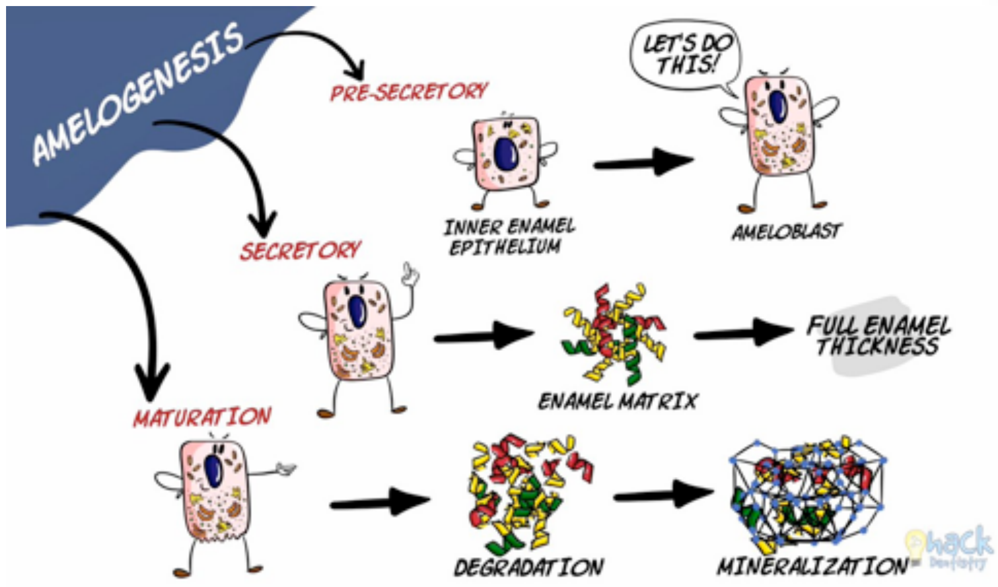
Stages of Enamel Formation9
Amelogenesis proceeds through the following stages:
- Pre-secretory — inner enamel epithelium → ameloblast
- Secretory — full enamel thickness; enamel matrix
- Maturation — degradation; mineralization
Lecturer — Amelogenesis Phases
The lecturer also described amelogenesis as involving an inductive phase before secretion.
- The initial enamel matrix contains a high protein content.
- Proteinases subsequently remove proteins before the matrix undergoes maturation and mineralisation.
Secretory Stage10
-
Secretory stage: a proteinaceous extracellular matrix is incrementally laid down by epithelially-derived ameloblasts via specialised cellular extensions termed Tomes’ processes.
-
This matrix partially mineralises to delineate the enamel architecture, which is characterised by bundles of enamel crystals (enamel prisms or rods) interdigitated with interprismatic enamel crystals.
-
As the enamel layer approaches full thickness, the Tomes’ process is retracted and the final outer enamel layer is aprismatic.
-
Disruption during the secretory stage may reduce the amount of enamel produced and contribute to hypoplastic defects.
Transition and Maturation Stages11
- The ameloblasts then enter the transition stage, characterised by reduced matrix protein secretion and internal reorganization.
- The cells then enter the maturation stage and begin secreting the serine protease KLK4 that completely degrades the proteinaceous matrix, leaving the immature enamel crystals bathed in enamel tissue fluid.
Mineralisation and Final Enamel Maturation12
- The significant increase in the active transport of mineral ions into the matrix during this stage accelerates enamel crystal growth in both width and thickness until the tissue volume is eventually occluded by mineral.
- Finally, the ameloblasts reduce, undergo apoptosis and, on tooth eruption, are completely lost.
Amelogenesis Imperfecta Types Overview13
-
Hypoplastic (60–73%)
-
Hypomaturation (20–40%)
-
Hypocalcification (7%)
-
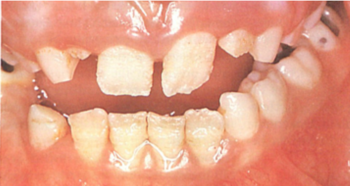
Hypoplastic enamel may be abnormally thin or absent in areas, with pits, fissures, vertical grooves, and failure of contact-point formation.
-
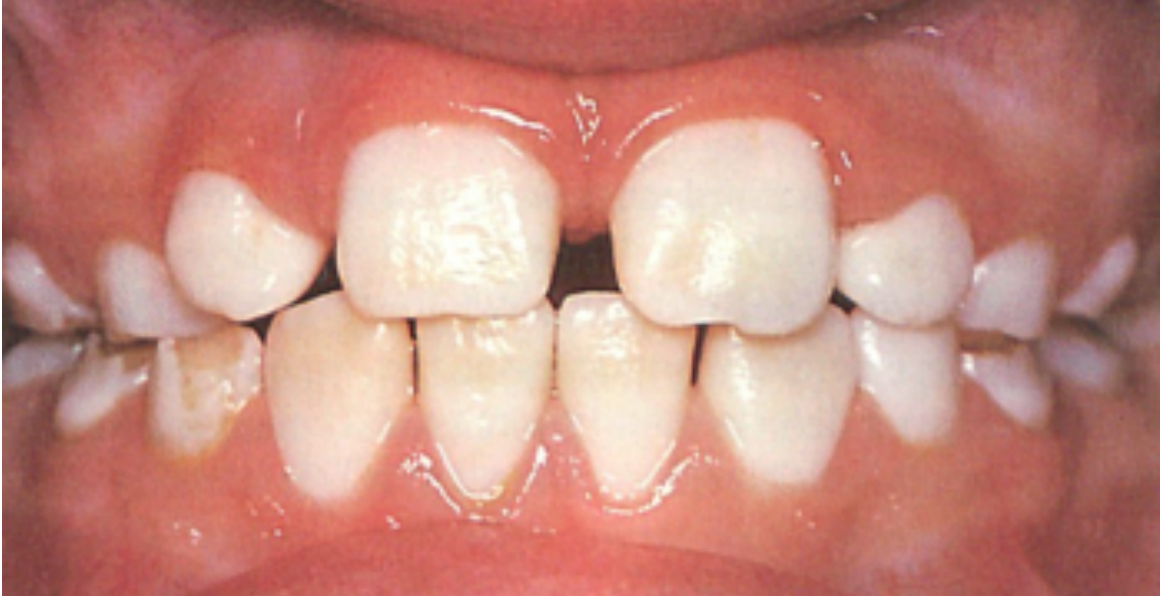
Hypomaturation enamel is generally of normal thickness but shows diffuse mottling or white opacities, with contact points present.
-
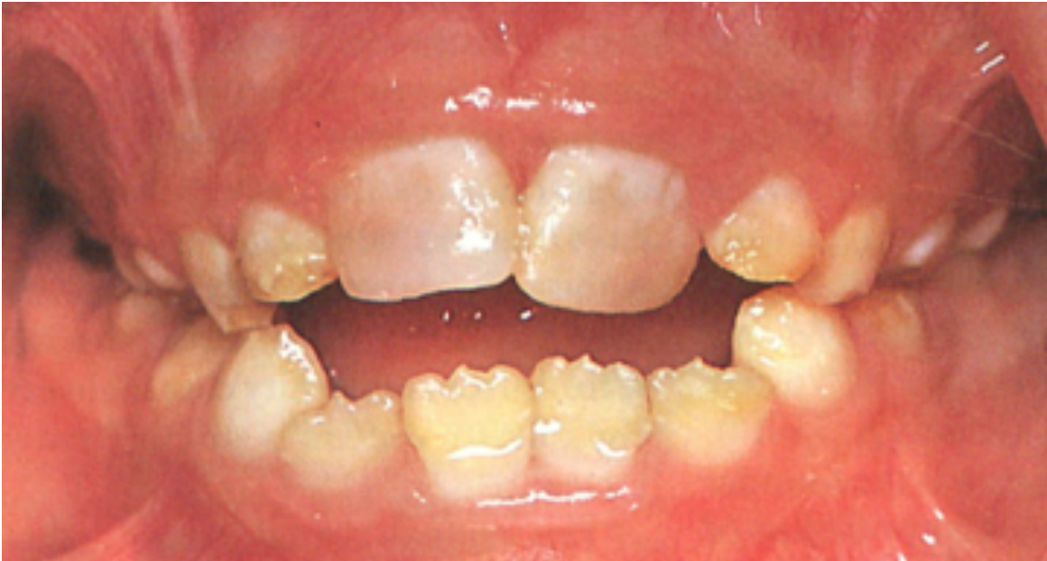
Hypocalcified enamel is extremely soft and may be lost soon after eruption, leaving exposed dentine.
-
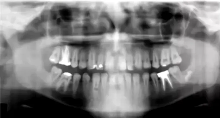
A hypoplastic-hypomaturation subtype with taurodontism may have an unusually large root canal space, with the pulp chamber extending almost to the apex.
-
This subtype may particularly severely affect the permanent dentition.
-
In severe hypocalcified cases, little or no enamel may remain within a few months of eruption, causing sensitivity and rapid breakdown. Chaudhary et al 2009; Kwok-Tung et al 2007.
Characteristics
Hypoplastic Form14
Bäckman (1988)
-
Enamel does not reach normal thickness during development.
-
Thin enamel, loss of mesiodistal contact, enamel pits or vertical or horizontal fissures in the enamel.
-
Enamel may be very thin or absent in areas.
-
Spaces may develop between teeth because contact points fail to form.
-
Severe cases may involve extensive post-eruptive breakdown.
Hypomaturation Form15
Bäckman (1988)
-
Mottled brown-yellow-white appearance; enamel is generally of normal thickness and teeth meet at contact points.
-
Female carriers may have vertical stripes of opaque white enamel alternating with bands of normal enamel (due to Lyonisation).
-
Diffuse white mottling or generalized white opacities may resemble fluorosis or widespread post-orthodontic white spot lesions.
-
Family and environmental histories help distinguish this form from other causes of diffuse opacities.
Hypocalcified Form16
Bäckman (1988)
-
Enamel tends to be so soft that it may be lost soon after eruption, leaving a crown composed only of dentin.
-
Radiographically, the enamel fails to contrast with the dentin.
-
The condition may cause significant pain and sensitivity.
-
A young child may have little or no enamel remaining on erupted teeth.
-
Rapid referral is important because extensive tooth destruction may occur within a few months.
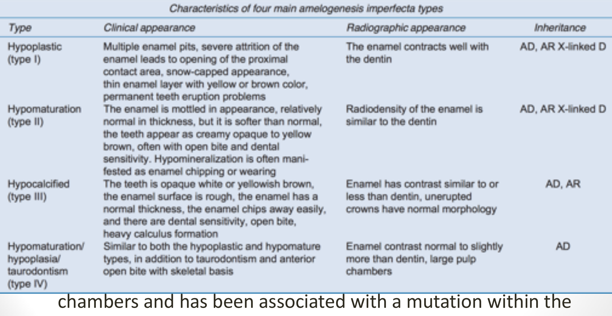
Witkop Classification Table17
Table 1: Witkop classification of amelogenesis imperfecta — characteristics of four main amelogenesis imperfecta types
| Type | Clinical appearance | Radiographic appearance | Inheritance |
|---|---|---|---|
| Hypoplastic (type I) | Multiple enamel pits, severe attrition of the enamel leads to opening of the proximal contact area, snow-capped appearance, thin enamel layer with yellow or brown color, permanent teeth eruption problems | The enamel contrasts well with the dentin | AD, AR, X-linked D |
| Hypomaturation (type II) | The enamel is mottled in appearance, relatively normal in thickness, but it is softer than normal; the teeth appear as creamy opaque to yellow brown, often with open bite and dental sensitivity. Hypomineralization is often manifested as enamel chipping or wearing | Radiodensity of the enamel is similar to the dentin | AD, AR, X-linked D |
| Hypocalcified (type III) | The teeth are opaque white or yellowish brown, the enamel surface is rough, the enamel has a normal thickness, the enamel chips away easily, and there are dental sensitivity, open bite, heavy calculus formation | Enamel has contrast similar to or less than dentin; unerupted crowns have normal morphology | AD, AR |
| Hypomaturation/hypoplasia/taurodontism (type IV) | Similar to both the hypoplastic and hypomaturation types, in addition to taurodontism and anterior open bite with skeletal basis | Enamel contrast normal to slightly more than dentin; large pulp chambers | AD |
Lecturer — Classification Limits
The Witkop table is a clinical overview only, and phenotype alone cannot reliably establish the precise subtype.
- Genetic testing is needed because there are multiple subtypes and considerable variability in clinical expression.
Large pulp chambers and taurodontism have been associated with a mutation within the human DLX3 gene (Dong et al 2005).
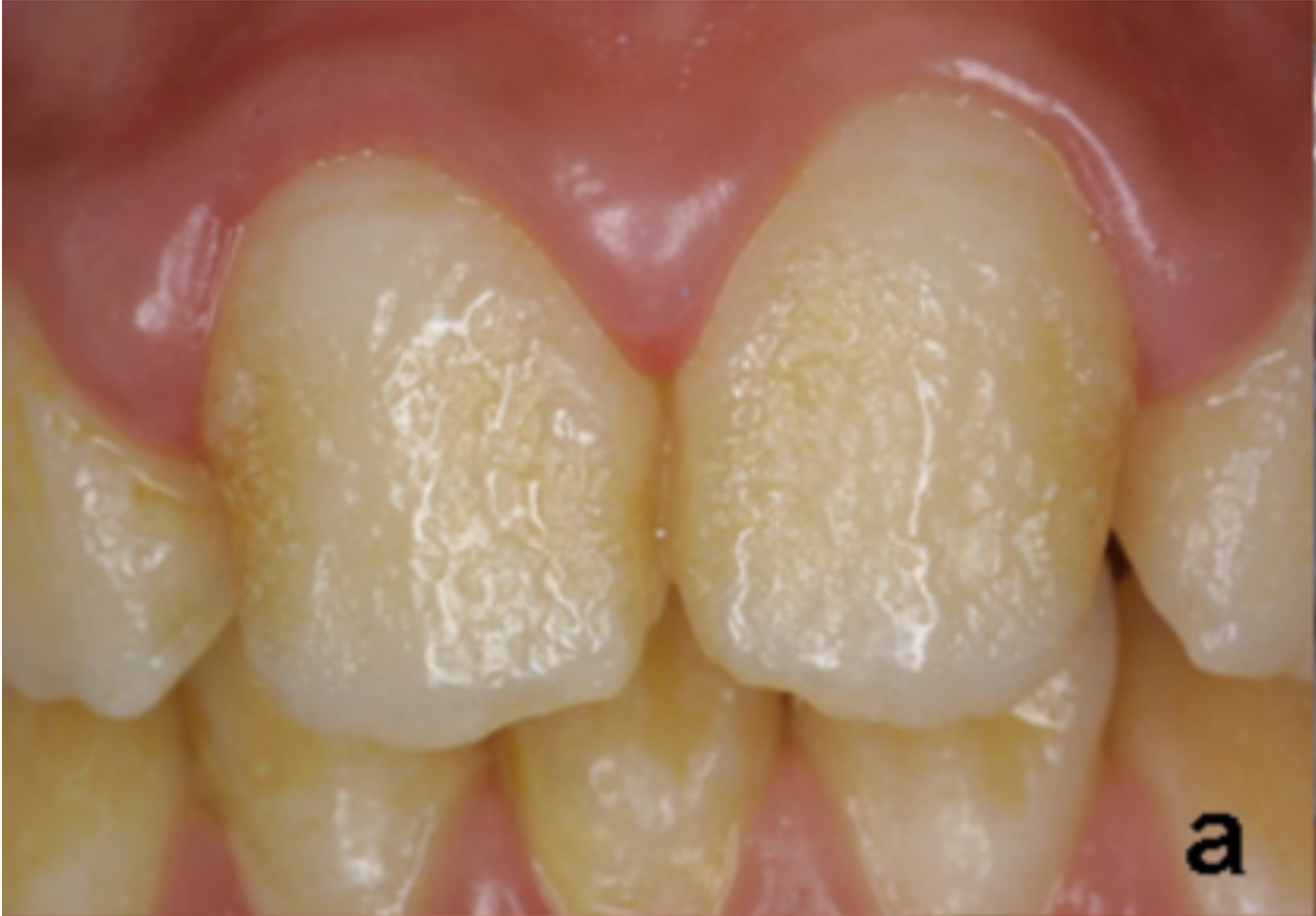
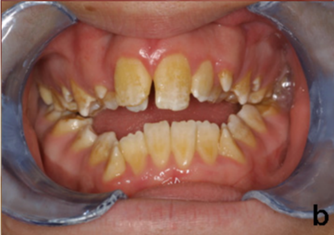
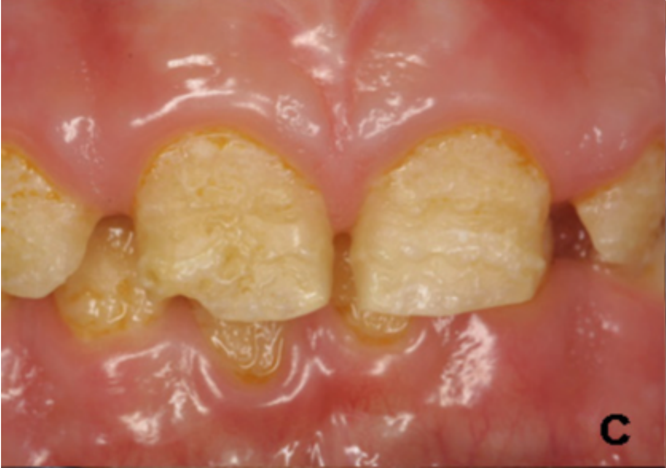
Clinical Figures1819202122232425
(Clinical figure page; no text content extracted.)
Panel b.
Panel c.
Panel e.
Panel f.
Panel g.
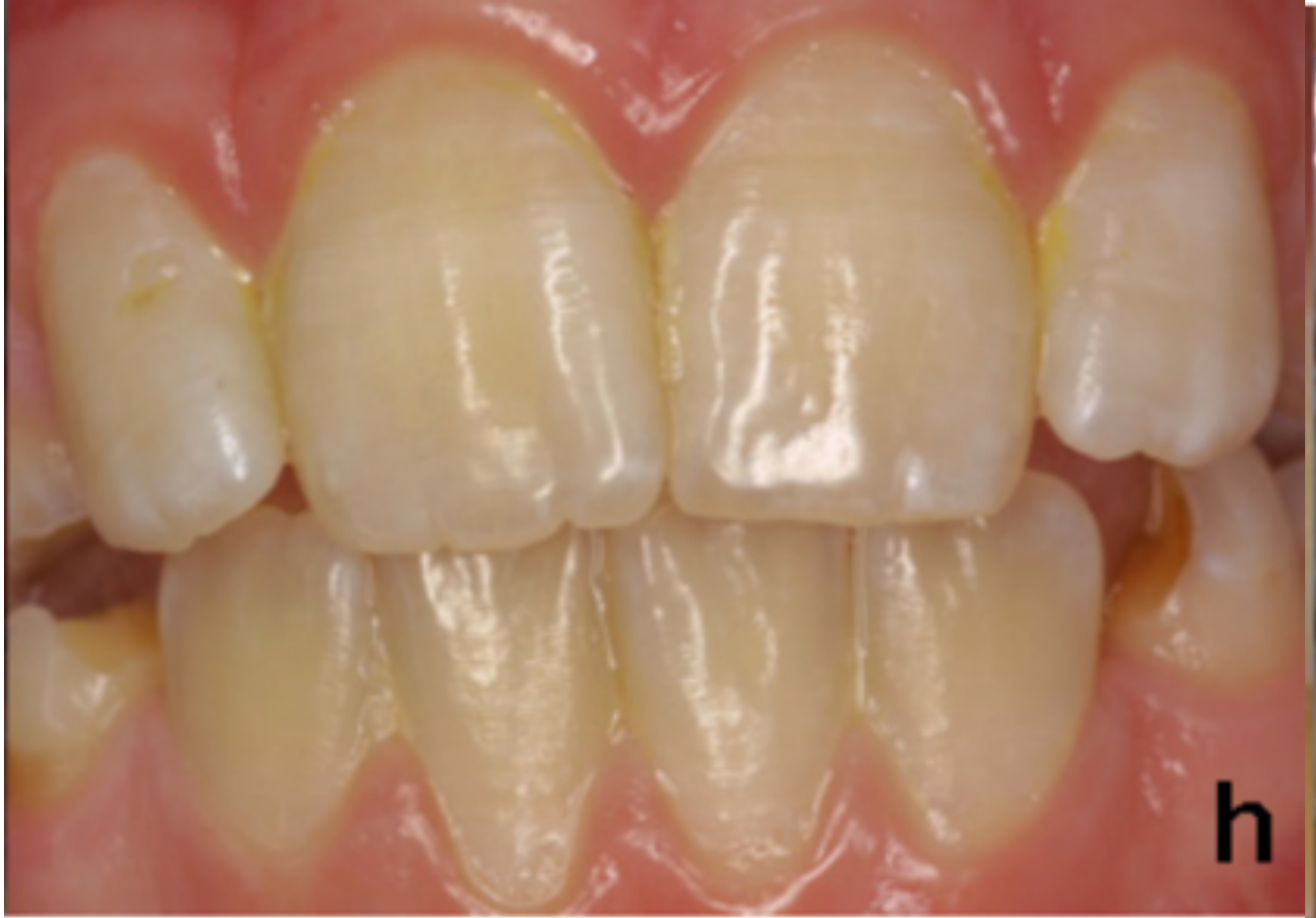
Panel h.
Clinical Variation and Management
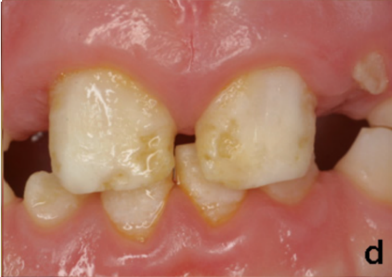
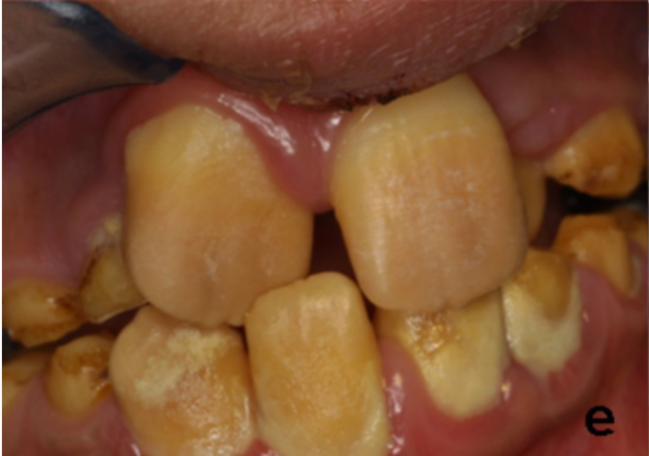
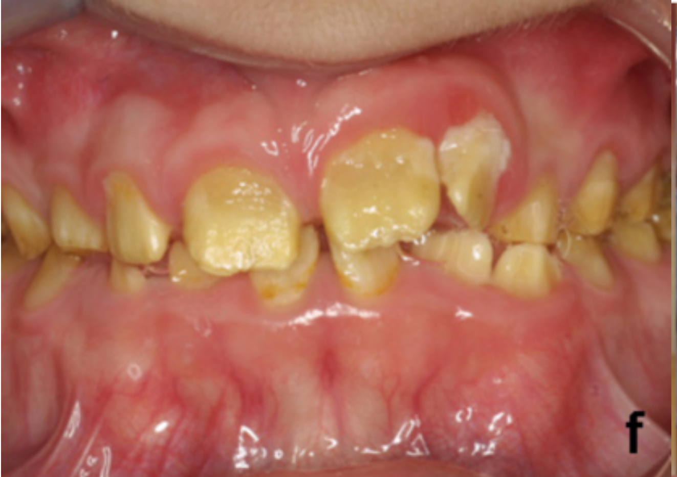
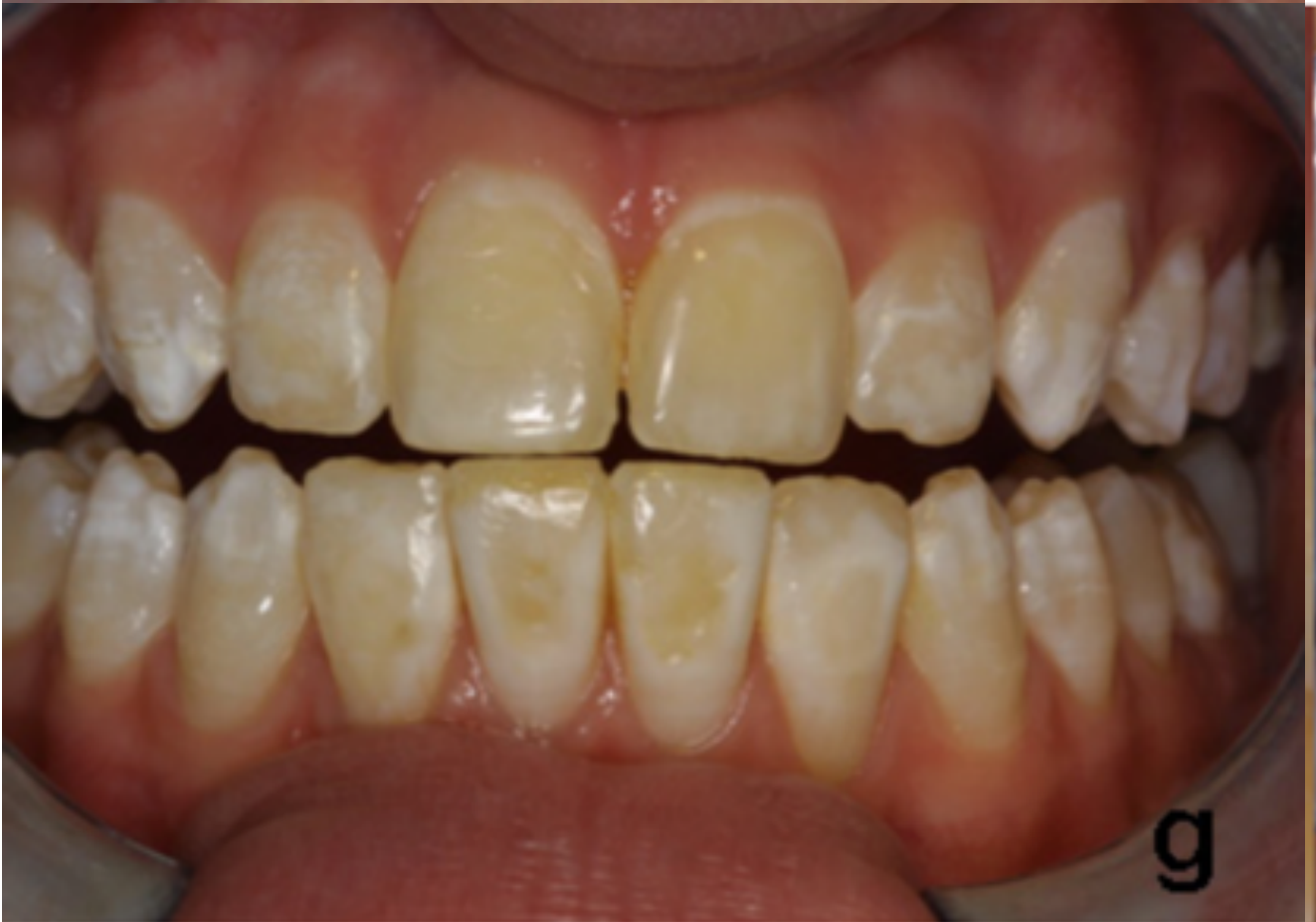
Clinical examples showed that amelogenesis imperfecta can vary substantially, from missing enamel and hypoplastic areas to mottling, generalized opacities, and severe breakdown.
- Some appearances could not be confidently assigned to a single type based on appearance alone.
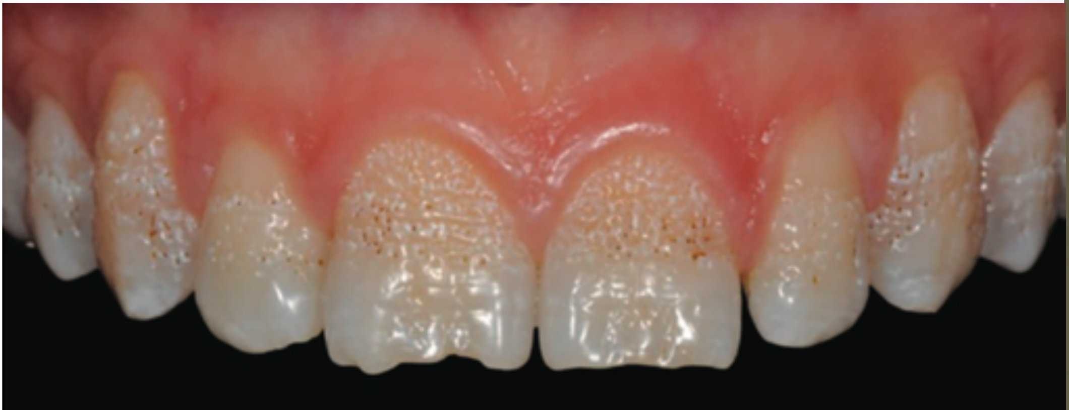
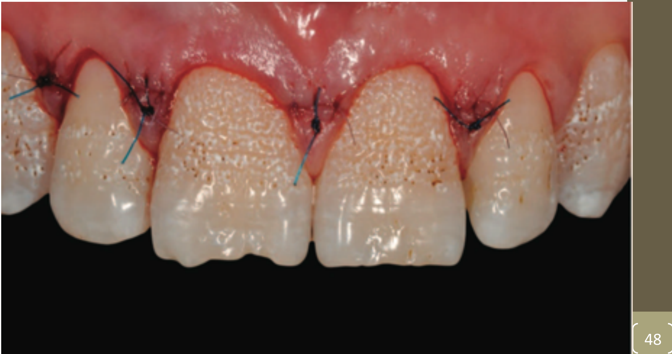
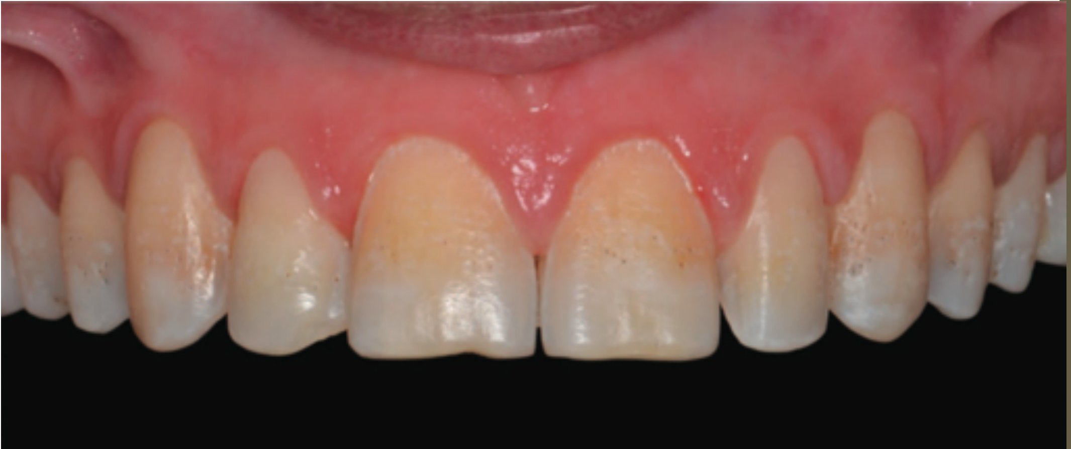
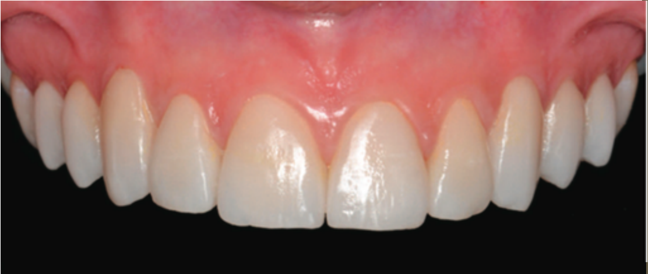
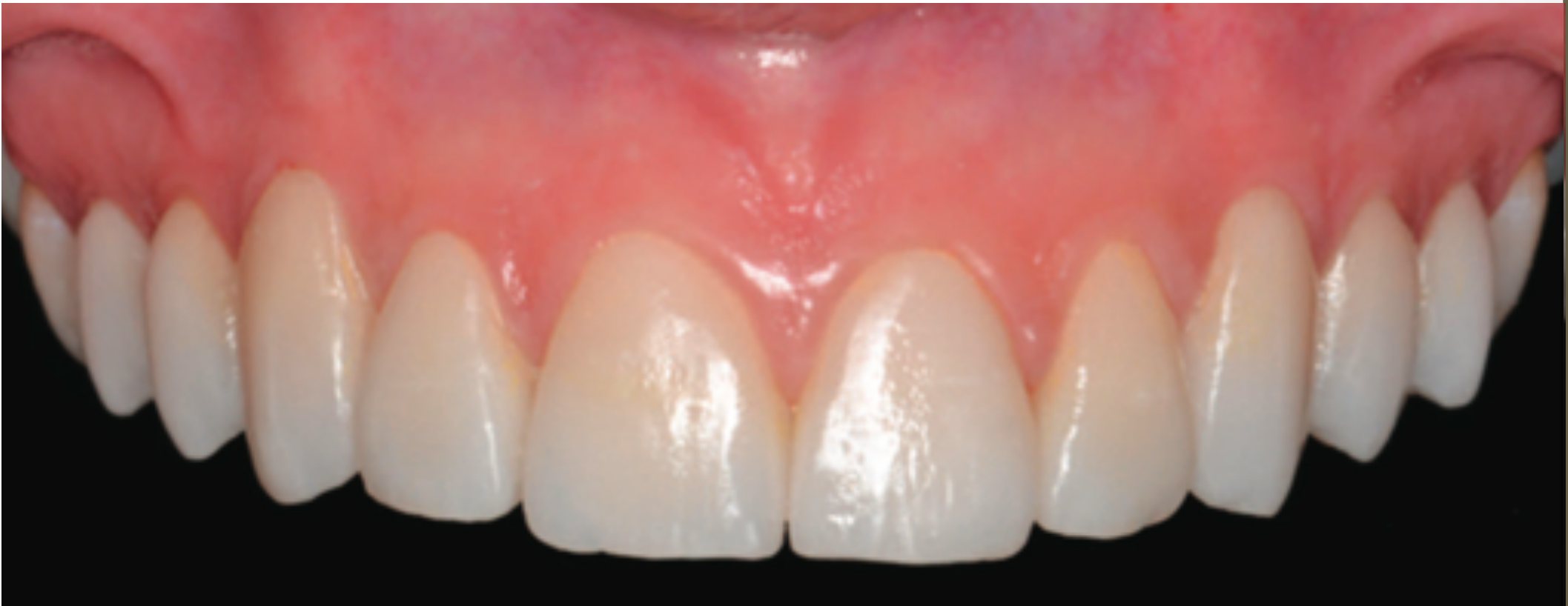
- A patient with rough hypoplastic amelogenesis imperfecta in the permanent dentition was managed with gingival recontouring and veneers to improve aesthetics.
Diagnostic Challenges
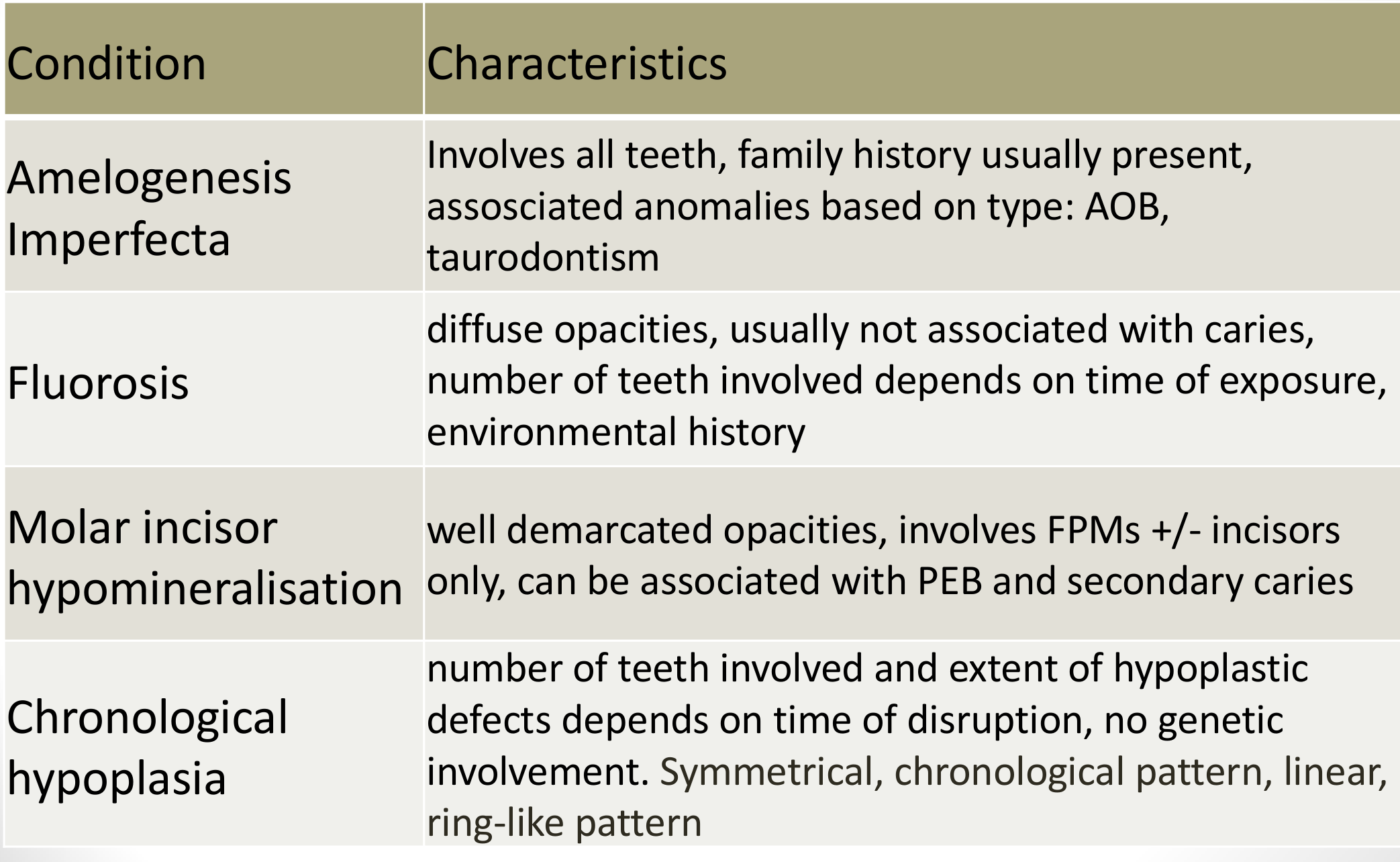
Differential Diagnosis of Enamel Defects2627
| Condition | Characteristics |
|---|---|
| Amelogenesis Imperfecta | Involves all teeth, family history usually present, associated anomalies based on type: AOB, taurodontism |
| Fluorosis | Diffuse opacities, usually not associated with caries, number of teeth involved depends on time of exposure, environmental history |
| Molar incisor hypomineralisation | Well demarcated opacities, involves FPMs +/- incisors only, can be associated with PEB and secondary caries |
| Chronological hypoplasia | Number of teeth involved and extent of hypoplastic defects depends on time of disruption, no genetic involvement. Symmetrical, chronological pattern, linear, ring-like pattern |
Lecturer — Differential Diagnosis
A family history is not essential for amelogenesis imperfecta because spontaneous mutations may occur, and associated findings can include failure of eruption and other tooth abnormalities.
- Fluorosis usually presents with diffuse opacities without significant breakdown, and the child’s environmental history is important.
- Molar-incisor hypomineralisation is localized to particular teeth, may produce yellow or brown opacities, and can break down in areas exposed to occlusal loading.
- Post-orthodontic white spot lesions may produce generalized white areas, but an orthodontic history helps distinguish them.
- A detailed medical, environmental, and family history is essential.
 |  |
Enamel Calcification Timeline28
Calcification begins at:
| Tooth | Onset of calcification |
|---|---|
| Central incisors | 3–4 months |
| Lateral incisors | Maxilla: 10–12 months; Mandible: 3–4 months |
| Canines | 4–5 months |
| First premolars | 18–24 months |
| Second premolars | 24–30 months |
| First molars | Birth |
| Second molars | 30–36 months |
| Third molars | Maxilla: 7–9 years; Mandible: 8–10 years |
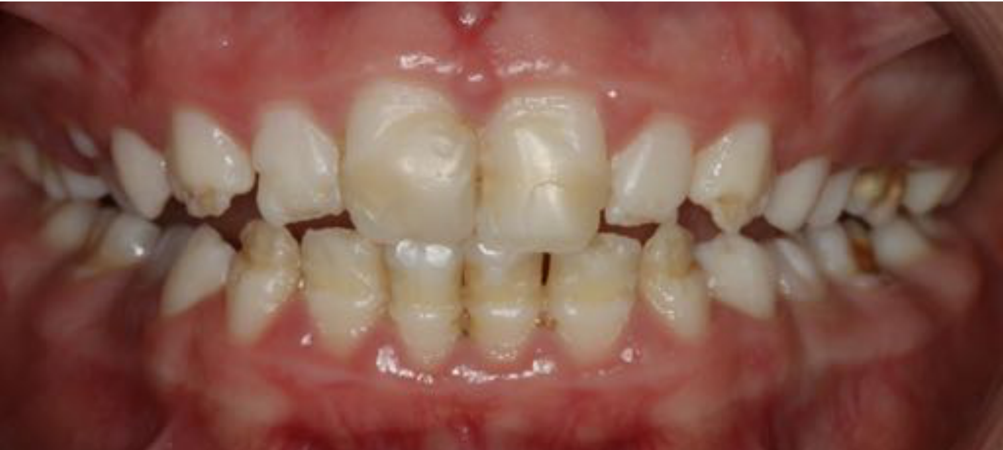
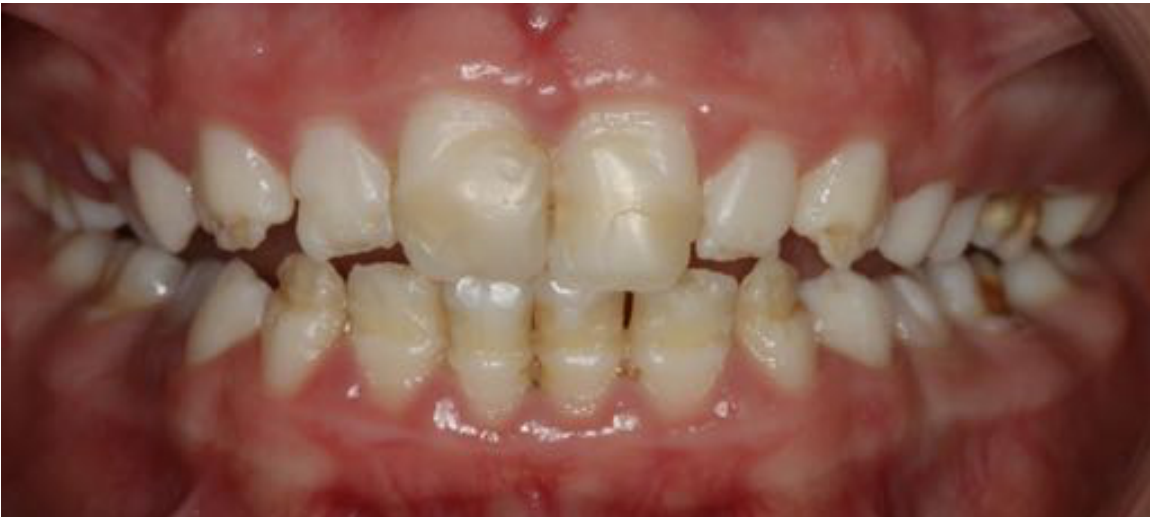
Chronological Hyperplasia After Illness
The distribution of enamel defects allowed the clinician to identify the likely period of a severe childhood illness.
- The first molars and premolars were relatively normal, while a band of hypoplasia crossed the lower incisors and lateral incisors.
- The canines were affected mainly at the cusp tips, and the incisal thirds of the central and lateral incisors were involved.
- The child had been hospitalized for approximately three months and had received intravenous antibiotics and extensive medical treatment.
- Canine calcification occurs approximately between three and five months of age; some molar areas calcify around birth, although the affected areas indicated a period slightly after birth.
- The chronological distribution distinguished this condition from amelogenesis imperfecta and demonstrated how tooth calcification dates can support clinical and forensic reconstruction of early childhood events.
Ultrastructural Characteristics29
- Histological sections of hypoplastic AI: pitted, rough with irregular cracked borders
- SEM analysis: irregular and disorganised superficial enamel layer (Gopinath et al 2004)
- Presence of external defects, incorrectly oriented enamel prisms and enlarged interprism spaces (Belcheva et al 2016)
- Reduced tooth mineral density
Lecturer — Enamel Ultrastructure
Scanning electron microscopy showed disorganized enamel with multiple defects and changes in prismatic spaces, supporting abnormal enamel structure rather than a purely localized surface lesion.
Associations
Intracoronal Resorption and Secondary Caries30
- Intracoronal resorption
- Aggressive, asymptomatic and often diagnosed incidentally from radiographs
- Secondary caries
- Pathogenesis unknown
- Contributing factors:
- Ectopic positioning of affected teeth
- Periapical inflammation of a primary precursor
- Developmental defect due to inclusion of uncalcified enamel matrix
Bhatia et al 2015
Lecturer — Intracoronal Resorption
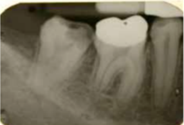
The lesion may be a pre-eruptive intracoronal resorption defect rather than ordinary caries.
- It can occur in an unerupted tooth and be identified incidentally on an orthopantomogram.
- A soft-tissue defect or pocket overlying the area may permit bacterial entry, leading to caries within or beneath the resorptive defect.
- Once the tooth erupts, the extensive lesion may be mistaken for occult or secondary caries, and it can rapidly approach the pulp.
- Treatment described involved raising a flap, isolating the tooth, restoring it and closing the area.
Failure of Eruption31
- Reported in several case reports
- Factors: concurrent follicular enlargements, abnormality in the molecular control of the eruption process, space deficiency and dental alignment problems
Williams et al 1988
Lecturer — Failure of Eruption
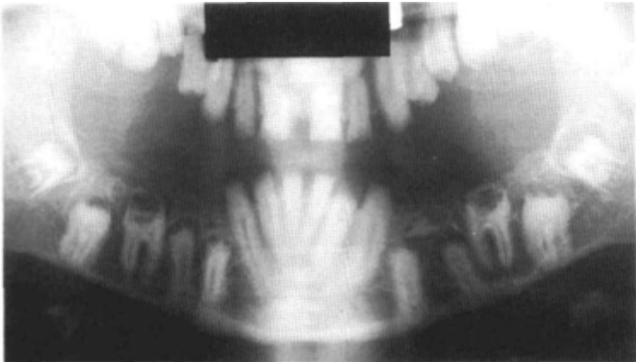
A tooth may have fully developed roots but remain unerupted.
- The unerupted tooth may show extensive breakdown and a radiolucency around it, so complications can include both enamel destruction and eruption abnormalities.
Nephrocalcinosis Syndrome32
- First reported in 1972 (MacGibbon 1972)
- Case reports reporting failure of eruption, enamel agenesis, unexplained nephrocalcinosis, and normal biochemical parameters
- Role of paediatric dentists in early referral of patients with AI to exclude renal pathology (Hunter et al 2007)
Lecturer — Nephrocalcinosis
The first report mentioned was from 1972 and described severe renal disease in siblings.
- One sibling died at 26 years of age after severe renal failure associated with nephrocalcinosis.
- Another sibling developed repeated urinary infections, hypertension and renal failure.
Management
General Principles and Clinical Problems
- Early diagnosis
Clinical Problems33
-
Esthetics
-
Dental sensitivity
-
Loss of occlusal vertical dimension due to loss of dental structure
-
Dental caries and/or gingival inflammation
-
Orthodontic considerations
-
Eruption abnormalities
-
Difficulty maintaining function Seow 1992, Gibbard 1974
Lecturer — Early Referral
Early referral is indicated when amelogenesis imperfecta is suspected or when associated developmental problems may be present.
- Referral is particularly appropriate with a relevant family history, severe enamel breakdown, or suspected eruption or tooth-development abnormalities.
- Perth Children’s Hospital was described as equipped for complex multidisciplinary conditions, but its dental outpatient department is not intended for routine general dental work.
- Amelogenesis imperfecta is included among the specified syndromes under the Medicare Schedule 7 scheme, and specialist multidisciplinary management is often required.
The primary aim in the management of AI is to ensure a pain-free, aesthetic and efficient masticatory apparatus for the patient (Gibbard 1974).
Lecturer — Management Aims
Sensitivity may compromise oral hygiene and contribute to calculus accumulation and gingival problems.
- The management aims are to keep the child free from pain and infection, restore aesthetics and function, and preserve tooth structure and the occlusal vertical dimension.
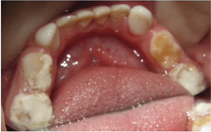
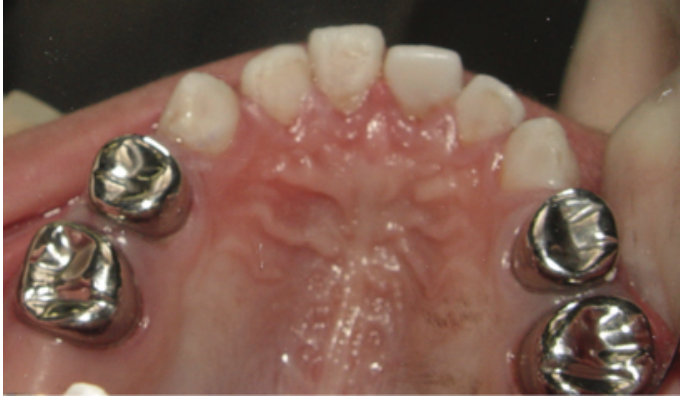
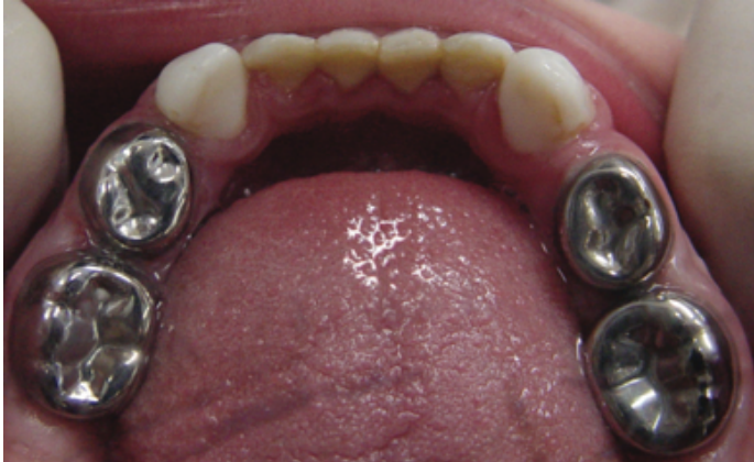
Management of the Primary Dentition34
Marquezin et al 2015
Lecturer — Primary Restorations
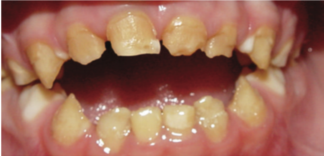
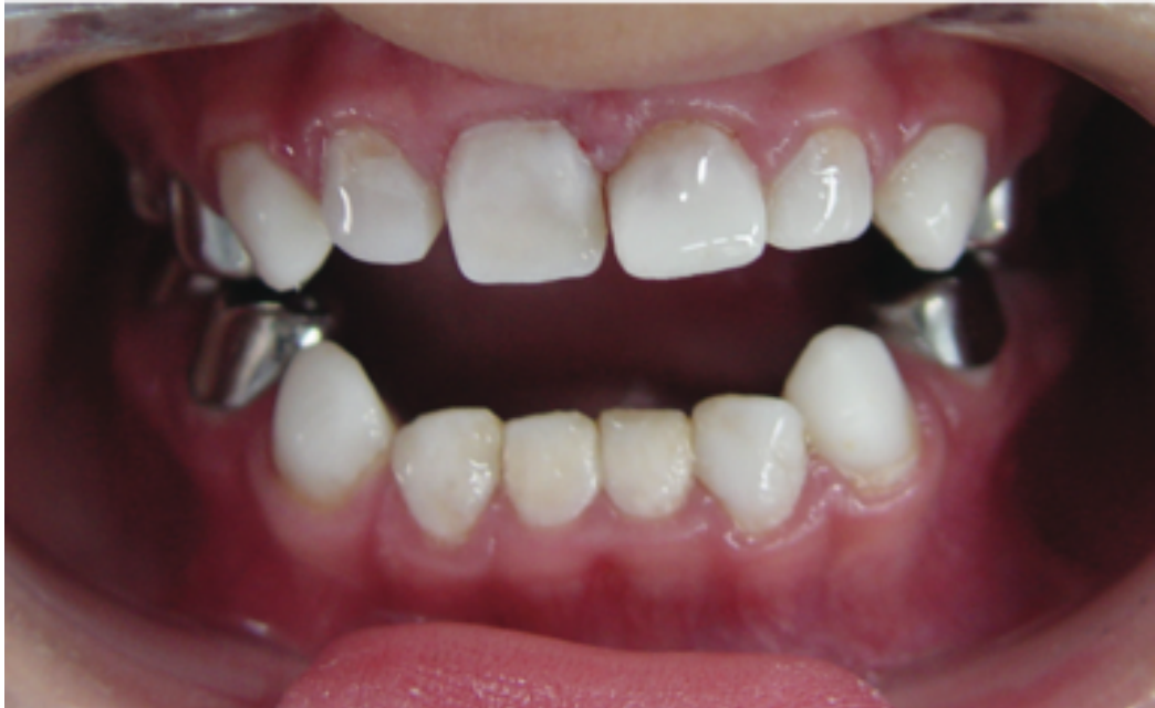
Management of the primary dentition is often based on full-coverage restorations because multisurface restorations may not withstand occlusal loading when the remaining tooth structure is weak.
- The first-line options described were stainless steel crowns for posterior teeth and composite strip crowns for anterior teeth.
- Tooth structure may break down around a multisurface restoration, so restorations should support function and help maintain the occlusal vertical dimension.
 |  |  |
 |  |
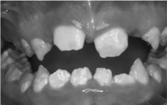
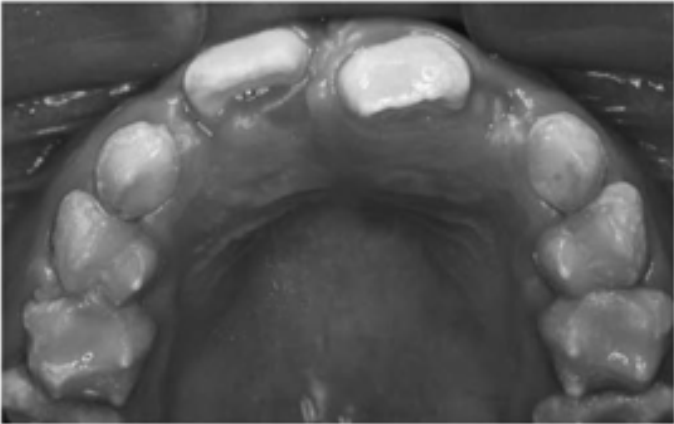
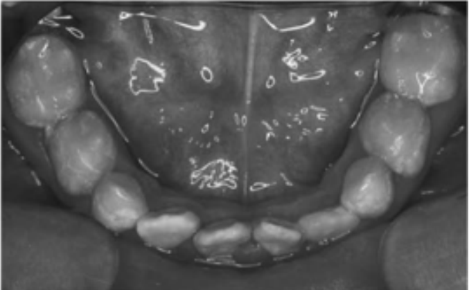
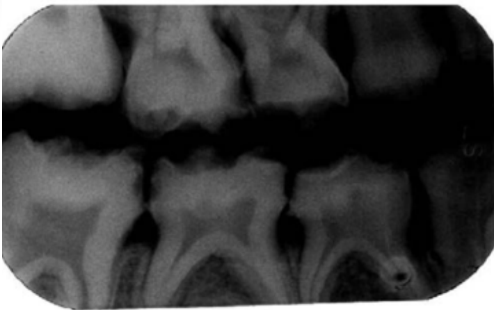
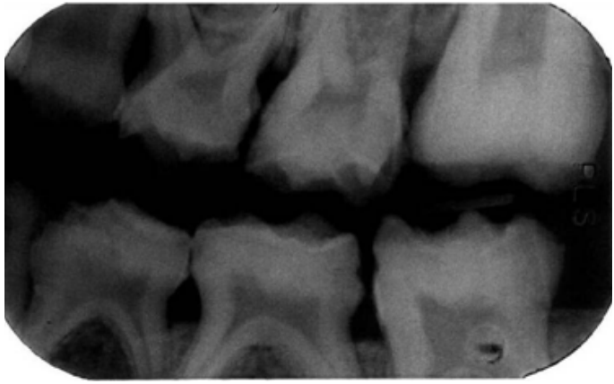
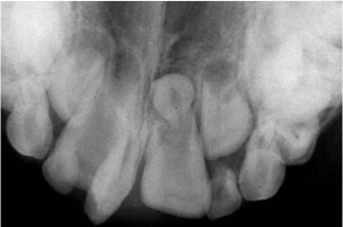
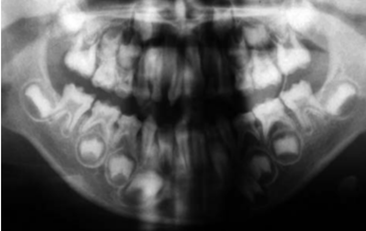
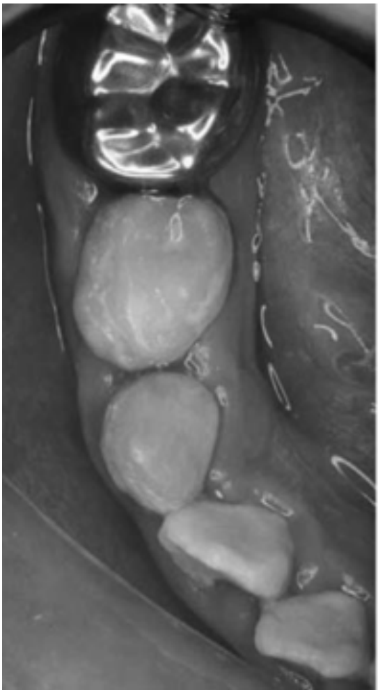
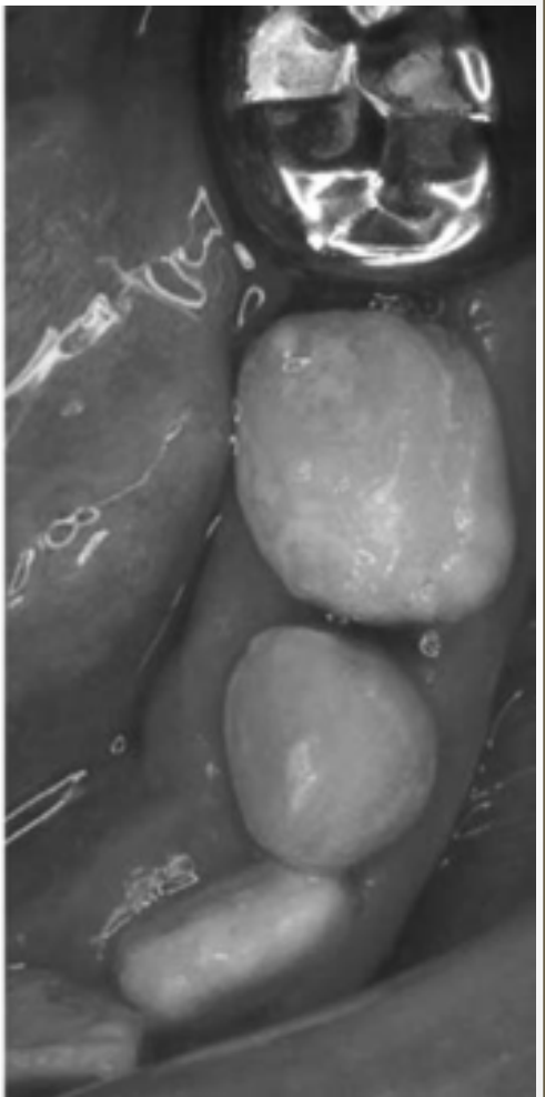
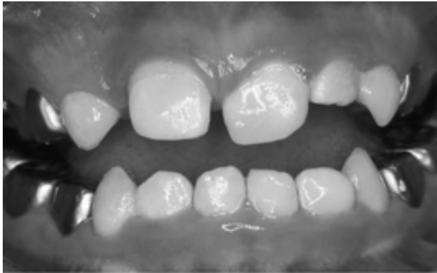
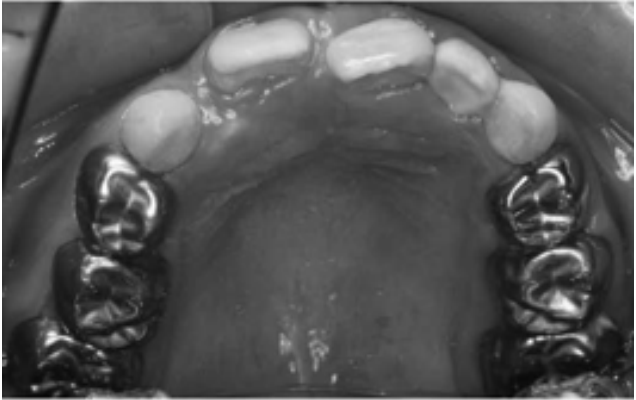
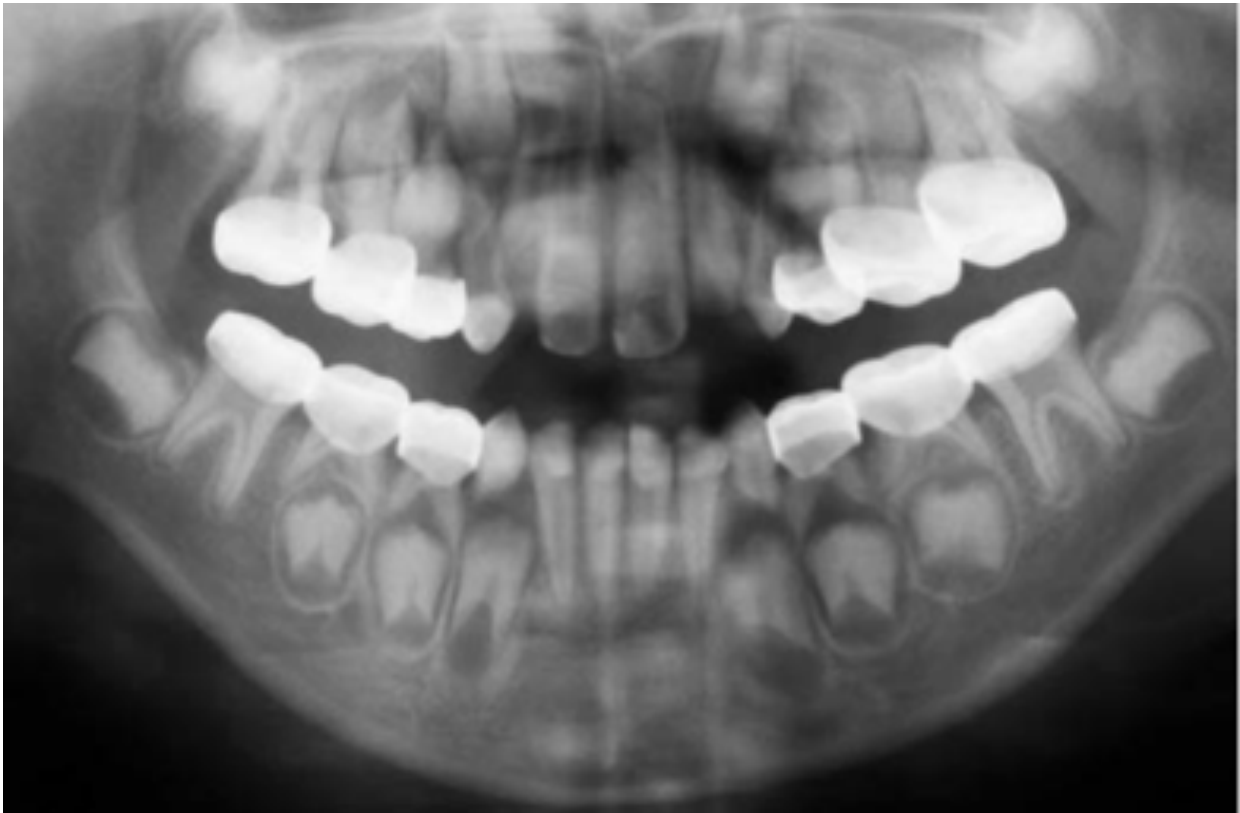
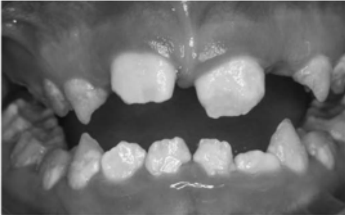
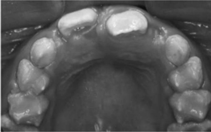
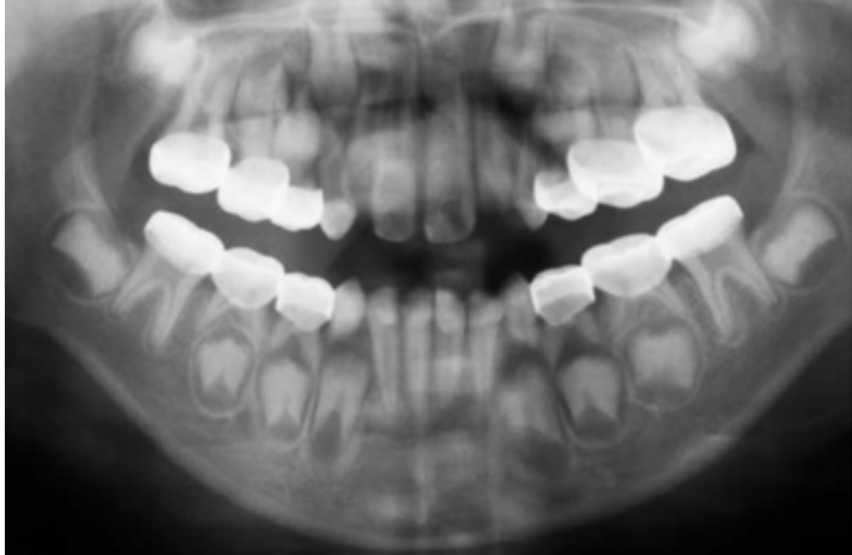
Management of the Mixed Dentition353637383940
Kwok-Tung and King 2006
Lecturer — Mixed Dentition Challenges
The mixed dentition is particularly challenging because permanent teeth are erupting while the anterior segments have important aesthetic implications.
- First permanent molars are important for stabilizing the vertical dimension.
- Aesthetics, sensitivity and function can significantly affect quality of life, particularly as the child transitions into high school.
- Treatment may include composite veneers on permanent incisors and stainless steel crowns on primary and permanent molars.
- Radiographic examination is important because additional anomalies may be present.
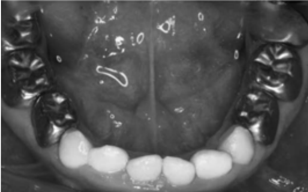
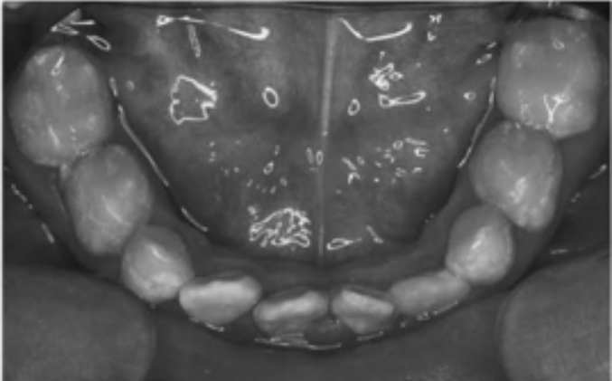
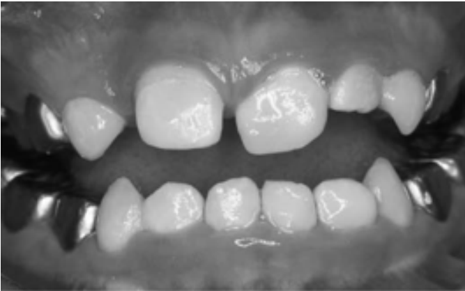
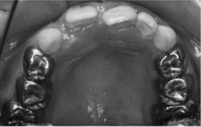
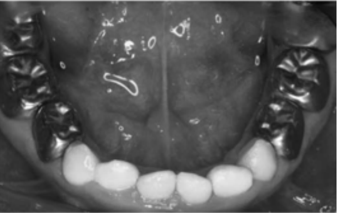
X-Linked Hypoplastic Amelogenesis Imperfecta
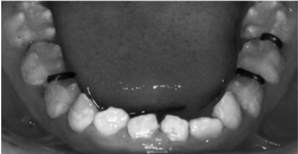
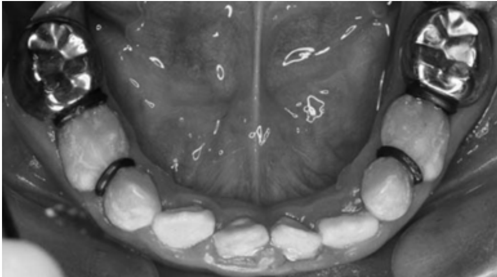
The child had a family history of similar dental manifestations in the paternal grandparents, father and uncles and was diagnosed with X-linked hypoplastic amelogenesis imperfecta.
- Loss of molar enamel caused loss of interproximal space as the teeth moved and contact relationships changed.
- Orthodontic separators were used to regain space, followed a few days later by stainless steel crowns.
- Teeth were restored bilaterally in the same appointment sequence to balance the occlusion and progressively alter the vertical dimension.
- Treatment included stainless steel crowns from the primary molars through the first permanent molars and composite resin strip crowns on the maxillary anterior teeth.
- The lecturer emphasized that this treatment should not be undertaken routinely without referral.
- Stainless steel crowns on permanent molars are substantially more difficult because of anaesthetic difficulties, access and preparation, proximal reduction, and wide pulp chambers in immature teeth.
- Permanent molars must not be over-prepared because the restorations are intended to last until completion of growth, after which definitive treatment may be provided.
- Back-to-back stainless steel crowns are technically difficult and highly technique-sensitive.
Management of the Permanent Dentition414243444546
Shibata et al. 2016
Lecturer — Permanent Dentition Management
Severe permanent dentition cases may involve post-eruptive breakdown, failure of eruption, missing teeth, microdontia, dysmorphic crowns, crowding and multiple developmental abnormalities.
- These cases require referral to specialist multidisciplinary teams.
- Treatment may include restorative, orthodontic and surgical management depending on the findings.
- In one case, gingival recontouring and veneers were used to improve aesthetics.
Severe Permanent Dentition Cases
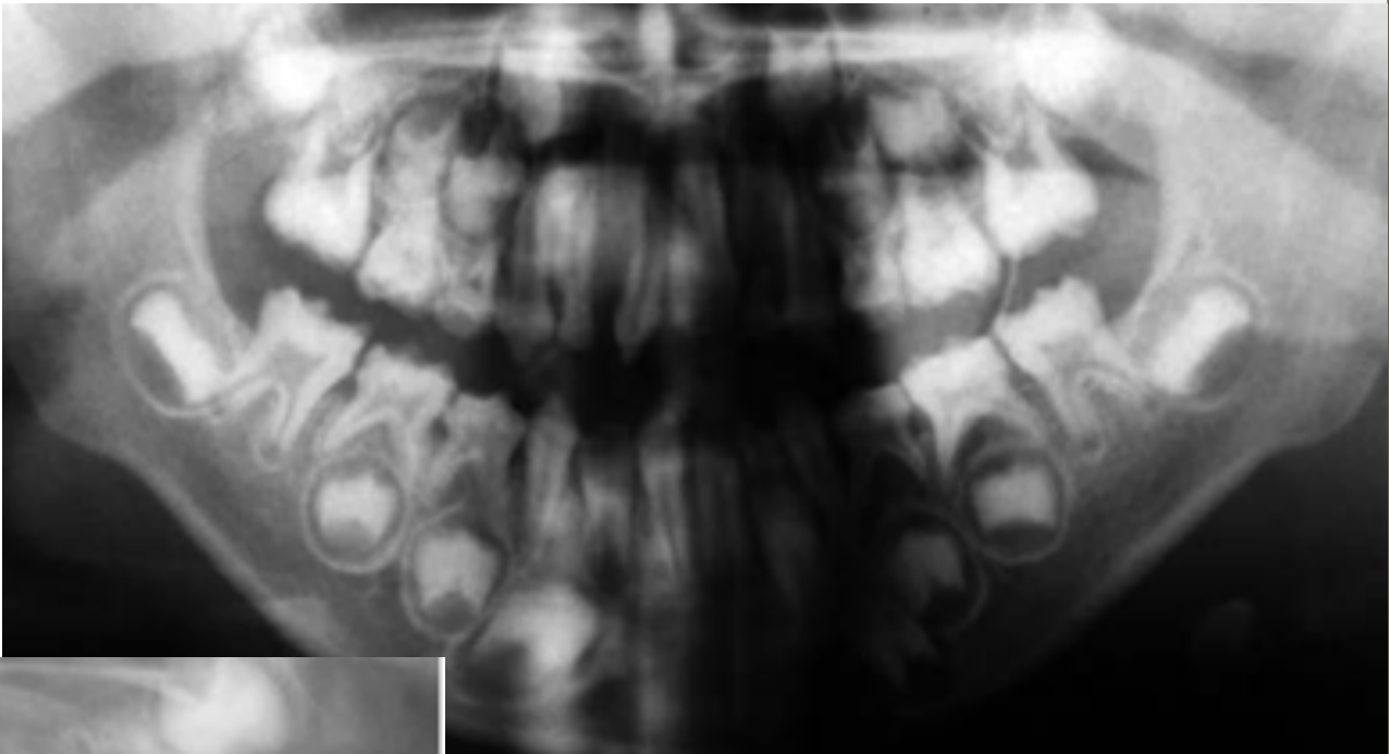
One child, approximately 11 years old at presentation, had extensive abnormalities on the orthopantomogram and severe non-carious breakdown of the first permanent molars.
- Findings included an unerupted canine with overlying soft tissue, a fully developed root in a tooth that had failed to erupt, a missing premolar, a dysmorphic crown in another premolar region, a microdont premolar, additional missing teeth and crowding.
- Clinical photographs showed unerupted permanent molars and substantial breakdown.
- Another case showed severe post-eruptive breakdown in both the primary dentition and first permanent molars.
- It was difficult to determine from appearance alone whether that condition was hypocalcified or hypoplastic.
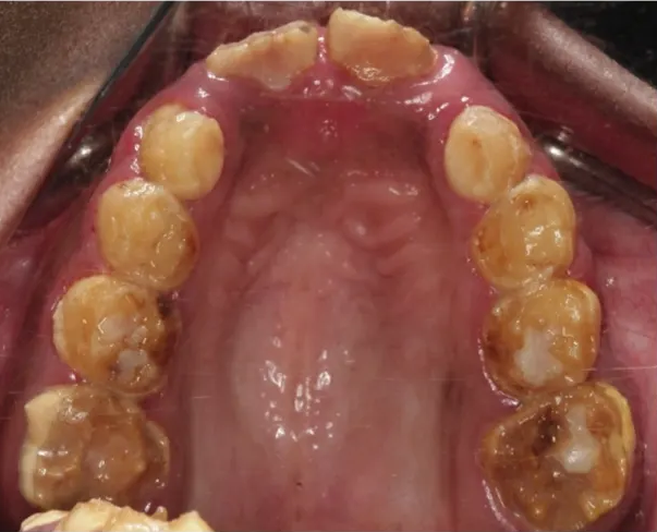
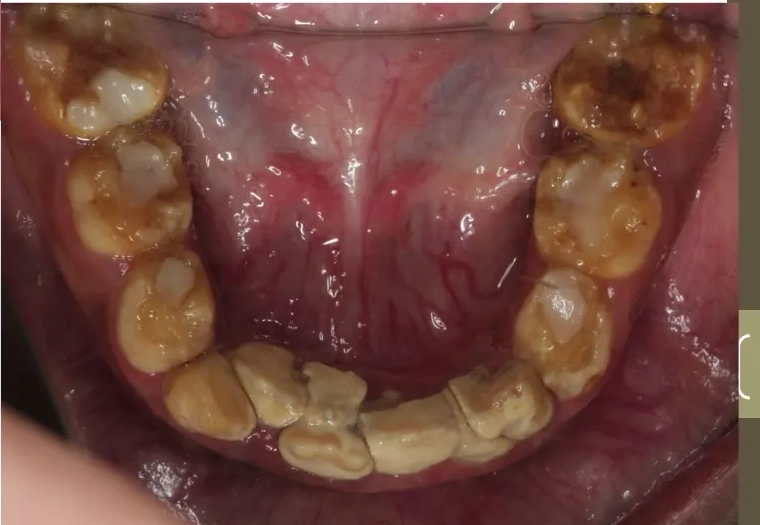
- Oral hygiene was significantly compromised, with substantial calculus in the lower anterior region; sensitivity from the severely broken-down enamel may have contributed to poor oral hygiene.
- These cases reinforced the need for early referral and multidisciplinary management.
Conclusion47
- AI is a rare condition and a number of classifications have been proposed over the years corresponding to advancements made in genetic testing and molecular biology
- The genomic origin of AI gives rise to a number of phenotypic characteristics and identifying the different variants of AI can be particularly challenging
- AI presents a number of clinical challenges including esthetics, attrition, dental sensitivity and orthodontic complications
- Management is focused on prevention, genetic counseling, early diagnosis and management
- Amelogenesis imperfecta comprises genetically originating conditions affecting the enamel of all or nearly all teeth.
- Historical classifications included hypoplastic, hypomaturation and hypocalcified types.
- Genetic testing is needed to identify the different subtypes accurately.
- Phenotypic assessment alone is unreliable because clinical appearances overlap and vary considerably.
- Proper history and examination are essential to exclude molar-incisor hypomineralisation, chronological hyperplasia, fluorosis and other localized or environmental enamel defects.
- Associated problems may include intracoronal resorption, taurodontism, failure of eruption and renal pathology associated with nephrocalcinosis syndrome.
- Management must also address pain, infection, function and preservation of the occlusal vertical dimension.
- Early diagnosis and referral are important.
- Severe cases require specialist multidisciplinary teams, particularly when the primary, mixed and permanent dentitions are affected together.
Clinical Cases
Case: Second molar with enlarged pulp chamber
Question
Scenario: A subset of amelogenesis imperfecta is described where the condition affects not only the enamel but also the root canal space.
What’s shown: An exhibit of a second molar.
Consider: What specific anatomical abnormality is visible in the pulp space of this tooth, and what is the term for this condition?
Answer
Observations:
- The pulp chamber extends all the way down to the apex of the tooth.
Reasoning: The lecturer points out that the pulp chamber goes all the way to the apex, identifying this feature as taurodontism. This is noted as a subset of amelogenesis imperfecta where the root canal space is affected in addition to the enamel.
Outcome: None stated.
Takeaway: Amelogenesis imperfecta can be associated with taurodontism, characterized by an enlarged pulp chamber that extends to the apex.
Case: Enamel defects in a child, clinical photographs
Question
Scenario: A child presents with enamel defects. The lecturer shows clinical photographs of the child’s teeth to determine the cause of the defects.
What’s shown: Clinical photos showing a band of hypoplasia across the lower incisors and lateral incisors, while the canines are only affected at the cusp tips. The premolars appear normal. The molars at the back show qualitative defects, appearing brown, with the defect located below the cusp tips.
Consider: What underlying event caused these specific enamel defects, and how is this determined from the clinical findings?
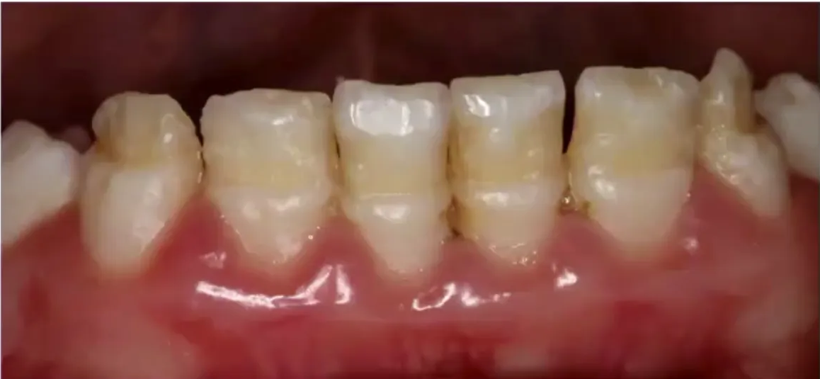
Answer
Observations:
- The defects do not involve every tooth equally.
- Premolars are normal.
- Lower incisors and laterals have a band of hypoplasia on the incisal third.
- Canines are affected only at the cusp tips.
- Molars show qualitative brown defects below the cusp tips.
Reasoning: The pattern of defects corresponds to the calcification dates of the affected teeth. The incisors and canines calcify between three and five months of age, while the molars calcify slightly after birth. This specific chronological pattern indicates a systemic disruption during those exact periods, diagnosing chronological hypoplasia rather than amelogenesis imperfecta.
Outcome: When asked what happened to the child between three and five months of age, the parent revealed the child was hospitalized for three months on IV antibiotics.
Takeaway: Enamel defects that follow a chronological pattern corresponding to tooth calcification dates indicate chronological hypoplasia due to systemic illness during specific periods of development, allowing clinicians to deduce the patient’s medical history.
Case: Unerupted lower first molar with radiolucency, radiograph
Question
Scenario: An 11 to 12-year-old patient presents with unerupted teeth. A radiograph is taken to investigate the cause.
What’s shown: A dental radiograph showing permanent lower canines with almost fully developed roots. A lower first molar has failed to erupt, appears half broken down, and has a radiolucency around it.
Consider: What is the cause of the radiolucency on the unerupted tooth, and what are the potential complications and management options?
Answer
Observations:
- Lower canines are present in the mouth with almost fully developed roots.
- A lower first molar has failed to erupt (should have erupted six years prior).
- The unerupted molar is half broken down with a radiolucency around it.
Reasoning: The resorptive defect on the unerupted tooth looks like caries but is subgingival in the bone, indicating pre-eruptive intra-coronal resorption rather than typical caries. This condition can lead to severe complications, including failure of eruption and extensive breakdown of the tooth.
Outcome: Management involves early detection; some clinicians raise a flap, isolate the tooth, restore it, and close the flap to prevent rapid pulp involvement.
Takeaway: Pre-eruptive intra-coronal resorption can present as a radiolucency on an unerupted tooth, mimicking caries but occurring in the bone, and can lead to failure of eruption and severe tooth breakdown if not managed early.
Case: Mixed dentition with supernumerary teeth, case report
Question
Scenario: A case report of a patient in the mixed dentition requiring management for amelogenesis imperfecta.
What’s shown: Bitewing radiographs, a maxillary occlusal radiograph, and an OPG.
Consider: What additional finding might you notice on the maxillary occlusal radiograph?
Answer
Observations:
- Two supernumerary teeth in the anterior maxilla.
- One supernumerary tooth erupted between the permanent central incisors.
- The other supernumerary tooth was inverted and palatally impacted.
Reasoning: The lecturer points out that identifying one syndrome or issue in a child does not exclude other concurrent anomalies, emphasizing the need for a thorough examination and radiographic diagnosis to catch all findings.
Outcome: Composite veneers were placed on the permanent incisors, and stainless steel crowns were placed on the primary and permanent molars.
Takeaway: When a syndrome or issue is identified in a child, a thorough examination and radiographic diagnosis are essential to rule out concurrent anomalies.
Case: Child with X-linked hypoplastic amelogenesis imperfecta, space regaining and crowns
Question
Scenario: A child with a family history of X-linked hypoplastic amelogenesis imperfecta presenting with lost intermolar space due to enamel loss.
What’s shown: Intraoral photographs showing space loss between molars, the use of orthodontic separators, and final restorations.
Consider: How can space be regained and the dentition restored in this hypoplastic case?
Answer
Observations:
- Space lost between molars due to enamel loss, with re-established contact points and tooth movement.
- Orthodontic separators used to regain space.
- Bilateral restoration to balance occlusion and alter vertical dimension.
- Final restorations include stainless steel crowns from the primary D’s to the permanent sixes, and composite resin strip crowns on the maxillary anterior teeth.
Reasoning: Preparing the tooth structure further to regain space risks pulp exposure. Orthodontic separators allow space regaining without over-preparation. Bilateral restoration balances the occlusion and vertical dimension. Stainless steel crowns on permanent molars are difficult but necessary to carry the child through to growth completion, avoiding over-preparation of wide pulp chambers in immature teeth.
Outcome: Space was regained using separators, followed by bilateral placement of stainless steel crowns on posterior teeth and composite strip crowns on anterior teeth.
Takeaway: Orthodontic separators can be used to regain space in hypoplastic AI without over-preparing teeth and risking pulp exposure, and bilateral restoration helps balance occlusion and vertical dimension.
Case: 11-year-old with severe hypoplastic amelogenesis imperfecta and multiple anomalies
Question
Scenario: An 11-year-old child presenting to the children’s hospital with a severe presentation of amelogenesis imperfecta, crowding, and compromised oral hygiene.
What’s shown: A panoramic radiograph (OPG) and intraoral photographs.
Consider: What radiographic and clinical findings are present in this severe case?
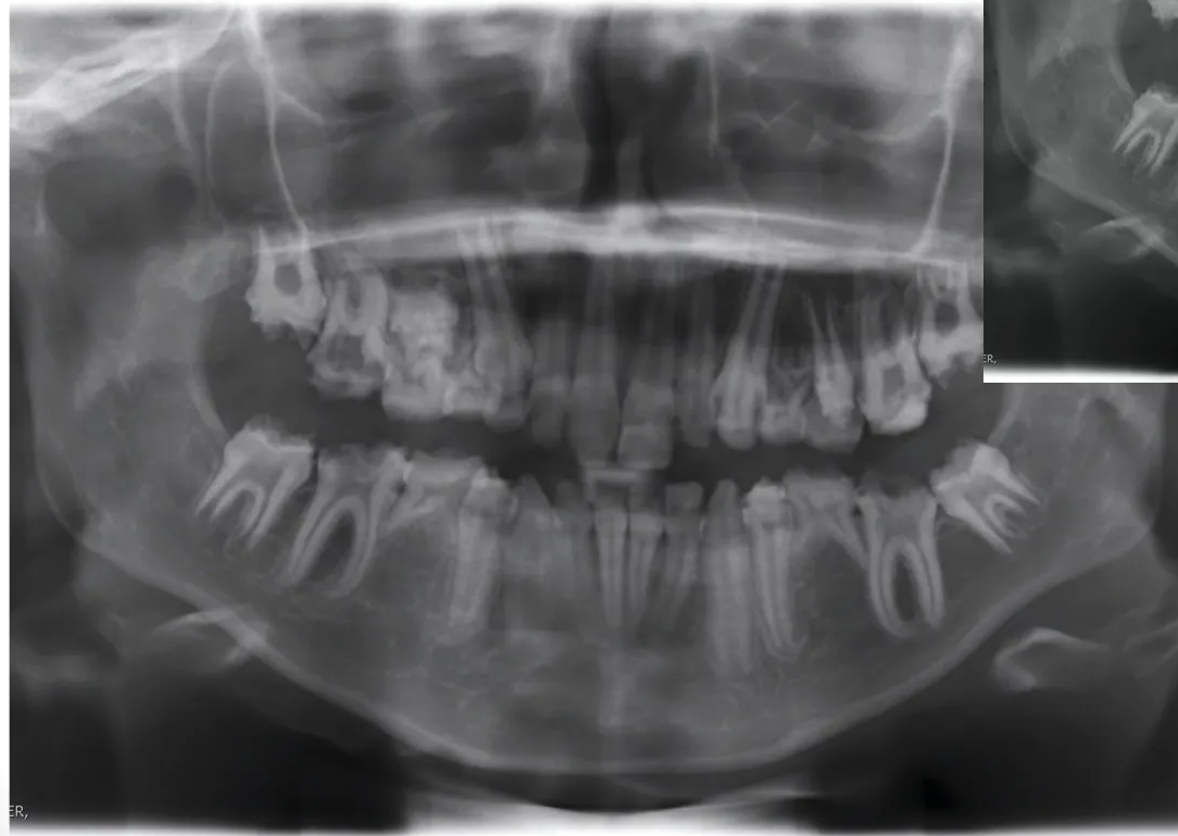
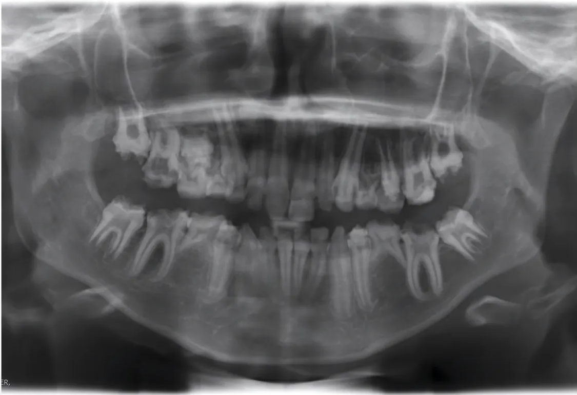
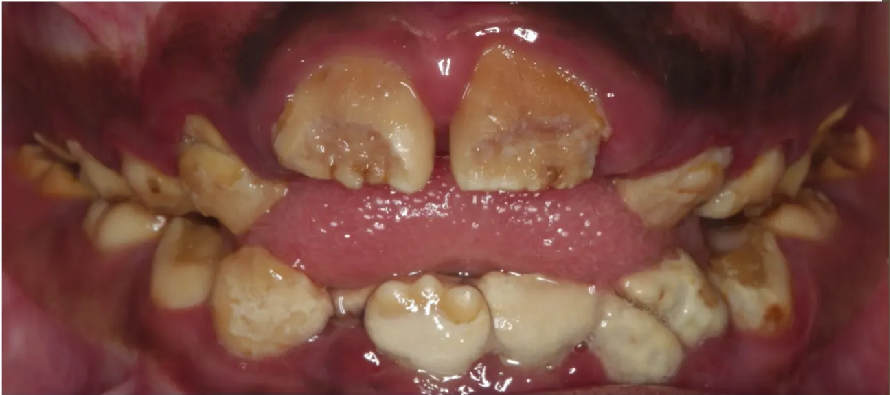
Answer
Observations:
- Severe posteruptive breakdown of the primary dentition and first permanent molars (not caries).
- Failure of eruption of the 3 and 6 (roots fully developed but unerupted, with soft tissue profile overlying).
- Missing teeth: 35, 45, and 14.
- Dysmorphic coronal features of 15.
- Microdont 25.
- Unerupted 36 and 46.
- Significant breakdown, crowding, and calculus buildup in the lower anterior segment.
Reasoning: The severe posteruptive breakdown is characteristic of hypoplastic AI. The failure of eruption despite full root development and the presence of multiple missing, dysmorphic, and microdont teeth highlight the complex, multi-factorial anomalies associated with severe AI. Compromised oral hygiene and calculus are likely secondary to dental sensitivity from severe enamel breakdown.
Outcome: The case requires referral to specialist multidisciplinary teams for efficient management.
Takeaway: Severe AI can present with complex concurrent anomalies including failure of eruption, missing teeth, and dysmorphic/microdont teeth, necessitating thorough radiographic diagnosis and multidisciplinary management.
Case: Rough hyperplastic amelogenesis imperfecta, gingival recontouring and veneers
Question
Scenario: A case report of a patient in the permanent dentition with rough hyperplastic amelogenesis imperfecta.
What’s shown: Clinical photographs of the maxillary anterior teeth.
Consider: What treatment was performed to restore aesthetics in this case?
Answer
Observations:
- Rough hyperplastic amelogenesis imperfecta in the permanent dentition.
- Gingival recontouring was performed.
- Veneers were placed.
Reasoning: The lecturer notes that gingival recontouring and veneers were used to address the aesthetic concerns caused by the rough hyperplastic AI in the permanent dentition.
Outcome: Gingival recontouring and veneers were placed to restore aesthetics.
Takeaway: Gingival recontouring and veneers are treatment options for restoring aesthetics in permanent dentition cases of rough hyperplastic AI.
Footnotes
-
Original PDF page 1: L7 Amelogenesis Imperfecta, p.1 ↩
-
Original PDF page 2: L7 Amelogenesis Imperfecta, p.2 ↩
-
Original PDF page 3: L7 Amelogenesis Imperfecta, p.3 ↩
-
Original PDF page 4: L7 Amelogenesis Imperfecta, p.4 ↩
-
Original PDF page 5: L7 Amelogenesis Imperfecta, p.5 ↩
-
Original PDF page 6: L7 Amelogenesis Imperfecta, p.6 ↩
-
Original PDF page 7: L7 Amelogenesis Imperfecta, p.7 ↩
-
Original PDF page 8: L7 Amelogenesis Imperfecta, p.8 ↩
-
Original PDF page 9: L7 Amelogenesis Imperfecta, p.9 ↩
-
Original PDF page 10: L7 Amelogenesis Imperfecta, p.10 ↩
-
Original PDF page 11: L7 Amelogenesis Imperfecta, p.11 ↩
-
Original PDF page 12: L7 Amelogenesis Imperfecta, p.12 ↩
-
Original PDF page 13: L7 Amelogenesis Imperfecta, p.13 ↩
-
Original PDF page 14: L7 Amelogenesis Imperfecta, p.14 ↩
-
Original PDF page 15: L7 Amelogenesis Imperfecta, p.15 ↩
-
Original PDF page 16: L7 Amelogenesis Imperfecta, p.16 ↩
-
Original PDF page 17: L7 Amelogenesis Imperfecta, p.17 ↩
-
Original PDF page 18: L7 Amelogenesis Imperfecta, p.18 ↩
-
Original PDF page 19: L7 Amelogenesis Imperfecta, p.19 ↩
-
Original PDF page 20: L7 Amelogenesis Imperfecta, p.20 ↩
-
Original PDF page 21: L7 Amelogenesis Imperfecta, p.21 ↩
-
Original PDF page 22: L7 Amelogenesis Imperfecta, p.22 ↩
-
Original PDF page 23: L7 Amelogenesis Imperfecta, p.23 ↩
-
Original PDF page 24: L7 Amelogenesis Imperfecta, p.24 ↩
-
Original PDF page 25: L7 Amelogenesis Imperfecta, p.25 ↩
-
Original PDF page 26: L7 Amelogenesis Imperfecta, p.26 ↩
-
Original PDF page 27: L7 Amelogenesis Imperfecta, p.27 ↩
-
Original PDF page 28: L7 Amelogenesis Imperfecta, p.28 ↩
-
Original PDF page 29: L7 Amelogenesis Imperfecta, p.29 ↩
-
Original PDF page 30: L7 Amelogenesis Imperfecta, p.30 ↩
-
Original PDF page 31: L7 Amelogenesis Imperfecta, p.31 ↩
-
Original PDF page 32: L7 Amelogenesis Imperfecta, p.32 ↩
-
Original PDF page 33: L7 Amelogenesis Imperfecta, p.33 ↩
-
Original PDF page 34: L7 Amelogenesis Imperfecta, p.34 ↩
-
Original PDF page 35: L7 Amelogenesis Imperfecta, p.35 ↩
-
Original PDF page 36: L7 Amelogenesis Imperfecta, p.36 ↩
-
Original PDF page 37: L7 Amelogenesis Imperfecta, p.37 ↩
-
Original PDF page 38: L7 Amelogenesis Imperfecta, p.38 ↩
-
Original PDF page 39: L7 Amelogenesis Imperfecta, p.39 ↩
-
Original PDF page 40: L7 Amelogenesis Imperfecta, p.40 ↩
-
Original PDF page 41: L7 Amelogenesis Imperfecta, p.41 ↩
-
Original PDF page 42: L7 Amelogenesis Imperfecta, p.42 ↩
-
Original PDF page 43: L7 Amelogenesis Imperfecta, p.43 ↩
-
Original PDF page 44: L7 Amelogenesis Imperfecta, p.44 ↩
-
Original PDF page 45: L7 Amelogenesis Imperfecta, p.45 ↩
-
Original PDF page 46: L7 Amelogenesis Imperfecta, p.46 ↩
-
Original PDF page 47: L7 Amelogenesis Imperfecta, p.47 ↩