Hereditary Dentine Disorders and Regional Odontodysplasia1
Dr Lisa Bowdin BDSc, DClinDent, MRACDS
Specialist Paediatric Dentist
Senior Lecturer, Paediatric Dentistry
UWA Dental School
Hereditary Dentine Disorders2
Dentinogenesis Imperfecta Introduction3
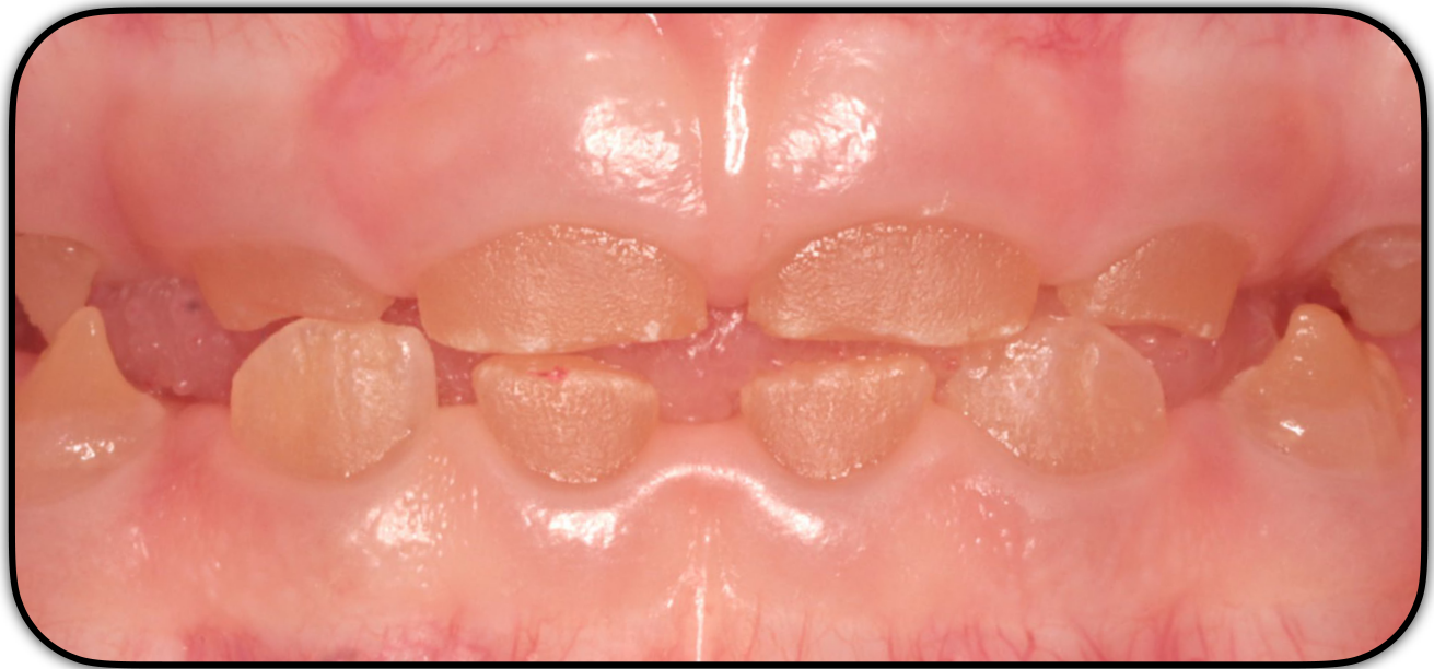
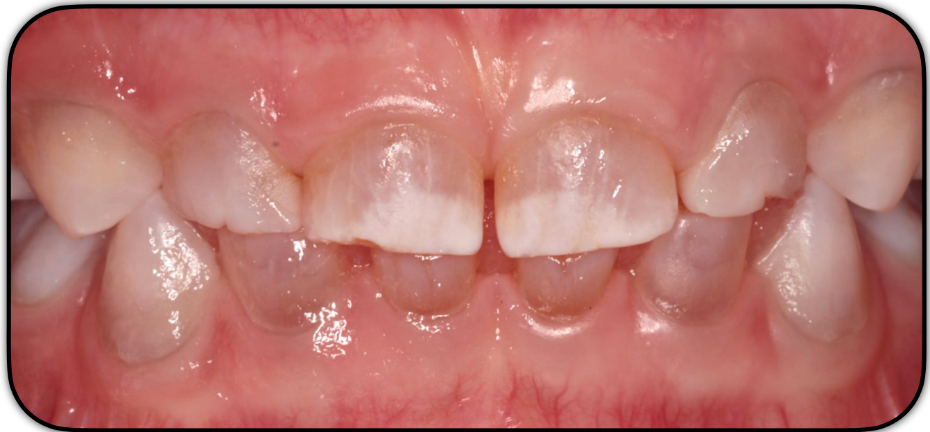
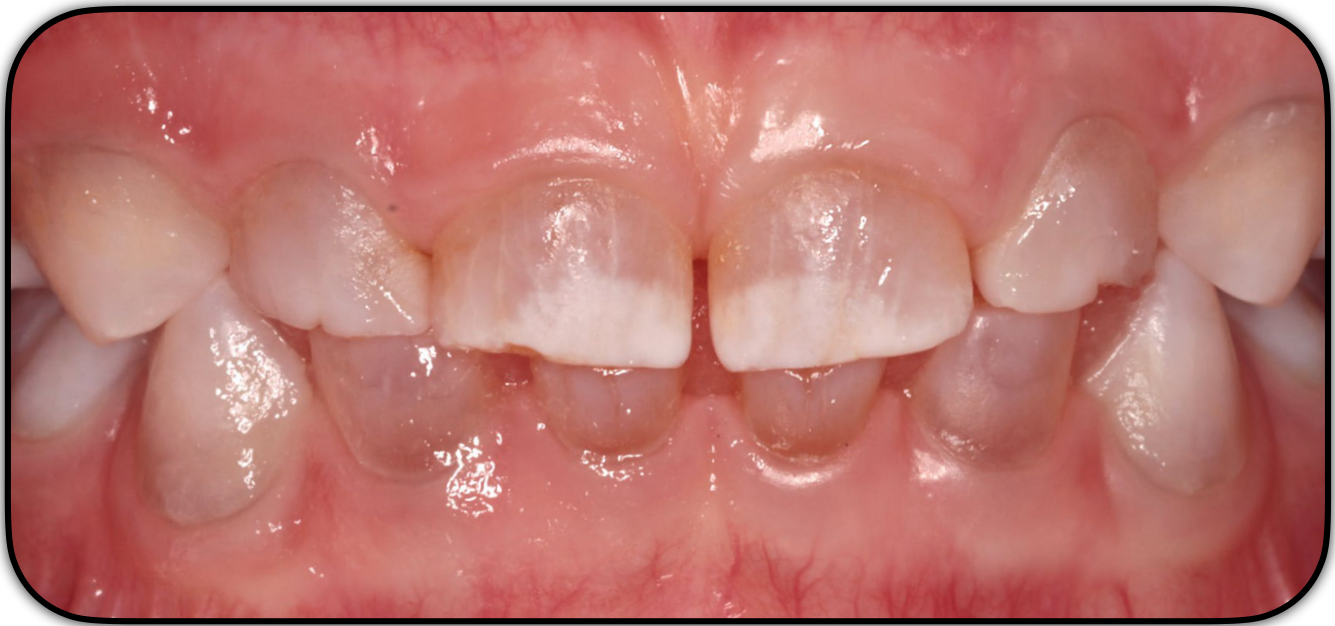
Dentinogenesis Imperfecta (DGI)
 |  |
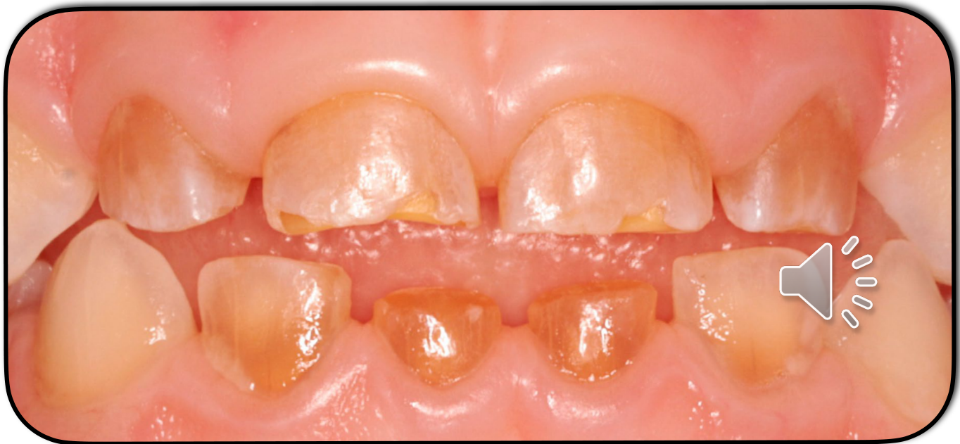 | |
| Genetic disorder of dnetine characterized by severe hypomineralization and loss of dentine structure. Leads to rapid deteriorration of teeth |
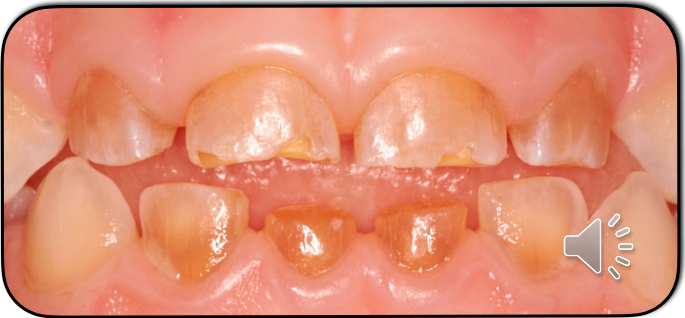
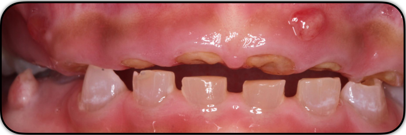
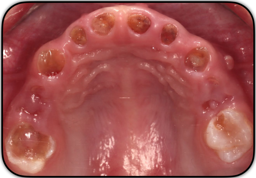
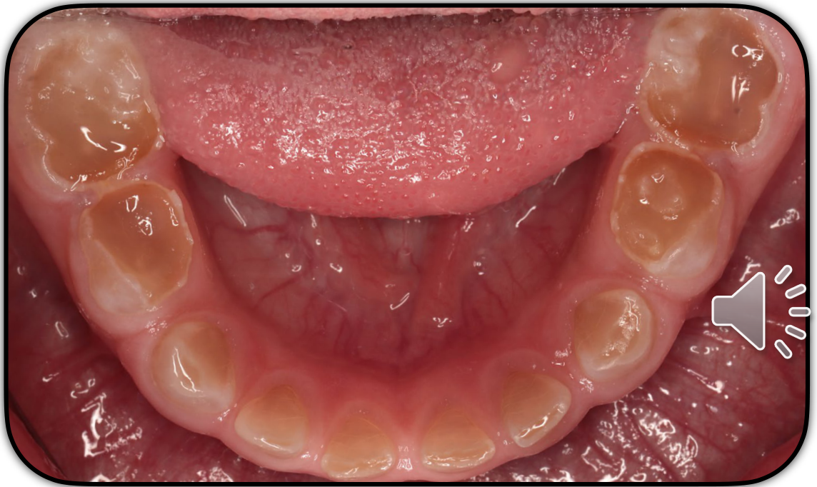
Clinical Presentation45
- Blue to brown discolouration of the dentition
- Enamel frequently separates from underlying dentine
- Variable expressivity
- Primary and/or permanent dentition
- Primary teeth more severely affected (Malmgren et al 2004)
Shields et al 1973
-
Accelerated attrition → severely worn dentition
-
Spontaneous abscesses
-
Pain
-
Infection
-
Poor aesthetics
-
Tooth mobility and premature tooth loss may occur, sometimes leading to repeated emergency dental visits and extensive restorative treatment.
 |  |  |
 |  |
Histomorphologic Characteristics6
Lecturer — Natal Teeth Presentation
- Dysplastic dentine
- Amorphous areas void of dentinal tubules
- Irregular dentinal tubules
- Embedded cells
- Interglobular dentine
- Continuous formation of abnormal circumpulpal dentine
- DEJ
- Straight with lack of scalloping
- Enamel
- Normal
- Some reports of aberrations
Davis et al 2015 Malmgren & Lindskog 2003 Malmgren et al 2004
Prevalence7
Lecturer — Natal Tooth Histology
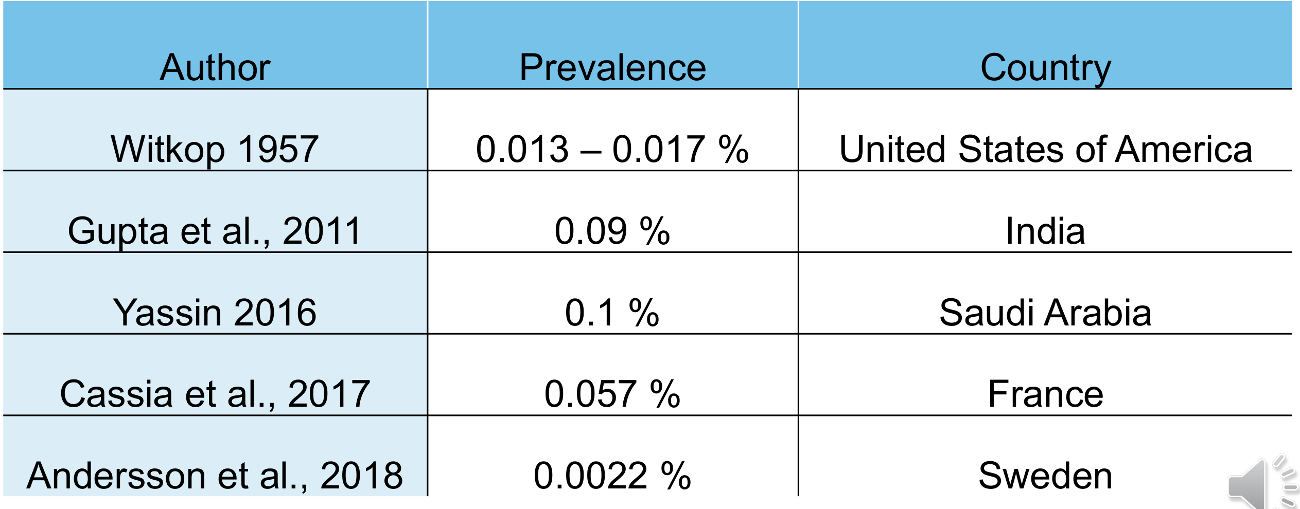
Prevalence ~ 1 in 8000 children
| Author | Prevalence | Country |
|---|---|---|
| Witkop 1957 | 0.013 – 0.017 % | United States of America |
| Gupta et al., 2011 | 0.09 % | India |
| Yassin 2016 | 0.1 % | Saudi Arabia |
| Cassia et al., 2017 | 0.057 % | France |
| Andersson et al., 2018 | 0.0022 % | Sweden |
 |  |
Classification8
Shields et al 1973:
-
Dentinogenesis Imperfecta (DGI) type I, II and III
-
Dentin dysplasia (DD) type I and II
-
Based on clinical phenotype rather than molecular data
-
Does not account for genetic aetiology of hereditary dentine defect
DGI-I9
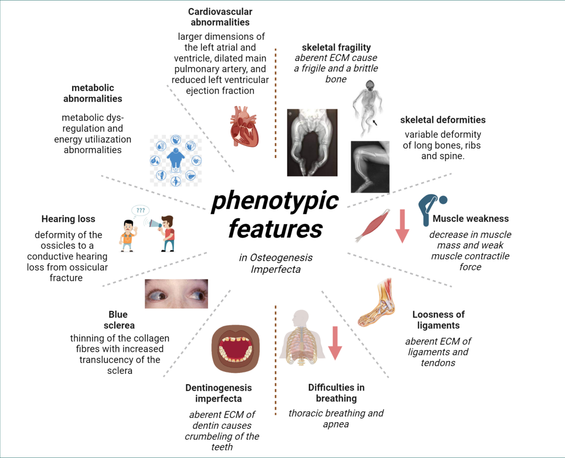
- Syndromic form of DGI associated with, and now classified within, osteogenesis imperfecta
- Discolouration in primary and permanent dentitions
- Radiographically — short, constricted roots, dentine hypertrophy → pulpal obliteration
- Variable expressivity
- Mild form of OI — risk of misdiagnosis
- DGI-I may be the only clinically apparent findings
Lecturer — DGI-I Diagnosis
Diagnosis of a neonatal oral lesion is based primarily on medical and dental history and clinical examination, with radiographic assessment when necessary.
The differential diagnosis includes Bohn’s nodules, Epstein pearls, congenital epulis of the newborn, eruption cysts, lymphangiomas, neuroectodermal tumor of infancy, and other swellings or abnormalities.
If the clinician is unsure of the diagnosis, referral is recommended rather than simply observing the lesion without clarification. DGI-I may show variable findings even within the same individual, ranging from complete pulpal obliteration to apparently normal dentine.
A careful medical history and examination are important because mild osteogenesis imperfecta may otherwise be missed.
Incorrectly diagnosing DGI-II or DD-II should be avoided when the dental findings may represent the only obvious feature of osteogenesis imperfecta.
DGI-II10
DGI-III11
- Defining feature: Shell teeth
Lecturer — DGI-III Background
DGI-III was originally identified in a triracial population of Maryland and Washington, D.C., and is also known as the Brandywine isolate. Large pulp chambers may result in multiple pulp exposures in primary teeth.
Sapir & Shapira et al 1973
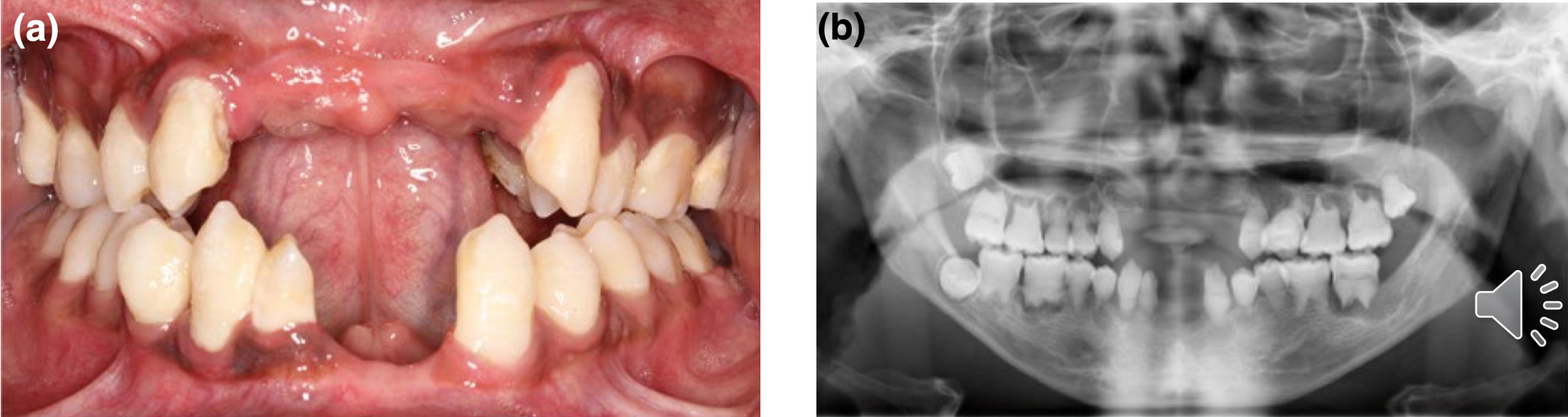
DD-I12
- Rarest of the hereditary dentine defects
- Prevalence of 1:100,000
- Witkop et al 1957
- Loss of organization of root dentine
- Sharp, conical and shortened root lengths
- Extreme tooth mobility and premature exfoliation
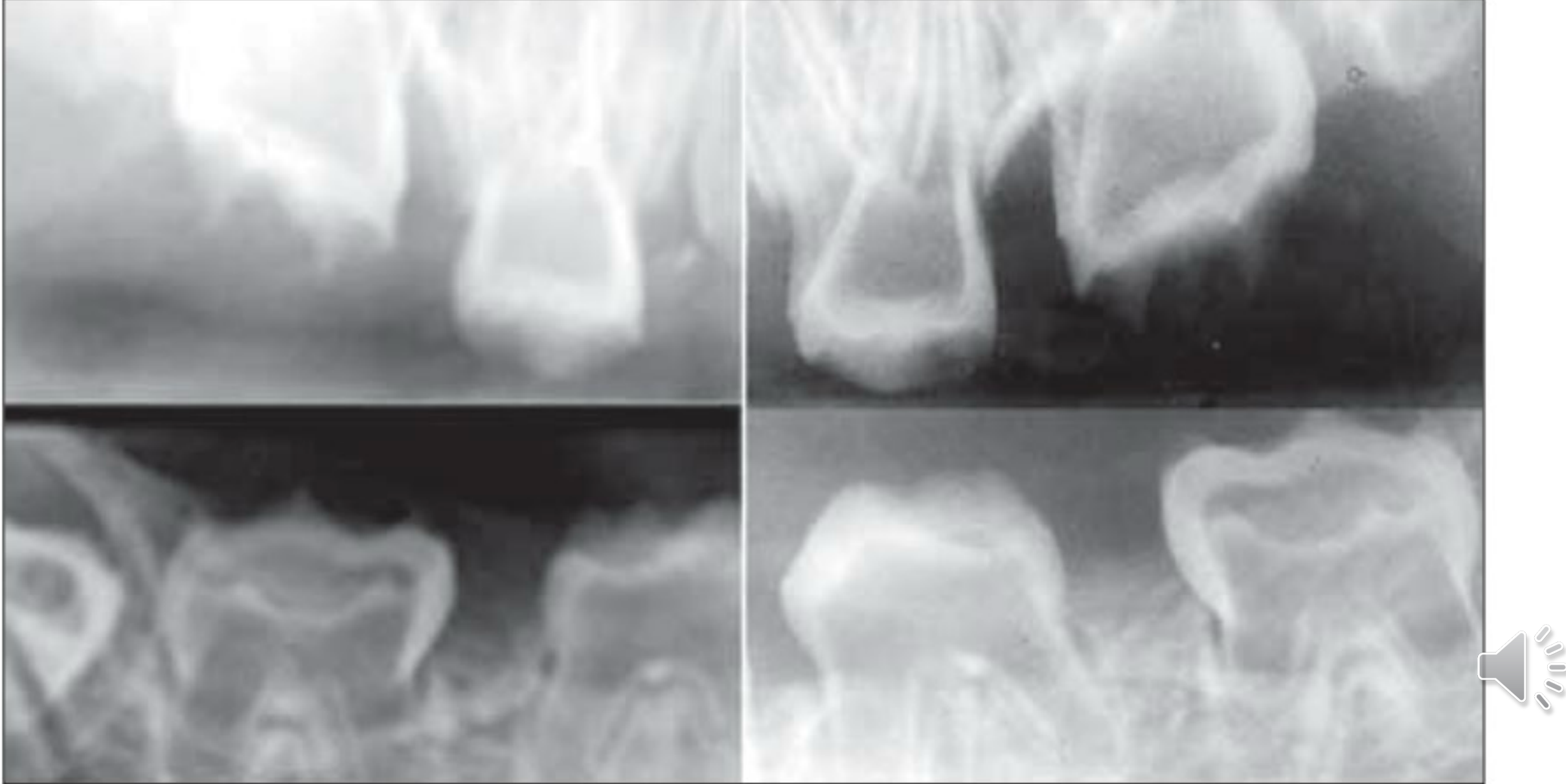
- Radiographs may show crescent-shaped pulp remnants parallel to the cementoenamel junction, complete pulpal obliteration, or periapical lesions without other associated pathology.
(a) (b)
Chen et al 2019
DD-II13
- Similar to DGI type II
- Primary teeth affected more severely than permanent teeth
- Primary teeth in DD-II similar to DGI-I
- Permanent teeth in DD-II mildly or not affected
- Thistle shaped pulp and pulp stones
- Thistle shaped pulp = large pulp chamber with thin root canals

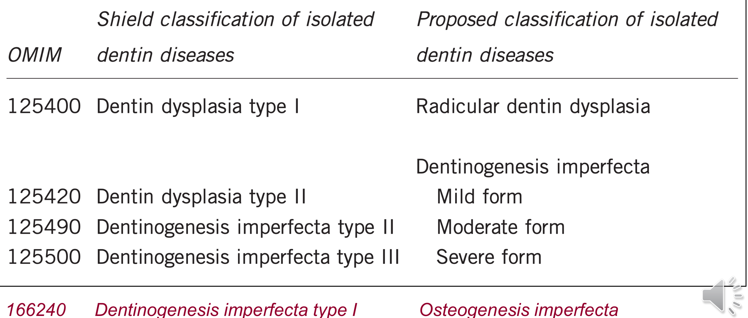
Proposed Classification of Isolated Dentine Diseases14
| OMIM | Shields classification of isolated dentin diseases | Proposed classification of isolated dentin diseases |
|---|---|---|
| 125400 | Dentin dysplasia type I | Radicular dentin dysplasia |
| 125420 | Dentin dysplasia type II | Dentinogenesis imperfecta — Mild form |
| 125490 | Dentinogenesis imperfecta type II | Moderate form |
| 125500 | Dentinogenesis imperfecta type III | Severe form |
| 166240 | Dentinogenesis imperfecta type I | Osteogenesis imperfecta |
de la Dure-Molla et al 2015
Lecturer — Proposed Classification
Classifications of natal teeth have been proposed according to whether the tooth is mature or immature and according to its morphology and degree of development.
- These classifications are mainly academic and help describe variation in the literature.
- Clinically, it may be difficult to determine how mature a tooth is or whether an apparently solid crown is actually only an enamel shell.
De la Dure-Molla introduced the genotype-based classification in 2015 because genetic studies made the Shields classification less relevant, although the older classification remains widely used. The lecture also referred to OMIM, the Online Mendelian Inheritance in Man database, which provides information about human genes, genetic phenotypes and associated conditions.
Clinical Findings15
- Radicular dentin dysplasia (DD type I) — teeth may appear clinically normal. Extreme tooth mobility and early loss of teeth.
- Dentinogenesis imperfecta
- Mild (DD type-II) — primary teeth more severely affected. Permanent teeth mildly or not affected. Primary teeth discolouration and attrition.
- Moderate (DGI type-II) — bulbous crowns with marked cervical constrictions. Teeth discolouration and attrition.
- Severe (DGI type-III) — opalescent primary and permanent teeth, marked attrition, multiple pulp exposures.
- Osteogenesis imperfecta with DGI (DGI type-I) — teeth discolouration and attrition.
Radiographic Findings16
- Radicular dentin dysplasia (DD type I)
- Sharp conical and shortened roots; short, constricted roots.
- Dentinogenesis imperfecta
- Mild (DD type-II) — thistle shaped pulp (large pulp chamber with thin root canals). Permanent teeth mildly or not affected.
- Moderate (DGI type-II) — early pulpal obliteration.
- Severe (DGI type-III) — normal thickness enamel in association with extremely thin dentine and dramatically enlarged pulps, aka “shell teeth”.
- Osteogenesis imperfecta with DGI (DGI type-I)
- Short, constricted roots.
- Pulpal obliteration.

Diagnosis17
Lecturer — Natal Tooth Classification
- Clinical presentation
- Family history
- Pedigree construction
- ? Genetic diagnosis ?
Lecturer — Diagnosis and Genetics
Management of a natal tooth should be based on mobility, ulceration, feeding difficulty, enamel breakdown, infection or abscess, and interference with cleft lip and palate treatment.
- Conservative management is preferred when the child is feeding well, the parent is comfortable with observation, and the tooth is mobile without causing significant problems.
- Parents should be reassured that the tooth is mobile because the root has not formed and that it erupted earlier than usual.
- Excessive mobility alone is not necessarily an indication for extraction, and the tooth is unlikely to suddenly detach and be swallowed.
Genetic diagnosis may become more useful as disease-causing mutations are identified, but this stage has not yet been reached for all hereditary dentine disorders. A careful medical history is particularly important when DGI-I is suspected because mild osteogenesis imperfecta may not otherwise be clinically apparent.
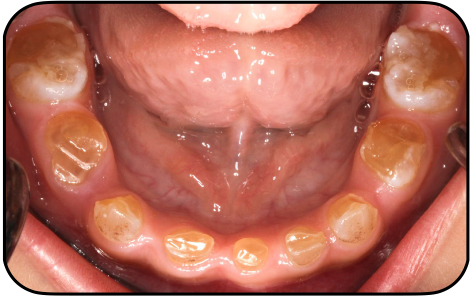
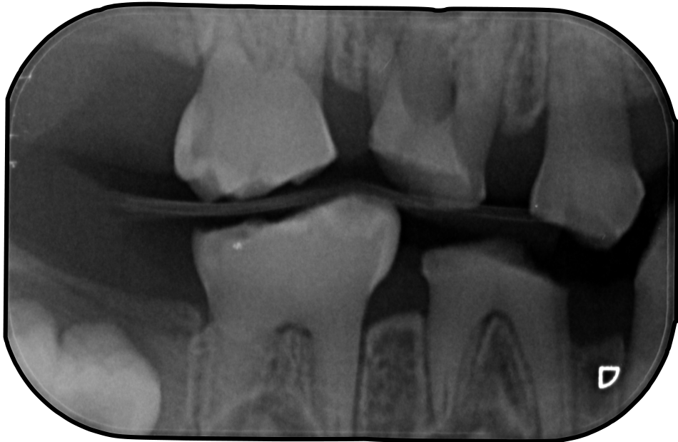
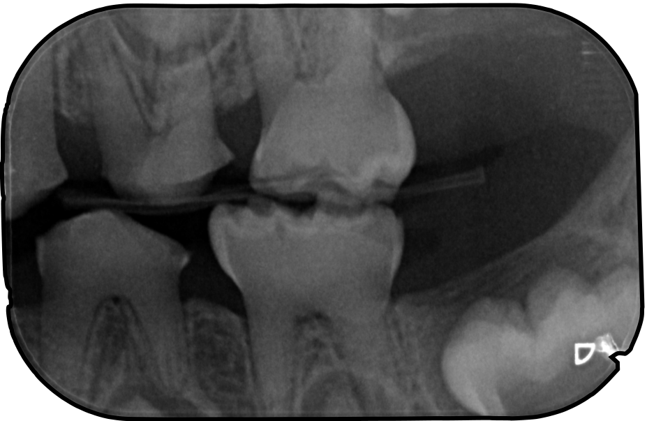
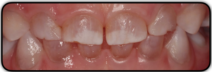
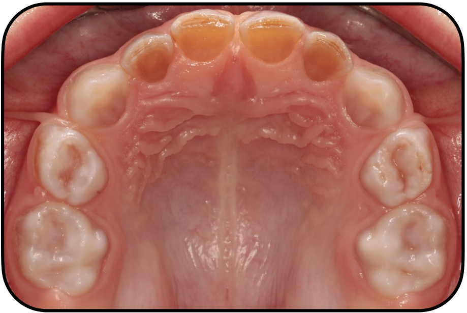
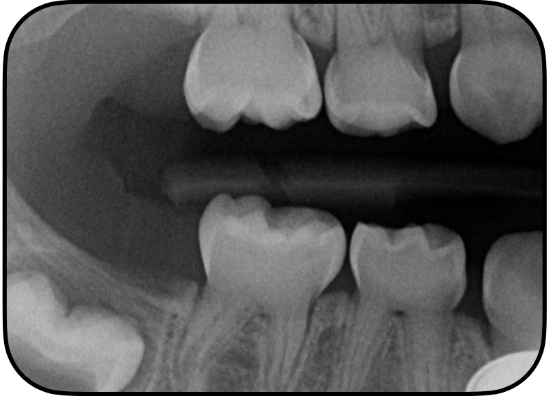
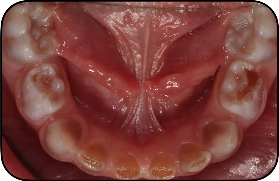
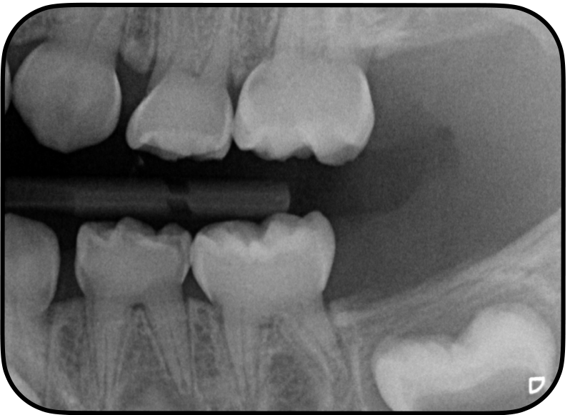
Case Images1819202122
Lecturer — Neonatal Radiographs
Images courtesy of Dr Maleeha Gilani
Family 3
Images courtesy of Dr Maleeha Gilani
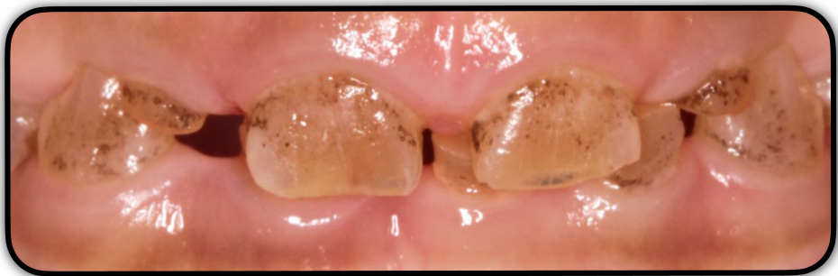
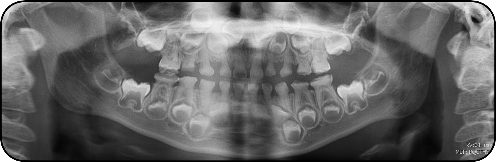
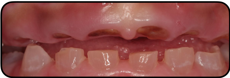
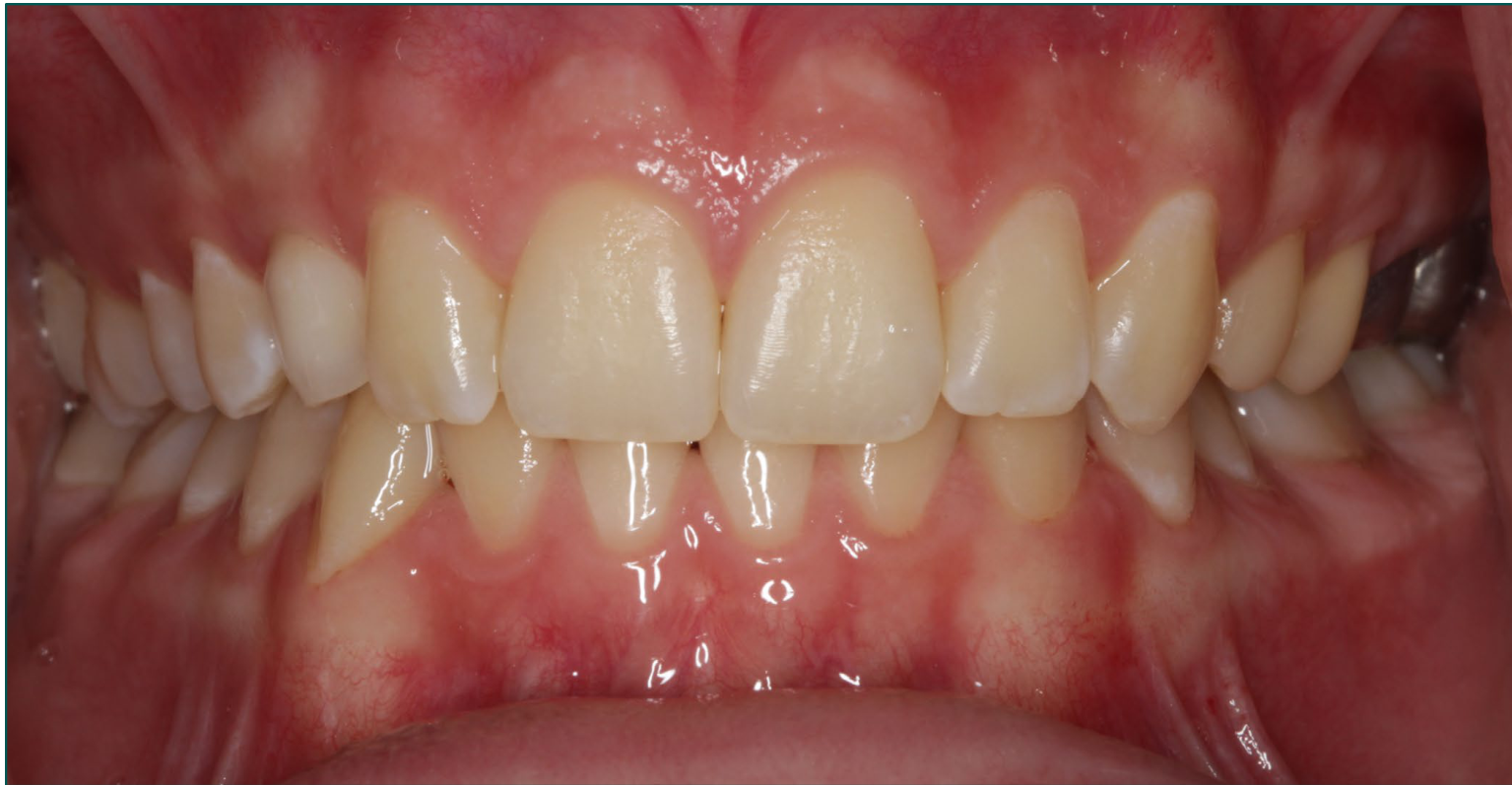
Family 4 — Twin 1
Images courtesy of Dr Maleeha Gilani
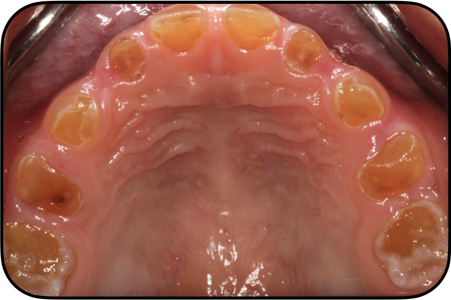
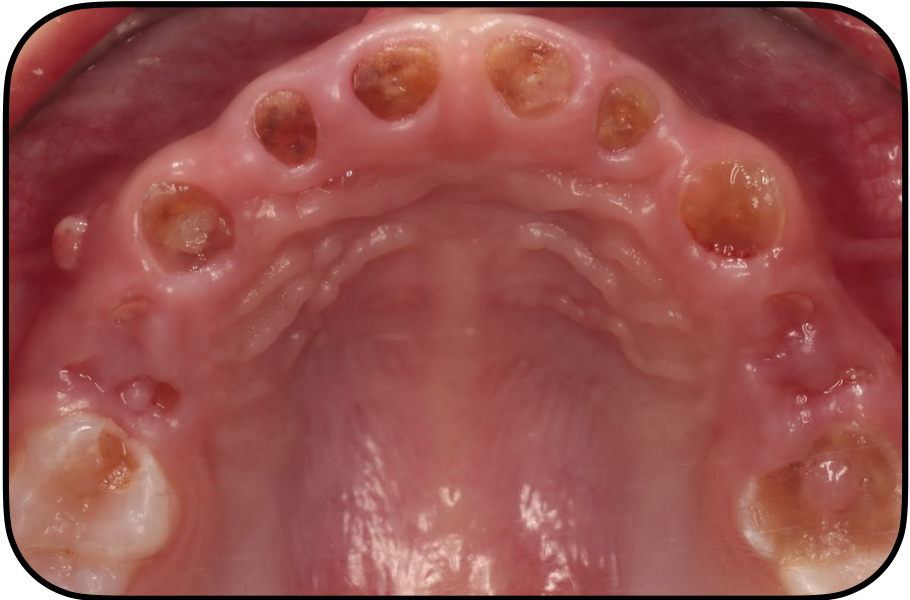
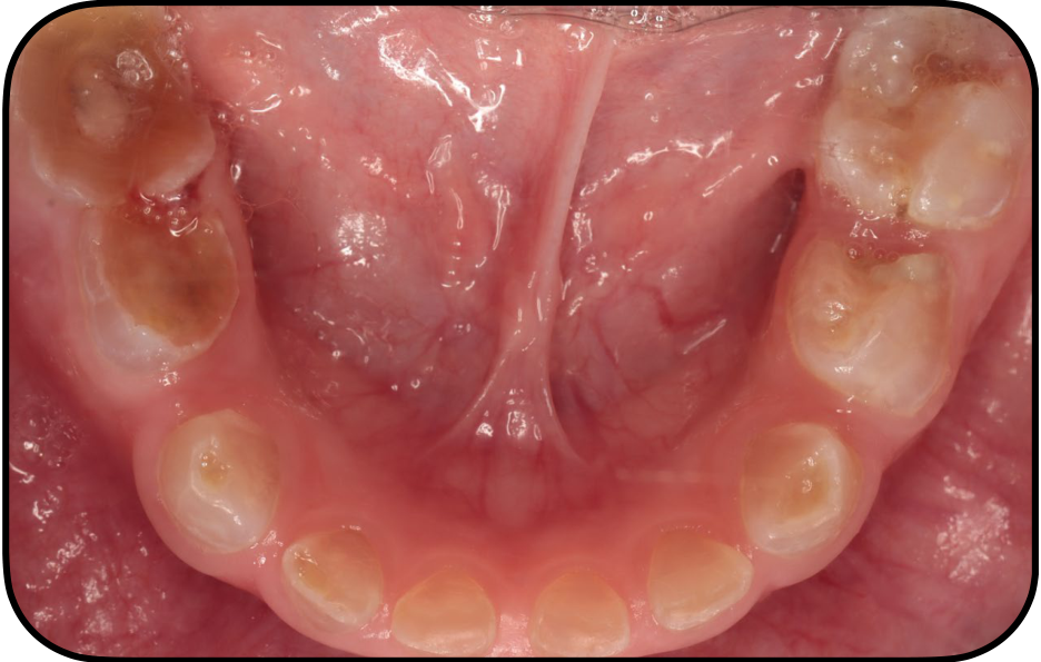
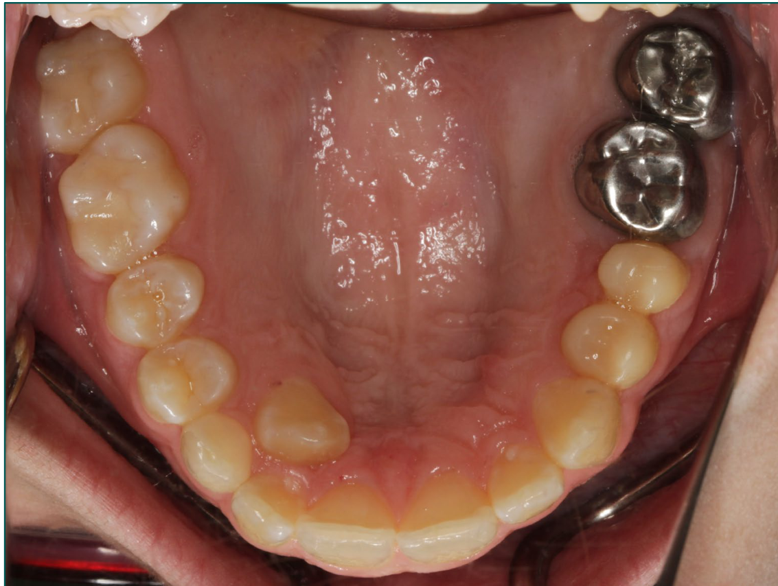
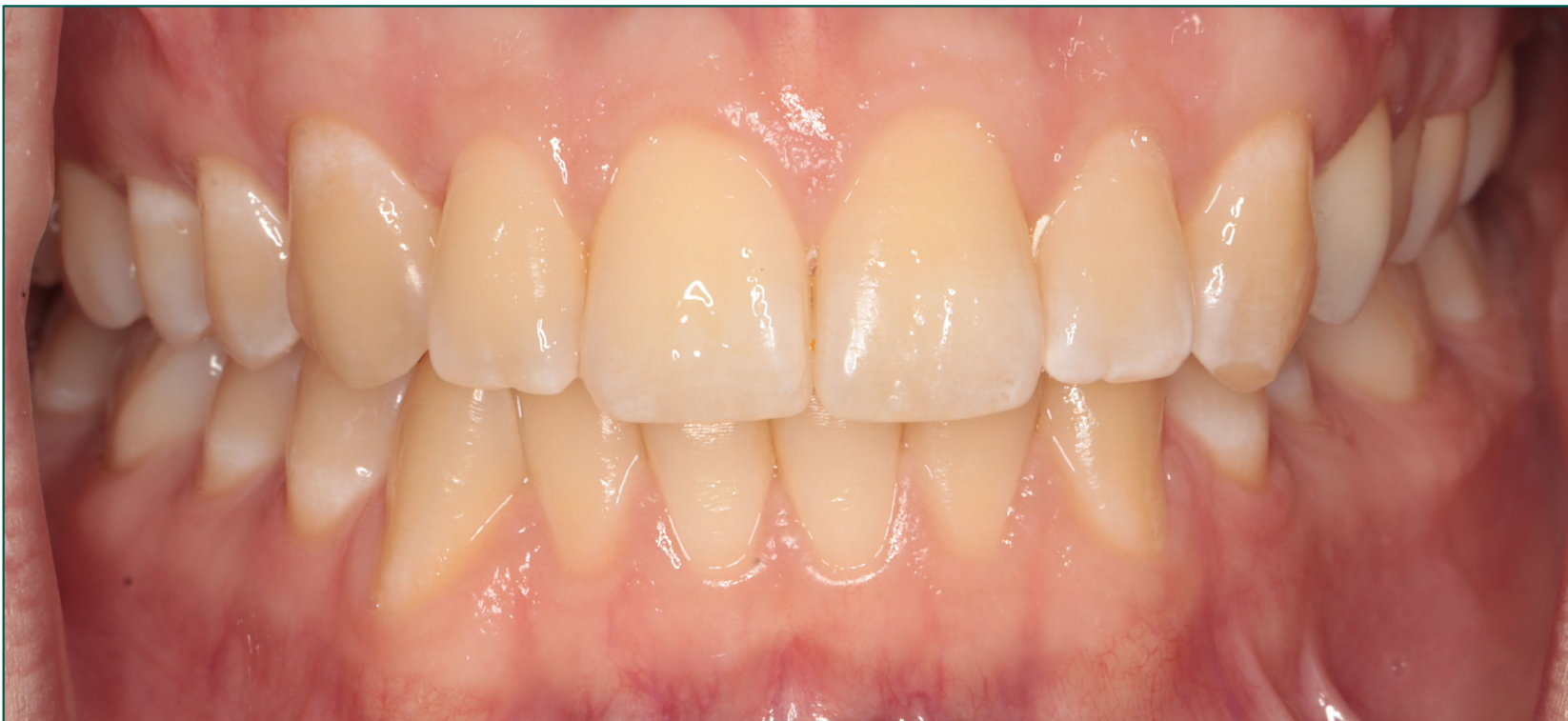
Family 4 — Twin 2
Family 4 Twins
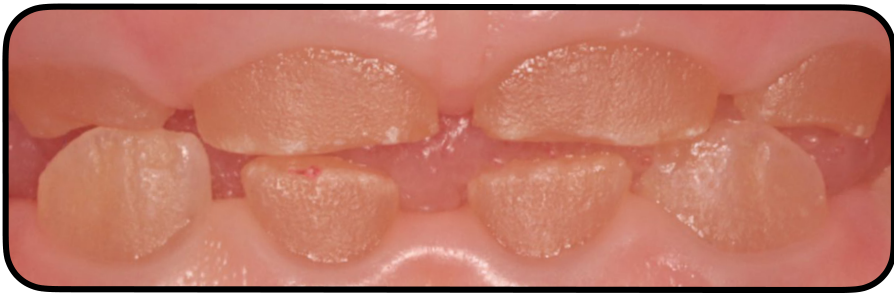
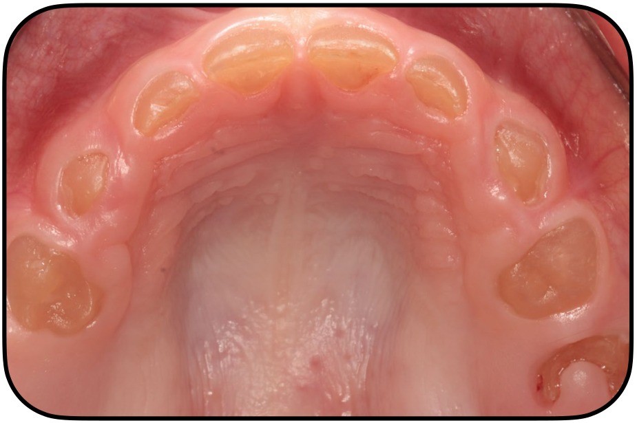
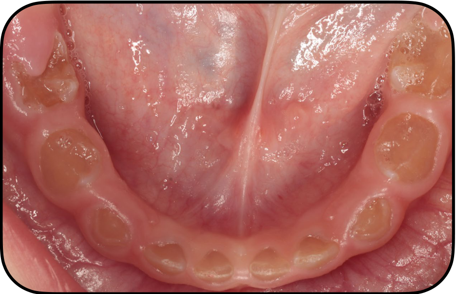
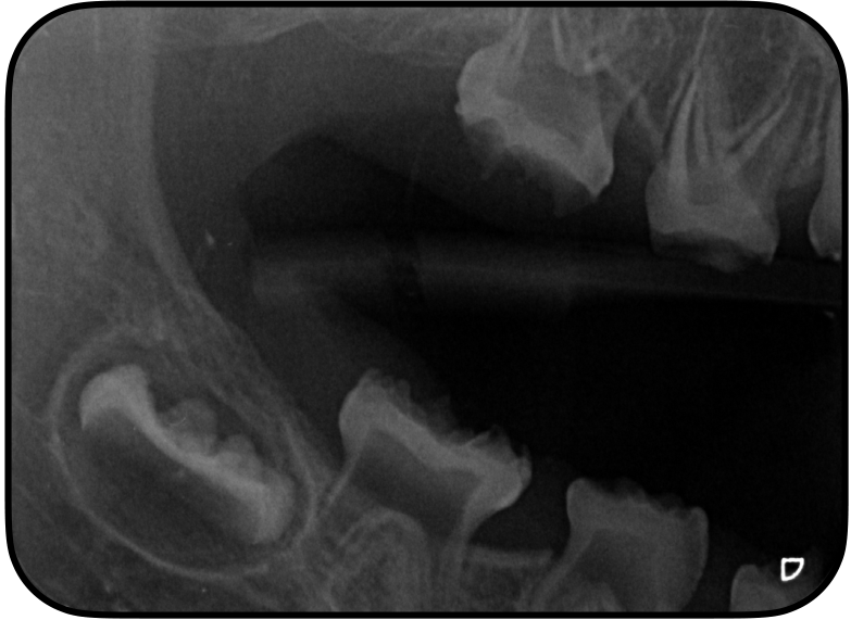
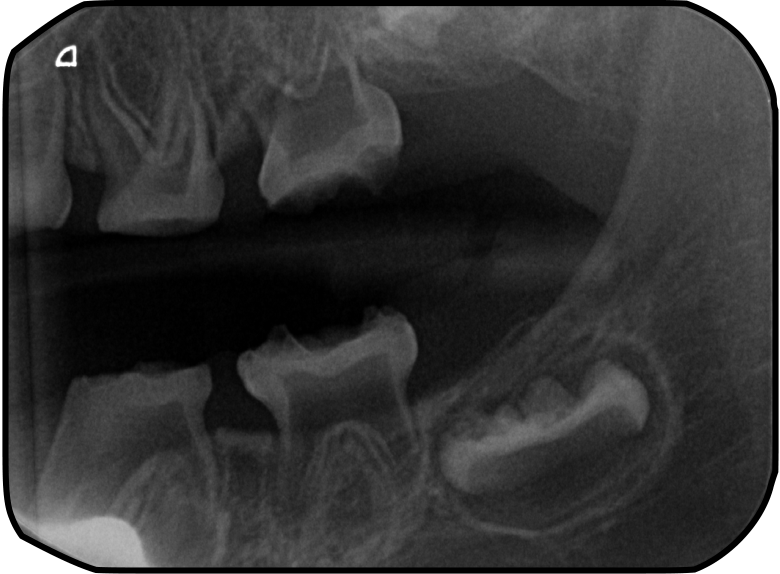
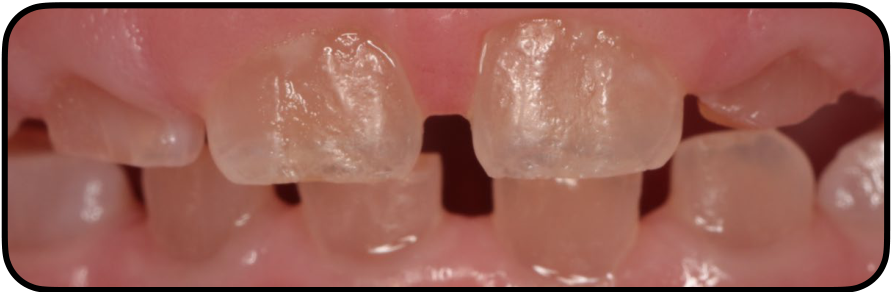
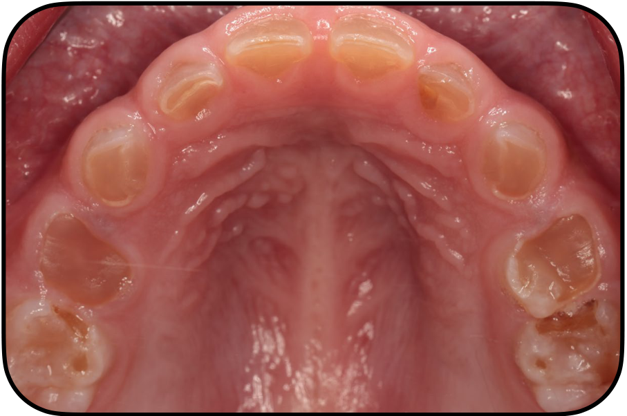
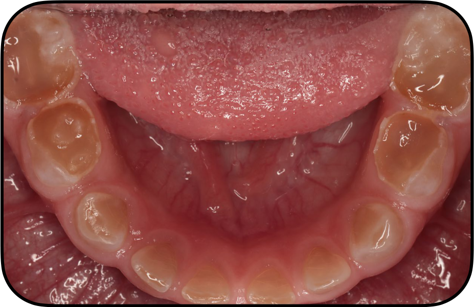
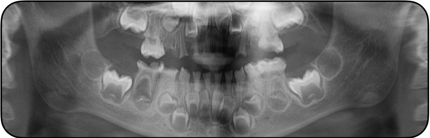
The affected twins were both classified as having moderate dentinogenesis imperfecta, but their severity differed. One child had significant attrition, amber discoloration throughout the dentition and substantial pulpal canal obliteration; her twin sister had more marked attrition, numerous abscesses and a history of severe pain.
Images courtesy of Dr Maleeha Gilani
Molecular Aetiology2324
Lecturer — Neonatal Differential Diagnosis
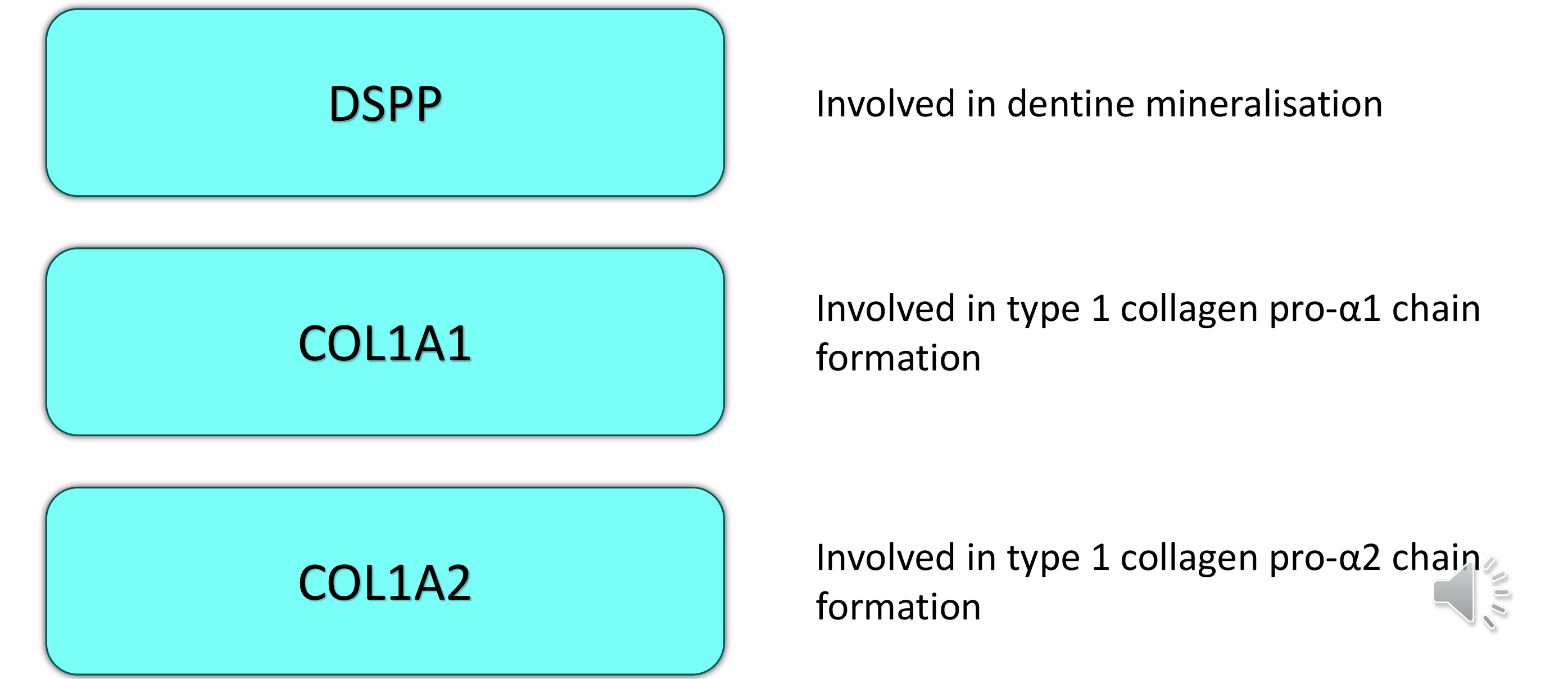
Implicated Genes25
- DSPP — involved in dentine mineralisation
- COL1A1 — involved in type 1 collagen pro-α1 chain formation
- COL1A2 — involved in type 1 collagen pro-α2 chain formation
Lecturer — Molecular Aetiology
Natal teeth commonly show structural differences from normally developed primary teeth.
- Enamel defects may include pitting, additional grooves, reduced thickness, yellow discoloration, and quantitative breakdown or loss of tooth structure.
- Enamel and dentine may be hypermineralized or lack normal histological structure; dentine may be irregular.
- The pulp may be larger and more vascularized, while the root is often poorly developed or absent because the tooth erupted before normal root development.
- Thin or defective enamel is more likely to break down, and a large pulp combined with defective hard tissues may increase the risk of infection, abscess formation, and further breakdown.
Approximately 91% of reported mutations associated with dentinogenesis imperfecta and dentine dysplasia are localised in DSPP. DSPP is expressed by odontoblasts and is required for dentine formation, whereas COL1A1 and COL1A2 encode type 1 collagen, the primary component of predentine.
DGI-I:
- Oral manifestation of osteogenesis imperfecta (OI)
- Mutations in genes COL1A1 and COL1A2
- Mild form of OI – risk of misdiagnosis
- DGI-I may be the only clinically apparent findings
DD-I:
- Pathogenesis largely unknown
Marini et al 2007
DGI-II, DGI-III and DD-II:
- Conditions are allelic and represent a single disease with variable expressions
- Mutations in the gene which encode DSPP
- DSPP – key non-collagenous proteins involved in tooth development and mineralisation
Barron et al 2008 de la Dure-Molla 2015 MacDougall et al 1999
Management2627
-
Dependent upon the clinical expression
- Standard “prescription” not possible
-
Multidisciplinary approach
-
Early identification critical
-
Preventative care and management of tooth sensitivity
-
Regular examinations allow teeth to be treated as they erupt, while meticulous oral hygiene improves periodontal health.
 |  |
Long Term Rehabilitation in Phases28
Lecturer — Natal Tooth Management
- Primary dentition
- Mixed dentition
- Permanent dentition
Primary Dentition29
-
Early stage
- ~18–20 months
- Treat and protect first primary molars and incisors
-
Second stage
- ~20–30 months
- Treat and protect primary canines and second primary molars
-
Treatment provided under general anaesthesia
Sapir & Shapira et al 2001
Images courtesy of Dr John Winters
Lecturer — Primary Rehabilitation
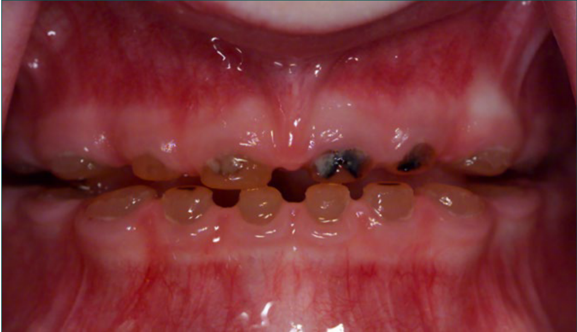
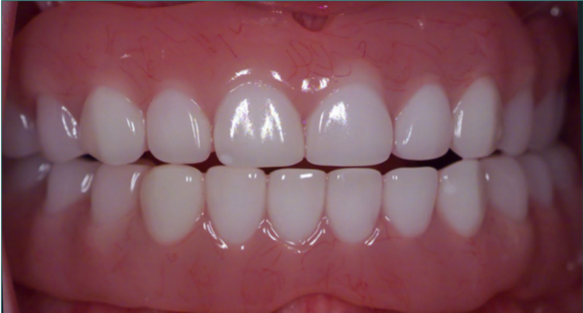
In severe tooth wear, overdentures may successfully re-establish function, but they require regular replacement as the child grows. One child who was very self-conscious about her teeth received overdentures and was described as very happy with the result.
Permanent Dentition30
- Tooth whitening has been used with varying success
- Long term evidence lacking on performance of adhesively bonded restorations
- Dental implants
- Endodontic treatment
- Often poor success due to partial or complete root canal obliteration
Lecturer — Adhesive Dentistry
When adhesive dentistry is used, enamel should be preserved where possible to improve bond strength. The lecturer recommended optimising the bond with air abrasion, using a universal adhesive, and reinforcing enamel and dentine with resin infiltration before restoration.
Hereditary Dentine Disorders Summary31
-
DGI type I = osteogenesis imperfecta
-
DGI type II, II and DD type II = single disease with variable expressions
-
DD type I = radicular dentin dysplasia
-
Early identification important
-
Multidisciplinary approach required for treatment
-
Eligible through Medicare Cleft Lip and Palate Scheme (PCH)
-
Further research required into genes responsible, and management options
Regional Odontodysplasia32
“Ghost Teeth”
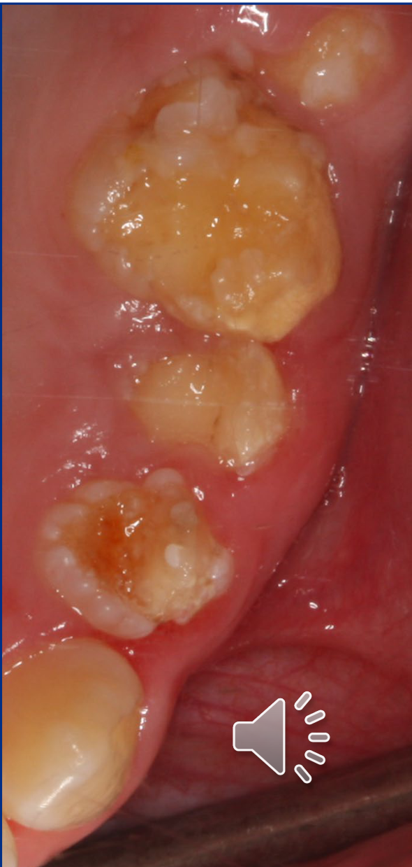
Regional Odontodysplasia Introduction33
- Localised non-inheritable developmental anomaly of dental tissues
- Affects both mesoderm and ectoderm tissue:
- enamel
- dentine
- pulp
- F > M
- No ethnic predilection
Lecturer — Tissue Comparison
Unlike amelogenesis imperfecta and dentinogenesis imperfecta, which predominantly affect one tissue type, regional odontodysplasia affects enamel, dentine and pulp. It therefore involves both mesodermal and ectodermal dental tissues.
Clinical Features34
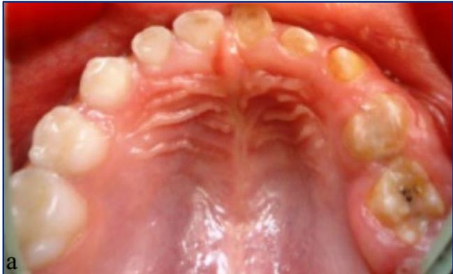
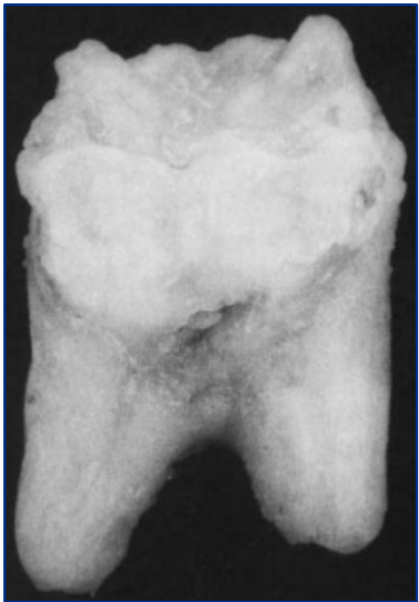
- Hypoplastic, small teeth
- surface pits and grooves
- yellow / brownish discolouration
- Eruption delay
- Maxilla > mandible
- Usually unilateral
- Abscesses in the absence of caries
Lecturer — Clinical Distribution
Natal teeth generally resemble primary teeth in size, shape, and morphology, but their development is often immature.
- Possible findings include enamel hypoplasia, dentine defects, a solitary tooth, two teeth occurring together, or a partially or completely erupted tooth.
- A tooth may be attached to the mucosa, very mobile, covered by an enamel shell, or show only its incisal edges; tissues may bulge over an unerupted tooth.
- The degree of development varies considerably, and a swelling in a newborn is not necessarily a natal tooth because other neonatal abnormalities may look similar.
The condition usually affects a particular segment of the dental arch, with the affected region commonly surrounded by teeth that appear normal. Because the enamel is defective and porous, bacteria can enter easily, so abscesses may occur frequently and soon after eruption.
 |  |
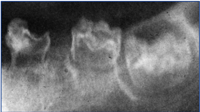
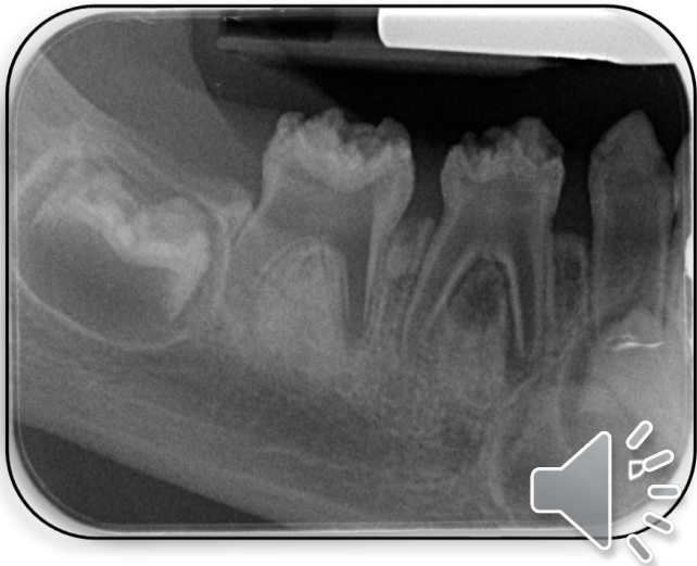
Radiographic Findings35
- Lack of contrast between thin enamel and dentine layers
- “Ghost-like” appearance
- Large pulp space
- pulp stones or denticles
 |  |
Histological Findings36
- Enamel is hypoplastic
- Hypocalcified areas and invaginations from the enamel surface into the dentin layer
- may result in bacterial contamination → pulpitis or necrosis
- Mixed areas of cellular, amorphous and interglobular dentin
- Bone not affected
Lecturer — Pulpal Complications
The routine use of radiographs in neonates should be questioned because the diagnostic value may be limited relative to the radiation exposure.
- A mandibular occlusal radiograph in a two-day-old child exposes the infant to radiation, and primary incisors may not yet be sufficiently calcified to show useful information about their relationship to developing permanent incisors.
- Radiographs may confirm the relationship to adjacent structures or whether a tooth is supernumerary, but these findings may not change management when removal is already required for ulceration or feeding problems.
- The ALARA principle should guide the decision; radiography may be more appropriate when the lesion does not clearly resemble a tooth or a foreign body is being considered.
The enamel-surface invaginations may allow bacterial contamination of the pulp. Pulpal contamination can lead to pulpitis, necrosis and abscess formation.
Aetiology37
Unknown – likely localised insult rather than inheritable condition:
- Local circulatory disorders
- Viral infections
- Local trauma
- Pharmacotherapy during pregnancy
- Facial asymmetry
- Combination of these factors
Treatment Options38
- Medicare Category 7 – Cleft Lip and Palate Scheme
- Treatment dependant on severity of defect
- Maintenance of teeth if possible
- Temporisation (GIC, composite, SSC)
- Hall crown technique
- Prosthodontic restoration
- Extraction
- Removable acrylic orthodontic appliance?
- Implant
- Autotransplantation
- Removable denture
Lecturer — Treatment Planning
Treatment may be staged as the child grows, beginning with temporisation and minimal-intervention techniques before eventual prosthodontic restoration. Because the enamel is thin and the pulp chambers are large, excessive tooth reduction should be avoided; a premolar from another quadrant may be suitable for autotransplantation because the remaining teeth are often normal.
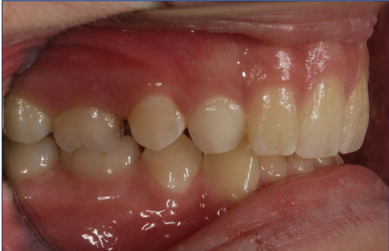
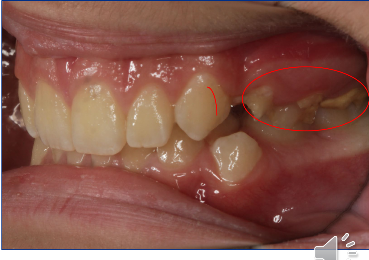
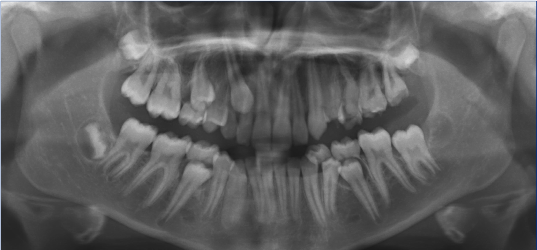
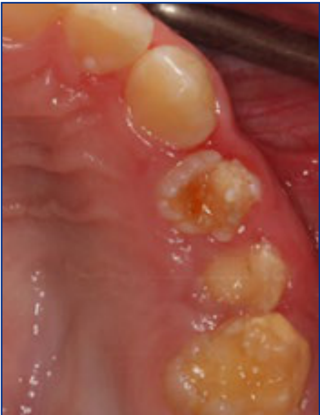
Patient 139404142434445
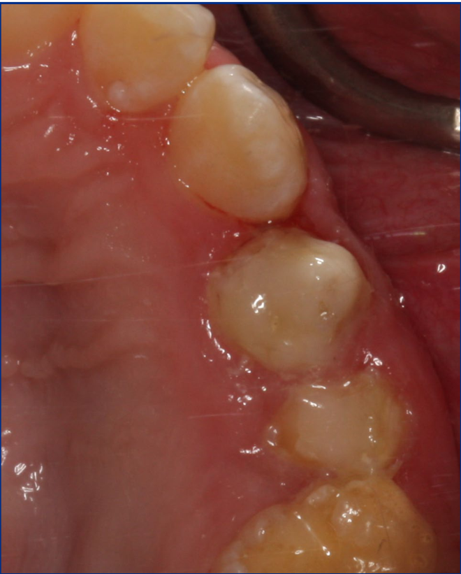
- 12-year-old girl
- Referred by private paediatric dentist for management of regional odontodysplasia
- No medical history of note
- Sensitivity to cold and toothbrushing – difficulties brushing Q2
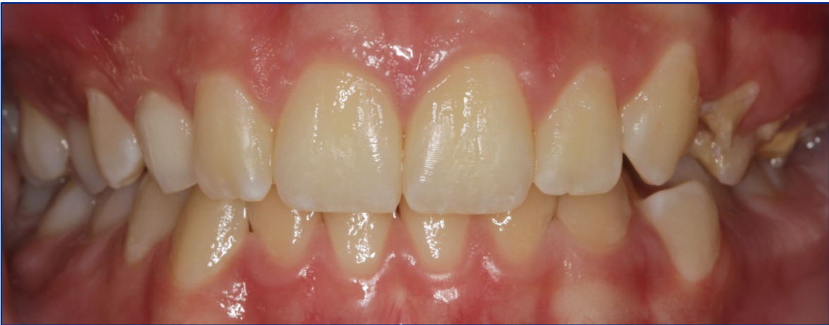
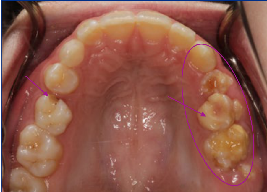
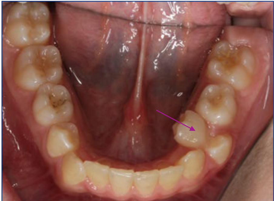
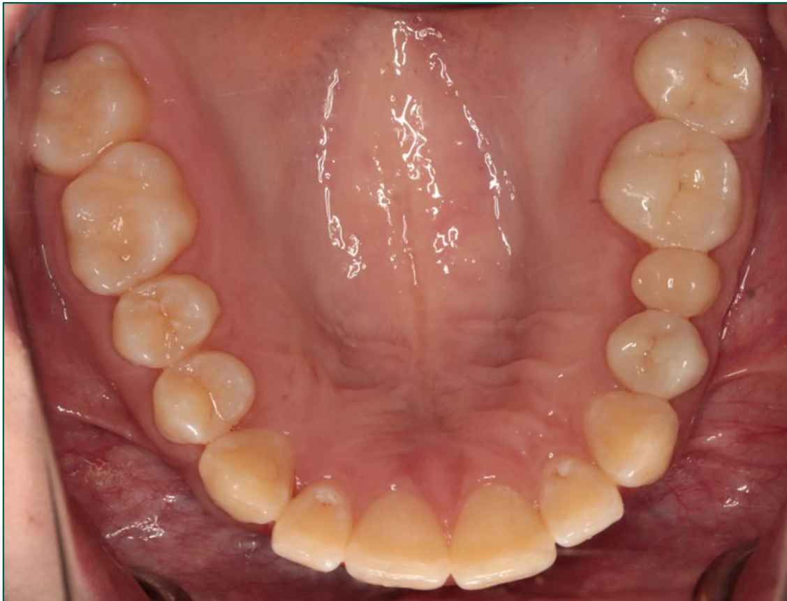
Intraoral findings (clinical photographs).
Radiographic findings (radiographs).
- Extraction of 65 and 74
- GIC sealant on 24 and 25
- Flowable composite sealant 23B
- Wait for further eruption of teeth
Patient 1 — Initial Findings
Detailed examination showed severe enamel hypoplasia affecting teeth 24 and 25, hyperplastic defects on the cusp tip of partially erupted tooth 26, plaque-induced gingivitis, an over-retained grade-two-mobile tooth 65, a carious over-retained tooth 74, and retained tooth 53 despite near-complete eruption of tooth 13. The premolars and molars in quadrant two were affected, while a small hypoplastic groove was present on the distobuccal surface of tooth 23.
- The panoramic radiograph showed defects in the premolars and molars of quadrant two and an ectopic tooth 13.
- The ectopic position of tooth 13 was considered incidental and unrelated to the regional odontodysplasia.
- Enamel, dentine and pulps in the remaining teeth appeared normal.
- Open surgical exposure of 13 (53 later extracted just prior to FFA)
- Composite crown 24 and 25 (3M bicuspid frasaco former)
- SSC 26 and 27
- Recalls during active ortho treatment
Patient 1 — Definitive Treatment
Once the teeth were stabilised and brushing was more comfortable, definitive treatment proceeded. Tooth 13 was surgically exposed using an open approach, and tooth 27 required exposure with diathermy to reduce the distal operculum.
- Composite crowns on teeth 24 and 25 provided complete tooth coverage, restored the vertical dimension and required minimal tooth removal.
- Tooth 53 was extracted when the orthodontist was ready to bring tooth 13 into the arch.
- The patient subsequently achieved a favourable orthodontic result.
- Monolithic zirconia crowns teeth 24, 26, 27
- Discharged from PCH to private dentist
- Aware 28 may require extraction or treatment
Patient 1 — Long-Term Management
At 19 years of age, tooth 24 appeared clinically sound, but its composite crown had an unsatisfactory distal margin, so crowning was recommended. Tooth 25 was relatively small with closed proximal contacts; because crown preparation would have required substantial tooth reduction and could have compromised it, the clinically and radiographically sound restoration was retained.
- The stainless steel crowns on teeth 26 and 27 were sound but were replaced with monolithic zirconia crowns for aesthetic reasons.
- The patient was very satisfied with the aesthetic result and continued regular maintenance with her general dentist after discharge from Perth Children’s Hospital.
Patient 246474849
- 3-year-old boy
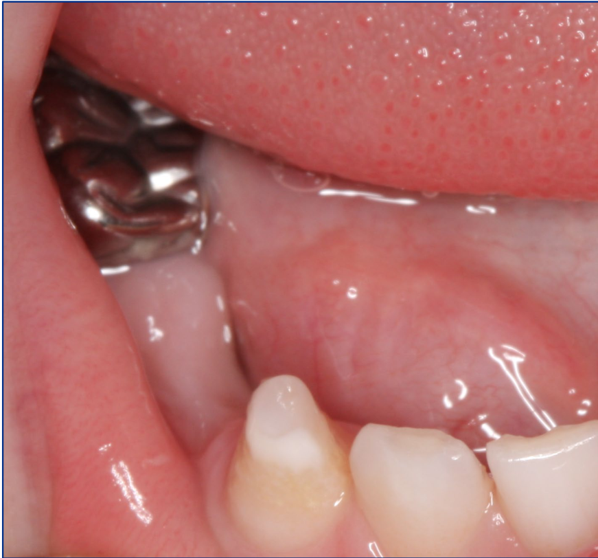
- Referred by community dentist for management of RHS facial swelling
- Medical history – multiple facial swellings in past month
- Prescribed 2 courses of antibiotics
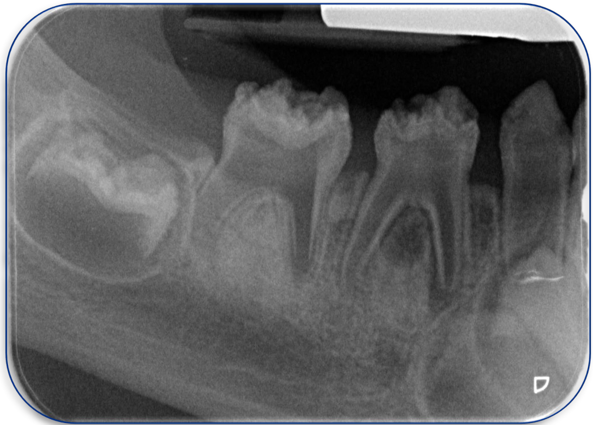
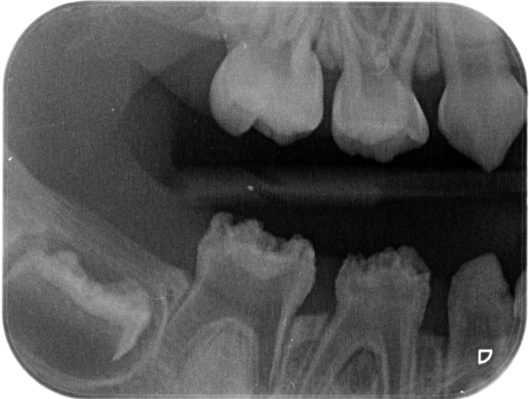
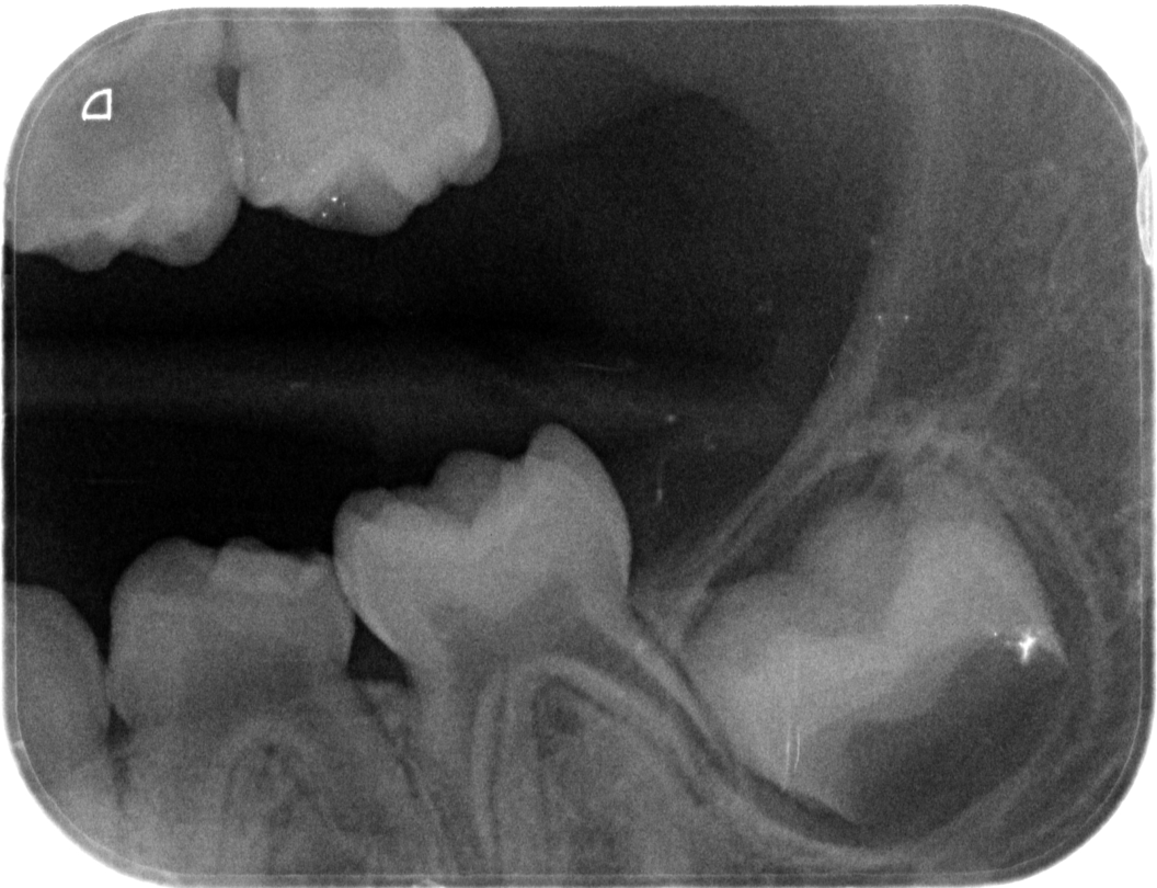
Radiographic findings (radiographs).
- Emergency treatment under general anaesthesia
- 84 extraction
- 85 SSC (Hall technique)
- Ongoing treatment
- 83 six monthly Duraphat applications
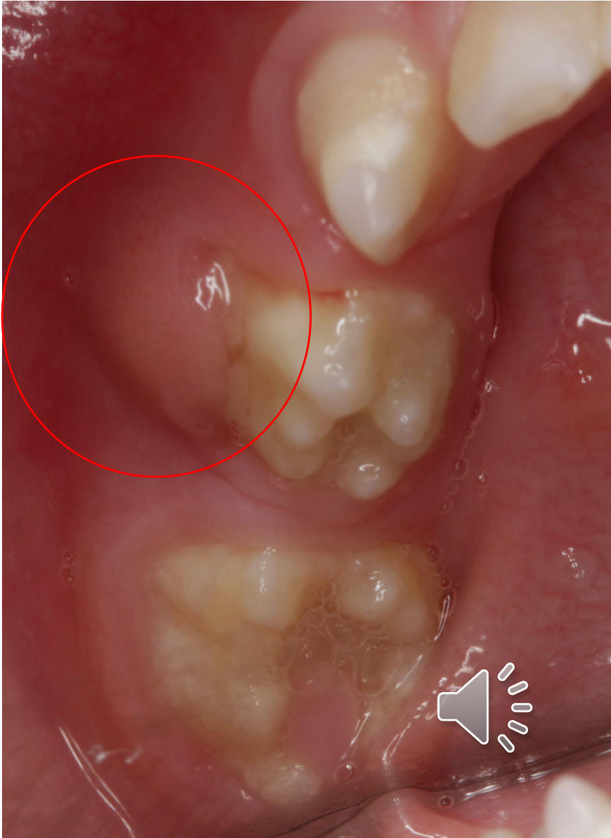
Patient 2 — Diagnostic Findings
Clinical examination showed a large buccal swelling and pus exudate from the lower first primary molar. Radiographs demonstrated hyperplastic enamel, thin dentine, very large pulp spaces and large pulp roots in the affected quadrant, with an obvious regional difference compared with the other quadrants.
- Unerupted tooth 46 also appeared hyperplastic compared with the contralateral tooth.
- Tooth 83 was impacted, with enamel on the incisal third and enamel hypoplasia in the more gingival area.
- Tooth 83 was smooth, easily cleansable and not sensitive, so no immediate restorative treatment was required.
- The parents were informed that tooth 46 also appeared affected and would require further treatment in the future.
Regional Odontodysplasia Summary50
- Localised non-inheritable developmental anomaly of dental tissues affecting adjacent teeth
- Affects both mesoderm and ectoderm tissue
- enamel
- dentine
- pulp
- Multidisciplinary care required
Clinical Cases
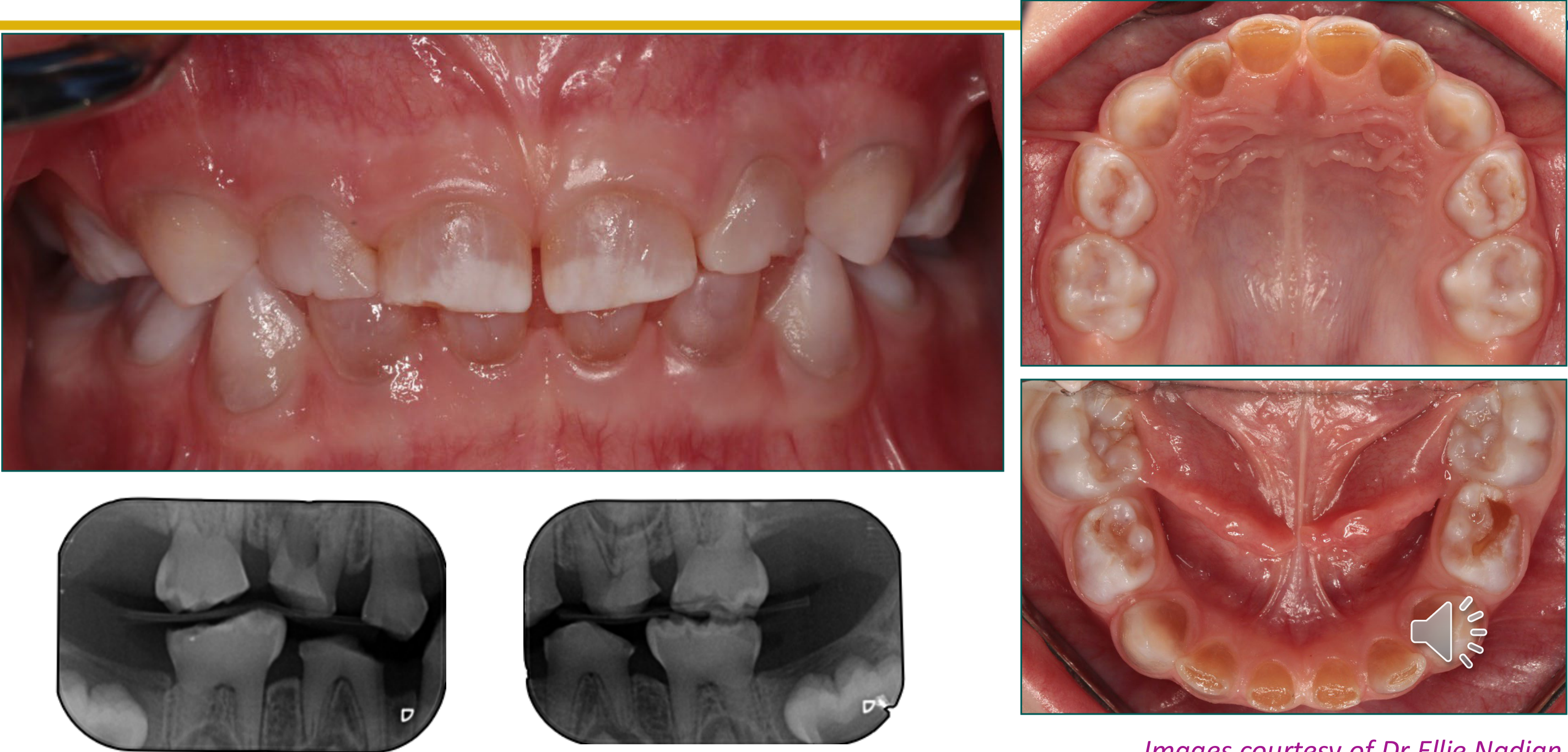
Case: Child from Family 1 with primary tooth attrition and discoloration
Question
Scenario: A child from the first of four families is presented as an example of the clinical and radiographic findings of a hereditary dentine disorder.
What’s shown: Intraoral and occlusal clinical photographs of the maxillary and mandibular dental arches, along with dental radiographs showing the anterior teeth and cross-sectional views.
Consider: Identify the clinical and radiographic features and determine the classification of the condition.
Answer
Observations:
- Significant attrition of the primary teeth.
- Dark amber discoloration of the dentition.
- Radiographs demonstrate complete pulpal obliteration with normal length roots.
Reasoning: The combination of primary tooth attrition, dark amber discoloration, and complete pulpal obliteration with normal root lengths is consistent with a moderate form of dentinogenesis imperfecta.
Outcome: Diagnosed as a moderate form of dentinogenesis imperfecta.
Takeaway: Moderate dentinogenesis imperfecta presents with primary tooth attrition, amber discoloration, and complete pulpal obliteration with normal root lengths.
Case: Child from Family 2 with opalescent teeth
Question
Scenario: A child from the second family is presented as an example of the clinical and radiographic findings of a hereditary dentine disorder.
What’s shown: Intraoral close-up and occlusal clinical photographs of the maxillary and mandibular dental arches, along with dental radiographs showing the crowns and roots.
Consider: Identify the clinical and radiographic features and determine the classification of the condition.
Answer
Observations:
- Opalescent teeth, less affected than the previous case.
- Radiographs show obliteration of the pulp chambers.
Reasoning: The opalescent appearance and pulpal obliteration, though milder than the first case, still indicate a milder form of moderate dentinogenesis imperfecta.
Outcome: Diagnosed as a milder form of moderate dentinogenesis imperfecta.
Takeaway: Moderate dentinogenesis imperfecta can present with varying degrees of severity, including milder opalescent discoloration and pulpal obliteration.
Case: Two-year-old child and mother from Family 3 with enlarged pulp chambers
Question
Scenario: An affected mother and son from the third family are presented to demonstrate a different radiographic presentation of a hereditary dentine disorder.
What’s shown: Close-up and intraoral occlusal clinical photographs of the primary anterior teeth and mandibular arch, along with a panoramic radiograph and posterior dental radiographs.
Consider: Identify the radiographic presentation and classify the condition based on the child’s age and findings.
Answer
Observations:
- Enlarged pulp chambers.
- Significant amounts of wear present in a two-year-old child.
Reasoning: The presence of enlarged pulp chambers with significant wear at a young age is characteristic of the severe form of dentinogenesis imperfecta.
Outcome: Diagnosed as a severe form of dentinogenesis imperfecta.
Takeaway: Severe dentinogenesis imperfecta is characterized by enlarged pulp chambers and marked attrition, often presenting in early childhood.
Case: First twin from Family 4 with amber discoloration and attrition
Question
Scenario: The first of a set of affected twins from the fourth family is presented as an example of a hereditary dentine disorder.
What’s shown: Close-up and intraoral clinical photographs of the upper, maxillary, and mandibular dental arches, along with a panoramic dental radiograph.
Consider: Identify the clinical and radiographic features and classify the condition.
Answer
Observations:
- Significant amounts of attrition.
- Amber discoloration of the entire dentition.
- Radiographs show significant pulpal canal obliteration.
Reasoning: The amber discoloration, significant attrition, and pulpal canal obliteration are consistent with moderate dentinogenesis imperfecta.
Outcome: Diagnosed as moderate dentinogenesis imperfecta.
Takeaway: Moderate dentinogenesis imperfecta can present with significant attrition, amber discoloration, and pulpal canal obliteration.
Case: Second twin from Family 4 with marked attrition and abscesses
Question
Scenario: The twin sister of the previous patient, also from the fourth family, is presented to show a different severity of the same hereditary dentine disorder.
What’s shown: Intraoral clinical photographs of the anterior dentition and maxillary arch, along with a panoramic dental radiograph.
Consider: Identify the clinical and radiographic features and classify the condition compared to her twin.
Answer
Observations:
- More marked attrition than her twin.
- Numerous abscesses.
- History of severe pain.
Reasoning: The more severe attrition, abscesses, and pain indicate a more severe presentation, but both twins are classified under the same condition of varying severity.
Outcome: Diagnosed as moderate dentinogenesis imperfecta of a more severe degree.
Takeaway: Dentinogenesis imperfecta can show varying degrees of severity even within the same family or between twins, with severe cases presenting with abscesses and pain.
Case: Child with severe tooth wear treated with overdentures
Question
Scenario: A young girl who is extremely self-conscious about her teeth after a teacher used her as an example in front of the class of what happens when one does not look after their teeth.
What’s shown: Clinical photographs of the child’s dentition before and after the insertion of a prosthodontic appliance.
Consider: What prosthodontic treatment was provided to address her severe tooth wear, and what specific considerations apply to this treatment in growing children?
Answer
Observations:
- The child presented with severe tooth wear.
- She experienced significant psychosocial impact, being extremely self-conscious about her appearance due to a teacher’s negative comments.
Reasoning: In severe cases of tooth wear, overdentures can be used successfully to re-establish function. However, when fabricated for children, they require regular replacement to accommodate the child’s ongoing growth.
Outcome: The child was treated with overdentures. Following their insertion, she was very happy with the result.
Takeaway: Overdentures can successfully re-establish function in children with severe tooth wear, but they must be regularly replaced as the child grows.
Case: 12-year-old girl with regional odontodysplasia in quadrant 2
Question
Scenario: A 12-year-old girl is referred to a children’s hospital by her private pediatric dentist for management of suspected regional odontodysplasia. She has no significant medical history or known pregnancy complications. Her main complaint is sensitivity to cold and difficulty brushing her teeth, particularly in the upper left quadrant.
What’s shown: Intraoral photographs and a panoramic radiograph (OPG) are presented.
Consider: What are the clinical and radiographic findings, and how would you manage this patient’s initial presentation and long-term restorative needs?
Answer
Observations:
- Intraoral photos show severe enamel defects (referred to as “hyperplasia” in the transcript) affecting the premolars and molars in quadrant 2, with a small defect on the distobuccal surface of tooth 23.
- Plaque-induced gingivitis is present due to brushing difficulties.
- An overretained, grade 2 mobile tooth 65 and a carious, mobile tooth 74 are present. Tooth 53 is retained while tooth 23 is nearly fully erupted.
- The OPG confirms the enamel and dentin defects in quadrant 2, with the remaining teeth appearing normal. It also shows an ectopic tooth 13, which is noted as an incidental finding.
Reasoning:
- The initial goal is to manage the presenting complaint of sensitivity and pain during brushing.
- Sealants are placed on teeth 24 and 25 to dome over them, which reduces plaque accumulation, prevents further enamel fracture, and reduces sensitivity by covering the dysplastic enamel.
- Teeth 65 and 74 are extracted, and a flowable composite sealant is placed in the buccal groove of tooth 23. The patient is placed on recall to allow for further eruption.
- Once stabilized, definitive treatment is planned. Due to the ectopic tooth 13, surgical exposure is performed under general anesthesia.
- Concurrently, composite crowns are placed on teeth 24 and 25 using formers to restore vertical dimension without significant tooth structure removal. Stainless steel crowns (SSCs) are placed on teeth 26 and 27, with tooth 27 requiring diathermy to reduce the distal operculum.
- During active orthodontic treatment, tooth 53 is extracted to allow tooth 13 to be brought into the arch.
- At age 19, final prosthodontic management is considered. Tooth 24 receives a monolithic zirconia crown because the distal margin of the composite crown is suboptimal. Tooth 25’s composite restoration is retained; as a microdont with closed proximal contacts, preparing it for a crown would remove too much tooth structure and compromise it long-term. Teeth 26 and 27 receive monolithic zirconia crowns to replace the SSCs for aesthetic purposes.
Outcome: The patient is extremely happy with her aesthetic results. She is aware that tooth 28 may also be affected upon eruption. She is discharged from the children’s hospital for regular maintenance with her general dentist.
Takeaway: Management of regional odontodysplasia requires a staged, multidisciplinary approach, starting with minimal intervention to manage sensitivity and prevent fracture, progressing to definitive restorations and prosthodontics as the patient grows, while preserving tooth structure where possible.
Case: 3-year-old boy with regional odontodysplasia and facial swelling
Question
Scenario: A 3-year-old boy is referred to a children’s hospital by a community dentist for a right-sided facial swelling. His mother reports multiple facial swellings over the past month, for which he received two courses of antibiotics from his GP.
What’s shown: A clinical intraoral photograph and dental radiographs of the affected quadrant, alongside comparison images of the contralateral side.
Consider: What do the clinical and radiographic findings indicate, and what is the appropriate immediate and ongoing management for this young patient?
Answer
Observations:
- The clinical photo shows a large buccal swelling with pus exudate associated with the lower right first primary molar (tooth 84).
- Radiographs of the affected quadrant show enamel defects (described as “hyperplastic enamel” in the transcript), thin dentine, very large pulp spaces, and short roots.
- Comparison with the contralateral side shows an obvious difference, and the unerupted tooth 46 also appears affected.
- Tooth 83 is impacted with an incisal third enamel defect, but the tooth is smooth, easily cleansable, and not sensitive.
Reasoning:
- The localized presentation of affected adjacent teeth with enamel, dentine, and pulp involvement confirms the diagnosis of regional odontodysplasia.
- The acute abscess associated with tooth 84 requires emergency management.
- Due to his young age, the extraction of tooth 84 is performed under general anesthesia.
- Tooth 85 is restored with a stainless steel crown using the Hall technique to minimize the removal of tooth structure.
- Tooth 83 does not require active intervention at this time due to the lack of symptoms and ease of cleaning, but it requires preventive management.
Outcome: The patient underwent emergency extraction of tooth 84 and placement of an SSC on tooth 85 under general anesthesia. Ongoing treatment includes six-monthly Duraphat applications to tooth 83 and regular recalls. The parents are counseled that the unerupted tooth 46 is affected and will require future treatment.
Takeaway: In young children with regional odontodysplasia presenting with acute abscesses, emergency extraction under general anesthesia may be necessary, followed by minimally invasive restorative techniques and preventive measures for affected but asymptomatic teeth.
Footnotes
-
Original PDF page 1: L8 Hereditary Dentine Disorders and Regional Odontodysplasia slides, p.1 ↩
-
Original PDF page 2: L8 Hereditary Dentine Disorders and Regional Odontodysplasia slides, p.2 ↩
-
Original PDF page 3: L8 Hereditary Dentine Disorders and Regional Odontodysplasia slides, p.3 ↩
-
Original PDF page 4: L8 Hereditary Dentine Disorders and Regional Odontodysplasia slides, p.4 ↩
-
Original PDF page 5: L8 Hereditary Dentine Disorders and Regional Odontodysplasia slides, p.5 ↩
-
Original PDF page 6: L8 Hereditary Dentine Disorders and Regional Odontodysplasia slides, p.6 ↩
-
Original PDF page 7: L8 Hereditary Dentine Disorders and Regional Odontodysplasia slides, p.7 ↩
-
Original PDF page 8: L8 Hereditary Dentine Disorders and Regional Odontodysplasia slides, p.8 ↩
-
Original PDF page 9: L8 Hereditary Dentine Disorders and Regional Odontodysplasia slides, p.9 ↩
-
Original PDF page 10: L8 Hereditary Dentine Disorders and Regional Odontodysplasia slides, p.10 ↩
-
Original PDF page 11: L8 Hereditary Dentine Disorders and Regional Odontodysplasia slides, p.11 ↩
-
Original PDF page 12: L8 Hereditary Dentine Disorders and Regional Odontodysplasia slides, p.12 ↩
-
Original PDF page 13: L8 Hereditary Dentine Disorders and Regional Odontodysplasia slides, p.13 ↩
-
Original PDF page 14: L8 Hereditary Dentine Disorders and Regional Odontodysplasia slides, p.14 ↩
-
Original PDF page 15: L8 Hereditary Dentine Disorders and Regional Odontodysplasia slides, p.15 ↩
-
Original PDF page 16: L8 Hereditary Dentine Disorders and Regional Odontodysplasia slides, p.16 ↩
-
Original PDF page 17: L8 Hereditary Dentine Disorders and Regional Odontodysplasia slides, p.17 ↩
-
Original PDF page 18: L8 Hereditary Dentine Disorders and Regional Odontodysplasia slides, p.18 ↩
-
Original PDF page 19: L8 Hereditary Dentine Disorders and Regional Odontodysplasia slides, p.19 ↩
-
Original PDF page 20: L8 Hereditary Dentine Disorders and Regional Odontodysplasia slides, p.20 ↩
-
Original PDF page 21: L8 Hereditary Dentine Disorders and Regional Odontodysplasia slides, p.21 ↩
-
Original PDF page 22: L8 Hereditary Dentine Disorders and Regional Odontodysplasia slides, p.22 ↩
-
Original PDF page 24: L8 Hereditary Dentine Disorders and Regional Odontodysplasia slides, p.24 ↩
-
Original PDF page 25: L8 Hereditary Dentine Disorders and Regional Odontodysplasia slides, p.25 ↩
-
Original PDF page 23: L8 Hereditary Dentine Disorders and Regional Odontodysplasia slides, p.23 ↩
-
Original PDF page 28: L8 Hereditary Dentine Disorders and Regional Odontodysplasia slides, p.28 ↩
-
Original PDF page 29: L8 Hereditary Dentine Disorders and Regional Odontodysplasia slides, p.29 ↩
-
Original PDF page 26: L8 Hereditary Dentine Disorders and Regional Odontodysplasia slides, p.26 ↩
-
Original PDF page 27: L8 Hereditary Dentine Disorders and Regional Odontodysplasia slides, p.27 ↩
-
Original PDF page 30: L8 Hereditary Dentine Disorders and Regional Odontodysplasia slides, p.30 ↩
-
Original PDF page 31: L8 Hereditary Dentine Disorders and Regional Odontodysplasia slides, p.31 ↩
-
Original PDF page 32: L8 Hereditary Dentine Disorders and Regional Odontodysplasia slides, p.32 ↩
-
Original PDF page 33: L8 Hereditary Dentine Disorders and Regional Odontodysplasia slides, p.33 ↩
-
Original PDF page 34: L8 Hereditary Dentine Disorders and Regional Odontodysplasia slides, p.34 ↩
-
Original PDF page 35: L8 Hereditary Dentine Disorders and Regional Odontodysplasia slides, p.35 ↩
-
Original PDF page 36: L8 Hereditary Dentine Disorders and Regional Odontodysplasia slides, p.36 ↩
-
Original PDF page 37: L8 Hereditary Dentine Disorders and Regional Odontodysplasia slides, p.37 ↩
-
Original PDF page 38: L8 Hereditary Dentine Disorders and Regional Odontodysplasia slides, p.38 ↩
-
Original PDF page 39: L8 Hereditary Dentine Disorders and Regional Odontodysplasia slides, p.39 ↩
-
Original PDF page 40: L8 Hereditary Dentine Disorders and Regional Odontodysplasia slides, p.40 ↩
-
Original PDF page 41: L8 Hereditary Dentine Disorders and Regional Odontodysplasia slides, p.41 ↩
-
Original PDF page 42: L8 Hereditary Dentine Disorders and Regional Odontodysplasia slides, p.42 ↩
-
Original PDF page 43: L8 Hereditary Dentine Disorders and Regional Odontodysplasia slides, p.43 ↩
-
Original PDF page 44: L8 Hereditary Dentine Disorders and Regional Odontodysplasia slides, p.44 ↩
-
Original PDF page 45: L8 Hereditary Dentine Disorders and Regional Odontodysplasia slides, p.45 ↩
-
Original PDF page 46: L8 Hereditary Dentine Disorders and Regional Odontodysplasia slides, p.46 ↩
-
Original PDF page 47: L8 Hereditary Dentine Disorders and Regional Odontodysplasia slides, p.47 ↩
-
Original PDF page 48: L8 Hereditary Dentine Disorders and Regional Odontodysplasia slides, p.48 ↩
-
Original PDF page 49: L8 Hereditary Dentine Disorders and Regional Odontodysplasia slides, p.49 ↩
-
Original PDF page 50: L8 Hereditary Dentine Disorders and Regional Odontodysplasia slides, p.50 ↩