Periodontal Conditions In Children
Lecturer — Periodontal Disease
Alveolar bone loss and premature loss of primary teeth are usually associated with chronic disease or syndromes, including immunological deficiencies.
- Periodontal disease may involve organisms such as A. actinomycetemcomitans, decreased host resistance to bacterial growth, neutropenia, or T-cell dysfunction.
- Inflammation is extremely common in children but is usually restricted to the gingiva, reversible, and not associated with attachment loss.
- Gingivitis and periodontitis are primarily initiated by plaque accumulation on the teeth and within the gingival crevices.
- Periodontitis is uncommon in children but may occur as a manifestation of systemic disease or as an aggressive molar-incisor pattern in the permanent dentition.
- Healthy gingiva may develop stippling from approximately two to three years of age in some children.
- Gingival papillae are generally flatter.
- The thinner, less keratinised epithelium makes the underlying vasculature more visible, so healthy gingiva may appear redder than adult gingiva.
- The junctional epithelium is more fragile, so probing around nearly erupted teeth is not recommended.
- The periodontal ligament is wider and more elastic, with fewer and less dense fibres, contributing to increased tooth mobility.
- Alveolar bone has a distinct but thin lamina dura, few trabeculae, large marrow spaces, and rich vascularisation.
- Cementum is relatively thin.
- Gingival inflammation in children is usually confined to the gingival tissues and does not involve attachment loss.
- Plaque accumulation is the primary cause. Periodontal disease in children
Dr Kelvin Jin BDS Dr Lee Lian Wong BDSc (Hons) UWA Dental School
The Healthy Periodontium In Children
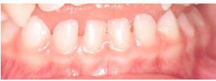
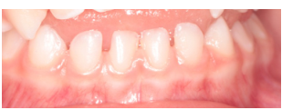
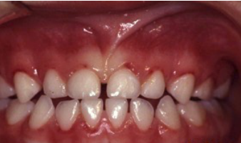
Bulky, Rounded Appearance1
- Bulky, rounded appearance
- Saddle-like interdental tissues in gaps
- Interdental papilla in proximal contacts
- Epithelium is thinner and less keratinised
Matsson. J Clin Periodontol 1978. Lindhe et al, Clinical periodontology and implant dentistry 5th Ed. 2008.
 |  |
 |
Structural Characteristics
- Thick junctional epithelium — resistant
- Shallow depth to junctional epithelium < 2 mm
- Wide, elastic PDL
- Porous and vascular alveolar bone
Might protect against attachment loss
Matsson. J Clin Periodontol 1978. Lindhe et al, Clinical periodontology and implant dentistry 5th Ed. 2008.
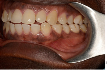
Physiological Pigmentation2
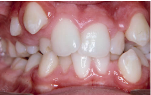
- Increased melanocyte activity that causes an increased amount of melanin within the epithelium
Lecturer — Physiological Pigmentation
Physiological pigmentation is normal anatomy and can occur in anyone, although it is more common in individuals with darker skin. Recognising it helps distinguish normal pigmentation from pigmentary disorders and more serious conditions such as malignant melanoma, which can also cause colour changes in the oral mucosa.
 |  |
 |
Gingivitis
Matson. J Clin Periodontol 1978 Bimstein et al J Dent Child 2002
Gingivitis In Children
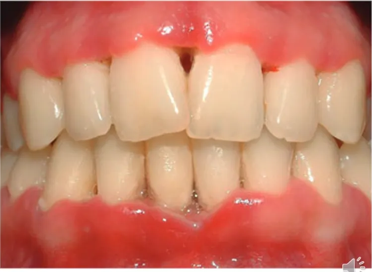
Plaque Induced Gingivitis
- Most common periodontal condition!
- Result of poor oral hygiene and plaque control
Lecturer — Plaque-Induced Gingivitis
Plaque-induced gingivitis is almost always found up to adolescence, and even short-term poor oral hygiene may cause inflammation.
- Colour change and swelling are more common clinical signs in children than bleeding on probing or increased pocket depths.
- In healthy children, the inflammation generally resolves and does not progress to attachment loss.
- Reversal may follow improved toothbrushing, flossing, and professional cleaning.
Key features
- Accumulation of plaque at gingival margin
- Colour change and swelling
- Red, puffy and shiny
- Reversible!
Eruption Gingivitis
- Transient inflammation in areas of erupting teeth
- Result of poor oral hygiene and plaque control due to tenderness
Key features
- Accumulation of plaque at gingival margin
- Colour change and swelling
- Red, puffy and shiny
- Subsides when the tooth erupts or exfoliates
Matson. J Clin Periodontol 1978 Bimstein et al J Dent Child 2002
Lecturer — Eruption Gingivitis
Eruption itself does not cause gingivitis; local plaque accumulation is the main cause when cleaning becomes difficult or uncomfortable around tender gingiva.
- Changes and weaknesses in the epithelium may increase permeability and bacterial accumulation.
- The long junctional epithelium around erupting teeth may create a pocket through which bacteria reach subgingivally.
- In the illustrated child, the eruption pattern suggested an age of approximately ten years, with cleaning difficulty and red, swollen gingiva along the cervical margins of the anterior teeth.
Local Factors
-
Plaque accumulation at the gingival margin
-
Poor oral hygiene
-
Erupting or exfoliating teeth
-
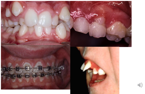
Malaligned teeth and crowding
-
Mouth breathing
-
Orthodontic appliances, enamel defects, rough enamel surfaces, unsatisfactory restorations, and carious lesions causing food impaction
Lecturer — Local Clinical Examples
Lack of lip closure and mouth breathing may dry the anterior gingiva and contribute to gingivitis.
- Examples included a poorly fitting buccal surface of a stainless-steel crown on tooth 85 and interproximal swelling mesial to tooth 62.
- The normal saddleback-shaped papilla was lost compared with the papilla between teeth 72 and 71.
Puberty Gingivitis
- Hormonal changes amplify gingival response to plaque
- Bacteria taking advantage of growth factors
Key features
- Minimal accumulation of plaque at gingival margin
- Colour change and swelling
- Red, puffy and shiny
- Subsides with adequate hygiene and normalising hormone levels
Matson. J Clin Periodontol 1978 Bimstein et al J Dent Child 2002
Lecturer — Puberty Gingivitis
Puberty gingivitis may appear as very red and oedematous gingiva despite small amounts of plaque. Increased levels of sex hormones can produce an exaggerated inflammatory reaction, while certain bacteria may use these hormones as growth factors.
Gingivitis in Children vs Adults
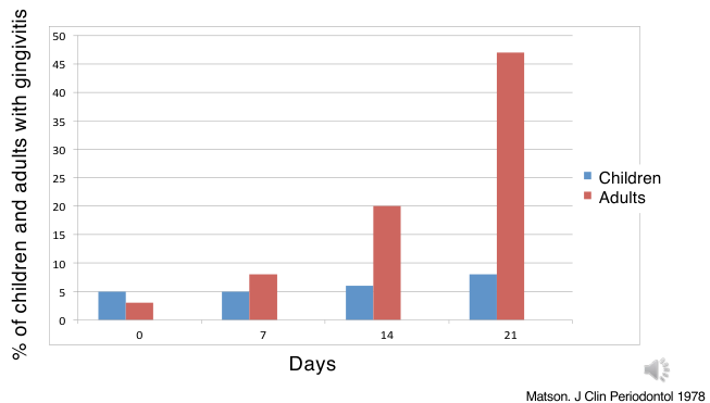
Percentage of children and adults with gingivitis measured over 21 days:
- Children show a markedly higher prevalence of gingivitis than adults across the 0–21 day observation period (y-axis: % of children and adults with gingivitis, 0–50; x-axis: days, 0–21)
Matson. J Clin Periodontol 1978
Lecturer — Children Versus Adults
Younger children are less susceptible to gingivitis than adolescents and adults. In Mattson’s study, plaque increased continuously in both groups during the 21-day period without tooth cleaning, but children showed a lower tendency to develop gingivitis than adults.
Age Differences In Gingival Response
Why Are There Age Differences?3
-
Differs in microbiology
-
Increased cell proliferation
-
Quick turnover of collagen
-
Altered immune response
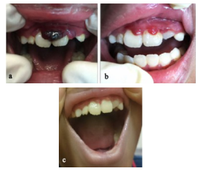
- T-lymphocytes — child
- B-lymphocytes — adult
-
Thicker junctional epithelium
-
Children may have lower proportions of some Fusobacterium and Lactobacillus species than adults
 |  |
 |
Poor Nutrition And Medications
- Vitamin deficiencies
- Vitamin C
- Medications:
- Anticonvulsants
- Blood pressure
- CCBs
- Typically involving interdental papillae
Lecturer — Nutrition and Medications
Severe nutritional deficiency is uncommon in developed countries, but severe vitamin C deficiency, or scurvy, can cause haemorrhagic and swollen gingiva.
- Cyclosporine and other immunosuppressants may cause gingival overgrowth, in addition to anticonvulsants and calcium-channel blockers.
- Medication-related enlargement is typically fibrous and nodular, and plaque exacerbates it.
Parental Responsibility to Brush
Bluey Tooth Brush Timer
Help parents help their children — give them a tool-box:
- Technique
- Tooth paste
- Tooth brush
- …any tricks
Lecturer — Parental Brushing Support
Parents should be educated and supported when children cannot yet brush effectively or consistently.
- Helpful approaches include timers, different toothbrushes, and toothpaste flavours that encourage toothbrushing.
Gingival Recession
Lecturer — Gingival Recession
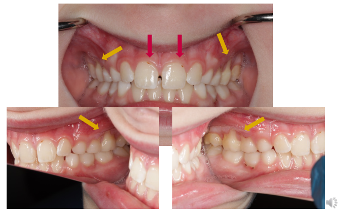
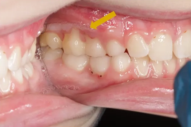
Localised gingival recession occurs in approximately 10–15% of adolescents.
- Possible causes include tooth position, traumatic toothbrushing, previous orthodontic treatment, and poor plaque control with heavy plaque or calculus deposits.
- Common sites are the labial surfaces of mandibular incisors in younger children and the buccal surfaces of upper molars and premolars in teenagers.
- Management includes a soft-bristled toothbrush, effective cleaning, correction of brushing technique, and discussion of orthodontic treatment where alignment contributes; the aim is to prevent progression while maintaining plaque removal.
Necrotising Periodontal Diseases
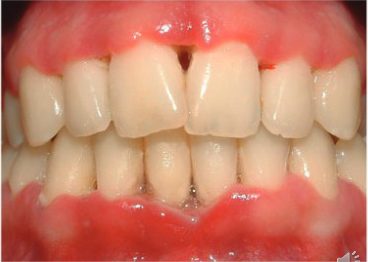
NUG/NUP/NUS — under the same disease but different stages.
- Ulceration of interdental papilla
- Painful
- Bleeding
- Fever, lymphadenopathy, malaise
Lecturer — Disease Distinctions
Necrotising ulcerative gingivitis, periodontitis and stomatitis are included within necrotising periodontal diseases, but they vary in the degree of tissue destruction.
- These diseases mainly affect the papillae and marginal gingiva, unlike primary herpetic gingivostomatitis, which affects a broader range of surfaces and has a viral pathogenesis.
- Necrotising periodontal disease may present similarly in children and adults.
Risk Factors For Necrotising Disease
Host And Environmental Factors4
- Necrotizing ulcerative disease may be observed among:
- Severe malnutrition
- Developed countries — young people (15–34 yrs)
- Developing countries — children
- Immunosuppressed patients
- Poor oral hygiene
- Smoking
- Insufficient sleep
- Psychological stress
Herrera et al 2014, Robinson et al. 1998
- Additional risk factors include inability to cope with stress, cancer therapy, and HIV infection.
Lecturer — Host Susceptibility
Both host susceptibility and environmental circumstances contribute to necrotising disease.
- Children with immunocompromise or severe nutritional problems require particular attention.
- If primary acute herpetic gingivostomatitis has been excluded, referral to a periodontist or paediatric dentist is advised because the condition may indicate a more serious underlying problem.
Treatment
- Gentle removal of plaque and necrotic debris (LA)
- Chlorhexidine 0.2% or hydrogen peroxide 3%
- Antibiotics — Metronidazole 400 mg, 3–5 days
- Analgesic
If too painful to perform oral hygiene:
- Hydrogen peroxide 3% solution, 5 ml mixed with 5 ml warm water; rinse 1 min twice a day until pain reduced
Therapeutic Guidelines Oral and Dental, version 3
- Referral should be considered in children with suspected necrotising periodontal disease.
Bone Loss in a Seven-Year-Old Child
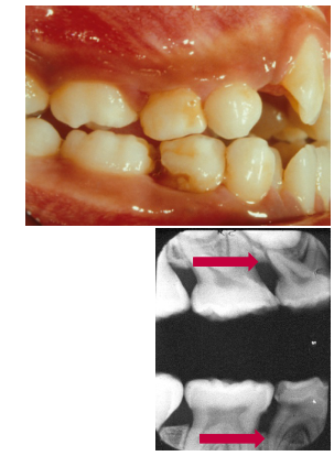
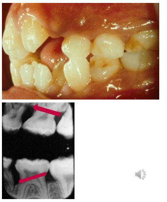
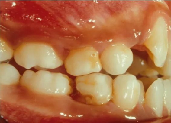
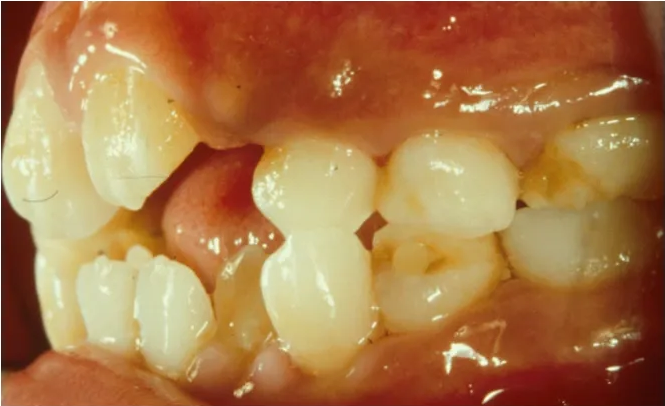
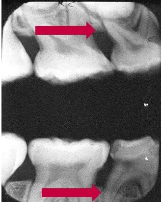
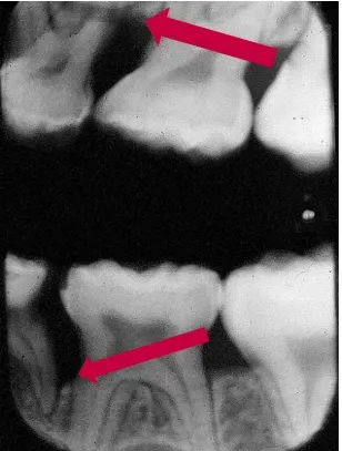
A seven-year-old child showed clear bone loss around the first primary molars. When bone loss is identified, possible causes include deep caries, a poorly contoured restoration, calculus that has not been removed, or an underlying medical condition or syndrome if local causes do not explain the finding.
Aggressive Periodontitis
Jasim M. Albandar & Eduardo M. R. Tinoco. Global epidemiology of periodontal diseases in children and young persons.
Lecturer — Periodontitis Diagnosis
Gingivitis is confined to soft tissues, whereas periodontitis extends deeper into the periodontium and destroys supporting structures, including alveolar bone. Diagnosis uses pocket depth, clinical attachment loss, marginal bone loss on radiographs, and bleeding on probing as an indicator of active disease.
- In younger children, probing depth measurements are less reliable because of primary tooth morphology and the eruption pattern of permanent teeth.
- Routine probing is therefore not usually useful until adolescence.
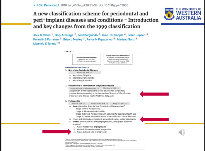
- The adult classification is also used in children: staging describes current severity, while grading estimates expected future behaviour.
- In children, periodontitis is often associated with systemic disease or an aggressive molar-incisor pattern in the permanent dentition, characterised by rapid progression with early bone and attachment loss.
Aggressive Periodontitis — Regional Prevalence5
- 0.4–0.8% in North America
- 0.3–1.0% in South America
- 0.1–0.5% in West Europe
- 0.5–5.0% in Africa
- 0.4–1.0% in Asia
Lecturer — Regional Prevalence
Population studies reviewed by Albandar and Tinoco in 2000 found that aggressive periodontitis in teenagers had a low prevalence of approximately 1%. Prevalence may differ between populations and regions.
- A particularly harmful strain of A. actinomycetemcomitans, known as JP2, was described as occurring mostly in patients of African origin.
 |  |  |
 |  |  |
 |
Prevalence
Primary Teeth
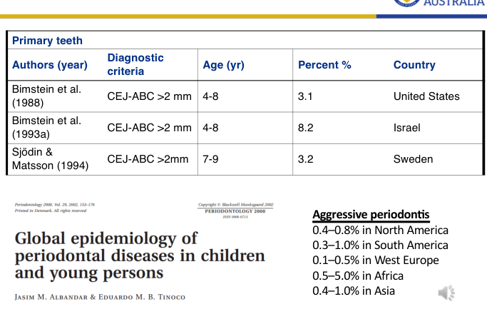
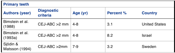
| Authors (year) | Diagnostic criteria | Age (yr) | Percent % | Country |
|---|---|---|---|---|
| Bimstein et al. (1988) | CEJ-ABC >2 mm | 4-8 | 3.1 | United States |
| Bimstein et al. (1993a) | CEJ-ABC >2 mm | 4-8 | 8.2 | Israel |
| Sjödin & Matsson (1994) | CEJ-ABC >2 mm | 7-9 | 3.2 | Sweden |
Lecturer — Primary Dentition
In younger children, bone loss often affects only one primary molar and may be associated with trauma or developmental factors. Children with this finding should be closely monitored.
- Periodontitis in the primary dentition should not be regarded only as a problem affecting primary teeth; it may indicate increased risk of disease in the permanent dentition.
Chronic Periodontitis — Regional Prevalence
- 2.0–5.0% in North America
- 4.0–8.0% in South America
- 1.0–3.0% in West Europe
- 10.0–20.0% in Africa
- 5.0–8.0% in Asia
Progressive Disease
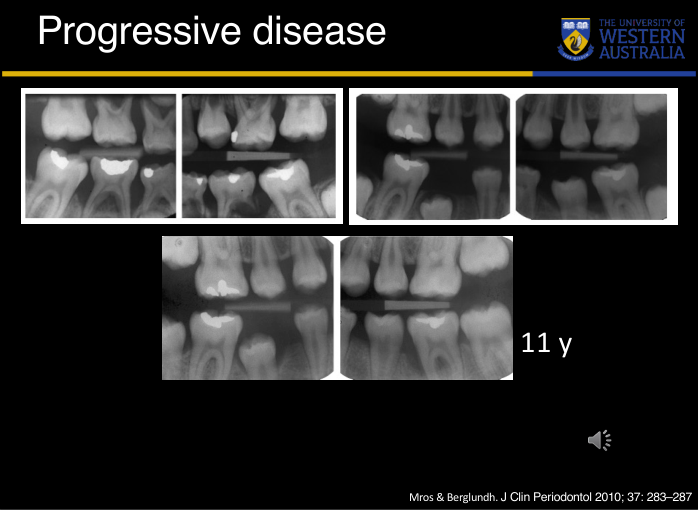
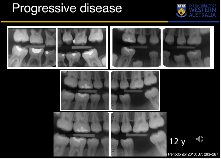
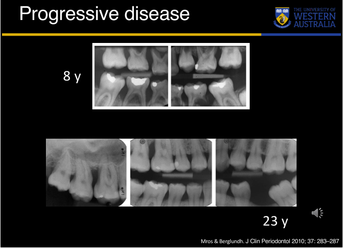
Disease Progression Over Time6
- 8 y
- 10 y
- 11 y
- 12 y
- 23 y
Mros & Berglundh. J Clin Periodontol 2010; 37: 283–287.
Lecturer — Disease Progression
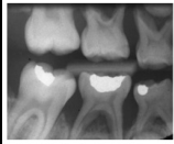
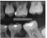
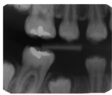
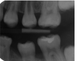
A bitewing study by Morass and Berlund followed one patient from eight to 23 years of age and demonstrated progressive periodontal destruction over time. At 11 years, bone loss was visible around tooth 36, and bone destruction around tooth 36 led to extraction at age 12.
- Bitewing radiographs in children should be examined not only for caries but also for alveolar bone levels.
- Small changes in the alveolar crest may be the first sign of periodontal risk.
 |  |  |
 |  |  |
Risk Factors Among Children
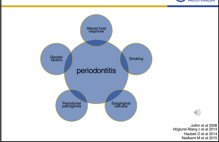
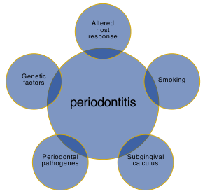
Risk factors contributing to periodontitis in children:
- Altered host response
- Smoking
- Subgingival calculus
- Periodontal pathogens
- Genetic factors
Julihn et al 2008
Höglund-Åberg C et al 2014
Haubek D et al 2014
Nadkarni M et al 2015
Lecturer — Risk Factors
Periodontitis in children and adolescents is multifactorial, with considerable variation in rate of progression, number of teeth involved, and clinical appearance.
- A. actinomycetemcomitans produces a toxin that damages polymorphonuclear leukocytes.
- Smoking impairs healing and alters the host response to infection.
- Periodontitis occurring in several family members suggests a hereditary component.
- The JP2 strain of A. actinomycetemcomitans is also an important risk factor.
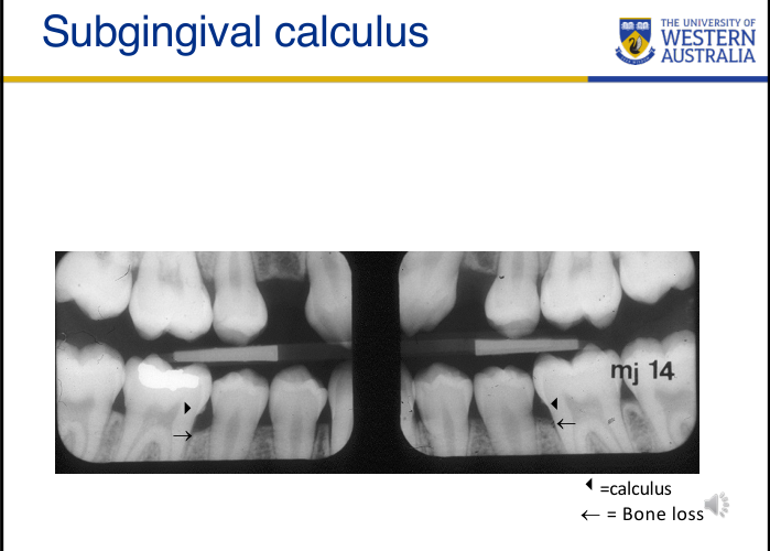
Subgingival Calculus
◀ = calculus
← = bone loss
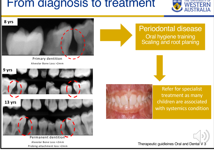
From Diagnosis to Treatment7
- 8 yrs — Primary dentition; alveolar bone loss > 2 mm
- 9 yrs
- 13 yrs — Permanent dentition; alveolar bone loss > 2 mm; probing attachment loss > 2 mm
Treatment pathway: periodontal disease → oral hygiene training, scaling and root planing → refer for specialist treatment, as many children are associated with systemic conditions.
 |  |
 |  |
Systemic Diseases and Conditions
- Blood disorders: Papillon–Lefèvre; Langerhans cell histiocytosis
- Metabolic disorders: Ehlers–Danlos
- Other conditions: Down syndrome; obesity; diabetes
Contributing factors again linked to periodontitis: altered host response, smoking, subgingival calculus, periodontal pathogens, and genetic factors.
Lecturer — Assessment and Management
Periodontal assessment should form part of the regular oral examination. From approximately 13 years of age, partial probing should focus on proximal sites around first permanent molars and incisors; in younger children, probing is indicated when disease is suspected.
- Bitewing radiographs should show the alveolar crest clearly, with vertical bitewings considered when bone levels cannot be seen.
- Initial therapy includes oral hygiene instruction, professional scaling, and root planing to remove supra- and subgingival calculus.
- Treatment outcomes should be reviewed after four to six weeks.
- Management of aggressive periodontal disease requires accurate diagnosis, a thorough medical and dental history, and collaboration with the patient’s physician.
Genetic And Systemic Conditions
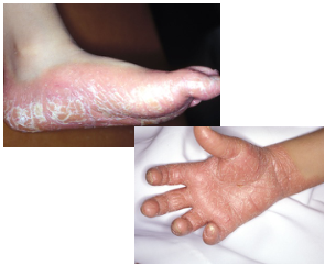
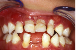
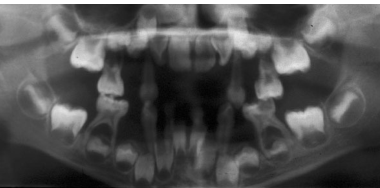
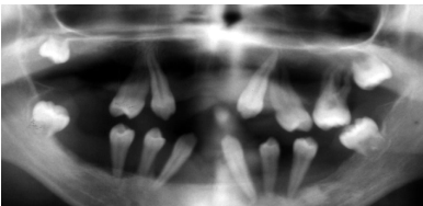
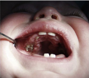
Papillon Lefevre Syndrome
- Autosomal recessive condition
- Hyperkeratosis of palms and feet
- Periodontitis — early loss of primary teeth
- A. actinomycetemcomitans (A.a.)
Ullbro et al 2004, Khocht et al 2014
Management of PLS8
- Intensive periodontal therapy
- Metronidazole and chlorhexidine
- Extractions to prevent bone loss
Lecturer — PLS Management
Treatment has not been shown to be consistently successful, so it should begin as early as possible, including during the primary dentition.
- Parents have an important role in maintaining good oral hygiene.
- Periodontally damaged primary teeth should be extracted before eruption of the first permanent molars to create a more favourable environment for the permanent teeth.
- In the permanent dentition, teeth with severe bone loss are usually extracted, while teeth with less severe bone loss may be managed with scaling, prophylactic measures and antibiotics.
- Extraction may help remove diseased teeth, preserve bone and maintain the alveolar crest.
 |  |  |
 |  |  |
 |  |  |
Langerhans Cell Histiocytosis
- Rare condition
Dental presentation
- Premature loss of primary molars
- “Floating in air”
- All 4 quadrants involved
- Primary molars, possible canine
- Incisors — rarely involved
Management
- Excision and curettage of oral lesions
- Extractions
- Multiagent chemotherapy
Merglová et al. Journal of Cranio-Maxillo-Facial Surgery 42 (2014) 93e10
Lecturer — Langerhans Management
Treatment is challenging, and early recognition and intervention are important because periodontal breakdown may be rapid and extensive.
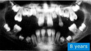
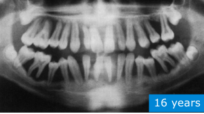
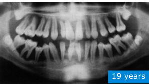
Congenital Neutropenia
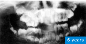
Figure: 16 years
Congenital Neutropenia Case
Congenital neutropenia is a primary immunodeficiency involving low levels of circulating neutrophils, with susceptibility to severe bacterial infections.
- Recurrent oral ulceration may be an early clinical sign.
- Periodontal involvement may begin as soon as teeth erupt, affecting both primary and permanent dentitions.
- Clinical features include severe gingivitis, increased tooth mobility and widespread alveolar bone destruction.
- Changes were already visible on an OPG at six years of age; by 16 years, severe bone loss affected the entire dentition.
- Management requires early prevention, ongoing dental care and close collaboration with the medical team.
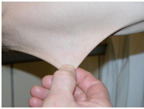
Ehlers Danlos Syndrome
Mitakides & Tinkle, 2017 Bowen et al 2017 Malfait et al 2017
Lecturer — Ehlers-Danlos Syndrome
Ehlers-Danlos syndrome is an inherited connective tissue disorder involving defects in collagen synthesis.
- It may present with joint hypermobility and hyperextensible skin.
- Gingival fragility and a predisposition to severe periodontal bone loss may also occur.
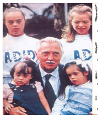
Down Syndrome
The most common recognizable genetic syndrome associated with immune defects
Lecturer — Down Syndrome
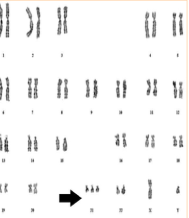
Down syndrome is the most common chromosomal abnormality and is caused by an extra chromosome 21.
- Associated features include intellectual disability, congenital heart disease and impaired immunity.
- These patients have a strong susceptibility to periodontal disease.
Cruz NV et al 2009
Periodontitis in Down Syndrome9
- Prevalence of periodontitis = 30%
- Early onset
- Increases with age
- More frequent in lower central incisors
 |  |  |
 |  | 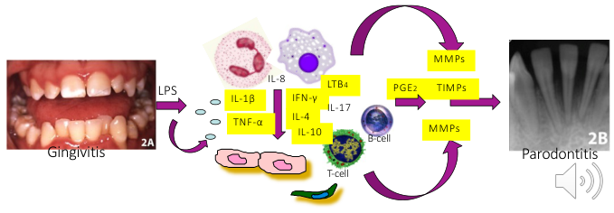 |
Altered Immune Response
- Weaker innate response
- Altered adaptive response
Lecturer — Down Syndrome Immunity
The altered immune response increases susceptibility to periodontal disease.
- The weaker innate response involves neutrophils and monocytes.
- The altered adaptive response contributes to increased infection risk and abnormal inflammatory responses.
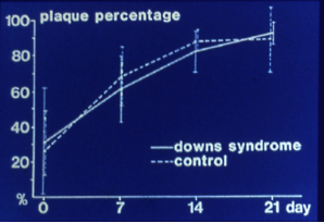
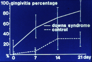
Experimental Gingivitis Around Deciduous Teeth in Children with Down’s Syndrome
Figure: plaque percentage vs gingivitis percentage over days 0, 7, 14, 21 — Down syndrome vs control (Reuland-Bosma et al 1986)
Lecturer — Experimental Gingivitis
In research by Rulin Bosmar in 1986, gingival inflammation developed earlier and was more extensive in children with Down syndrome, even when plaque levels were comparable with those of other children.
Clinical Implications
-
Inform parents with DS children about differences in immuno-response and the increased risk of periodontal disease
-
Prevention on regular basis will reduce plaque, gingivitis and pocket depth (Cheng RHW et al 2008)
-
Early and regular preventive treatment is important even before clinical signs appear.
-
Individualised ongoing prophylaxis may slow disease progression.
-
Regular periodontal treatment involving scaling and chlorhexidine can reduce plaque, gingivitis and pocket depth. Figure: inflammatory pathway — LPS → Gingivitis → Parodontitis, involving mediators IL-1β, TNF-α, IFN-γ, IL-4, LTB4, PGE2, IL-10, IL-8, IL-17, MMPs, TIMPs, and T-cell / B-cell involvement.
Obesity
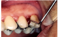
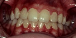
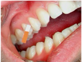
- Enhanced risk for exhibiting pathological periodontal pockets
- Enhanced level of cytokines
- Increased plaque accumulation and bleeding on probing
- Increased levels of periodontal pathogens
Owens S et al 2014; Modeer T et al 2011; Zeigler et al 2012; de Castilhos et al 2012.
Lecturer — Obesity and Periodontitis
Childhood and adolescent obesity is associated with multiple health risks, including an increased likelihood of adult obesity; the lecture gave a current prevalence of approximately 20%.
- Only a small number of studies have examined obesity and periodontal disease in children.
- The increased risk of pathological periodontal pockets in obese adolescents is not explained solely by poor oral hygiene.
- Adipose tissue produces cytokines that may amplify the inflammatory response to periodontal pathogens.
- Higher levels of pathogens such as Porphyromonas gingivalis and A. actinomycetemcomitans may be present.
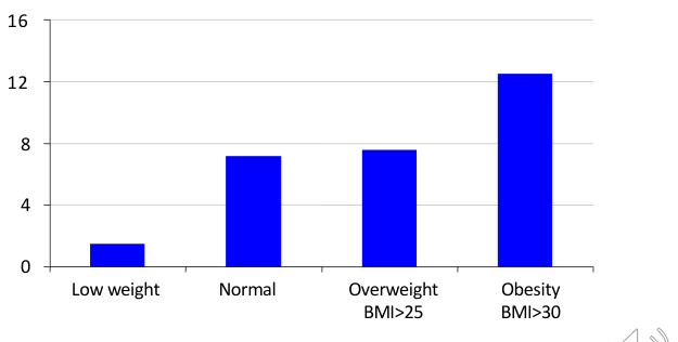
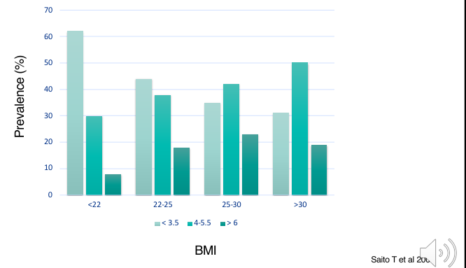
Prevalence Data10
Chart: Prevalence (%) by weight category — Low weight, Normal, Overweight (BMI>25), Obesity (BMI>30); axis values 0, 4, 8, 12, 16.
Al-Zahrani MS et al 2003.
Chart: Prevalence (%) by BMI — categories <22, 22-25, 25-30, >30; axis values 0-70; additional groupings < 3.5, 4-5.5, > 6.
Saito T et al 2001.
- Al Zahrani and colleagues found a significant association between body fat and periodontal disease in younger adults, but not in middle-aged or older adults.
- The BMI chart showed that higher BMI was associated with an increase in pockets deeper than 4 mm.
 |  |  |
 |  |  |
Rationale for Treatment
-
Prevention
-
Information
-
Help parents to help their children
-
Regular re-calls
-
Monitor periodontal status and intervene early if problems develop.
References And Further Reading11
 |  |
 |
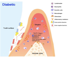
Diabetes
-
4x increased risk of loss of periodontal support
-
Increased gingival inflammation
-
Changes in bacterial composition with enhanced pathogenicity
-
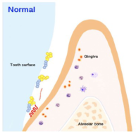
Diabetes and periodontitis have a two-way relationship, with periodontitis affecting blood sugar control in people with diabetes.
-
Diabetes may increase leukocyte and cytokine expression.
-
The relationship involves increased chronic inflammation in the periodontium.
-
These changes accelerate tissue destruction and increase the risk of periodontal disease. Tooth surface / Tooth surface (figure labels)
Graves et al 2019.
Handbook of Pediatric Dentistry. Edited by Angus C. Cameron and Richard P. Widmer. Foreword by Roger A. Hall.
Read more in textbook!
Clinical Cases
Case: Erupting anterior teeth with gingival inflammation
Question
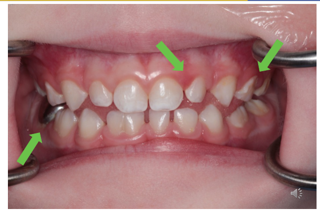
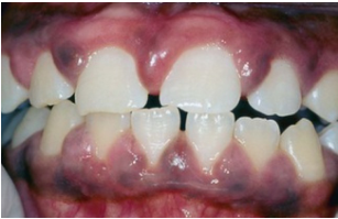
Scenario: A child, estimated to be around 10 years of age, presenting with gingival inflammation around erupting anterior teeth.
What’s shown: An intraoral photograph showing a red, swollen appearance along the cervical margin of the front teeth, with an eruption pattern visible.
Consider: What local and anatomical factors are contributing to the gingival inflammation in this child?
Answer
Observations:
- Red, swollen gingiva along the cervical margin of the front teeth.
- Eruption pattern of the anterior teeth is visible.
Reasoning: The inflammation is due to erupting gingivitis. The eruption process makes plaque control difficult and uncomfortable on tender gums. Additionally, changes in the epithelium create weaknesses allowing permeability and bacterial accumulation. The long junctional epithelium of the erupting teeth creates a pocket for bacteria to reach subgingivally.
Takeaway: Erupting gingivitis is a transient inflammation caused by difficult plaque control and anatomical changes, such as a long junctional epithelium creating subgingival pockets for bacteria.
Case: Gingival inflammation adjacent to stainless steel crowns
Question
Scenario: A patient presenting with localized gingival inflammation associated with dental restorations.
What’s shown: Clinical photographs showing a poor fit on the buccal surface of stainless steel crowns and proximal gingival swelling that has lost its normal saddleback shape compared to adjacent healthy areas.
Consider: What local factors are contributing to the gingival inflammation and altered papilla shape in this patient?
Answer
Observations:
- Poor fit on the buccal surface of stainless steel crowns.
- Proximal gingival swelling that is no longer saddleback shaped compared to other interdental areas.
Reasoning: Unsatisfactory restorations, such as poorly fitting stainless steel crowns, create irregularities that aid in plaque accumulation and food impaction, leading to localized gingivitis and proximal swelling.
Takeaway: Poorly fitting restorations like stainless steel crowns can cause localized plaque accumulation and gingival inflammation, altering the normal saddleback shape of the interdental papilla.
Case: Localised gingival recession, clinical photo
Question
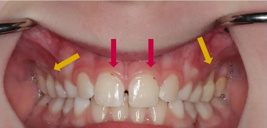
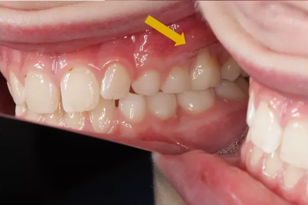
Scenario: A teenager presenting with localised gingival recession.
What’s shown: An intraoral clinical photograph showing recession on the buccal surfaces of the first permanent upper molars, with remnants of plaque visible on the upper incisors.
Consider: What are the clinical findings, and what management strategies are recommended to prevent progression while ensuring effective cleaning?
Answer
Observations:
- Recession on the buccal surfaces of the first permanent upper molars.
- Remnants of plaque on the upper incisors.
Reasoning: The recession in teenagers commonly affects the buccal surfaces of upper molars and premolars, often caused by tooth position, toothbrush trauma, previous orthodontics, or poor plaque control. A soft bristle toothbrush is recommended to prevent the recession from progressing while still allowing effective cleaning of the labial surfaces of the incisors, particularly if the recession is related to dental alignment.
Outcome: Management includes using a soft bristle toothbrush, providing oral hygiene instructions and techniques, and discussing orthodontic treatment to prevent further recession.
Takeaway: Localised gingival recession in teenagers requires a balanced management approach that prevents further tissue loss while maintaining effective plaque control, often necessitating orthodontic evaluation.
Case: Longitudinal bitewing radiographs of a patient with progressive periodontal disease
Question
Scenario: A single patient from a study by Morass and Berlund is presented with a follow-up spanning from 8 to 23 years of age to illustrate the progression of periodontal disease.
What’s shown: A series of bitewing radiographs, including a specific set taken at 11 years of age.
Consider: Observe the progression of the disease on the radiographs and note the outcome for the affected tooth.
Answer
Observations:
- Bitewing radiographs from age 8 through 23 years demonstrate progressive periodontal destruction.
- Radiographs at 11 years of age reveal bone loss around tooth 36.
Reasoning:
- The sequential images illustrate how periodontal destruction can begin early and gradually progress if the condition is not identified and managed in time.
- The progressive bone loss around tooth 36 ultimately compromised the tooth’s support.
Outcome:
- Tooth 36 was extracted at 12 years of age due to the destruction of the surrounding bone.
Takeaway:
- Periodontal destruction can begin early and progress if not identified and managed in time, emphasizing the importance of early detection and intervention.
Case: Seven-year-old child with bone loss on primary molars
Question
Scenario: A seven-year-old child is presented for dental evaluation.
What’s shown: Radiographic images demonstrating clear bone loss on the first primary molars.
Consider: What is the clinical significance of finding periodontitis in the primary dentition, and how should this influence the clinical approach?
Answer
Observations:
- Clear bone loss is visible on the first primary molars.
Reasoning:
- Periodontitis in the primary dentition should not be viewed merely as an isolated problem with the primary teeth.
- It serves as a significant risk factor for developing periodontal disease later in the permanent dentition.
- This highlights the critical need for early diagnosis and for clinicians to carefully evaluate alveolar bone levels on bitewing radiographs, as small changes can be the very first sign of periodontal risk.
Outcome:
- Not explicitly stated for this specific patient, but the lecturer emphasizes that early diagnosis improves prognosis and that underlying local or systemic causes must be investigated.
Takeaway:
- Periodontitis in the primary dentition is a strong indicator of future periodontal risk in the permanent dentition, making early recognition and careful evaluation of alveolar bone levels essential.
Case: Panoramic radiographs of a patient at age 6 and 16
Question
Scenario: A pediatric patient with a primary immunodeficiency is presented to illustrate the periodontal manifestations of their condition.
What’s shown: Panoramic radiographs (OPGs) of the patient at 6 years of age and at 16 years of age.
Consider: Identify the radiographic changes over time and the clinical features associated with this condition.
Answer
Observations:
- The patient has congenital neutropenia, a primary immunodeficiency characterized by low neutrophil levels.
- At 6 years of age, early changes are already visible on the OPG.
- By 16 years of age, there is severe bone loss affecting the entire dentition.
- Both the primary and permanent dentitions are affected, showing severe gingivitis, increased tooth mobility, and widespread alveolar bone destruction.
- Early clinical signs include recurrent oral ulceration.
Reasoning: The low neutrophil count makes the patient highly susceptible to severe bacterial infections. Because of this, periodontal involvement typically begins as soon as the teeth erupt, leading to rapid and extensive breakdown.
Outcome: Management focuses on early prevention and ongoing dental care, always in close collaboration with the medical team.
Takeaway: In congenital neutropenia, periodontal involvement begins as soon as teeth erupt, necessitating early prevention and ongoing dental care in close collaboration with the medical team.
Footnotes
-
Original PDF page 1: L9 Periodontal lesions 2025, p.1 ↩
-
Original PDF page 2: L9 Periodontal lesions 2025, p.2 ↩
-
Original PDF page 4: L9 Periodontal lesions 2025, p.4 ↩
-
Original PDF page 5: L9 Periodontal lesions 2025, p.5 ↩
-
Original PDF page 6: L9 Periodontal lesions 2025, p.6 ↩
-
Original PDF page 7: L9 Periodontal lesions 2025, p.7 ↩
-
Original PDF page 8: L9 Periodontal lesions 2025, p.8 ↩
-
Original PDF page 9: L9 Periodontal lesions 2025, p.9 ↩
-
Original PDF page 10: L9 Periodontal lesions 2025, p.10 ↩
-
Original PDF page 11: L9 Periodontal lesions 2025, p.11 ↩
-
Original PDF page 12: L9 Periodontal lesions 2025, p.12 ↩