Case 41
Worksheet prompts (Suggested Approach, applies to every case)
- Describe the image/radiograph.
- Identify relevant soft tissue findings.
- Identify relevant hard tissue findings.
- Estimate the patient’s dental age.
- Develop a problem list.
- Formulate differential diagnoses.
- Consider additional investigations.
- Develop a management plan.
Clinical Case
Answered from the lecturer’s comments in the recording only. The student presentations are left out.
1. Image / radiograph
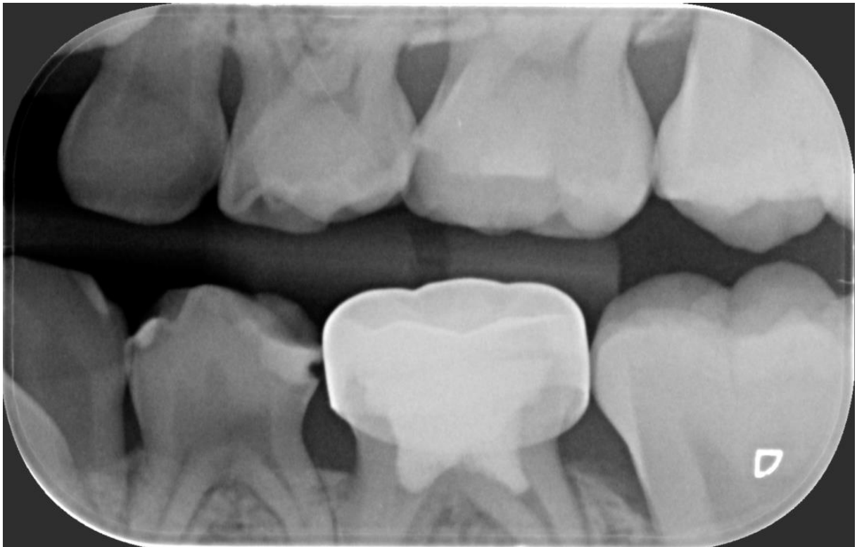
- Bitewing radiograph.
2. Soft tissue findings
- Not discussed (radiograph only).
3. Hard tissue findings
- 74: previous restoration with a defect (a bubble), so it is unsatisfactory.
- A pulp projection into the mid-occlusal area suggests dens evaginatus on the occlusal surface.
- Be wary even if the pulp seems to end lower down. In roughly 40% of dens evaginatus teeth, a very thin pulp extension isn’t visible at radiographic resolution.
- 64: mesio-occlusal and distal caries.
- 65: a very straight-edged, “perfectly shaped” cavity. It was probably a previous preparation whose filling has fallen out, since caries rarely has such straight lines.
- Furcation radiolucency (a student’s question): the lecturer thought it was probably just the PDL space or an odd projection.
4. Dental age
- Not discussed by the lecturer.
5. Problem list
- High caries risk: caries in multiple teeth.
- 74: defective restoration, plus dens evaginatus with a high pulp horn.
- 64 and 65: carious, and 65 has a lost restoration.
6. Differential diagnoses
- 74: dens evaginatus (occlusal tubercle). Nothing further was discussed.
7. Additional investigations
- Check the pulp anatomy on the radiograph before any crown preparation. No other investigations were discussed by the lecturer.
8. Management plan
- 74, option 1: conventional stainless steel crown.
- Consent must cover a possible pulp exposure, which would then need a pulpotomy.
- Remove the old restoration at the prep.
- Usually place a GIC core so there is no negative space or air bubble under the crown. If in doubt, place the core.
- 74, option 2: redo the DO composite. It is less invasive, but warn the parents that retention may be poorer.
- 74, option 3: Hall crown, which avoids drilling the occlusal surface.
- It is fine over a small, sound defect or a GIC temporisation, because the luting GIC flows into a small open defect.
- If the defect is large, remove the old filling, place a Fuji II core, then the Hall crown.
- It is not possible with a very pronounced tubercle: the crown rocks on it and won’t seat. A slight dome is OK.
- Dens evaginatus without caries:
- Fissure-seal around the tubercle for support.
- Or use selective grinding, done circumferentially to stimulate tertiary dentine more than grinding from the top down.
- Or (increasingly common) amputate it in one appointment under local anaesthetic and do a Cvek partial pulpotomy.
- Cvek partial pulpotomy steps:
- Remove about 2 mm of pulp.
- Apply a damp cotton pellet with pressure for 5 minutes for haemostasis. If haemostasis isn’t achieved, remove more pulp to get past the inflamed pulp. Expect little inflammation after an amputation; more after trauma.
- Place a bioactive silicate cement (MTA, Biodentine or EndoSequence) to promote a hard-tissue barrier.
- Place GIC over it (preferred to Cavit), then a core and crown, or a localised composite.
- The same procedure is used after trauma to a permanent incisor.
- Carious pulp exposure in a primary tooth: do a full pulpotomy instead. There is more inflammation, so a partial pulpotomy or direct pulp cap is less predictable.
- 64 and 65: restore. Conventional SSCs can be placed on opposing teeth because the occlusal surface is reduced.
- Prevention (high caries risk): fissure sealants on the first permanent molars.
- Workflow: quadrant dentistry is reasonable as speed and confidence build. Full-mouth or opposing-quadrant treatment is done under GA.
Footnotes
-
Original PDF page 1: T1 Case 4, p.1 ↩