Case 6: R1
R
Worksheet prompts (Suggested Approach, applies to every case)
- Describe the image/radiograph.
- Identify relevant soft tissue findings.
- Identify relevant hard tissue findings.
- Estimate the patient’s dental age.
- Develop a problem list.
- Formulate differential diagnoses.
- Consider additional investigations.
- Develop a management plan.
Clinical Case
Answered from the lecturer’s comments in the recording only. The student presentations are left out.
1. Image / radiograph
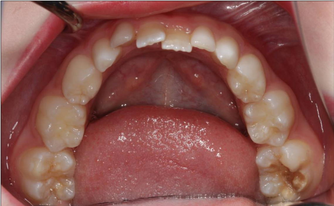
- Intraoral occlusal photograph. The image type wasn’t discussed in the recording.
2. Soft tissue findings
- Not discussed by the lecturer.
3. Hard tissue findings
- Lower anterior crowding: a tooth-size/arch-length discrepancy, with overlapping teeth and no primate spaces.
- 46: a well-demarcated yellow opacity. You can draw a line around it. Most likely hypomineralised enamel with post-eruptive breakdown. Hypoplasia is possible but less likely.
- Other first permanent molar: affected but not broken down. The lecturer referred to it only as “that tooth”, so it isn’t named here. It could be very sensitive (see investigations).
4. Dental age
- Not discussed by the lecturer.
5. Problem list
- MIH-affected first permanent molar(s); 46 has post-eruptive breakdown.
- Possible hypersensitivity of the affected molars.
- Lower anterior crowding.
6. Differential diagnoses
- Diagnosis: molar incisor hypomineralisation (MIH). Definition: one or more first permanent molars affected, with or without the permanent incisors.
7. Additional investigations
- Sensitivity history: an affected tooth that hasn’t broken down may still be hypersensitive and need treatment.
8. Management plan
- Restorative margins: two schools of thought.
- Recommended: leave the margins on sound enamel. Colour is a good indicator of what is normal tooth structure.
- Alternative: remove the hypomineralised enamel until it feels firm to the slow-speed bur. This is more acceptable for white defects, which are closer to normal enamel.
- Both were accepted in the exam if justified.
- Pre-treatment: deproteinisation, most commonly by rubbing sodium hypochlorite on the surface, has been proposed. Hypomineralised enamel has a high protein content. There is no strict protocol yet (“one to watch”).
- Bonding: use total-etch rather than self-etch, since this enamel is harder to etch.
- 46 restoration: you don’t have to jump to a stainless steel crown.
- An onlay keeps more tooth structure.
- A large composite may also be possible, depending on the defect.
- Remove secondary caries or deep defects first if they are present.
- Stainless steel crown: minimal preparation with feather-edge margins. Tell the parents its goal is to last until adulthood (about 18–20), when it is replaced with a gold or ceramic crown, or until an extraction decision is made.
- Avoid zirconia crowns in children: the pulp is large, and every replacement removes more tooth structure. Preserve structure for the future.
- Anaesthesia is often difficult in MIH.
- Supplement the infiltration with a PDL (intraligamentary) injection, or use a block. This is personal preference.
- Consider nitrous, and paracetamol/ibuprofen before the appointment.
- Hall crowns are not for permanent molars: the crowns are too big, and opening the bite at the back of the jaw opens it even more anteriorly.
Footnotes
-
Original PDF page 1: T1 Case 6, p.1 ↩