Case 41
Worksheet prompts (Suggested Approach, applies to every case)
- Describe the image/radiograph.
- Identify relevant soft tissue findings.
- Identify relevant hard tissue findings.
- Estimate the patient’s dental age.
- Develop a problem list.
- Formulate differential diagnoses.
- Consider additional investigations.
- Develop a management plan.
Clinical Case
Answered from the lecturer’s comments in the recording only. The student presentations are left out.
Partly unintelligible
Much of the W2.4 audio is off-mic chatter. Only the points below are audible.
1. Image / radiograph
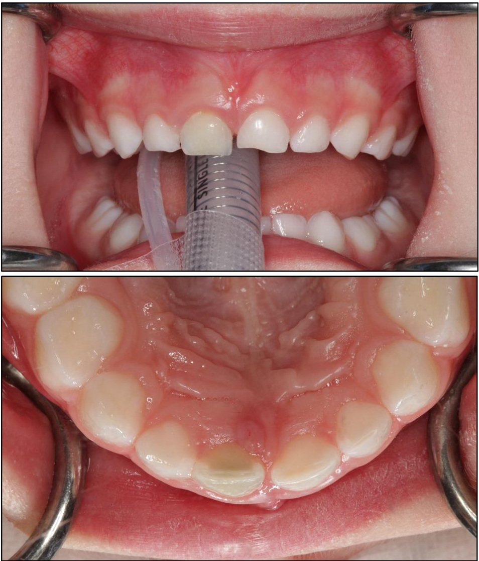
- Two intraoral photographs: a frontal view, and a palatal (occlusal) view.
- Where was it taken? Under GA: an airway tube is visible. An oral rather than a nasal tube is typically used when only an extraction is required.
2. Soft tissue findings
- Draining sinus on the palatal surface. This is unusual; sinuses are usually found on the buccal.
3. Hard tissue findings
- Discoloured upper central incisor. There are no clear caries that would explain the infection.
4. Dental age
- Not discussed in an audible part of the recording.
5. Problem list
- Discoloured upper central incisor with a palatal draining sinus, so it is infected.
- Likely past trauma.
6. Differential diagnoses
- Trauma is likely (“something must have happened”), given the sinus without caries.
7. Additional investigations
- A more detailed history: the lecturer agreed with the group.
8. Management plan
- Extraction: the lecturer agreed (“Yep, definitely”). It was done under GA (see image).
- Children’s fear: needles are the most commonly reported fear. The Wand (computer-controlled local anaesthetic delivery) and injecting very slowly were mentioned.
Footnotes
-
Original PDF page 1: T2 Case 4, p.1 ↩