Case 51
R
Worksheet prompts (Suggested Approach, applies to every case)
- Describe the image/radiograph.
- Identify relevant soft tissue findings.
- Identify relevant hard tissue findings.
- Estimate the patient’s dental age.
- Develop a problem list.
- Formulate differential diagnoses.
- Consider additional investigations.
- Develop a management plan.
Clinical Case
Answered from the lecturer’s comments in the recording only. The student presentations are left out.
1. Image / radiograph
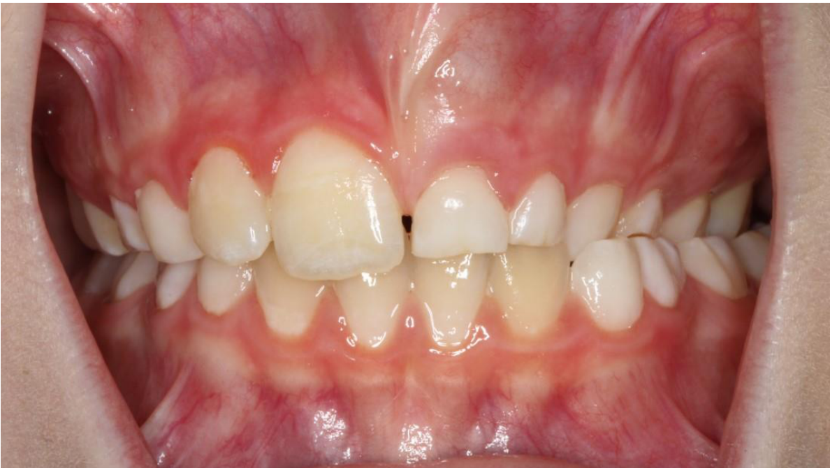
- Frontal intraoral photograph.
2. Soft tissue findings
- Swelling high in the upper anterior region.
3. Hard tissue findings
- Huge eruption discrepancy: 21 and 22 are very delayed compared with the contralateral side.
4. Dental age
- Not discussed by the lecturer.
5. Problem list
- Delayed eruption of 21 and 22, with an asymmetry between sides.
- Anterior maxillary swelling. The lecturer said it kept increasing over time while the patient attended recalls without it being noticed. Follow-up was also delayed because the family moved around.
6. Differential diagnoses
- Outcome: the radiograph showed a large radiolucent lesion, a cyst. The transcript most likely reads “radicular cyst”.
7. Additional investigations
- Radiograph.
8. Management plan
- Not discussed beyond the radiograph.
- Take-home: if the eruption asymmetry between sides is more than 6 months, take a radiograph, so pathology like this isn’t missed.
Footnotes
-
Original PDF page 1: T2 Case 5, p.1 ↩