Case 71
R
R
 |  |
Worksheet prompts (Suggested Approach, applies to every case)
- Describe the image/radiograph.
- Identify relevant soft tissue findings.
- Identify relevant hard tissue findings.
- Estimate the patient’s dental age.
- Develop a problem list.
- Formulate differential diagnoses.
- Consider additional investigations.
- Develop a management plan.
Clinical Case
Answered from the lecturer’s comments in the recording only. The student presentations are left out.
Partly unintelligible
Only a short part of the W2.7 audio is about this case. The lecturer said the groups had “covered everything” for all the cases, but those answers aren’t audible.
1. Image / radiograph
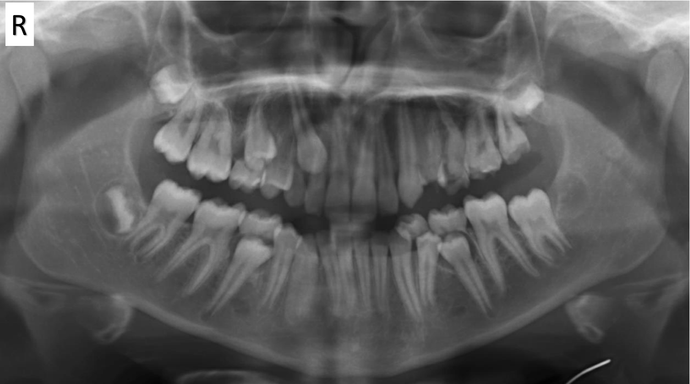
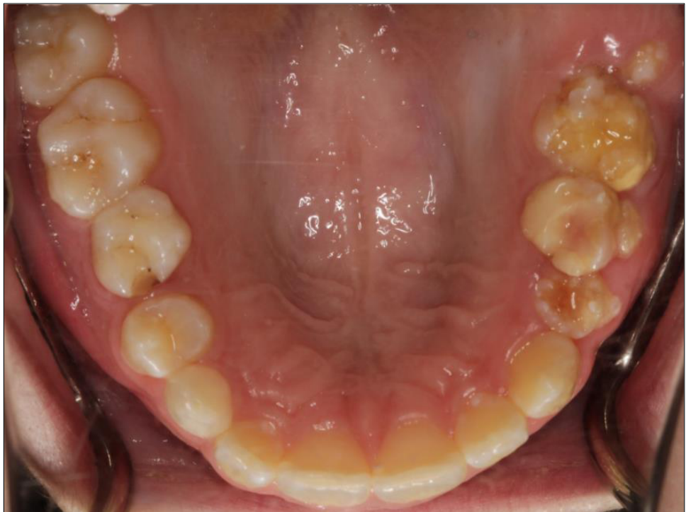
- OPG and a lower occlusal photograph.
2. Soft tissue findings
- Not discussed in an audible part of the recording.
3. Hard tissue findings
- Not described in an audible part of the recording.
4. Dental age
- Not discussed in an audible part of the recording.
5. Problem list
- Not discussed in an audible part of the recording.
6. Differential diagnoses
- Amelogenesis imperfecta vs regional odontodysplasia.
- AI is hereditary, so ask whether anyone else in the family has the condition.
- Regional odontodysplasia is regional, which explains its radiographic appearance (“ghost teeth”).
7. Additional investigations
- Family history (for AI).
8. Management plan
- Not discussed in an audible part of the recording.
Separate slide shown at the end: impacted canines (not this case's images)
- Extract the primary canine when impaction of the permanent canine is suspected from its angulation, and refer to orthodontics. Extracting at the right indication avoids many surgical exposures.
- The example on the slide covered about half of the lateral. Extracting the primary canine may let it come through, but given the angulation and root development it will likely still need an exposure with bonding and traction.
Footnotes
-
Original PDF page 1: T2 Case 7, p.1 ↩