Renal Disease and Dentistry1
by Dr David Lim
21 Jul 2026 (Tuesday) 9–10
Acknowledgement of Country2
We acknowledge the Traditional Custodians of this land. We acknowledge the waterways, the land, the sky and all who inhabit this place, Country.
We acknowledge their ancestors, and Elders and recognise those who continue to protect and promote Aboriginal and Torres Strait Islander cultures. We acknowledge the past and stand together for our future.
Lecturer — Local Landscape
The acknowledgement situated teaching and learning on the land of the Traditional Custodians. It also described the land around the Swan River, including a rainbow over the river and mist over the Perth Hills from Kings Park.
Learning Objectives
IMPT!
- Discuss the specific characteristics and dental needs of individuals with kidney disease
- Perform a risk assessment and make appropriate treatment adaptations to ensure safe and effective dental care.
What Is It About XXX Disease/Condition?3
- Definition
- Aetiology
- Epidemiology
- General Manifestations/Clinical Features
- General Management/Treatment
- Oral Manifestations
- Oral Management = Risk Assessment and ACCESS acronym
Lecturer — Renal Disease Focus
The renal section focused on chronic kidney disease and its implications for general health and dental management.
- The section addressed the effects of chronic kidney disease on general health, renal replacement therapy and dental management.
- Dental care should take account of dialysis and transplantation.
- Treatment modifications depend on the patient’s renal disease, dialysis schedule, medications, bleeding risk, anaemia and immune status.
ADC: 5.1 iADH: 5A, 5B, 5C, 6Ci 5.1 Scientific and Clinical Knowledge apply the social, cultural, biological, biomedical, physical and behavioural sciences in relation to oral health care provision and disease prevention
Renal Anatomy and Physiology
Source: https://www.usz.ch/en/organs/kidney/
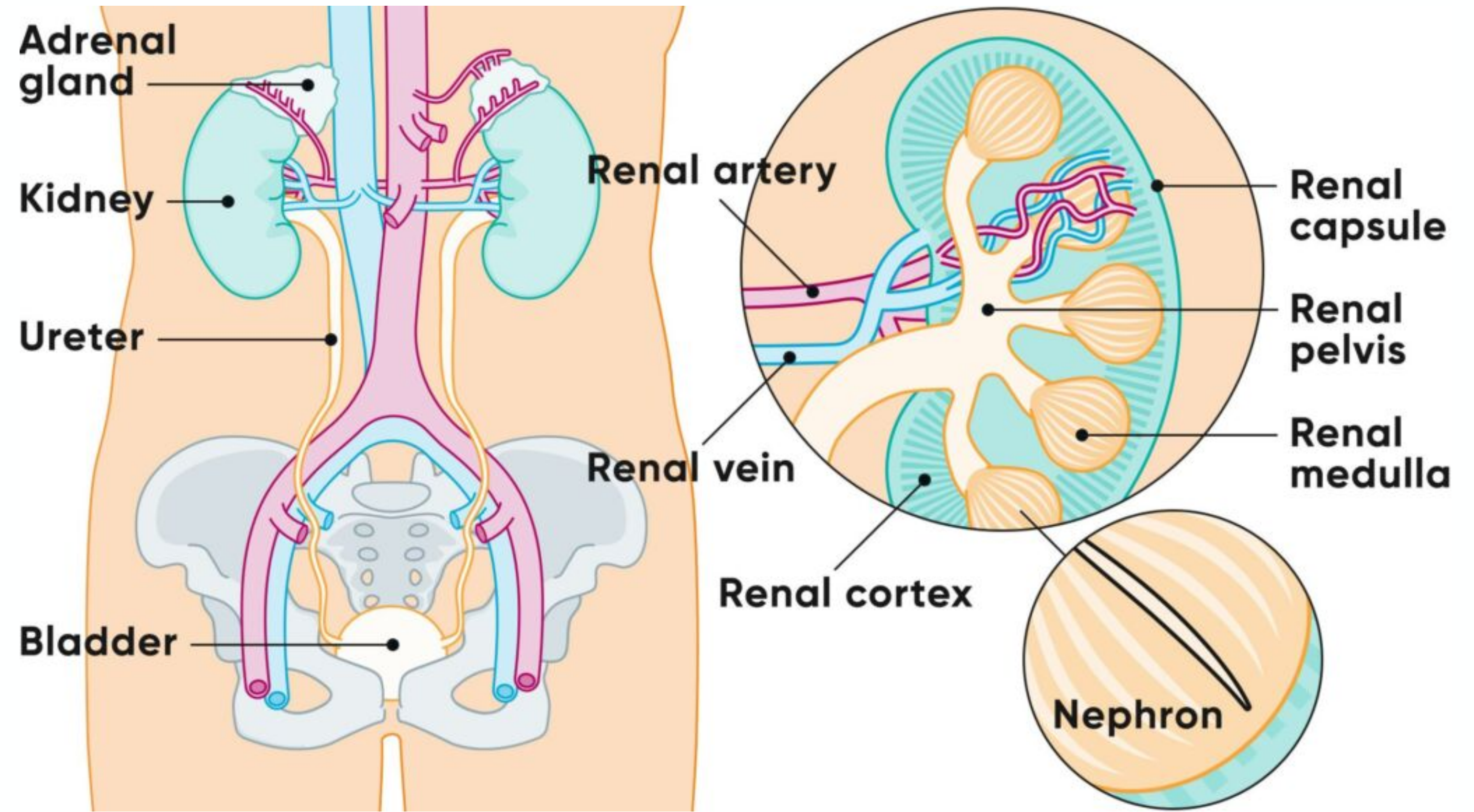
Recap on Structures4
- Adrenal gland
- Kidney
- Ureter
- Bladder
- Renal artery
- Renal vein
- Renal capsule
- Renal pelvis
- Renal medulla
- Renal cortex
- Nephron
Lecturer — Kidney Location
The kidneys are approximately fist-sized organs located in the abdomen towards the back.
Renal Physiology
Maintaining Homeostasis5
- Fluid-electrolyte balance — balance Na+ electrolytes, regulate fluid
- Acid-base balance — excrete hydrogen ions and regenerate HCO3-
- Excrete waste — urea, drugs
Lecturer — Kidney Functions
The kidneys also filter blood and regulate blood pressure through the renin-angiotensin-aldosterone pathway.
- They excrete certain waste ions in addition to urea and drugs.
- When kidney function deteriorates, waste-product levels, electrolytes, acid-base balance, blood pressure and drug elimination can all be affected.
Endocrine Function
-
Renin
-
Erythropoietin (stimulate formation of haemoglobin)
-
Calcitriol (active form of Vitamin D, regulating Ca2+ balance and others)
-
Others (minute amounts), e.g. thrombopoietin
-
Calcitriol activation affects bone density through its relevance to calcium metabolism
-
Thrombopoietin contributes to the production of thrombocytes, also known as platelets
Lecturer — Reduced Hormones
Reduced erythropoietin production can contribute to anaemia, while reduced thrombopoietin production may cause problems with platelet production.
Common Renal Diseases
Overview of Three Common Renal Diseases
- Chronic Kidney Disease — caused mainly by HBP and DM
- Treated with adapted diet/fluids, controlling BP and blood glucose (diabetes), dialysis, or renal transplant
- Pyelonephritis — affects mainly women due to shorter urethra. Fever, vomiting, back pain
- Kidney stones (nephrolithiasis, urolithiasis/urethral stone, cystolithiasis/bladder stone) — cause severe pain (renal colic)
- Passed out in urine, or dissolved with medication, or removed with surgery
Lecturer — Early CKD Management
Early chronic kidney disease management may also involve controlling other risk factors and adapting lifestyle.
- Excessive water intake increases the amount of fluid that the kidneys need to filter.
- Polycystic kidney disease was mentioned as another renal disease, although it was not covered in detail.
- The lecture focused mainly on chronic kidney disease rather than covering all renal diseases in detail.
Overview of Three Common Renal Diseases6
KDIGO Classification of Chronic Kidney Disease
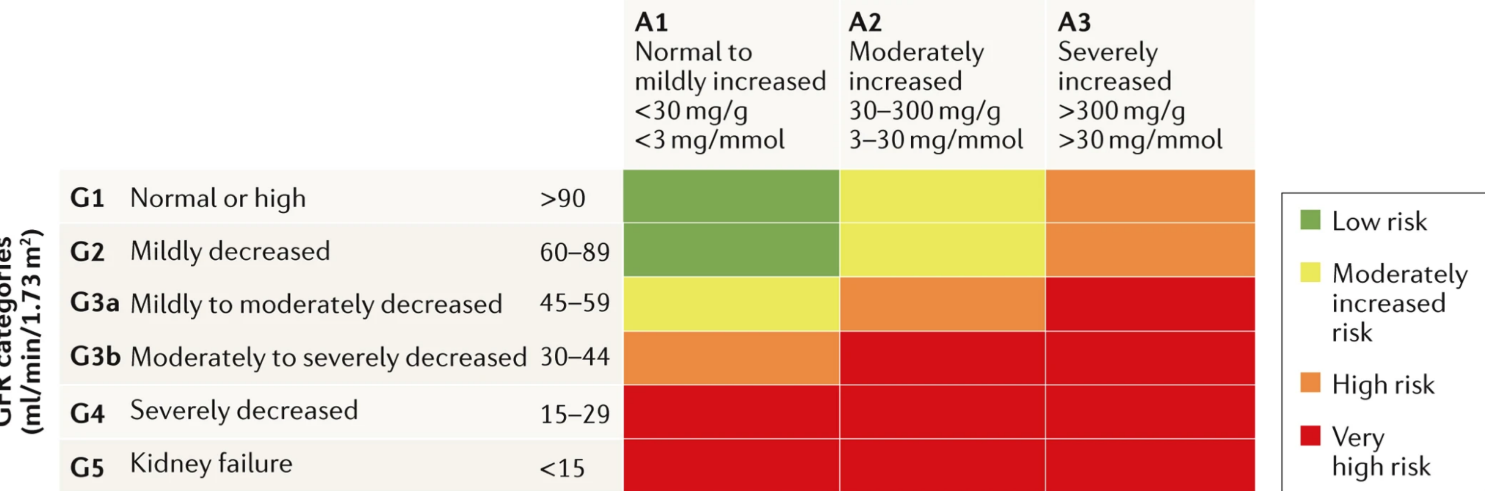
Kidney Disease Improving Global Outcomes (KDIGO) 2D matrix classification of CKD, based on risk of end stage renal disease.
Persistent Albuminuria Categories7
- A1 — Normal to mildly increased: <30 mg/g (<3 mg/mmol)
- A2 — Moderately increased: 30–300 mg/g (3–30 mg/mmol)
- A3 — Severely increased: >300 mg/g (>30 mg/mmol)
Lecturer — Albuminuria Meaning
Albuminuria refers to the amount of albumin found in the urine. When kidney function is impaired, proteins may not be filtered effectively, so albumin can be detected in the urine; lower levels are positioned towards the left of the classification and higher levels towards the right.
 | |
GFR Categories (ml/min/1.73 m²)
- G1 — Normal or high: >90
- G2 — Mildly decreased: 60–89
- G3a — Mildly to moderately decreased: 45–59
- G3b — Moderately to severely decreased: 30–44
- G4 — Severely decreased: 15–29
- G5 — Kidney failure: <15
Lecturer — Filtration Categories
Glomerular filtration reflects the amount of waste being filtered by the kidneys. Healthy kidneys filter more waste and have a higher filtration rate, whereas diseased kidneys filter less waste and progress towards the lower G5 category.
Risk levels across the matrix: low risk, moderately increased risk, high risk, very high risk.
Lecturer — Risk Colouring
Risk is represented across a colour range in the classification. The green area represents relatively low risk, the red area represents very high risk, and the intermediate areas represent progressively increasing risk.
Measuring the exact GFR is a complex, invasive procedure. Instead, doctors use an estimate (eGFR) calculated through a simple formula. [1]
How eGFR Is Determined
- The Blood Test: The lab measures the level of waste products in your blood, primarily creatinine (a byproduct of muscle breakdown) or occasionally cystatin C. Healthy kidneys filter these wastes out into your urine. If kidney function declines, wastes build up in the bloodstream. [1, 2, 3, 4]
- The Calculation: The creatinine level is plugged into a mathematical formula. To ensure accuracy, the calculation also accounts for your age and gender.
Source: Nature Reviews | Disease Primers
Types of Dialysis
- Haemodialysis — AV fistula on arm; blood drawn into a machine, filtered, and returned. Done usually in a clinic, with anticoagulation (usually LMW heparin)
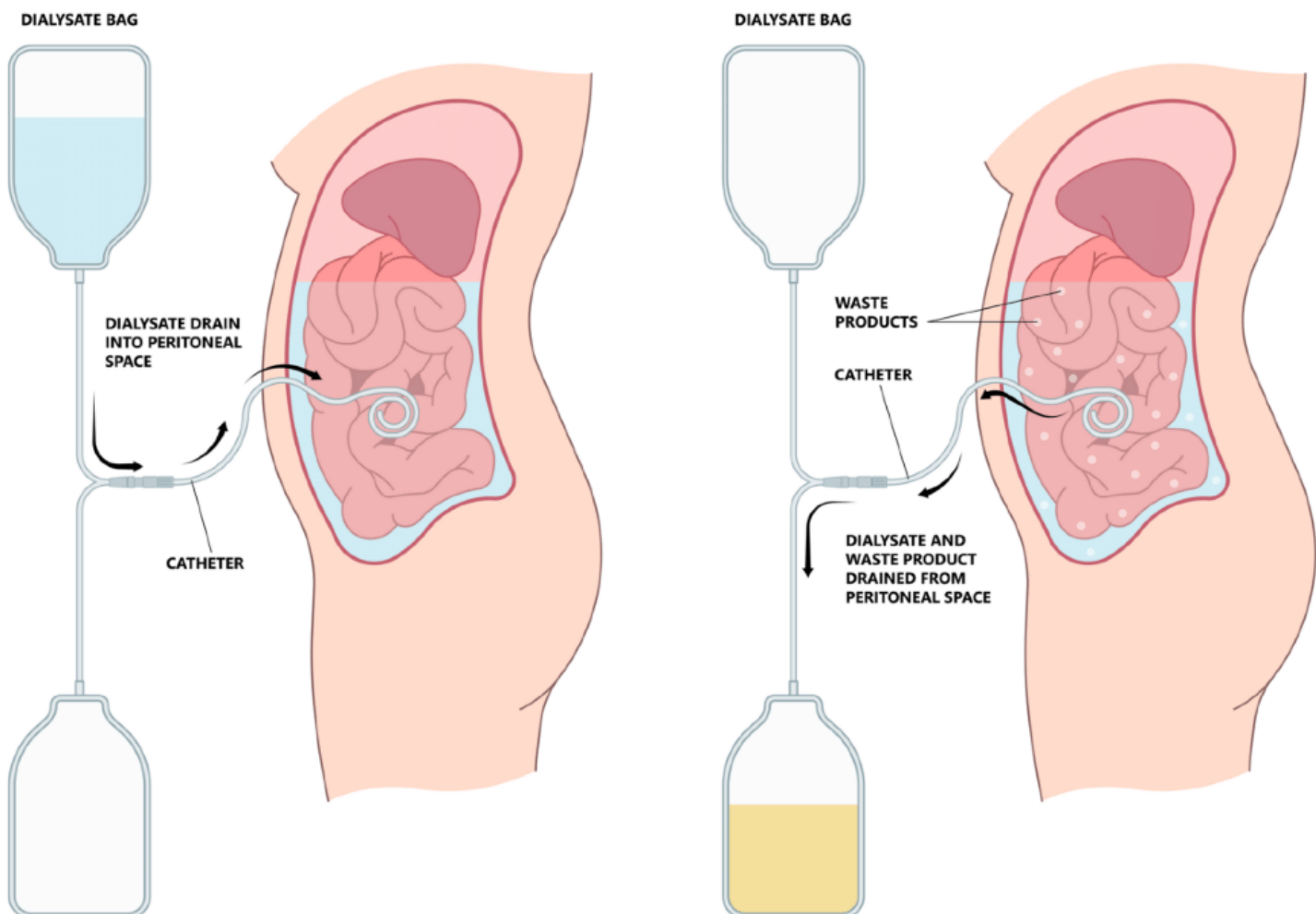
- Peritoneal dialysis — catheter inserted into peritoneal cavity where dialysis fluid is introduced. This cleanses waste and fluid is replaced. Repeated several times a day or during sleep; done at home
- Continuous renal replacement therapy (in ICU)
Lecturer — Dialysis and Dentistry
Haemodialysis and peritoneal dialysis have dental implications, particularly because of bleeding risk, fatigue, scheduling, anticoagulation, and the patient’s overall medical condition.
Peritoneal Dialysis (Diagram Labels)8
- Dialysate bag; dialysate drain into peritoneal space; catheter; drainage bag
- Dialysate bag; waste products; catheter; dialysate and waste product drained from peritoneal space; drainage bag
Lecturer — Peritoneal Dialysis
Peritoneal dialysis allows passive diffusion of waste products into fluid introduced into the peritoneal cavity, which is drained after several hours. It can usually be performed at home, may allow greater mobility and easier attendance at the dental clinic, but is less effective than haemodialysis.
- Home haemodialysis is also possible, although patients must learn how to connect themselves to the machine.
https://www.kidneyhealth.com.sg/peritoneal-dialysis/
 | 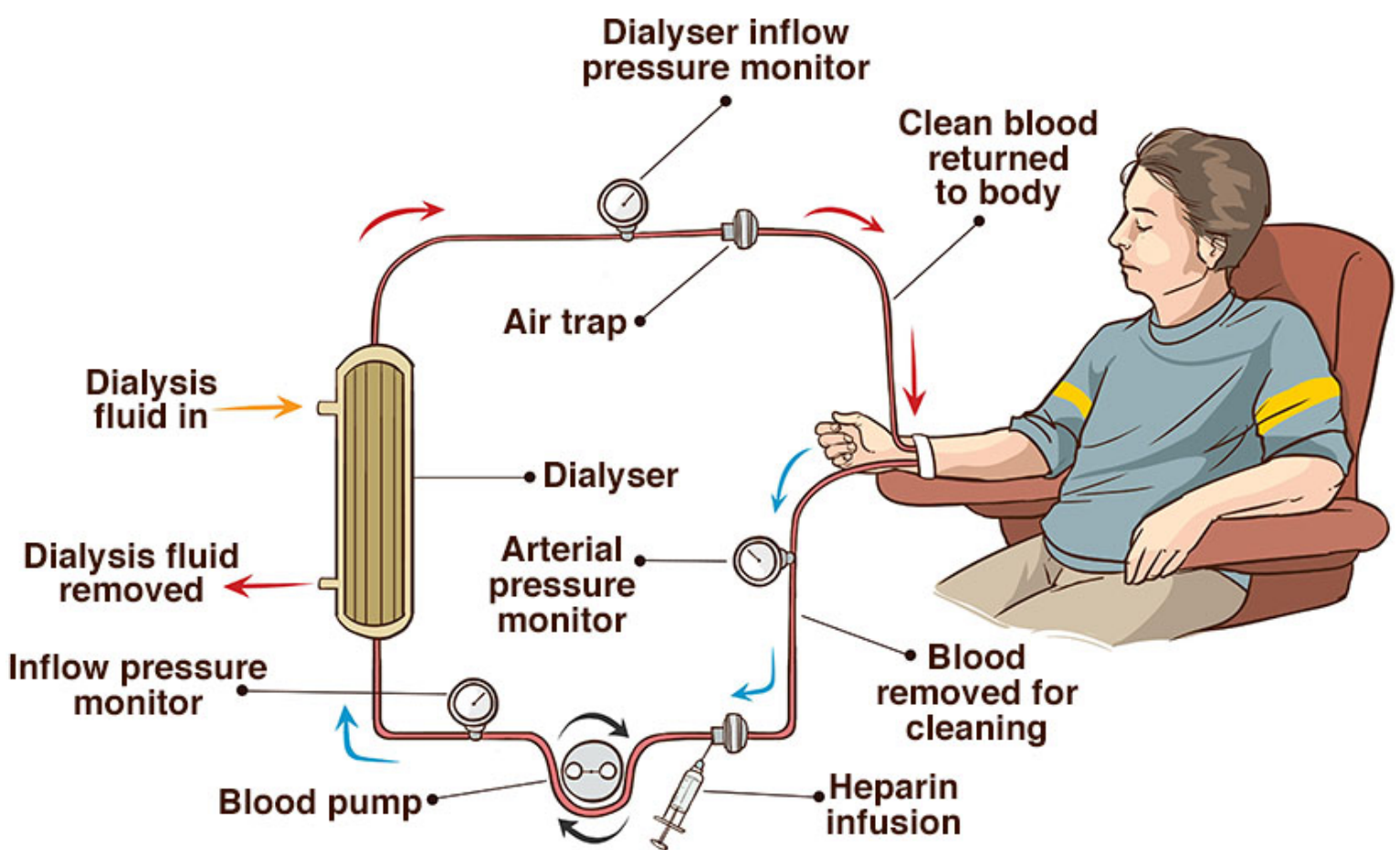 |
Haemodialysis (Diagram Labels)
- Dialyser inflow pressure monitor; clean blood returned to body; air trap; dialysis fluid in; dialyser; arterial pressure monitor; dialysis fluid removed; inflow pressure monitor; blood pump; heparin infusion; blood removed for cleaning
https://www.healthdirect.gov.au/dialysis
Lecturer — Haemodialysis Effects
During haemodialysis, heparin is administered and a dialysis filter removes waste products before the blood is pumped back into the body. Haemodialysis removes many electrolytes and other components from the blood, but it is not as effective as a healthy, functioning kidney.
- Patients may be tired after haemodialysis.
- On the day of haemodialysis, increased bleeding risk, fatigue, and effects from electrolyte removal may occur.
Clinical Cases
CKD Patient Case9
- A 49 y.o. male underwent hemodialysis for End Stage Kidney/Renal Disease (ESRD). He was referred by a nephrologist for an oral assessment to eliminate potential oral foci of infection prior to planned kidney transplant.
Lecturer — Dental Clearance Timing
Dental clearance may also be required before bisphosphonates, radiotherapy, or treatment for haematological conditions. If transplantation is expected soon, more treatment may need to be completed during the appointment; if it is expected much later, treatment can be staged over time.
Medical History10
- CKD-ESRD, Hyperuricaemia
- HTN, HLD
- Osteopenia (renal osteodystrophy)
- Fe deficiency anaemia
Medications
HD 3 days a week for 5 years (heparin), Nifedipine, Irbesartan, Furosemide, Cal acetate Vitamin D3, Iron, Atorvastatin, Allopurinol.
Case Questions
- The patient reports halitosis despite brushing TDS. There is also a metallic taste that improves after HD. What do you think is the reason?
- The patient has multiple decays and feels tired easily. How are these related or not to CKD?
- When prescribing analgesics, what medications should be avoided?
- CKD-ESRD, Hyperuricaemia
- HTN, HLD
- Osteopenia (renal osteodystrophy)
- Fe deficiency anaemia
Lecturer — Bone and Anaemia
Reduced activation of vitamin D affects bone calcification. Osteopenia means reduced bone mineral density that has not reached the level of osteoporosis.
- Reduced erythropoietin-related red blood cell production and haemoglobin effects contribute to fatigue and anaemia.
HD 3 days a week for 5 years (heparin), Nifedipine, Irbesartan, Furosemide, Cal acetate Vitamin D3, Iron, Atorvastatin, Allopurinol.
Lecturer — Haemodialysis and Heparin
Patients are likely to have increased anticoagulation on the day of haemodialysis, which is relevant to dental treatment.
Case Questions and Answers11
The patient reports halitosis despite brushing TDS. There is also a metallic taste that improves after HD. What do you think is the reason?
- High salivary urea levels decomposing to ammonia. Xerostomia additionally contributes to dysguesia (abnormal taste).
Lecturer — Dry Mouth Terminology
Dry mouth is a sign that can be tested, whereas xerostomia is the patient’s subjective report of oral dryness. A patient may feel dry despite measurable moisture, or may not report dryness despite being objectively dry.
The patient has multiple decays and feels tired easily. How are these related or not to CKD?
- Saliva pH should be alkaline, protecting against decay. Tiredness due to low erythropoietin, and shortened Hb survival due to uraemia. Plus Fe deficiency.
Lecturer — Salivary pH and Caries
An increased salivary pH does not necessarily prevent demineralisation because calcium content may still be low.
- Remineralisation requires calcium ions, hydroxide ions, phosphate, and a collagen matrix in the tooth.
- Simply increasing oral pH with bicarbonate rinses therefore does not reliably stop decay.
- Some organisms, including P. gingivalis, thrive at a higher pH, while Streptococcus species are more comfortable in a lower-pH acidic environment.
When prescribing analgesics, what medications should be avoided?
- Ibuprofen, diclofenac, Tramadol are contraindicated in ESRD. In late stage CKD, amoxicillin can be given at half dose.
Lecturer — Analgesic Selection
Paracetamol was identified as a suitable choice for pain relief in this context. In patients with end-stage liver disease, its dose may need to be reduced.
- In late-stage CKD, amoxicillin elimination may take longer, so a lower dose may be used.
Transplant Case
Medical History12
- History of Renal failure due to bilateral polycystic kidney disease
- Rt kidney transplant 9 years ago
- HTN, Osteopenia
- Absolute polycythaemia (Dx 3 years ago)
- Fe deficiency anaemia
Medications
Mycophenolate mofentil, Tacrolimus, Prednisolone (5mg OD). Irbesartan, Trimethoprim/sulfamethoxazole
Case Questions
- Why is the patient taking 3 immunosuppressants?
- What dental risks should we note for these immunosuppressants?
- Will you consider antibiotic prophylaxis when conducting multiple extractions?
- History of Renal failure due to bilateral polycystic kidney disease
- Rt kidney transplant 9 years ago
- HTN, Osteopenia
- Absolute polycythaemia (Dx 3 years ago)
- Fe deficiency anaemia
Mycophenolate mofentil, Tacrolimus, Prednisolone (5mg OD). Irbesartan, Trimethoprim/sulfamethoxazole
Case Questions and Answers13
Why is the patient taking 3 immunosuppressants?
- Reduce risk of T-cell alloimmune rejection.
What dental risks should we note for these immunosuppressants?
- Risk of myelosuppression, poor healing, infections, MRONJ (e.g. sirolimus), steroid cover, gingival hyperplasia (cyclosporin etc), candida.
Lecturer — Immunosuppression Risks
Immunosuppressive therapy may cause delayed healing after periodontal debridement or extraction, increased risk of dry socket, poor implant healing, and implant failure during the surgical stage.
- Oral complications may include denture stomatitis and angular cheilitis.
- Chronic or latent infections may become active, including shingles, hepatitis B, tuberculosis, and herpes simplex virus-related problems.
- The patient might be on long-term prophylactic antiviral (HSV, VZV), antifungal, or antibiotics.
Will you consider antibiotic prophylaxis when conducting multiple extractions?
- There is a significantly increased risk of post-extraction infection. Some transplant centres might indicate AB cover but evidence is limited. Case-by-case consult please.
Lecturer — Antibiotic Prophylaxis
Antibiotic prophylaxis should be considered case by case, with local hospital or service protocols consulted. Previous antibiotic exposure and the possibility of resistant organisms may affect whether a previously used antibiotic remains effective during later medical treatment.
Consider graft-v-host disease also.
Lecturer — Alloimmune Lesions
Alloimmune reactions may produce graft-versus-host disease-type manifestations, including leukoplakia, keratotic lesions, and other lesions associated with a type IV hypersensitivity reaction. When unsure about an oral lesion, take a photograph.
CKD and Dentistry
Oral Findings14
-
Brittle bone during extractions — inability to convert 25-OH Vitamin D to 1,25-dihydroxyl Vitamin D (active form), phosphate retention and calcium loss
-
Osteitis fibrosa cystica (brown tumor, below) from secondary hyperparathyroidism
-
Transplant-related
-
Risk of bleeding (thrombopoietin produced by kidney, heparin)
-
Halitosis and a metallic or abnormal taste
-
Xerostomia or objectively reduced oral moisture
-
Multiple caries and periodontal disease
-
Candidiasis in immunosuppressed transplant patients
-
Angular cheilitis and denture stomatitis
-
Poor healing after dental procedures and dry socket
-
Gingival hyperplasia associated with certain immunosuppressants
-
Lesions related to graft-versus-host disease-type reactions
-
High urea levels and ammonia production, altered salivary bicarbonate and pH, anaemia, and immunosuppression after transplantation
Lecturer — Salivary Protection
A higher salivary pH may offer some protection against decay, but it does not prevent caries by itself.
- Calcium, phosphate, hydroxide and the collagen matrix are also required for remineralisation.
Other Considerations
-
Immunosuppressant effects
-
Dental scheduling with dialysis (heparin, tiredness)
-
Prescription and drug elimination considerations
-
Consider the stage of kidney disease and whether the patient receives haemodialysis or peritoneal dialysis
-
Consider whether the patient is being assessed before kidney transplantation and the expected timing of transplantation
-
Consider anaemia, reduced erythropoietin, and reduced platelet production related to reduced thrombopoietin
-
Consider the use of nephrotoxic medications, the patient’s immune status after transplantation, and infection risk
-
Consider healing after periodontal treatment, extractions or implant placement
-
Schedule haemodialysis patients’ dental appointments between dialysis sessions
-
Bleeding risk may be increased on the day of dialysis because of heparin
-
Oral assessment or dental clearance may be required before kidney transplantation
-
Treatment may be staged if transplantation is not expected soon, but more extensive treatment may need to be completed promptly if it is expected shortly
-
Avoid NSAIDs because of renal clearance and renal toxicity concerns; paracetamol was identified as suitable in the renal patient discussed
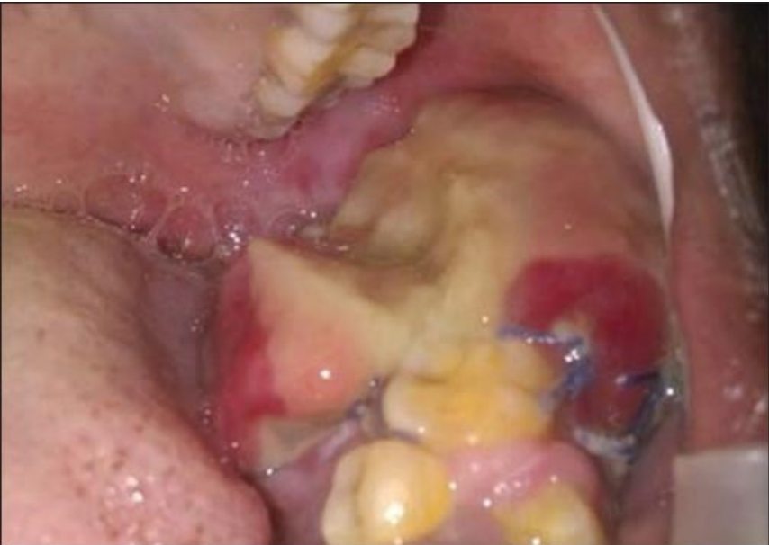
Brown Tumors
Brown tumors are giant cell focal lesions that arise as a result of abnormal bone metabolism in patients with hyperparathyroidism (HPT). The lesions localize in areas of extensive bone resorption, which is replaced by fibrovascular tissue and giant cells with abundant deposits of hemorrhage and hemosiderin.
Shetty AD, Namitha J, James L. Brown tumor of mandible in association with primary hyperparathyroidism: a case report. J Int Oral Health. 2015;7(2):50-52.
Lecturer — Brown Tumor Findings
Chronic kidney disease can result in secondary hyperparathyroidism, which differs from primary hyperparathyroidism described as being related to an autoimmune function.
- A brown tumor may present as increased bone mass or a large bony swelling pressing against the upper teeth.
- This can produce an indentation and frictional keratosis around the bony area.
- If the nature of a bony mass is uncertain, photograph it and refer the patient to an oral medicine specialist.
- Brown tumors may enlarge and swell outwards; if the lesion comes into occlusion, it may be displaced in relation to the occluding structures.
Clinical Cases
Case: 49-year-old male with ESRD referred for pre-transplant oral assessment
Question
Scenario: A 49-year-old male with end-stage renal disease (ESRD) on hemodialysis is referred by a nephrologist for an oral assessment (dental clearance) prior to a planned kidney transplant.
What’s shown: The patient’s medical history includes CKD-ESRD, hyperuricemia, hypertension, hyperlipidemia, osteopenia (renal osteodystrophy), and iron deficiency anemia. He undergoes hemodialysis three days a week using heparin, and takes multiple medications including nifedipine, irbesartan, furosemide, calcium acetate, vitamin D3, iron, atorvastatin, and allopurinol. Clinically, he reports halitosis despite brushing three times a day, a metallic taste that improves after hemodialysis, multiple dental decays, and easy fatigue.
Consider:
- What is the reason for the patient’s halitosis and metallic taste?
- How are the multiple decays and tiredness related to his chronic kidney disease?
- When prescribing analgesics, what medications should be avoided?
Answer
Observations:
- Halitosis and a metallic taste that improves after hemodialysis.
- Multiple dental decays.
- Easy fatigue.
- Medical history of ESRD on hemodialysis (with heparin), osteopenia, and iron deficiency anemia.
Reasoning:
- Halitosis and metallic taste: High salivary urea levels decompose into ammonia in the mouth. Xerostomia (subjective dry mouth) additionally contributes to dysgeusia (abnormal taste).
- Multiple decays: Changes in bicarbonate levels cause salivary pH to become more alkaline. This altered pH allows bacteria like Porphyromonas gingivalis, which thrive in higher pH environments, to flourish compared to Streptococcus species that prefer acidic environments.
- Tiredness: Caused by low erythropoietin production (leading to anemia) and shortened hemoglobin survival due to uremia-induced changes in blood pH.
- Analgesics: NSAIDs (e.g., ibuprofen, diclofenac) and tramadol are contraindicated in ESRD. Paracetamol is a safe choice. Amoxicillin is also safe but may require a reduced dose in late-stage CKD.
Outcome:
- The patient is undergoing dental clearance in preparation for a kidney transplant, which may involve a waiting period. Dental appointments are scheduled in between his hemodialysis sessions to manage bleeding risks (due to heparin) and fatigue. Analgesic prescribing is adjusted to avoid nephrotoxic drugs.
Takeaway:
- In ESRD, uremia causes high salivary urea (leading to ammonia-related halitosis and metallic taste) and alkaline salivary pH (favoring periodontal pathogens). Fatigue is driven by anemia and uremia. NSAIDs and tramadol are contraindicated; paracetamol is the analgesic of choice, and amoxicillin may require dose reduction.
Case: CKD patient presenting with halitosis, metallic taste, and fatigue
Question
Scenario: A patient with end-stage chronic kidney disease (CKD-ESRD) on hemodialysis for 5 years, with a history of hyperuricaemia, hypertension, hyperlipidaemia, osteopenia, and iron deficiency anaemia. The patient reports halitosis despite brushing three times a day, a metallic taste that improves after dialysis, multiple dental caries, and easy fatigue.
What’s shown: The patient’s medical history, current medications (including hemodialysis with heparin, antihypertensives, phosphate binders, vitamin D, iron, statins, and allopurinol), and their oral and systemic symptoms.
Consider: What is the reason for the halitosis and metallic taste? How are the multiple decays and tiredness related to chronic kidney disease? Which analgesic medications should be avoided or adjusted?
Answer
Observations:
- Halitosis despite brushing three times a day.
- Metallic taste (dysgeusia) that improves after hemodialysis.
- Multiple dental decays.
- Easy tiredness and fatigue.
- Medical history of CKD-ESRD on hemodialysis, osteopenia, and iron deficiency anaemia.
Reasoning:
- The halitosis and metallic taste are caused by high salivary urea levels decomposing into ammonia, with xerostomia additionally contributing to the dysgeusia.
- The multiple decays occur because, although saliva pH may be alkaline (which typically protects against decay), the calcium content can be low, leading to demineralization since hydroxyapatite formation requires calcium, hydroxide, and phosphate ions.
- Tiredness is due to low erythropoietin production, shortened hemoglobin survival from uraemia, and concurrent iron deficiency.
Outcome:
- Analgesics to avoid: Ibuprofen, diclofenac, and tramadol are contraindicated in ESRD.
- Amoxicillin can be prescribed in late-stage CKD but should be given at half the standard dose.
Takeaway:
- In end-stage renal disease, oral symptoms like halitosis and dysgeusia are linked to urea decomposition and xerostomia, while caries risk remains high due to altered salivary composition beyond just pH, and analgesic prescribing must be carefully adjusted to avoid contraindicated drugs.
Case: Renal transplant patient on triple immunosuppression
Question
Scenario: A patient with a history of renal failure due to bilateral polycystic kidney disease, who received a right kidney transplant 9 years ago. The patient has hypertension, osteopenia, absolute polycythaemia, and iron deficiency anaemia, and is currently taking three immunosuppressants along with other medications.
What’s shown: The patient’s medical history, transplant status, and current medication list including mycophenolate mofetil, tacrolimus, prednisolone, irbesartan, and trimethoprim/sulfamethoxazole.
Consider: Why is the patient taking three different immunosuppressants? What dental risks should be noted for these immunosuppressants? Will you consider antibiotic prophylaxis when conducting multiple extractions?
Answer
Observations:
- History of bilateral polycystic kidney disease and right kidney transplant 9 years ago.
- Current medications include three immunosuppressants: mycophenolate mofetil, tacrolimus, and prednisolone.
- Comorbidities include hypertension, osteopenia, absolute polycythaemia, and iron deficiency anaemia.
Reasoning:
- Three immunosuppressants are used to reduce the risk of T-cell alloimmune rejection by targeting different pathways of the immune function.
- Dental risks include poor healing (e.g., post-extraction, dry socket, implant failures), increased risk of infections (including candidiasis and reactivation of latent infections like shingles, Hep B, and TB), medication-related osteonecrosis of the jaw (MRONJ), need for steroid cover, and gingival hyperplasia.
- There is a significantly increased risk of post-extraction infection due to the patient being immunocompromised.
Outcome:
- Antibiotic prophylaxis for multiple extractions is considered on a case-by-case basis; some transplant centres indicate it, but evidence is mixed, so consultation with the hospital or medical team is required.
- The patient may also be on long-term prophylactic antivirals, antifungals, or antibiotics to prevent latent infections from reactivating.
- Graft-versus-host disease should also be considered, which can present with leukoplakia or keratosis.
Takeaway:
- Renal transplant patients on multiple immunosuppressants require careful dental management due to risks of poor healing, opportunistic infections, and MRONJ, and antibiotic prophylaxis for invasive procedures should be decided via case-by-case medical consultation.
Case: Mandibular brown tumor in a patient with hyperparathyroidism
Question
Scenario: An intraoral clinical photograph from a case report showing a large lesion in the mandible associated with hyperparathyroidism.
What’s shown: An intraoral view of the mandible showing a large bony mass with an indentation on the occlusal surface and surrounding tissue changes.
Consider: What is the nature of this lesion, what causes it, and how does it present clinically in the mouth?
Answer
Observations:
- A large bony mass in the mandible with increased bone mass.
- An indentation on the bony area where it presses against the upper teeth.
- Frictional keratosis surrounding the bony area.
Reasoning:
- The lesion is a brown tumor, which is a giant cell focal lesion arising from abnormal bone metabolism.
- It is typically seen in patients with hyperparathyroidism. While the case report label mentions primary hyperparathyroidism (autoimmune), in the context of chronic kidney disease, it forms a secondary type of hyperparathyroidism due to the kidney’s inability to produce active vitamin D (calcitriol), leading to phosphate retention and calcium loss.
Outcome:
- If a bone mass is seen and the diagnosis is uncertain, a photo should be taken and the case referred to an oral medicine specialist or oral surgeon for evaluation.
Takeaway:
- Brown tumors are giant cell lesions resulting from abnormal bone metabolism in hyperparathyroidism (often secondary to CKD) and can present as large bony expansions causing frictional keratosis from occlusal trauma.
Case: Mandibular bone mass, intraoral photo
Question
Scenario: An intraoral clinical photo from a case report of a brown tumor of the mandible associated with primary hyperparathyroidism is presented.
What’s shown: An intraoral view showing a large bony mass in the mandible.
Consider: Identify the clinical features visible on the bony mass and its effect on the adjacent structures.
Answer
Observations:
- The bone has increased in mass, forming a large area.
- The mass is pressing against the upper teeth.
- There is a small indentation on the mass.
- Frictional keratosis is present around the bony area.
Reasoning: The lesion tends to swell out on its own, but when it comes into occlusion with the opposing teeth, it becomes displaced, leading to indentation and frictional keratosis from the pressure.
Takeaway: Brown tumors can present as large bony masses that swell out, and when they interfere with occlusion, they can cause indentation and frictional keratosis.
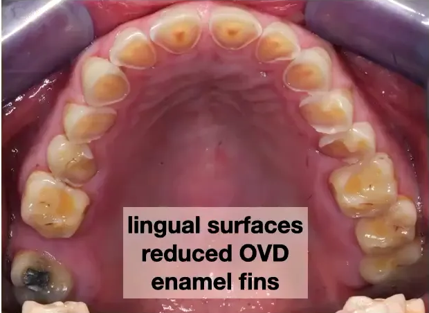
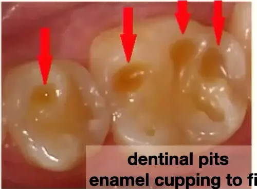
Case: Severe dental erosion and attrition in a GERD patient
Question
Scenario: A patient with gastroesophageal reflux disease (GERD) presenting for dental evaluation.
What’s shown: Clinical photographs showing the palatal surfaces of the incisors with significant wear.
Consider: What are the specific clinical findings on the teeth, and what treatment might be required as a result?
Answer
Observations:
- Erosion and attrition are present together.
- Little valleys and cups are visible on the palatal surfaces of the incisors.
- The pulp chambers are visible.
Reasoning: The chronic acid exposure from GERD causes erosion, which softens the enamel and tooth structure. This makes the teeth highly susceptible to attrition, where even gentle contact causes the softened structure to wear away, creating cupped lesions and eventually exposing the pulp.
Outcome: Some of the affected teeth may require root canal treatment.
Takeaway: GERD can lead to severe dental erosion and attrition, potentially resulting in pulp exposure and the need for endodontic therapy.
Case: Patient with epidermolysis bullosa
Question
Scenario: A patient with a history of epidermolysis bullosa (EB) being discussed in the context of chronic inflammatory and fibrotic conditions.
What’s shown: The patient’s clinical history of widespread mucosal involvement and systemic management.
Consider: What were the patient’s main clinical challenges, and how did the disease course change over time?
Answer
Observations:
- Fibrosis of all mucosal tissues.
- Significant difficulties with swallowing and digestion.
- Required treatment with multiple immunosuppressants.
Reasoning: The extensive fibrosis of the mucosal tissues in EB severely compromises gastrointestinal function, necessitating aggressive immunosuppressive management to control the disease activity.
Outcome: After the age of 35, the disease activity slowed down, which improved the patient’s condition enough to allow her to have children.
Takeaway: While epidermolysis bullosa can cause severe mucosal fibrosis and systemic complications requiring immunosuppressants, the disease activity may naturally slow down after the age of 35.
Footnotes
-
Original PDF page 1: L3 Renal Disease and Dentistry DLim 2026, p.1 ↩
-
Original PDF page 2: L3 Renal Disease and Dentistry DLim 2026, p.2 ↩
-
Original PDF page 3: L3 Renal Disease and Dentistry DLim 2026, p.3 ↩
-
Original PDF page 4: L3 Renal Disease and Dentistry DLim 2026, p.4 ↩
-
Original PDF page 5: L3 Renal Disease and Dentistry DLim 2026, p.5 ↩
-
Original PDF page 6: L3 Renal Disease and Dentistry DLim 2026, p.6 ↩
-
Original PDF page 7: L3 Renal Disease and Dentistry DLim 2026, p.7 ↩
-
Original PDF page 8: L3 Renal Disease and Dentistry DLim 2026, p.8 ↩
-
Original PDF page 9: L3 Renal Disease and Dentistry DLim 2026, p.9 ↩
-
Original PDF page 10: L3 Renal Disease and Dentistry DLim 2026, p.10 ↩
-
Original PDF page 11: L3 Renal Disease and Dentistry DLim 2026, p.11 ↩
-
Original PDF page 12: L3 Renal Disease and Dentistry DLim 2026, p.12 ↩
-
Original PDF page 13: L3 Renal Disease and Dentistry DLim 2026, p.13 ↩
-
Original PDF page 14: L3 Renal Disease and Dentistry DLim 2026, p.14 ↩