Haematology and Dentistry 1: Red Blood Cells (Anaemia)1
2026 DENT5311 DMD3
Dr Jee-Yun Leung
- Specialist in Special Needs Dentistry
- Senior Lecturer | University of Western Australia
- Consultant | Fiona Stanley Hospital
Lecture Outcomes2
- Review the role of RBCs, systemic disorders and treatments which affect their structure and function, and general management
- Review the oral manifestations of disorders of RBCs
- Risk assess the implications of RBC disorders, and the treatment modifications required, when planning dental procedures
- This lecture forms the first of three haematology lectures, covering red blood cells before white blood cells and platelets, coagulation and bleeding disorders
Blood Components and Haemopoiesis
Blood
Blood Components
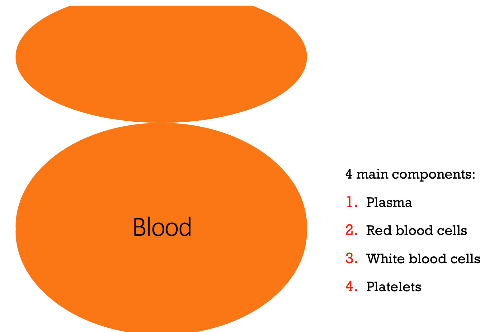
4 main components:
- Plasma
- Red blood cells
- White blood cells
- Platelets
Lecturer — Blood Separation
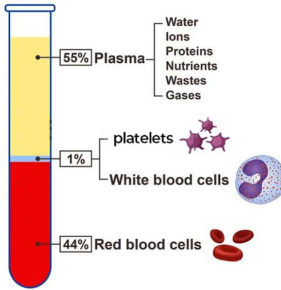
Blood can be separated by centrifugation into three visible layers.
- Plasma forms the pale yellow liquid matrix carrying the blood cells.
- The buffy coat is less than 1% of total blood volume and contains leukocytes and platelets.
- Leukocytes in the buffy coat include monocytes, lymphocytes, eosinophils, basophils and neutrophils.
Composition by Volume3
- 55% Plasma
- Water
- Ions
- Proteins
- Nutrients
- Wastes
- Gases
- 1% Platelets and white blood cells
- 44% Red blood cells
Lecturer — Plasma Composition
Plasma carries antibodies, while its water and ions help provide osmotic balance. Erythrocytes comprise approximately 45% of total blood volume.
 |  |
Haemopoietic Stem Cells and System
Adapted from Kumar and Clark’s Clinical Medicine (11th ed, 2025)
Cell Lineage4
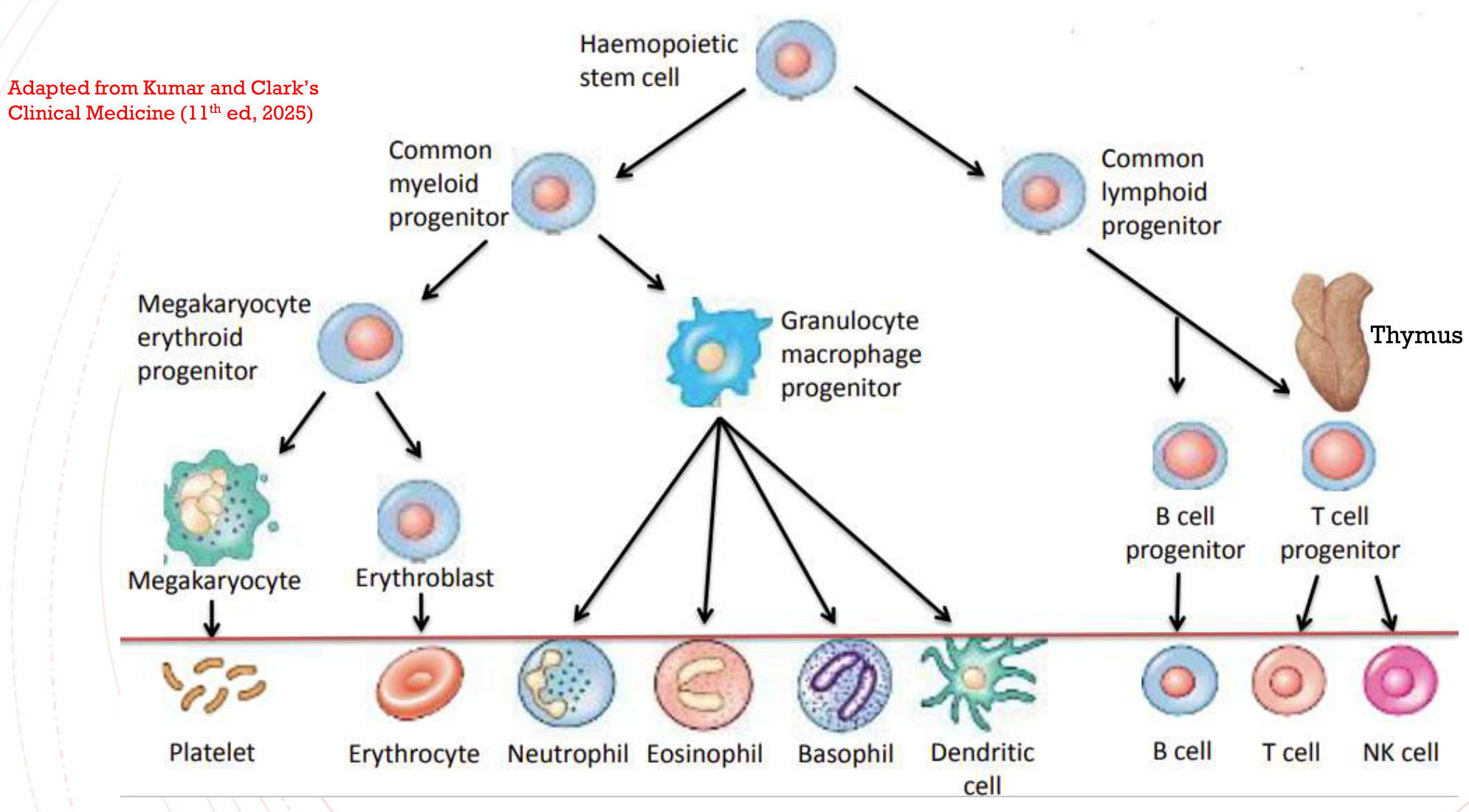
- Haemopoietic stem cell
- Common myeloid progenitor
- Megakaryocyte erythroid progenitor
- Megakaryocyte → Platelet
- Erythroblast → Erythrocyte
- Granulocyte macrophage progenitor
- Neutrophil
- Eosinophil
- Basophil
- Dendritic cell
- Megakaryocyte erythroid progenitor
- Common lymphoid progenitor
- Thymus
- T cell progenitor → T cell
- B cell progenitor → B cell
- NK cell
- Thymus
- Common myeloid progenitor
Lecturer — Haemopoietic Disorders
Blood disorders can be organised according to the affected cell type, including red blood cells, white blood cells and the coagulation system.
- Anaemias are the most common red blood cell disorders and involve a deficiency.
- Many significant white blood cell disorders involve excessive proliferation that results in malignant transformation.
- Platelet abnormalities may result in bleeding disorders.
- These categories are closely linked: a disorder affecting one cell type can affect the entire haemopoietic system and alter other cell types.
Haemopoietic System5
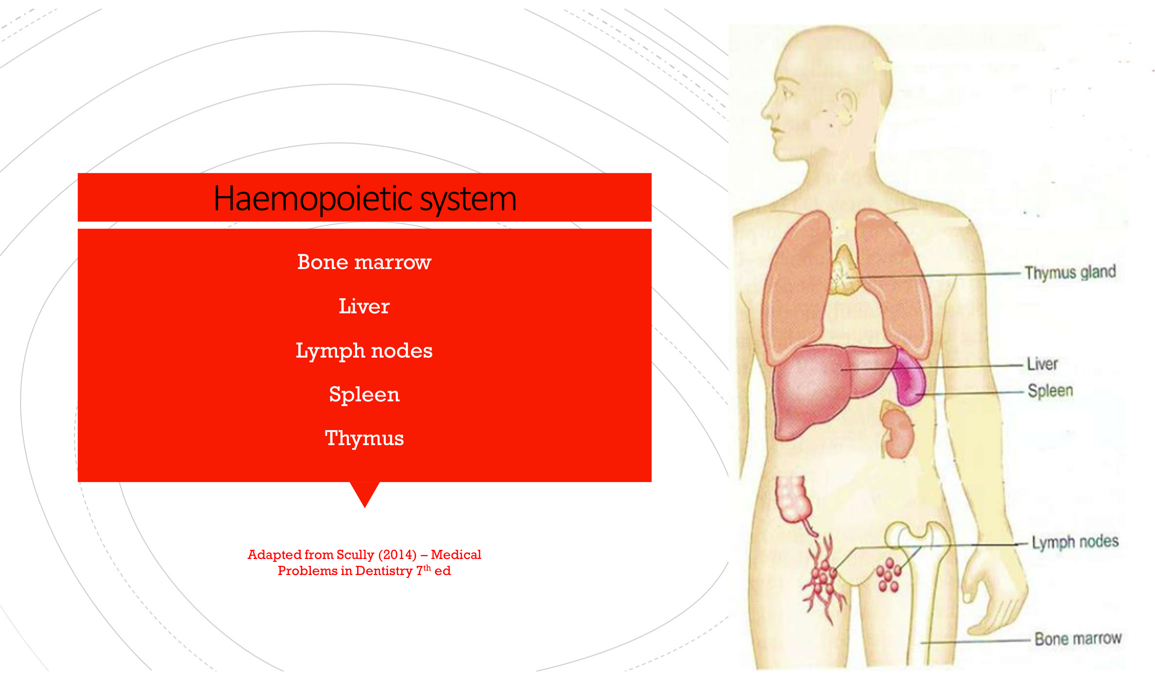
- Bone marrow
- Liver
- Lymph nodes
- Spleen
- Thymus
Adapted from Scully (2014) – Medical Problems in Dentistry 7th ed
Haemopoiesis
- Blood cell formation
- Occurs in haemopoietic system
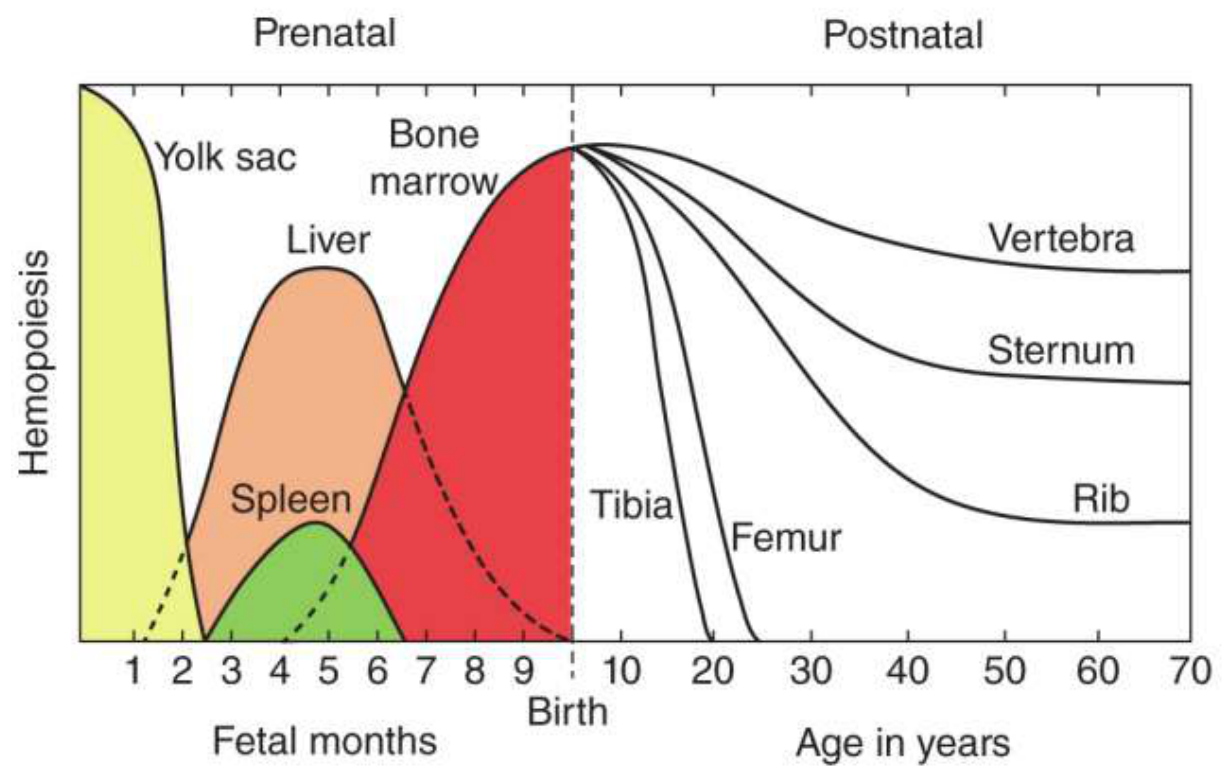
Prenatal and Postnatal Haemopoiesis6
- Prenatal: yolk sac, liver, spleen
- Postnatal: bone marrow
Sites of haemopoiesis over time include bone marrow, liver, spleen, tibia, femur, vertebra, sternum, and rib, spanning fetal months, birth, and age in years (1–70).
Lecturer — Prenatal Haemopoiesis
Blood cell production begins before birth while the embryo is in the uterus. By approximately week three, the yolk sac produces a less-developed type of red blood cell; during months two and three, red blood cells and platelets are produced in the liver and spleen, while white blood cells are produced in the liver, spleen and thymus.
- By month five, most blood cell production occurs in the bone marrow, although the thymus, spleen and other lymphoid tissues continue producing some white blood cells.
- After birth, most production takes place in the bone marrow from infancy into adulthood, while lymphocytes are produced and develop in the thymus.
Blood Tests Reference Values78
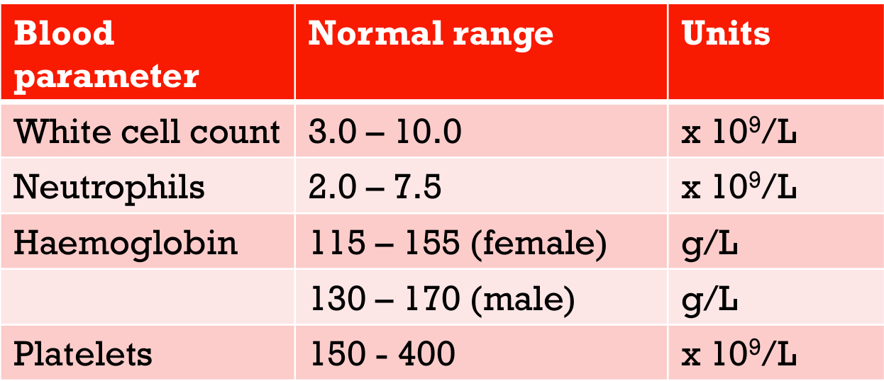
| Blood parameter | Normal range | Units |
|---|---|---|
| White cell count | 3.0 – 10.0 | x 10⁹/L |
| Neutrophils | 2.0 – 7.5 | x 10⁹/L |
| Haemoglobin | 115 – 155 (female) | g/L |
| 130 – 170 (male) | g/L | |
| Platelets | 150 – 400 | x 10⁹/L |
Lecturer — Blood Test Interpretation
Haemoglobin differs between females and males partly because of menstruation. Blood results are clinically important because values outside the expected range can affect dental treatment.
Red Blood Cells and Haemoglobin91011
Red Blood Cells (Erythrocytes)
- Erythrocytes: responsible for oxygen carriage from lungs to tissues
- 120 day lifespan
- Broken down in spleen
- Erythropoiesis: red blood cell production
Haemoglobin (Hb)
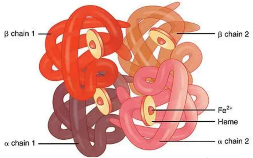
- Protein contained by mature erythrocytes
- 4 globin chains (2 alpha, 2 beta)
- Degradation:
- Hb → haem → bilirubin in the liver
Lecturer — Red Cell Regulation
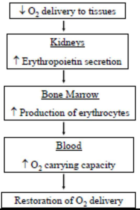
When oxygen delivery to tissues decreases, the kidneys detect the reduction and trigger erythropoietin production, increasing red blood cell production in the bone marrow.
- Red blood cells develop from undifferentiated pluripotent stem cells through immature intermediate cells and reticulocytes.
- After approximately 120 days, damaged red blood cells are phagocytosed by macrophages in the bone marrow, liver and spleen, and their components are recycled.
 |  |
Anaemia Overview and Classification
Anaemia: Disorder of red blood cells, defined as a reduction in the oxygen carrying capacity of the blood.
Definition and Classification12
Classification on the basis of:
Lecturer — Classification Focus
The lecture focused primarily on classification by cause. This approach helps explain how the anaemias develop.
- Cause:
- blood loss anaemia
- deficiency anaemia (reduced erythropoiesis)
- haemolytic anaemia (increased rate of RBC destruction)
- Red-cell size:
- microcytic (MCV <78fl)
- macrocytic (MCV >99fl)
- normocytic (79 < MCV < 98fl)
Microcytic anaemia (MCV <78fl)13
- Iron deficiency
- Thalassemia
- Chronic disease
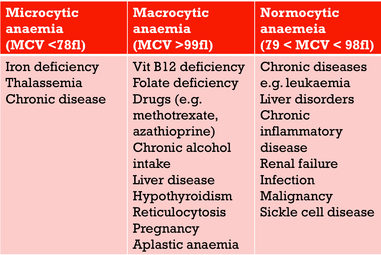
Macrocytic anaemia (MCV >99fl)
- Vit B12 deficiency
- Folate deficiency
- Drugs (e.g. methotrexate, azathioprine)
- Chronic alcohol intake
- Liver disease
- Hypothyroidism
- Reticulocytosis
- Pregnancy
- Aplastic anaemia
Normocytic anaemia (79 < MCV < 98fl)
- Chronic diseases e.g. leukaemia
- Liver disorders
- Chronic inflammatory disease
- Renal failure
- Infection
- Malignancy
- Sickle cell disease
Clinical Manifestations
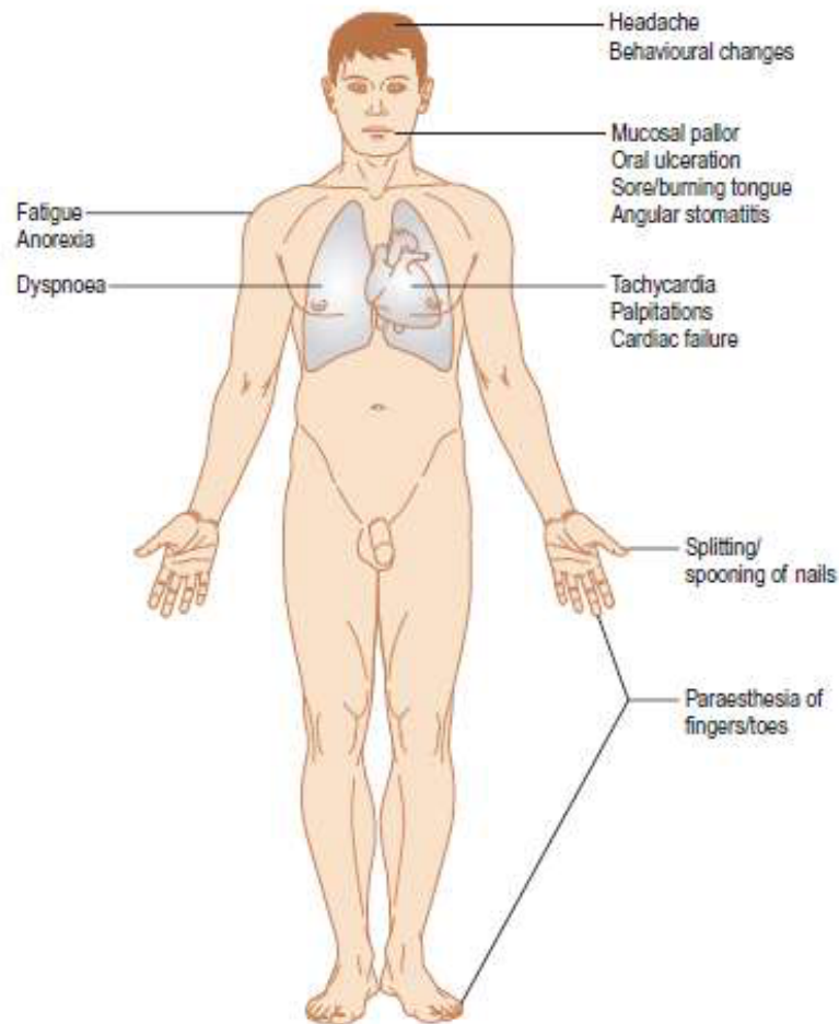
Clinical Features of Anaemia (Box 8.9)14
-
Typically none in the early stages
-
General lassitude
- Fatigue
- Anorexia
-
Neurological/behavioural
- Headache
- Behavioural changes
- Paraesthesia of fingers/toes
-
Cardiorespiratory
- Dyspnoea
- Tachycardia
- Congestive cardiac failure
- Murmurs
- Palpitations
- Angina pectoris
- Cardiac failure
-
Cutaneous
- Pallor (mucosal pallor)
- Brittle nails
- Koilonychia (spoon-shaped nails – Iron deficiency)
- Splitting/spooning of nails
-
Oral
- Sore mouth
- Ulceration
- Angular stomatitis
- Glossitis
- Burning mouth symptoms
- Sore/burning tongue
-
Breathlessness or dyspnoea due to reduced oxygen carriage
-
Muscle cramps
-
Headaches, although these were described as less usual
-
Tachycardia, particularly when anaemia is severe, as the cardiovascular system attempts to circulate more oxygen
Oral Manifestations
Low iron levels can lead to early oral signs!
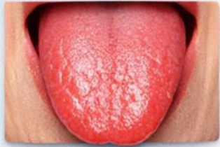
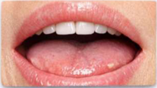
- Smooth, shiny, “beefy red” tongue
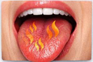
- Burning & tingling pain
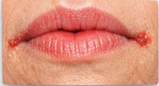
- Cracks at the mouth corners
- Pale gums & mucosa
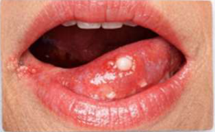
- Painful mouth sores
Cause: ↓ haemoglobin → ↓ oxygen supply → ↓ epithelial regeneration and immunity
Lecturer — Oral Manifestations
The lecturer associated reduced haemoglobin and reduced oxygen supply with increased epithelial regeneration and changes in immunity.
Iron Deficiency Anaemia — Oral Manifestations15
- Atrophic Glossitis — Smooth, shiny, “beefy red” tongue due to loss of filiform papillae. Caused by impaired epithelial turnover. May cause altered taste (dysgeusia).
- Burning Mouth Sensation — Burning or tingling pain without obvious lesions. Due to mucosal atrophy + neural sensitivity.
- Angular Cheilitis — Cracks at mouth corners. Epithelial thinning + secondary Candida/Staph infection. Often linked with iron ± B-complex deficiency.
- Pale Oral Mucosa — Pale gums and inner lips. Reduced hemoglobin → decreased tissue oxygenation.
- Recurrent Oral Ulcers — Painful, slow-healing ulcers. Impaired oxygen delivery delays mucosal repair.
 |  |  |
 |  |
Blood Loss Anaemia16
Definition: occurs after sustaining a wound in which there is acute blood loss
Compensatory Mechanism17
- Immediate threat at this stage is hypovolaemic shock (cf. anaemia) and surviving is the primary objective.
- Kidneys retain salt and water to increase the blood volume.
- Once stabilised, haemodilution begins and achieves its full effect within 2-3 days, masking the extent of the red cell loss.
- Healthy patient with adequate iron stores: full recovery from this transient anaemia by utilising iron stores to increase erythropoiesis → complete cell replacement and correction of the anaemia.
Deficiency Anaemias
- Iron
- Folate
- Vit B12
Overview of Deficiency Anaemia18
Definition: Caused by inadequate supply to the bone marrow of some substances necessary for haemopoiesis (Hb synthesis).
Haematinics19
Nutrients required to make red blood cells:
-
Vitamin B12 = 3 year store
-
Iron = 12 month store
-
Folate = 3 month store
-
The shorter folate store means folate deficiency can develop more quickly than some other deficiency anaemias.
Iron Deficiency Anaemia
Causes20
- Malabsorption (e.g. Coeliac disease, Crohn’s disease)
- Nutritional deficiency (low iron intake – malnutrition, reduced meat diet)
- Excess demand (e.g. pregnancy, infancy)
- Chronic blood loss (e.g. especially GIT – peptic ulcers, haemorrhoids, colon cancer; menstruation)
Features
- Weakness
- Pallor
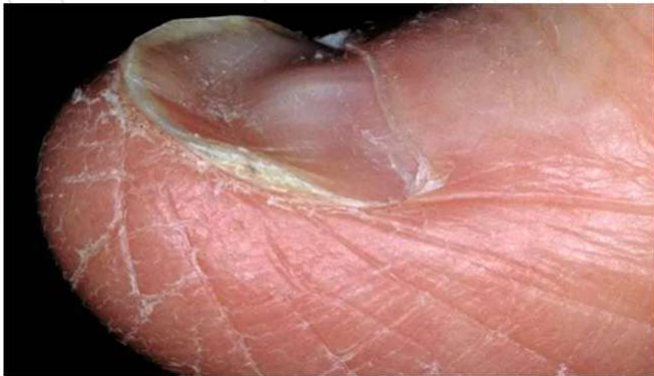
- Koilonychia (nail spooning)
- Hair loss
General Management21
-
Dietary iron
- Heme iron: animal tissues – higher absorption rate
- Non-heme iron: plants, fortified foods – lower absorption rate
- Enhancers of non-heme absorption: Vitamin C
- Inhibitors of non-heme absorption: phytates (in grains/beans), oxalates, phosphates present in plant foods; tannin in tea/coffee/wine; calcium in dairy, oxalates
-
Iron supplements
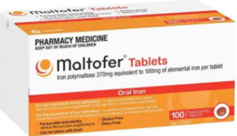
- Oral intake, may take few months to correct, constipation
-
Iron infusions (IV)
-
Multiferr was described as causing less constipation than some other oral iron preparations.
-
Iron infusions are frequently required during pregnancy.
Vitamin B12 Deficiency Anaemia
Vit B12: role in synthesising and breaking down amino acids, and synthesising DNA/RNA.
- Required to build new cells, especially blood, skin and mucosal cells.
Causes22
- Usually defect in intrinsic factor (as a result of pernicious anaemia or gastrectomy)
- Occasionally ileum disease or resection
- Rarely congenital ileum absorption defect
Pernicious Anaemia
Autoimmune condition where the body cannot absorb vitamin B12 due to autoantibodies against gastric parietal cells, and/or intrinsic factor. Without intrinsic factor, Vit B12 is unable to form the complex to absorb in the terminal ileum.
Lecturer — Pernicious Anaemia
Pernicious anaemia is a type of vitamin B12 deficiency anaemia. Gastric parietal cells are involved in producing intrinsic factor, so their impairment can prevent effective vitamin B12 absorption.
Additional Features23
-
Burning tongue
-
Neurological symptoms (paraesthesia of extremities) e.g. loss of toe positional sense and diminished perception of tuning fork vibration, unsteady movement, muscle weakness
-
Shortness of breath
-
Slower healing
-
Premature greying of hair
-
Increased homocysteine
-
Burning tongue is particularly relevant in dentistry.
Treatment
Parenteral or high-dose oral replacement.
Folic Acid Deficiency Anaemia
Folic Acid24
- Needed by body to synthesise and break down amino acids, and synthesise DNA/RNA to build new cells, especially blood, skin and mucosal cells.
- Plays a role in: myelination (liposynthesis to maintain healthy CNS), RBC production, DNA/RNA synthesis → joint metabolism with Vit B12
- Found in fresh leafy vegetables (spinach, kale, Brussel sprouts, asparagus).
- Absorbed from proximal small intestine
Causes
- Usually dietary deficiency
- Some caused by disease of small intestine (e.g. Coeliac)
- Cancer patients treated with methotrexate
Lecturer — Methotrexate Treatment
Methotrexate inhibits an enzyme involved in folate synthesis. In cancer treatment, this inhibition is used to reduce DNA, RNA and protein synthesis.
Features25
-
Similar to B12 deficiency
-
In pregnancy, predisposes foetus to neural tube defects and cleft lip/palate
-
Predisposes to IHD in adults
-
Daily folic acid is recommended during pregnancy to help protect against neural tube defects.
Treatment
- Folic acid 5mg daily for at least 4 months (rapidly restores)
Haemolytic Anaemias
Sickle Cell, Thalassaemia
Overview of Haemolytic Anaemias2627
- Haemoglobinopathies (inherited abnormalities of Hb formation)
- Sickle cell anaemia / sickle cell trait (globin structure affected)
- Thalassaemia (globin chain number diminished)
- Inherited abnormalities of erythrocyte structure or function
- Hereditary spherocytosis (cell shape change)
- Glucose-6-phosphate dehydrogenase deficiency (cell membrane enzyme defect)
- Damage to erythrocytes
- Aplastic anaemia
- Malaria
- Anaemia of chronic disease
- Others
- Pure red-cell aplasia
- Paroxysmal nocturnal haemoglobinuria
Lecturer — Aplastic Anaemia
Aplastic anaemia may cause general features of anaemia, including weakness, pallor and shortness of breath.
- White cells and platelets may also be affected, causing increased susceptibility to infection, bleeding, purpura and petechiae.
- Management may include removal of the cause where possible, antibiotics, immunosuppression, blood transfusions and stem cell transplantation.
- Reported five-year survival was approximately 90% for patients under 40 and 70% for patients under 60, with prognosis decreasing with age.
Sickle Cell Anaemia and Trait
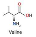
- Sickling disorders: hereditary disorder characterised by the presence of structurally abnormal Hb (point mutation in gene encoding for β-chain; Hb → HbS)
- Sickle cell anaemia or disease (HbSS): homozygous, usually a serious disease with widespread complications, frequently apparent in 3rd month of life. Sickle Cell Anaemia is most severe type of the group of sickle cell diseases.
- Sickle cell trait (HbSA): heterozygous and 10 times more common. Frequently asymptomatic, but sickle cell crises can be caused by low oxygen tension (GA, high altitudes, unpressurised aircraft).
Lecturer — Sickle Cell Complications
Sickle cell disease can produce multiple systemic complications through microemboli and vascular obstruction.
- Involvement may extend to the lungs, kidneys, eyes, brain, liver, spleen and bone marrow.
- Splenic damage affects immunity because the spleen has an important role in immune function.
Epidemiology28
-
Sickle cell trait carriage: Afro-Caribbean (1/10), then India / Middle East / Mediterranean (1/100)
-
The frequency encountered in dental practice depends on the treated population, migration patterns and the country in which the practitioner works; more sickle cell anaemia was encountered in the United Kingdom and Sydney than in Perth.
Cause and Consequences of Sickling
Cause29
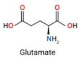
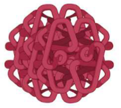
- On deoxygenation, HbS molecules undergo polymerisation (crystallisation).
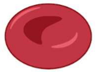
- The change in physical shape distorts the RBC, which assumes a sickle shape.
- Initially this shape deformation is reversible with reoxygenation, but damage occurs with every episode of sickling (e.g. loss of water and potassium, and accumulation with calcium).
- Eventually the sickle shape becomes permanent.
Sickled RBC with mutated hemoglobin (HbS) — glutamate, β6 position
 |  |
 |  |
Consequences of Sickle Cells30
- Membrane damage and dehydration of RBC: renders them rigid and irreversibly sickled → mean lifespan reduced from 120 to 20 days as mononuclear phagocytes remove damaged RBCs → chronic extravascular haemolytic anaemia
- Widespread microvascular obstructions: occurs in kidney & spleen due to congestion of red pulp with masses of sickled RBC packed together → splenic erythrostasis → hypoxia → shrunken/fibrous spleen
Features and General Management
Features31
- Chronic anaemia
- Chronic hyperbilirubinaemia
- Painful crisis (i.e. vaso-occlusive crisis)
- Infarcts as a result of sickling: spleen, lungs, kidneys, eyes, cerebrovascular stroke
- Brought on by infection, dehydration, hypoxia, acidosis, cold.
- Haematological crises: caused by parovirus infection
- Infections: due to immune defect resulting from splenic infarction and dysfunction.
- Sequestration syndromes: sequestration of sickle cells in lungs (impeding gas exchange → chest syndrome), spleen (→ septicaemia), liver (→ bowel dilatation)
Lecturer — Aplastic Crises
During an aplastic crisis, erythropoiesis suddenly and temporarily stops.
- Reticulocytes disappear from the blood.
- The anaemia consequently worsens.
Box 8.13 — Features of Sickle Cell Anaemia
- Anaemia
- Crises
- Painful crises
- Aplastic crises
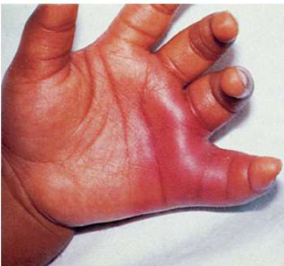
- Dactylitis – painful osteitis in the hands (Fig. 8.35)
- Infarcts of central nervous system, lungs, kidney, spleen, bone
- Skin ulcers
- Haemolysis
- Jaundice
- Gallstones
- Reticulocytosis
- Impaired growth
- Skeletal deformities
- Susceptibility to infections
General Management32
Main principles in treatment are to prevent trauma, infection, hypoxia, acidosis or dehydration:
- regular blood pictures
- comprehensive care programmes
- regular folic acid
- exchange transfusions (remove sickled cells and replace defective cells) — heparinised
- fluids
- keep warm (clothes)
- long term antibiotics
- most pts on prophylactic penicillin too
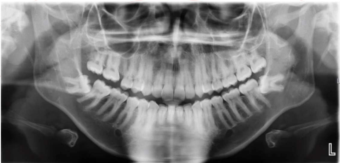
Oral Findings in Sickle Cell Anaemia
- Increased radiolucency in posterior body of mandible
- Enlarged haemopoietic maxilla with excessive OJ and OB (due to bone marrow hyperplasia)
- Trigeminal neuropathy (due to osteomyelitis)
- Painful infarcts in jaws (can be mistaken for toothache)
- Enamel hypomineralisation and calcified pulp canals
- Pulp symptoms and pulp necrosis in absence of obvious dental disease
- Delayed skeletal maturation
Radiological Features33
- Hypercementosis (dense lamina dura)
- Sparse mandibular trabecular bone pattern (due to bone marrow hyperplasia)
- Thickening/expansion of skull diploe with “hair-on-end” appearance
- Dense radio-opacities with “stepladder” trabeculae pattern (due to previous infarcts)
- Increased radiolucency of the bone in the inferior body of the mandible — due to micro-embolisms in sickle cell artery
Grossman S, Bailey E, Makdissi J. Oral aspects of sickle cell disease, Br Dent J. 2019; 226: 818.
Thalassaemia
Definition and Cause
- Definition: group of heterogenous autosomal dominant genetic disorders of Hb synthesis, characterized by a lack or decreased synthesis of globin chains.
- May be severe (major; homozygous) or mild (minor; heterozygous)
- α-thalassemia: α-globin chain synthesis is ↓
- β-thalassemia: β-globin synthesis is ↓ or non-existent
Lecturer — Sickle Cell and Thalassaemia
A key distinction is that sickle cell disease is associated particularly with infarcts, whereas thalassaemia is associated particularly with iron deposition.
Lecturer — Alpha Globin Chains
Alpha thalassaemia may be relatively mild because there are four alpha-globin chains.
- If all four alpha-globin chains are affected, the condition is not compatible with life and the individual dies in infancy.
- There are only two beta-globin chains, so abnormalities can produce more severe effects.
Epidemiology34
- Alpha: south-east Asian, southern Chinese, sub-Saharan Africa, Middle East
- Beta: Mediterranean
Cause — Beta Thalassemia35
β-globin synthesis is ↓ or non-existent:
- Excess of unpaired α-chains → chains aggregate within the RBC and form insoluble precipitates → damages RBC → sequestration
- Systemic iron overload due to mass destruction of RBCs
- Can be life threatening in homozygous form
Clinical Presentation and General Management
Clinical Presentation — Homozygotes36
- Iron Overload from repeated transfusions:
- Thyroid & parathyroid damage
- Pituitary (delayed puberty, short stature)
- Liver & kidney damage
- Cardiac failure
- Pancreas (secondary diabetes)
- Skin (green/bronze pigmentation due to iron deposition)
- Low levels estrogen or testosterone
- Impact of Treatment:
- Regular hospital admissions for transfusions
- Disruption to family/school/social life
- Psychological implication of side effects
- Compliance with chelation (particularly adolescence)
Lecturer — Thalassaemia Complications
Thalassaemia can be considered a disease associated with multiple secondary conditions because iron is deposited throughout the body.
- Iron deposition may affect the liver, kidneys, heart, pancreas, thyroid, parathyroid and pituitary system.
- Cardiac deposition may cause cardiomyopathy and cardiac failure, while liver disease may include cirrhosis.
- Endocrine effects may include thyroid dysfunction and reduced sex hormones.
General Management37
-
Regular blood transfusion (to treat anaemia)
-
Folic acid supplements (builds new blood cells)
-
Iron-chelating agents, ascorbic acid
-
Splenectomy (if hypersplenism causing greater blood destruction → accumulation of iron elsewhere) → daily antibiotic prophylaxis
-
Alpha and beta globin chains are normally intended to match; unpaired alpha chains aggregate inside red blood cells and form insoluble precipitates that damage them.
Management of Other Affected Organs
- Pancreas → diabetes testing
- Hypothyroidism → thyroid hormone
- Osteoporosis → bisphosphonates, calcium
- Liver → MRI monitoring
- Hormone replacement
- Gallbladder → gallbladder removal if gallstones
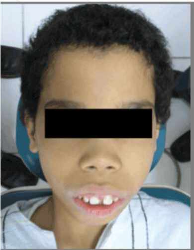
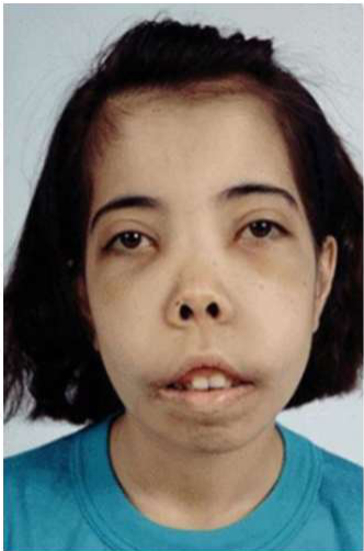
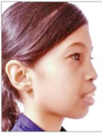
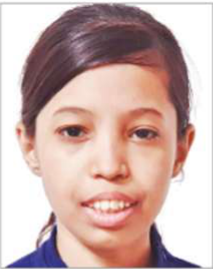
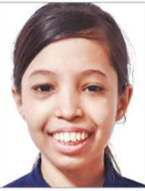
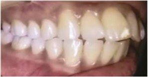
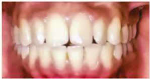
Oral Findings in Thalassaemia38
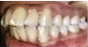
- Enlargement of maxilla (“chipmunk face”) due to extramedullary haemopoiesis → OJ and OB
- Migration and spacing of upper anterior teeth
- Painful swelling of parotids and hyposalivation (due to iron deposits)
- Delayed pneumatisation of maxillary sinuses
- Teeth discoloured with short crowns/roots
- Alveolar bone with “chicken wire” radiographical appearance (extramedullary haematopoiesis)
- Sore/burning tongue (folate deficiency)
- Pale gingiva and mucosa (anaemia)
 |  |  |
 |  |  |
 |  |  |
Aplastic Anaemia
- Aplastic anaemia: Macrocytic pancytopenia with a non-functioning bone marrow. A rare disease, characterised by suppression of multipotent myeloid stem cells causing leukopenia, thrombocytopenia and refractory anaemia.
- Fanconi anaemia: inherited type of aplastic anaemia associated with progressive deficiency of all bone marrow production blood cells and increased risk of AML
Definition and Causes
- Idiopathic: poor prognosis
- Secondary: after whole body irradiation/chemo, chemicals, drugs, hepatitis viruses, GVHD
Box 8.14 Causes of aplastic anaemia39
- Genetic
- Fanconi anaemia
- Dyskeratosis congenita
- Immunological
- Autoimmune
- Graft-versus-host disease
- Drugs
- Allopurinol
- Anticonvulsants
- Antithyroids
- Chloramphenicol
- Cytotoxic agents
- Gold
- Non-steroidal anti-inflammatory drugs (NSAIDs)
- Penicillamine
- Phenylbutazone
- Sulphonamides
- Chemicals
- Benzene
- Glue-sniffing
- Heavy metals
- Toluene
- Viruses
- Hepatitis
- Irradiation
Features and General Management4041
Features: those of anaemia + abnormal susceptibility to infection/bleeding
- Purpura, petechiae, ecchymoses
- Predisposition to infection
- Weakness, pallor, dyspnoea
General Management:
-
Remove cause
-
Isolation and antibiotics to control infection
-
Immunosuppression
-
Blood transfusion
-
Stem cell transplant
-
Patients with aplastic anaemia may be encountered in hospital settings during assessment for stem cell transplantation. Prognosis — if treated:
-
90% for 19–39yo
-
70% for 40–59yo
-
38% for >60yo
-
Can evolve into AML
Dental Management Considerations4243
Dental management
Oral features:
- Similar to leukaemia (ulceration, infection susceptibility, haemorrhaging tendencies)
- Oral lichenoid lesions
- Sjogren-like syndrome of GVHD
- Gingiva enlargement (ciclosporin, or AML transformation)
Risk Assessment
Red blood cell count (>80–100 g/L)
Questions for Patient44
-
Are you transfusion-dependent?
-
How many times per week/month?
-
Do you experience shortness of breath? (if so, when)
-
Transfusion dependence gives an indication of disease control and severity.
-
Shortness of breath has implications for airway management and positioning.
-
Ask about triggers for shortness of breath.
-
Establish whether the patient has had a splenectomy.
-
Ask about other affected organs and relevant complications.
-
Establish whether the patient is receiving exchange transfusions.
-
Determine whether the patient may be heparinised or otherwise anticoagulated.
Risks Specific to Thalassaemia (Especially Beta-Thalassaemia Major)
-
Splenectomy = infection risks
-
Osteoporosis (possible MRONJ risk)
-
Cardiomyopathy and cardiac failure
-
Liver cirrhosis
-
Kidney
-
Diabetes (secondary to pancreas iron deposition)
-
Thyroid/parathyroid damage
-
Cardiomyopathy from iron deposition may progress to cardiac failure.
-
The risks depend on which organs have been affected.
Risks Specific to Sickle Cell Anaemia
-
Timing around transfusions
-
Oxygen support
-
Infection risks
-
Carrying out treatment during phase when haemolysis is minimal
-
Depends on other organs affected
-
Heparinised for exchange transfusions
-
Dental treatment should not be carried out on the same day as an exchange transfusion.
-
Treatment may be possible the following day once the heparin has cleared.
-
Anticoagulation may increase bleeding risks during dental treatment.
Dental Management
Access and Prevention
Access45
- Short appointments in morning to ensure patient not too fatigued.
- If patient receives regular exchange/blood transfusions, avoid treatment on same day (will also be heparinised).
Lecturer — Transfusion Timing
Treatment should be coordinated around transfusions. Treatment may be carried out the following day after heparin has cleared.
Communication/Consent
- Nil significant
Education/Prevention
- Ensure strict oral hygiene regime and prevention education to avoid need for dental treatment.
- Removable prosthetics may be unable to be tolerated when oral ulcers present.
Lecturer — Communication and Prevention
Visual disturbances caused by vascular complications may create communication difficulties. The patient’s systemic disease, transfusion dependence and organ involvement should be established before treatment, and consent should account for the increased risks associated with anaemia, haemolysis, infection, bleeding and affected organs.
- Anaemia can be associated with oral ulceration, and the mouth may be more sore during dental procedures.
Pre-Operative and Peri-Operative Care
Surgery – Pre-Operative46
- Haemoglobin assay/complete blood count performed.
- Ideally correct anaemia prior to dental treatment and check Hb level raised, if necessary by transfusion in emergency.
- Patient stabilised at least 24 hrs pre-operatively.
- Hb estimations are unreliable for 12 hrs post-transfusion or after acute blood loss.
Caution advised if <80 g/L: ideally only emergency dental treatment to stabilise dentition with peri-operative nasal oxygen/pulse oximeter.
Lecturer — Blood Count Interpretation
Blood counts should be interpreted cautiously because they do not necessarily show the functional status of red blood cells or the function of other blood cell lines. Blood counts do not rise immediately after transfusion, and the same principle applies after acute blood loss because compensatory mechanisms take time to develop.
Surgery – Peri-Operative47
- Consider supplemental nasal oxygen (3 L/min) if Hb <80–100 g/L.
- [SCA] Avoid prilocaine (risk: methemoglobinemia).
- Nitrous oxide contraindicated in Vit B12 deficiency — interferes with B12 metabolism.
- Deeper levels of sedation — risk of hypoxia; reconsider elective IV/GA’s if Hb <80–100 g/L.
- [SCA/Th] Consider prophylactic/surgical antibiotics in post-splenectomy patients.
Lecturer — Anaesthesia Risks
The myocardium may be unable to respond adequately to the demands of anaesthesia or sedation. Severe anaemia therefore increases the risk of hypoxia during deeper sedation, including intravenous sedation and general anaesthesia.
Infection Control and Antibiotic Prophylaxis
Infection Control Considerations48
- Standard infection control: risk of BBVs in older patients receiving recurrent transfusions prior to current blood product screening standards.
Lecturer — Blood-Borne Viruses
Some older patients who received regular transfusions throughout their lives may have acquired blood-borne viruses when blood-product screening was less rigorous. Possible blood-borne viruses include HIV, hepatitis B and hepatitis C.
Antibiotic Prophylaxis
Consider antibiotic prophylaxis if:
- Other cell lines affected
- Splenectomy
- Sickle cell disease
Lecturer — Antibiotic Prophylaxis
Patients with thalassaemia who have had a splenectomy require antibiotic prophylaxis. Long-term penicillin may be used in sickle cell disease to reduce infection risk, and patients with sickle cell disease may have more profound immunocompromise.
Questions and Contact49
Questions?
Clinical Cases
Case: Fingernail exhibiting spooning
Question
Scenario: A clinical photograph of a fingernail is presented to illustrate the physical features of a specific type of anaemia.
What’s shown: A close-up clinical photograph of a fingernail.
Consider: Identify the specific nail abnormality visible on the image and the associated type of anaemia.
Answer
Observations:
- The fingernail exhibits a concave, spoon-like shape (spooning/koilonychia).
Reasoning: The lecturer points out the spooning of the nail on the image, identifying it as a classic clinical feature of iron deficiency anaemia, alongside other symptoms such as weakness and hair loss.
Outcome: None stated for this exhibit.
Takeaway: Spooning of the nails (koilonychia) is a classic clinical feature of iron deficiency anaemia.
Case: Panoramic radiograph showing mandibular and skull changes
Question
Scenario: A panoramic radiograph is presented to illustrate the radiological features associated with sickle cell anemia.
What’s shown: A panoramic dental radiograph (OPG).
Consider: Identify the radiological findings visible in the image and relate them to the underlying pathophysiology of the condition.
Answer
Observations:
- Increased radiolucency in the inferior body of the mandible, indicating a sparse mandibular trabecular bone pattern.
- Thickening and expansion of the skull diploë (the middle layer of spongy bone in the flat bones of the skull).
- A “hair on end” appearance of the skull.
- Dense radiopacities with a stepladder trabecular pattern.
Reasoning: The increased radiolucency in the mandible and the expansion of the skull diploë are a result of bone marrow hyperplasia, as the body struggles to produce red blood cells normally. The “hair on end” appearance is a classic sign of this diploë expansion. The dense radiopacities with a stepladder trabecular pattern correlate with previous bone infarcts from sickle crises.
Outcome: None stated for this specific exhibit.
Takeaway: Sickle cell anemia causes bone marrow hyperplasia that leads to distinct radiological features, including sparse mandibular trabeculation, expansion of the skull diploë with a “hair on end” appearance, and stepladder trabecular patterns resulting from previous infarcts.
Footnotes
-
Original PDF page 1: L5.1 haematology 1 RBC, p.1 ↩
-
Original PDF page 2: L5.1 haematology 1 RBC, p.2 ↩
-
Original PDF page 3: L5.1 haematology 1 RBC, p.3 ↩
-
Original PDF page 4: L5.1 haematology 1 RBC, p.4 ↩
-
Original PDF page 5: L5.1 haematology 1 RBC, p.5 ↩
-
Original PDF page 6: L5.1 haematology 1 RBC, p.6 ↩
-
Original PDF page 7: L5.1 haematology 1 RBC, p.7 ↩
-
Original PDF page 8: L5.1 haematology 1 RBC, p.8 ↩
-
Original PDF page 9: L5.1 haematology 1 RBC, p.9 ↩
-
Original PDF page 10: L5.1 haematology 1 RBC, p.10 ↩
-
Original PDF page 11: L5.1 haematology 1 RBC, p.11 ↩
-
Original PDF page 12: L5.1 haematology 1 RBC, p.12 ↩
-
Original PDF page 13: L5.1 haematology 1 RBC, p.13 ↩
-
Original PDF page 14: L5.1 haematology 1 RBC, p.14 ↩
-
Original PDF page 15: L5.1 haematology 1 RBC, p.15 ↩
-
Original PDF page 16: L5.1 haematology 1 RBC, p.16 ↩
-
Original PDF page 17: L5.1 haematology 1 RBC, p.17 ↩
-
Original PDF page 18: L5.1 haematology 1 RBC, p.18 ↩
-
Original PDF page 19: L5.1 haematology 1 RBC, p.19 ↩
-
Original PDF page 20: L5.1 haematology 1 RBC, p.20 ↩
-
Original PDF page 21: L5.1 haematology 1 RBC, p.21 ↩
-
Original PDF page 22: L5.1 haematology 1 RBC, p.22 ↩
-
Original PDF page 23: L5.1 haematology 1 RBC, p.23 ↩
-
Original PDF page 24: L5.1 haematology 1 RBC, p.24 ↩
-
Original PDF page 25: L5.1 haematology 1 RBC, p.25 ↩
-
Original PDF page 26: L5.1 haematology 1 RBC, p.26 ↩
-
Original PDF page 27: L5.1 haematology 1 RBC, p.27 ↩
-
Original PDF page 28: L5.1 haematology 1 RBC, p.28 ↩
-
Original PDF page 29: L5.1 haematology 1 RBC, p.29 ↩
-
Original PDF page 30: L5.1 haematology 1 RBC, p.30 ↩
-
Original PDF page 31: L5.1 haematology 1 RBC, p.31 ↩
-
Original PDF page 32: L5.1 haematology 1 RBC, p.32 ↩
-
Original PDF page 33: L5.1 haematology 1 RBC, p.33 ↩
-
Original PDF page 34: L5.1 haematology 1 RBC, p.34 ↩
-
Original PDF page 35: L5.1 haematology 1 RBC, p.35 ↩
-
Original PDF page 36: L5.1 haematology 1 RBC, p.36 ↩
-
Original PDF page 37: L5.1 haematology 1 RBC, p.37 ↩
-
Original PDF page 38: L5.1 haematology 1 RBC, p.38 ↩
-
Original PDF page 39: L5.1 haematology 1 RBC, p.39 ↩
-
Original PDF page 40: L5.1 haematology 1 RBC, p.40 ↩
-
Original PDF page 41: L5.1 haematology 1 RBC, p.41 ↩
-
Original PDF page 42: L5.1 haematology 1 RBC, p.42 ↩
-
Original PDF page 43: L5.1 haematology 1 RBC, p.43 ↩
-
Original PDF page 44: L5.1 haematology 1 RBC, p.44 ↩
-
Original PDF page 45: L5.1 haematology 1 RBC, p.45 ↩
-
Original PDF page 46: L5.1 haematology 1 RBC, p.46 ↩
-
Original PDF page 47: L5.1 haematology 1 RBC, p.47 ↩
-
Original PDF page 48: L5.1 haematology 1 RBC, p.48 ↩
-
Original PDF page 49: L5.1 haematology 1 RBC, p.49 ↩