Haematology and Dentistry 2: Immunocompromise and Haematological Malignancies1
Immunocompromise (WBC) and Haematological Malignancies
2026 DMD3 DENT5311 – Integrated Dental Practice 2 (Special Needs Dentistry)
Dr Jee-Yun Leung BDSc(Hons) MSc(SCD) DSCD(RCSEng) DClinDent(SND) MRACDS(SND)
Lecture Outcomes2
-
Review the role of WBCs, systemic disorders and treatments which affect their structure and function, and general management
-
Review the oral manifestations of disorders of WBCs
-
Risk assess the implications of WBC disorders, and the treatment modifications required, when planning dental procedures
Overview of White Cell Disorders3
- Non-neoplastic disorders of white cells
- Leukopenia
- Reactive Leukocytosis
- Reactive Lymphadenitis
- Neoplastic proliferations of white cells
- Leukaemias
- Myelodysplastic syndromes
- Myeloproliferative neoplasms
- Lymphomas: Hodgkin’s and Non-Hodgkin’s
- Plasma cell neoplasms (e.g. multiple myeloma)
- Histiocytic neoplasms
- Post-transplant lymphoproliferative disorder
Adapted from Robins Basic Pathology ed11 2023 (Kumar, Abbas, Aster, Robbins)
Lecturer — White Cell Disorders
White-cell disorders may involve deficiencies, but proliferations or excess cells are more common.
- The lecture focused mainly on neoplastic proliferations.
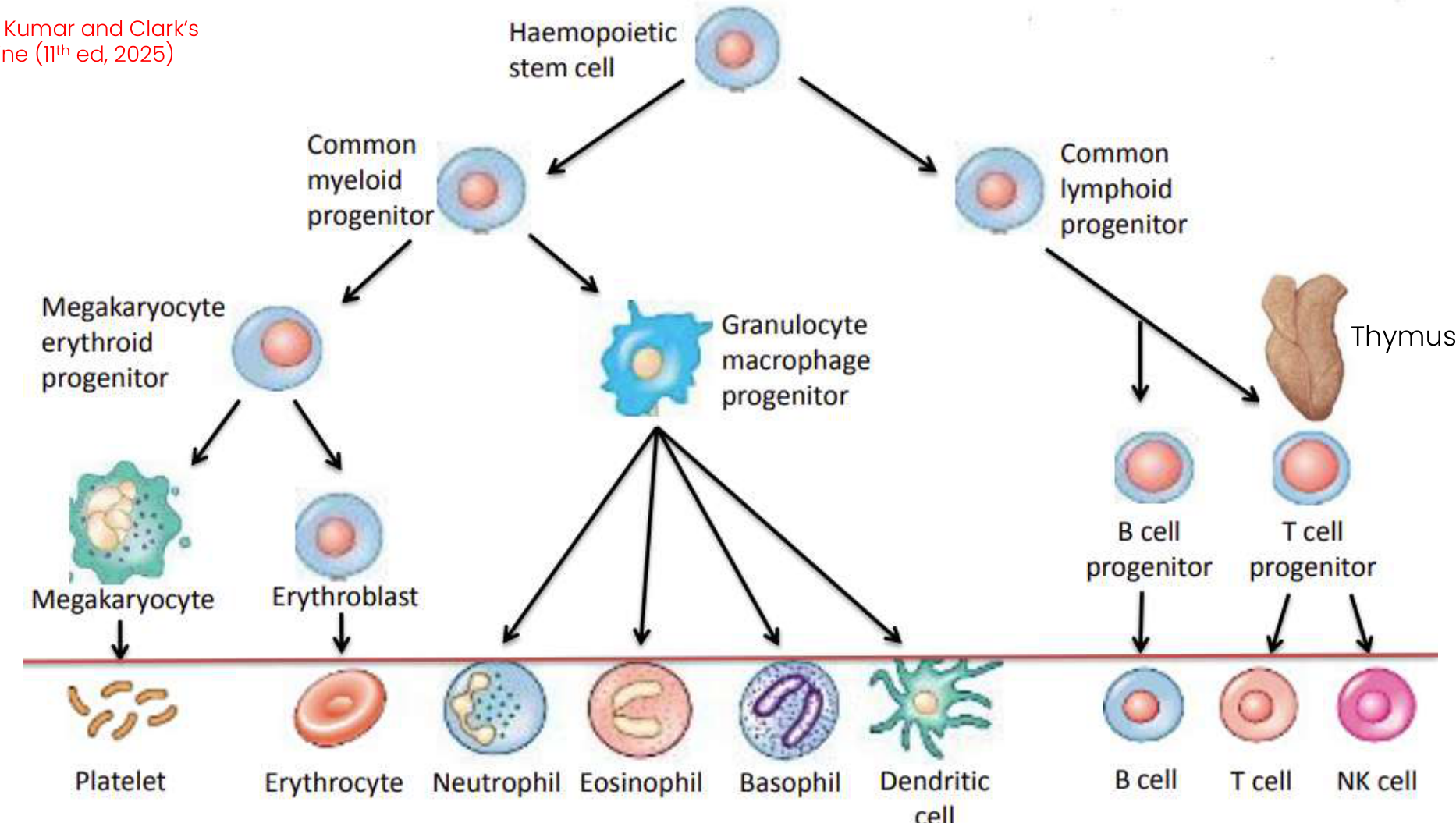
Formation of Blood Cells
Adapted from Kumar and Clark’s Clinical Medicine (11th ed, 2025)
Haemopoietic Cell Lineage4
- Haemopoietic stem cell
- Common myeloid progenitor
- Megakaryocyte erythroid progenitor
- Megakaryocyte → Platelet
- Erythroblast → Erythrocyte
- Granulocyte macrophage progenitor
- Neutrophil
- Eosinophil
- Basophil
- Dendritic cell
- Megakaryocyte erythroid progenitor
- Common lymphoid progenitor
- B cell progenitor → B cell
- T cell progenitor (thymus) → T cell
- NK cell
- Common myeloid progenitor
Lymphoid Organs
Primary Lymphoid Organs5
- Thymus
- Bone marrow
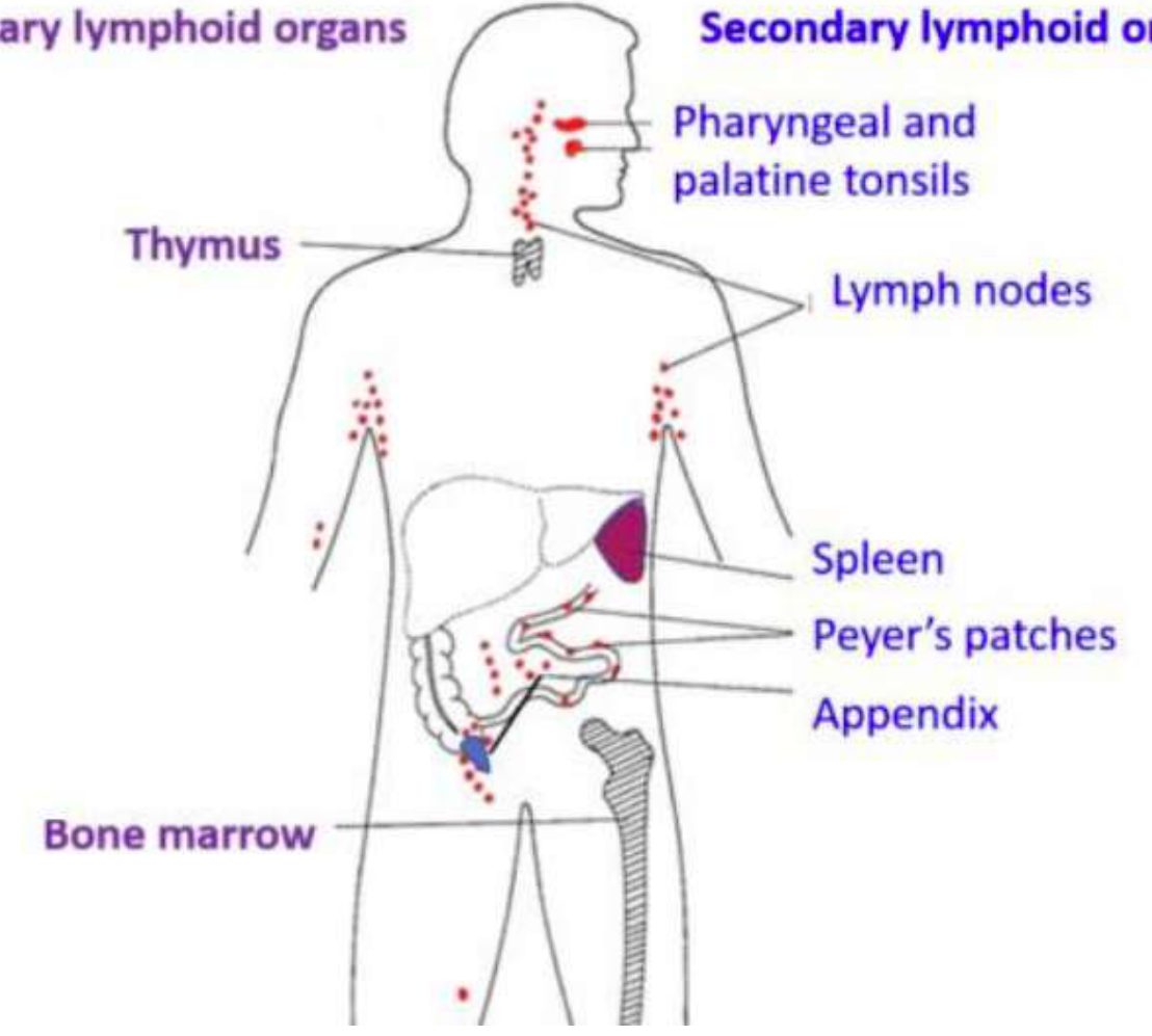
Secondary Lymphoid Organs
- Pharyngeal and palatine tonsils
- Lymph nodes
- Spleen
- Peyer’s patches
- Appendix
Lecturer — Lymphatic System
The lymphatic system collects excess fluid from tissues, returns it to the venous circulation, circulates lymphocytes, and contributes to adaptive, lymphocyte-mediated immunity.
- It is therefore part of both the circulatory system and the immune system.
Lymph Node Groups
Palpable Nodes6
- Cervical
- Pre-auricular
- Upper cervical
- Median or lower cervical
- Posterior cervical
- Mediastinal
- Paratracheal
- Hilar
- Epitrochlear
- Axillary
- Inguinal
- Femoral
- Popliteal
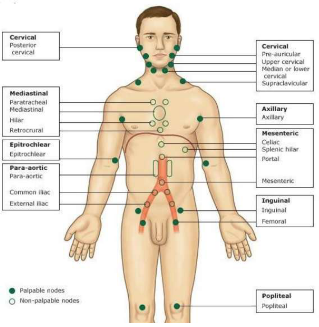
Non-palpable Nodes
- Mediastinal
- Retrocrural
- Para-aortic
- Common iliac
- External iliac
- Mesenteric
- Celiac
- Splenic hilar
- Portal
Lecturer — Lymph Node Examination
Cervical lymph nodes are routinely assessed during extraoral examination. Other lymph-node clusters are distributed throughout the body and may not be palpable during routine dental examination.
Non-Neoplastic Disorders of White Cells7
Leukopenia89
-
Most commonly due to neutropenia (reduced granulocyte count)
- Agranulocytosis: severe reduction in granulocyte count
- Mechanisms:
- Decreased granulocyte production: marrow hypoplasia (chemo, aplastic anaemia), certain drugs
- Increased granulocyte destruction: immune-mediated injury (e.g. drugs), overwhelming infections (increased peripheral “use”), sequestration from splenomegaly
-
Lymphopenia: less common — rare congenital immunodeficiency diseases, advanced HIV infection, high dose corticosteroids
-
Granulocytes are the most numerous circulating white cells.
Reactive Leukocytosis
-
Inflammatory states caused by microbial and nonmicrobial stimuli
-
Infectious mononucleosis → lymphocytosis
-
Reactive leukocytosis is an increased number of white cells in the blood.
Reactive Lymphadenitis
-
Infections and nonmicrobial inflammatory stimuli activate immune cells residing in lymph nodes → lymphadenopathy
-
Lymph nodes act as defence barriers.
-
Infections causing lymphadenitis are varied and numerous and may be acute or chronic.
Classification Systems
- WHO (World Health Organisation) — very comprehensive
- FAB (French American British) — myeloproliferative disorders
Lecturer — Classification Scope
These classification systems were introduced for awareness rather than detailed memorisation. The WHO system is used for malignant disorders of white cells.
WHO and FAB Classification
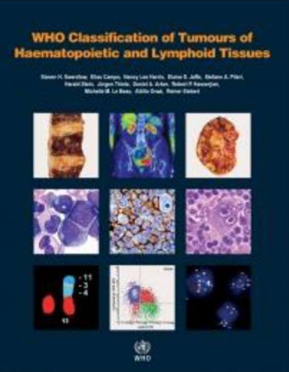
WHO 200810
WHO Classification of Tumours of Haematopoietic and Lymphoid Tissues
French American British (FAB) — 1976 & 1982
British Journal of Haematology, 1976, 33, 451. Proposals for the Classification of the Acute Leukaemias. French-American-British (FAB) Co-operative Group. J. M. Bennett, D. Catovsky, Marie-Thérèse Daniel, G. Flandrin, D. A. G. Galton, H. R. Grainick and C. Sultan. (Received 23 January 1976; accepted for publication 26 January 1976)
British Journal of Haematology, 1982, 51, 189–199. Proposals for the classification of the myelodysplastic syndromes. J. M. Bennett, D. Catovsky, M. T. Daniel, G. Flandrin, D. A. G. Galton, H. R. Grainick and C. Sultan, The French-American-British (FAB) Co-operative Group. (Received 12 November 1981; accepted for publication 18 December 1981)
- Chronic myelomonocytic leukaemia was referenced in relation to oral manifestations as a subtype of myelodysplastic syndrome.
REAL and EORTC Classification11
- REAL (Revised European and American Lymphoma), 1994 — lymphomas
- EORTC (European Organisation for Research and Treatment of Cancer), 1997 — lymphoma
Common Haematological Malignancies1213
- Leukaemias
- Lymphomas
- Hodgkin’s lymphoma
- Non-Hodgkin’s lymphomas
- Myelodysplastic syndromes
- [Non-leukaemic] Myeloproliferative neoplasms
- Plasma cell tumours
- Particularly multiple myeloma
Leukaemias
- Definition: Malignant neoplasms of haematopoietic tissue that progressively displace normal blood-forming elements of bone marrow with neoplastic cells
Definition and Main Types1415
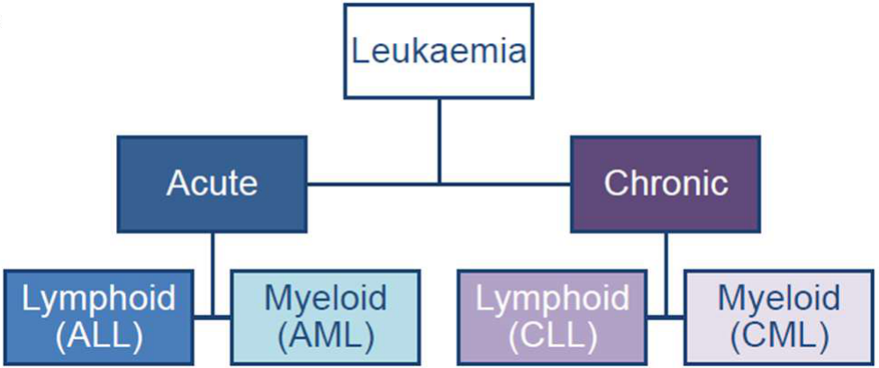
- Classification: on basis of cell type and maturity
- Lymphoid or myeloid
- Acute or chronic
- Acute: >20% myeloblasts or lymphoblasts in bone marrow at clinical presentation
- Chronic: cells more differentiated
Main types of leukaemia:
- Acute lymphoblastic leukaemia (ALL): most common type of childhood leukaemia
- Acute myeloblastic leukaemia (AML): can occur at any age, more often older people
- Chronic lymphocytic leukaemia (CLL): most common type of leukaemia in adults. Slow-growing, may be asymptomatic for months–years.
- Chronic myeloid leukaemia (CML): mostly affects adults >40 years old. Progresses over weeks or months.
 |  |
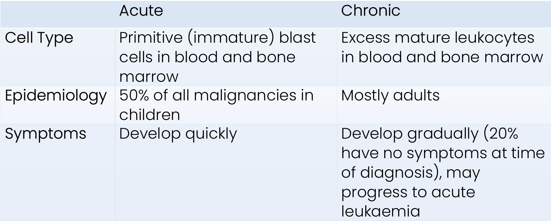
Acute Versus Chronic Leukaemia16
| Acute | Chronic | |
|---|---|---|
| Cell Type | Primitive (immature) blast cells in blood and bone marrow | Excess mature leukocytes in blood and bone marrow |
| Epidemiology | 50% of all malignancies in children | Mostly adults |
| Symptoms | Develop quickly | Develop gradually (20% have no symptoms at time of diagnosis), may progress to acute leukaemia |
Acute Leukaemia Clinical Features
Clinical features associated with:
- Bone marrow failure
- Tissue infiltration
Bone Marrow Failure17
| Anaemia | Neutropenia | Thrombocytopenia |
|---|---|---|
| Fatigue | Infections | Bleeding |
| Breathlessness | Fever | Bruising |
| Pallor | Oral ulceration | Gingival bleeding |
-
In congenital disorders, AML may develop during childhood or adolescence. Clinical features associated with:
-
Bone marrow failure
-
Tissue infiltration

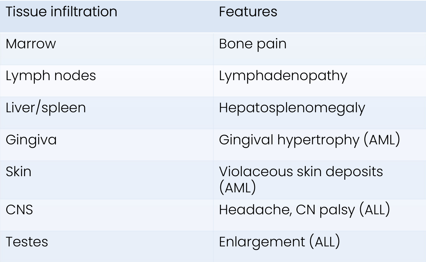
Tissue Infiltration18
| Tissue infiltration | Features |
|---|---|
| Marrow | Bone pain |
| Lymph nodes | Lymphadenopathy |
| Liver/spleen | Hepatosplenomegaly |
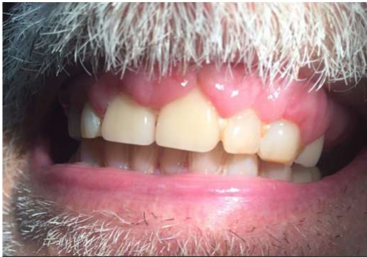
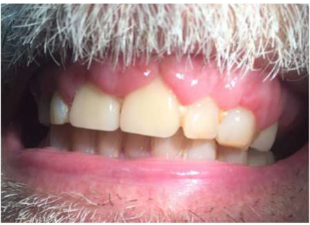
| Gingiva | Gingival hypertrophy (AML) |
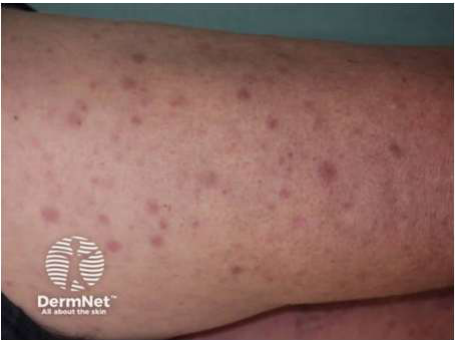
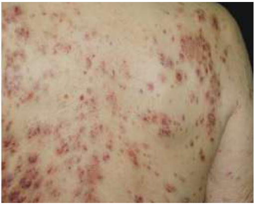
| Skin | Violaceous skin deposits (AML) |
| CNS | Headache, CN palsy (ALL) |
| Testes | Enlargement (ALL) |
DermNet NZ — https://dermnetnz.org/topics/leukaemia-cutis
 |  |
 |
Acute Lymphoblastic Leukaemia19
- 65% of childhood acute leukaemia (most common leukaemia in children)
- Predominantly children (peak incidence 0–4 years old), but can affect adults
- Risk factors:
- Some congenital disorders: Down Syndrome (33-fold ↑ risk), Fanconi’s anaemia
- Philadelphia chromosome (25% adult cases, 3–5% of children)
- Exposure to radiation, chemicals, reaction to viral infections
Features20
-
CNS involvement (cranial nerve palsies, sensory disturbances)
-
Testes enlargement
-
Pain in bone/joints
-
Acute serious illness, infections
-
Purpura
-
Gingival bleeding on brushing
-
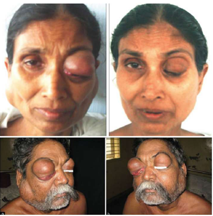
Bilateral salivary ± lacrimal gland swelling (Mikulicz syndrome)
-
Oral ulceration
Prognosis21
- Complete remission / 5-year survival = 70–80% (combined children and adult)
- <15 years old: 90%
- 15–40 years old: 65%
-
40 years old: 25%
Lecturer — ALL Prognosis
Prognosis influences dental treatment planning.
- A favourable long-term prognosis may support comprehensive treatment.
- A poor prognosis may support a more palliative or symptom-focused approach.
Acute Myeloblastic Leukaemia22
- Most common acute leukaemia in adults (especially males)
- Peak incidence older age (60–70 years old)
- Risk factors:
- Some congenital disorders: Down Syndrome (150-fold ↑ risk), Fanconi’s anaemia, Bloom syndrome
- Some blood disorders: myelodysplastic syndromes and myelofibrosis, aplastic anaemia, paroxysmal nocturnal haemoglobinuria
- Exposure to radiation, chemicals, chemotherapy, tobacco
Features23
- Violaceous skin deposits
- Gingival hypertrophy
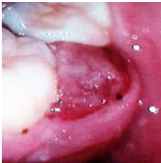
- Neutropenic ulceration
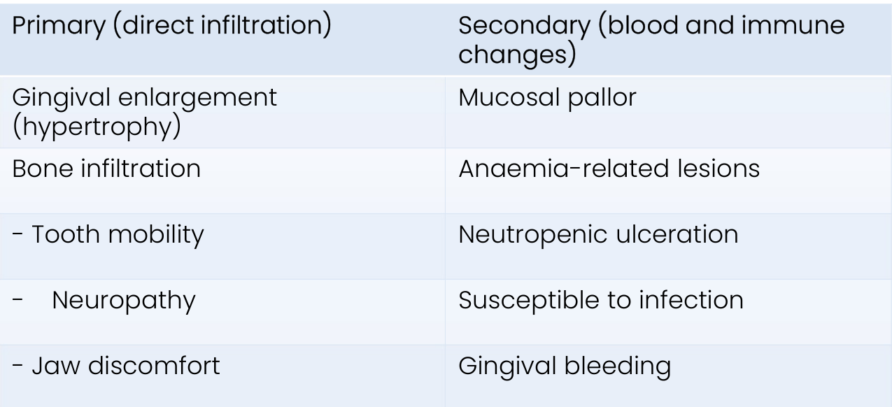
| Primary (direct infiltration) | Secondary (blood and immune changes) |
|---|---|
| Gingival enlargement (hypertrophy) | Mucosal pallor |
| Bone infiltration | Anaemia-related lesions |
| - Tooth mobility | Neutropenic ulceration |
| - Neuropathy | Susceptible to infection |
| - Jaw discomfort | Gingival bleeding |
 |  |
 |  |
Prognosis24
5-year survival:
- <40 years old: 55%
- 40–49 years old: 50%
- 50–59 years old: 25%
- 60–69 years old: 15%
- 70–79 years old: 5%
-
80 years old: 1%
Chronic Lymphocytic Leukaemia25
- Overpopulation of lymphocytes (B-cells) in blood and bone marrow
- Shared features with small lymphocytic lymphoma (but cancer cells in lymph nodes)
- Most common type of leukaemia in adults
- 2× as common as CML
- 25% of all leukaemias
- Median age at diagnosis 65 years old (mostly older adults)
- Risk factors:
- Gender – M:F = 2:1
- Ethnicity = white European origin
- Family history = 10% have parent, sibling, child affected
- Exposure to high-risk chemicals e.g. pesticides, benzene, heavy solvents
Lecturer — CLL Classification
The WHO classifies chronic lymphocytic leukaemia as a slow-growing non-Hodgkin lymphoma because it affects lymphocytes. It is traditionally called leukaemia because the malignant cells mainly accumulate in the blood and bone marrow.
Features26
-
Often asymptomatic (1/3 diagnosed following routine FBC)
-
Recurrent/severe infections (e.g. shingles)
-
Painless lymphadenopathy
-
Anaemia
-
Fever
-
Splenomegaly
-
Thrombocytopenia
-
Gingival or palatal mucosal enlargement
-
Spontaneous bleeding
-
Petechiae
Progression27
- Can live with slow-growing CLL for long periods (indolent)
- Richter’s syndrome (7% of patients with CLL): transformation into aggressive NHL (diffuse large B-cell lymphoma) or HL
Prognosis
- 1 in 10 people may never require treatment (active monitoring, “watch and wait”)
- Immune system fights to keep CLL under control
- No survival/symptom benefit, avoid side-effects of treatment
- No cure for CLL
- But treatments are very effective at controlling symptoms (supportive care)
- 5-year survival = 88%
- Median survival = >10 years
Lecturer — Treatment Mechanisms
Chemotherapy kills cancer cells or slows their growth and may be administered intravenously, by injection under the skin, or as tablets. Stem-cell transplantation introduces healthy stem cells after high-dose chemotherapy.
- Radiotherapy may be used before stem-cell transplantation as conditioning.
- It may also treat disease involving the central nervous system or testes, or relieve symptoms such as swollen organs or bone pain.
- Targeted therapies attack specific molecular targets while avoiding healthy cells, including tyrosine-kinase inhibitors and monoclonal antibodies.
Lecturer — CLL Monitoring
Some patients undergo several rounds of treatment followed by remission, and some return to watchful monitoring after treatment.
- Active treatment may begin if the disease becomes symptomatic or problematic.
- Treatment may include chemotherapy or immunotherapy.
Chronic Myeloid Leukaemia28
- Overpopulation of granulocytes (neutrophils, eosinophils, basophils, mast cells)
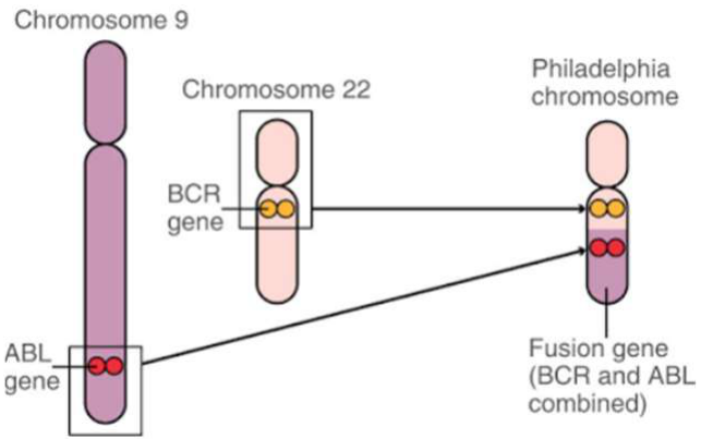
- Caused by genetic abnormality: Philadelphia chromosome (abnormal chromosome 22)
- Affects adults almost exclusively (usually >60 years old)
- 14% of all leukaemias
- Risk factors:
- Gender – M > F (slightly)
- Exposure to radiation
Lecturer — Philadelphia Chromosome
The Philadelphia chromosome forms when parts of chromosomes 9 and 22 exchange places. This produces the BCR-ABL1 gene, which produces tyrosine kinase, an enzyme involved in controlling cell growth.
Features29
- Symptomatic anaemia (shortness of breath)
- Fever and sweats in the absence of infection
- Weight loss
- Retinal damage
- Lymphadenopathy – uncommon, suggests transformation to acute leukaemia
- Progressive splenomegaly (>75%) and associated abdominal discomfort
Progression30
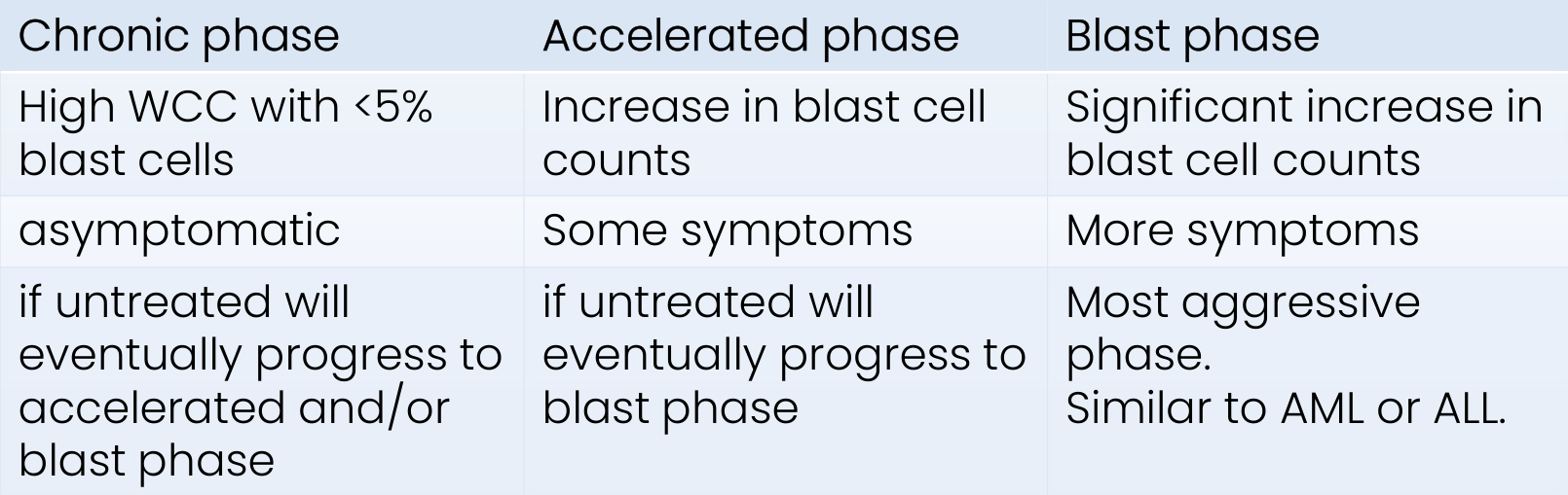
- Can transform to acute phase similar to AML
- Classified by phase, defined by number of blasts in blood/bone marrow
Prognosis
- 5-year survival: 80%
| Chronic phase | Accelerated phase | Blast phase |
|---|---|---|
| High WCC with <5% blast cells | Increase in blast cell counts | Significant increase in blast cell counts |
| Asymptomatic | Some symptoms | More symptoms |
| If untreated will eventually progress to accelerated and/or blast phase | If untreated will eventually progress to blast phase | Most aggressive phase. Similar to AML or ALL. |
Leukaemia Treatments3132
Treatment depends on:
- Type of leukaemia
- Stage of disease
- Severity of symptoms
- Age
- General health
- Patient choice
| Acute leukaemia | Chronic leukaemia | Palliative Care |
|---|---|---|
| - Chemotherapy - Stem cell or bone marrow transplant - Radiotherapy - Steroids - Targeted therapy - Leukapheresis - Immunotherapy (e.g. CAR-T cell therapy) | - As for acute - Active monitoring - Splenectomy | - Radiotherapy and/or chemotherapy - Analgesics - Blood transfusions - Nutritional supplements / medications for nausea and appetite loss |
Lecturer — Immunotherapy Details
Leukapheresis uses a machine to remove white cells from the blood, while immunotherapy supports the immune system in recognising and attacking cancer cells.
- CAR T-cell therapy modifies a patient’s own T cells to attack cancer cells.
- T-cell therapy may cause profound immunosuppression and may require consideration of antibiotic prophylaxis.
Lecturer — Advanced Disease Care
Advanced disease may require shorter or less intensive radiotherapy or chemotherapy.
- Supportive care may include analgesics, blood transfusions, nutritional supplements, and management of nausea and appetite loss.
Lymphomas33
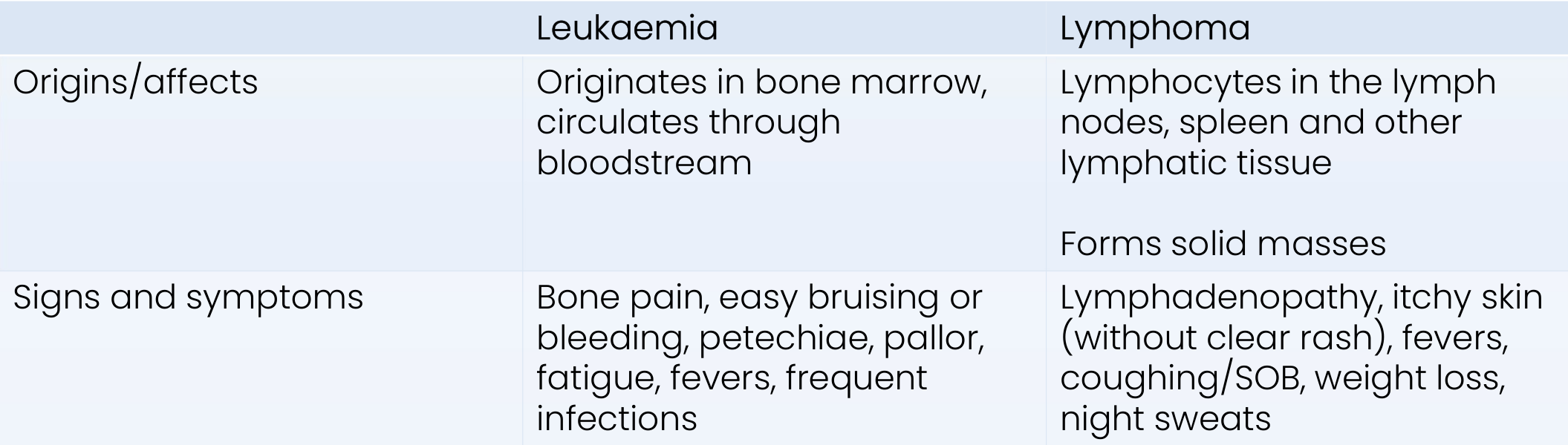
Leukaemia Versus Lymphoma34
| Leukaemia | Lymphoma | |
|---|---|---|
| Origins/affects | Originates in bone marrow, circulates through bloodstream | Lymphocytes in the lymph nodes, spleen and other lymphatic tissue; forms solid masses |
| Signs and symptoms | Bone pain, easy bruising or bleeding, petechiae, pallor, fatigue, fevers, frequent infections | Lymphadenopathy, itchy skin (without clear rash), fevers, coughing/SOB, weight loss, night sweats |
Definitions and Aetiology35
- Lymphoma = B- and T-cell (lymphocyte) malignancies of the lymphoid system
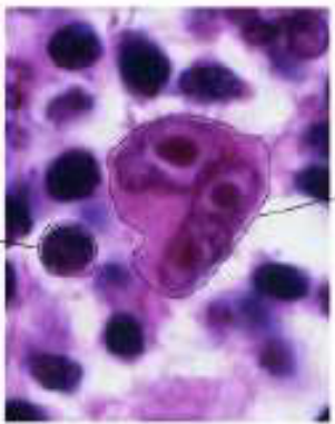
- Hodgkin Lymphoma = haematological malignancy arising from mature B-cells, with presence of Reed-Sternberg cells and Hodgkin cells
- Non-Hodgkin Lymphoma = haematological malignancy arising from mature B or T-cells (lymphocytes), without presence of Reed-Sternberg cells
- More than 60 subtypes of NHL, including:
- Follicular lymphoma
- Diffuse Large B-cell lymphoma (DLBCL)
- Burkitt’s lymphoma
- More than 60 subtypes of NHL, including:
- Divided into B-cell and T-cell NHLs
- T-cell lymphomas generally harder to cure
Normal lymphocyte; Reed-Sternberg cell
- Unknown exact cause
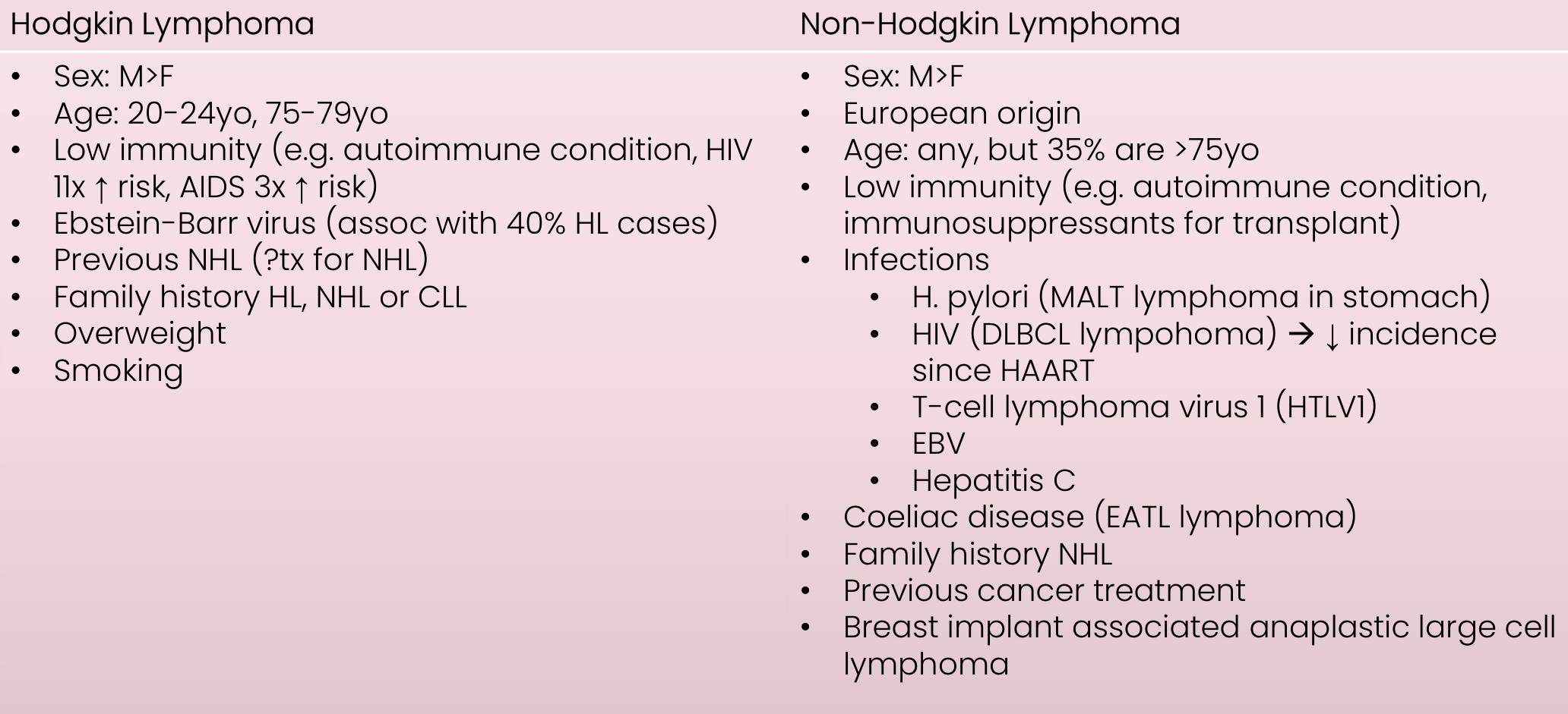
Risk Factors36
| Hodgkin Lymphoma | Non-Hodgkin Lymphoma |
|---|---|
| Sex: M>F | Sex: M>F |
| Age: 20-24yo, 75-79yo | European origin |
| Low immunity (e.g. autoimmune condition, HIV 11x ↑ risk, AIDS 3x ↑ risk) | Age: any, but 35% are >75yo |
| Epstein-Barr virus (assoc with 40% HL cases) | Low immunity (e.g. autoimmune condition, immunosuppressants for transplant) |
| Previous NHL (?tx for NHL) | Infections |
| Family history HL, NHL or CLL | H. pylori (MALT lymphoma in stomach) |
| Overweight | HIV (DLBCL lymphoma) → ↓ incidence since HAART |
| Smoking | T-cell lymphoma virus 1 (HTLV1) |
| EBV | |
| Hepatitis C | |
| Coeliac disease (EATL lymphoma) | |
| Family history NHL | |
| Previous cancer treatment | |
| Breast implant associated anaplastic large cell lymphoma |
Clinical Manifestations37
| Hodgkin Lymphoma | Non-Hodgkin Lymphoma |
|---|---|
| Lymphadenopathy — contiguous spread | Lymphadenopathy — non-contiguous spread (often disseminated disease); predilection for GIT and CNS |
- Fatigue
- B-symptoms: unexplained fevers, night sweats, weight loss
- Itchy skin
- Pressure on other organs/ducts/nerves

Hodgkin Lymphoma38
- Rarely affects mouth
- If does, clinically indistinguishable from NHL
Non-Hodgkin Lymphoma
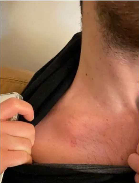
- Enlarged cervical lymph nodes (discrete, rubbery, painless) — initial complaint in 50% of cases
- Swellings and ulcers
- especially palate and fauces (often associated with AIDS)
- also gingiva, FOM
- Isolated tooth mobility or lip paraesthesia
- Rarely: involvement of Waldeyer’s ring (more commonly in HIV-infected, NHL>HL)
Management and Prognosis
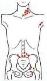
Staging39
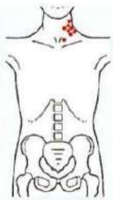
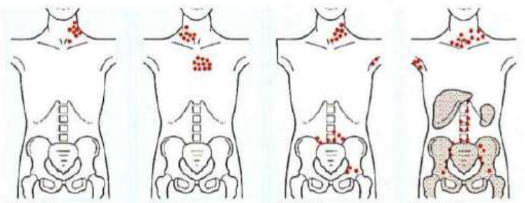
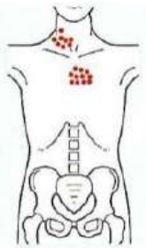
Ann Arbor staging:
- How many areas affected
- Above, below or on both sides of diaphragm
- Whether has spread to bone marrow or other organs
 |  |
 |  |
Treatment
- Watch and wait (active monitoring) — low-grade slow-growing lymphomas
- Radiotherapy
- Chemotherapy
- Immunotherapy / targeted therapies
- Stem cell transplant
- Palliative care
Hodgkin Lymphoma40
- 85–95% 5-year survival (stage 1-4)
- 80% 10-year survival
Non-Hodgkin Lymphoma
- Prognosis variable based on type
- Generally 65% 5-year survival, 55% 10-year survival
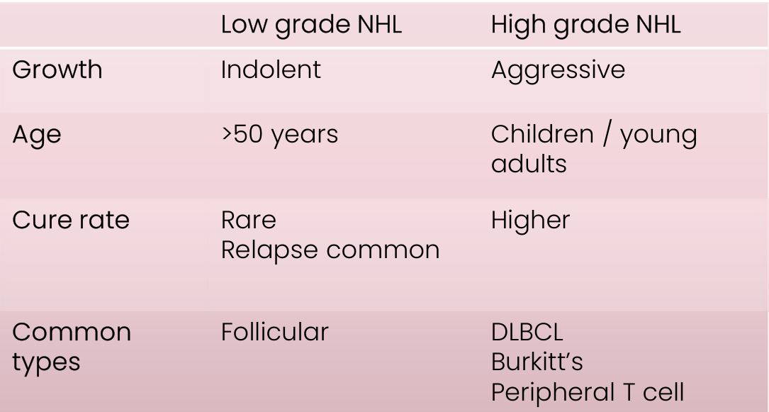
| Low grade NHL | High grade NHL | |
|---|---|---|
| Growth | Indolent | Aggressive |
| Age | >50 years | Children / young adults |
| Cure rate | Rare; relapse common | Higher |
| Common types | Follicular | DLBCL, Burkitt’s, Peripheral T cell |
Lecturer — NHL Prognosis
Low-grade lymphoma may be difficult to eradicate completely but can remain controlled for several years.
- High-grade lymphoma requires more intensive treatment but often responds better.
- Many patients with high-grade disease are cured.
Myelodysplastic Syndromes41
Myelodysplastic Syndromes (MDS)
(Myelodysplasia)
Definition and Aetiology
Definition42
A group of clonal haematopoietic stem cell disorders caused by a mutation that results in abnormal proliferation of myeloid progenitor cells, usually in the presence of a hypercellular bone marrow and multilineage dysplasia.
Aetiology
- Mutation in ≥1 genes that control blood cell development
- Affected stem cell divides → “clones” of defective stem cell produced (preservation of mutation)
- Bone marrow gradually overtaken by abnormally functioning immature blood cells
- Results in lower production of mature myeloid cells (ineffective haematopoiesis), with progressive cytopenia
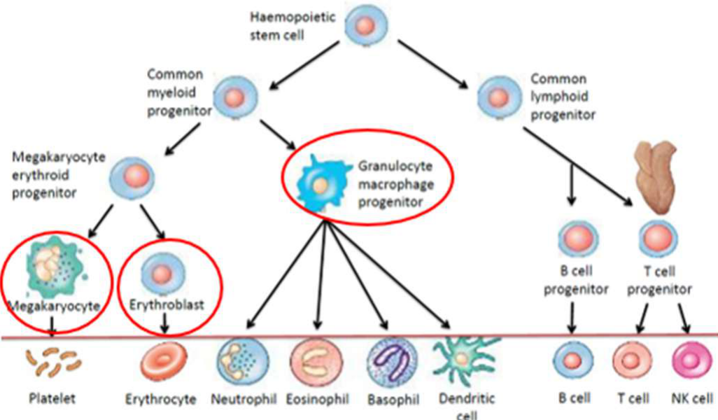
Usually all 3 cell lines (erythroid, megakaryocyte, granulocyte) involved, with increased risk of AML development.
Haematopoietic Cell Lineage
- Haemopoietic stem cell
- Common myeloid progenitor
- Megakaryocyte erythroid progenitor
- Megakaryocyte → Platelet
- Erythroblast → Erythrocyte
- Granulocyte macrophage progenitor
- Neutrophil, Eosinophil, Basophil
- Dendritic cell
- Megakaryocyte erythroid progenitor
- Common lymphoid progenitor
- B cell progenitor → B cell
- T cell progenitor → T cell
- NK cell
- Common myeloid progenitor
Manifestations
General Findings43
Related to increasing bone marrow failure:
- Red blood cells (macrocytic anaemia): weakness, tiredness/fatigue, SOB with minimal exercise, pallor
- White blood cells (neutropenia): frequent infections (especially chest infections), fevers
- Platelets (thrombocytopenia): bruising, easy bleeding (nosebleeds), purpura/petechiae (lower limbs initially)
- Enlargement of spleen or liver
Oral Findings
Related to increasing bone marrow failure:
-
White blood cells: sore mouth (mouth ulcers), oral infections (herpes, fungal, bacterial), exacerbation of chronic periodontitis and/or dental infection
-
Platelets: spontaneous gingival bleeding, petechiae
-
Gingival infiltration and oral ulceration may develop in CMML subtype. May be first sign of acceleration of a stable disease process.
-
Paraesthesia, burning mouth, xerostomia reported
Management
Supportive Treatment44
-
Growth factor injections (erythropoietin, G-CSF)
-
Blood transfusions (RBCs, platelets)
-
Iron chelating agents
-
Antibiotics (if low WBCs)
-
Supplements: folic acid, Vit B6
-
Systemic corticosteroids
-
Iron-chelating agents reduce excess iron after repeated transfusions.
Active Treatment
- Targeted therapy (e.g. lenalidomide)
- Chemotherapy
- Stem cell transplant
- Clinical trials
Myeloproliferative Neoplasms45
Myeloproliferative disorders
Definition and Classification46
- Definition: A heterogenous group of clonal haematopoietic stem cell disorders caused by a mutation whereby abnormal myeloid progenitor cells (erythroid, myeloid, megakaryocyte) produce excess numbers of one or more cell types.
- Classification: FAB / WHO
- Usually described according to most affected blood cell type. Four commonest subtypes:
Lecturer — Blood Cell Production
The marrow rapidly produces functional mature blood cells, causing blood-cell counts to become high.
- Blood may become thick as a result.
- Polycythaemia vera (PV): excessive RBCs
- Essential thrombocythaemia (ET): excessive platelets
- Chronic myelogenous leukaemia (CML): excessive WBCs
- Myelofibrosis (MF): replacement of the stem cells in the bone marrow by scar and fibrous tissue (fibroblasts), resulting in too few blood cells and often an enlarged spleen.
General Features47
-
Symptoms related to deficient blood cells (RBC, WBC, platelets)
-
Thrombosis, blood hyperviscosity (MF, PV, ET): brain (stroke/TIA), eyes (vision), heart (MI), legs (DVT), lungs (PE)
-
Splenomegaly and hepatomegaly (MF, PV, ET): secondary to extramedullary haematopoiesis → abdominal pain, appetite loss, pain on respiration
-
Gout (MF, PV, ET): increased production and breakdown of blood cells → build up of uric acid
-
Aquagenic pruritis (PV): itchy skin following a contact with water
-
Bone pain (MF)
-
Night sweats, pyrexia, weight loss, pruritic, dizziness, weakness (MF)
-
Vasomotor symptoms: headaches, dizziness, tingling, erythromelalgia, visual disturbances (ET)
-
Aquagenic pruritus may be unbearable after contact with water
-
Erythromelalgia may cause burning pain in the hands and feet with redness and warmth
Management and Dental Implications4849
General management depends on the specific subtype.
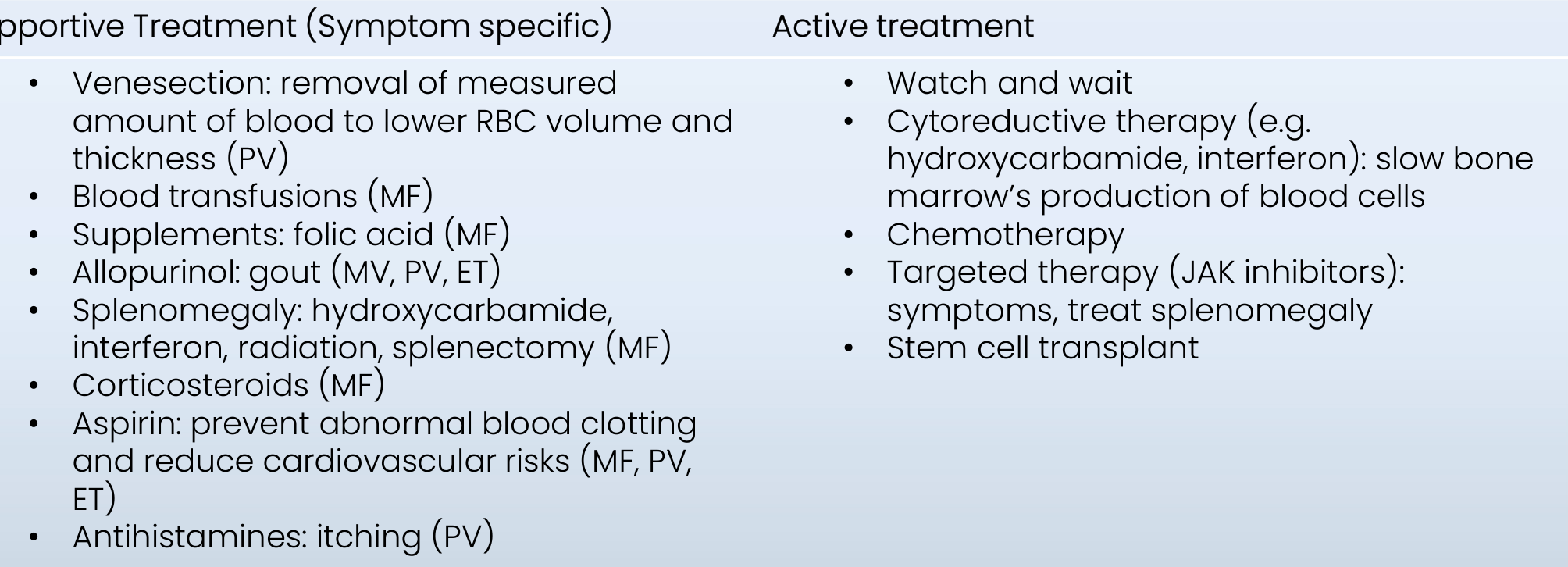
Supportive Treatment (Symptom Specific)50
- Venesection: removal of measured amount of blood to lower RBC volume and thickness (PV)
- Blood transfusions (MF)
- Supplements: folic acid (MF)
- Allopurinol: gout (MF, PV, ET)
- Splenomegaly: hydroxycarbamide, interferon, radiation, splenectomy (MF)
- Corticosteroids (MF)
- Aspirin: prevent abnormal blood clotting and reduce cardiovascular risks (MF, PV, ET)
- Antihistamines: itching (PV)
Active Treatment
-
Watch and wait
-
Cytoreductive therapy (e.g. hydroxycarbamide, interferon): slow bone marrow’s production of blood cells
-
Chemotherapy
-
Targeted therapy (JAK inhibitors): symptoms, treat splenomegaly
-
Stem cell transplant
-
Oral manifestations and risk management as per blood cells affected
-
Related to treatments provided
-
Paradoxical clotting and bleeding tendency
- Haemostasis
- Cardiovascular risks (e.g. MI, stroke)
-
Aquagenic pruritic (PV): ability to operate, oral hygiene at home
-
Erythromelalgia (burning pain of hands): oral hygiene ability
-
Splenic changes: infection risk
-
Anticoagulant or aspirin therapy may affect dental treatment
-
Dental treatment may need to be performed slowly and carefully when contact with water is difficult because of aquagenic pruritus
Plasma Cell Tumours51
Plasma cell tumours comprise:
- Multiple myeloma
- Solitary myeloma
- Waldenstrom’s macroglobulinaemia
- Heavy chain disease
- Monoclonal gammopathy of undetermined significance (MGUS)
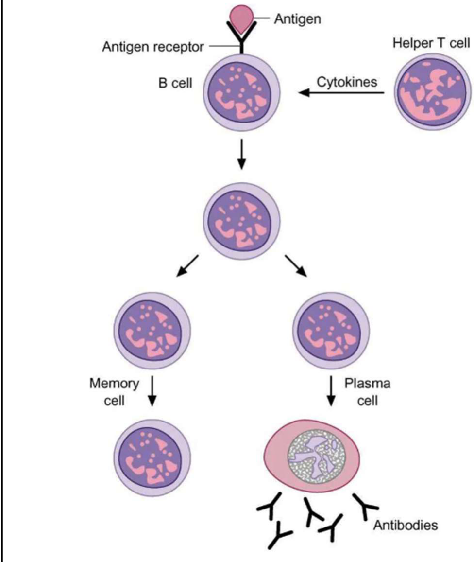
- Plasma-cell tumours are also called paraproteinaemias. Diagram labels: B cell → antigen receptor (antigen); cytokines from helper T cell; differentiation to memory cell and plasma cell → antibodies.
Multiple Myeloma52
-
Definition: Disseminated plasma cell neoplasm, predominantly causing bone lesions. Malignant plasma cells produce defective immunoglobulins and release osteoclast-activation factors, causing bone resorption and pain.
-
Multiple myeloma is the most common plasma-cell malignancy discussed.
-
Aetiology: Exact reason unknown.
- Occasionally related to ionising radiation and petroleum products exposure.
- Associated with MGUS (1/100 develop MM), ethnicity (twice as common in black population than white/Asians)
- Mostly middle-age and elderly
- Slight predilection for males
Clinical Features of Relevance to Dentistry5354
- Neoplastic proliferation of plasma cells in bone marrow → release of cytokines →
- Osteolysis and bone destruction
- Mandibular lesions: 5–14% of patients
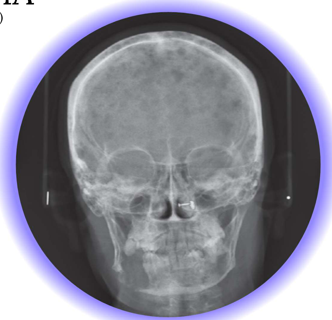
- Multiple punched-out radiolucencies.
- Promote dental mobility, pain, paraesthesia, soft tissue swelling, pathological jaw fractures
- Suppression of haemopoiesis and bone marrow replacement →
- RBC: anaemia
- WBC: increased risk of infections (leukopenia, abnormal antibody production)
- Platelets: bleeding tendency (thrombocytopenia)
- Defective immunoglobulin (hyperglobulinemia with paradoxical hypogammaglobulinemia) →
- Plasma hyperviscosity with clotting or bleeding tendency
- Renal failure
- Increased liability to infections
- Neurological sequelae
Raghavan S, Nagaraj P, Ramaswamy B, Nayak D. Multiple Myeloma of the jaw: A Case Report. J Indian Acad Oral Med Radiol, 2014, 26(4):454-457.
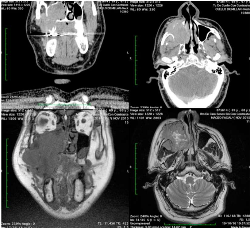
- Associated with extramedullary plasmacytoma
Merino F, Portillo Hernandez I, Sanchez Aniceto G, Ballestin C. Extramedullary plasmacytoma in the maxillofacial region: A review of the literature and case report. Oral Maxillofac Surg Cases. 2018; 4(4): 141-145.
Lecturer — Extramedullary Plasmacytomas
Extramedullary plasmacytomas are uncommon solitary tumours composed of neoplastic plasma cells occurring outside bone.
- They primarily arise in the head and neck, accounting for 80–90% of extramedullary plasmacytomas in that region.
- They represent approximately 1% of head and neck tumours and commonly involve the nasal cavity, nasopharynx, and maxillary sinus.
- Oral-cavity involvement is rare, and they occur in approximately 20% of multiple-myeloma recurrences.
- Extramedullary plasmacytomas may progress to multiple myeloma.
 |  |
 |  |
Treatment of Relevance to Dentistry
Treatment for Cancer55
- Chemotherapy
- Corticosteroids
- Targeted therapy, immunotherapy, CAR-T cell therapy
- Thalidamide-derived agents (e.g. lenalidomide)
- Bortezomib
- Stem cell / bone marrow transplant (typically autologous)
- Radiotherapy (localised plasmacytic infiltration)
Treatment for Symptoms (of Cancer or Cancer Treatment)
- Bisphosphonates
- Reduce bone pain
- Prevent pathological fracture
- Have implications because of medication-related osteonecrosis of the jaws.
- Anticoagulants
- Hyperviscosity of blood
- Lenalidomide – clotting tendency
- Dialysis
- Renal failure
Dental Implications and Management56
Pre-Cancer Therapy Dental Assessment57
- Patients should have a comprehensive dental assessment prior to commencing chemotherapy or a stem cell / bone marrow transplant
- Comprehensive oral examination and x-rays
- Goal: identify indications for dental treatment before cancer therapy starts
- Infection
- Teeth with poor prognosis
- Potential causes of mucosal trauma (e.g. sharp teeth)
- Institute appropriate oral care program (oral side effects of cancer therapy)
- Dental interventions ideally completed before cancer therapy starts, but this may not be feasible
- Cancer therapy should not be delayed
Lecturer — Treatment Priorities
If time is limited, prioritise the most urgent and infection-prone dental problems first. Sometimes blood counts make dental treatment more hazardous than beneficial.
Disease and Treatment Effects
Disease Effects58
-
RBC: anaemia
-
WBC: infections
-
Platelets: bleeding
-
Hyperglobulinaemia / hypogammaglobulinaemia: infection [MM]
-
Renal failure: dialysis [MM]
-
Malignancies may also cause mucosal ulceration, gingival enlargement or infiltration, bone pain, tooth mobility, paraesthesia, and pathological fractures.
Treatment Effects
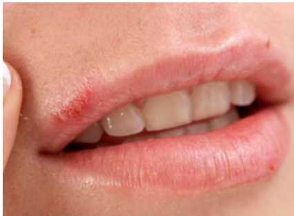
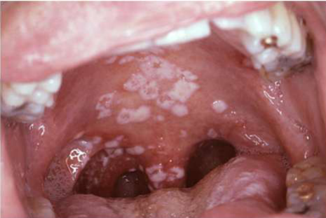
- Chemotherapy: ↓ RBC/WBC/platelets, mucositis, hyposalivation, candidiasis, taste disturbances
- Stem Cell / bone marrow transplant: ↓ RBC/WBC/platelets, mucositis, hyposalivation, candidiasis, HSV (cold sores), gingival hyperplasia (ciclosporin), dental developmental disturbances (paeds), GVHD (allo), SCC
- Corticosteroids: adrenal suppression, fat redistribution, impaired wound healing, susceptibility to infection, mood changes
- Bisphosphonates [MM] + emerging targeted therapies: MRONJ
- Anticoagulants: bleeding [MM]
- Radiotherapy: mucositis, hyposalivation, candidiasis, increased caries risk, taste disturbances, osteoradionecrosis
Access and Communication59
Fatigue60
- Time of day considerations; may fatigue easily
Lecturer — Managing Fatigue
Consider how much treatment the patient can tolerate in one visit. Shorter or modified appointments may be appropriate.
Timing of Dental Treatment
- Consult haematologist/oncologist
- Defer elective treatment until remission phase
- Dental pain: medical risks/timing weighed up against benefit of prompt dental treatment (usually infective source)
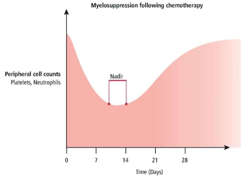
- Chemotherapy: perform dental treatment day(s) before starting new cycle → chemotherapy nadir
- [MM] Haemodialysis: book dental treatment on non-dialysis days (heparinisation)
Lecturer — Chemotherapy Timing
The nadir generally occurs 7–14 days after treatment begins, when blood-cell counts are lowest and the risks of infection, anaemia, and bleeding increase.
- Dental treatment is usually best timed toward the end of the cycle, when counts have recovered and before the next cycle.
- A blood test before the next cycle can help determine whether treatment is safe.
- For an extraction requiring about 10–14 days of healing, schedule it so healing occurs before the next nadir.
- Usually no issues
- Visual disturbances (written material)
- Cognitive/neurological sequelae
- Setting expectations re: treatment approach
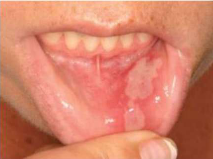
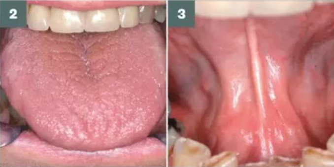
Education and Prevention61
 |  |
 |  |
Key Guideline Sources62
- MASCC/ISOO/EBMT 2015: Basic Oral Care for Haematology-Oncology Patients and HSCT Recipients position paper
- Royal College of Surgeons England / BSDH 2018: Oral Management of Oncology Patients Requiring Radiotherapy, Chemotherapy and/or Bone Marrow Transplantation
- MASCC/ISOO 2020: Clinical Practice Guidelines for the Management of Mucositis Secondary to Cancer Therapy
Supporting Literature
Elad, S., Raber-Durlacher, J. E., Brennan, M. T., Saunders, D. P., Mank, A. P., Zadik, Y., Quinn, B., Epstein, J. B., Bijllevens, N. M. A., Wahlmio, T., Passweg, J. R., Correa, M. E. P., Dahlöf, G., Garming-Legert, K. U. E., Logan, R. M., Potting, C. M. J., Shapira, M. Y., Soga, Y., Stringer, J., Stokman, M. A., Vokurka, S., Wallhult, E., Yarom, N., and Beier Jensen, S. (2015). Basic oral care for hematology–oncology patients and hematopoietic stem cell transplantation recipients: a position paper from the joint task force of the Multinational Association of Supportive Care in Cancer/International Society of Oral Oncology (MASCC/ISOO) and the European Society for Blood and Marrow Transplantation (EBMT), Support Care Cancer, 23, 223–236.
Elad, S., Cheng, K. K. F., Lalla, R. V., Yarom, N., Hong, C., Logan, R. M., Bowen, J., Gibson, R., Saunders, D. P., Zadik, Y., Ariyawarnana, A., Correa, M. E., Ranma, V., and Bossi, P., for the Mucositis Guidelines Leadership Group of the Multinational Association of Supportive Care in Cancer and International Society of Oral Oncology (MASCC/ISOO). (2020). MASCC/ISOO Clinical Practice Guidelines for the Management of Mucositis Secondary to Cancer Therapy, Cancer, 126, 4423–4431.
Royal College of Surgeons of England / The British Society for Disability and Oral Health (2018). The Oral Management of Oncology Patients Requiring Radiotherapy, Chemotherapy and / or Bone Marrow Transplantation — Clinical Guidelines, Updated 2018.
The MASCC/ISOO 2020 guidelines cover evidence from 1197 publications related to oral or gastrointestinal mucositis; thirteen new guidelines were developed for or against the use of various interventions in specific treatment settings, 11 previous guidelines were confirmed after a review of new evidence, and 13 previously established guidelines were carried over because there was no new evidence for these interventions.
Prevention of Infection63
Before HSCT/CT — Oral/Dental examination
- Refer for dental evaluation
- Focus: eliminate foci of infection, traumatic surfaces
During HSCT/CT
- Oral hygiene: ensure cleaning teeth and tongue — soft bristled toothbrush — flossing if capable without trauma
- Decontamination:
- Advise using bland rinses (e.g., saline), repeated rinses/day
- Advise rinsing with chlorhexidine in alcohol-free solution, x2/day
- If unable to rinse, apply solution to sterile gauze or toothette
- Prophylaxis: apply the institute protocol for prophylaxis for oral candidiasis and viral reactivation
After HSCT/CT
- Viral infection: consider anti-viral prophylaxis
- Candidal infection: consider anti-fungal prophylaxis — topical (sugar-free) or systemic
- Dental infection: encourage the patient to have frequent dental recalls
- Oral hygiene: educate and reinforce optimal oral hygiene and dental prevention
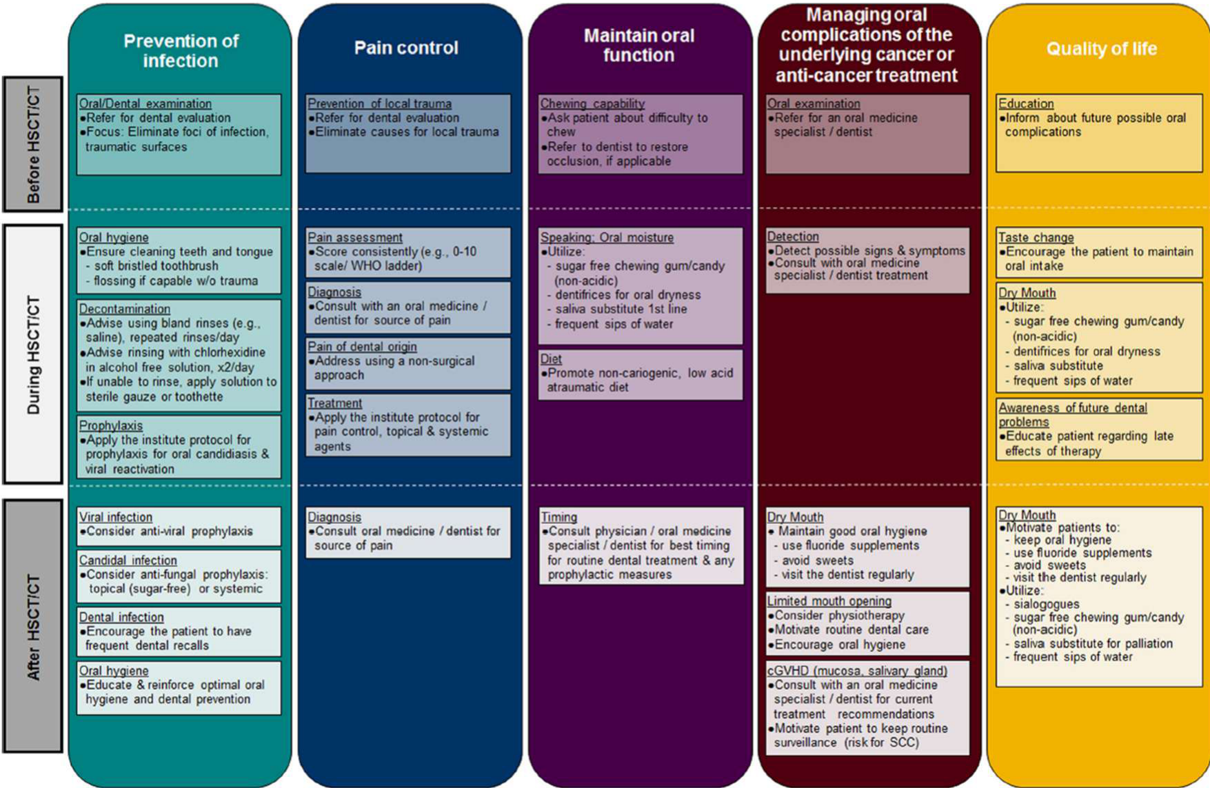
Pain Control
- Prevention of local trauma: refer for dental evaluation; eliminate causes for local trauma
- Pain assessment: score consistently (e.g., 0–10 scale / WHO ladder)
- Diagnosis: consult with an oral medicine specialist / dentist for source of pain
- Pain of dental origin: address using a non-surgical approach
- Treatment: apply the institute protocol for pain control, topical and systemic agents
Maintain Oral Function
- Chewing capability: ask patient about difficulty to chew; refer to dentist to restore occlusion, if applicable
- Speaking / oral moisture — utilize:
- Sugar-free chewing gum/candy (non-acidic)
- Dentifrices for oral dryness
- Saliva substitute (first line)
- Frequent sips of water
- Diet: promote non-cariogenic, low acid, atraumatic diet
- Timing: consult physician / oral medicine specialist / dentist for best timing for routine dental treatment and any prophylactic measures
Managing Oral Complications of the Underlying Cancer or Anti-Cancer Treatment
- Oral examination: refer to an oral medicine specialist / dentist
- Detection: detect possible signs and symptoms; consult with oral medicine specialist / dentist re: treatment
- Dry mouth:
- Maintain good oral hygiene
- Use fluoride supplements
- Avoid sweets
- Visit the dentist regularly
- Limited mouth opening: consider physiotherapy; motivate routine dental care; encourage oral hygiene
- cGVHD (mucosa, salivary gland): consult with an oral medicine specialist / dentist for current treatment recommendations; motivate patient to keep routine surveillance (risk for SCC)
Quality of Life
- Education: inform about future possible oral complications; educate patient regarding late effects of therapy
- Taste change: encourage the patient to maintain oral intake
- Dry mouth:
- Motivate patients to keep oral hygiene, use fluoride supplements, avoid sweets, visit the dentist regularly
- Utilize sialogogues, sugar-free chewing gum/candy (non-acidic), saliva substitute for palliation, frequent sips of water
Source: MASCC/ISOO/EBMT 2015: Basic Oral Care for Haematology-Oncology Patients and HSCT Recipients position paper
- Counsel on possible oral side effects of chemotherapy
- Each chemotherapy agent has different adverse effects
- Common examples: mucositis, reduced salivary flow, immune suppression
- Increased risk of oral infections: oral and oropharyngeal candidiasis, herpes simplex virus, post-operative infection
Chemotherapy / Radiotherapy / HSCT — Pain64
-
Encourage compliance with oral hygiene (brush 2–3 times/day)
-
Suck ice chips 30 min prior to chemotherapy
-
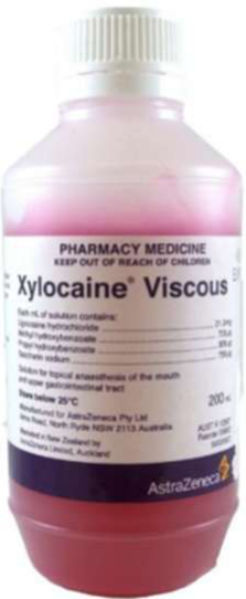
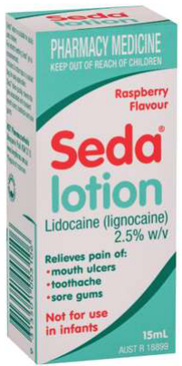
Topical anaesthetics: xylocaine viscous, Seda Lotion
-
Surgical ultra-soft toothbrush (Curaprox)
- Air-dry between uses
- Sponge brushes better than nothing, but not as effective
-
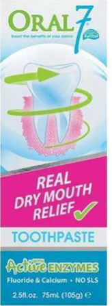
SLS-free toothpaste (e.g. Oral 7, Biotene)
-
Avoid spicy/rough/crunchy/acidic food and drinks
-
A bland rinse can be made with half a teaspoon of salt and half a teaspoon of bicarbonate of soda in a glass of water, and used as often as required.
 |  |  |
 |  |
Hyposalivation
-
Caries risk (buffering capacity; food stagnation; modified diet)
-
Bicarb + salt mouthrinses after meals
-
Saliva substitutes (e.g. Oral 7, Biotene gel, MW, spray)
-
Regular lip balm
-
Oral 7 gel for denture retention
-
Plant-based or animal-derived lip products were preferred over petroleum-based products because petroleum-based products do not moisturise the lips.
Chemotherapy / Radiotherapy / HSCT — Opportunistic Infections65
Opportunistic infections (candida, periodontal pathogens, suprainfection of mucositis):
- Maintain oral hygiene
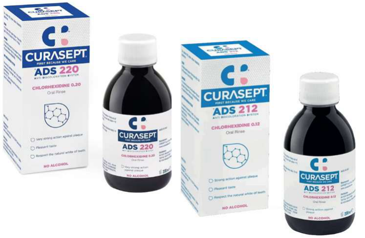
- Alcohol-free chlorhexidine rinse
- Add as short-term oral antiseptic if oral hygiene compromised
- 0.2% ideal, substantivity at 0.1% (dilute 50:50 with water)
- Bland rinses
- Denture hygiene
 |  |
Flossing?
“Measures to remove plaque from between teeth, crown & bridges or implants, and from tongue, should be continued using familiar devices to patient to avoid self-injury”
- History of regular interdental cleaning? Or raging gingivitis?
Source: MASCC/ISOO/EBMT 2015: Basic Oral Care for Haematology-Oncology Patients and HSCT Recipients position paper
Lecturer — Individualised Flossing
Flossing recommendations should be individualised according to platelet count, immunosuppression, gingival health, and familiarity with flossing.
- Some centres advise no flossing during stem-cell transplantation or severe thrombocytopenia.
- Regular flossers with good gingival health may benefit from continuing, whereas starting flossing during severe gingivitis is inappropriate.
Surgery Considerations
RBC (Anaemia)66
- Check Hb levels prior to appointment — caution <80 g/L
- Desaturation — stress of dental procedure
- Consider pulse oximetry, nasal oxygen, RBC transfusion
- Pallor, atrophic glossitis, angular cheilitis, ulcers, glossodynia
Platelets (Bleeding)
- Ideally check within 24 hrs of dental procedure — caution <50 x 10⁹/L
- Consider platelet transfusion — rapid sequestration
- Caution regional LA blocks and lingual infiltrations
- Consider cushioning retraction instruments during extractions
- Lip emollient to prevent cracking/bleeding
- Local haemostatic measures post-extractions
Check bloods → Consider specialist setting if deranged
WBC (Infections)67
- Ideally check counts within 24 hrs of dental procedures
- WBC: caution <3 x 10⁹/L, need to see profiling
- Neutrophils: <1 x 10⁹/L — risks of infection, poor wound healing, bacteraemia
- Liaise with haematologist/oncologist to support haematologically (e.g. G-CSF)
- Surgical gown and sterile environment/equipment
- Consider post-op 1 wk chlorhexidine rinse
- Consider antibiotic prophylaxis
Aerosol-Generating Procedures
- Aerosol-generating procedures (e.g. S/C with u/s scaler) should be minimised if immunosuppressed immediately post-CT/HSCT
Lecturer — Aerosol Risk
Plaque contains bacteria, so aerosolising substantial plaque in severely immunosuppressed patients may increase bacterial exposure and pneumonia risk.
Check bloods → Consider specialist setting if deranged
Immune Compromise and Antibiotic Prophylaxis68
Therapeutic Guidelines – Oral and Dental v4 2026
A patient’s degree of immune compromise affects treatment decisions, including decisions on indications for antibiotic treatment of established infection or prophylaxis for a dental procedure.
Patients with profound immune compromise are at increased risk of infection which can spread rapidly. Their dental management requires a multidisciplinary approach that considers how invasive a procedure is and the associated risk of infection.
Risk of bleeding also requires evaluation because patients may have impaired haemostasis.
Causes of Profound Immune Compromise69
- Kidney failure (end-stage kidney disease)
- Liver failure (end-stage liver disease)
- Active haematological malignancies (leukaemias, lymphomas, myelomas)
- Some chemotherapies and immunotherapies (e.g. Chimeric Antigen Receptor T-Cell therapy [CAR T-cell therapy]) for malignancies, particularly for leukaemia and lymphoma
- Immune suppressive drugs for peripheral blood stem cell and bone marrow transplants
- Immune suppressive drugs for solid organ transplantation or autoimmune and inflammatory conditions (e.g. rheumatoid arthritis, psoriasis, inflammatory bowel disease, systemic lupus erythematosus, vasculitides)
- Untreated or end-stage human immunodeficiency virus (HIV) infection
- Primary immunodeficiency syndromes (see the Royal Children’s Hospital guideline for features of these syndromes)
Assessing Severity of Immune Suppression
Not all immune compromise is profound. For example, suboptimally managed diabetes can compromise the immune system but is not a cause of profound immune compromise that may warrant broader indications for antibiotic use (than apply to patients who are immunocompetent). Parameters that can guide medical practitioners in assessing the severity of immune suppression include:
- A low neutrophil count — severe neutropenia is a manifestation of profound immunosuppression in many of the conditions listed above
- Reduced antibody production (hypogammaglobulinaemia) (e.g. in patients with myeloma or lymphoma)
- Other immune parameters (e.g. CD4 cell counts)
If a patient has a condition that may cause profound immune compromise, consult the patient’s medical practitioner, specialist or multidisciplinary team to assess the severity of the immune compromise, and to determine an appropriate treatment plan (e.g. treatment for established infection, decisions on surgical antibiotic prophylaxis, assessment and management of bleeding risk). For classification of dental procedures by invasiveness and implications for risk management, see Table 13.29.
Lecturer — Prophylaxis Decisions
The choice of antibiotic may require discussion when the patient is already taking background antibiotics. Local infection also matters: extracting a tooth with a large periapical lesion introduces an infected site into the bloodstream.
Therapeutic Guidelines – Oral and Dental v4 2026
Recommendations on surgical antibiotic prophylaxis involve evaluating the efficacy of antibiotics in reducing postoperative infections (for specific procedures and patient groups) against the risk of harms. Potential harms of antimicrobial use include diarrhoea, hypersensitivity reactions and bacterial resistance.
- In patients without profound immune compromise, surgical antibiotic prophylaxis is indicated for some oral and maxillofacial surgical procedures, but not for most other dental procedures because the potential benefit is outweighed by potential harms (see Table 13.12 for a summary of which procedures require surgical prophylaxis and which do not).
- In patients with profound immune compromise, the need to consider broader indications for surgical antibiotic prophylaxis (than for patients with intact immune function) is uncertain because evidence of the balance of benefits and harms is lacking. Consider consulting the treating specialist or multidisciplinary team about surgical antibiotic prophylaxis in patients with profound immune compromise; for examples of relevant dental procedures, see Table 13.29.
Surgical antibiotic prophylaxis is rarely indicated for dental procedures.
Therapeutic Guidelines – Oral and Dental v4 2026
Table 13.29: Classification of Dental Procedures by Invasiveness and Considerations for Risk Management70
Noninvasive procedures
Minimally invasive procedures: gingival retraction cord, subgingival matrix band placement, stainless steel crowns, rubber dam clamps; supragingival debridement; dental restorations requiring local anaesthetic or closed root canal procedure
- Bleeding risk considerations: unlikely to cause prolonged bleeding, but see Table 13.30 for considerations to minimise risk
- Antibiotic prophylaxis: surgical antibiotic prophylaxis is not required for most patients; consider (with the patient’s medical team) whether it is appropriate for patients with gingivitis and profound immune compromise; infective endocarditis prophylaxis is indicated in patients with specific cardiac conditions
- Other considerations: an increase in glucocorticoid replacement is not indicated for patients at risk of adrenal crisis
Invasive procedures: tooth extraction, periodontal surgery, biopsies, implant placement, subgingival debridement
- Bleeding risk considerations: prolonged bleeding is likely with invasive procedures; see Table 13.30 for considerations to minimise risk
- Antibiotic prophylaxis: for patients without profound immune compromise, surgical antibiotic prophylaxis is not indicated for most dental procedures (see Table 13.12); for patients with profound immune compromise, consider (with the patient’s medical team) whether surgical antibiotic prophylaxis is appropriate; infective endocarditis prophylaxis is recommended only for specific oral and dental procedures in patients with specific cardiac conditions
- Other considerations: an increase in glucocorticoid dose is recommended in patients at risk of adrenal crisis (including patients on chronic glucocorticoids [e.g. prednisolone 5 mg or more daily for more than 3 weeks]); seek advice from the patient’s medical practitioner; risk of triggering medication-related osteonecrosis of the jaw (MRONJ) may be increased for patients taking certain medications
 |  |
Medication Considerations
Bisphosphonates [MM] and Emerging Targeted Therapies71
- MRONJ (coming up soon!)
- Well-fitting dentures
- Consider if possible to avoid extraction (treat periodontally, endodontically)
Antithrombotic Therapy [MM]
- Invasive dental procedures
- Consult with haematologist/oncologist, since MM-induced thrombocytopenia, chemotherapy and antithrombotic drugs all contribute to bleeding risk
- Consider withholding just prior to dental procedure (separate lecture — Haemostasis)
Therapeutic Guidelines – Oral and Dental v4 2026
Corticosteroids72
- Adrenal crisis — physiological stress/anxiety from dental procedure
- Risk of adrenal suppression depends on dose, duration, potency and route of administration of glucocorticoid, individual susceptibility
- Oral prednisolone >5 mg daily for >3 weeks
- Multiple corticosteroid formulations concurrently (e.g. inhaled and oral)
Determining Risk of Adrenal Crisis
- Patient factors
- Procedural factors
Lecturer — Adrenal Crisis
Adrenal crisis is life-threatening and may occur during physiological stress from a dental procedure. Symptoms can include abdominal pain, vomiting, diarrhoea, hypotension, confusion, and acute circulatory failure.
Therapeutic Guidelines – Oral and Dental v4 2026
Minimally Invasive Procedures: Dental Restorations Requiring Local Anaesthetic or Closed (Nonsurgical) Root Canal Procedure73
- Bleeding risk considerations: unlikely to cause prolonged bleeding if local infiltration alone is used; if an inferior alveolar nerve block is required, see Table 13.30 for considerations to minimise risk
- Antibiotic prophylaxis: surgical antibiotic prophylaxis is not indicated; infective endocarditis prophylaxis is not indicated
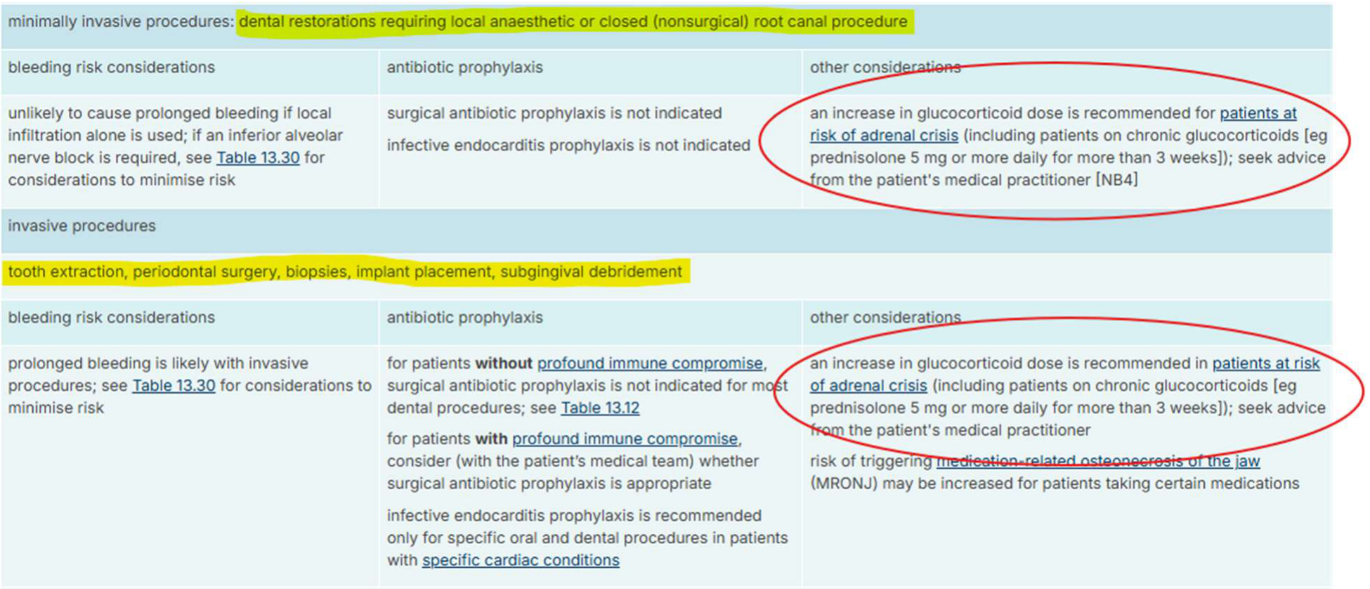
- Other considerations: an increase in glucocorticoid dose is recommended for patients at risk of adrenal crisis (including patients on chronic glucocorticoids [e.g. prednisolone 5 mg or more daily for more than 3 weeks]); seek advice from the patient’s medical practitioner
Invasive Procedures: Tooth Extraction, Periodontal Surgery, Biopsies, Implant Placement, Subgingival Debridement
- Bleeding risk considerations: prolonged bleeding is likely with invasive procedures; see Table 13.30 for considerations to minimise risk
- Antibiotic prophylaxis: for patients without profound immune compromise, surgical antibiotic prophylaxis is not indicated for most dental procedures (see Table 13.12); for patients with profound immune compromise, consider (with the patient’s medical team) whether surgical antibiotic prophylaxis is appropriate; infective endocarditis prophylaxis is recommended only for specific oral and dental procedures in patients with specific cardiac conditions
- Other considerations: an increase in glucocorticoid dose is recommended in patients at risk of adrenal crisis (including patients on chronic glucocorticoids [e.g. prednisolone 5 mg or more daily for more than 3 weeks]); seek advice from the patient’s medical practitioner; risk of triggering medication-related osteonecrosis of the jaw (MRONJ) may be increased for patients taking certain medications
Therapeutic Guidelines – Oral and Dental v4 2026
Glucocorticoid Dosing Around Dental Procedures74
For dental procedures that do not warrant an increase in glucocorticoid dosing (see Table 13.29), patients must take their usual glucocorticoid dose on the day of the dental procedure.
For dental restorations requiring local anaesthetic, closed (nonsurgical) root canal procedures, or any invasive procedures, an increase in glucocorticoid dose is recommended to minimise the risk of precipitating adrenal crisis. Patients may have a written action plan addressing the need for periprocedural dose increases; if not, seek advice from the patient’s medical practitioner on glucocorticoid dosing adjustment. Usually, the patient’s regular glucocorticoid dose is doubled on the morning of the dental procedure (and in some situations, for up to 48 hours postoperatively). Dental practitioners should consult the patient’s medical practitioner when a dose increase is required, to ensure all clinicians involved in the patient’s care can consider the impact of any proposed short-term changes to their glucocorticoid regimen. Seek endocrinologist advice for patients at risk of adrenal crisis who are undergoing general anaesthetic, sedation or fasting.
Post-Procedural Care
Perform dental treatment in the morning so that if an adrenal crisis occurs, symptoms present while the patient is awake. After dental treatment, ensure the patient remains in the care of a responsible adult for the rest of the day, and the carer remains in contact with the patient for the following 2 to 3 days. Advise the patient and the carer to seek urgent medical attention if the patient experiences symptoms of adrenal crisis.
Therapeutic Guidelines – Oral and Dental v4 2026
Renal Dialysis [MM]75
- Drug doses may require reduction
- Haemodialysis: treat on non-dialysis days (heparinisation)
- Bleeding tendency, anaemia, infection tendency…
- (Topic of another lecture: Renal Disease)
Infection Control and Cross-Infection76
- Susceptibility to infection → infection control protocols
- Transfusions: screened for BBVs
- Immunocompromise/immunosuppression = may be on systemic antifungal (e.g. fluconazole), antiviral (e.g. valaciclovir), antibiotics (e.g. sulfamethoxazole/trimethoprim)
- Predisposed to oral candida, herpes simplex, VZV, etc
Questions and Contact77
QUESTIONS?
Lecturer — Antibiotic Consultation
Antibiotic prophylaxis should be assessed jointly by the dentist and medical team. The dentist should explain the anticipated bacteraemia risk, particularly when the choice of antibiotic is uncertain or the patient is already taking background antibiotics.
Clinical Cases
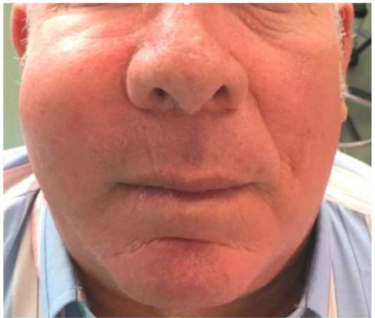
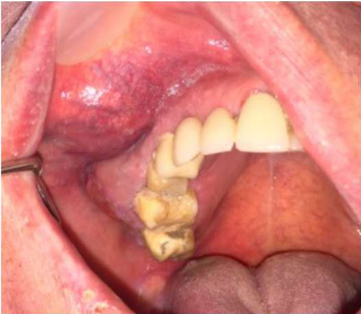
Case: Patient with polycythemia vera and aquagenic pruritus
Question
Scenario: A male patient with polycythemia vera and a history of aquagenic pruritus presents for dental treatment.
What’s shown: The patient’s medical history of aquagenic pruritus, a condition characterized by itchy skin following contact with water.
Consider: How the patient’s specific condition affects the delivery and management of dental treatment.
Answer
Observations:
- The patient has polycythemia vera.
- The patient experiences aquagenic pruritus (itchy skin following contact with water), which can be a chief complaint and is considered unbearable in about 15% of cases with this symptom.
Reasoning: Dental treatment typically involves a lot of water. For a patient with aquagenic pruritus, exposure to water can trigger severe itching, making standard dental procedures highly problematic and uncomfortable for the patient.
Outcome: The dental team had to take it quite easy and carefully modify the treatment approach to minimize the patient’s exposure to water.
Takeaway: Aquagenic pruritus, a symptom associated with polycythemia vera, is triggered by water contact, requiring the dental team to carefully manage and minimize water use during dental procedures.
Case: Skull radiograph showing multiple myeloma features
Question
Scenario: A PA skull radiograph is presented to illustrate the skeletal manifestations of multiple myeloma.
What’s shown: A PA skull view revealing multiple punched-out radiolucencies in the skull and a right-sided mandibular pathological fracture.
Consider: Identify the radiographic findings and explain the underlying pathophysiology and clinical implications for the jaws and systemic health.
Answer
Observations:
- Multiple punched-out radiolucencies in the skull.
- A right-sided mandibular pathological fracture.
Reasoning: In multiple myeloma, neoplastic proliferation of plasma cells in the bone marrow releases cytokines that lead to osteolysis and bone destruction. This process causes mandibular lesions in about 5 to 14% of patients, which can present clinically as dental mobility, pain, paresthesia, soft tissue swellings, and pathological jaw fractures. Additionally, the proliferation can cause suppression of hemopoiesis and bone marrow replacement, leading to anemia, increased infection risk, and bleeding tendencies. Defective immunoglobulins can also cause plasma hyperviscosity, renal failure, and neurological sequelae.
Takeaway: Multiple myeloma causes osteolytic bone destruction via cytokine release, which can present radiographically as multiple punched-out skull radiolucencies and mandibular pathological fractures, alongside systemic complications from bone marrow suppression.
Footnotes
-
Original PDF page 1: L5.2 Haematology 2 WBC, p.1 ↩
-
Original PDF page 2: L5.2 Haematology 2 WBC, p.2 ↩
-
Original PDF page 3: L5.2 Haematology 2 WBC, p.3 ↩
-
Original PDF page 4: L5.2 Haematology 2 WBC, p.4 ↩
-
Original PDF page 5: L5.2 Haematology 2 WBC, p.5 ↩
-
Original PDF page 6: L5.2 Haematology 2 WBC, p.6 ↩
-
Original PDF page 7: L5.2 Haematology 2 WBC, p.7 ↩
-
Original PDF page 8: L5.2 Haematology 2 WBC, p.8 ↩
-
Original PDF page 9: L5.2 Haematology 2 WBC, p.9 ↩
-
Original PDF page 10: L5.2 Haematology 2 WBC, p.10 ↩
-
Original PDF page 11: L5.2 Haematology 2 WBC, p.11 ↩
-
Original PDF page 12: L5.2 Haematology 2 WBC, p.12 ↩
-
Original PDF page 13: L5.2 Haematology 2 WBC, p.13 ↩
-
Original PDF page 14: L5.2 Haematology 2 WBC, p.14 ↩
-
Original PDF page 15: L5.2 Haematology 2 WBC, p.15 ↩
-
Original PDF page 16: L5.2 Haematology 2 WBC, p.16 ↩
-
Original PDF page 17: L5.2 Haematology 2 WBC, p.17 ↩
-
Original PDF page 18: L5.2 Haematology 2 WBC, p.18 ↩
-
Original PDF page 19: L5.2 Haematology 2 WBC, p.19 ↩
-
Original PDF page 20: L5.2 Haematology 2 WBC, p.20 ↩
-
Original PDF page 21: L5.2 Haematology 2 WBC, p.21 ↩
-
Original PDF page 22: L5.2 Haematology 2 WBC, p.22 ↩
-
Original PDF page 23: L5.2 Haematology 2 WBC, p.23 ↩
-
Original PDF page 24: L5.2 Haematology 2 WBC, p.24 ↩
-
Original PDF page 25: L5.2 Haematology 2 WBC, p.25 ↩
-
Original PDF page 26: L5.2 Haematology 2 WBC, p.26 ↩
-
Original PDF page 27: L5.2 Haematology 2 WBC, p.27 ↩
-
Original PDF page 28: L5.2 Haematology 2 WBC, p.28 ↩
-
Original PDF page 29: L5.2 Haematology 2 WBC, p.29 ↩
-
Original PDF page 30: L5.2 Haematology 2 WBC, p.30 ↩
-
Original PDF page 31: L5.2 Haematology 2 WBC, p.31 ↩
-
Original PDF page 32: L5.2 Haematology 2 WBC, p.32 ↩
-
Original PDF page 33: L5.2 Haematology 2 WBC, p.33 ↩
-
Original PDF page 34: L5.2 Haematology 2 WBC, p.34 ↩
-
Original PDF page 35: L5.2 Haematology 2 WBC, p.35 ↩
-
Original PDF page 36: L5.2 Haematology 2 WBC, p.36 ↩
-
Original PDF page 37: L5.2 Haematology 2 WBC, p.37 ↩
-
Original PDF page 38: L5.2 Haematology 2 WBC, p.38 ↩
-
Original PDF page 39: L5.2 Haematology 2 WBC, p.39 ↩
-
Original PDF page 40: L5.2 Haematology 2 WBC, p.40 ↩
-
Original PDF page 41: L5.2 Haematology 2 WBC, p.41 ↩
-
Original PDF page 42: L5.2 Haematology 2 WBC, p.42 ↩
-
Original PDF page 43: L5.2 Haematology 2 WBC, p.43 ↩
-
Original PDF page 44: L5.2 Haematology 2 WBC, p.44 ↩
-
Original PDF page 45: L5.2 Haematology 2 WBC, p.45 ↩
-
Original PDF page 46: L5.2 Haematology 2 WBC, p.46 ↩
-
Original PDF page 47: L5.2 Haematology 2 WBC, p.47 ↩
-
Original PDF page 49: L5.2 Haematology 2 WBC, p.49 ↩
-
Original PDF page 50: L5.2 Haematology 2 WBC, p.50 ↩
-
Original PDF page 48: L5.2 Haematology 2 WBC, p.48 ↩
-
Original PDF page 51: L5.2 Haematology 2 WBC, p.51 ↩
-
Original PDF page 52: L5.2 Haematology 2 WBC, p.52 ↩
-
Original PDF page 53: L5.2 Haematology 2 WBC, p.53 ↩
-
Original PDF page 54: L5.2 Haematology 2 WBC, p.54 ↩
-
Original PDF page 55: L5.2 Haematology 2 WBC, p.55 ↩
-
Original PDF page 56: L5.2 Haematology 2 WBC, p.56 ↩
-
Original PDF page 57: L5.2 Haematology 2 WBC, p.57 ↩
-
Original PDF page 58: L5.2 Haematology 2 WBC, p.58 ↩
-
Original PDF page 60: L5.2 Haematology 2 WBC, p.60 ↩
-
Original PDF page 59: L5.2 Haematology 2 WBC, p.59 ↩
-
Original PDF page 63: L5.2 Haematology 2 WBC, p.63 ↩
-
Original PDF page 61: L5.2 Haematology 2 WBC, p.61 ↩
-
Original PDF page 62: L5.2 Haematology 2 WBC, p.62 ↩
-
Original PDF page 64: L5.2 Haematology 2 WBC, p.64 ↩
-
Original PDF page 65: L5.2 Haematology 2 WBC, p.65 ↩
-
Original PDF page 66: L5.2 Haematology 2 WBC, p.66 ↩
-
Original PDF page 67: L5.2 Haematology 2 WBC, p.67 ↩
-
Original PDF page 69: L5.2 Haematology 2 WBC, p.69 ↩
-
Original PDF page 68: L5.2 Haematology 2 WBC, p.68 ↩
-
Original PDF page 70: L5.2 Haematology 2 WBC, p.70 ↩
-
Original PDF page 71: L5.2 Haematology 2 WBC, p.71 ↩
-
Original PDF page 72: L5.2 Haematology 2 WBC, p.72 ↩
-
Original PDF page 73: L5.2 Haematology 2 WBC, p.73 ↩
-
Original PDF page 74: L5.2 Haematology 2 WBC, p.74 ↩
-
Original PDF page 75: L5.2 Haematology 2 WBC, p.75 ↩
-
Original PDF page 76: L5.2 Haematology 2 WBC, p.76 ↩
-
Original PDF page 77: L5.2 Haematology 2 WBC, p.77 ↩