Musculoskeletal Disorders and Dentistry1
Dr Jee-Yun Leung BDSc(Hons) MSc(SCD) DSCD(RCSEng) DClinDent(SND) MRACDS(SND) FANZASND
- Specialist in Special Needs Dentistry
- Senior Lecturer, The University of Western Australia
- Consultant, Maxillofacial & Dental Dept, Fiona Stanley Hospital
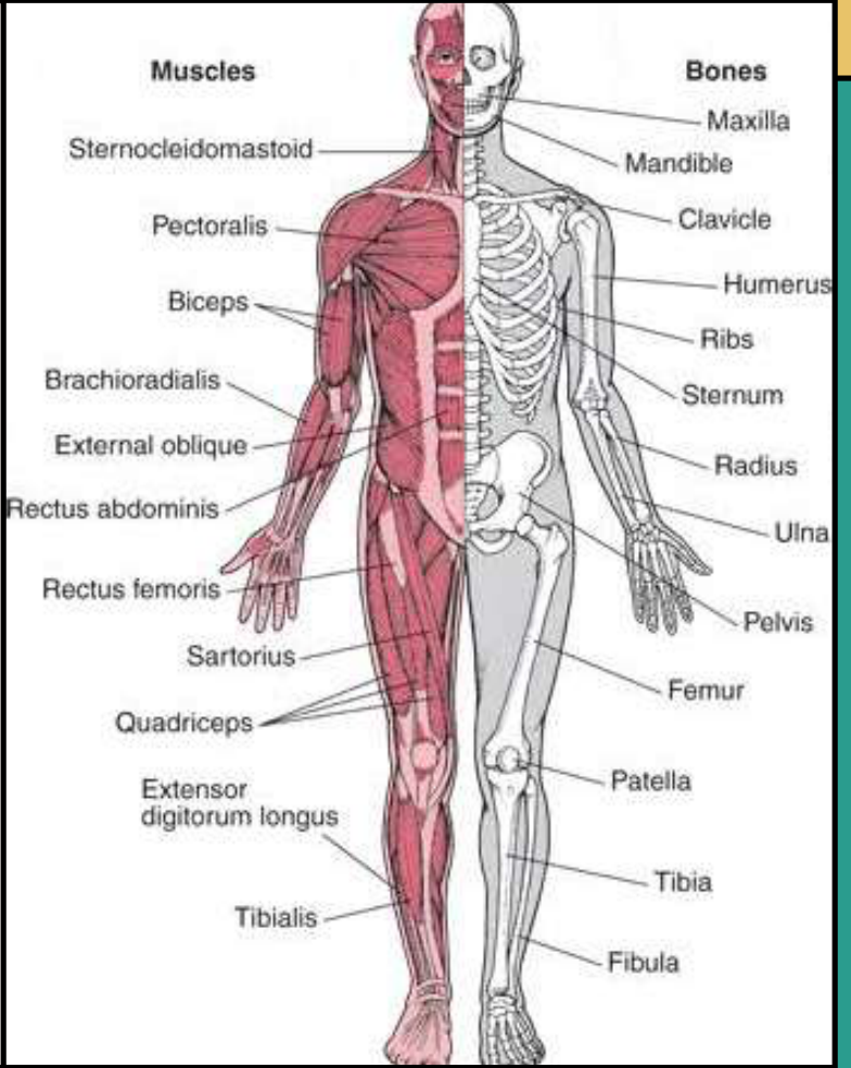
Lecture Outcomes2
-
Discuss the specific characteristics and dental needs of individuals with joint (e.g. osteoarthritis, rheumatoid arthritis) and bone (e.g. osteoporosis) disorders
-
Perform a risk assessment and make appropriate treatment adaptations to ensure safe and effective dental care
-
Work a patient up prior to commencement of bone-modifying drugs (e.g. bisphosphonates, denosumab, romosozumab)
-
Assess MRONJ risk prior to invasive dental procedures, and institute preventive measures
-
Appreciate management options of MRONJ
 |  |
Chronic Musculoskeletal Disorders3
Chronic musculoskeletal disorders affect:
-
Muscles (e.g. sarcopenia)
-
Joints (e.g. osteoarthritis, rheumatoid arthritis)
-
Bones (e.g. osteoporosis, osteonecrosis, traumatic fractures)
-
Spine (e.g. back and neck pain)
-
Multiple body systems and locations (e.g. widespread and regional pain disorders, inflammatory or connective tissue diseases, systemic vasculitides, fibromyalgia).
-
Muscles may also be affected by muscular dystrophy.
-
The most common symptom is pain.
-
Another common symptom is restricted mobility.
Joint Disorders4
- Osteoarthritis
- Rheumatoid Arthritis
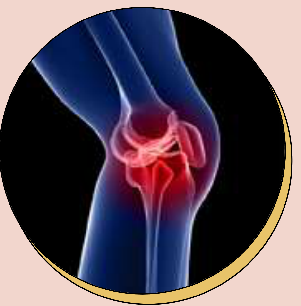
Arthritis Overview56789
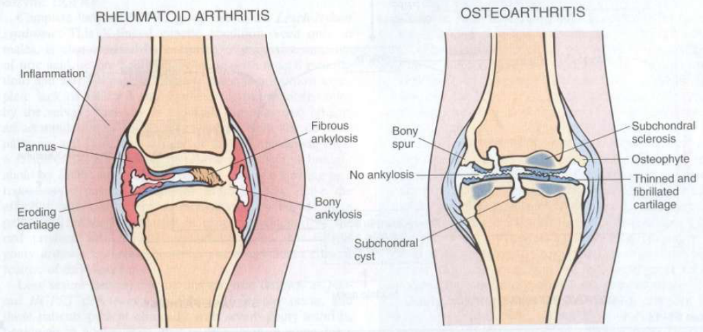
- Osteoarthritis: Degenerative joint disease. Key feature is degeneration of articular cartilage (“wear and tear”).
- Rheumatoid arthritis: Systemic chronic immunologically mediated inflammatory disease affecting multiple tissues, but principally attacking the joints. Characterised by an autoantibody directed against IgG (rheumatoid factor, RF) → formation of immune complexes and activation of complement → synovial inflammation and destructive joint disease.
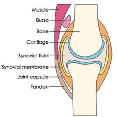
Normal Joint10
Muscle, bursa, bone, cartilage, synovial fluid, synovial membrane, joint capsule, tendon.
 | 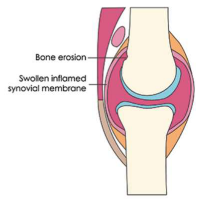 |
Inflamed Joint
Eroded cartilage; bone ends rub together. Bone erosion; swollen inflamed synovial membrane.
Lecturer — Joint Damage
Rheumatoid arthritis can affect the synovium, adjacent tissues and articular surfaces, with consequences beyond the joints. Osteoarthritis may also produce subchondral cysts and sclerosis in addition to deformed articular surfaces and bone spurs.
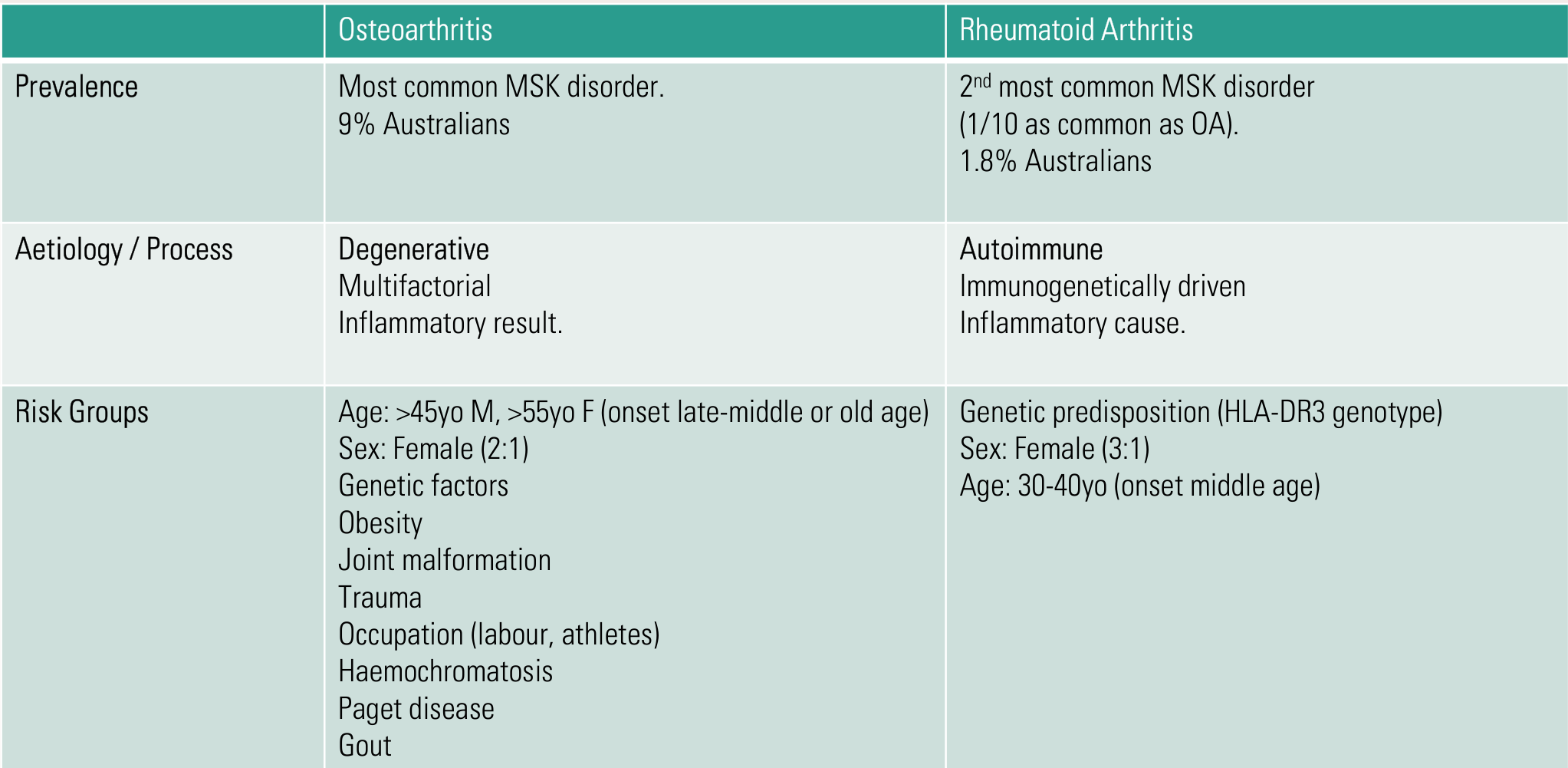
| Osteoarthritis | Rheumatoid Arthritis | |
|---|---|---|
| Prevalence | Most common MSK disorder. 9% Australians | 2nd most common MSK disorder (1/10 as common as OA). 1.8% Australians |
| Aetiology / Process | Degenerative. Multifactorial. Inflammatory result. | Autoimmune. Immunogenetically driven. Inflammatory cause. |
| Risk Groups | Age: >45yo M, >55yo F (onset late-middle or old age); Sex: Female (2:1); Genetic factors; Obesity; Joint malformation; Trauma; Occupation (labour, athletes); Haemochromatosis; Paget disease; Gout | Genetic predisposition (HLA-DR3 genotype); Sex: Female (3:1); Age: 30-40yo (onset middle age) |
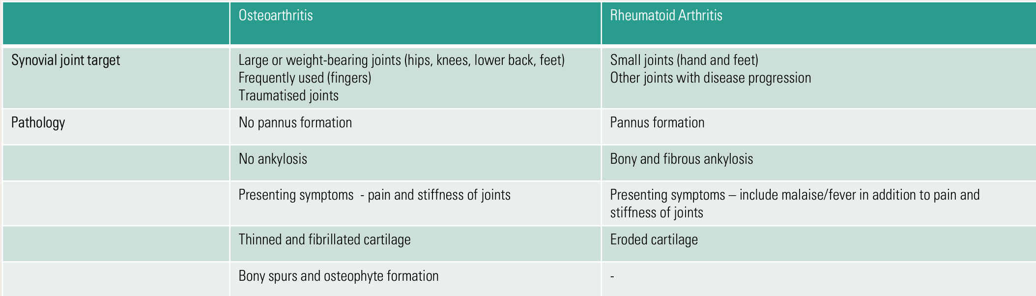
| Osteoarthritis | Rheumatoid Arthritis | |
|---|---|---|
| Synovial joint target | Large or weight-bearing joints (hips, knees, lower back, feet); frequently used (fingers); traumatised joints | Small joints (hand and feet); other joints with disease progression |
| Pathology | No pannus formation | Pannus formation |
| No ankylosis | Bony and fibrous ankylosis | |
| Presenting symptoms – pain and stiffness of joints | Presenting symptoms – include malaise/fever in addition to pain and stiffness of joints | |
| Thinned and fibrillated cartilage | Eroded cartilage | |
| Bony spurs and osteophyte formation | – |
| Osteoarthritis | Rheumatoid Arthritis | |
|---|---|---|
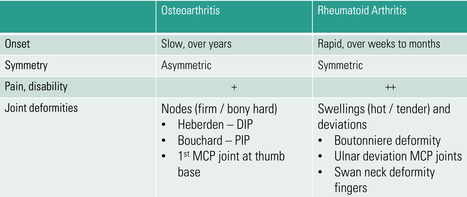
| Onset | Slow, over years | Rapid, over weeks to months |
| Symmetry | Asymmetric | Symmetric |
| Pain, disability | + | ++ |
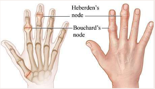
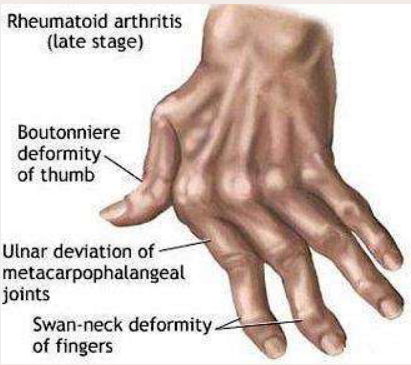
| Joint deformities | Nodes (firm / bony hard): Heberden – DIP; Bouchard – PIP; 1st MCP joint at thumb base | Swellings (hot / tender) and deviations: Boutonniere deformity; Ulnar deviation MCP joints; Swan neck deformity fingers |
| Osteoarthritis | Rheumatoid Arthritis | |
|---|---|---|
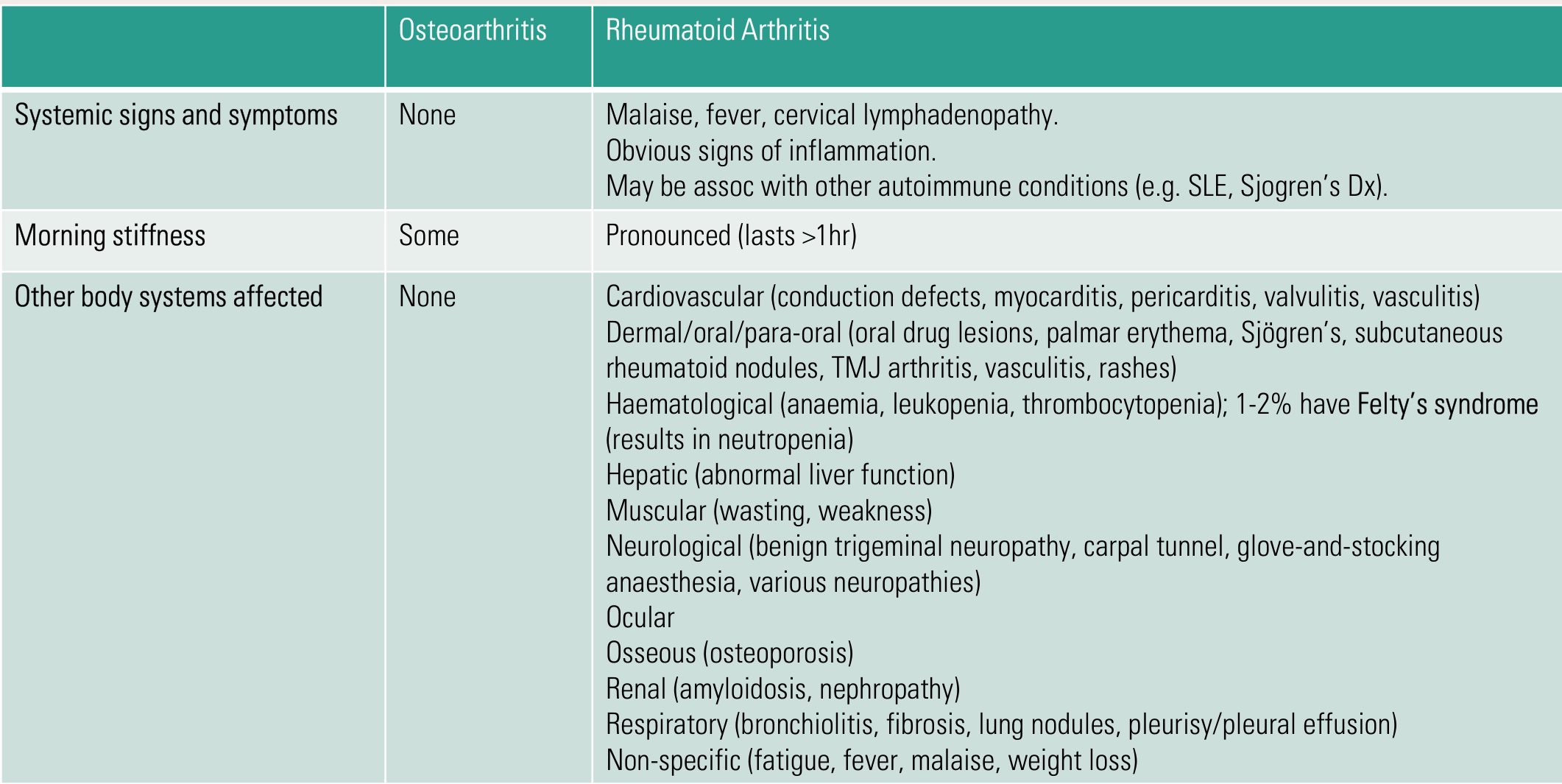
| Systemic signs and symptoms | None | Malaise, fever, cervical lymphadenopathy. Obvious signs of inflammation. May be assoc with other autoimmune conditions (e.g. SLE, Sjogren’s Dx). |
| Morning stiffness | Some | Pronounced (lasts >1hr) |
| Other body systems affected | None | Cardiovascular (conduction defects, myocarditis, pericarditis, valvulitis, vasculitis); Dermal/oral/para-oral (oral drug lesions, palmar erythema, Sjögren’s, subcutaneous rheumatoid nodules, TMJ arthritis, vasculitis, rashes); Haematological (anaemia, leukopenia, thrombocytopenia); 1-2% have Felty’s syndrome (results in neutropenia); Hepatic (abnormal liver function); Muscular (wasting, weakness); Neurological (benign trigeminal neuropathy, carpal tunnel, glove-and-stocking anaesthesia, various neuropathies); Ocular; Osseous (osteoporosis); Renal (amyloidosis, nephropathy); Respiratory (bronchiolitis, fibrosis, lung nodules, pleurisy/pleural effusion); Non-specific (fatigue, fever, malaise, weight loss) |
| Osteoarthritis | Rheumatoid Arthritis | |
|---|---|---|
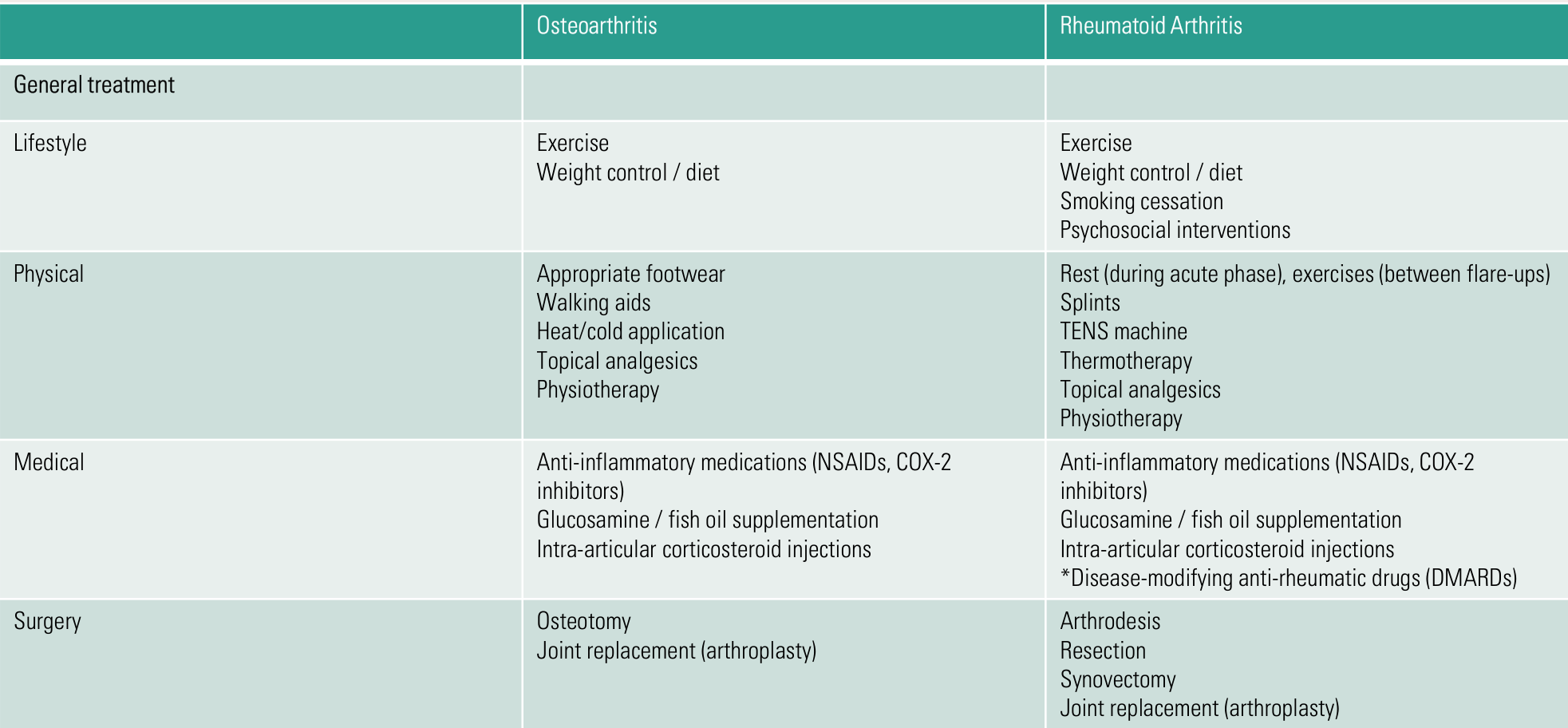
| General treatment – Lifestyle | Exercise; weight control / diet | Exercise; weight control / diet; smoking cessation; psychosocial interventions |
| Physical | Appropriate footwear; walking aids; heat/cold application; topical analgesics; physiotherapy | Rest (during acute phase), exercises (between flare-ups); splints; TENS machine; thermotherapy; topical analgesics; physiotherapy |
| Medical | Anti-inflammatory medications (NSAIDs, COX-2 inhibitors); glucosamine / fish oil supplementation; intra-articular corticosteroid injections | Anti-inflammatory medications (NSAIDs, COX-2 inhibitors); glucosamine / fish oil supplementation; intra-articular corticosteroid injections; disease-modifying anti-rheumatic drugs (DMARDs) |
| Surgery | Osteotomy; joint replacement (arthroplasty) | Arthrodesis; resection; synovectomy; joint replacement (arthroplasty) |
Oral Implications of Arthritis11
Association with periodontitis: 2017 world workshop (AAP + EFP)
Albandar JM, Susin C, Hughes FJ. Manifestations of systemic diseases and conditions that affect the periodontal attachment apparatus: Case definitions and diagnostic considerations. J Periodontol. 2018 Jun;89 Suppl 1:S183-S203.
| Disorder | Strength of association | Quality of evidence | Biologic mechanisms | Case definitions | Diagnostic considerations |
|---|---|---|---|---|---|
| Arthritis | Significant | Animal models, systematic review | Rheumatoid arthritis is an autoimmune disease; osteoarthritis is due to gradual deterioration of cartilage | Joint pain, swelling, stiffness, redness, and limited motion; increased risk for loss of periodontal attachment and alveolar bone | Clinical history and physical examination for arthritis |
Lecturer — Shared Inflammation
The association between periodontitis and arthritis was related to inflammatory mechanisms shared by both conditions.

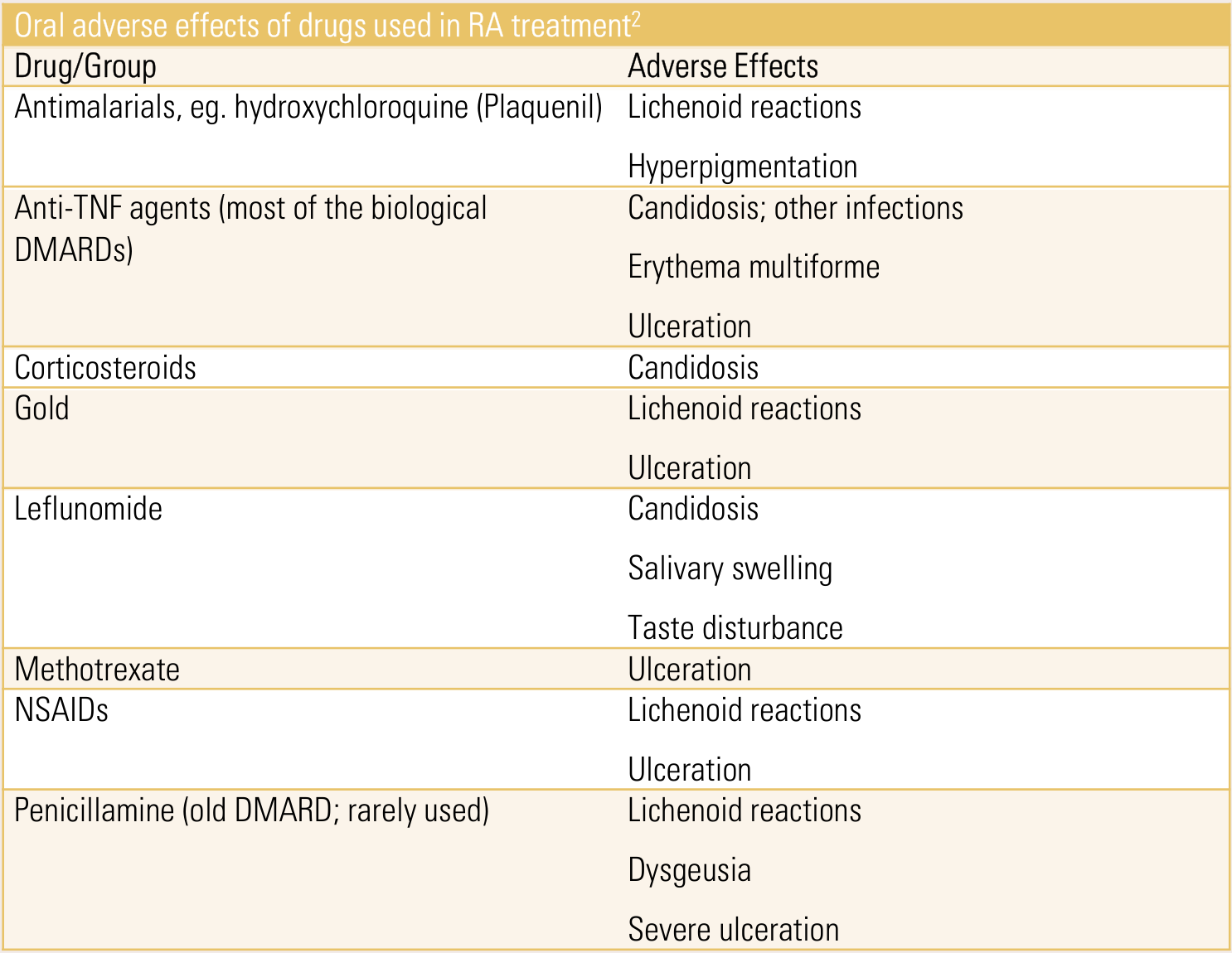
Rheumatoid Arthritis Drugs and Oral Side Effects1213
Oral adverse effects of drugs used in RA treatment:
| Drug/Group | Adverse Effects |
|---|---|
| Antimalarials, e.g. hydroxychloroquine (Plaquenil) | Lichenoid reactions; hyperpigmentation |
| Anti-TNF agents (most of the biological DMARDs) | Candidosis; other infections; erythema multiforme; ulceration |
| Corticosteroids | Candidosis |
| Gold | Lichenoid reactions; ulceration |
| Leflunomide | Candidosis; salivary swelling; taste disturbance |
| Methotrexate | Ulceration |
| NSAIDs | Lichenoid reactions; ulceration |
| Penicillamine (old DMARD; rarely used) | Lichenoid reactions; dysgeusia; severe ulceration |
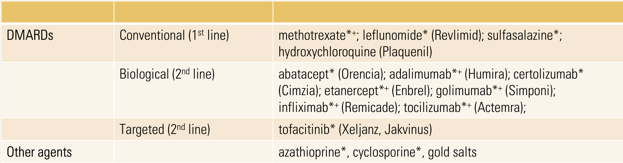
| DMARDs | Conventional (1st line) | methotrexate*+; leflunomide* (Revlimid); sulfasalazine*; hydroxychloroquine (Plaquenil) |
| Biological (2nd line) | abatacept* (Orencia); adalimumab*+ (Humira); certolizumab* (Cimzia); etanercept*+ (Enbrel); golimumab*+ (Simponi); infliximab*+ (Remicade); tocilizumab*+ (Actemra) | |
| Targeted (2nd line) | tofacitinib* (Xeljanz, Jakvinus) | |
| Other agents | azathioprine*, cyclosporine*, gold salts |
Lecturer — Targeted DMARDs
Targeted agents may produce fewer side effects than broadly immunosuppressive drugs because they act on more specific disease targets.
May combine with corticosteroids in induction phase.
- * agents causing immunocompromise
- + emerging case reports of MRONJ association
- Associated with DMARDs
 |  |
Dental Implications of Arthritis1415
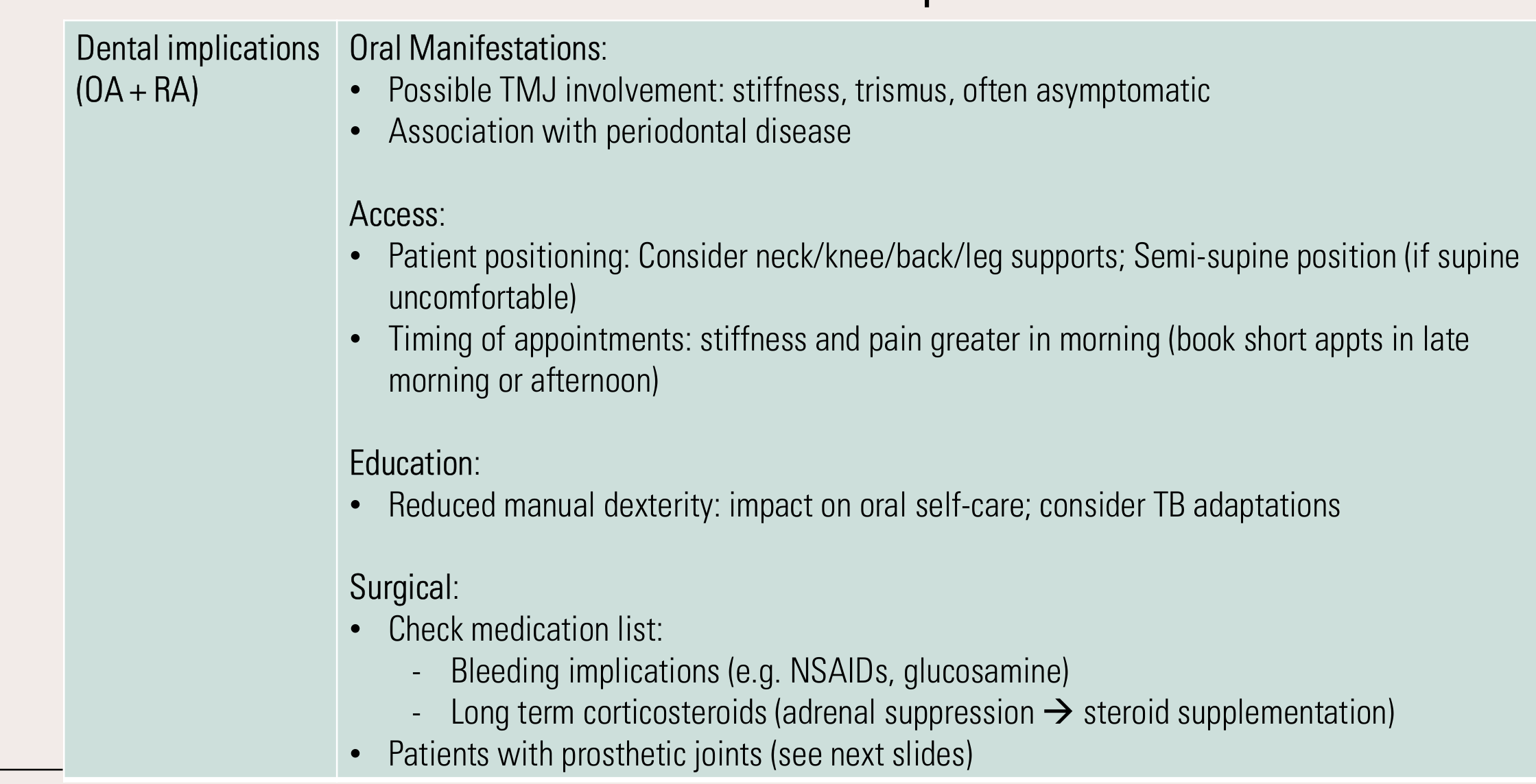
Dental implications (OA + RA):
- Oral Manifestations:
- Possible TMJ involvement: stiffness, trismus, often asymptomatic
- Association with periodontal disease
- Access:
- Patient positioning: consider neck/knee/back/leg supports; semi-supine position (if supine uncomfortable)
- Timing of appointments: stiffness and pain greater in morning (book short appts in late morning or afternoon)
- Gradually recline the chair and check comfort at intervals; use towels, cushions or neck supports where appropriate.
- Education:
- Reduced manual dexterity: impact on oral self-care; consider TB adaptations
- Patients may need assistance with toothbrushing and interproximal cleaning.
- Reduced manual dexterity: impact on oral self-care; consider TB adaptations
- Surgical:
- Check medication list:
- Bleeding implications (e.g. NSAIDs, glucosamine)
- Long term corticosteroids (adrenal suppression → steroid supplementation)
- Patients with prosthetic joints (see next slides)
- Check medication list:
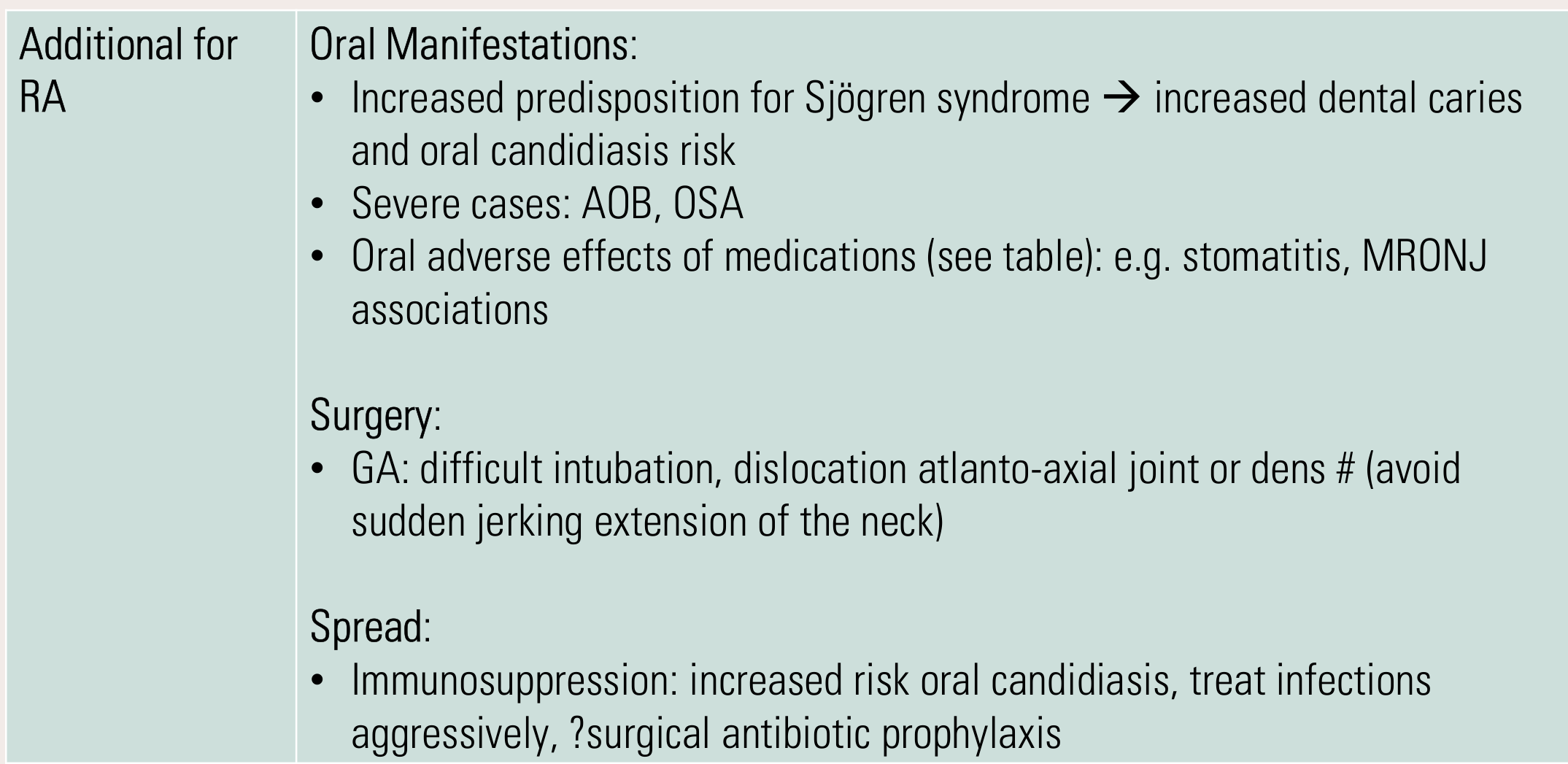
Additional for RA:
- Oral Manifestations:
- Increased predisposition for Sjögren syndrome → increased dental caries and oral candidiasis risk
- Severe cases: AOB, OSA
- Oral adverse effects of medications (see table): e.g. stomatitis, MRONJ associations
- Surgery:
- GA: difficult intubation, dislocation atlanto-axial joint or dens # (avoid sudden jerking extension of the neck)
- Spread:
- Immunosuppression: increased risk oral candidiasis, treat infections aggressively, ?surgical antibiotic prophylaxis
Lecturer — Anaesthetic Risk
Intubation and positioning may be hazardous when rheumatoid arthritis affects the atlantoaxial joint or dens. This is particularly important during general anaesthesia because the patient cannot provide feedback about discomfort.
 |  |
Prosthetic Joints
Is surgical antibiotic prophylaxis indicated for patients with a pre-existing joint prosthesis?
Principles of Surgical Antibiotic Prophylaxis16
Although surgical procedures can result in incidental bacteraemia, there is a low risk of seeding of the joint prosthesis and subsequent infection. Accumulated evidence supports the same approach to surgical antibiotic prophylaxis for patients with and without a pre-existing joint prosthesis; neither the indication for prophylaxis nor the choice of antibiotic regimen is altered by the presence of a joint prosthesis. This is true even for procedures that commonly cause bacteraemia (e.g. tooth extractions, periodontal procedures, cystoscopy, insertion of an indwelling urinary catheter, perforating dermatological procedures). For patients with pre-existing joint prosthesis, the potential adverse effects of prophylaxis outweigh the potential benefits unless indicated [Note 4] [30].
Only give surgical antibiotic prophylaxis to patients with a pre-existing joint prosthesis if prophylaxis is indicated.
Note 4: For a detailed discussion of prevention of prosthetic joint infection in patients undergoing dental procedures, see the American Dental Association Council on Scientific Affairs evidence-based guideline on prevention of orthopaedic implant infection in patients undergoing dental procedures.
Upcoming lecture: Antibiotic Prophylaxis
Is surgical antibiotic prophylaxis indicated for patients with a pre-existing joint prosthesis?
American Dental Association Council on Scientific Affairs evidence-based guideline on prevention of orthopaedic implant infection in patients undergoing dental procedures: https://www.ada.org/resources/research/science/evidence-based-dental-research/antibiotics-to-prevent-prosthetic-joint-infection/
Shared Decision-Making Is the Standard17
Consider:
- Patient’s overall health & medical status
- Recovery progress and healing
- Risk factors for infection or complications
- Urgency and nature of the dental condition
- Patient values and preferences
Work with the patient and their orthopedic provider when needed to determine the best timing for care.
The ADA does not support any standard time frame for delaying dental care after joint replacement. Routine antibiotics are usually not indicated for patients with prosthetic joints.
Clinical Evaluation — For Patients with Prosthetic Joints Undergoing Dental Procedure (Individualize · Collaborate · Document)
Does the patient have a prosthetic joint? Consult orthopedic provider if uncertain.
- Move away from “automatic prescribing of antibiotics” — current evidence shows no proven benefit of routine antibiotic prophylaxis in preventing prosthetic joint infection for patients undergoing dental procedures (JADA, 2026). Minimal evidence of benefit with chances of:
- Antibiotic resistance
- Adverse drug reactions
- C. difficile infection
- Does the patient have high-risk medical factors?
- History of complications with joint replacement
- Immunocompromised status
- No: Most dental procedures are low risk for causing prosthetic joint infection. Focus on excellent oral hygiene; prevention and treatment of oral disease to reduce bacteremia.
- Yes: Initiate shared decision-making — dentist + patient + orthopedic provider collaborate:
- Discuss patient values & concerns
- Review medical history & joint status
- Consider risks vs. benefits
- Make a joint decision on antibiotic prophylaxis
Chairside script (quick use): “Current evidence shows antibiotics aren’t routinely needed for joint replacements. Let’s review your health history and decide together what’s safest for you.”
Documentation tip: Document the conversation, factors considered, who was involved in the decision, and the final plan.
Source: Shared decision making in oral health care for patients with prosthetic joint replacements. American Dental Association Council on Scientific Affairs position statement. Journal of the American Dental Association, 2026.
© 2026 American Dental Association. All Rights Reserved. Updated: 6/11/26.
Bone Disorders18
Osteopenia, Osteoporosis
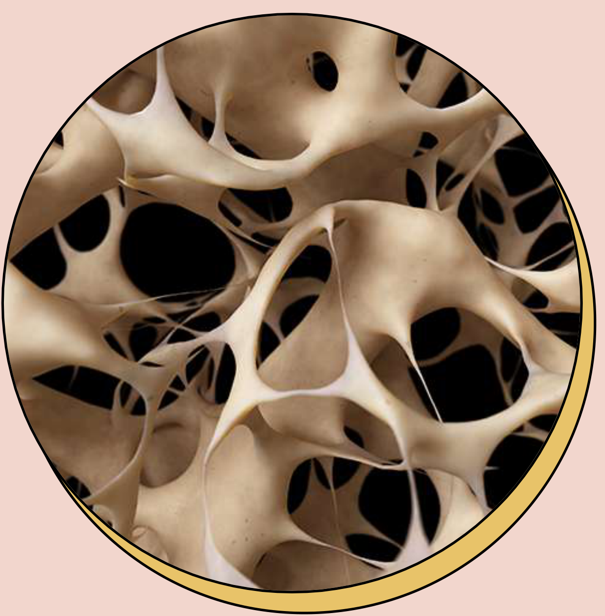
Osteopenia and Osteoporosis19
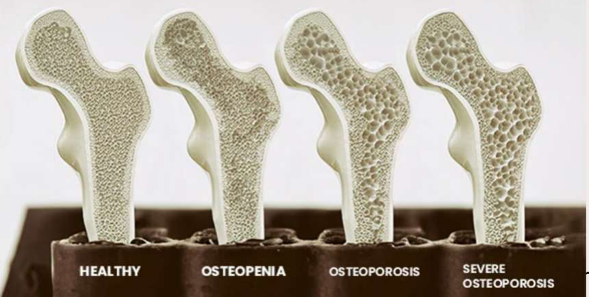
Definition: Loss of bone mineral density characterised by reduced mineralised bone mass, especially common in older women.
-
Osteopenia: Bone densitometry: BMD 1 to 2.5 standard deviations (T score -1 to -2.5) below the peak bone mass of a 25yo individual.
- Precursor to osteoporosis
-
Osteoporosis: Bone densitometry: BMD >2.5 standard deviations (T score < -2.5) below the peak bone mass of a 25yo individual.
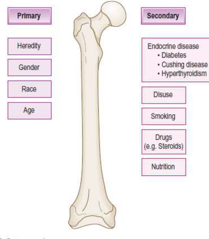
Osteoporosis Aetiology20
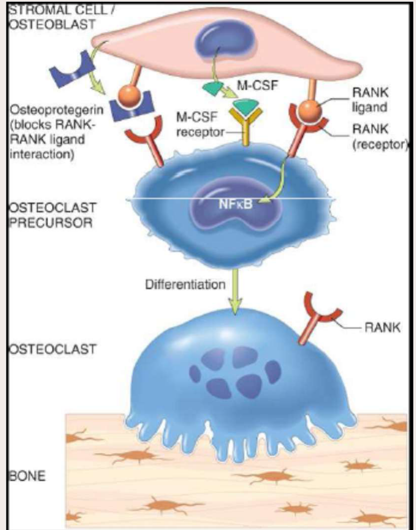
Metabolic bone disease that results from an imbalance in osteoblastic and osteoclastic activities.
 |  |
RANK–RANK Ligand Pathway21
-
Stromal cell / osteoblast
- Osteoprotegerin (blocks RANK–RANK ligand interaction)
- M-CSF
- M-CSF receptor
- RANK ligand
-
Osteoclast precursor
- RANK (receptor)
- NFκB
- Differentiation
-
Osteoclast
- RANK
- Bone
-
Primary osteoporosis: androgen-oestrogen deficiency, lack of Vit D, lack of exercise
-
Secondary osteoporosis: corticosteroid use, hyperparathyroidism, diabetes, chronic liver disease, malabsorption or hyperthyroidism, excessive smoking/alcohol
Lecturer — RANK Pathway Dysregulation
Dysregulation of the RANK–RANK ligand pathway contributes to osteoporosis.
- Possible influences include ageing, changes in the cytokine environment and oestrogen deficiency.
Lecturer — Bone Loss Over Time
Bone formation during the first approximately 28 years of life and subsequent bone remodelling influence later osteopenia and osteoporosis.
- From approximately 30 years of age, bone loss may occur at about 1% per year.
- Bone loss may increase to approximately 5% per year after menopause.
Osteoporosis Clinical Manifestations22
- Usually asymptomatic.
- Increased bone fragility →
- Vertebral crush fractures
- Femoral neck, distal radius, humerus fractures
- Hip fractures: 20% one-year mortality risk in older adults
- Persistent lower back pain
- Kyphosis and loss of height
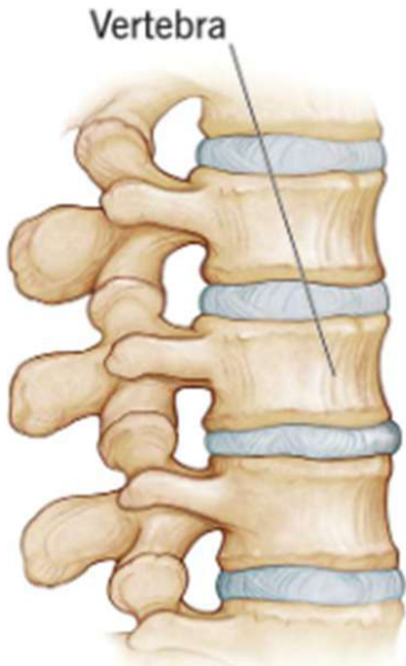 | 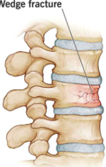 |
Osteoporosis General Management
Prevention23
- Weight-bearing exercise
- Calcium supplementation (dairy, green veg)
- Hormone replacement therapy (HRT)
- Avoiding risk factors
- During the first 20 years of life, bone formation is the major protective factor; later, prevention of bone loss becomes more important.
Treatment
- Calcitonin
- Vitamin D
- Bisphosphonates
- Denosumab
Dental Implications of Osteoporosis24
Association with Periodontitis: 2017 World Workshop (AAP + EFP)25
Albandar JM, Susin C, Hughes FJ. Manifestations of systemic diseases and conditions that affect the periodontal attachment apparatus: Case definitions and diagnostic considerations. J Periodontol. 2018 Jun;89 Suppl 1:S183-S203.
| Disorder | Strength of association | Quality of evidence | Biologic mechanisms | Case definitions | Diagnostic considerations |
|---|---|---|---|---|---|
| Osteoporosis | Significant | Animal models, surveys, longitudinal follow-up, case-control study, systematic reviews | Increased bone turnover leading to net bone loss, which can also be associated with other factors (such as estrogen level, vitamin D and calcium deficiency, lifestyle and behavioral factors) | • Decrease in bone mineral density and weakening of bone microarchitecture, leading to a high risk for bone fracture • Higher prevalence and severity of radiographic alveolar bone loss • No clear association with periodontitis (probing depth or clinical attachment loss) | • Clinical diagnosis |

Imaging
Jaws are thinned and granular. Lamina dura appears thinned. Maxillary sinus may extend between roots of molars. OPG may display spinal osteoporosis.
Lecturer — Dental Associations
Osteoporosis has been associated with periodontitis in women, excessive alveolar bone loss, pathological fracture and possible effects involving the jaw bones.
- Treatment of systemic osteoporosis might improve some aspects of jaw-bone osteoporosis in older adults.
- Dental implications include extractions, denture fit and implant placement.
- Oral manifestation:
- Association with periodontitis
- Radiographic findings
- Access:
- Provide adequate mobility and positioning support (depending on severity of OP)
- Surgery:
- MRONJ risk (bone modifying drugs) – extractions, dentures, implants
Bone Modifying Drugs26
Referrals to a Special Needs Dentistry Clinic2728
A common referral to the dental clinic…
Consult - Dental - for ongoing poor dental hygiene and care. Will need ongoing review and guidance with maintenance. Possibility of osteo medications that may require further follow up in the future. Thank you kindly.
Consult Progress: Requested
Consult - Dental - for 89F from home w/ daughter, admitted for nof due to mechanical fall. Poor dentition. Ceased denosumab 1/52 ago. Seen by orthogeriatrics who would like to assess for suitability of resuming denosumab.
Priority Level: Non-Urgent
Consult Progress: Requested
Example referrals to a SND clinic…
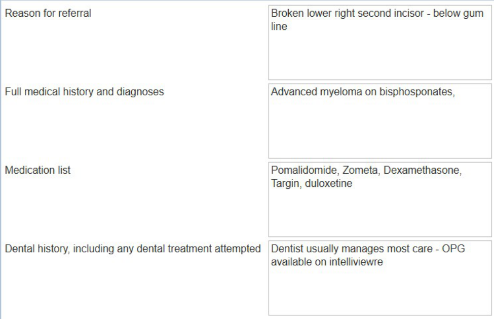
- Reason for referral: Broken lower right second incisor - below gum line
- Full medical history and diagnoses: Advanced myeloma on bisphosphonates
- Medication list: Pomalidomide, Zometa, Dexamethasone, Targin, duloxetine
- Dental history, including any dental treatment attempted: Dentist usually manages most care - OPG available on intelliviewer
64 YO M admitted under Haematology for CAR T Cell Monitoring CARTITUDE10 Trial
PMHx:
- Multiple myeloma diagnosed November 2025.
- Pancytopenia, leukoerythroblastic blood film, large IgA paraprotein.
- Bone marrow shows diffuse myeloma.
- Compression fractures in the spine.
- Commenced VRd therapy (Dr Hutchinson).
- Consented for CARTITUDE-10 clinical trial.
- Osteoporosis.
- Fatty liver disease.
- Hyperlipidaemia.
- Ischaemic heart disease, on aspirin therapy.
- Atraumatic compression fractures T5, T7, T12, L1, L5 (from approximately February 2025).
- Multiple previous vertebroplasties.
On review today raised concern of loose tooth, right molar.
On examination:
- Loose right upper molar
- Nil petechiae
- Nil jaw pain
- Nil fevers
- Isolated area of gum recession
- Nil other loose teeth
We have requested an OPG prior to discharge and would appreciate a review from yourselves given the patient’s active immunotherapy and neutropaenia.
Bloods:
- Neut 0.64
- Hb 105
- Lymp 0.16
MRONJ Definition29
- Current or previous treatment with antiresorptive therapy alone or in combination with immune modulators or anti-angiogenic medications
- Exposed bone, or bone that can be probed through an intraoral or extraoral fistula in the maxillofacial region that has persisted for more than 8 weeks
- No history of radiation therapy or no metastatic disease to the jaws.
(AAOMS 2022)
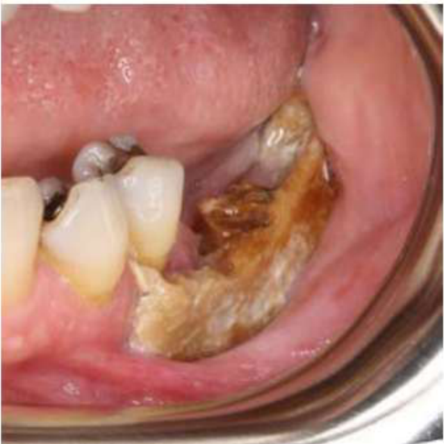
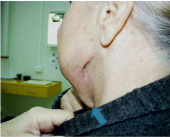
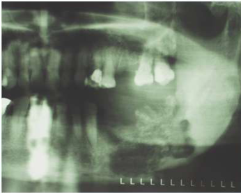
Clinical Presentation of MRONJ
Symptoms30
- Jaw pain or toothache
- Halitosis
- Numbness or altered sensation (paraesthesia)
 |  |
Signs
- Exposed bone in mandible or maxilla
- Localised swelling, tenderness of mucosa/gingiva
- Non-healing extraction sockets
- Purulent discharge
- Pathological fracture
- Oro-cutaneous fistulae
MRONJ Guidelines
AAOMS 2022 Guidelines31
Ruggiero SL, Dodson TB, Agbalao T, Carlson ER, Ward BB, Kademani D. American Association of Oral and Maxillofacial Surgeons’ Position Paper on Medication-Related Osteonecrosis of the Jaws—2022 Update.
SDCEP 2017 Guidelines – Updated 2022
NHS Education for Scotland. Oral Health Management of Patients at Risk of Medication-related Osteonecrosis of the Jaw. Dental Clinical Guidance.
RACGP 2024 Guidelines
RACGP / Healthy Bones Australia. Osteoporosis management and fracture prevention in postmenopausal women and men over 50 years of age. March 2024.
MASCC/ISOO/ASCO 2019
Yarom N, Shapiro CL, Peterson DE, Van Poznak CH, Bohike K, Ruggiero SL, Migliorati CA, Khan A, Morrison A, Anderson H, Murphy BA, Alton-Johnson D, Amaral Mendes R, Beadle BM, Beier Jensen S, Saunders DP. Medication-Related Osteonecrosis of the Jaw: MASCC/ISOO/ASCO Clinical Practice Guideline. Multinational Association of Supportive Care in Cancer, International Society of Oral Oncology, American Society of Clinical Oncology 2019.
International Task Force on ONJ 2015 (ASBMR, AAOMS, CAOMS, CAOMP, ECTS, IBMS, IOF, ISCD, IAOMS, JSBMR, NOF, Osteoporosis Canada, PAOS, TES)
Khan AA, Morrison A, Hanley DA, Felsenberg D, McCauley LK, O’Ryan F, Reid IR, Ruggiero SL, Taguchi A, Tetrakis S, Watts NB, Brandi ML, Peters E, Guise T, Eastell R, Cheung AM, Morin SN, Masri B, Cooper C, Morgan SL, Obermayer-Pietsch B, Langdahl BL, Al Dabagh R, Davison KS, Kendler DL, Sándor GK, Josse RG, Bhandari M, El Rabbany M, Pierroz DD, Sulimani R, Saunders DP, Brown JP, Compston J. Diagnosis and Management of Osteonecrosis of the Jaw: A Systematic Review and International Consensus. JBMR (Review), on behalf of the International Task Force on Osteonecrosis of the Jaw.
American Dental Association Council on Scientific Affairs 2011
Hellstein JW, Adler RA, Edwards B, Jacobsen PL, Kalmar JR, Koka S, Migliorati CA, Ristic H. Managing the care of patients receiving antiresorptive therapy for prevention and treatment of osteoporosis. Executive summary of recommendations from the American Dental Association Council on Scientific Affairs.
Healthy Bones Australia 2026 (ANZBMS, ADA, ESA, RHA)
Healthy Bones Australia Consensus Statement: Medication-related Osteonecrosis of the Jaw (MRONJ) and Osteoporosis.
Dr Jee-Yun Leung
Mechanisms of Action of Anti-Resorptives32
| Bisphosphonates | Denosumab | Romosozumab |
|---|---|---|
| Inhibits enzymes essential to formation, recruitment and function of osteoclasts | Fully human monoclonal antibody which inhibits osteoclast function | Monoclonal antibody |
| Binds to bone = high affinity for hydroxyapatite | Binds to receptor activator nuclear κB factor ligand (RANKL) | Wnt pathway = binds to and inhibits sclerostin (produced by osteocytes) |
| Persists in skeletal tissue for significant periods (e.g. alendronate ½ life ~10 years) | Does not bind to bone → effects on bone turnover diminish within 6 months | Potent bone anabolic agent (builds bone) |
| Very high risk men and post-menopausal women of osteoporotic minimal trauma fracture |
Bisphosphonates
Chemical Structures and Relative Potency33
- A: Inorganic pyrophosphate vs Bisphosphonate (chemical structures shown)
- B: Etidronate, Clodronate, Tiludronate (chemical structures shown) — relative potency: 1, 10, 10
- C: Alendronate, Risedronate, Ibandronate, Pamidronate, Zoledronic acid (chemical structures shown) — relative potency: 500, 2000, 1000, 100, 10,000
Bisphosphonate structures and approximate relative potencies for osteoclast inhibition.
Lecturer — Bisphosphonate Persistence
Bisphosphonates are commonly prescribed for approximately five to ten years for osteoporosis, but their long terminal persistence means they may remain relevant for approximately ten years afterward.
Generations and MRONJ Association
- Non-nitrogenous (1st gen): Direct action on osteoblastic cells to ↑ bone formation without anti-angiogenic effects → NOT associated with MRONJ
- Nitrogenous (2nd, 3rd gen): ↓ osteoclastic bone resorption by entering osteoclast → associated with MRONJ
Denosumab34
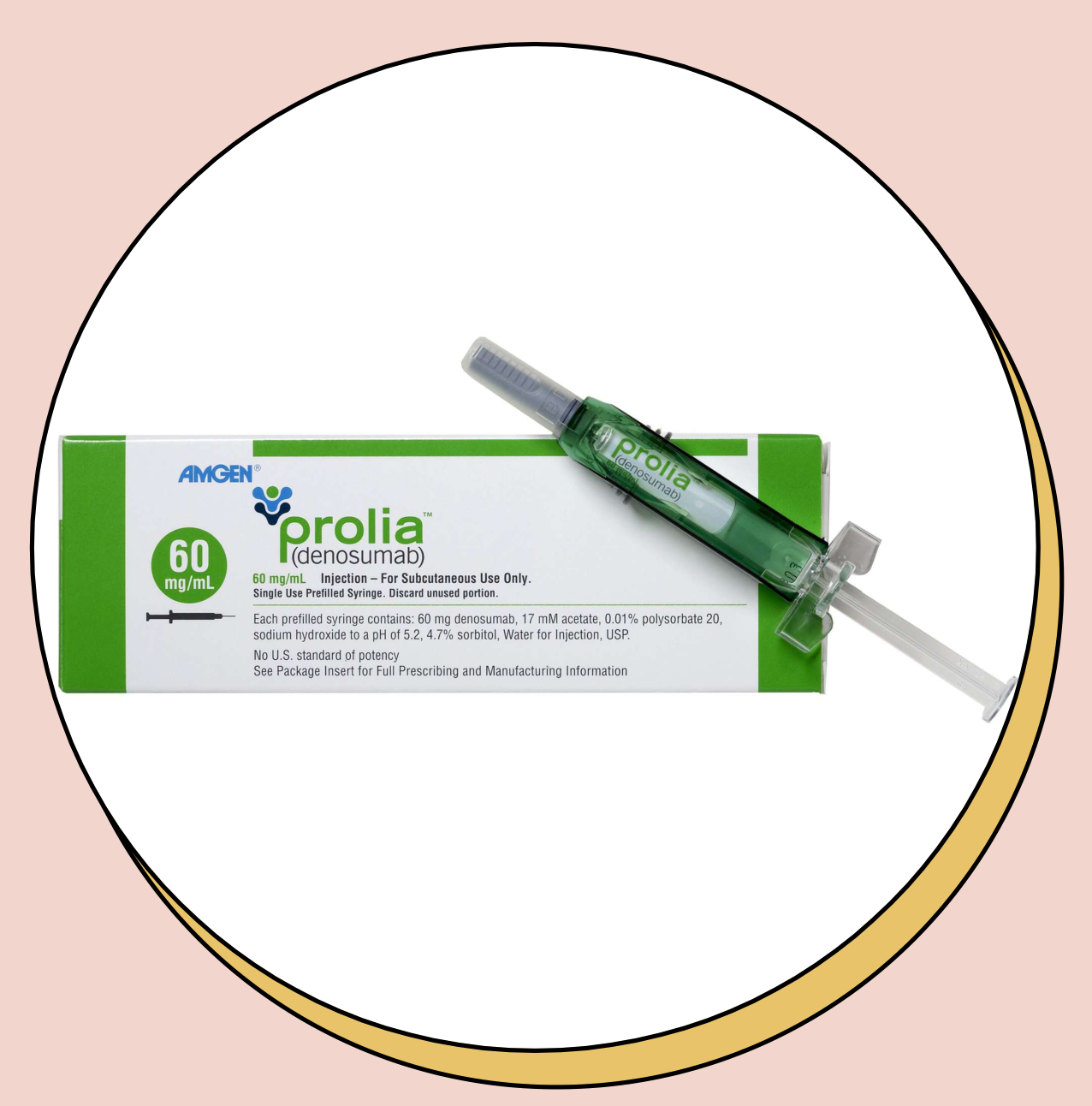
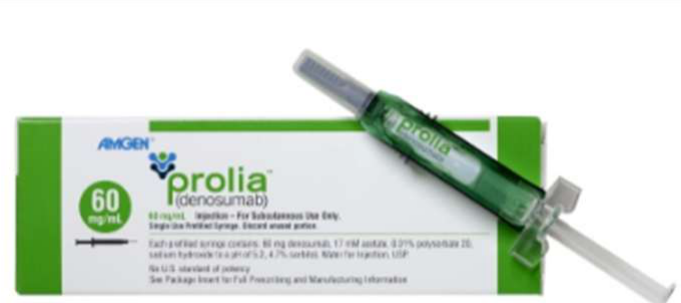
- Denosumab (Prolia®) – 6 monthly 60 mg subcutaneous injection
- Osteoporosis
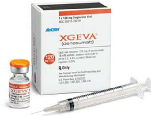
- Denosumab (Xgeva®) – 4 weekly 120 mg subcutaneous injection
- Solid cancer metastasis to bone
- Giant cell tumour of bone
- Hypercalcaemia of malignancy
 |  |
Other Drugs Associated with MRONJ3536
- Well-documented:
- Antiangiogenic drugs: bevacizumab, sunitinib, aflibercept
- Possible association:
- Protein kinase inhibitors: axitinib, cabozantinib, dasatinib, everolimus, imatinib, osimertinib, pazopanib, regorafenib, sorafenib, temsirolimus
- Monoclonal antibodies: adalimumab, infliximab, ipilimumab, nivolumab, ranibizumab, rituximab, romosozumab, tocilizumab
- Antimetabolites: azacitidine, methotrexate
- Tumour necrosis factor alpha inhibitor: etanercept
Scottish Dental Clinical Effectiveness Programme. Oral Health Management of Patients at Risk of Medication-related Osteonecrosis of the Jaw – Supplement March 2024 [Internet]. NHS Education for Scotland; 2024. Available from: https://www.sdcep.org.uk/media/d0sb4flf/sdcep-mronj-guidance-supplement_march-2024.pdf
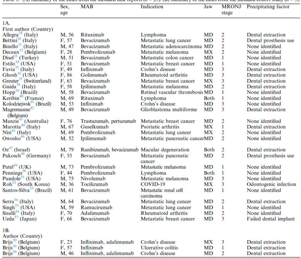
Table 1. (A) Summary of the cases from the included case reports (n = 29). (B) Summary of the cases from the included cohort study (n = 3).
(A) Case reports
| First author (Country) | Sex, age | MAB | Indication | Jaw | MRONJ stage | Precipitating factor |
|---|---|---|---|---|---|---|
| Allegra (Italy) | M, 56 | Rituximab | Lymphoma | MD | 2 | Dental extraction |
| Bettini (Italy) | F, 57 | Bevacizumab | Metastatic lung cancer | MD | 2 | Dental prosthesis use |
| Binello (Italy) | M, 47 | Bevacizumab | Metastatic adenocarcinoma | MD | 2 | None identified |
| Decaux (Belgium) | F, 28 | Pembrolizumab | Metastatic melanoma | MX | 2 | None identified |
| Disel (Turkey) | M, 51 | Bevacizumab | Metastatic colon cancer | MD | 1 | None identified |
| Estio (USA) | F, 51 | Bevacizumab | Metastatic breast cancer | MD | 1 | None identified |
| Favia (Italy) | F, 49 | Infliximab | Crohn’s disease | MD | 3 | Dental extraction |
| Ghosh (USA) | F, 86 | Golimumab | Rheumatoid arthritis | MD | 3 | Dental extraction |
| Greuter (Switzerland) | F, 63 | Bevacizumab | Metastatic breast cancer | MX | 3 | Dental extraction |
| Guida (Italy) | F, 58 | Ipilimumab | Metastatic melanoma | MD | 2 | Dental extraction |
| Hopp (Brazil) | M, 58 | Bevacizumab | Retinal vascular thrombosis | MD | 1 | None identified |
| Keribin (France) | M, 69 | Rituximab | Lymphoma | Both | 1 | None identified |
| Kolodziejewski (Brazil) | M, 53 | Infliximab | Crohn’s disease | MD | 3 | None identified |
| Magrenanne (Belgium) | M, 49 | Bevacizumab | Glioblastoma multiforme | MD | 3 | Dental extraction |
| Manzie (Australia) | F, 76 | Trastuzumab, pertuzumab | Metastatic breast cancer | MD | 2 | None identified |
| Marotta (Italy) | M, 67 | Guselkumab | Psoriatic arthritis | MX | 1 | Dental extraction |
| Nisi (Italy) | M, 69 | Pembrolizumab | Metastatic lung cancer | MX | 2 | None identified |
| Owoshi (USA) | M, 52 | Ipilimumab | Metastatic pancreatic cancer | MD | 2 | None identified |
| Oz (Israel) | M, 79 | Ranibizumab, bevacizumab | Macular degeneration | Both | 2 | Dental extraction |
| Pakosch (Germany) | F, 53 | Bevacizumab | Metastatic pancreatic cancer | MD | 2 | Dental prosthesis use |
| Patel (UK) | M, 73 | Pembrolizumab | Metastatic melanoma | MD | 1 | None identified |
| Penning (USA) | F, 44 | Pembrolizumab | Lymphoma | Both | 1 | None identified |
| Pundole (USA) | M, 75 | Nivolumab | Metastatic melanoma | MD | 3 | None identified |
| Roh (South Korea) | M, 36 | Tocilizumab | COVID-19 | MX | 3 | Odontogenic infection |
| Santos-Silva (Brazil) | M, 61 | Bevacizumab | Metastatic renal cell carcinoma | MD | 1 | None identified |
| Serra (Italy) | M, 64 | Bevacizumab | Metastatic lung cancer | MD | 2 | Dental extraction |
| Singh (USA) | M, 59 | Ramucirumab | Metastatic lung cancer | MD | 1 | None identified |
| Sisalli (Italy) | F, 70 | Adalimumab | Rheumatoid arthritis | MD | 2 | None identified |
| Ueda (Japan) | F, 66 | Bevacizumab | Metastatic breast cancer | MD | 3 | Failed dental implant |
(B) Cohort study
| Author (Country) | Sex, age | MAB | Indication | Jaw | MRONJ stage | Precipitating factor |
|---|---|---|---|---|---|---|
| Brijs (Belgium) | F, 23 | Infliximab, adalimumab | Crohn’s disease | MX | 3 | Dental extraction |
| Brijs (Belgium) | F, 57 | Infliximab | Ulcerative colitis | MD | 1 | Dental extraction |
| Brijs (Belgium) | M, 46 | Infliximab, adalimumab | Crohn’s disease | MD | 2 | Dental extraction |
F, female; M, male; MAB, monoclonal antibody; MD, mandible; MRONJ, medication-related osteonecrosis of the jaw; MX, maxilla.
- Most common: bevacizumab
- Followed by: infliximab, pembrolizumab, adalimumab and rituximab
Pathogenesis of MRONJ37
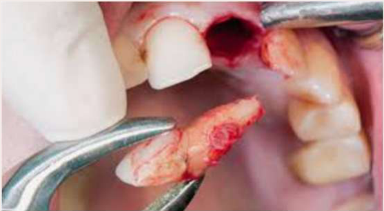
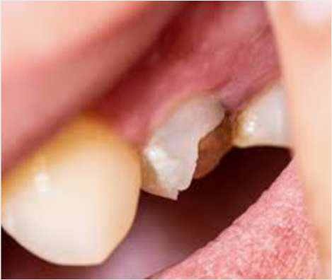
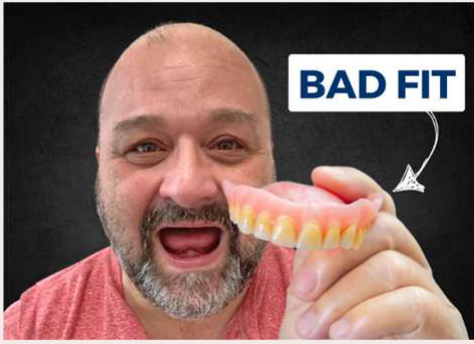
- Majority: dental intervention impacting bone
- e.g. extractions, ill-fitting dentures, sharp teeth
- May occur spontaneously
- Likely multi-factorial - current hypotheses:
- Suppression of bone turnover
- Inhibition of angiogenesis
- Toxic effect on soft tissues
- Inflammation or infection
 |  |
 |
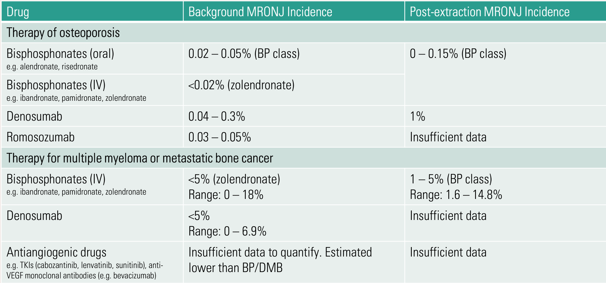
Incidence of MRONJ3839
| Drug | Background MRONJ Incidence | Post-extraction MRONJ Incidence |
|---|---|---|
| Therapy of osteoporosis | ||
| Bisphosphonates (oral), e.g. alendronate, risedronate | 0.02 – 0.05% (BP class) | 0 – 0.15% (BP class) |
| Bisphosphonates (IV), e.g. ibandronate, pamidronate, zolendronate | <0.02% (zolendronate) | |
| Denosumab | 0.04 – 0.3% | 1% |
| Romosozumab | 0.03 – 0.05% | Insufficient data |
| Therapy for multiple myeloma or metastatic bone cancer | ||
| Bisphosphonates (IV), e.g. ibandronate, pamidronate, zolendronate | <5% (zolendronate); range: 0 – 18% | 1 – 5% (BP class); range: 1.6 – 14.8% |
| Denosumab | <5%; range: 0 – 6.9% | Insufficient data |
| Antiangiogenic drugs, e.g. TKIs (cabozantinib, lenvatinib, sunitinib), anti-VEGF monoclonal antibodies (e.g. bevacizumab) | Insufficient data to quantify. Estimated lower than BP/DMB | Insufficient data |
Ruggiero SL, Dodson TB, Aghaloo T, et al. American Association of Oral and Maxillofacial Surgeons’ Position Paper on Medication-Related Osteonecrosis of the Jaws – 2022 Updates. J Oral Maxillofac Surg. 2022; 80(5): 920-943.
Know these numbers
-
Osteoporosis < Cancer = 0.05–1% vs <5%
-
Osteoporosis: bisphosphonates < denosumab (1 order higher)
- Background: <0.05% vs <0.3%
- Extractions: <0.15% vs 1%
-
Cancer: bisphosphonates ≈ denosumab (5%)
- Ranges up to 18% BP and 7% DMB
-
Risk/consequence of skeletal events > MRONJ risk
-
Dental alveolar operations were identified as a predisposing event in approximately 62–82% of MRONJ cases.
Risk Factors for MRONJ
Key Risk Factors40
- History of MRONJ
- Cancer regimen drugs that increase MRONJ risk
-
4 years bisphosphonate therapy
Significant Risk Factors
- Poor oral health / periodontal disease
- Invasive dental procedures
- Ill-fitting dentures
- Uncontrolled diabetes mellitus
- Tobacco use
Other Possible Risk Factors
- Chemotherapy
- Profound immunocompromise
- Corticosteroids
- Cancer site
- Renal disease / dialysis
- Erythropoietin therapy / anaemia
- Hyperthyroidism
- Gender, ethnicity, increasing age
Prevention of MRONJ41
- Medical practitioner to refer for dental assessment prior to commencing drug
- Dentist should:
- Undertake a comprehensive oral examination (including pulp tests and radiographs)
- Eliminate dental caries (e.g. restorations, extractions)
- Establish periodontal health (e.g. debridement, extractions)
- Encourage good oral hygiene
- Invasive dental procedures should be completed prior to commencing drug
- Osteoporosis: less ideally, complete invasive dental procedures within 6 months of the first dose
- Regular dental review is essential to monitor oral health
Achieving Dental Clearance Prior to Drug Therapy42434445
“Dentally Fit?” — “Dental Clearance”
Patients at Risk of MRONJ: Prior to Drug Therapy
Prioritise care that will reduce mucosal trauma, and/or may help avoid foreseeable oral surgery that may impact bone, and/or may risk infection/bacteraemia:
- Radiographs to identify possible areas of infection/pathology
- Undertake remedial dental work
- Extract any teeth of poor prognosis without delay
- Minimise periodontal / dental infection or disease
- Adjust/replace poorly fitting dentures
Dr Jee-Yun Leung
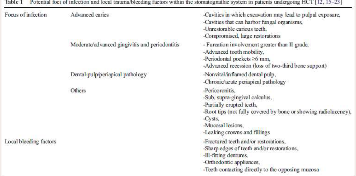
Potential sources of oral infection prior to urgent medical treatment
Table I. Potential foci of infection and local trauma/bleeding factors within the stomatognathic system in patients undergoing HCT
- Focus of infection — Advanced caries:
- Cavities in which excavation may lead to pulpal exposure
- Cavities that can harbor fungal organisms
- Unrestorable carious teeth
- Compromised, large restorations
- Moderate/advanced gingivitis and periodontitis:
- Furcation involvement greater than II grade
- Advanced tooth mobility
- Periodontal pockets ≥6 mm
- Advanced recession (loss of two-third bone support)
- Dental-pulp/periapical pathology:
- Nonvital/inflamed dental pulp
- Chronic/acute periapical pathology
- Others:
- Pericoronitis
- Sub-, supra-gingival calculus
- Partially erupted teeth
- Root tips (not fully covered by bone or showing radiolucency)
- Cysts
- Mucosal lesions
- Leaking crowns and fillings
- Local bleeding factors:
- Fractured teeth and/or restorations
- Sharp edges of teeth and/or restorations
- Ill-fitting dentures
- Orthodontic appliances
- Teeth contacting directly to the opposing mucosa
Boguslawska-Kapala et al. Oral health of adult patients undergoing hematopoietic cell transplantation. Pre-transplant assessment and care. 2017:96;1135-1145.
Therefore, if systemic conditions permit, initiation of antiresorptive therapy should be delayed until dental health is optimized. This decision must be made in conjunction with the treating physician and dentist and other specialists involved in the care of the patient. There is widespread consensus that optimizing dental health prior to initiating therapy is efficacious and of paramount importance.
AAOMS 2022 MRONJ Guidelines46
Ruggiero SL, Dodson TB, Aghaloo T, Carlson ER, Ward BB, Kademani D. American Association of Oral and Maxillofacial Surgeons’ Position Paper on Medication-Related Osteonecrosis of the Jaws—2022 Update. J Oral Maxillofac Surg. 2022;80(5):920–43.
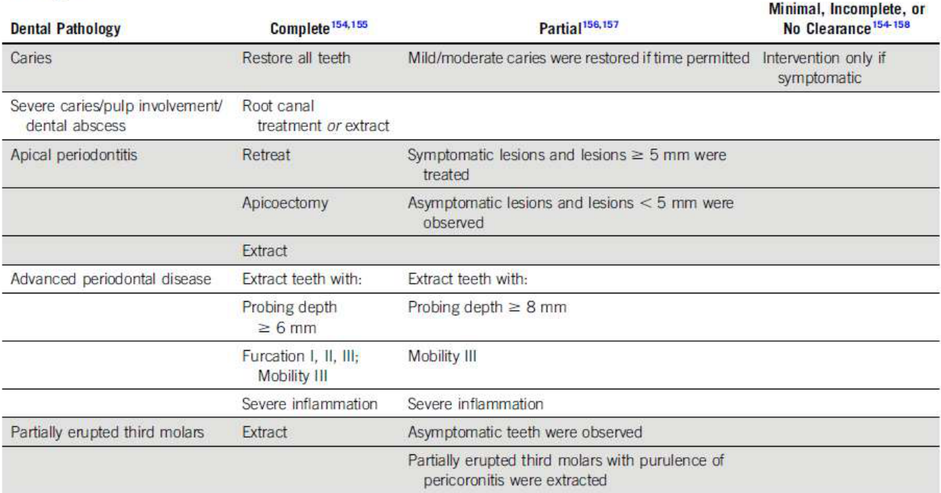
When medical condition requires prompt initiation of treatment…
Table 3. Descriptions of Complete, Partial, and Minimal Dental Evaluation Protocols Based on the Type of Dental and/or Periodontal Pathology
| Dental Pathology | Complete | Partial | Minimal, Incomplete, or No Clearance |
|---|---|---|---|
| Caries | Restore all teeth | Mild/moderate caries were restored if time permitted | Intervention only if symptomatic |
| Severe caries/pulp involvement/dental abscess | Root canal treatment or extract | ||
| Apical periodontitis | Retreat; apicoectomy; extract | Symptomatic lesions and lesions ≥5 mm were treated; asymptomatic lesions and lesions <5 mm were observed | |
| Advanced periodontal disease | Extract teeth with: probing depth ≥6 mm; furcation I, II, III; mobility III; severe inflammation | Extract teeth with: probing depth ≥8 mm; mobility III; severe inflammation | |
| Partially erupted third molars | Extract | Asymptomatic teeth were observed; partially erupted third molars with purulence or pericoronitis were extracted |
NOTE. The proper protocol should be selected by the oncologist and dentist according to the patient’s medical status.
- Yarom et al. Medication-Related Osteonecrosis of the Jaw: MASCC/ISOO/ASCO Clinical Practice Guideline. J Clin Oncol. 2019;37(25):2270-2290.
- Hong et al. A systematic review of dental disease management in cancer patients. Support Care Cancer. 2018;26:155-174.
Dr Jee-Yun Leung 2025
Counselling and Ongoing Preventive Care47
Bone-Modifying Drugs: Work-Up
- Advise patient of MRONJ with medication they are taking, but ensure they understand the risk is small, so that they are not discouraged from taking their BMD or from undergoing dental treatment.
- Personalised preventive advice to optimise oral health (healthy diet, reduce sugar, maintain oral hygiene, fluoride, cease smoking, limit alcohol, regular dental checks)
- Ensure dentures are well-fitting
Dr Jee-Yun Leung
Assessing MRONJ Risk Prior to Invasive Dental Procedures4849
Current BMDs - Assessing MRONJ Risk prior to Invasive Dental Procedures
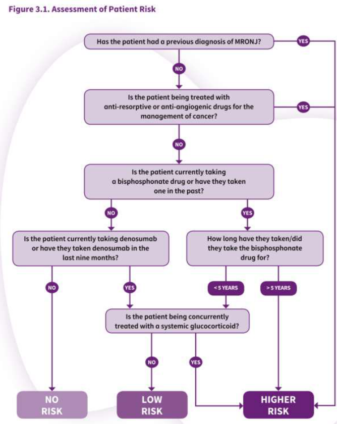
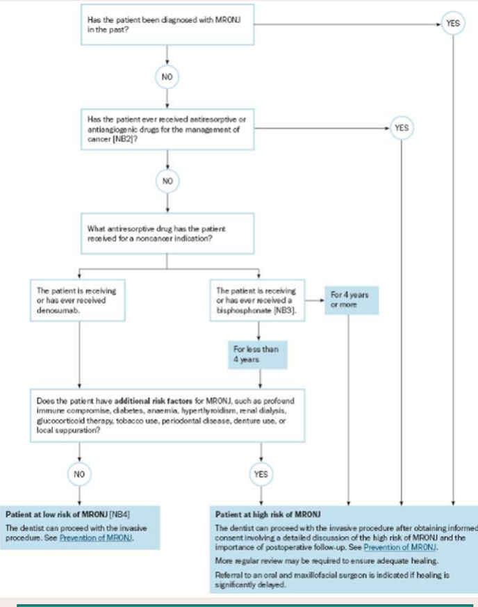
- Assess if patient is at low or higher risk of developing MRONJ based on their medical condition, type and duration of therapy, and any other complicating factors (e.g. glucocorticoid medication and length of exposure to drugs).
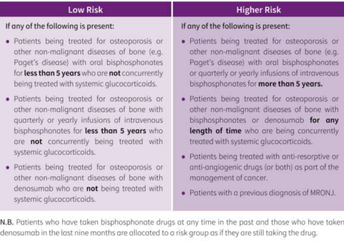
Table 3.1 Patient Risk Categories
| Low Risk | Higher Risk |
|---|---|
| If any of the following is present: • Patients being treated for osteoporosis or other non-malignant diseases of bone (e.g. Paget’s disease) with oral bisphosphonates for less than 5 years who are not concurrently being treated with systemic glucocorticoids. • Patients being treated for osteoporosis or other non-malignant diseases of bone with quarterly or yearly infusions of intravenous bisphosphonates for less than 5 years who are not concurrently being treated with systemic glucocorticoids. • Patients being treated for osteoporosis or other non-malignant diseases of bone with denosumab who are not being treated with systemic glucocorticoids. | If any of the following is present: • Patients being treated for osteoporosis or other non-malignant diseases of bone (e.g. Paget’s disease) with oral bisphosphonates or quarterly or yearly infusions of intravenous bisphosphonates for more than 5 years. • Patients being treated for osteoporosis or other non-malignant diseases of bone with bisphosphonates or denosumab for any length of time who are being concurrently treated with systemic glucocorticoids. • Patients being treated with anti-resorptive or anti-angiogenic drugs (or both) as part of the management of cancer. • Patients with a previous diagnosis of MRONJ. |
N.B. Patients who have taken bisphosphonate drugs at any time in the past and those who have taken denosumab in the last nine months are allocated to a risk group as if they are still taking the drug.
SDCEP 2017 Guidelines – Updated 2022
Therapeutic Guidelines – Oral & Dental v4 2026
Dr Jee-Yun Leung 2025
Current BMDs – Assessing MRONJ Risk prior to Invasive Dental Procedures
Patients can undergo invasive dental procedures in general dental practice, after a discussion of the risk assessment and precautions.
Figure 13.75 Precautions for managing patients at increased risk of MRONJ undergoing an invasive dental procedure
- Inform the patient of the risk of medication-related osteonecrosis of the jaw and obtain informed consent for the procedure.
- See advice on drug holidays and scheduling of procedures in patients at risk of MRONJ.
- Do not use antibiotic prophylaxis to reduce the risk of medication-related osteonecrosis of the jaw – there is insufficient evidence to support this practice. However, an active infection should be treated.
- Ensure optimal oral hygiene before and after the procedure.
- Reduce the plaque load with mechanical debridement and pre- and post-procedural chlorhexidine mouthwash.
- Consider root retention techniques to avoid extractions.
- Minimise trauma and periosteum stripping, and use sutures to close any mucosal flaps that are raised.
- Monitor the oral wound until it heals – healing may be slow.
- Do not debride non-healing wounds.
- Refer to an oral and maxillofacial surgeon if bone is still visible 8 weeks after the invasive dental procedure.
Lecturer — Infection Management
Immunosuppressed patients may have an increased risk of oral candidiasis. Oral infections should be treated promptly because infection may progress to bacteraemia.
Dr Jee-Yun Leung 2025
Healthy Bones Australia Consensus Statement: Medication-Related Osteonecrosis Of The Jaw (MRONJ) and Osteoporosis
 |  |
 |
Patient Factors50
- Low Risk:
- Anti-resorptive therapy for osteoporosis
- Duration of BP and/or Dmba for <4 years
- No adverse systemic conditions
- No other adverse drugs
- Good baseline dentition
- High Risk:
- Duration of BP and/or Dmba ≥4 years
- Systemic conditions: diabetes with poor control; anaemia, poor nutrition; inflammatory rheumatic disorders; smoking
- Adverse drugs: anti-angiogenic or tyrosine kinase inhibitor (TKI) for cancer; glucocorticoids
- Active dental infection, poor oral health, periodontal disease, protracted dental infection
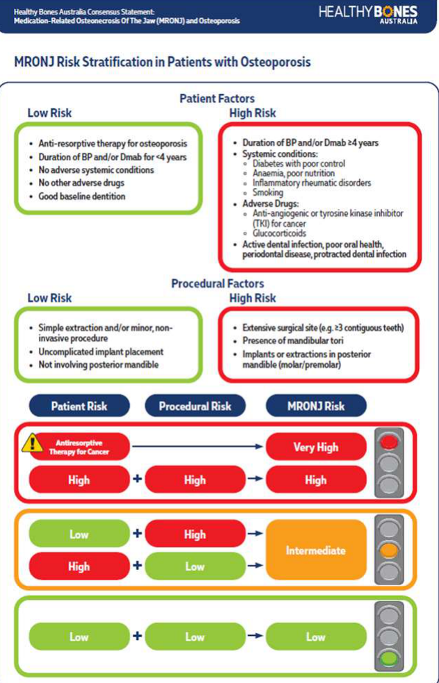
Procedural Factors
- Low Risk:
- Simple extraction and/or minor, non-invasive procedure
- Uncomplicated implant placement
- Not involving posterior mandible
- High Risk:
- Extensive surgical site (e.g. ≥3 contiguous teeth)
- Presence of mandibular tori
- Implants or extractions in posterior mandible (molar/premolar)
MRONJ Risk Stratification
| Patient Risk | Procedural Risk | MRONJ Risk |
|---|---|---|
| Antiresorptive therapy for cancer | — | Very High (red) |
| High | High | High (red) |
| Low | High | Intermediate (orange) |
| High | Low | Intermediate (orange) |
| Low | Low | Low (green) |
Management Precautions by MRONJ Risk
- Red traffic light:
- Refer to specialist centre or practitioner with MRONJ expertise
- Discussion with osteoporosis specialist regarding ongoing use of antiresorptives and treatment goals
- Orange traffic light:
- Minimise active dental infection
- Antibacterial mouthwash ≥1 week prior and following extraction
- Consider periprocedural antibiotics*
- Atraumatic surgical approach
- Consider primary closure with tension-free sutures (if appropriate)**
- Close wound safely, monitor oral hygiene, frequent follow-up
- Consider referral to specialist centre or practitioner with MRONJ experience
- Green traffic light:
- General precautions
- Advise low MRONJ risk
- Proceed with routine extractions and implants
- Consider primary closure (if appropriate)**
- Close wound safely, monitor oral hygiene, frequent follow-up
* The benefit of postoperative antibiotic use to prevent MRONJ is unclear. Individual patient risk assessment is warranted.
** Primary closure is not routinely recommended. This is appropriate in select cases.
Dr Jee-Yun Leung 2025
Drug Holidays with Bisphosphonates51
AAOMS 201452
Asymptomatic patients – oral BP for osteoporosis
- Oral BP <4 years + no risk factors = no delays/alteration.
- Oral BP <4 years + concomitant corticosteroids or anti-angiogenics = consider discontinuation at least 2 months pre-op, if systemic conditions permit. Restart when osseous healing (3 months).
- Oral BP >4 years ± concomitant medical therapy = consider discontinuation for 2 months pre-op, if systemic conditions permit. Restart when osseous healing (3 months).
AAOMS 2022
- Difficult to establish or refute efficacy of drug holidays → working group evenly split
- Rarity of MRONJ in these patient populations → insufficient data for RCTs
- 2020 systematic review: variety of papers had differing conclusions
- Factors for consideration: disease-related risk (cancer vs OP), drug-dosing frequency, therapy duration, comorbidities, other medications (chemo, steroids, antiangiogenics), degree of infection/inflammation, extent of surgery
Ruggiero SL, Dodson TB, Aghaloo T, Carlson ER, Ward BB, Kademani D. American Association of Oral and Maxillofacial Surgeons’ Position Paper on Medication-Related Osteonecrosis of the Jaws—2022 Update. J Oral Maxillofac Surg. 2022;80(5):920–43.
Ottesen C, Schiodt M, Gotfredsen K. Efficacy of a high-dose antiresorptive drug holiday to reduce the risk of medication-related osteonecrosis of the jaw (MRONJ): A systematic review. Heliyon. 2020;6(4):e03795–e03795.
Dr Jee-Yun Leung
Current BMDs: Drug Holidays prior to Invasive Dental Procedures
- Controversial – evidence to support or refute inconclusive
- Responsibility for drug holiday is prescribing practitioner, together with clinical judgement of the dentist, and in discussion with the patient
Ruggiero SL, Dodson TB, Aghaloo T, Carlson ER, Ward BB, Kademani D. American Association of Oral and Maxillofacial Surgeons’ Position Paper on Medication-Related Osteonecrosis of the Jaws—2022 Update. J Oral Maxillofac Surg. 2022;80(5):920–43.
Ottesen C, Schiodt M, Gotfredsen K. Efficacy of a high-dose antiresorptive drug holiday to reduce the risk of medication-related osteonecrosis of the jaw (MRONJ): A systematic review. Heliyon. 2020;6(4):e03795–e03795.
Yarom N, et al. Medication-Related Osteonecrosis of the Jaw: MASCC/ISOO/ASCO Clinical Practice Guideline. 2019;37(25):2270-2290
Therapeutic Guidelines: Oral & Dental version 4 2026
Dr Jee-Yun Leung
Drug Holidays with Denosumab535455
Current BMDs - Denosumab
Current BMDs - Denosumab
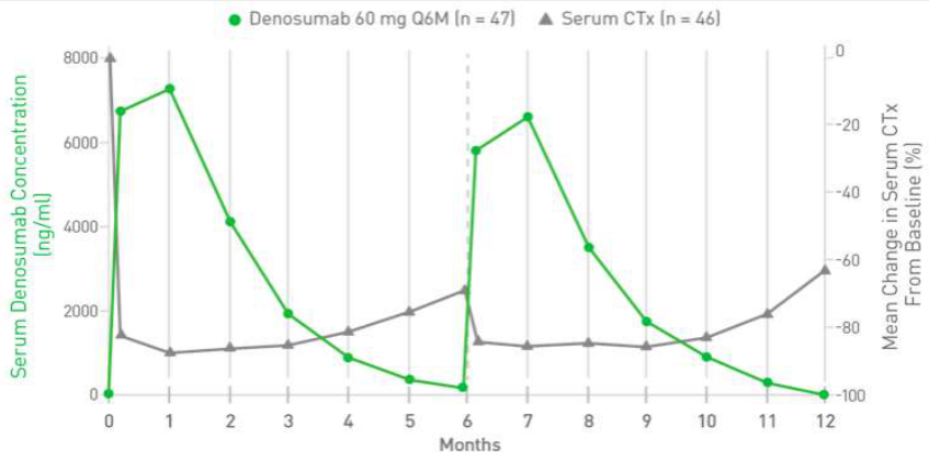
Osteoporotic patients with 6-monthly S.C. denosumab can delay any non-urgent invasive dental Tx in an asymptomatic tooth until 3–4 months after the last dose.
This allows 6–8 weeks for healing prior to resumption of denosumab.
Denosumab discontinuation → rebound risk of spontaneous vertebral fractures
- Onset of bone loss and fracture risk starts within 4 weeks of the missed dose
- ↑ risk occurred mostly within the first 2–6 months of discontinuation
- Fractures mostly occur within 6–20 months of denosumab discontinuation
Ruggiero SL, Dodson TB, Aghaloo T, Carlson ER, Ward BB, Kademani D. American Association of Oral and Maxillofacial Surgeons’ Position Paper on Medication-Related Osteonecrosis of the Jaws—2022 Update. J Oral Maxillofac Surg. 2022;80(5):920–43.
Cummings SR, Ferrari S, Eastell R, et al. Vertebral fractures after discontinuation of denosumab: A Post Hoc Analysis of the Randomized Placebo-Controlled FREEDOM Trial and Its Extension. J Bone Miner Res. 2018;33(2):190-198
Lamy O, Stoll D, Aubrey-Rozir et al. Stopping denosumab. Curr Osteoporos Rep. 2019;17:8-15
Dr Jee-Yun Leung
Current BMDs: Drug Holidays - Denosumab
- Denosumab discontinuation → rebound risk of multiple spontaneous vertebral fractures
- FREEDOM study and its extension (2018): risk of rebound vertebral fractures ↑ from 1.2 (on-treatment period) to 7.1 (denosumab cessation) per 100 participant-years of discontinuation.
- ↑ occurred mostly within the first 2–6 months of discontinuation
- Lamy et al (2019):
- Onset of bone loss and fracture risk starts within 4 weeks of the missed dose
- Significant fracture risk occurs after 2nd dose of denosumab
- Fractures mostly occur within 6–20 months of denosumab discontinuation
- If denosumab is discontinued, bone density declines to pre-treatment levels within 12 months
Ruggiero SL, Dodson TB, Aghaloo T, Carlson ER, Ward BB, Kademani D. American Association of Oral and Maxillofacial Surgeons’ Position Paper on Medication-Related Osteonecrosis of the Jaws—2022 Update. J Oral Maxillofac Surg. 2022;80(5):920–43.
Cummings SR, Ferrari S, Eastell R, et al. Vertebral fractures after discontinuation of denosumab: A post hoc analysis of the randomized placebo controlled FREEDOM trial and its extension. J Bone Miner Res 2018;33(2):190–98.
Dr Jee-Yun Leung
 |  |
Drug Holidays in Therapeutic Guidelines56
- There is no evidence that drug holidays reduce risk of MRONJ.
- If bone-invasive dental procedure cannot be avoided but is not urgent, temporary discontinuation of AR therapy, or timing procedure to coincide with a low serum drug concentration, have been suggested.
- These practices are based on extrapolation of the drug pharmacokinetics in the serum and on bone physiology; however no available outcome evidence to support.
- Clinical judgement of treating doctors/dentists is required to determine appropriate management of each patient.
- Recommendations in international guidelines are inconsistent. Vary depending on indication for AR drugs, duration of therapy, and presence of additional risk factors.
- Decision to alter AR therapy lies with practitioner responsible for managing the AR drug.
- AR drugs for osteoporosis:
- Benefits of continued therapy outweigh low risk of MRONJ.
- Bisphosphonates = although stopping for short period unlikely to cause harm in a patient at low risk of fracture, no evidence that this reduces MRONJ risk.
- AR drugs for cancer:
- Decision to alter AR therapy lies with specialist managing the cancer.
Dr Jee-Yun Leung 2025
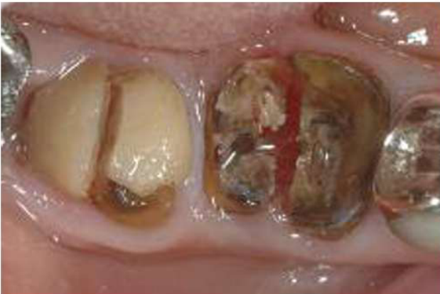
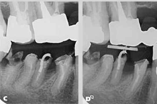
Alternative Options for Unrestorable Teeth57
 |  |
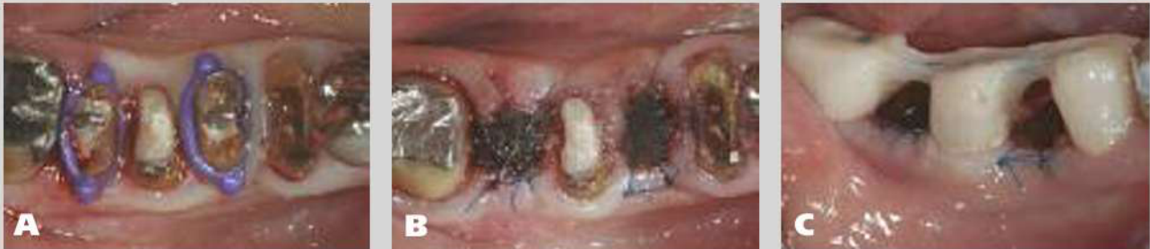 |
Root Canal Treatment and Tissue-Level Restoration58
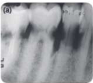
Zamparini F, Pelliccioni GA, Spinelli A, Gissi DB, Gandolfi MG, Prati C. Root canal treatment of compromised teeth as alternative treatment for patients receiving bisphosphonates: 60-month results of a prospective clinical study. International Endodontic Journal. First published: 09 September 2020. https://doi.org/10.1111/iej.13405
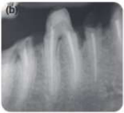
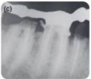
Figure 3. Series of periapical radiographs of patients under BPs therapy enrolled in the study and followed for 60 months. (a) 68-year patient with zoledronate-endovenous therapy started for multiple myeloma. Root canal treatment was performed on teeth 4.7, 4.6, 4.5 and 4.4 due to multiple deep caries lesion with no periapical radiolucency (initial PAI was 1 in all cases). (b) roots were shaped with manual step-back instrumentation and filled with carrier-based technique and epoxy resin based sealer. Due to the large coronal destruction of tooth 4.7 and 4.5, these two teeth were restored at tissue level (TL group), the other teeth were rehabilitated with occlusal contact restorations (OC group). (c) At the end-line all teeth demonstrated no peri-apical radiolucency (PAI ≤ 2).
Aim
This 60-month prospective study aimed to evaluate tooth survival and healing rates after root canal treatment in patients taking bisphosphonates (BPs). Secondary outcomes were complications and clinical variables observed during and after the treatment.
Methods
Root canal treatment was performed using manual K-file canal instrumentation, a carrier-based filling technique and epoxy resin–based sealer. Teeth without adequate root/crown integrity were restored by trained operators at the tissue level (TL group) to prevent occlusal/mechanical stress and to enable periapical lesion healing without the risk of root fracture. Other teeth were restored with normal occlusal contact (OC group). Healthy patients who had undergone one or more root canal treatments of the same type constituted the control group. The relationships of the following variables to survival and health status were examined (chi-squared test and multivariate analysis, p=0.05): age, gender, smoking habit, tooth location, treatment type, BPs treatment, BPs exposure, initial periapical index (PAI) and occlusal restoration. Survival curves were constructed using Kaplan–Meier analysis, with extraction serving as endpoint.
Results
In total, 65 patients with 109 root canal treated teeth who were taking BPs were included. At 60 months, data from 57 patients (52F, 5M; median-age 65.7±8.6 years) who had undergone 96 root canal treatments were analysed (drop-out rate=16.9%). The survival rate was 85% and the success rate was 76%.
The control group consisted of 46 patients (21F, 25M; median-age 60.3±7.2 years) who had undergone 102 root canal treatments. The survival rate was 88%, with 12 teeth lost during follow-up. The success rate was 73%. In the BPs group, 55 teeth were restored normally (OC group) and 41 teeth were restored at the tissue level (TL group). No difference in the success or survival rate was observed between the BP and control groups (p>0.05). Univariate Kaplan-Meier analysis revealed that only tooth type significantly affected survival status in the BP group. The analysis revealed the clinical relevance of smoking, tooth location and initial PAI on patients’ health status (p<0.05).
Conclusion
Root canal treatments and post-endodontic restoration with tissue level filling procedures represent a safe approach for severely-damaged teeth in patients receiving bisphosphonates having comparable results to occlusal contact and control group.
Lecturer — Root Retention
Root canal treatment may be considered for retained roots or teeth that would otherwise be extracted.
- A small glass ionomer cement cap may be placed when a crown or post is not feasible; this can retain the root while avoiding trauma to the jaw bone, although the restoration may not withstand normal occlusal or masticatory forces.
Dr Jee-Yun Leung
 |  |  |
 |  | 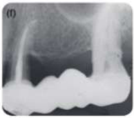 |
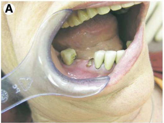
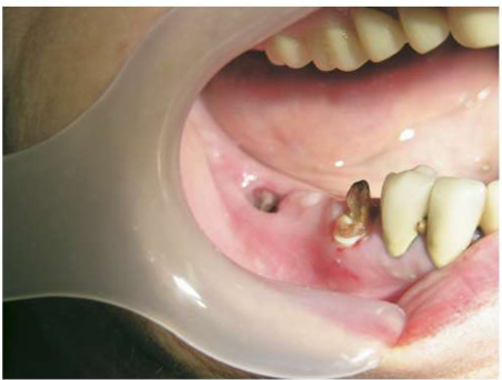
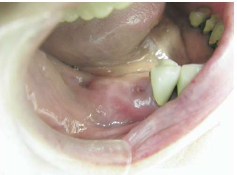
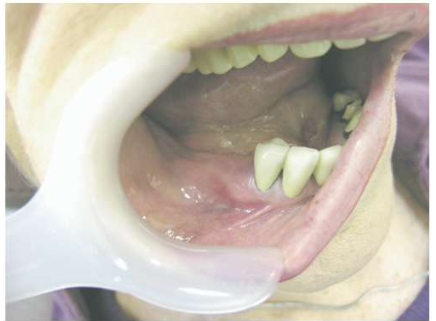
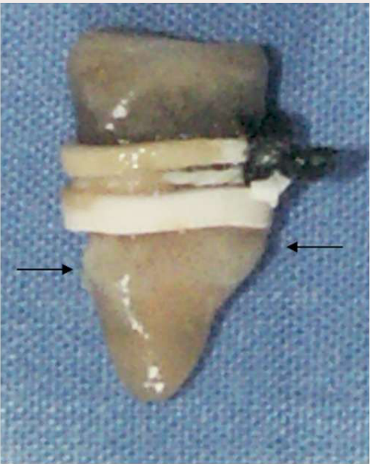
Orthodontic Extrusion59
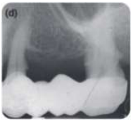
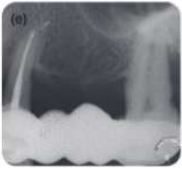
- Figure 3. Teeth after exfoliation of the second molar and extrusion of the first premolar.
- Figure 5. Complete healing 2 months after exfoliation.
- Figure 6. Dentin bulge (arrows) preventing elastics from sliding apically.
Regev E, Lustmann J, Nashef R. Atraumatic Teeth Extraction in Bisphosphonate-Treated Patients. Journal of Oral and Maxillofac Surgery. 2008;66(6):1157-61.
Dr Jee-Yun Leung
Smidt A, Lipovetsky-Adler M, Sharon E. Forced eruption as an alternative to tooth extraction in long-term use of oral bisphosphonates: Review, risks and technique. JADA. 2012;143(12):1303-12
Dr Jee-Yun Leung
 |  |  |
 |  |
Treatment Planning Considerations60
Thinking it through… BMDs
| Medical, Dental and Social Considerations | Optimal Timeframes | Treatment Approaches |
|---|---|---|
| • Chemotherapy cycle and blood counts • Long-term prognosis of tooth • Compliance with ongoing preventive oral care • Previous history BMD vs BMD naïve • Concomitant oral disease (periodontal disease, periapical pathology) = infection risk factor for MRONJ • Tobacco use | • Urgency to commence drug • Prioritise care that reduces mucosal trauma or need for future oral surgery impacting on bone (e.g. poor prognosis dental extractions, poorly fitting dentures) • Future-proof for foreseeable future: bisphosphonate osteoporosis tx: 5–10 years treatment period + 10-year terminal half-life; denosumab: long-term, or cessation followed by another BMD (12 months oral bisphosphonate or 1+ zolendronate infusion) • Dental extractions just prior to 6-monthly injection • Avoid delaying next dose = risk spontaneous vertebral fractures | • Aggressive vs conservative: typically aggressive pre-BMD, conservative options if time permits. Typically conservative post-BMD. • Long-term medical prognosis, physiological tolerance • Resulting QOL |
Dr Jee-Yun Leung 2025
Lecturer — Dental Clearance
“Dental clearance” does not necessarily mean removing all teeth, achieving an ideal 28-tooth dentition, replacing or aligning every tooth, or eliminating every carious or periodontal lesion.
- Treatment should instead be prioritised according to mucosal trauma, bone involvement, infection, risk of bacteraemia, and the urgency of medical treatment.
Lecturer — Long-Term Planning
For bisphosphonates, treatment planning may need to anticipate approximately fifteen to twenty years of future dental needs because patients may receive treatment for five to ten years followed by residual drug persistence for a further ten years.
- In patients with a poor long-term medical prognosis, the teeth only need to outlast the patient; extensive extractions may not improve quality of life and may create a prolonged rehabilitation burden.
Management of MRONJ61
Goals: improved quality of life, disease control, prevention of secondary infection
Lecturer — Management Goals
Cure is possible in some patients, but treatment choice must balance symptom and infection control against treatment morbidity.
- Surgery may cause substantial morbidity.
- Non-operative management may sometimes provide better symptom and infection control.
- Symptoms should be maintained at a tolerable level.
- Non-operative
- Operative
Staging of MRONJ
Stage 0 (non-exposed bone variant)62
- No clinical evidence of necrotic bone
- Non-specific symptoms or clinical and radiographic findings
Symptoms
- Odontalgia unexplained by an odontogenic cause
- Dull, aching jaw bone pain, which may radiate to the TMJ region
- Sinus pain, inflammation and thickening of the maxillary sinus wall
- Altered neurosensory function
Clinical Findings
- Loosening of teeth unexplained by chronic periodontal disease
- Intraoral or extraoral swelling
Radiographic Findings
- Alveolar bone loss or resorption unattributable to chronic periodontal disease
- Changes to trabecular pattern, sclerotic bone and no new bone in extraction sockets
- Regions of osteosclerosis involving alveolar bone and/or surrounding basilar bone
- Thickening/obscuring of PDL
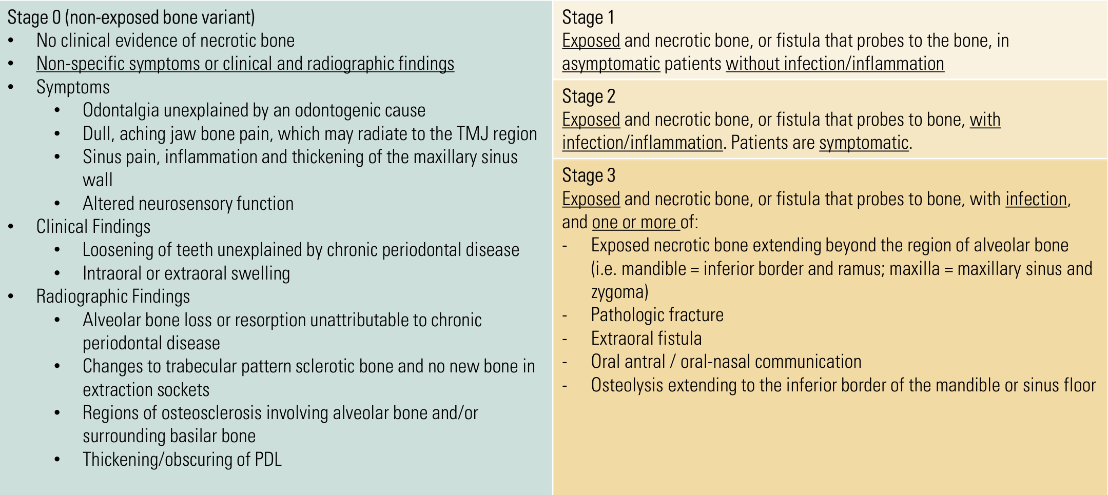
Stage 1
Exposed and necrotic bone, or fistula that probes to the bone, in asymptomatic patients without infection/inflammation
Stage 2
Exposed and necrotic bone, or fistula that probes to bone, with infection/inflammation. Patients are symptomatic.
Stage 3
Exposed and necrotic bone, or fistula that probes to bone, with infection, and one or more of:
- Exposed necrotic bone extending beyond the region of alveolar bone (i.e. mandible = inferior border and ramus; maxilla = maxillary sinus and zygoma)
- Pathologic fracture
- Extraoral fistula
- Oral antral / oral-nasal communication
- Osteolysis extending to the inferior border of the mandible or sinus floor
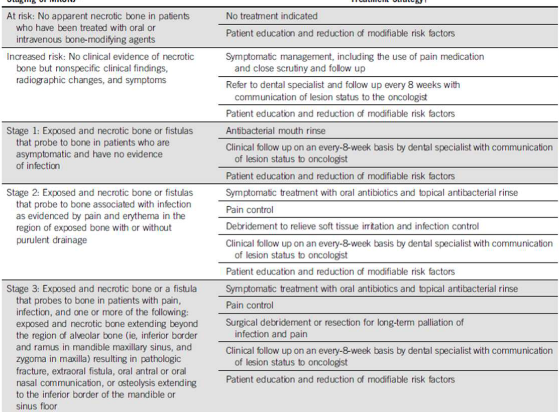
Treatment Strategies by Stage63
| Staging of MRONJ* | Treatment Strategy† |
|---|---|
| At risk: No apparent necrotic bone in patients who have been treated with oral or intravenous bone-modifying agents | No treatment indicated; patient education and reduction of modifiable risk factors |
| Increased risk: No clinical evidence of necrotic bone but nonspecific clinical findings, radiographic changes, and symptoms | Symptomatic management, including the use of pain medication and close scrutiny and follow up; refer to dental specialist and follow up every 8 weeks with communication of lesion status to the oncologist; patient education and reduction of modifiable risk factors |
| Stage 1: Exposed and necrotic bone or fistulas that probe to bone in patients who are asymptomatic and have no evidence of infection | Antibacterial mouth rinse; clinical follow up on an every-8-week basis by dental specialist with communication of lesion status to oncologist; patient education and reduction of modifiable risk factors |
| Stage 2: Exposed and necrotic bone or fistulas that probe to bone associated with infection as evidenced by pain and erythema in the region of exposed bone with or without purulent drainage | Symptomatic treatment with oral antibiotics and topical antibacterial rinse; pain control; debridement to relieve soft tissue irritation and infection control; clinical follow up on an every-8-week basis by dental specialist with communication of lesion status to oncologist; patient education and reduction of modifiable risk factors |
| Stage 3: Exposed and necrotic bone or a fistula that probes to bone in patients with pain, infection, and one or more of the following: exposed and necrotic bone extending beyond the region of alveolar bone (ie, inferior border and ramus in mandible, maxillary sinus, and zygoma in maxilla) resulting in pathologic fracture, extraoral fistula, oral antral or oral nasal communication, or osteolysis extending to the inferior border of the mandible or sinus floor | Symptomatic treatment with oral antibiotics and topical antibacterial rinse; pain control; surgical debridement or resection for long-term palliation of infection and pain; clinical follow up on an every-8-week basis by dental specialist with communication of lesion status to oncologist; patient education and reduction of modifiable risk factors |
NOTE. Adapted from Ruggiero et al.²
Abbreviation: MRONJ, medication-related osteonecrosis of the jaw.
*Exposed or probable bone in the maxillofacial region without resolution for longer than 8 weeks in patients who were treated with an antiresorptive or an angiogenic inhibitor and who have not received radiation therapy to the jaws.
†Regardless of disease stage, mobile segments of bony sequestrum should be removed without exposing uninvolved bone. Extraction of symptomatic teeth within exposed necrotic bone should be considered because it is unlikely that extraction will exacerbate the established necrotic process.
Adapted from AAOMS 2014.
Yarom N, et al. Medication-Related Osteonecrosis of the Jaw: MASCC/ISOO/ASCO Clinical Practice Guideline. 2019;37(25):2270-2290
Non-Operative Management
General Dental Practitioner64
Non-operative management:
-
Improve patient’s oral hygiene
- Biofilm (tooth brushing)
- Chlorhexidine wound care: dip cotton swab or soft small toothbrush in 0.12% CHX solution, gently mechanically scrub exposed bone or fistula
-
Monitoring wound healing
-
Management of generalised periodontal disease
-
Adjusting dentures to alleviate pressure on non-healing area
-
N/B. Stage 2 patients may struggle with local wound care and require antibiotics for symptom control
-
Temporarily remove dentures if they traumatise the MRONJ site.
Specialist Measures
- Removal of loose bone sequestra
- Adjunctive medications/treatments?
- Teriparatide
- Vitamin E and pentoxifylline
- Hyperbaric oxygen
- Ozone therapy
Lecturer — Adjunctive Therapies
Evidence for some adjunctive approaches is still emerging in MRONJ.
Surgical Management65
- Refer all patients with established MRONJ to a surgical dental specialist for assessment
- Surgery: marginal or segmental resection (including sequestrectomy)
- Progression of MRONJ can be unpredictable
- Decision to proceed is patient-specific
- Quality of life, symptoms, ability to clean wound to prevent infection and disease spread, morbidity of procedure, oral function, rehabilitation required after procedure
Lecturer — Surgical Decision-Making
Surgery may not improve quality of life and should therefore be deferred unless the expected benefits outweigh its morbidity.
- It may require prolonged rehabilitation.
- It may cause sensory loss.
- It may affect the ability to eat.
- Surgery may be considered when there is significant loss of jaw function, such as a fracture.
Dental Assessment Before Bone Modifying Therapy
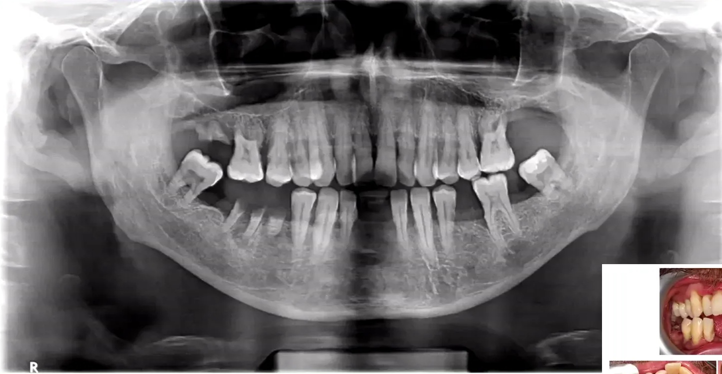
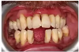
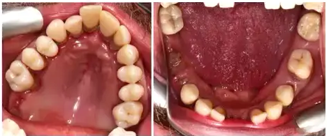
A 47-year-old man had pulled out a lower incisor and an upper-right molar with his fingers. He had generalised periodontal disease, heavy calculus, substantial bone loss involving teeth 26 and 48, caries, retained roots, and some minor restorable caries.
- For osteoporosis treatment, definite root remnants should be removed; grade 3 mobile teeth and, in some circumstances, grade 2 mobile teeth could be removed, while other teeth could be retained if the patient was motivated to maintain them.
- A shortened dental arch could be considered, with assessment of whether the patient would attend regular recalls and maintain oral hygiene.
- For cancer treatment, a more aggressive approach may be appropriate because of the higher MRONJ risk and the urgency of treating possible infection sources. Teeth with deep probing depths, significant periodontal involvement, poor long-term prognosis, or poor expected maintenance might be removed when there is limited time before therapy.
- Patient motivation and preferences should be discussed frankly and non-judgementally, including willingness to brush regularly, clean interproximally, attend dental reviews, and maintain periodontal treatment.
- Restorative treatment could generally be completed after starting the bone-modifying drug, whereas invasive procedures affecting bone should be prioritised beforehand.
Conclusion and Contact66
Thank you
Clinical Cases
Case: 47-year-old male with multiple myeloma and poor dentition, OPG and clinical findings
Question
Scenario: A 47-year-old male with a history of multiple myeloma is being assessed for dental fitness prior to starting bone-modifying drugs (denosumab/bisphosphonates).
What’s shown: An OPG and clinical findings of a patient who extracted a lower incisor and an upper right molar with his own fingers.
Consider: Identify the dental findings on the OPG and clinical examination, and consider how the dental workup differs based on the specific medical condition the patient is being worked up for.
Answer
Observations:
- Generalized periodontal disease with heavy calculus.
- Substantial bone loss and likely mobility in teeth 2, 6, and 48.
- Presence of caries and retained roots.
- Possible minor restorable caries.
Reasoning: The lecturer walks through the patient’s OPG and clinical findings to demonstrate a dental workup for a patient needing clearance for bone-modifying drugs. The assessment highlights that the dental workup and subsequent management must be tailored to the specific medical condition (e.g., myeloma, cardiovascular disease, or solid organ transplant) the patient is being worked up for.
Outcome: The patient is being worked up for denosumab/bisphosphonates, and the case will be revisited later in the lecture.
Takeaway: A dental workup for patients starting bone-modifying drugs must be individualized, as the assessment and management differ depending on the underlying medical condition being treated.
Case: Patient workup for bone-modifying drugs, extraction planning
Question
Scenario: A patient is being worked up for osteoporosis or cancer and requires bone-modifying drugs. The lecturer reviews the patient’s dentition to decide which teeth to extract to future-proof the patient.
What’s shown: The patient’s dentition, showing mobile teeth, increased probing depths, and compromised anterior teeth.
Consider: Which teeth should be extracted to minimize the risk of MRONJ and account for the patient’s compliance and motivation?
Answer
Observations:
- Grade 3 and grade 2 mobile teeth.
- Teeth with increased probing depths (greater than 8 mm, or >6 mm).
- Compromised (“dodgy”) anterior teeth in the aesthetic zone.
- The patient has poor motivation, is non-compliant, and pulls out his own teeth with his fingers.
Reasoning:
- Grade 3 mobile teeth should definitely be extracted.
- For a cancer workup, a more aggressive approach is taken, extracting teeth with probing depths >8 mm.
- The lower mandible has a higher risk of MRONJ, so preventing it there is prioritized.
- A shortened dental arch approach is considered because the patient is more likely to maintain the remaining teeth.
- A frank discussion is held with the patient about compliance; if they are unlikely to maintain their teeth or attend regular recalls, it is better to extract them now rather than refer for periodontal treatment.
- Compromised anterior teeth may also be extracted if the patient is non-compliant.
Outcome: The plan involves extracting the compromised teeth (including grade 3 mobile teeth and those with deep probing depths) and adopting a shortened dental arch approach, with restorations to be completed after the bone-modifying drugs are delivered.
Takeaway: When planning extractions prior to starting bone-modifying drugs, clinical findings must be balanced against the patient’s motivation and compliance; a more aggressive extraction or shortened dental arch approach may be necessary if the patient is unlikely to maintain their dentition.
Footnotes
-
Original PDF page 1: L8 Musculoskeletal Disorders, p.1 ↩
-
Original PDF page 2: L8 Musculoskeletal Disorders, p.2 ↩
-
Original PDF page 3: L8 Musculoskeletal Disorders, p.3 ↩
-
Original PDF page 4: L8 Musculoskeletal Disorders, p.4 ↩
-
Original PDF page 6: L8 Musculoskeletal Disorders, p.6 ↩
-
Original PDF page 7: L8 Musculoskeletal Disorders, p.7 ↩
-
Original PDF page 8: L8 Musculoskeletal Disorders, p.8 ↩
-
Original PDF page 9: L8 Musculoskeletal Disorders, p.9 ↩
-
Original PDF page 10: L8 Musculoskeletal Disorders, p.10 ↩
-
Original PDF page 5: L8 Musculoskeletal Disorders, p.5 ↩
-
Original PDF page 11: L8 Musculoskeletal Disorders, p.11 ↩
-
Original PDF page 12: L8 Musculoskeletal Disorders, p.12 ↩
-
Original PDF page 13: L8 Musculoskeletal Disorders, p.13 ↩
-
Original PDF page 14: L8 Musculoskeletal Disorders, p.14 ↩
-
Original PDF page 15: L8 Musculoskeletal Disorders, p.15 ↩
-
Original PDF page 16: L8 Musculoskeletal Disorders, p.16 ↩
-
Original PDF page 17: L8 Musculoskeletal Disorders, p.17 ↩
-
Original PDF page 18: L8 Musculoskeletal Disorders, p.18 ↩
-
Original PDF page 19: L8 Musculoskeletal Disorders, p.19 ↩
-
Original PDF page 21: L8 Musculoskeletal Disorders, p.21 ↩
-
Original PDF page 20: L8 Musculoskeletal Disorders, p.20 ↩
-
Original PDF page 22: L8 Musculoskeletal Disorders, p.22 ↩
-
Original PDF page 23: L8 Musculoskeletal Disorders, p.23 ↩
-
Original PDF page 25: L8 Musculoskeletal Disorders, p.25 ↩
-
Original PDF page 24: L8 Musculoskeletal Disorders, p.24 ↩
-
Original PDF page 26: L8 Musculoskeletal Disorders, p.26 ↩
-
Original PDF page 27: L8 Musculoskeletal Disorders, p.27 ↩
-
Original PDF page 28: L8 Musculoskeletal Disorders, p.28 ↩
-
Original PDF page 29: L8 Musculoskeletal Disorders, p.29 ↩
-
Original PDF page 30: L8 Musculoskeletal Disorders, p.30 ↩
-
Original PDF page 31: L8 Musculoskeletal Disorders, p.31 ↩
-
Original PDF page 32: L8 Musculoskeletal Disorders, p.32 ↩
-
Original PDF page 33: L8 Musculoskeletal Disorders, p.33 ↩
-
Original PDF page 34: L8 Musculoskeletal Disorders, p.34 ↩
-
Original PDF page 35: L8 Musculoskeletal Disorders, p.35 ↩
-
Original PDF page 36: L8 Musculoskeletal Disorders, p.36 ↩
-
Original PDF page 37: L8 Musculoskeletal Disorders, p.37 ↩
-
Original PDF page 38: L8 Musculoskeletal Disorders, p.38 ↩
-
Original PDF page 39: L8 Musculoskeletal Disorders, p.39 ↩
-
Original PDF page 40: L8 Musculoskeletal Disorders, p.40 ↩
-
Original PDF page 41: L8 Musculoskeletal Disorders, p.41 ↩
-
Original PDF page 42: L8 Musculoskeletal Disorders, p.42 ↩
-
Original PDF page 43: L8 Musculoskeletal Disorders, p.43 ↩
-
Original PDF page 44: L8 Musculoskeletal Disorders, p.44 ↩
-
Original PDF page 46: L8 Musculoskeletal Disorders, p.46 ↩
-
Original PDF page 45: L8 Musculoskeletal Disorders, p.45 ↩
-
Original PDF page 47: L8 Musculoskeletal Disorders, p.47 ↩
-
Original PDF page 48: L8 Musculoskeletal Disorders, p.48 ↩
-
Original PDF page 49: L8 Musculoskeletal Disorders, p.49 ↩
-
Original PDF page 50: L8 Musculoskeletal Disorders, p.50 ↩
-
Original PDF page 52: L8 Musculoskeletal Disorders, p.52 ↩
-
Original PDF page 51: L8 Musculoskeletal Disorders, p.51 ↩
-
Original PDF page 53: L8 Musculoskeletal Disorders, p.53 ↩
-
Original PDF page 54: L8 Musculoskeletal Disorders, p.54 ↩
-
Original PDF page 55: L8 Musculoskeletal Disorders, p.55 ↩
-
Original PDF page 56: L8 Musculoskeletal Disorders, p.56 ↩
-
Original PDF page 59: L8 Musculoskeletal Disorders, p.59 ↩
-
Original PDF page 57: L8 Musculoskeletal Disorders, p.57 ↩
-
Original PDF page 58: L8 Musculoskeletal Disorders, p.58 ↩
-
Original PDF page 60: L8 Musculoskeletal Disorders, p.60 ↩
-
Original PDF page 61: L8 Musculoskeletal Disorders, p.61 ↩
-
Original PDF page 62: L8 Musculoskeletal Disorders, p.62 ↩
-
Original PDF page 63: L8 Musculoskeletal Disorders, p.63 ↩
-
Original PDF page 64: L8 Musculoskeletal Disorders, p.64 ↩
-
Original PDF page 65: L8 Musculoskeletal Disorders, p.65 ↩
-
Original PDF page 66: L8 Musculoskeletal Disorders, p.66 ↩