Haematopoietic Stem Cell Transplants1
2026 DENT5311 DMD3
Special Needs Dentistry Module
Dr Jee-Yun Leung
BDSc(Hons), MSc(SCD), DScD(RCSEng), DClinDent(SND), MRACDS(SND), FANZASND
Lecture Outcomes2
-
Understand the types of, and common indications for, HSCT/BMT
-
Outline the patient journey to HSCT/BMT
-
Describe the oral complications of HSCT/BMT, and basic oral care before, during and after transplant
-
Discuss the systemic and oral manifestations of graft vs host disease, and appreciate its management options
-
Appreciate the dental work-up required prior to stem cell transplant
- Modifications required to safely provide dental treatment

Introduction to Haematopoietic Stem Cell Transplant3
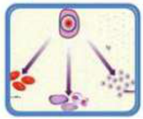
The process of transplanting healthy blood-forming cells into an individual to replace cells that have been destroyed by disease, radiation or cytotoxic drugs.
Lecturer — Transplant Use
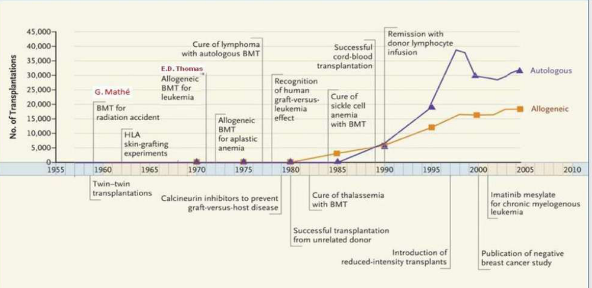
Stem cell transplantation was previously considered a treatment of last resort, but its use has increased.
- Peripheral blood stem cell transplantation and more rapid engraftment have contributed to this increase.
- Reduced-intensity conditioning regimens and increasing numbers of patients suitable for transplantation have also expanded its use.
Definitions and Abbreviations4
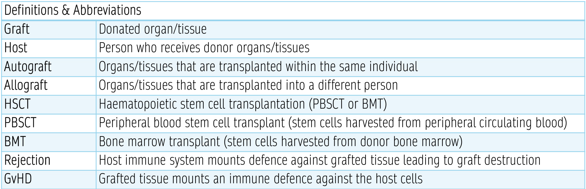
| Term | Definition |
|---|---|
| Graft | Donated organ/tissue |
| Host | Person who receives donor organs/tissues |
| Autograft | Organs/tissues that are transplanted within the same individual |
| Allograft | Organs/tissues that are transplanted into a different person |
| HSCT | Haematopoietic stem cell transplantation (PBSCT or BMT) |
| PBSCT | Peripheral blood stem cell transplant (stem cells harvested from peripheral circulating blood) |
| BMT | Bone marrow transplant (stem cells harvested from donor bone marrow) |
| Rejection | Host immune system mounts defence against grafted tissue leading to graft destruction |
| GvHD | Grafted tissue mounts an immune defence against the host cells |
Lecturer — Rejection and GvHD
Rejection occurs when the host immune system recognises the graft as foreign and attacks it, whereas graft-versus-host disease occurs when donor-derived immune cells attack host tissues.
- In rejection, the host attacks the graft.
- In GVHD, the graft attacks the host.
Types and Indications5
Types of Stem Cell Transplant6
Stem cell transplants are classified by:
- Source of the stem cells
- Donor type
- The conditioning regimen
Source of Stem Cells
Peripheral blood7
-
Preferred method of harvest
-
Administration of G-CSF → harvest from peripheral blood circulation
-
Advantages: superior engraftment, reduced early infection, shorter hospitalisation
-
Disadvantages: higher incidence of GVHD and CMV infection
-
G-CSF stimulates granulocyte production and mobilises stem cells into the circulating blood.
-
Earlier recovery of granulocytes and platelets than with bone marrow.
-
Reduced cost compared with bone marrow transplantation.
-
Lower complication rates associated with mobilisation and collection.
Bone marrow
-
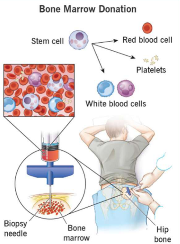
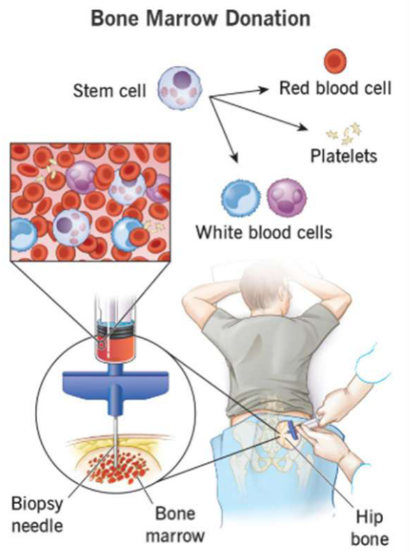
Aspiration of bone marrow tissue from posterior iliac crest
-
Advantages: lower incidence of GVHD, less immunosuppression
-
Disadvantages: frequently complicated by mild back or hip pain, fatigue, transient changes in peripheral blood cell counts
-
Bone marrow is collected from the posterior iliac crest while the donor is under anaesthesia.
-
The graft contains fewer T cells than a peripheral blood graft, reducing the incidence and severity of acute and chronic GVHD.
-
Bone marrow is commonly used for non-malignant disease, including aplastic anaemia.
Umbilical cord blood
-
Predominantly for paediatric setting (low volume of available cells)
-
Advantages: less stringent matching required, expanded donor pool, ease of procurement, less GVHD
-
Disadvantages: slower engraftment, increased graft failure, unavailability of donor for additional donations
-
Cord blood has minimal donor risk and no donor attrition.
-
Cord blood may have delayed immune reconstitution.
-
Cord blood from two matching donors may be required in some cases.
-
The donor is unavailable for additional donations such as donor lymphocyte infusion if the transplant fails.
Donor Types8
- Autologous (self)
- Allogeneic (other)
- Matched donors
- Haplo-identical donor
- Syngeneic (identical twin)
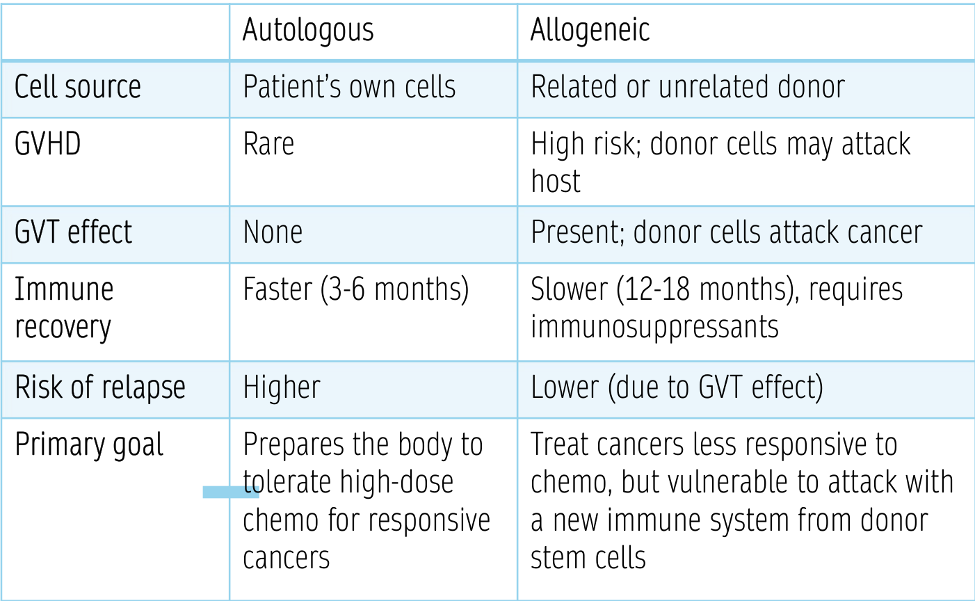
| Autologous | Allogeneic | |
|---|---|---|
| Cell source | Patient’s own cells | Related or unrelated donor |
| GVHD | Rare | High risk; donor cells may attack host |
| GVT effect | None | Present; donor cells attack cancer |
| Immune recovery | Faster (3–6 months) | Slower (12–18 months), requires immunosuppressants |
| Risk of relapse | Higher | Lower (due to GVT effect) |
| Primary goal | Prepares the body to tolerate high-dose chemo for responsive cancers | Treat cancers less responsive to chemo, but vulnerable to attack with a new immune system from donor stem cells |
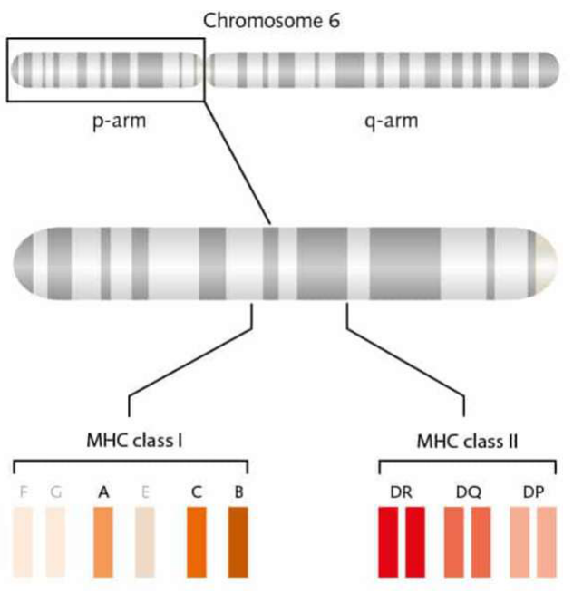
Chromosome 6 — HLA regions9
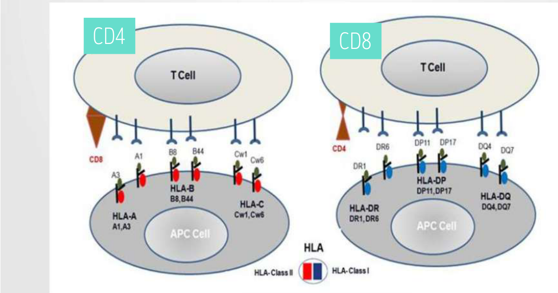
- MHC class I: HLA-A (A1, A3), HLA-B (B8, B44), HLA-C (Cw1, Cw6)
- MHC class II: HLA-DR (DR1, DR6), HLA-DP (DP11, DP17), HLA-DQ (DQ4, DQ7)
- Gene loci on the p-arm and q-arm: F, G, A, E, C, B (class I); DR, DQ, DP (class II)
Lecturer — HLA Function
HLA is a cell-surface marker found on most cells that the immune system uses to distinguish self from foreign cells.
- If T cells recognise an antigen as self, they do not respond; recognition as foreign may trigger an immune response.
- Depending on the transplant centre, matching may use eight-out-of-eight, ten-out-of-ten, or twelve-out-of-twelve criteria.
 |  |
Antigen presentation
- APC cell presents HLA class I antigens (A1, A3; B8, B44; Cw1, Cw6) to CD8 and CD4 T cells
- APC cell presents HLA class II antigens (DR1, DR6; DP11, DP17; DQ4, DQ7) to CD4 and CD8 T cells
Matching = HLA complex (human leukocyte antigen) / MHC (major histocompatibility complex)
Lecturer — Minor Histocompatibility
Even when major HLA markers are well matched, mismatched minor histocompatibility complex genes can cause rejection or GVHD.
- Immunosuppressive regimens are therefore required for allogeneic transplantation.
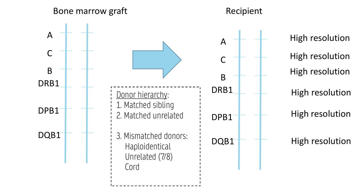
Donor hierarchy10
- Matched sibling
- Matched unrelated
- Mismatched donors:
- Haploidentical
- Unrelated (7/8)
- Cord
- Allogeneic (other)
- Matched donors
- Matched related donor → siblings have 1/4 chance
- Matched unrelated donor
- Haplo-identical donor
- Haploidentical transplantation uses high-dose chemotherapy several days after transplantation to reduce active T cells and minimise GVHD.
- An identical twin donor is described as a syngeneic transplant; these patients do not develop GVHD but have less graft-versus-tumour effect and a higher relapse risk than comparable allogeneic recipients.
- Matched donors
Loci tested (high resolution): HLA-A, HLA-C, HLA-B, DRB1, DPB1, DQB1 — assessed between bone marrow graft donor and recipient.
Lecturer — Donor Matching
Matched unrelated donors may be identified through a worldwide registry containing approximately 40 million donors.
- Biological parents and children are automatically haploidentical matches.
- Full siblings have approximately a 50% chance of being haploidentical matches.
- Potential donors undergo HLA matching using a buccal swab or blood test; donation usually takes approximately half a day and is similar to donating plasma.
- Matching is more difficult for East African, First Nations Australian, and Torres Strait Islander patients because of greater HLA diversity and fewer registered donors.
Conditioning Regimens1112
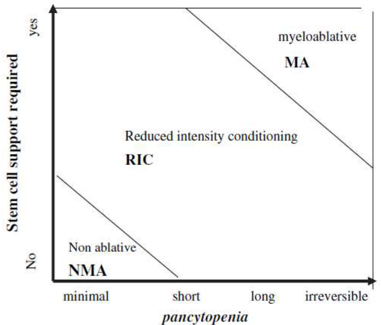
- Myeloablative (MA) conditioning
- Irreversible pancytopenia
- Stem cell support required to rescue marrow function and prevent aplasia-related death
- Myeloablative conditioning is generally used for younger or fitter patients and provides high disease control with rapid donor-cell engraftment, but has higher toxicity and transplant-related mortality.
- Reduced intensity conditioning
- Spectrum between MA and NMA conditioning
- Induced cytopenia: may be prolonged requiring stem cell support, OR host-recovery possible with significant morbidity
- Reduced-intensity conditioning uses enough immunosuppression to permit donor-cell engraftment without completely eradicating the recipient’s bone marrow.
- It is used more often in older or medically compromised patients and has less immediate organ toxicity and reduced transplant-related mortality.
- Non-myeloablative (NMA) conditioning
- Minimal cytopenia, does not require stem cell support
- Non-myeloablative conditioning relies on the graft-versus-tumour effect rather than high-dose chemotherapy or irradiation.
- Donor T cells eventually eliminate and replace host blood stem cells.
- Minimal cytopenia, does not require stem cell support
Bacigalupo A, et al. Defining the intensity of conditioning regimens: working definitions. Biol Blood Marrow Transplant. 2009 Dec;15(12):1628-33.
Lecturer — Conditioning Purpose
Conditioning before transplantation for malignant disease reduces tumour burden and provides sufficient immunosuppression to prevent graft rejection.
- Traditional regimens used total-body irradiation and chemotherapy beyond the level at which the patient’s own blood-forming cells could recover.
- The transplant then acted as a rescue for the immune and bone marrow systems.
Regimen selection13
-
Underlying condition
-
Disease status
-
Recipient co-morbidities
-
Risk of rejection (graft source)
-
Regimen choice also depends on whether the marrow is malignant or hyperplastic, the recipient’s fitness for intensive treatment, and the degree of HLA disparity between donor and recipient.
Indications for Stem Cell Transplants1415
-
Haematological malignancy
- ALL, CLL (usually allo)
- Plasma cell disorders, HL, NHL (usually auto)
-
Solid tumours (usually auto)
- Neuroblastoma
- Soft tissue / Ewing sarcoma
- Germ cell tumours
- Breast cancer
- Other
-
Immunological disease
- Primary immune deficiency (usually allo)
- Autoimmune disorders (usually auto)
-
Other (usually allo)
- Thalassemia
- Sickle cell disease
- Paroxysmal nocturnal haemoglobinuria
- Inherited disorders of metabolism
- Bone marrow failure
-
Number of HSCT increasing over time
-
Notable increase in autologous HSCT for autoimmune disease since 2009
-
Western Australia allo-HSCT: 70 in 2025, 55 in 2024
Lecturer — Transplant Services
In Western Australia, allogeneic transplants are performed at Fiona Stanley Hospital.
- Autologous transplants may be performed at Royal Perth Hospital and Sir Charles Gairdner Hospital.
Patient Journey and Transplant Process1617
patient journey.*
- Diagnosis and start initial treatment (induction chemotherapy)
- Referral to HSCT program, concurrent family donor search
- Initial consult (HSCT clinic) – may be phone consult
- Donor search (family → unrelated donor)
- Presentation at HSCT MDT meeting (initial)
- Referrals / investigations
- Echocardiogram & cardiologist review
- Pulmonary function tests
- Dental review
- Blood tests, kidney and liver function, viruses (HIV, hepatitis, CMV)
- CT or bone scans
- Pregnancy test
- Recovery from induction chemo
Lecturer — Treatment Timeline
Induction chemotherapy generally lasts approximately four weeks and may require hospitalisation. If the patient has a good response and no longer requires transplantation, they may be discharged from the transplant pathway.
- Consolidation chemotherapy while the donor is being prepared is generally lower-toxicity treatment delivered over three to five days.
- This is followed by approximately three weeks at home for recovery, with patients receiving approximately three to four cycles.
- Approximately six weeks may be required between completion of consolidation chemotherapy and commencement of conditioning chemotherapy to allow healing.
Referral and Work-Up to HSCT Program1819
- Disease re-assessment (e.g. bone marrow biopsy)
- Remission → discharged
- Second HSCT clinic appointment – face-to-face
- Discuss family search result, refer for unrelated donor search if required
- Consolidation chemotherapy cycle(s) whilst awaiting donor
- Final face-to-face clinic review (review of results, donor confirmation, consent)
- Presentation at HSCT MDT meeting (subsequent)
- Donor available → Ward schedules admission for HSCT
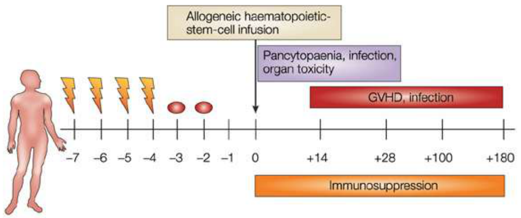
Allogeneic transplant.
 |  |
Allogeneic Transplant2021
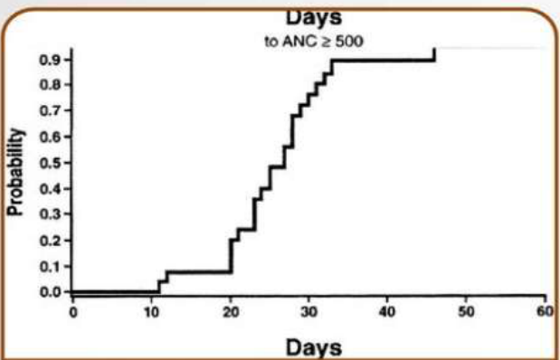
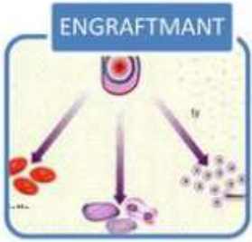
Engraftment: blood count recovery
Neutrophil engraftment22
- Figure: probability curve of days to ANC ≥ 500 (x-axis 0–60 days; y-axis probability 0.0–0.9)
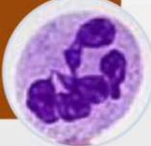
- Neutrophil engraftment occurs when neutrophils ≥ 0.5 × 10⁹
- Thomson B G et al. Blood 2000;96:2703-2711.
Lecturer — Engraftment Recovery
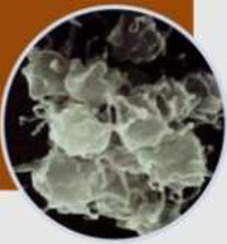
Neutrophil recovery reduces, but does not immediately eliminate, the risk associated with infection. Platelet recovery may take longer, so patients may remain platelet-transfusion dependent after neutrophils have returned to safer levels.
 |  |
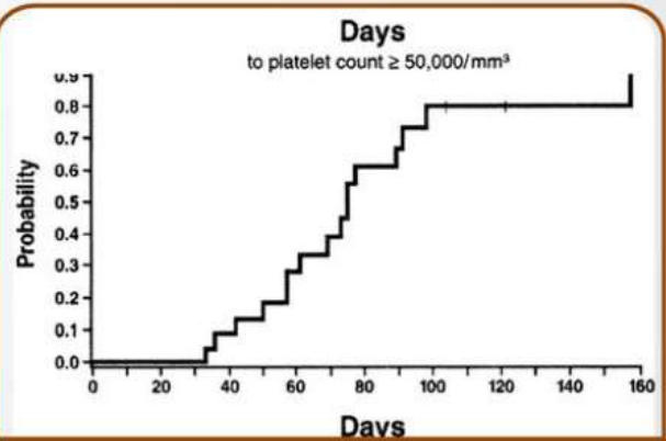 |  |
Platelet engraftment
- Figure: probability curve of days to platelet count ≥ 50,000/mm³ (x-axis 0–160 days; y-axis probability 0.0–0.9)
- Platelet engraftment occurs when platelets ≥ 20 × 10⁹
Allogeneic – from another person:
- Sibling donor – HLA matched brother or sister
- Syngeneic – from an identical twin
- Unrelated donor – found using a donor registry
- Haploidentical – half-matched family member
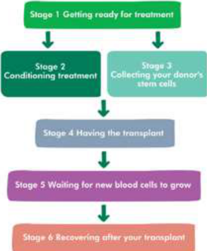
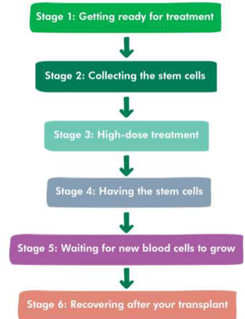
Stages of the Transplant23
- Getting ready for treatment
- Conditioning treatment
- Collecting your donor’s stem cells
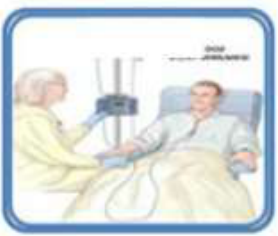
- Having the transplant
- Waiting for new blood cells to grow
- Recovering after your transplant
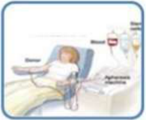
(Figure: patient and donor apheresis.)
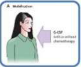
- Mobilisation — Administer pre-treatment to release blood stem cells from bone marrow into the bloodstream
- G-CSF with or without chemotherapy
- Apheresis
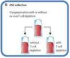
- Cryopreservation with or without ex vivo T cell depletion (without T cell depletion / with T cell depletion)
- Conditioning — Conditioning the blood stem cells in the laboratory until they are required
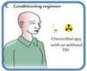
- Chemotherapy with or without TBI, with or without ex vivo T cell depletion
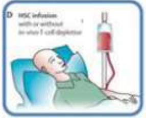
- HSC Infusion
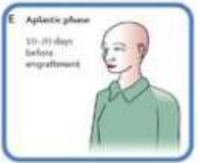
- Aplastic Phase — Provide supportive medical treatment for at least 4 weeks as the immune system rebuilds
- 10–21 days before engraftment
- Recovery
Lecturer — Stem Cell Collection
The donor’s stem cells are mobilised using injections of a stimulating factor for approximately five to ten days. The cells are then collected by apheresis or bone marrow aspiration, and may be cryopreserved if collected at another hospital or imported from another country.
Lecturer — Conditioning Treatment
Conditioning usually lasts approximately six to eight days and finishes on day minus two or minus three. Day zero is the day of stem cell infusion.
- Conditioning may be myeloablative, reduced-intensity, or non-myeloablative.
- Its purposes are to destroy remaining cancer cells, make room in the bone marrow for donor stem cells, and reduce the risk of graft rejection.
Lecturer — Early Post-Transplant Course
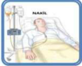
Stem cells are usually infused on a weekday through a central line, with approximately 200–300 mL of graft infused over 20–30 minutes. After infusion, the cells migrate to the bone marrow and immunosuppression is required to prevent graft rejection.
- Toxicity develops over the following days, including diarrhoea, nausea, stomach cramps, oedema, and mucositis.
- Blood counts become profoundly reduced, neutrophils may fall to zero, and patients may require blood and platelet transfusions and significant analgesia.
- Patients usually remain in hospital for approximately one to four weeks, followed by initially weekly outpatient reviews.
- Even after neutrophil recovery, immunity remains low for several months and some patients may continue to require transfusions.
 |  |  |
 |  |
Autologous Transplant242526
- Pre-HSCT testing
- Choice of conditioning regimen and immunosuppression
- Antibacterial and pneumocystis prophylaxis (e.g. trimethoprim-sulfamethoxazole = Bactrim®)
- Antifungal prophylaxis (e.g. fluconazole)
- Antiviral prophylaxis (e.g. valaciclovir)
- Infection control measures
- Immunisation
Lecturer — Autologous Transplant
In an autologous transplant, the patient’s own stem cells are collected before high-dose chemotherapy, generally cryopreserved, and later reinfused to restore blood-cell production.
- Advantages include no graft rejection risk, no significant GVHD risk, reduced need for immunosuppression, faster immune recovery, less waiting time for a donor, and lower overall risk than allogeneic transplantation.
- Autologous transplantation has less graft-versus-tumour effect than allogeneic transplantation.
 |  |  |
 |  |  |
 |
Risk Reduction Strategies27
- Inactivated vaccines — usually commence 6–12 months post-HSCT
- Live vaccines — usually commence at least 24 months post-HSCT
- Not if active GVHD or receiving immunosuppressive agents
Lecturer — Post-Transplant Vaccination
During transplantation, patients lose their memory B cells and need to be revaccinated. Patients may describe this as losing their previous blood and repeating childhood vaccination schedules; day zero may be described as a new birthday because the blood and immune system are being rebuilt.
Vaccinations2829
Mortality
- Non-relapse mortality ~10-12%
- Infection or toxicity
- Sharpest increase in first year
- Relapse mortality ~20-25% at 2 years
- Greatest risk of death in first 4 years post-transplant: recurrence
- Other common causes of death: infection*, second cancers*, organ system dysfunction
Lecturer — Long-Term Survival
Relapse mortality is related to the graft-versus-tumour effect. Overall survival remains lower than in the general population, with increased mortality for approximately ten to thirty years after transplantation before approaching that of the general population over time.
- Squamous cell carcinoma
- Oral cancer
Increased Risk of Transplant-Related Mortality30
-
Allogeneic transplant
-
Advanced disease status at time of transplant
-
Type of donor
-
Intensity of conditioning regimen
-
Patient age
-
Infection
-
Treatment toxicity
-
Significant comorbidities
-
Ongoing immunosuppression
-
GVHD Mortality data as above.
Oral Complications31
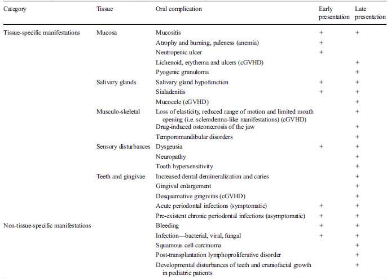
Oral complications occur before, after, and immediately post-transplant.
Patients at Risk32
Myelosuppression and Immunosuppression33
- Disease
- Cancer therapy
- Immunosuppressive regime
Oral complications affect ~80% of patients.
- Damage to blood cells and immunosuppression
- Infection, bleeding and anaemia-related oral manifestations
- Cytotoxic therapies to oral tissues
- Direct and indirect effects
Patients at risk are those with myelosuppression and immunosuppression due to disease, cancer therapy, or immunosuppressive regime; oral complications affect ~80% of patients.
Lecturer — Transplant Dental Risk
Haematology and oncology patients undergoing transplantation are a high-risk dental population. Inadequate management may contribute to:
- Interruption of cancer treatment.
- Sepsis.
- Effects on overall prognosis.
Lecturer — Additional Risk Factors
Risk also results from conditioning therapy, post-transplant immune-mediated treatment, neutropenia, thrombocytopenia, and anaemia.
Acute Side Effects34
- Susceptibility to infection (including oral)
- Flare of chronic infection (e.g. odontogenic, periodontal)
- Reactivation (e.g. oral herpes simplex virus)
- Opportunistic (e.g. oral candidiasis)
- Systemic (immunocompromised + mucositis)
- Oral & gastrointestinal mucositis
- Implications: pain, infection, nutrition, speech, ability for oral care
- Cytopenias
- Thrombocytopenia = bleeding risk
- Neutropenia / leukopenia = infection risk
- Anaemia
- Disseminated intravascular coagulation
- Anticoagulant prophylaxis
- Taste alterations
- Acute graft-vs-host disease (allo only)
Lecturer — Mucositis Progression
Mucositis may progress from oral erythema to ulceration within approximately two weeks of chemotherapy initiation.
- Susceptibility to infection (including oral): flare of chronic infection, reactivation, opportunistic and systemic infection
- Oral & gastrointestinal mucositis
- Cytopenias
- Disseminated intravascular coagulation
- Taste alterations
- Acute graft-vs-host disease (allo only)
Chronic Side Effects3536
- Immunosuppression (allo only)
- Ongoing infection risk
- Risk of secondary malignancy – heightened at cGVHD sites
- Hyposalivation (and associated sequelae)
- Chemo damage, polypharmacy, cGVHD
- Osteoporosis risk
- Bone-altering medications
- Chronic GVHD (allo only)
Lecturer — Osteoporosis Causes
Osteoporosis risk may be related to altered endocrine function from conditioning regimens and long-term corticosteroid therapy.
- Immunosuppression (allo only): ongoing infection risk; risk of secondary malignancy – heightened at cGVHD sites
- Hyposalivation (and associated sequelae): chemo damage, polypharmacy, cGVHD
- Osteoporosis risk: bone-altering medications
- Chronic GVHD (allo only)
 |  |  |
 |  |  |
Graft Versus Host Disease37
“Caused by reaction of the donor-derived immunocompetent cells against the recipient’s tissues”
Overview and Pathophysiology of GVHD3839
Immune cells in the donated tissue (graft) identify the recipient (host) tissues as ‘foreign’, mounting an immune response to ‘reject’ the host’s cells.
Tissue damage is caused by the proliferation of donor T cells and release of inflammatory cytokines.
Lecturer — Transplant Setting
GVHD is primarily an issue in allogeneic transplantation.
Severity depends on:
- Degree of histocompatibility matching
- Number of donor T-cells infused
- Use of immunosuppressant medication
- Conditioning regimen
Previously designated purely based on the time frame of occurrence:
- Acute had its onset <100 days after transplant
- Chronic commenced or persisted >100 days after transplant
Acute Versus Chronic GVHD40
Now recognised that they have distinct clinical and pathologic features. Four subclassifications:
Lecturer — Changing Definitions
The former time-based definitions are less useful with reduced-intensity conditioning.
- Acute symptoms may be delayed.
- Chronic GVHD may occur earlier than expected.
- Classic acute (<100 days; only acute features)
- Persistent/recurrent/late onset acute (>100 days; only acute features)
- Classic chronic (>100 days; only chronic features)
- Overlap syndrome (present at any time post transplant; mixed acute/chronic features)
-
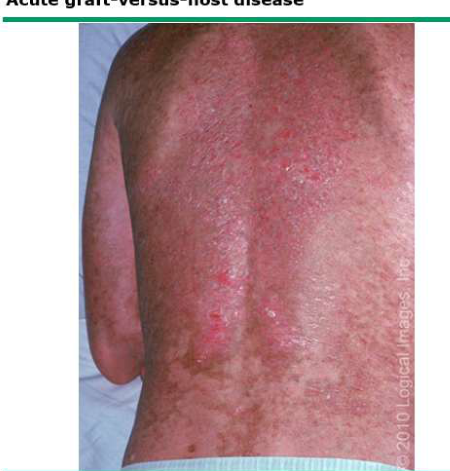
Principal target organs = skin, GIT, liver
-
Skin: usually first and most common site
- Maculopapular rash
- May develop into bullous lesions with toxic epidermal necrolysis
-
GIT: diarrhoea, abdominal pain, nausea, vomiting, anorexia, oral mucosal involvement
- Weight loss
-
Liver: elevated LFTs, elevated cholesterol, painful hepatomegaly, dark urine, pale stool, fluid retention, pruritus
-
Less commonly involved organ systems: haematopoietic system, eyes, kidneys, and lung
-
Rash may begin at the nape of the neck, ears, shoulders, palms, or soles
- The rash may have a sunburn-like appearance
- Itching and pain may occur
Features of Acute GVHD41
- Immunoregulatory disorder with features of both autoimmunity and immunodeficiency; can affect any organ
- Clinical manifestations resemble autoimmune collagen vascular diseases
- ~50% of allo-HSCT patients surviving >1yr develop cGVHD
 |  |
Cutaneous Forms42
Two main cutaneous forms:
- Lichenoid – predominates in early cGvHD; generally affects peri-orbital skin, ears, palms, soles, genital mucosa
- Sclerodermatous – inflammatory skin plaques (esp. on the extremities) are replaced by areas of fibrosis, causing dimpling and contracture; skin of trunk can contract causing secondary restrictive lung disease
Features of Chronic GVHD43
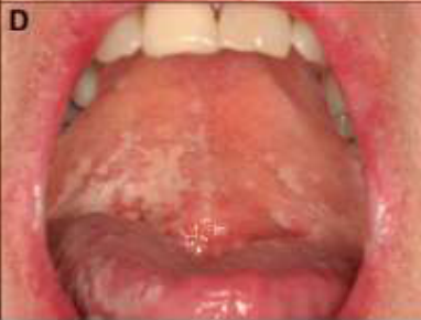
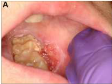
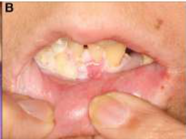
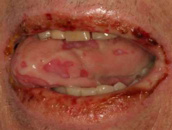
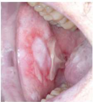
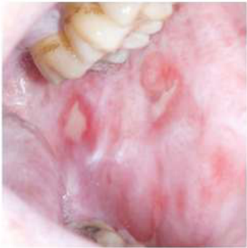
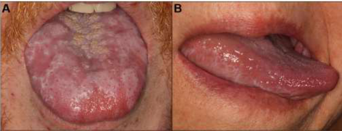
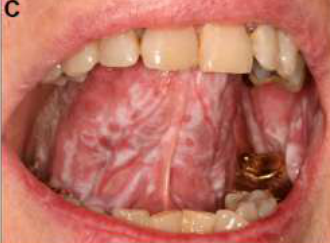
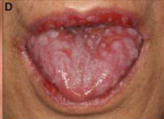
The oral mucosa is the most commonly involved site.
Other Organ Systems
- Eyes (60%)
- GIT (dysmotility, pseudo-obstruction, dysphagia, oesophageal ulceration/fibrosis/webbing/stricture)
- Lungs
- Liver
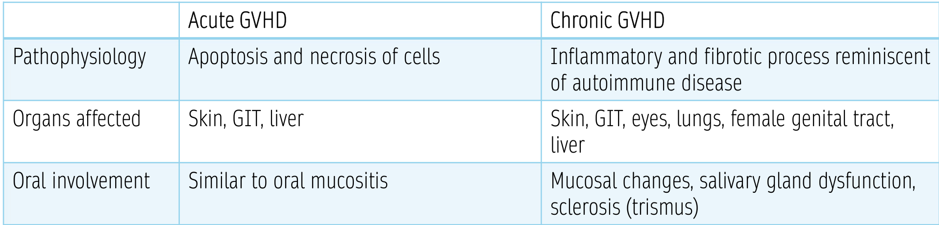
| Acute GVHD | Chronic GVHD | |
|---|---|---|
| Pathophysiology | Apoptosis and necrosis of cells | Inflammatory and fibrotic process reminiscent of autoimmune disease |
| Organs affected | Skin, GIT, liver | Skin, GIT, eyes, lungs, female genital tract, liver |
| Oral involvement | Similar to oral mucositis | Mucosal changes, salivary gland dysfunction, sclerosis (trismus) |
Reported to occur in 45–83% of patients with cGvHD (Imanguli, 2008).
Classic manifestations (mimic immune-mediated conditions):
Oral Chronic GVHD44
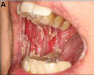
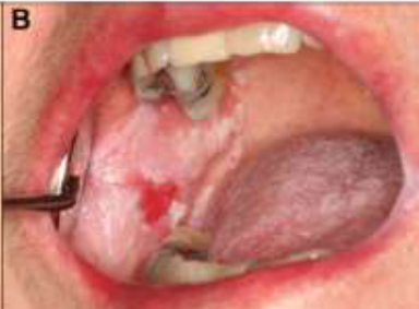
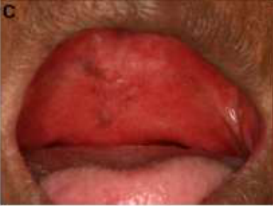
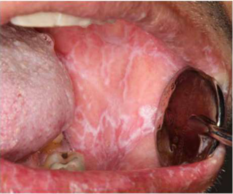
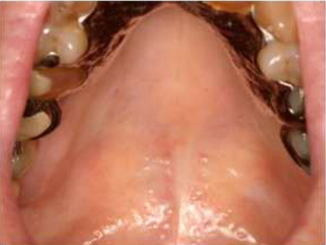
- Lichenoid changes, mucosal atrophy and ulceration – resembles oral lichen planus
- Salivary gland dysfunction – salivary gland infiltrates resemble Sjögren’s syndrome
- Trismus – resembles scleroderma
(Treister, 2012)
- Most common on buccal/labial mucosae and tongue, but can affect any site
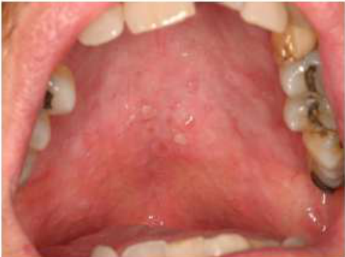
- Commonly presents as white striations or plaques, erythema, atrophy, and/or ulceration
- May be asymptomatic, uncomfortable, or painful; may limit oral intake
- Prominent buccal reticular changes → sensation of mouth tightness or trismus
Lecturer — Oral Manifestations
Oral chronic GVHD may also cause microstomia, difficulty eating, and an increased risk of dental caries.
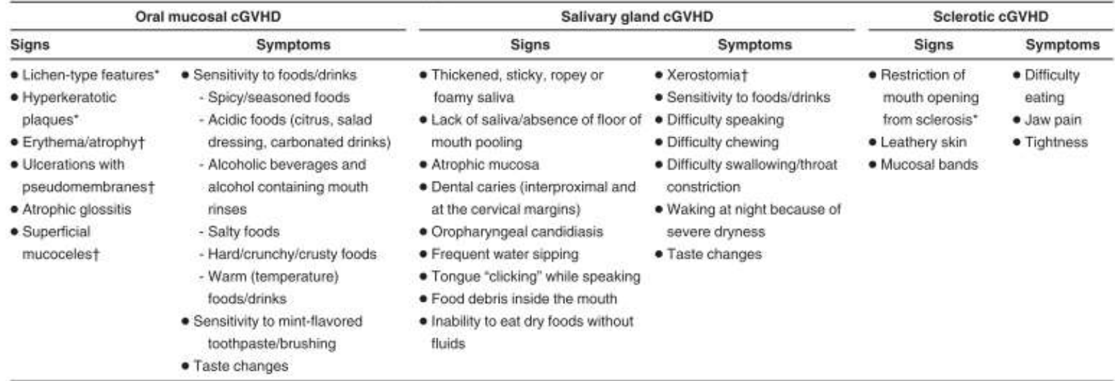
Mucosal Changes in Oral cGVHD45
Changes may include chronic salivary gland inflammation, formation of mucoceles and duct blockage, reduced volume of saliva and increased viscosity.
Extensive salivary gland involvement can result in total destruction of acinar units, leading to permanent and profound hyposalivation.
 |  |  |
 |  |  |
Salivary Changes in Oral cGVHD46
Both qualitative and quantitative changes are observed.
Signs and symptoms include: trismus, pain, secondary ulceration, impaired oral hygiene.
Can be primarily cutaneous or primarily mucosal:
- Cutaneous: perioral and facial skin sclerosis
- Mucosal: chronic mucosal inflammation → fibrosis
 |  |
 |
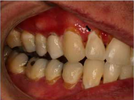
Sclerotic and Fibrotic Changes in Oral cGVHD47
Secondary periodontal recession may result → dental sensitivity.
Lecturer — Mouth Opening Baseline
At the pre-transplant examination, maximum inter-incisal opening can be recorded as a baseline. This can be compared with the measurement at approximately 100 days after transplantation, when chronic GVHD may begin to develop and oral involvement becomes more likely.
(Illustrated images labelled A and B.)
- Allogenic SCT survivors face a significant risk of secondary malignancy:
- 2–6% develop a secondary solid malignancy at 10 years
- 1/6 are SCCs of the oral cavity
- Risk factors: male, Fanconi anaemia, more extensive cGvHD, advanced age at time of transplant, TBI
- Unclear whether due to the cGvHD itself or prolonged immunosuppression
- Mucosal surveillance required
Lecturer — Ongoing Surveillance
Mucosal surveillance remains important after the patient returns to routine community dental care.
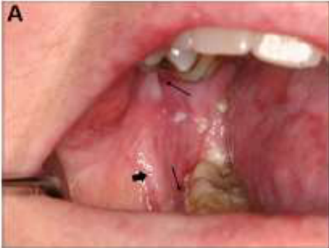 | 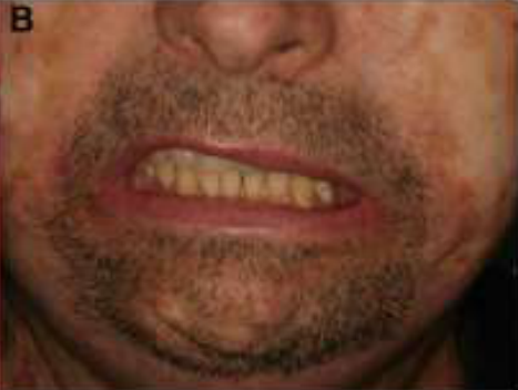 |
Oral SCC as a Complication of cGVHD48
Medical Management49
- Systemic management
- Topical immunomodulatory therapy:
- Topical corticosteroids (gels, ointments, rinses, puffers)
- Cyclosporin rinse or paste
- Tacrolimus and pimecrolimus (calcineurin inhibitors)
Sclerosis Management
Management of Oral cGVHD
- Physical therapy – low load passive stretching
- Intralesional steroid therapy
- Surgery to disrupt mucosal bands or strictures in focal areas
Dental Considerations
- Caries prevention (tailored)
- Minimise oral trauma and irritation
- Well-fitting dentures
- Smoothing of sharp teeth/fillings
- Avoidance of SLS, ETOH mouthwash
- Avoidance of dietary irritants
- Mucosal surveillance & biopsy as indicated
- Symptom palliation
- Topical analgesics
- Mucosal lubricants
- Sialagogues (avoid: pulmonary or gastrointestinal GvHD, significant comorbidities)
- Smoking cessation counselling; moderation of alcohol consumption
Lecturer — Treatment Goals
Treatment is generally directed toward symptom control and patient comfort rather than cure, because chronic GVHD may not be curable.
Haematopoietic stem cell and bone marrow transplants
Basic Oral Care50
Elad S., Raber-Durlacher J.E., Brennan M.T., Saunders D.P., Mank A.P., Zadik Y., Quinn B., Epstein J.B., Bijlevens N.M.A., Waltimo T., Passweg J.R., Correa M.E.P., Dahllöf G., Garming-Legert K.U.E., Logan R.M., Potting C.M.J., Shapira M.Y., Soga Y., Stringer J., Stokman M.A., Vokurka S., Wallbult E., Yarom N., Beier Jensen S. (2014). Basic oral care for hematology-oncology patients and hematopoietic stem cell transplantation recipients: a position paper from the joint task force of the Multinational Association of Supportive Care in Cancer/International Society of Oral Oncology (MASCC/ISOO) and the European Society for Blood and Marrow Transplantation (EBMT), Support Care Cancer, 23, 223-236.
Received: 24 May 2014 / Accepted: 30 July 2014 / Published online: 5 September 2014 © Springer-Verlag Berlin Heidelberg 2014
Hong C.H.L., Gueiros L.A., Fulton J.S., Cheng K.K.F., Kandwal A., Galiti D., Fall-Dickson J.M., Johansen J., Ameringer S., Kataoka T., Weikel D., Eilers J., Ranna V., Vaddi A., Lalla R.V., Bossi P., Elad S., on behalf of the Mucositis Study Group of MASCC/ISOO (2019). Systematic review of basic oral care for the management of oral mucositis in cancer patients and clinical practice guidelines, Support Care Cancer, 27, 3949-3967.
Received: 20 January 2019 / Accepted: 30 April 2019 / Published online: 8 July 2019 © Springer-Verlag GmbH Germany, part of Springer Nature 2019
Basic Oral Care Guidelines51
Abstract52
- Purpose: The aim of this study was to update the clinical practice guidelines for the use of basic oral care (BOC) interventions for the prevention and/or treatment of oral mucositis (OM).
- Methods: A systematic review was conducted by the Mucositis Study Group of MASCC/ISOO. The body of evidence for each intervention in each cancer treatment setting was assigned an evidence level. The findings were added to the database used to develop the 2013 MASCC/ISOO clinical practice guidelines. Based on the evidence level, one of the following three guideline determinations was possible: Recommendation, Suggestion, No guideline possible.
- Results: A total of 17 new papers across six interventions were examined and merged with a previous database. Based on the literature, the following guidelines were possible: the panel suggests that the implementation of multi-agent combination oral care protocols is beneficial for the prevention of OM during chemotherapy, head and neck (H&N) radiation therapy (RT), and hematopoietic stem cell transplantation (Level of Evidence III). The panel suggests that chlorhexidine not be used to prevent OM in patients undergoing H&N RT (Level of Evidence III). No guideline was possible for professional oral care, patient education, saline, and sodium bicarbonate, and expert opinion complemented these guidelines.
Lecturer — Transplant Complications
Patients undergoing transplantation are at risk of oral complications that may cause substantial morbidity and potentially contribute to mortality.
The Oral & Dental Management of Patients Before, During and After Cancer Therapy — Faculty of Dental Surgery, February 2026, v.10 — full guidance available at www.rcseng.ac.uk/dental-facilities/dfs/publications-guidelines/clinical-guidelines
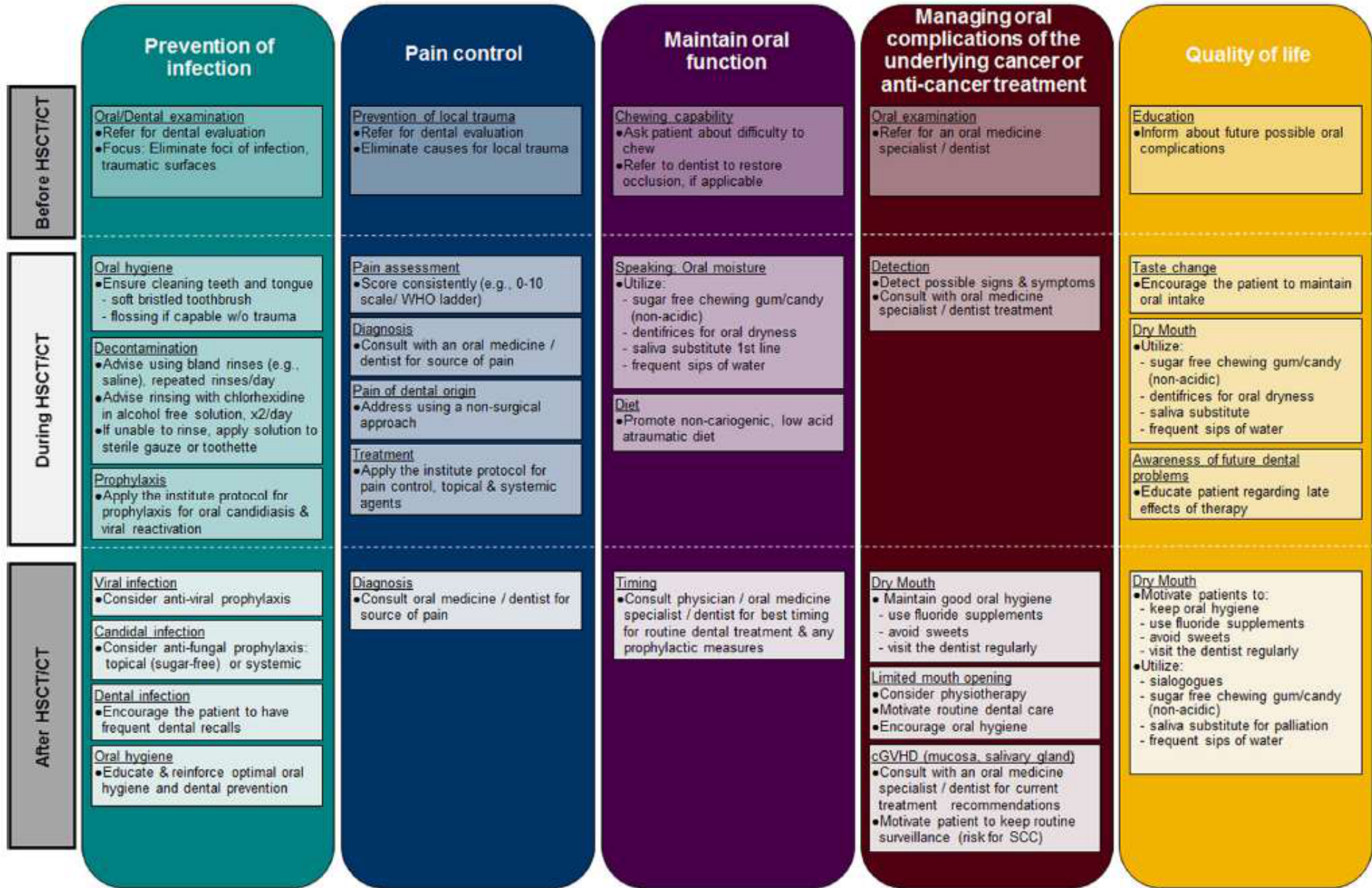
“the activities that should be part of the patient’s routine care during periods of cancer treatment in order to maintain good oral health and reduce the risk for local inflammation and infection, as well as systemic infection originating from the oral cavity”
Before HSCT/CT53
Prevention of infection — Oral/dental examination
- Refer for dental evaluation
- Focus: eliminate foci of infection, traumatic surfaces
Pain control — Prevention of local trauma
- Refer for dental evaluation
- Eliminate causes for local trauma
Maintain oral function — Chewing capability
- Ask patient about difficulty to chew
- Refer to dentist to restore occlusion, if applicable
Managing oral complications of the underlying cancer or anti-cancer treatment — Oral examination
- Refer for an oral medicine specialist / dentist
Quality of life — Education
- Inform about future possible oral complications
During HSCT/CT
Oral hygiene
- Ensure cleaning teeth and tongue — soft bristled toothbrush — flossing if capable w/o trauma
Decontamination
- Advise using bland rinses (e.g., saline), repeated rinses/day
- Advise rinsing with chlorhexidine in alcohol-free solution, x2/day
- If unable to rinse, apply solution to sterile gauze or toothette
Prophylaxis
- Apply the institute protocol for prophylaxis for oral candidiasis & viral reactivation
Pain assessment
- Score consistently (e.g., 0-10 scale / WHO ladder)
Diagnosis
- Consult with an oral medicine specialist / dentist for source of pain
Pain of dental origin
- Address using a non-surgical approach
Treatment
- Apply the institute protocol for pain control, topical & systemic agents
Speaking, oral moisture
- Utilize:
- sugar-free chewing gum/candy (non-acidic)
- dentifrices for oral dryness
- saliva substitute (1st line)
- frequent sips of water
Diet
- Promote non-cariogenic, low-acid atraumatic diet
Care Before HSCT
Detection
- Detect possible signs & symptoms
- Consult with oral medicine specialist / dentist for treatment
Taste change
- Encourage the patient to maintain oral intake
Dry mouth
- Utilize:
- sugar-free chewing gum/candy (non-acidic)
- dentifrices for oral dryness
- saliva substitute
- frequent sips of water
Awareness of future dental problems
- Educate patient regarding late effects of therapy
After HSCT/CT
Viral infection
- Consider anti-viral prophylaxis
Candidal infection
- Consider anti-fungal prophylaxis: topical (sugar-free) or systemic
Dental infection
- Encourage the patient to have frequent dental recalls
Oral hygiene
- Educate & reinforce optimal oral hygiene and dental prevention
Diagnosis
- Consult oral medicine specialist / dentist for source of pain
Timing
- Consult physician / oral medicine specialist / dentist for best timing for routine dental treatment & any prophylactic measures
Dry mouth
- Motivate patients to:
- keep oral hygiene
- use fluoride supplements
- avoid sweets
- visit the dentist regularly
- Utilize:
- sialogogues
- sugar-free chewing gum/candy (non-acidic)
- saliva substitute for palliation
- frequent sips of water
Limited mouth opening
- Consider physiotherapy
- Motivate routine dental care
- Encourage oral hygiene
cGVHD (mucosa, salivary gland)
- Consult with an oral medicine specialist / dentist for current treatment recommendations
- Motivate patient to keep routine surveillance (risk for SCC)
Elad S., et al. Basic oral care for hematology-oncology patients and hematopoietic stem cell transplantation recipients: a position paper from the joint task force of the Multinational Association of Supportive Care in Cancer/International Society of Oral Oncology (MASCC/ISOO) and the European Society for Blood and Marrow Transplantation (EBMT). Support Care Cancer. 2015 Jan;23(1):223-36.
Elad S., et al. Basic oral care for hematology-oncology patients and hematopoietic stem cell transplantation recipients: a position paper from the joint task force of the Multinational Association of Supportive Care in Cancer/International Society of Oral Oncology (MASCC/ISOO) and the European Society for Blood and Marrow Transplantation (EBMT). Support Care Cancer. 2015 Jan;23(1):223-36.
Before HSCT54
- Oral/dental examination: eliminate foci of infection and trauma ASAP to allow time for treatment and healing (ideally 2 weeks)
Lecturer — Dental Planning
Dental planning should account for the patient’s neutropenia, thrombocytopenia, anaemia, conditioning schedule, potential treatment delays, and ability to heal.
During HSCT
-
If oral hygiene is compromised, add CHX as an oral antiseptic
-
Brushing should be continued 2-3 times daily with ultra-soft or soft toothbrush, bristles softened in hot water
-
Brushing with sponge brushes is optional, but less effective in removing plaque (though the addition of CHX increases effectiveness)
-
Air-dry toothbrush between uses
-
Toothbrush replaced regularly (e.g. after each neutropenic cycle)
-
Measures to remove plaque from between teeth, C&Bs or implants, and from the tongue should be continued using devices familiar to the patient to avoid self-injury
-
A surgically soft toothbrush may be useful for severe mucositis or fragile oral mucosa because its bristles are softer than those generally available in supermarkets.
Lecturer — Interdental Cleaning
Patients who are not already familiar with flossing or interdental brushes should not necessarily begin these methods during severe thrombocytopenia. Self-injury may occur.
After HSCT
-
May need to avoid invasive dental treatment
-
Aerosol-generating procedures (e.g. S/C with u/s scaler) should be minimised if immunosuppressed
-
Immune reconstitution may take months after HSCT (even when haematological status is apparently normal)
-
Ultrasonic scaling aerosolises bacterial biofilm from the teeth and is generally avoided while the immune system is recovering.
-
Appropriate vaccination and medical follow-up should continue after transplantation. Haematopoietic stem cell and bone marrow transplants
Dental Work-Up Prior to Transplant55
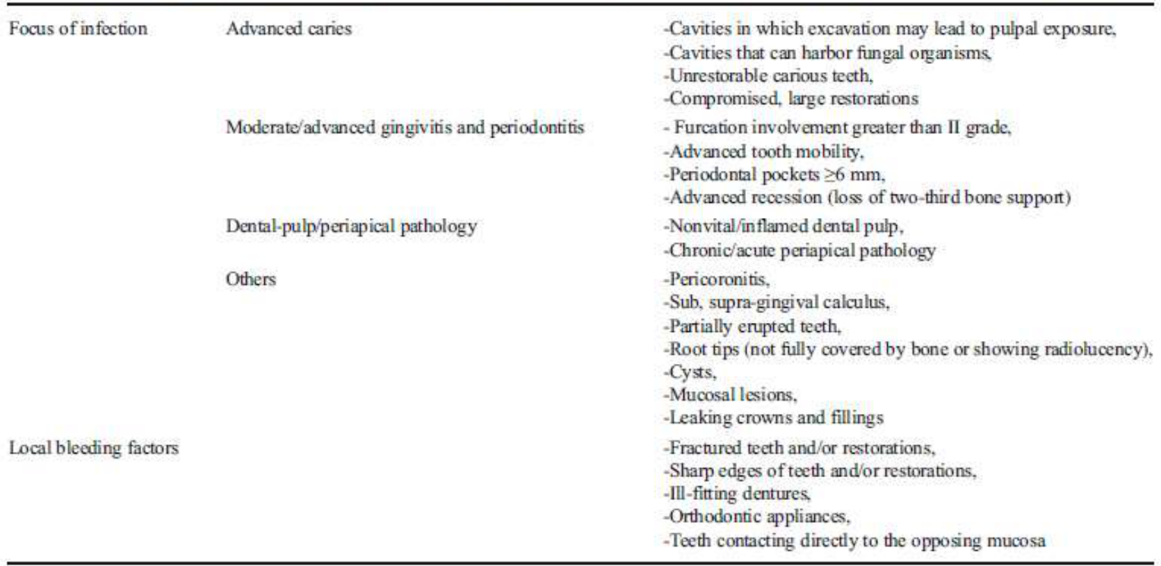
Foci of infection
- Advanced caries: cavities in which excavation may lead to pulpal exposure; cavities that can harbor fungal organisms; unrestorable carious teeth; compromised, large restorations
- Moderate/advanced gingivitis and periodontitis: furcation involvement greater than grade II; advanced tooth mobility; periodontal pockets ≥6 mm; advanced recession (loss of two-thirds bone support)
- Dental-pulp/periapical pathology: nonvital/inflamed dental pulp; chronic/acute periapical pathology
- Others: pericoronitis; sub-/supra-gingival calculus; partially erupted teeth; root tips (not fully covered by bone or showing radiolucency); cysts; mucosal lesions; leaking crowns and fillings
Local bleeding factors
Oral Foci of Infection of Concern56
-
Fractured teeth and/or restorations
-
Sharp edges of teeth and/or restorations
-
Ill-fitting dentures
-
Orthodontic appliances
-
Teeth contacting directly with the opposing mucosa
-
Non-healing extraction sites
-
Potential sources of infection during acute immunosuppression Boguslawska-Kapala et al. (2017). Oral health of adult patients undergoing hematopoietic cell transplantation. Pre-transplant assessment and care, Support Care Cancer, 96, 1135-1145.
Lecturer — Treatment Priorities
The dental work-up focuses on urgent problems rather than necessarily completing all treatment before transplantation.
- Treatment may need to be limited when transplantation is urgent, the patient is medically unstable, or cytopenias make procedures unsafe.
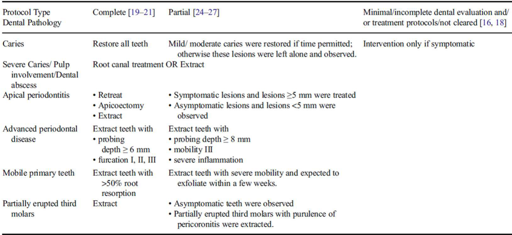
- Delayed healing and the risks of treatment may outweigh the risks of observing the dental condition.
| Dental pathology | Complete [19-21] | Partial [24-27] | Minimal/incomplete dental evaluation and/or treatment protocols / not cleared [16, 18] |
|---|---|---|---|
| Caries | Restore all teeth | Mild/moderate caries were restored if time permitted; otherwise these lesions were left alone and observed. | Intervention only if symptomatic |
| Severe caries / pulp involvement / dental abscess | Root canal treatment OR extract | ||
| Apical periodontitis | Retreat; apicoectomy; extract | Symptomatic lesions and lesions ≥5 mm were treated; asymptomatic lesions and lesions <5 mm were observed | |
| Advanced periodontal disease | Extract teeth with probing depth ≥6 mm, furcation I, II, III | Extract teeth with probing depth ≥8 mm, mobility III, severe inflammation | |
| Mobile primary teeth | Extract teeth with >50% root resorption | Extract teeth with severe mobility expected to exfoliate within a few weeks | |
| Partially erupted third molars | Extract | Asymptomatic teeth were observed; partially erupted third molars with purulence of pericoronitis were extracted |
Time-Limited and Alternative Dental Protocols57
Hong et al. (2018). A systematic review of dental disease management in cancer patients, Support Care Cancer, 26, 155-174.
Lecturer — Time-Limited Treatment
When treatment time is limited, prioritise acute infection, pain, and bleeding risks, and consider watchful waiting for less urgent disease.
- Treat only if the patient becomes symptomatic when definitive treatment cannot be safely completed.
- Coordinate procedures with the medical team and consider platelet support where appropriate.
- Platelet transfusions may not produce a sufficient platelet increment in patients with HLA-related issues.
- Weigh dental infection risk against poor healing, bleeding, or delaying transplantation.
[!example] Dental Management in Severe Aplastic Anaemia
A man in his early 30s with aplastic anaemia was assessed approximately five months after 15 teeth had been extracted under general anaesthesia for extensive dental decay.
- He had very poor oral hygiene, brushed his teeth only every few months, and had plaque-related periodontal disease with gingival bleeding.
- Tooth 25 had grade-three mobility, and the extraction socket at tooth 37 had not healed five months after extraction.
- He had significant pancytopenia and a platelet count of approximately 8.
- Ordinarily, tooth 25 would have been extracted before transplantation, but the platelet count was too low for safe extraction. Some guidelines use 30 as a threshold, while 50 was preferred in this clinical setting.
- Platelets can be transfused immediately before or during dental treatment because they sequester rapidly. In this case, HLA-matching issues prevented a satisfactory platelet increment and the count could not be raised above 30.
- The non-healed extraction socket raised concern that another extraction might also fail to heal and delay transplantation.
- The case illustrates that avoiding an invasive procedure can be safer than attempting treatment that may cause severe bleeding, poor healing, or delay transplantation.
Closing and References58
Thank you.
Dr Jee-Yun Leung | jeeyun.leung@uwa.edu.au
- Triester N, et al (2012). How we treat oral chronic graft-versus-host disease. Blood 120(17): 3407-3418.
- Hull K, et al (2015). Oral chronic graft-versus-host disease in Australia: clinical features and challenges in management. International Medicine Journal 45(7): 702-710.
- Yeoh, S-C et al (2019). Oral manifestations of systemic diseases and their treatments. Contemporary oral medicine, ed. Farah, C. S et al. Switzerland, Springer International, pp. 1523-1639.
- Elad S, et al. Basic oral care for hematology-oncology patients and hematopoietic stem cell transplantation recipients: a position paper from the joint task force of the Multinational Association of Supportive Care in Cancer/International Society of Oral Oncology (MASCC/ISOO) and the European Society for Blood and Marrow Transplantation (EBMT). Support Care Cancer. 2015 Jan;23(1):223-36.
- Hong CHL, et al. Systematic review of basic oral care for the management of oral mucositis in cancer patients and clinical practice guidelines. Support Care Cancer. 2019 Oct;27(10):3949-3967.
- Kumar N, et al. The Oral Management of Oncology Patients Requiring Radiotherapy, Chemotherapy and/or Bone Marrow Transplantation. London: RCS; 2026. Available from: https://www.rcseng.ac.uk/dental-faculties/fds/publications-guidelines/clinical-guidelines/
Lecturer — Transplant Dental Care
Stem cell transplant patients are complex, and dental care must be adapted throughout the transplant journey.
- Disease status
- Cytopenias
- Immunosuppression
- Conditioning regimen
- Timing within the transplant journey
- Risk of infection
- Risk of bleeding
- Healing capacity
- GVHD
- Long-term oral complications
References59
Clinical Cases
Case: Pre-transplant dental assessment in a patient with severe thrombocytopenia
Question
Scenario: A gentleman in his early 30s with aplastic anemia is evaluated for dental clearance prior to a stem cell transplant. He has a history of poor oral hygiene and underwent 15 extractions under general anesthesia five months ago for extensive decay.
What’s shown: Clinical examination reveals poor oral hygiene with florid plaque, periodontal disease, and bleeding. Tooth 25 is grade 3 mobile. There is a non-healed extraction socket at tooth 37 from the extractions five months ago. Blood tests reveal severe pancytopenia with a platelet count of 8.
Consider: How should the grade 3 mobile tooth 25 be managed in this patient prior to his stem cell transplant?
Answer
Observations:
- Poor oral hygiene with florid plaque, periodontal disease, and bleeding.
- Tooth 25 is grade 3 mobile.
- Non-healed extraction socket at tooth 37 from five months prior.
- Severe pancytopenia with a platelet count of 8.
Reasoning:
- Normally, a grade 3 mobile tooth would be extracted prior to a stem cell transplant to remove a focus of infection.
- However, the patient’s platelet count is only 8 (guidelines suggest 30, and the lecturer prefers 50 for extractions).
- The patient cannot achieve adequate platelet increments with transfusions due to HLA matching issues (maximum count achieved is 30).
- There is a high risk of poor wound healing, as evidenced by the non-healed socket at tooth 37, which could delay his stem cell transplant.
- The risks of bleeding and non-healing from an extraction outweigh the risks of leaving a potential source of infection during acute immunosuppression.
Outcome:
- The extraction of tooth 25 was not performed. Dental treatment was deferred because the risks of the procedure and subsequent non-healing were deemed worse than the risk of infection.
Takeaway:
- When a patient has severe, uncorrectable thrombocytopenia and a demonstrated history of poor wound healing, the risks of invasive dental treatment may outweigh the risks of retaining potential foci of infection prior to a stem cell transplant.
Footnotes
-
Original PDF page 1: L9.1 Transplant Medicine 1 HSCT - DMD3 DENT5311, p.1 ↩
-
Original PDF page 2: L9.1 Transplant Medicine 1 HSCT - DMD3 DENT5311, p.2 ↩
-
Original PDF page 3: L9.1 Transplant Medicine 1 HSCT - DMD3 DENT5311, p.3 ↩
-
Original PDF page 4: L9.1 Transplant Medicine 1 HSCT - DMD3 DENT5311, p.4 ↩
-
Original PDF page 5: L9.1 Transplant Medicine 1 HSCT - DMD3 DENT5311, p.5 ↩
-
Original PDF page 6: L9.1 Transplant Medicine 1 HSCT - DMD3 DENT5311, p.6 ↩
-
Original PDF page 7: L9.1 Transplant Medicine 1 HSCT - DMD3 DENT5311, p.7 ↩
-
Original PDF page 8: L9.1 Transplant Medicine 1 HSCT - DMD3 DENT5311, p.8 ↩
-
Original PDF page 9: L9.1 Transplant Medicine 1 HSCT - DMD3 DENT5311, p.9 ↩
-
Original PDF page 10: L9.1 Transplant Medicine 1 HSCT - DMD3 DENT5311, p.10 ↩
-
Original PDF page 11: L9.1 Transplant Medicine 1 HSCT - DMD3 DENT5311, p.11 ↩
-
Original PDF page 13: L9.1 Transplant Medicine 1 HSCT - DMD3 DENT5311, p.13 ↩
-
Original PDF page 12: L9.1 Transplant Medicine 1 HSCT - DMD3 DENT5311, p.12 ↩
-
Original PDF page 14: L9.1 Transplant Medicine 1 HSCT - DMD3 DENT5311, p.14 ↩
-
Original PDF page 15: L9.1 Transplant Medicine 1 HSCT - DMD3 DENT5311, p.15 ↩
-
Original PDF page 16: L9.1 Transplant Medicine 1 HSCT - DMD3 DENT5311, p.16 ↩
-
Original PDF page 17: L9.1 Transplant Medicine 1 HSCT - DMD3 DENT5311, p.17 ↩
-
Original PDF page 18: L9.1 Transplant Medicine 1 HSCT - DMD3 DENT5311, p.18 ↩
-
Original PDF page 19: L9.1 Transplant Medicine 1 HSCT - DMD3 DENT5311, p.19 ↩
-
Original PDF page 20: L9.1 Transplant Medicine 1 HSCT - DMD3 DENT5311, p.20 ↩
-
Original PDF page 23: L9.1 Transplant Medicine 1 HSCT - DMD3 DENT5311, p.23 ↩
-
Original PDF page 21: L9.1 Transplant Medicine 1 HSCT - DMD3 DENT5311, p.21 ↩
-
Original PDF page 22: L9.1 Transplant Medicine 1 HSCT - DMD3 DENT5311, p.22 ↩
-
Original PDF page 24: L9.1 Transplant Medicine 1 HSCT - DMD3 DENT5311, p.24 ↩
-
Original PDF page 25: L9.1 Transplant Medicine 1 HSCT - DMD3 DENT5311, p.25 ↩
-
Original PDF page 26: L9.1 Transplant Medicine 1 HSCT - DMD3 DENT5311, p.26 ↩
-
Original PDF page 27: L9.1 Transplant Medicine 1 HSCT - DMD3 DENT5311, p.27 ↩
-
Original PDF page 28: L9.1 Transplant Medicine 1 HSCT - DMD3 DENT5311, p.28 ↩
-
Original PDF page 29: L9.1 Transplant Medicine 1 HSCT - DMD3 DENT5311, p.29 ↩
-
Original PDF page 30: L9.1 Transplant Medicine 1 HSCT - DMD3 DENT5311, p.30 ↩
-
Original PDF page 31: L9.1 Transplant Medicine 1 HSCT - DMD3 DENT5311, p.31 ↩
-
Original PDF page 33: L9.1 Transplant Medicine 1 HSCT - DMD3 DENT5311, p.33 ↩
-
Original PDF page 32: L9.1 Transplant Medicine 1 HSCT - DMD3 DENT5311, p.32 ↩
-
Original PDF page 34: L9.1 Transplant Medicine 1 HSCT - DMD3 DENT5311, p.34 ↩
-
Original PDF page 35: L9.1 Transplant Medicine 1 HSCT - DMD3 DENT5311, p.35 ↩
-
Original PDF page 36: L9.1 Transplant Medicine 1 HSCT - DMD3 DENT5311, p.36 ↩
-
Original PDF page 37: L9.1 Transplant Medicine 1 HSCT - DMD3 DENT5311, p.37 ↩
-
Original PDF page 38: L9.1 Transplant Medicine 1 HSCT - DMD3 DENT5311, p.38 ↩
-
Original PDF page 39: L9.1 Transplant Medicine 1 HSCT - DMD3 DENT5311, p.39 ↩
-
Original PDF page 40: L9.1 Transplant Medicine 1 HSCT - DMD3 DENT5311, p.40 ↩
-
Original PDF page 41: L9.1 Transplant Medicine 1 HSCT - DMD3 DENT5311, p.41 ↩
-
Original PDF page 42: L9.1 Transplant Medicine 1 HSCT - DMD3 DENT5311, p.42 ↩
-
Original PDF page 43: L9.1 Transplant Medicine 1 HSCT - DMD3 DENT5311, p.43 ↩
-
Original PDF page 44: L9.1 Transplant Medicine 1 HSCT - DMD3 DENT5311, p.44 ↩
-
Original PDF page 45: L9.1 Transplant Medicine 1 HSCT - DMD3 DENT5311, p.45 ↩
-
Original PDF page 46: L9.1 Transplant Medicine 1 HSCT - DMD3 DENT5311, p.46 ↩
-
Original PDF page 47: L9.1 Transplant Medicine 1 HSCT - DMD3 DENT5311, p.47 ↩
-
Original PDF page 48: L9.1 Transplant Medicine 1 HSCT - DMD3 DENT5311, p.48 ↩
-
Original PDF page 49: L9.1 Transplant Medicine 1 HSCT - DMD3 DENT5311, p.49 ↩
-
Original PDF page 50: L9.1 Transplant Medicine 1 HSCT - DMD3 DENT5311, p.50 ↩
-
Original PDF page 52: L9.1 Transplant Medicine 1 HSCT - DMD3 DENT5311, p.52 ↩
-
Original PDF page 51: L9.1 Transplant Medicine 1 HSCT - DMD3 DENT5311, p.51 ↩
-
Original PDF page 53: L9.1 Transplant Medicine 1 HSCT - DMD3 DENT5311, p.53 ↩
-
Original PDF page 54: L9.1 Transplant Medicine 1 HSCT - DMD3 DENT5311, p.54 ↩
-
Original PDF page 55: L9.1 Transplant Medicine 1 HSCT - DMD3 DENT5311, p.55 ↩
-
Original PDF page 56: L9.1 Transplant Medicine 1 HSCT - DMD3 DENT5311, p.56 ↩
-
Original PDF page 57: L9.1 Transplant Medicine 1 HSCT - DMD3 DENT5311, p.57 ↩
-
Original PDF page 58: L9.1 Transplant Medicine 1 HSCT - DMD3 DENT5311, p.58 ↩
-
Original PDF page 59: L9.1 Transplant Medicine 1 HSCT - DMD3 DENT5311, p.59 ↩