Cardiovascular Disease and Dentistry1
DR JEE-YUN LEUNG
Learning Outcomes2
PART 1
- Review common vascular, valvular, electrical, muscular and congenital cardiac conditions and their management (e.g. hypertension, coronary artery disease, rheumatic heart disease, infective endocarditis, arrhythmias, congenital structural defects, heart failure)
- Describe potential oral findings associated with cardiac conditions
- Describe the oral manifestations and manage the risks associated with common treatments used to manage cardiac conditions
- Understand the classification of cardiac conditions and their dental implications.
- Identify risk factors, manifestations, and management strategies for hypertension and ischaemic heart disease.
- Recognize the relationship between oral health and conditions such as infective endocarditis and rheumatic heart disease.
- Manage dental treatment for patients with electrical dysrhythmias and cardiac electronic devices.
- Assess the associated risks of common dental procedures and modify dental treatment appropriately to safely provide care
PART 2
- Work a patient up prior to cardiac surgery
- Manage angina or an acute coronary syndrome in dental practice
- Evaluate the risks and benefits of preoperative dental treatment for patients undergoing cardiac surgery.

Classification of Cardiac Conditions34
COMMON CARDIAC CONDITIONS
Hypertension, Coronary Artery Disease, Rheumatic Heart Disease, Infective Endocarditis, Arrhytmias, Congenital Structural Defects, Heart Failure
WHO INTERNATIONAL CLASSIFICATION OF DISEASES - 11TH REVISION
11 Diseases of the circulatory system
- Hypertensive diseases
- Hypotension
- Ischaemic heart diseases
- Diseases of coronary artery
- Pulmonary heart disease or diseases of pulmonary circulation
- Pericarditis
- Acute or subacute endocarditis
- Heart valve diseases
- BC20 Chronic rheumatic heart diseases, not elsewhere classified
- Diseases of the myocardium or cardiac chambers
- Cardiac arrhythmia
- Heart failure
- Diseases of arteries or arterioles
- Diseases of veins
Disorders of lymphatic vessels or lymph nodes Postprocedural disorders of circulatory system Neoplasms of the circulatory system Developmental anomalies of the circulatory system Infections of the circulatory system Symptoms, signs or clinical findings of the circulatory system Cerebrovascular diseases Functional vascular disorders of the skin JB64.4 Diseases of the circulatory system complicating pregnancy, childbirth or the puerperium
BE2Y Other specified diseases of the circulatory system BE2Z Diseases of the circulatory system, unspecified
Types of Cardiac Conditions5
Vascular / Ischaemic
- Hypertension
- Hypotension
- Ischaemic heart disease (Coronary artery disease) – angina, myocardial infarction
- Thromboembolic complications of CVD
Valvular
- Rheumatic fever and Rheumatic heart disease
- Infective endocarditis
- Aortic and mitral valve stenosis, regurgitation and prolapse
Electrical (Dysrhythmias)
- Extrasystoles (ectopics)
- Tachycardia (including Atrial fibrillati
- Disturbances in heart rate or rhythm, such as atrial fibrillation.on)
- Bradycardias
Muscular (cardiomyopathies)
- Primary – genetic/inherited, acquired
- Secondary
- Diseases of the heart muscle affecting pumping ability.
Congenital
- Cyanotic
- Acyanotic
- Structural defects present from birth.
Heart Failure
Definition
Structural or functional impairment where blood output is insufficient for body demands.
Vascular and Ischaemic Conditions6
- Hypotension
- Hypertension
- Ischaemic heart disease (Coronary artery disease) – angina, myocardial infarction
- Thromboembolic complications of CVD
Hypotension Definitions and Aetiology7
- Hypotension (chronic arterial hypotension) = systolic BP <100 mmHg
- Pre-syncope (“near-fainting”) = condition in which a patient feels syncope is imminent
- Syncope (“fainting”) = acute fall in blood pressure, resulting in transient global cerebral hypoperfusion, causing a loss of consciousness which usually leads to loss of postural tone and falling
- Vasovagal syncope = reaction to pain, or to anxiety and fear before, during or after a dental procedure
- Orthostatic hypotension = fall in blood pressure when standing up after sitting or lying down for an extended period of time in the dental chair
Causes8
- Cardiac conditions: ability for heart to pump blood (e.g. abnormal heart rhythm, heart failure, MI)
- Autonomic nervous system: involved in returning BP to normal (e.g. Parkinson’s disease)
- Long-term bed rest (e.g. hospitalised, pregnancy)
- Low blood volume (e.g. dehydration, anaemia, injury)
- Medications (e.g. antihypertensives, antidepressants)
Table 5.12 Causes of orthostatic hypotension
| Primary autonomic causes | Secondary autonomic causes | Non-autonomic causes |
|---|---|---|
| Familial dysautonomia (Riley-Day syndrome) | B vitamin deficiency | Hypovolaemia |
| Pure autonomic failure (idiopathic orthostatic hypotension) | Alcoholism | Ageing |
| Shy-Drager syndrome | Diabetes | Prolonged bed rest |
| Dopamine beta-hydroxylase deficiency | Parkinsonism | Pregnancy |
| Porphyrin | Drugs (e.g. antihypertensives) |
Scully C. Scully’s medical problems in dentistry. 7th ed. Edinburgh: Churchill Livingstone/Elsevier; 2014
General Manifestations9
- Early symptoms: vertigo, dizziness, visual blurring, headache, syncope
- Later: tachycardia, paleness, cold extremities, sweating
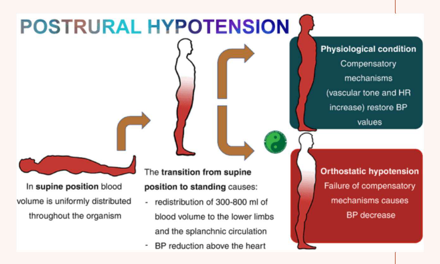 |  |
Postural Hypotension
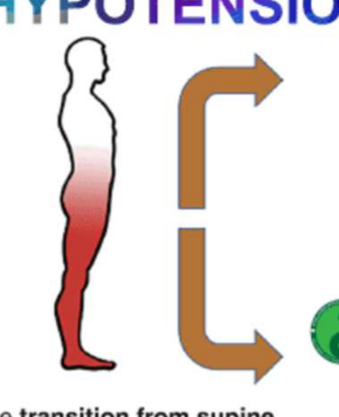
In supine position blood volume is uniformly distributed throughout the organism. The transition from supine position to standing causes:
- redistribution of 300-800 ml of blood volume to the lower limbs and the splanchnic circulation
- BP reduction above the heart
Physiological condition: Compensatory mechanisms (vascular tone and HR increase) restore BP values
Orthostatic hypotension: Failure of compensatory mechanisms causes BP decrease
General Management of Hypotension
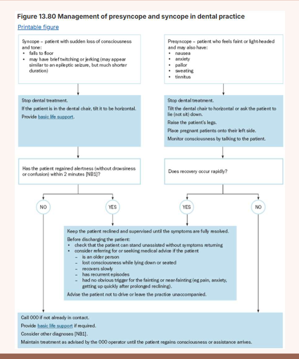
Syncope – patient with sudden loss of consciousness and tone10
- falls to floor
- may have brief twitching or jerking (may appear similar to an epileptic seizure, but much shorter duration)
Stop dental treatment. If the patient is in the dental chair, tilt it to be horizontal. Provide basic life support.
- If a patient does not respond, raise their legs and call for emergency help.
Has the patient regained alertness (without drowsiness or confusion) within 2 minutes [NB1]?
NO:
- Call 000 if not already in contact.
- Provide basic life support if required.
- Consider other diagnoses [NB1].
- Maintain treatment as advised by the 000 operator until the patient regains consciousness or assistance arrives.
YES:
- Keep the patient recumbent and supervised until the symptoms are fully resolved.
- Before discharging the patient:
- check that the patient can stand unassisted without symptoms returning
- consider referring for or seeking medical advice if the patient:
- is an older person
- lost consciousness while lying down or seated
- recovers slowly
- has recurrent episodes
- had no obvious trigger for the fainting or near-fainting (e.g. pain, anxiety, getting up quickly after prolonged reclining)
- Advise the patient not to drive or leave the practice unaccompanied.
Presyncope – patient who feels faint or light-headed and may also have
- nausea
- anxiety
- pallor
- sweating
- tinnitus
Stop dental treatment. Tilt the dental chair to horizontal or ask the patient to lie (not sit) down. Raise the patient’s legs. Place pregnant patients onto their left side. Monitor consciousness by talking to the patient.
Does recovery occur rapidly?
YES:
- Keep the patient recumbent and supervised until the symptoms are fully resolved.
- Before discharging the patient:
- check that the patient can stand unassisted without symptoms returning
- consider referring for or seeking medical advice if the patient:
- is an older person
- lost consciousness while lying down or seated
- recovers slowly
- has recurrent episodes
- had no obvious trigger for the fainting or near-fainting (e.g. pain, anxiety, getting up quickly after prolonged reclining)
- Advise the patient not to drive or leave the practice unaccompanied.
NO:
- Call 000 if not already in contact.
- Provide basic life support if required.
- Consider other diagnoses [NB1].
- Maintain treatment as advised by the 000 operator until the patient regains consciousness or assistance arrives.
NB1: Many people with chest pain do not have angina or a heart attack, but anyone with acute chest pain requires rapid medical evaluation for potentially life-threatening causes. The likelihood of angina or heart attack increases with the number of suggestive features.
NB2: Sublingual glyceryl trinitrate (GTN) dosing is a 400 microgram spray (or 300 to 600 microgram tablet) generally administered every 5 minutes to a maximum of 3 doses. Ask patients who use GTN to bring their GTN to their dental appointment so that it can be administered if chest pain occurs.
NB3: If symptoms (pain or other features that may indicate angina) last longer than the patient’s usual angina symptoms, urgent medical evaluation of a possible heart attack or other urgent condition is required.
Hypertension Classification and Aetiology11
Essential (primary) hypertension (95%): High blood pressure for which a secondary cause cannot be found.
Secondary hypertension (5%): Systolic BP >140mmHg and diastolic BP >90mmHg with an identifiable cause.
National Heart Foundation of Australia – Classification of Blood Pressure12
| Diagnostic category* | Systolic (mmHg) | Diastolic (mmHg) |
|---|---|---|
| Optimal | <120 and <80 | - |
| Normal | 120–129 and/or 80–84 | - |
| High-normal | 130–139 and/or 85–89 | - |
| Grade 1 (mild) hypertension | 140–159 and/or 90–99 | - |
| Grade 2 (moderate) hypertension | 160–179 and/or 100–109 | - |
| Grade 3 (severe) hypertension | ≥180 and/or ≥110 | - |
| Isolated systolic hypertension | >140 and <90 | - |
*When a patient’s systolic and diastolic blood pressure levels fall into different categories, the higher diagnostic category and recommended actions apply.
Essential Hypertension13
- Genetic predisposition
- High basal metabolic index
- Sympathetic overactivity (40% have raised catecholamines and noradrenaline)
- High alcohol, salt intake
Secondary Hypertension
- Renal disease (80%)
- Endocrine conditions: pregnancy, Cushing syndrome, corticosteroids, hyperaldosteronism, acromegaly
- Cerebral disease: cerebral oedema (mainly strokes, head injuries or tumours)
- Coarctation of aorta
- Sleep apnoea
- Drugs: oral contraceptive pill, HRT, corticosteroids, NSAIDs, sympathomimetics (decongestants, diet pills, cocaine), stimulants (amphetamine), cyclosporine / tacrolimus, excess alcohol, SNRIs and MAOIs, haemopoietic drugs
Epidemiology14
- Australian Institute of Health and Welfare
- 39% of Australians >18yo have hypertension (~7.2 million adults)
- Of adults with hypertension:
- 80-82% of those <54yo had uncontrolled hypertension
- 65% had been dispensed antihypertensive medications
- Hypertensive disease contributed to 13% of all Australian deaths in 2023
- Hypertension is disproportionately high in First Nations adults.
- Nearly 1 in 20 (5%) Australian women aged 15-44 giving birth had new onset hypertension in pregnancy
- 24% of ATSI adults >18yo had hypertension 2022-2023
- Source: https://www.aihw.gov.au/reports/risk-factors/hypertension/contents/summary
| Primary | Complications |
|---|---|
| Usually asymptomatic | Arteriosclerosis |
| Advanced cases: headache, blurred vision, tinnitus, fatigue, dizziness, angina | Cerebrovascular disease (stroke) |
| Peripheral vascular disease | |
| Hypertensive retinopathy | |
| Hypertensive heart disease | |
| Hypertensive renal disease | |
| Hypertensive crisis |
Clinical Manifestations
The disease itself has few oral manifestations, with the exception of facial palsy during a hypertensive crisis.
General Management of Hypertension1516
- Identify and manage risk factors: physical activity, smoking cessation, weight control, diet, alcohol intake, etc.
- Assess presence/absence of organ damage
- Pharmacologic strategies: most widely used first-line ACEIs and ARBs
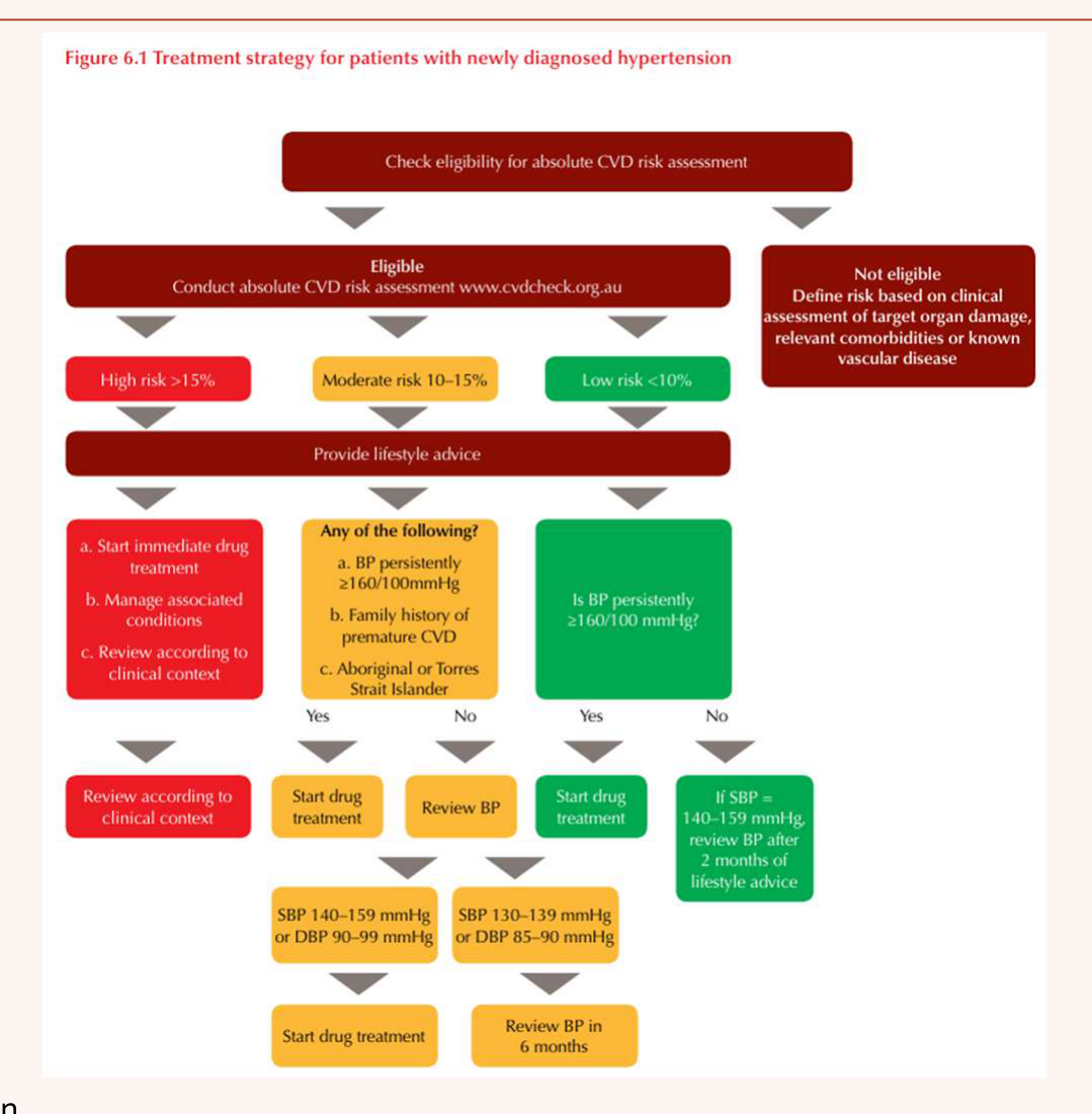
FIGURE 6.1 Treatment strategy for patients with newly diagnosed hypertension
Source: https://www.heartfoundation.org.au/for-professionals/hypertension
Effective Drug Combinations17
| First drug | Second drug | Comment |
|---|---|---|
| Effective combination | ||
| ACE inhibitor or ARB* | plus Calcium channel blocker | Particularly useful in presence of diabetes and/or lipid abnormalities1,24 |
| ACE inhibitor or ARB* | plus Thiazide diuretic | Useful in presence of heart failure or post stroke |
| ACE inhibitor or ARB* | plus Beta-blocker | Recommended post myocardial infarction or in patients with heart failure2 |
| Beta-blocker | plus Dihydropyridine calcium channel blocker | Useful in presence of symptomatic coronary heart disease |
| Thiazide diuretic | plus Calcium channel blocker | |
| Thiazide diuretic | plus Beta-blocker | Not recommended in presence of glucose intolerance, metabolic syndrome or established diabetes |
| Combinations to use with care | ||
| Diltiazem | plus Beta-blocker | Due to risk of heart block, but risk is less than with verapamil |
| ACE inhibitor or ARB | plus Potassium-sparing diuretic | Due to risk of hyperkalaemia |
| Combinations to avoid | ||
| ACE inhibitor | plus ARB | Increased risk of renal dysfunction1,20 |
| Verapamil | plus Beta-blocker | Due to risk of heart block |
- ACE, angiotensin-converting enzyme; ARB, angiotensin receptor blocker a In head-to-head trials ACE inhibitors and ARB’s are equally effective in blood pressure reduction and prevention of cardiovascular events overall, however may have important differences in their efficacy, so that they are not interchangeable, in some clinical conditions.
- Carvedol; bisoprolol (beta-1 selective antagonist); metoprolol extended release (beta-1 selective antagonist); nebivolol.
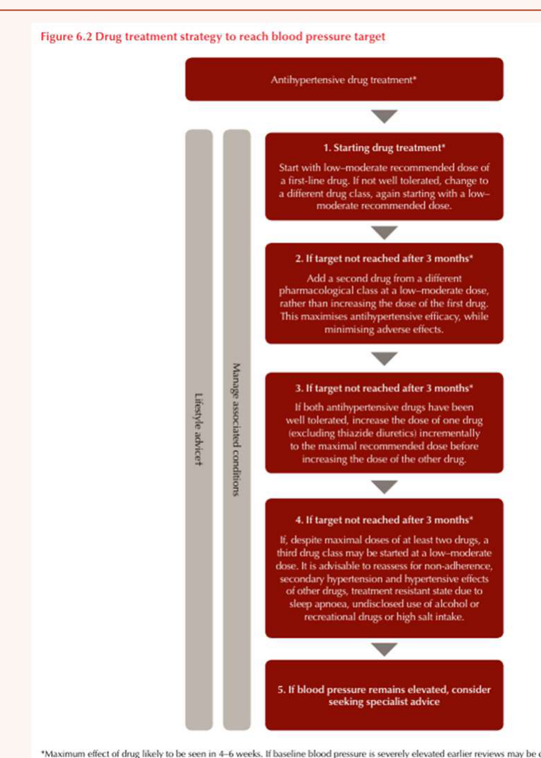
Antihypertensive Drug Treatment – Manage Associated Conditions
- Starting drug treatment: Start with low-moderate recommended dose of a first-line drug. If not well tolerated, change to a different drug class, again starting with a low-moderate recommended dose.
- If target not reached after 3 months: Add a second drug from a different pharmacological class at a low-moderate dose, rather than increasing the dose of the first drug. This maximizes antihypertensive efficacy, while minimising adverse effects.
- If target not reached after 3 months: If both antihypertensive drugs have been well tolerated, increase the dose of one drug (excluding thiazide diuretic) incrementally to the maximal recommended dose before increasing the dose of the other drug.
- If target not reached after 3 months: If, despite maximal doses of at least two drugs, a third drug class may be started at a low-moderate dose. It is advisable to reassess for non-adherence, secondary hypertension and hypertension effects of other drugs, treatment resistant state due to sleep apnoea, underdosed use of alcohol or recreational drugs or high salt intake.
- If blood pressure remains elevated, consider seeking specialist advice.
*Maximum effect of drug likely to be seen in 4-6 weeks. If baseline blood pressure is severely elevated earlier recess may be considered. For steps 1-4, review every 4-6 weeks for tolerance, efficacy and adverse effects. TAD patients should receive intensive advice with follow-up based on clinical context.
| Drug Class | Examples | Oral Side Effects |
|---|---|---|
| ACEI | Captopril, Ramipril | Burning mouth syndrome, angioedema, ulceration, dysguesia, dry mouth, lichenoid reactions, coughing, taste disturbances |
| ARB | Candesartan, Irbesartan, Losartan, Valsartan | Facial flushing, taste disturbances, dry mouth, increased gag reflex, lupoid reactions |
| β-adrenergic blockers | β₁ selective (not bronchioles): Atenolol, Bisoprolol, Metoprolol; Non-selective: Propranolol | Dry mouth, lichenoid lesions, paraesthesia, taste changes |
| Calcium channel blockers | Dihydropyridines (vascular selective): Amlodipine, Nifedipine, Nicardipine; Non-dihydropyridines (heart rate lowering): Phenylalkylamine class: Verapamil, Gallopamil; Benzothiazepine class: Diltiazem | Gingival hyperplasia (dihydropyridines), angioedema, facial flushing, xerostomia, metallic taste (diltiazem) |
| Diuretics | Hydrochlorothiazide, Furosemide, Spironolactone | Dry mouth |
Oral Management of Hypertensive Patients1819
- General dental treatment possible if blood pressure is controlled
- Drug interactions and side effects
- Decrease endogenous catecholamine release: dental anxiety, “white coat syndrome”, manage pain
- Underlying disease or problems (cardiac/renal/endocrine)
- Avoid elective dental treatment if >180/110 mmHg → refer for medical assessment
- Anaesthetic and sedation risks:
| BP (Systolic/Diastolic) | Stage | ASA Grade |
|---|---|---|
| <140 and <90 | - | I |
| 140-159 and 90-99 | 1 Borderline/Mild | II |
| 160-179 and 95-109 | 2 Moderate | III |
| >180 and >110 | 3 Severe | IV |
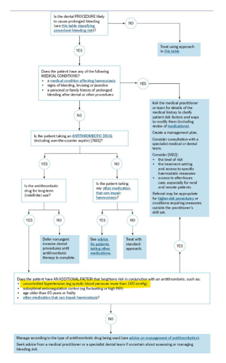
Risk factor when evaluating peri-procedural bleeding risk (Therapeutic Guidelines v4 2026 – Figure 13.73)
Is the dental PROCEDURE likely to cause prolonged bleeding (see this table classifying procedural bleeding risk)?
- YES
- NO → Treat using approach in this table
Does the patient have any of the following MEDICAL CONDITIONS?
-
a medical condition affecting haemostasis
-
signs of bleeding, bruising or purpura
-
a personal or family history of prolonged bleeding after dental or other procedures
-
YES → Ask the medical practitioner or team for details of the medical history to clarify patient risk factors and ways to modify them (including review of medications). Create a management plan. Consider consultation with a specialist medical or dental team. Consider:
- the level of risk
- the treatment setting and access to specific haemostatic measures
- access to after-hours care, especially for rural and remote patients.
- Referral may be appropriate for higher-risk procedures or conditions requiring measures outside the practitioner’s skill set.
-
NO
Is the patient taking an ANTITHROMBOTIC DRUG (including over-the-counter aspirin)?
- YES → Is the anticoagulation drug for long-term (indefinite) use?
- YES → Does the patient have AN ADDITIONAL FACTOR that heightens risk in conjunction with an antithrombotic, such as:
- uncontrolled hypertension (e.g. systolic blood pressure more than 160 mmHg)
- suboptimal anticoagulation control (e.g. fluctuating or high INR)
- age older than 65 years or frailty
- other medication that can impair haemostasis?
- NO
- NO → Manage according to the type of antithrombotic drug being used (see advice on management of antithrombotics). Seek advice from a medical practitioner or a specialist dental team if uncertain about assessing or managing bleeding risk.
- YES → Does the patient have AN ADDITIONAL FACTOR that heightens risk in conjunction with an antithrombotic, such as:
- NO → Is the patient taking any other medication that can impair haemostasis?
- YES → See advice for patients taking other medications.
- NO → Treat with standard approach.
Access20
- Timing / Appointments: Short, minimally stressful, ideally late morning
- Late morning appointments are preferred to avoid early morning catecholamine peaks.
- Positioning: orthostatic hypotension (ACEIs, ARBs)
- Setting: hospital if severe/uncontrolled hypertension and urgent dental care needed
Education / Prevention
- Common risk factor approach: smoking, alcohol, diet
- Antihypertensives: dry mouth management
- Calcium channel blockers (e.g. amlodipine) → gingiva enlargement
- Minimise with good oral hygiene and perio debridement - Emphasize oral hygiene to mitigate drug-induced hyperplasia.
- Extensive enlargement → specialist periodontal and medical management
Surgery21
- Check BP control prior to treatment
- Manage dental anxiety (exacerbates hypertension)
- Avoid NSAIDs = diuretic + either of:
- Angiotensin converting enzyme inhibitor (e.g. perindopril)
- Angiotensin II receptor blocker (e.g. candesartan)
- → risk of acute kidney injury
- Sedation may be helpful to control stress/blood pressure
- Pending nature and severity of underlying disease
Vasoconstrictors and Local Anaesthesia in Hypertension2223
How Much Adrenaline-Containing LA Can We Use?24
-
Profound anaesthesia for pain and anxiety control is important to ↓ endogenous catecholamine release
-
LA with adrenaline:
- Delays systemic absorption
- Increases duration of anaesthesia
- Provides local haemostasis
- Reduces the dose required
-
Poses a theoretical risk:
- Stimulates α and β adrenergic receptors (tachycardia, increase BP)
- Irritates cardiac pacemaker cells (arrhythmias)
-
Evidence regarding maximum safe dose of adrenaline is lacking
-
Malamed 2004: Most frequently quoted. Recommended max 0.04mg adrenaline per dental appt.
- ~2 cartridges containing 1:100,000 adr (0.018mg per 1.8ml cartridge, or 0.022mg per 2.2ml cartridge).
-
Tolas et al 1982, Cioffi et al 1985: Evaluated healthy pts following 2% lignocaine 1:100,000 adr injection
- After 1.8ml (1 cartridge): plasma levels incr 2-3x, no signif changes in BP or HR
- After 5.4ml (3 cartridges): plasma levels incr 5-6x, signif incr HR and BP
-
Godzieba et al 2014: systematic review of 11 RCTs
- Use of ≤4 cartridges of lignocaine with 1:100,000 adrenaline seems relatively safe for cardiovascular compromised patients
-
Serrera Figallo et al 2012: systematic review of RCTs
- Possible to use a dose between 1.8ml and 3.6ml on a general basis in patients with CVD
-
Conclusion:
- Use of >2 cartridges of vasoconstrictor-containing LA should be considered a relative rather than absolute contraindication.
- Current consensus seems to favour judicious use of local anaesthetics containing vasoconstrictors to ensure profound anaesthesia
Recommendations25
- If supplementation with plain LA is required, use vasoconstricting LA first
- Aspirating syringe
- Intraligamentary injection contraindicated
- Some report felypressin has fewer haemodynamic effects
- Felypressin is a safe alternative with minimal myocardial effects.
- ?Adrenaline-containing retraction cord astringent
Therapeutic Guidelines v4 2026
- Local anaesthetics containing adrenaline = limited effect on BP elevation
- Avoid in patients with uncontrolled hypertension
- Consider felypressin (Citanest® = prilocaine + felypressin)
- Also avoid: MI in previous 6 months, unstable angina, uncontrolled arrhythmias, uncontrolled hyperthyroidism
Ischaemic Heart Disease Definition and Aetiology26272829
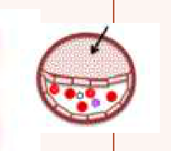
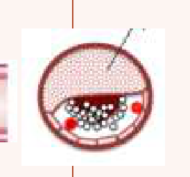
- Definition: Group of syndromes resulting from myocardial ischaemia – an imbalance between cardiac blood supply (perfusion) and myocardial oxygen demand.
- Classification / Aetiology:
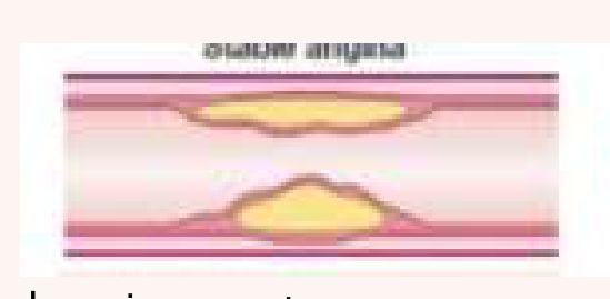
- Stable angina: chronic symptoms
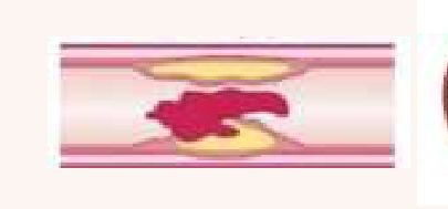
- Stable atherosclerotic plaque → Fixed coronary obstruction
- ACS: new or increasing ischaemic symptoms
- Unstable atherosclerotic plaque or endothelial disruption → transient/permanent thrombotic occlusion → ischemia and infarction
- Stable angina: chronic symptoms
 |  |  |
 |  | |
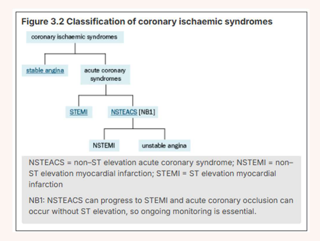
Classification / Aetiology30
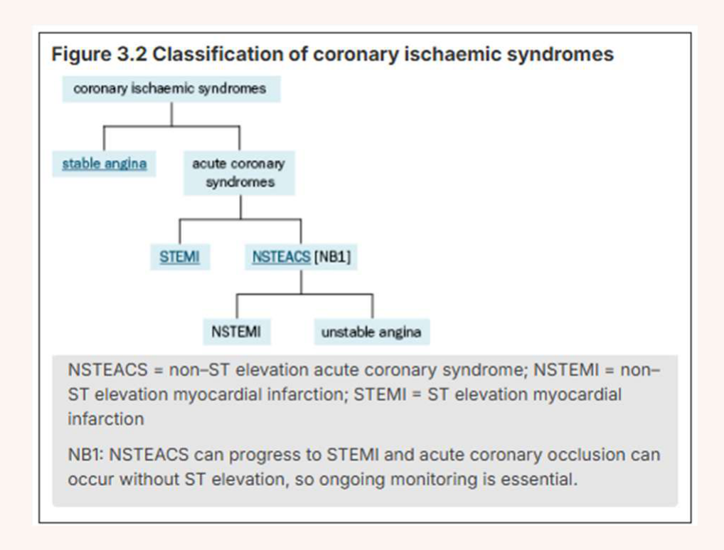
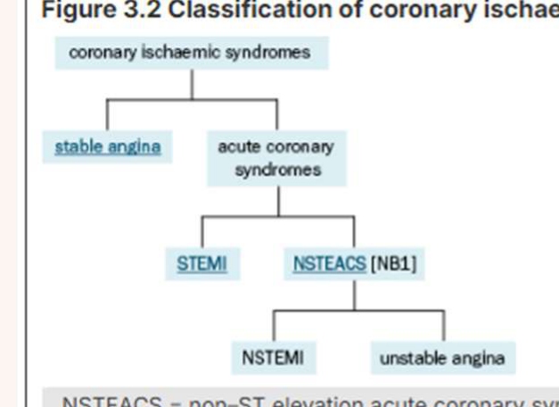
Figure 3.2 Classification of coronary ischaemic syndromes
| Coronary Ischaemic Syndromes | |
|---|---|
| Stable Angina | Acute Coronary Syndromes |
| STEMI | |
| NSTEMI |
NSTEACS = non–ST elevation acute coronary syndrome; NSTEMI = non–ST elevation myocardial infarction; STEMI = ST elevation myocardial infarction.
NB1: NSTEACS can progress to STEMI and acute coronary occlusion can occur without ST elevation, so ongoing monitoring is essential.
 |  |  |
 |  |  |
 |
Acute coronary syndromes
- Non-ST elevation acute coronary syndrome
- Unstable angina = without elevated cardiac biomarkers
- Non-ST elevation myocardial infarction = pts with MI as determined by elevated cardiac biomarkers
- ST-elevation myocardial infarction = medical emergency
- NSTEACS can progress to STEMI
Thygesen K, Alpert JS, Jaffe AS, Chaitman BR, Bax JJ, Morrow DA, et al. Fourth Universal Definition of Myocardial Infarction (2018). J Am Coll Cardiol 2018;72(18):2231-64
1) Type 1 myocardial infarction: 1. Atherothrombotic coronary occlusion compromises myocardial blood flow
2) Type 2 myocardial infarction: 1. Secondary to an imbalance between myocardial oxygen supply and demand (arises from e.g. tachyarrhythmias, sustained bradyarrhythmias, anaemia, respiratory failure and hypotension) 2. Coronary artery spasm 3. Spontaneous coronary artery dissection 4. Coronary embolism
| Risk Factor | Description |
|---|---|
| H | Heredity |
| A | Age (older) |
| S | Sex (male) |
| L | Lipidaemia |
| I | Increased weight (obesity) |
| P | Pressure (arterial hypertension) |
| I | Inactivity (sedentary lifestyle) |
| D | Diabetes |
| S | Smoking tobacco |
Dr Jee-Yun Leung
Epidemiology:
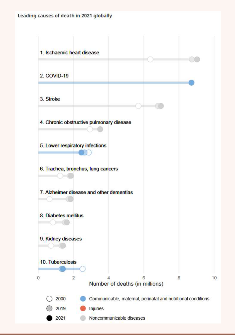
- Leading cause of death globally: 13%
- Prevalence: 3% in Australia (higher amongst males)
- Significant decrease in age-standardised IHD mortality rates
- 1960s: 7/10 MI’s fatal ; Today: 7/10 survive a MI
- CVD-related prescriptions increased 7-8 fold in this time
General Manifestations of Ischaemic Heart Disease313233
Figure 13.81 Probability that chest pain is ischaemic in origin based on common descriptions of pain
| High Probability of Ischaemia | Moderate Probability | Low Probability |
|---|---|---|
| pressure | sharp | |
| squeezing | fleeting | |
| gripping | shifting | |
| heaviness | pleuritic | |
| tightness | positional | |
| exertional or stress-related | ||
| retrosternal | ||
| left-sided | ||
| dull | ||
| aching | ||
| stabbing | ||
| right-sided | ||
| tearing | ||
| ripping | ||
| burning |
Source: Gulati M, Levy PD, Mukherjee D, Amsterdam E, Bhatt DL, Birtcher KK, et al. 2021 AHA/ACC/ASE/CHEST/SAEM/SCCT/SCMR Guideline for the Evaluation and Diagnosis of Chest Pain: A Report of the American College of Cardiology/American Heart Association Joint Committee on Clinical Practice Guidelines. Circulation 2021;144(22):e368-e454.
| Stable Angina | Acute Coronary Syndromes (e.g. MI) | Cardiac Arrest |
|---|---|---|
| • Episodic retrosternal chest discomfort (pain, ache or tightness) | • “Crushing” or heavy central chest pain (more severe) | • Sudden loss of consciousness |
| • May radiate to arms, neck, back, shoulder, jaw | • Shortness of breath, nausea, sweating, pale/clammy skin | • No pulse |
| • Commonly triggered by physical activity or emotional stress | • Decrease pulse & BP, vomiting, “impending sense of doom” | • Not breathing |
| • Predictable, consistent over time | • May occur at rest | |
| • Lasts <10min if provoking stimulus removed | • Lasts 20min to hours | |
| • Subsides with rest or GTN | • Not relieved by rest or GTN | |
| • NB: Silent Angina = some may have no pain due to psychological factors persistently inhibiting pain perception (elderly) or autonomic neuropathy (diabetics) |

Ischaemic Heart Disease: General Management34
| Lifestyle | Pharmacological | Revascularisation |
|---|---|---|
| • ↓ Smoking | • Beta-blockers | • Coronary artery bypass grafting (CABG) |
| • Diet | • Calcium-channel blockers | • Percutaneous coronary intervention (PCI) = “coronary angioplasty and stent” |
| • ↓ Alcohol | • Nitrates | |
| • Physical Activity | • Nicorandil | |
| • Healthy weight | • ACE inhibitors | |
| • Psychosocial | • ARBs | |
| • Antiplatelets | ||
| • Lipid-lowering drugs | ||
| • Ivabradine |
| Therapeutic goal | Pharmacological strategy |
|---|---|
| Inhibit progression atherosclerosis | Statins - ↓lipids |
| ↑coronary circulation | Nitrates, Ca channel blockers |
| ↓heart workload | B blockers |
| Prevent thrombus formation | Anti-thrombotic agents |
Oral Manifestations of Ischaemic Heart Disease35
- Antihypertensive drugs: as per previous section
- Nicorandil: oral/skin/eye ulceration, facial flushing, lupoid lesions
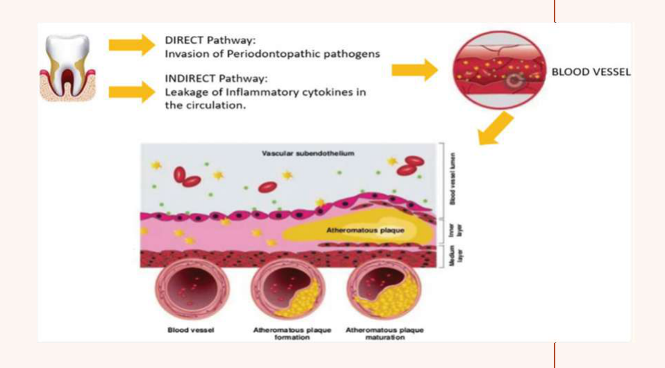
- Possible association with periodontitis (growing evidence)
- Direct pathway – bacteraemia
- Indirect pathway – induces chronic systemic inflammatory state
- Possible association with endodontic disease (less investigated and inconclusive evidence)
- apical periodontitis – comparable inflammatory and microbial profile (gram –ve anaerobes) to periodontal disease
- pulp stones – similarity to calcified stones forming in coronary and renal arteries
- Rarely, angina pain referred to lower jaw/teeth mistakenly thought to be of dental origin (clue: onset with exertion)
Periodontitis and Atherosclerotic Cardiovascular Disease36
| Category | Details |
|---|---|
| Journal | Circulation |
| Author | Andrew H. Tran, MD, MPH, MS, FAHA, Chair; Abbas H. Zaidi, MD, MS, Vice Chair; Ann F. Bolger, MD, FAHA; Oscar H. Del Brutto, MD, Rashmi Hegde, BDS, MS, Lauren L. Patton, DDS, Jamie Rausch, PhD, RN, and Justin P. Zachariah, MD, PhD, FAHA on behalf of the American Heart Association Cardiovascular Disease Prevention Committee |
| Publish | Volume 153, Number 6 |
Atherosclerotic cardiovascular diseases (ACVDs) are a leading cause of death and disability across the developed world and impose a vast burden on healthcare resources. This category of diseases includes:
- Coronary heart disease, which presents as angina or heart attack (myocardial infarction).
- Ischaemic cerebrovascular disease, which may manifest as stroke or mini-stroke (transient ischaemic attack or TIA).
- Peripheral vascular disease.
Periodontitis, although not a cause of mortality, is also a major public health concern because of its high prevalence in the population and its negative impact on oral health, ability to chew, appearance, quality of life, dental care costs and tooth loss. In addition, as a progressive, chronic inflammatory disease, untreated periodontitis has the potential to undermine overall health and even exacerbate other inflammatory conditions, such as ACVD.
European Federation of Periodontology: https://www.bsperio.org.uk/assets/downloads/EFP_cardiovascular-disease-gum-disease.pdf
Dental Management of Ischaemic Heart Disease3738
Careful medical history at first appointment. Ask about indicators of risk:
- History of chest pain at rest
- Triggers/frequency of episodes
- Carry GTN?
- How episodes are usually managed
- Number of anti-anginal medications
- Hx of CABG/stent placement with ongoing symptoms
- Hx of heart failure
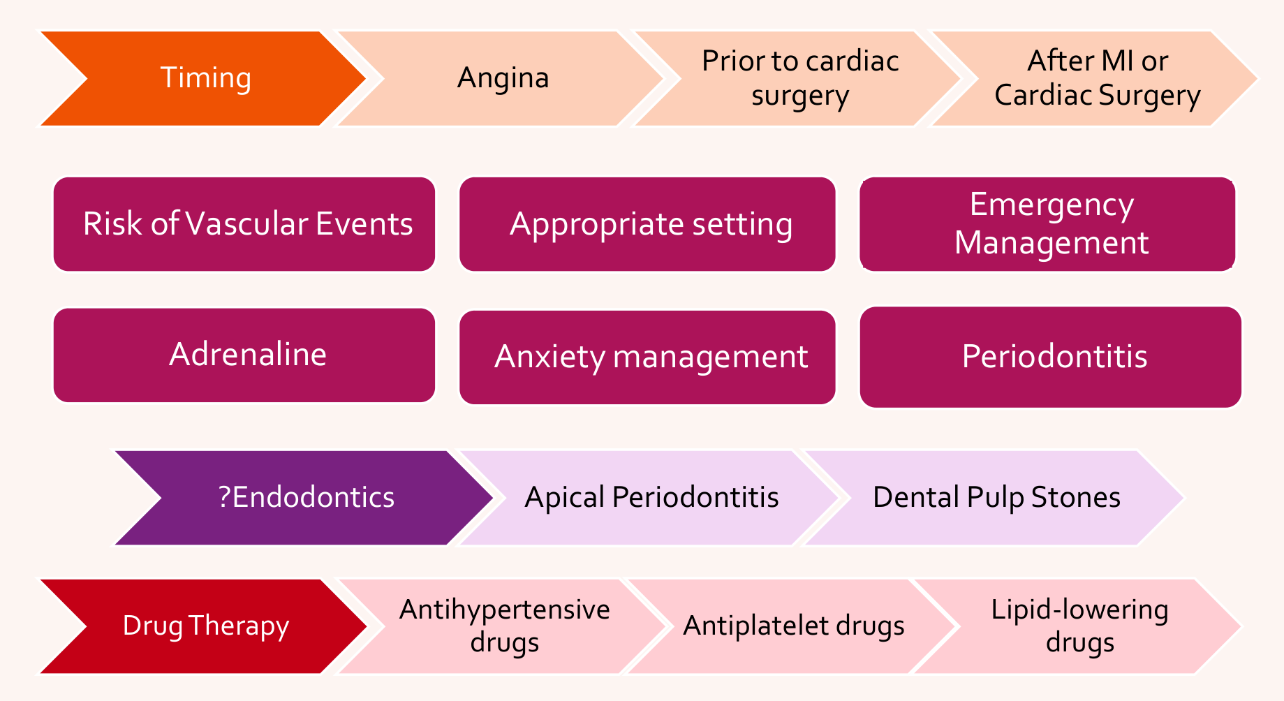
| Timing | Angina | Prior to cardiac surgery | After MI or Cardiac Surgery |
|---|---|---|---|
| Risk of Vascular Events | Appropriate setting | Emergency Management | Adrenaline |
| Drug Therapy | Anxiety management | Periodontitis | |
| ?Endodontics | Apical Periodontitis | Dental Pulp Stones | |
| Antihypertensive drugs | Antiplatelet drugs | Lipid-lowering drugs |
Access39
- Access to clinic / Mobility: Functional capacity (cardiopulmonary reserve)
- Appointments: Short, minimally stressful appointments. Late morning appointments.
- Positioning: Orthostatic hypotension (antihypertensives) → semi-supine and slowly upright
- Location / setting: Hospital setting may be required for severe cardiac disease when emergency dental treatment required.
- Timing:
- Patients with history of angina (or MI >6 months earlier) can undergo dental treatment if medically stable
- Defer elective dental care if unstable angina
- Defer elective dental procedures for 6 months after MI, stent placement or CABG.
- Pain and infection within 6 months of above:
- provide adequate emergency treatment
- avoid use of adrenaline-containing LA
- consider specialist advice re: appropriate treatment and timing of procedures
Capacity40
- Nil significant
Consent
- Nil significant
Education/Prevention
- Oral side effects of medications
- Possible association with periodontitis +/- endodontic disease
Surgery41
Pre-operative
- Instruct patients with angina to bring their medication (e.g. GTN spray or tablets), and have it readily accessible.
- Ensure GTN and oxygen readily available
- Monitor SpO, BP, HR
Peri-operative
- Use relaxation techniques and consider other measures for anxiety management
- Limit the duration of dental procedures
- Bleeding: may be on antithrombotics (see Haematology – Bleeding Disorders lecture)
- LA: Ensure effective analgesia → use of adrenaline (see Hypertension)
- Limit adrenaline-containing local anaesthetic to a maximum of 2 cartridges.
- Sedation: anxiety relief helpful (see Hypertension)
Medication Safety
Avoid NSAIDs in patients taking a “triple whammy” (diuretic + ACE inhibitor/ARB + NSAID) due to the high risk of acute kidney injury. Uncontrolled hypertension (systolic >160 mmHg) is a risk factor for increased bleeding, especially in patients taking antithrombotics.
Post-operative
- Caution with NSAIDs
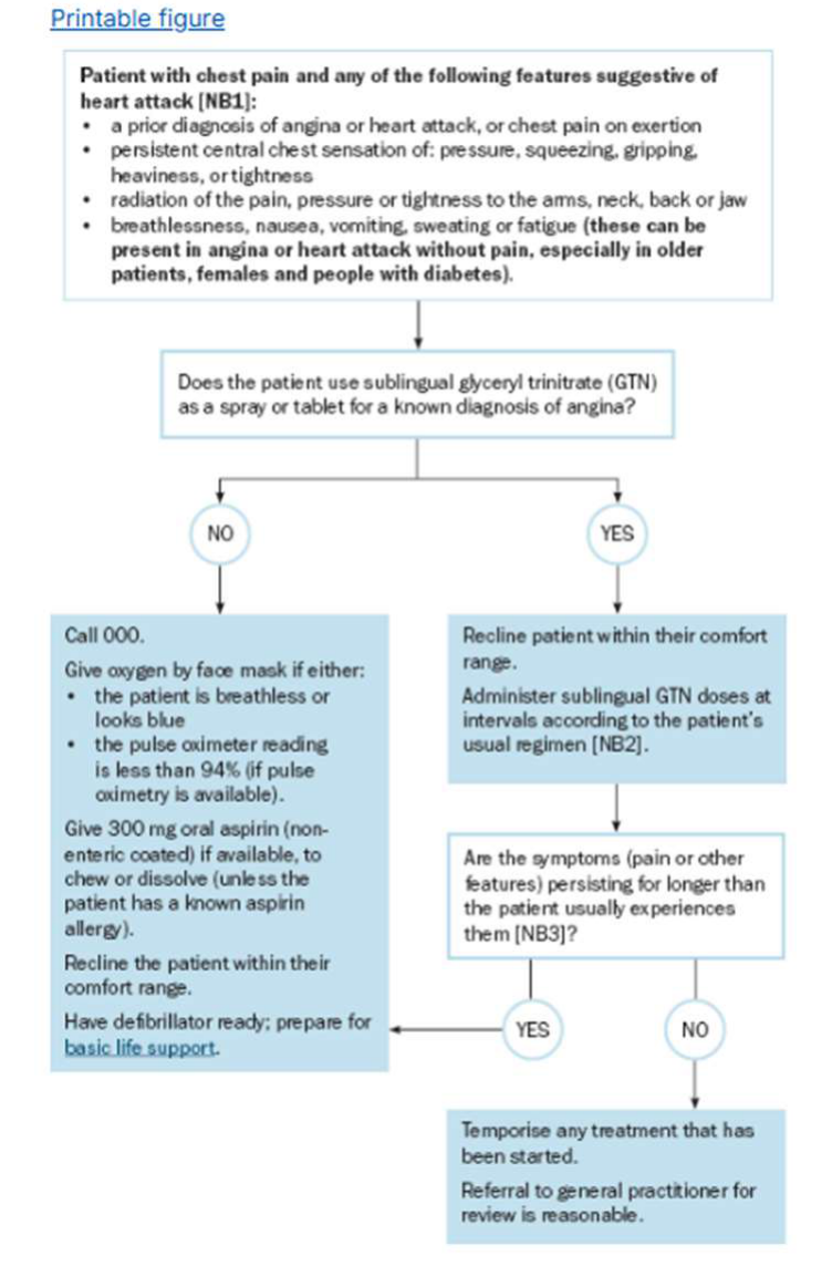
Management of Chest Pain in Dental Practice42
NB1: Many people with chest pain do not have angina or a heart attack, but anyone with acute chest pain requires rapid medical evaluation for potentially life-threatening causes. The likelihood of angina or heart attack increases with the number of suggestive features.
NB2: Sublingual glyceryl trinitrate (GTN) dosing is a 400 microgram spray (or 300 to 600 microgram tablet) generally administered every 5 minutes to a maximum of 3 doses. Ask patients who use GTN to bring their GTN to their dental appointment so that it can be administered if chest pain occurs.
NB3: If symptoms (pain or other features that may indicate angina) last longer than the patient’s usual angina symptoms, urgent medical evaluation of a possible heart attack or other urgent condition is required.
 |  |
 |
Management of chest pain in dental practice43
Patient with chest pain and any of the following features suggestive of heart attack [NB1]:
- a prior diagnosis of angina or heart attack, or chest pain on exertion
- persistent central chest sensation of: pressure, squeezing, gripping, heaviness, or tightness
- radiation of the pain, pressure or tightness to the arms, neck, back or jaw
- breathlessness, nausea, vomiting, sweating or fatigue (these can be present in angina or heart attack without pain, especially in older patients, females and people with diabetes).
NO → Call 000.
- Give oxygen by face mask if either:
- the patient is breathless or looks blue
- the pulse oximeter reading is less than 94% (if pulse oximetry is available).
- Give 300 mg oral aspirin (non-enteric coated) if available, to chew or dissolve (unless the patient has a known aspirin allergy).
- Recline the patient within their comfort range.
- Have defibrillator ready; prepare for basic life support.
YES → Recline patient within their comfort range.
- Administer sublingual GTN doses at intervals according to the patient’s usual regimen [NB2].
- Are the symptoms (pain or other features) persisting for longer than the patient usually experiences them [NB3]?
- YES → (connects back to Call 000 box as described above)
- NO → Temporise any treatment that has been started.
- Referral to general practitioner for review is reasonable.
If administering nitrates, recline the patient because blood pressure can be reduced by administration. To manage chest pain in a patient with a history of angina, use:
- glyceryl trinitrate spray 400 micrograms sublingually, repeat every 5 minutes if pain persists, up to a total of 3 doses if tolerated
OR
- glyceryl trinitrate tablet 300 to 600 micrograms sublingually, repeat every 5 minutes if pain persists, up to a total of 3 doses if tolerated.
To manage chest pain if angina or heart attack is suspected, provided no contraindication (e.g. aspirin allergy) is present, use:
- aspirin (non-enteric coated) 300 mg orally, chewed or dissolved before swallowing.
Thromboembolic Complications of Cardiovascular Disease44
- Arterial thrombi: platelet aggregation + fibrin + erythrocytes
- Venous thrombi: slow & turbulent blood flow, ↑ risk small emboli detaching & blocking pulmonary arteries
General Manifestations45
Arterial thrombi
- TIA/stroke
- Occlusion of coronary artery grafts
- MI & death in unstable angina
- Systemic embolism with prosthetic heart valves/AF
Venous thrombi
- DVT
- Pulmonary embolism
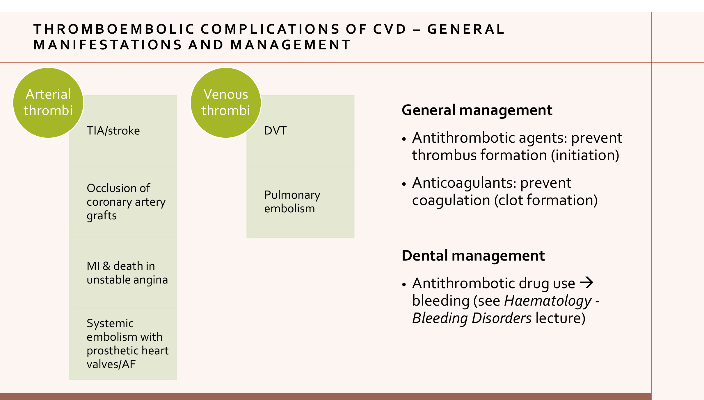
General management
- Antithrombotic agents: prevent thrombus formation (initiation)
- Anticoagulants: prevent coagulation (clot formation)
Dental management
- Antithrombotic drug use → bleeding (see Haematology - Bleeding Disorders lecture)
Valvular Conditions46
- Rheumatic fever and Rheumatic heart disease
- Infective endocarditis
- Aortic and mitral valve stenosis, regurgitation and prolapse
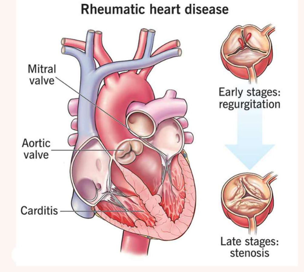
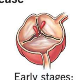
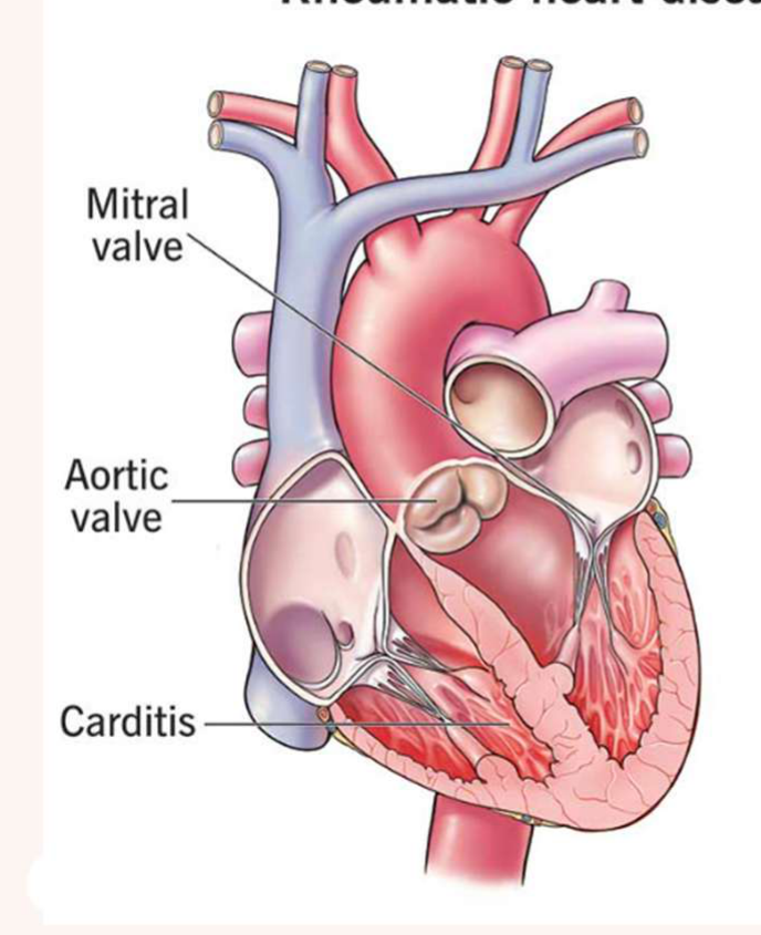
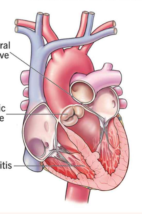
Rheumatic Fever and Rheumatic Heart Disease4748
 |  |
 |  |
Definition49
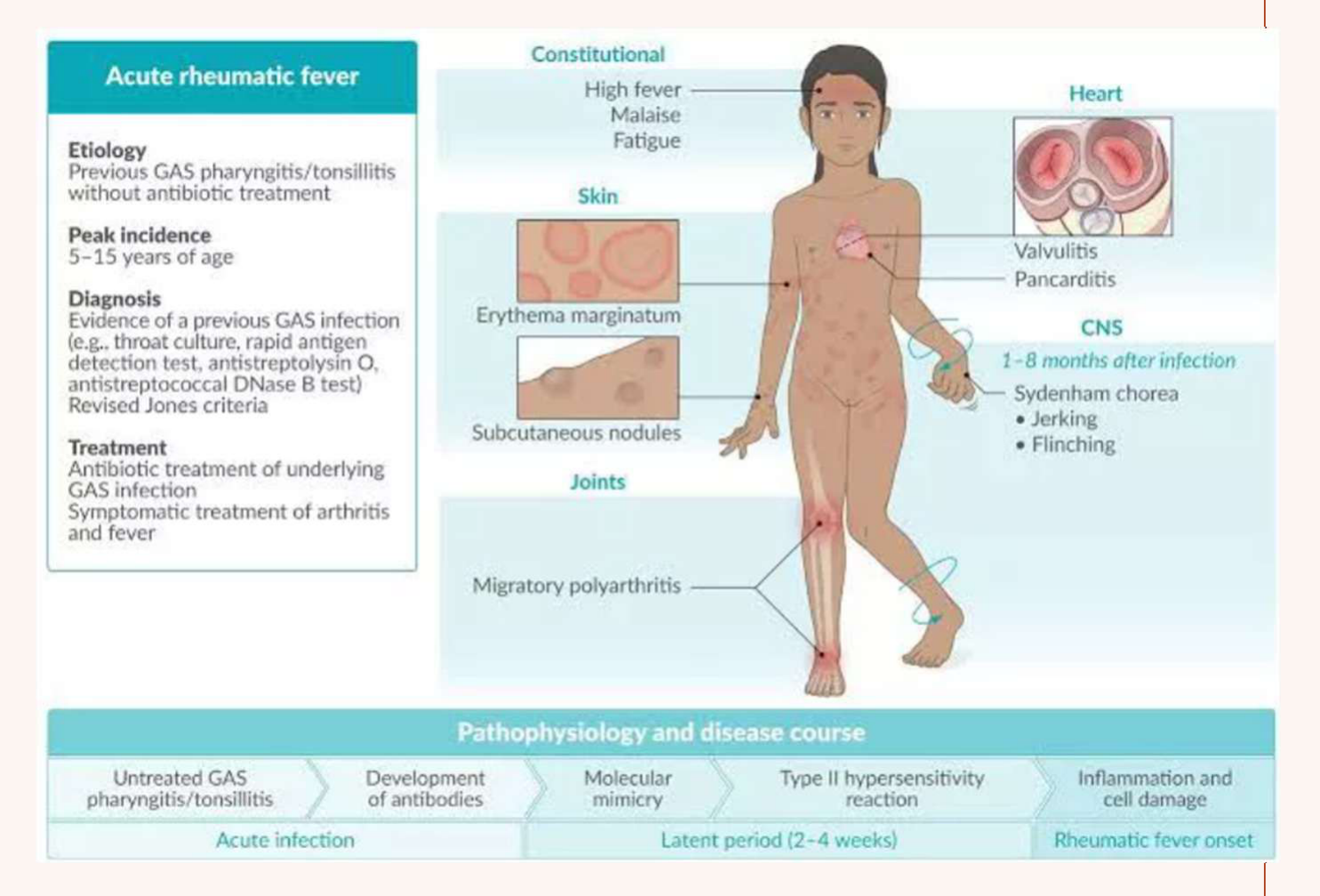
- Rheumatic Fever: An acute, immunologically-mediated, systemic inflammatory condition characterised by fever and arthritis 2-4 weeks after pharyngitis. Caused by certain strains of Group A beta-haemolytic streptococci (e.g. following impetigo and scabies).
Epidemiology
- 1.06 Acute rheumatic fever and rheumatic heart disease - AIHW Indigenous HPF
- RHD is a disease of disadvantage that is both preventable and treatable.
- First Nations people in Australia have one of the highest recorded rates of ARF and RHD in the world (119 per 100,000), with incidence rates increasing (50% increase 2012-2021)
- First Nations people accounted for 92% of the total number of ARF cases in Australia recorded over the 5-year period 2017–2021
- Otherwise rare disease in Western world, common in Indian subcontinent, Middle East, some Caribbean islands.
Aetiology50
- Group A streptococci
- RHD: repeated episodes of RF
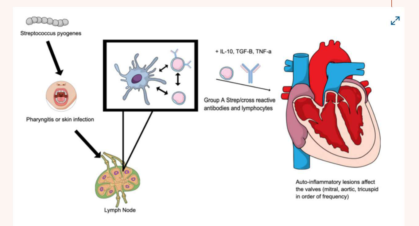
- Autoimmune reaction: antibodies produced to fight streptococci cross react with enzymes of the heart
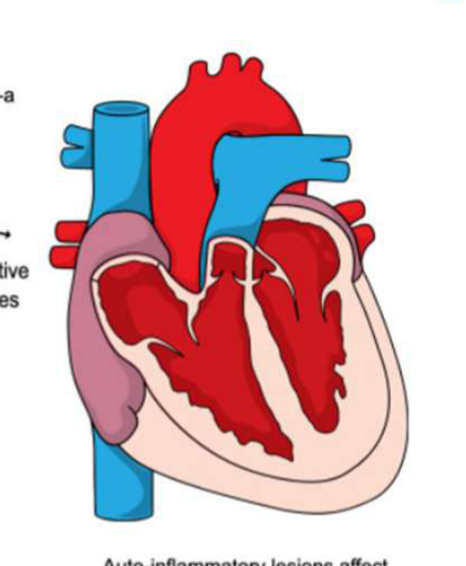
- Antibodies against streptococci cross-react with heart valves, causing scarring and permanent damage (most commonly the mitral valve).
Figure 2 Pathophysiology of RHD. RHD occurs as a sequela of GAS infection, especially pharyngitis. Social determinants of health and genetic predisposition play a role in repeated infection as well as in the progression to RHD. Cross-reactive antibodies and immune cells form in response to GAS infection and attack endothelial cells throughout the body; however, damage to the endothelial surface of the valves is not repaired effectively.
Image: Simpson MT, et al. Rheumatic Heart Disease in the Developing World. Structural Heart. 2023; 7(6): 100219
 |  |
General Manifestations51
- Acute Rheumatic Fever
- Subacute/chronic Rheumatic Fever
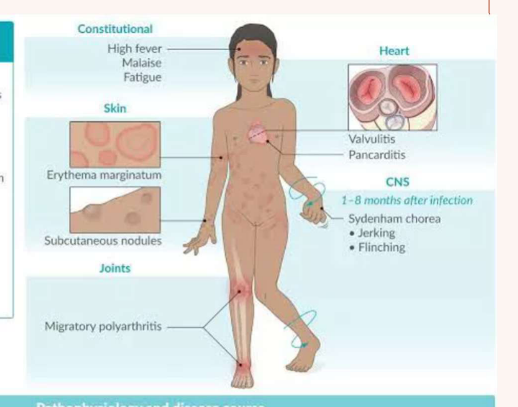
Acute rheumatic fever
| Etiology | Previous GAS pharyngitis/tonsillitis without antibiotic treatment | |||
|---|---|---|---|---|
| Peak incidence | 5–15 years of age | |||
| Diagnosis | Evidence of a previous GAS infection (e.g., throat culture, rapid antigen detection test, antistreptolysin O, antistreptococcal DNase B test) Revised Jones criteria | Constitutional High fever Malaise Fatigue | Heart Valvulitis Pancarditis | |
| Treatment | Antibiotic treatment of underlying GAS infection Symptomatic treatment of arthritis and fever | CNS 1–8 months after infection Sydenham chorea Jerking Flinching | ||
| Skin Erythema marginatum Subcutaneous nodules | ||||
| Joints Migratory polyarthritis |
Pathophysiology and disease course
| Untreated GAS pharyngitis/tonsillitis | Development of antibodies | Molecular mimicry | Type II hypersensitivity reaction | Inflammation and cell damage |
|---|---|---|---|---|
| Acute infection | Latent period (2~4 weeks) | Rheumatic fever onset |
- Rheumatic Heart Disease
- Valvular damage → regurgitation, stenosis → IE risk
- Early heart disease: Heart murmur
- Late effects: heart enlargement, cardiac failure
- Other:
- Shortness of breath
- Chest pain
- Fatigue
- Swollen legs and feet
| Diagnosis | Treatment | Prognosis |
|---|---|---|
| Physical examination History collection Echocardiogram Electrocardiogram Blood tests | Antibiotic prophylaxis (benzathine benzylpenicillin G) Complications: - Anticoagulants - Balloon valvuloplasty - Heart valve repair or replacement - Heart failure management - Lifestyle and prevention | - Acute phase lasts 6-12 weeks, definitive cure up to 6 months - RF recurrence rate is high (50% in first 5 years) → prophylactic therapy with penicillin - Mortality rate: 0.5 per 100,000 |
 |  |
Infective Endocarditis52
Definition53
A rare, but dangerous, infection of the endocardial surface of the heart. Predominantly affects one or more heart valves, especially where there is turbulent blood flow because of valve damage (typically left sided valves).
Epidemiology
- Relatively uncommon illness with high morbidity and mortality
- 2010: Australian incidence 4.7 cases per 100 000 person-years
- In-hospital mortality variable – increases with age (up to 15% in 80-89 yos)
Aetiology54
- Endothelial sites with turbulent blood flow
- Platelet and fibrin deposits accumulate (non-thrombotic endocarditis)
- sterile “vegetations” produced
- can be readily infected if a subsequent bacteraemia occurs
Causative organisms: Streptococci viridans (oral) 50-60%, Enterococci (faecal strep) 10%, Staphylococci (skin) 20-30%, Others 10% (Gram negative bacilli – mainly HACEK group, fungi)
Association with Dentistry55
- Dental procedures involving bleeding can initiate a bacteraemia that may lead to endocarditis (usually transient and <15min, but occasionally up to 1 hour)
- Most common types of bacteraemia in IE:
- S mutans and S sanguis (both Strep viridans) from dental plaque [40% of IE causative organisms due to complex attachment mechanisms enabling them to adhere to endocardium]
- Gram negative bacilli (HACEK group, including AA) occasionally implicated
- Chance of dental extraction causing IE in patient with valvular disease may be as low as 1 in 3000, therefore bacteraemia not synonymous with IE (or would need antibiotic cover for brushing).
Dental treatment is not the only source of IE-bacteraemia
Susceptible surface + high circulating bacterial loads
Risk Categories56
High risk: previous IE, prosthetic heart valves or valves repaired with prosthetic material, unrepaired cyanotic congenital heart disease, or some repaired congenital heart defects (up to 6 months after the procedure or lifelong if residual shunt or valvular regurgitation remains), +/- cardiac transplantation with cardiac valvulopathy.
Moderate risk: previous rheumatic fever, heart murmur, or evidence of native valve disease.
Table 5.32: Main groups affected by infective endocarditis
| Aetiology | Approximate % of all cases of infective endocarditis |
|---|---|
| No obvious cardiac valve disease | 40 |
| Chronic rheumatic heart disease | 30 |
| Congenital heart disease | 10 |
| Prosthetic cardiac valves | 10 |
| Intravenous drug abuse | 10 |
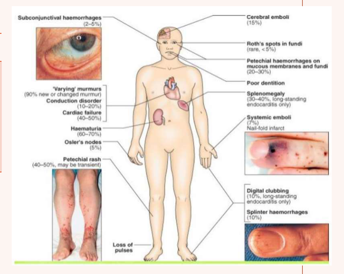
| Variable (flu-like) symptoms | Septic emboli (20-45%) – Systemic manifestations | Cardiac manifestations | Immune complex formation from antigens’ resultant antibodies |
|---|---|---|---|
| Fever | Infarcts brain (→ stroke), limbs, spleen (→ splenomegaly), kidney (→ haematuria), pulmonary. | Sepsis | Vasculitis |
| Headache | Retinal necrosis and blindness | Tachycardia | Arthritis |
| Joint pain | Mycotic aneurysms | Hypotension | Renal damage |
| Malaise | Focal glomerulonephritis | Valve destruction | |
| SOB/cough | Murmur | ||
| Chest pain (esp IVDUs) | Pericarditis | ||
| Coronary artery embolism |
Common signs: 90% fever, 85% heart murmurs
Management Complexity
Infective endocarditis requires long-term IV antibiotics and often urgent valve replacement due to high mortality rates (15-20% during admission).
General Management57
| Treatment | Prognosis |
|---|---|
| - Hospital admission for early treatment | - 15-20% die during initial hospital admission; further 10-15% die over the following year |
| - Antibiotic therapy | - 40-45% require surgery during initial hospital admission; further 10% require surgery within 1 yr post-discharge |
| - Severe cases: valve replacement | - Good prognostic factors: young, penicillin-sensitive isolates, early tx |
Aortic and Mitral Valve Disorders58
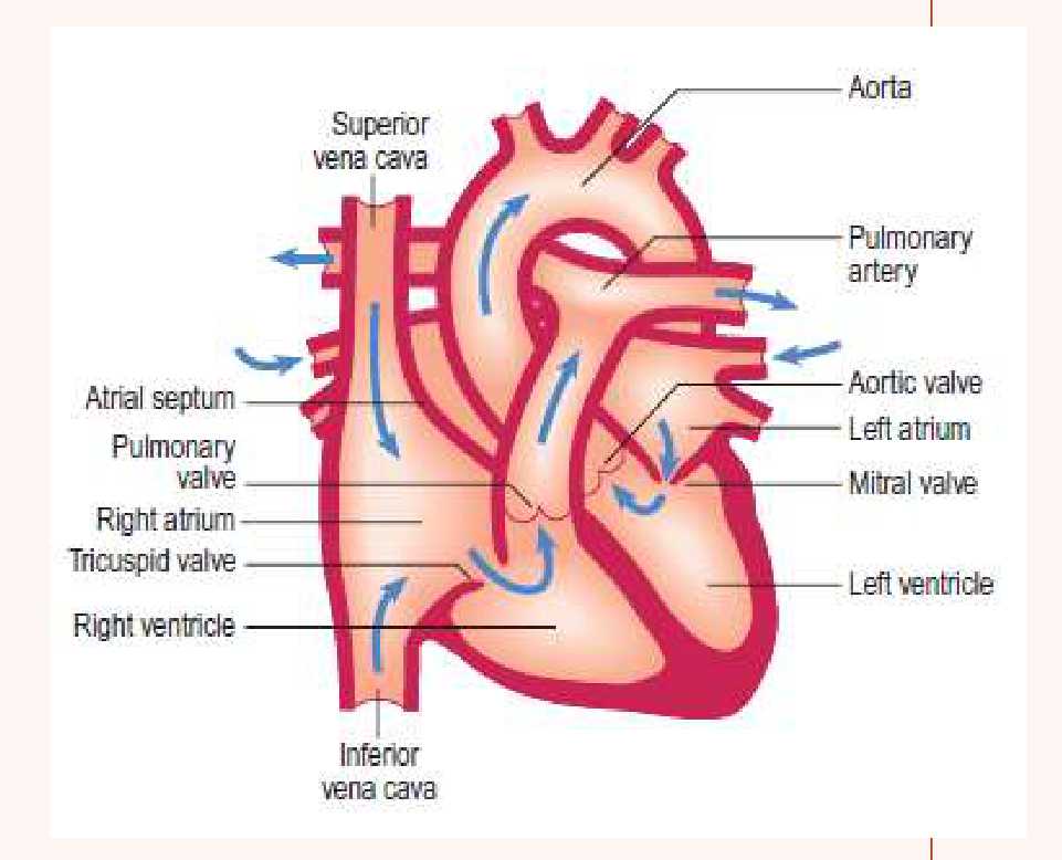
- Aortic stenosis: narrowing of the aortic valve; this generates an increased pressure in the left ventricle to maintain the blood volume per beat
- Aortic regurgitation: blood propelled to the aorta during systole returns to the left ventricle during diastole due to a defective aortic valve
- Mitral valve stenosis: Narrowing of the mitral valve that generates an increasing pressure in the left atrium of the heart
- Mitral valve regurgitation: The blood propulsed to the left ventricle during systolic contraction returns into the left atrium during diastolic dilatation due to a valve closure defect
- Mitral valve prolapse: Protrusion of the valves into the left atrium during systole. May exist in isolation, but sometimes associated with mitral regurgitation. May be associated with atrial septal defect, patent ductus arteriosus.
→ DO NOT REQUIRE ANTIBIOTIC PROPHYLAXIS
Valve Replacement59
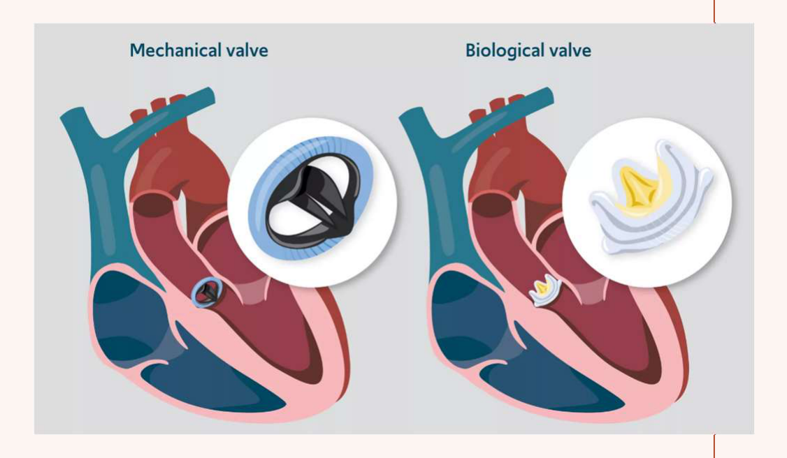
- Tissue valve
- Material: porcine, bovine, human
- Lifespan = 10–20 years
- Usually for older patients
- No turbulence = no anticoagulation required
- Unless required for other reason e.g. AF, DVT
- No noise
- Mechanical valve
- Material: carbon, titanium
- Lifespan = highly durable, can last lifetime (30+ years)
- Usually for younger patients
- Turbulence = anticoagulation required
- Requires lifelong warfarin due to turbulence.
- Noisy
Dental Management of Valvular Heart Disease60
Risk Assessment61
- Bacteraemia to heart valves
- Oral hygiene
- Timing to perform dental treatment
- Anticoagulation
- Oral side effects of diuretics (e.g., furosemide), ACE inhibitors, and beta-blockers
- GA/sedation risks: myocarditis
- Concurrent antibiotic therapy
- Identify patients requiring antibiotic prophylaxis by referring to Therapeutic Guidelines Version 4.
Access62
- Postpone elective dental care 6 months after episode of RF
- RHD → IE or valve surgery = urgent management of dental infections (bacteraemia)
- May be hospital in-patient setting
- IVDUs: less reliable attendance unless in pain
Capacity/Consent
- Nil significant
Education/Prevention
- Good oral hygiene most important factor in reducing risk in susceptible individuals
- Preventive measures to avoid caries and periodontal disease
- OHI for home maintenance, regular dental attendance, diet, etc
- Patient educated to alert dental providers of RHD/IE history, and need for antibiotic prophylaxis
Surgery63
- Antibiotic prophylaxis
- Liaise with Infectious Diseases physician if on concurrent antibiotics
- Consult with physician re: anticoagulation
- LA: use adrenaline with caution, aspirating syringe (IV injection may precipitate arrhythmias)
- Liaise with physicians regarding anticoagulation management, specifically for patients on Warfarin or DOACs.
- Conscious sedation / GA: avoid when acute RF (myocarditis), hospital environment if required
Spread
- Infective endocarditis bacteraemia
- Possible antibiotic resistance issues
Table 2.5 Indications for antibiotic prophylaxis for endocarditis
Antibiotic prophylaxis against infective endocarditis is recommended only for people who meet both of the following criteria:
- are undergoing a procedure associated with a high risk of a bacteraemia and subsequent infective endocarditis
- have a cardiac condition associated with an increased risk of developing infective endocarditis and the highest risk of adverse outcomes from endocarditis.
Procedures for which endocarditis prophylaxis is recommended for patients with a cardiac condition listed below [NB1]
- Dental procedures – only those involving manipulation of the gingival or periapical tissue or perforation of the oral mucosa (eg dental extractions, implant placement, biopsy, removal of soft tissue or bone, subgingival debridement, replanting avulsed teeth) [NB2] [NB3] [12] [33] [44].
NB1: Endocarditis prophylaxis is not recommended for procedures other than those covered in this topic. However, surgical prophylaxis may be indicated even if endocarditis prophylaxis is not – see Surgical antibiotic prophylaxis recommendations for specific procedures.
NB2: For a list of dental procedures classified by invasiveness and their indications for endocarditis prophylaxis, see Table 13.29.
NB3: Evidence on the risk of bacteraemia and subsequent infective endocarditis following supra- and subgingival debridement is conflicting. Pending further evidence, endocarditis prophylaxis is only recommended for supra- and subgingival debridement that involves manipulation of the gingival tissue [49] [50].
Commentary (NB6): Endocarditis prophylaxis is not recommended for patients with forms of valvular or structural heart disease not listed in this table, including patients with mitral valve prolapse, septal defects or cardiac implantable electronic devices.
NB7: Patients with a heart transplant who have developed cardiac valvulopathy may also be at high risk of adverse outcomes from endocarditis; consult the patient’s cardiologist for specific recommendations.
Cardiac conditions for which endocarditis prophylaxis is recommended for patients undergoing a procedure listed above [NB5] [NB6]
- prosthetic cardiac valve, including transcatheter-implanted prosthesis or homograft
- prosthetic material used for cardiac valve repair, such as annuloplasty rings and chords
- previous infective endocarditis
- ventricular assist devices
- congenital heart disease but only if it involves:
- unrepaired cyanotic defects, including palliative shunts and conduits
- repaired defects with residual defects at or adjacent to the site of a prosthetic patch or device (which inhibit endothelialisation) [53] [48]
- rheumatic heart disease [NB7].
EBI4: Symptoms are not sufficient for diagnosis; imaging is essential for confirmation.
NB7: Endocarditis prophylaxis was previously only recommended for rheumatic heart disease in Aboriginal and Torres Strait Islander peoples, or considered for people with rheumatic heart disease at significant socioeconomic disadvantage. However, studies have demonstrated that rheumatic heart disease is an independent risk factor for the development of and adverse outcomes from infective endocarditis [1] [3] [20].
Electrical Dysrhythmias64
- Extrasystoles (ectopics)
- Tachycardia (including Atrial fibrillation)
- Bradycardias
Aetiology and Manifestations of Dysrhythmias65
- Definition: An alternation in the normal rhythm of the heartbeat.
- Epidemiology: 15% of the population
- Aetiology: disorders in heart automaticity (impulse formation) or conductivity (block or delay)
| Abnormal impulse generation | Abnormal impulse conduction |
|---|---|
| Sinus bradycardia <60 bpm | AV Block – delay/failure conduction from atria to ventricles |
| Sinus tachycardia >100 bpm | |
| Atrial flutter | |
| Atrial fibrillation – acute/chronic | |
| Premature ventricular contractions | |
| Ventricular tachycardia | |
| Ventricular fibrillation | |
| Ventricular extrasystole |
Causes66
- Cardiac disease
- Drugs: caffeine, alcohol, smoking, β-2-agonists, digoxin, dopamine, tricyclics, tobacco
- Metabolic changes: catecholamines, autonomic/endocrine disease, hyperthyroidism, fever, hypoxia, surgery, persistent vegetative neurological states
- Electrolyte imbalance: e.g. secondary to CKD
- Respiratory: COPD
Signs/Symptoms67
- Most frequent symptom: Palpitations.
- Others: sickness, syncope, dyspnoea, anxiety, fatigue, chest pain, dizziness.
- May have no symptoms
Classification
- Rate:
- Tachycardia (fast rate >100 bpm)
- Bradycardia (slow rate <60 bpm day, <50 bpm night)
- Irregular
- Mechanism: automaticity, re-entry, fibrillation
- Origin: atrium, ventricle
Types of Arrhythmias68
Extrasystoles
- Premature beats
- Atrial extrasystoles
- Ventricular extrasystoles
Inherited Disorders
- Long QT syndrome
- Brugada syndrome
Bradycardias
- Sinus bradycardia
- Sinus arrest
- Atrioventricular block, bundle branch block, heart block
Tachycardias
- Sinus tachycardia
- Sinus node re-entrant tachycardia
- Atrioventricular tachyarrhythmia
- Supraventricular tachycardia
- Atrial flutter
- Atrial fibrillation
- Paroxysmal AF
- Persistent AF
- Permanent AF
- Paroxysmal atrial tachycardia
- Paroxysmal supraventricular tachycardia
- Wolff-Parkinson-White syndrome - Atrial Fibrillation (AF) is the most common arrhythmia (1% of population) and carries a high risk of stroke; patients are usually anticoagulated.
- Ventricular tachycardia
- Ventricular fibrillation
General Management and Cardiac Electronic Devices6970
Identify cause – May only need treatment for this
If no underlying cause – Treatment depends on type/severity of arrhythmia. Focus: controlling RATE and RHYTHM
Treatments
- Medications
- Pharmacological cardioversion
- Electric cardioversion
- Pacemaking / Defibrillation
- Surgery
Cardioversion71
- Restores regular rhythm
- Pharmacological (chemical) cardioversion
- Electrical cardioversion
Cardiovascular Implantable Electronic Devices (CIEDs)
- Permanent pacemakers
- External cardiac defibrillators
- Ventricular assistive devices (VAD)
- Implantable Cardioverter Defibrillator (ICD)
- Pacemakers: Stimulate the heart when the rate is too slow.
- ICDs: Administer shocks to stop dangerous rhythms.
Radiofrequency Ablation (Catheter Ablation)
Dental Management of Dysrhythmias727374
Anti-arrhythmic Drugs75
-
Gingival swelling = verapamil/diltiazem (Ca-channel blockers), enalapril (ACE inhibitor)
-
Lichenoid ulceration and oral ulceration related to agranulocytosis = some β-blockers
-
Lupus-like reaction and mucosal ulcers due to agranulocytosis = procainamide
-
Xerostomia = disopyramide
-
Anticoagulation (see Bleeding Disorders lecture)
-
Oral side effects of drugs (see Hypertension section)
-
CIED interference
-
Patients with CIED can undergo most general dental treatment.
-
Surgical diathermy can interfere with some CIEDs (especially monopolar).
-
With modern CIEDs, interference from most other dental electronic and ultrasonic devices is usually not clinically significant.
- Interference from ultrasonic scalers or apex locators is rare with modern shielded devices.
-
Consult cardiologist if in doubt.
-
CIED is not an indication for surgical nor IE antibiotic prophylaxis.
Patient Safety
Be aware that an ICD shock may cause the patient to flinch or bite down suddenly during treatment.
- LVADs increase risk of thrombotic events ∴ increased risk of post-op bleeding.
- Patients may be on antithrombotics.
- LVAD-induced blood pressure changes.
Further info on interference (American Dental Association): https://www.ada.org/resources/ada-library/oral-health-topics/cardiac-implanted-devices-and-electronic-dental-instruments
Access76
- Peak endogenous epinephrine levels in early morning → ideally appointments late morning or early afternoon.
- High risk patients – treated in hospital.
Education/Prevention
- Oral side effects of medication.
Surgery
- Liaise with cardiologist if concerned about CIED interference.
- Pre-operative: anxiety management, check INR if warfarinated, defibrillator readily available.
- Peri-operative:
- Positioning: supine if pacemaker.
- LA: adequate provision, judicious use of vasoconstrictor, avoid intravenous injection.
- Sedation: oxygen saturation monitoring, may be helpful to minimise stress.
- Post-operative: haemostasis if anticoagulated.
Muscular Cardiomyopathies777879
Primary – genetic/inherited, acquired
Secondary
CARDIOMYOPATHIES
Definition: broad term for diseases of the heart muscle that result in reduced ability for the heart to pump blood to the rest of the body.
- Manifests as fatigue, shortness of breath, and peripheral edema.
Aetiology
Primary
Genetic/Inherited
- Hypertrophic cardiomyopathy (HCM)
- Dilated cardiomyopathy (DCM)
- Arrhythmogenic R ventricular (ARVC)
Acquired
- Alcohol, chemotherapeutics (most common acquired cause)
- Tako-tsubo cardiomyopathy
- Tachycardia induced
- Muscular dystrophies
Secondary
- Storage: haemochromatosis, Fabry disease
- Infiltrative: amyloidosis, Hurlers syndrome
- Toxicity: alcohol, radiation
- Nutrition: Beri-beri, Kwashiorkor
- Autoimmune: SLE, RA
- Neuromuscular: Friedrich’s ataxia
- Endocrine: Diabetes, thyroid disease, phaeochromocytoma
| Signs/Symptoms | Management |
|---|---|
| - Oedema (pulmonary, peripheral – ankles & legs, abdominal) - Thrombi/emboli/clot formation - Exertional and paroxysmal nocturnal dyspnoea (SOB) - Arrhythmias - Nausea - Chest pain - Palpitations - Fainting - Sudden cardiac death | - Lifestyle changes: diet, exercise, stress, avoid tobacco/alcohol - Medications: hypertension drugs, anti-arrhythmic drugs, oral anticoagulants. Often: beta blockers, potassium channel blockers. Other: CCBs (verapamil, diltiazem) +/- ACEIs, diuretics, digoxin. - Devices to correct arrhythmias: pacemakers, ICDs - Devices to improve blood flow: LVAD, cardiac resynchronisation therapy (CRT) - Procedures: catheter ablation, heart transplant |
Congenital Heart Disease80
CONGENITAL
Cyanotic
Acyanotic
Definition81
- Any congenital structural defects of the heart (including valve stenosis or septal defects) or adjacent great vessels that lead to malfunction (arrhythmias or flow problems). Can be alone or in combinations.
Epidemiology
- Is the most common type of heart problem in children (1% live births). 20% also have other congenital anomalies.
- Diagnosed in at least 1 in 180 births (1-2% of the population).
Aetiology82
- Often unidentified, multifactorial, genetic, and environmental factors.
- Acquired causes:
- maternal infections (Rubella, Cytomegalovirus)
- maternal drug use (alcohol, anticonvulsants, lithium, thalidomide, warfarin, other drugs)
- maternal systemic disease (SLE, diabetes); maternal irradiation (rare).
- Genetic syndromes with cardiac defects:
- Chromosomal: Downs, Edwards, Turners
- Hereditary: Ehlers-Danlos syndrome, Marfan syndrome, osteogenesis imperfecta, mucopolysaccharidosis, Holt-Oram syndrome, Noonan syndrome, Williams syndrome
Classification83
- Cyanotic (R to L shunting): shunting deoxygenated blood from the right ventricle into the left side of the heart and the systemic circulation (bypassing the lungs)
- Acyanotic (obstruction, or L to R shunting): blood contains enough oxygen but is pumped abnormally around body, forcing the heart to work harder
| Cyanotic | Acyanotic, no shunt | Acyanotic with shunt |
|---|---|---|
| Tetralogy of Fallot | Aortic stenosis | Atrial septal defect |
| Eisenmenger syndrome | Bicuspid aortic valve | Patent ductus arteriosus |
| Pulmonary atresia | Coarctation of the aorta | Ventricular septal defect |
| Pulmonary valve stenosis | Dextrocardia | |
| Total anomalous venous drainage | Mitral valve prolapse | |
| Transposition of the great vessels | ||
| Tricuspid atresia |
Complications84
- Arrhythmias
- Infective endocarditis
- Stroke (cerebral abscess)
- Pulmonary hypertension and oedema
- Polycythaemia/bleeding tendency/thrombotic tendency
- Heart failure
Oral Manifestations8586
- Delayed eruption of both dentitions
- Positional anomalies
- Enamel hypoplasia
- Gross vasodilatations in the pulps
- Cleft palate
- Fissure tongue
- Greater caries and periodontal disease (associated to poor oral hygiene and lack in dental attention).
- Increased risk of periodontal disease can be attributed to “treatment burnout” in patients and caregivers.
Cardiac Failure8788
Definition: When structural or functional impairment of the pumping action of the heart leads to blood output insufficient to meet the body’s demands → lack of tissue and organ perfusion
Aetiology: any structural or functional cardiac or non-cardiac disorder that impairs ability of heart to respond to physiological demands for CO
Main causes of heart failure89
| Left-sided mainly | Right-sided mainly | Biventricular |
|---|---|---|
| Ischaemic heart disease | Chronic obstructive pulmonary disease | Ischaemic heart disease |
| Aortic valve disease | Pulmonary embolism | Aortic valve disease |
| Mitral valve disease | Mitral valve disease | |
| Hypertension | Hypertension | |
| Cardiomyopathies | ||
| Hyperthyroidism | ||
| Chronic anaemias | ||
| Arrhythmias |
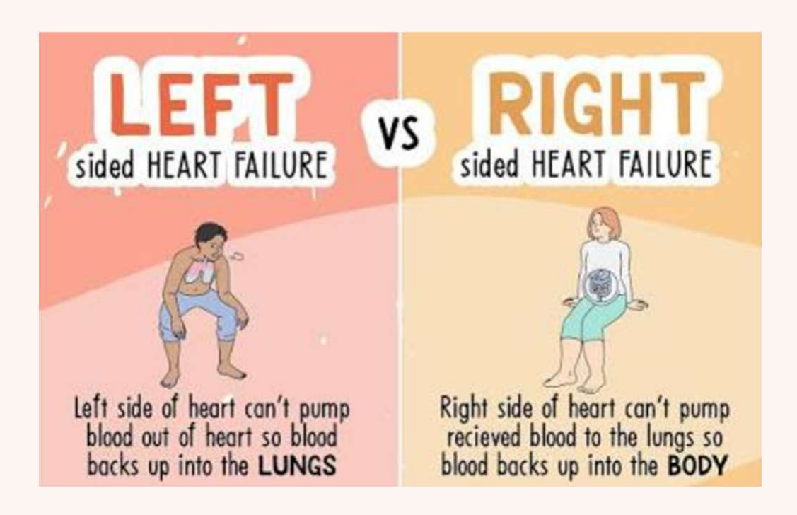
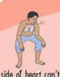
Left-sided heart failure90
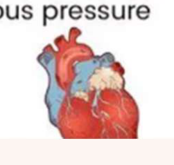
Left side of heart can’t pump blood out of heart so blood backs up into the LUNGS
- Left-sided failure primarily presents with pulmonary issues such as cough, crackles, and shortness of breath.
 |  |  |
 |  |
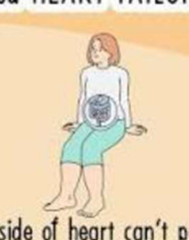
Right-sided heart failure
Right side of heart can’t pump received blood to the lungs so blood backs up into the BODY
- Right-sided failure primarily presents with systemic issues like peripheral edema and liver enlargement.
Symptoms
- Pulmonary congestion:
- Cough
- Crackles
- Pink-tinged sputum
- Tachypnea
- Tachycardia
- Fatigue
- Cyanosis
- Exertional dyspnea
- Peripheral edema
- Ascites
- Enlarged liver & spleen
- JVD
- Weight gain
- Increased peripheral venous pressure
Edema
| Lifestyle changes | Pharmacologic | Devices | Surgical intervention |
|---|---|---|---|
| - Fluid & salt management (limit intake) |
-
Minimise alcohol intake
-
Gentle exercise | - Inotropes e.g. digoxin (AF)
-
Diuretics e.g. thiazide
-
ACE inhibitors
-
β blockers
-
Vasodilators
-
Aldosterone antagonists | - Pacemaker
-
Cardiac resynchronisation device
-
ICD | - Revascularisation procedures (CABG)
-
Valve replacement / repair (due to valve damage: CHD, IE, MI)
-
Heart transplantation (end stage) |
-
Do not undertake dental treatment unless medical practitioner advised HF is stable
-
Limit duration of dental procedures
-
Patient may be unable to lie horizontally
- Position dental chair with head higher than heart
- Treat the patient in a semi-supine position to avoid respiratory distress.
- Comfortable position for sleeping → “how many pillows do you use when sleeping?”
- Position dental chair with head higher than heart
-
Increased risk with sedation and GA → hospital environment with anaesthetist
-
Avoid NSAIDs if possible
- Especially if diuretic + (ACEI or A2RB) → risk of acute kidney injury
-
Diuretics + drugs with diuretic effect (e.g. dapagliflozin, empagliflozin) → dry mouth
-
May be candidate for heart transplant
- Require dental assessment before being waitlisted
- Optimise oral health
- May seek hospital dental specialist for advice for pre-transplant dental treatment plan
Cardiac Surgery and Perioperative Management
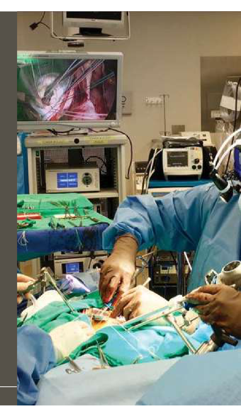
Cardiac Surgery Work-Up91
Types of Cardiac Surgery92
- Coronary angioplasty and stent (bare metal or drug-eluting) = Percutaneous coronary intervention (PCI)
- Improve blood flow through occluded coronary arteries
- Coronary artery bypass grafting (CABG)
- Redirect blood flow around occluded coronary arteries
- Valve replacement or Transcatheter aortic valve implantation (TAVI)
- Replace faulty or damaged heart valves
- Congenital correction
- Repair abnormal or damaged structures
- Pacemaker or Implantable Cardioverter Defibrillator (ICD) placement
- Implant devices to control heart beat or support function / blood flow
- Cardiac Transplant
- Replace a damaged heart, severe heart failure
Risks and Benefits of Preoperative Dental Treatment
Cardiac Risks of Dental Treatment9394
- Risk stratification guidelines: Peri-operative risks for CVD patients undergoing non-cardiac surgical procedures
- H&N surgery 1-5%, dental surgery / superficial surgery <1%
- European Society of Cardiology (ESC) and European Society of Anaesthesiology (ESA) (Halvorsen et al., 2022)
- American College of Cardiology (ACC) and American Heart Association (AHA) (Thompson et al, 2024)
- Smith et al 2014:
- Prophylactic dental extractions prior to cardiac surgery carries a 3% risk of death and 8% risk of major adverse outcome before cardiac operation
Smith MM, Barbara DW, Mauermann WJ, Viozzi CF, Dearani JA, Grim KJ. Morbidity and Mortality Associated with Dental Extraction Before Cardiac Operation. Ann Thorac Surg. 2014; 97: 838-44.
”… raises the question whether we should in fact get on with our cardiac operations and deal with the dental work at some other time, if at all, or risk killing patients with good intent!”
Unsworth-White 2014 (commentary of Smith et al 2014)
Unsworth-White MJ. Invited Commentary - Morbidity and Mortality Associated with Dental Extraction Before Cardiac Operation. Ann Thorac Surg. 2014;97,844.
Management of perioperative acute pain and infection (pus, swelling, cellulitis) needs to be a joint decision between the dentist and the physicians caring for the patient planned for cardiothoracic surgery.
But when should we treat chronic, asymptomatic pathology?95
Risks outweigh benefits
- Ischaemic disease (eg. CABG, PCI, stent, angioplasty)
- Rhythmic disease (eg. ICD/pacemaker, ablation)
Benefits may outweigh Risks
- Valvular disease (eg. replacement, repairs, TAVI)
- Infective endocarditis (often leads to valvular surgery)
- Heart transplant (heart failure)
 |  |  |
 |  |  |
Ischaemic Indications – Risks vs Benefits of Pre-Op Dental Treatment96979899
Potential benefits:
- Decrease endogenous catecholamine release (resulting in increased BP) if patient is in pain
- Reduce pre-operative infection risk or graft rejection
- Limited literature regarding dental extractions prior to CABG or heart transplant surgery
- Lack of clear evidence to support the reduced risk of post-operative infection (Meyer et al., 1999)
| Risks outweigh benefits | Benefits may outweigh Risks |
|---|---|
| Ischaemic disease (eg. CABG, PCI, stent, angioplasty) |
- <mark>It is generally better to defer dental treatment until after revascularization (CABG/Stent) when the heart has better oxygen supply.</mark> | Valvular disease (eg. replacement, repairs, TAVI)
- <mark>More aggressive dental treatment is often performed preoperatively to prevent postoperative infective endocarditis.</mark>
Rhythmic disease (eg. ICD/pacemaker, ablation) | Infective endocarditis (often leads to valvular surgery) | Heart transplant (heart failure)
Management of perioperative acute pain and infection (pus, swelling, cellulitis) needs to be a joint decision between the dentist and the physicians caring for the patient planned for cardiothoracic surgery.
Clin Oral Invest (1999) 3:79–83 © Springer-Verlag 1999
ORIGINAL ARTICLE
U. Meyer · D. Weingart · M.C. Deng · H.H. Scheld · U. Joos Heart transplants – assessment of dental procedures
Received: 25 September 1998 / Accepted: 26 February 1999
Abstract The object of this study was to evaluate the effects of dental foci on survival rates and rejection episodes in heart transplant recipients. Therefore, in a retrospective longitudinal study we studied 74 heart transplant recipients at the Department of Maxillofacial Surgery and Department of Thoracic and Cardiovascular Surgery, University of Münster. Study patients were divided into groups; those in which dental foci had been verified (n=31) and those without dental foci (n=43). Statistical analysis was performed using the chi-square test, Kaplan-Meier life table analysis, and the log-rank test. Before heart transplantation, patients were screened clinically and radiographically to determine the extent of dental foci. Postoperatively, patients were evaluated dentally and medically to identify the impact of dental foci on the incidence of systemic and oral infections, frequency and severity of rejection episodes, mortality, and complications arising during dental treatment. By comparing the mortality, infection and rejection rates in the various groups no statistically significant differences (P>0.05) were found between patients. Despite immunosuppression, extended inflammatory processes such as abscess formation or viral stomatitis were not found in the oral cavity. We therefore suggest that patients suffering from the symptoms of severe heart failure need not be subjected to rigorous preoperative dental treatment.
Key words Heart transplant · Dental treatment · Mortality
Introduction The survival rate of patients after heart transplantation has improved due to recent advances in perioperative management and immunosuppression [7]. Preoperative evaluation includes assessment of dental foci and their subsequent eradication [13, 3, 5]. So far, no study has examined the need for such a radical approach. Most microorganisms associated with bacteriemia in patients with heart transplantation are commensal organisms of the oral cavity [5]. Systemic infections and rejection dramatically decrease the survival rate in patients subjected to heart transplantation. Since odontogenic inflammation may favor transplant rejection [5], cause bacterial endocarditis and may lead to life-threatening postoperative infections [2], all existing or potential sources of dental infection should be eliminated before heart transplantation. To our knowledge, specific guidelines specifying the extent of such dental procedures do not exist. The purpose of this study was to analyze the complication rates in patients in whom dental foci had been verified as compared with patients without dental foci so that the role of these foci on the postoperative outcome of heart transplantation could be determined. The influence of various dental foci on differences in survival and rejection probabilities of heart transplant recipients was investigated to reevaluate our dental treatment protocols.
 |  |  |
 |  |  |
 |  | |
 |  |  |
|
Clinical Decision Making and Future Proofing
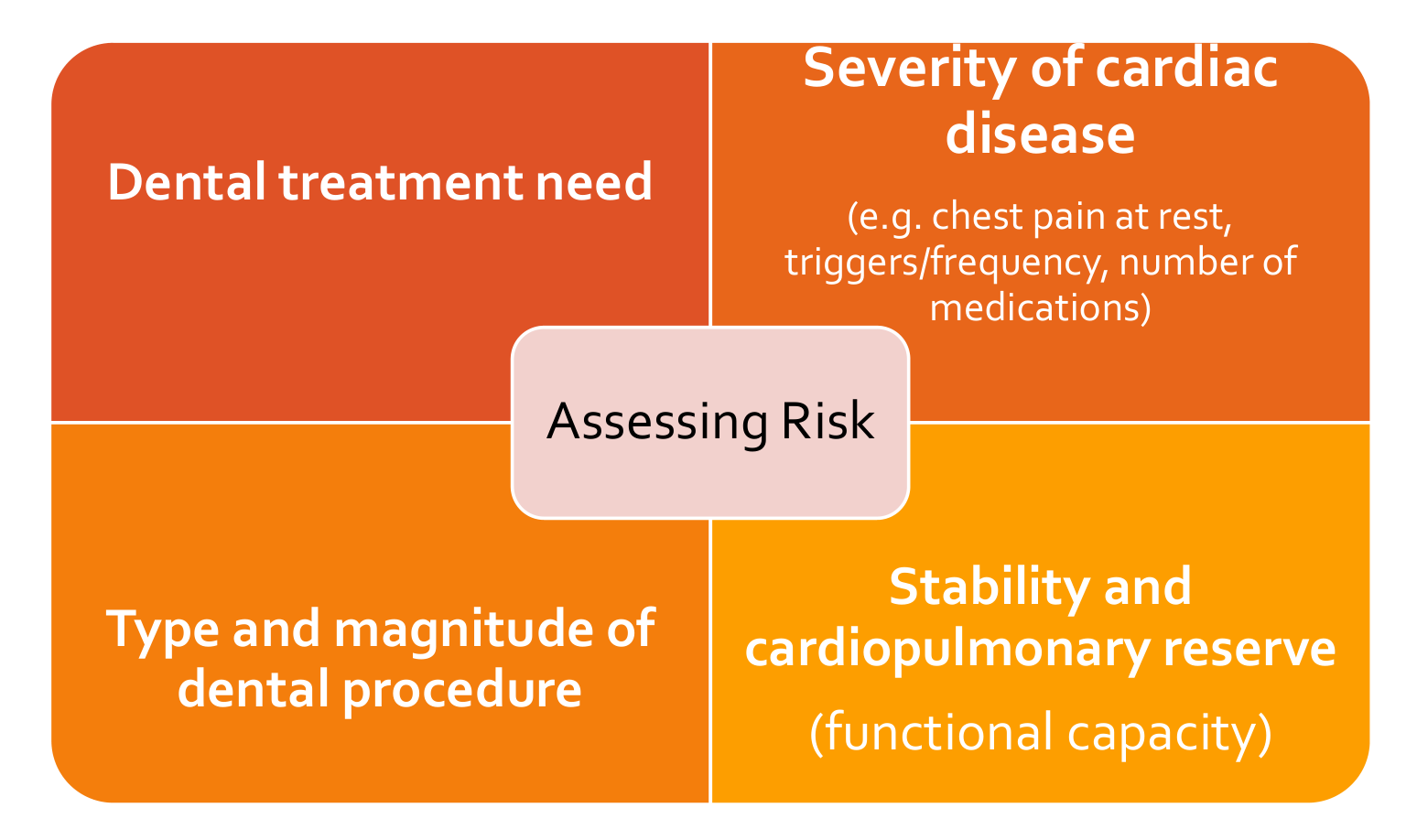
To Proceed or Not to Proceed?100
- Recommendation: integrated team decision based on individual situation of the patient (stable vs unstable condition)
- Dental treatment need
- Severity of cardiac disease (e.g. chest pain at rest, triggers/frequency, number of medications)
- Type and magnitude of dental procedure
- Weigh the “treatment burden” against the severity of cardiac disease.
- Stability and cardiopulmonary reserve (functional capacity)
How Far Ahead to Future Proof?101
“IN THE 12 MONTHS AFTER MYOCARDIAL INFARCTION, STENT PLACEMENT, CORONARY ARTERY BYPASS SURGERY, OR VALVE PROCEDURES (INCLUDING TRANSCATHETER AORTIC VALVE IMPLANTATION [TAVI]), PATIENTS ARE AT INCREASED RISK OF A MAJOR ADVERSE CARDIAC EVENT (e.g. SUDDEN DEATH).
DEFER ELECTIVE DENTAL TREATMENT FOR 6 MONTHS AFTER MYOCARDIAL INFARCTION, STENT PLACEMENT, CORONARY ARTERY BYPASS SURGERY OR VALVE PROCEDURE.
IF DENTAL PAIN OR INFECTION OCCURS WITHIN 6 MONTHS FOLLOWING THE EVENT, PROVIDE ADEQUATE EMERGENCY TREATMENT BUT AVOID THE USE OF ADRENALINE-CONTAINING LOCAL ANAESTHETICS. CONSIDER SEEKING SPECIALIST ADVISE ABOUT APPROPRIATE TREATMENT AND TIMING OF PROCEDURES.”
Oral & Dental Therapeutic Guidelines v4 2026
Thinking It Through… Cardiac Surgery102
Rationale
- Does need for dental treatment outweigh the cardiac risks?
- Direct effect on cardiac outcome = Endocarditis risk
Medical, Dental and Social Considerations
- Bacteraemia risk of dental pathology
- Amount of treatment patient can tolerate in one go vs risks of performing over multiple visits
- Anticoagulation / antiplatelet drugs (prophylactic vs therapeutic)
- Antibiotic prophylaxis
- Focus on preventive oral hygiene to reduce daily bacteraemia
Entire or Localised Areas of the Mouth
- Entire dentition
Optimal Timeframes
- Urgency for cardiac surgery
- Future-proof for next 6 months (i.e. optimise for the short-term)
- Aim to make the patient dentally stable for at least 6 months post-surgery, as elective care is deferred during this high-risk recovery period.
Treatment Approaches
- Aggressive (e.g. typically insufficient time to assess periapical response to RCT)
- Treatment is often more aggressive (extractions over RCT) because there is no time to monitor healing before urgent surgery.
Clinical Cases
Case: Gingival Hyperplasia from Calcium Channel Blockers
Question
The lecturer presents a scenario involving patients taking calcium channel blockers (specifically dihydropyridines). What are the characteristic oral findings associated with this medication, and what factors exacerbate the condition?
Findings and Reasoning
- Observation: The lecturer describes “really swollen gums” or gingival hyperplasia.
- Mechanism: This is a known side effect of dihydropyridine calcium channel blockers (and sometimes ACE inhibitors).
- Exacerbating Factors: The presence of poor oral hygiene and plaque can significantly worsen the extent of the gingival enlargement.
- Management: If the enlargement is extensive, it may require specialist periodontal and medical management.
Case: Oral Ulceration from Nicarandil
Question
What specific oral manifestation is “classically known” to be associated with the drug Nicarandil, often used for ischemic heart disease?
Findings and Reasoning
- Observation: Nicarandil is associated with oral ulceration.
- Other Potential Signs: It can also lead to facial flushing and lupoid lesions.
- Clinical Significance: When a patient with ischemic heart disease presents with oral ulcers, the clinician should consider Nicarandil as a potential contributing cause.
Case: Angina Radiating to the Jaw
Question
How might a cardiac event be mistaken for a dental problem, and what is the key clinical “clue” to differentiate them?
Findings and Reasoning
- Scenario: Angina pain can radiate to the lower jaw and the teeth.
- Differential Diagnosis: This can be mistakenly identified as being of dental origin.
- Key Diagnostic Clue: If the onset of the pain is associated with physical exertion, it is much more likely to be angina rather than a primary dental or maxillofacial problem.
Case: Oral Manifestations of Congenital Heart Disease
Question
In children with congenital heart disease, what specific dental and oral anomalies might be observed during a clinical examination?
Findings and Reasoning
- Developmental Findings:
- Delayed eruption of both primary and permanent dentitions.
- Positional anomalies of the teeth.
- Enamel hypoplasia.
- Structural Findings:
- Grossly enlarged (dilated) pulp chambers.
- Association with cleft palate or fissured tongue.
- Reasoning: These anomalies occur because congenital heart disease affects the overall growth and development of the child.
Case: Left-Sided vs. Right-Sided Heart Failure
Question
The lecturer contrasts the clinical presentations of left-sided and right-sided heart failure. What are the observable signs for each?
Findings and Reasoning
- Left-Sided Heart Failure (Pulmonary focus):
- Cough and crackles in the lungs.
- Pink-tinged sputum.
- Tachycardia and cyanosis.
- Right-Sided Heart Failure (Systemic focus):
- Peripheral edema (swelling in the limbs).
- Liver issues and splenomegaly (enlarged spleen).
- Weight gain.
- Clinical Application: For patients with heart failure, the lecturer notes they may not tolerate a horizontal position. A clinical tip is to ask the patient how many pillows they use to sleep and recline the dental chair only to that comfortable level.
Footnotes
-
Original PDF page 1: L2 Cardiovascular Disease DENT5311 - JY Leung, p.1 ↩
-
Original PDF page 2: L2 Cardiovascular Disease DENT5311 - JY Leung, p.2 ↩
-
Original PDF page 3: L2 Cardiovascular Disease DENT5311 - JY Leung, p.3 ↩
-
Original PDF page 4: L2 Cardiovascular Disease DENT5311 - JY Leung, p.4 ↩
-
Original PDF page 5: L2 Cardiovascular Disease DENT5311 - JY Leung, p.5 ↩
-
Original PDF page 6: L2 Cardiovascular Disease DENT5311 - JY Leung, p.6 ↩
-
Original PDF page 7: L2 Cardiovascular Disease DENT5311 - JY Leung, p.7 ↩
-
Original PDF page 8: L2 Cardiovascular Disease DENT5311 - JY Leung, p.8 ↩
-
Original PDF page 9: L2 Cardiovascular Disease DENT5311 - JY Leung, p.9 ↩
-
Original PDF page 10: L2 Cardiovascular Disease DENT5311 - JY Leung, p.10 ↩
-
Original PDF page 14: L2 Cardiovascular Disease DENT5311 - JY Leung, p.14 ↩
-
Original PDF page 11: L2 Cardiovascular Disease DENT5311 - JY Leung, p.11 ↩
-
Original PDF page 12: L2 Cardiovascular Disease DENT5311 - JY Leung, p.12 ↩
-
Original PDF page 13: L2 Cardiovascular Disease DENT5311 - JY Leung, p.13 ↩
-
Original PDF page 15: L2 Cardiovascular Disease DENT5311 - JY Leung, p.15 ↩
-
Original PDF page 17: L2 Cardiovascular Disease DENT5311 - JY Leung, p.17 ↩
-
Original PDF page 16: L2 Cardiovascular Disease DENT5311 - JY Leung, p.16 ↩
-
Original PDF page 18: L2 Cardiovascular Disease DENT5311 - JY Leung, p.18 ↩
-
Original PDF page 19: L2 Cardiovascular Disease DENT5311 - JY Leung, p.19 ↩
-
Original PDF page 20: L2 Cardiovascular Disease DENT5311 - JY Leung, p.20 ↩
-
Original PDF page 21: L2 Cardiovascular Disease DENT5311 - JY Leung, p.21 ↩
-
Original PDF page 23: L2 Cardiovascular Disease DENT5311 - JY Leung, p.23 ↩
-
Original PDF page 25: L2 Cardiovascular Disease DENT5311 - JY Leung, p.25 ↩
-
Original PDF page 22: L2 Cardiovascular Disease DENT5311 - JY Leung, p.22 ↩
-
Original PDF page 24: L2 Cardiovascular Disease DENT5311 - JY Leung, p.24 ↩
-
Original PDF page 26: L2 Cardiovascular Disease DENT5311 - JY Leung, p.26 ↩
-
Original PDF page 28: L2 Cardiovascular Disease DENT5311 - JY Leung, p.28 ↩
-
Original PDF page 29: L2 Cardiovascular Disease DENT5311 - JY Leung, p.29 ↩
-
Original PDF page 30: L2 Cardiovascular Disease DENT5311 - JY Leung, p.30 ↩
-
Original PDF page 27: L2 Cardiovascular Disease DENT5311 - JY Leung, p.27 ↩
-
Original PDF page 31: L2 Cardiovascular Disease DENT5311 - JY Leung, p.31 ↩
-
Original PDF page 32: L2 Cardiovascular Disease DENT5311 - JY Leung, p.32 ↩
-
Original PDF page 34: L2 Cardiovascular Disease DENT5311 - JY Leung, p.34 ↩
-
Original PDF page 33: L2 Cardiovascular Disease DENT5311 - JY Leung, p.33 ↩
-
Original PDF page 35: L2 Cardiovascular Disease DENT5311 - JY Leung, p.35 ↩
-
Original PDF page 36: L2 Cardiovascular Disease DENT5311 - JY Leung, p.36 ↩
-
Original PDF page 37: L2 Cardiovascular Disease DENT5311 - JY Leung, p.37 ↩
-
Original PDF page 38: L2 Cardiovascular Disease DENT5311 - JY Leung, p.38 ↩
-
Original PDF page 39: L2 Cardiovascular Disease DENT5311 - JY Leung, p.39 ↩
-
Original PDF page 40: L2 Cardiovascular Disease DENT5311 - JY Leung, p.40 ↩
-
Original PDF page 41: L2 Cardiovascular Disease DENT5311 - JY Leung, p.41 ↩
-
Original PDF page 43: L2 Cardiovascular Disease DENT5311 - JY Leung, p.43 ↩
-
Original PDF page 42: L2 Cardiovascular Disease DENT5311 - JY Leung, p.42 ↩
-
Original PDF page 44: L2 Cardiovascular Disease DENT5311 - JY Leung, p.44 ↩
-
Original PDF page 45: L2 Cardiovascular Disease DENT5311 - JY Leung, p.45 ↩
-
Original PDF page 46: L2 Cardiovascular Disease DENT5311 - JY Leung, p.46 ↩
-
Original PDF page 50: L2 Cardiovascular Disease DENT5311 - JY Leung, p.50 ↩
-
Original PDF page 51: L2 Cardiovascular Disease DENT5311 - JY Leung, p.51 ↩
-
Original PDF page 47: L2 Cardiovascular Disease DENT5311 - JY Leung, p.47 ↩
-
Original PDF page 48: L2 Cardiovascular Disease DENT5311 - JY Leung, p.48 ↩
-
Original PDF page 49: L2 Cardiovascular Disease DENT5311 - JY Leung, p.49 ↩
-
Original PDF page 56: L2 Cardiovascular Disease DENT5311 - JY Leung, p.56 ↩
-
Original PDF page 52: L2 Cardiovascular Disease DENT5311 - JY Leung, p.52 ↩
-
Original PDF page 53: L2 Cardiovascular Disease DENT5311 - JY Leung, p.53 ↩
-
Original PDF page 54: L2 Cardiovascular Disease DENT5311 - JY Leung, p.54 ↩
-
Original PDF page 55: L2 Cardiovascular Disease DENT5311 - JY Leung, p.55 ↩
-
Original PDF page 57: L2 Cardiovascular Disease DENT5311 - JY Leung, p.57 ↩
-
Original PDF page 58: L2 Cardiovascular Disease DENT5311 - JY Leung, p.58 ↩
-
Original PDF page 59: L2 Cardiovascular Disease DENT5311 - JY Leung, p.59 ↩
-
Original PDF page 63: L2 Cardiovascular Disease DENT5311 - JY Leung, p.63 ↩
-
Original PDF page 60: L2 Cardiovascular Disease DENT5311 - JY Leung, p.60 ↩
-
Original PDF page 61: L2 Cardiovascular Disease DENT5311 - JY Leung, p.61 ↩
-
Original PDF page 62: L2 Cardiovascular Disease DENT5311 - JY Leung, p.62 ↩
-
Original PDF page 64: L2 Cardiovascular Disease DENT5311 - JY Leung, p.64 ↩
-
Original PDF page 65: L2 Cardiovascular Disease DENT5311 - JY Leung, p.65 ↩
-
Original PDF page 66: L2 Cardiovascular Disease DENT5311 - JY Leung, p.66 ↩
-
Original PDF page 67: L2 Cardiovascular Disease DENT5311 - JY Leung, p.67 ↩
-
Original PDF page 68: L2 Cardiovascular Disease DENT5311 - JY Leung, p.68 ↩
-
Original PDF page 69: L2 Cardiovascular Disease DENT5311 - JY Leung, p.69 ↩
-
Original PDF page 70: L2 Cardiovascular Disease DENT5311 - JY Leung, p.70 ↩
-
Original PDF page 71: L2 Cardiovascular Disease DENT5311 - JY Leung, p.71 ↩
-
Original PDF page 73: L2 Cardiovascular Disease DENT5311 - JY Leung, p.73 ↩
-
Original PDF page 74: L2 Cardiovascular Disease DENT5311 - JY Leung, p.74 ↩
-
Original PDF page 75: L2 Cardiovascular Disease DENT5311 - JY Leung, p.75 ↩
-
Original PDF page 72: L2 Cardiovascular Disease DENT5311 - JY Leung, p.72 ↩
-
Original PDF page 76: L2 Cardiovascular Disease DENT5311 - JY Leung, p.76 ↩
-
Original PDF page 77: L2 Cardiovascular Disease DENT5311 - JY Leung, p.77 ↩
-
Original PDF page 78: L2 Cardiovascular Disease DENT5311 - JY Leung, p.78 ↩
-
Original PDF page 79: L2 Cardiovascular Disease DENT5311 - JY Leung, p.79 ↩
-
Original PDF page 80: L2 Cardiovascular Disease DENT5311 - JY Leung, p.80 ↩
-
Original PDF page 81: L2 Cardiovascular Disease DENT5311 - JY Leung, p.81 ↩
-
Original PDF page 82: L2 Cardiovascular Disease DENT5311 - JY Leung, p.82 ↩
-
Original PDF page 83: L2 Cardiovascular Disease DENT5311 - JY Leung, p.83 ↩
-
Original PDF page 84: L2 Cardiovascular Disease DENT5311 - JY Leung, p.84 ↩
-
Original PDF page 85: L2 Cardiovascular Disease DENT5311 - JY Leung, p.85 ↩
-
Original PDF page 86: L2 Cardiovascular Disease DENT5311 - JY Leung, p.86 ↩
-
Original PDF page 89: L2 Cardiovascular Disease DENT5311 - JY Leung, p.89 ↩
-
Original PDF page 90: L2 Cardiovascular Disease DENT5311 - JY Leung, p.90 ↩
-
Original PDF page 87: L2 Cardiovascular Disease DENT5311 - JY Leung, p.87 ↩
-
Original PDF page 88: L2 Cardiovascular Disease DENT5311 - JY Leung, p.88 ↩
-
Original PDF page 91: L2 Cardiovascular Disease DENT5311 - JY Leung, p.91 ↩
-
Original PDF page 92: L2 Cardiovascular Disease DENT5311 - JY Leung, p.92 ↩
-
Original PDF page 93: L2 Cardiovascular Disease DENT5311 - JY Leung, p.93 ↩
-
Original PDF page 94: L2 Cardiovascular Disease DENT5311 - JY Leung, p.94 ↩
-
Original PDF page 95: L2 Cardiovascular Disease DENT5311 - JY Leung, p.95 ↩
-
Original PDF page 96: L2 Cardiovascular Disease DENT5311 - JY Leung, p.96 ↩
-
Original PDF page 97: L2 Cardiovascular Disease DENT5311 - JY Leung, p.97 ↩
-
Original PDF page 98: L2 Cardiovascular Disease DENT5311 - JY Leung, p.98 ↩
-
Original PDF page 99: L2 Cardiovascular Disease DENT5311 - JY Leung, p.99 ↩
-
Original PDF page 100: L2 Cardiovascular Disease DENT5311 - JY Leung, p.100 ↩
-
Original PDF page 101: L2 Cardiovascular Disease DENT5311 - JY Leung, p.101 ↩
-
Original PDF page 102: L2 Cardiovascular Disease DENT5311 - JY Leung, p.102 ↩