Head And Neck Cancer: The Patient Journey, Multidisciplinary Team And Managing Chronic Side Effects1
2026 DENT5311 DMD3
Dr Jee-Yun Leung
BDSc(Hons) MSc(SCD) DSCD(RCSEng) DClinDent(SND) MRACDS(SND) FANZASND
- Specialist in Special Needs Dentistry
- Consultant, Maxillofacial & Dental Dept, Fiona Stanley Hospital
- Senior Lecturer, The University of Western Australia
Lecture Outcomes2
- Outline cancer therapies and the interprofessional multidisciplinary team involved in providing management
- Appreciate some of the surgical considerations for head and neck cancer resection and reconstruction
- Describe the oral complications of head and neck cancer therapies and their management
- Explain the importance of oral care for head and neck radiotherapy patients
- Discuss the factors to be considered when performing a dental assessment for patients prior to commencing head and neck radiotherapy
- Understand how treatment approaches may differ for palliative patients
Lecturer — Patient Journey
The lecture focused on the practical, day-to-day patient journey through head and neck cancer care, particularly from a dental and multidisciplinary perspective.
- The journey includes referral with suspected head and neck cancer and the role of the head and neck multidisciplinary team.
- Rehabilitation, chronic treatment effects and survivorship are important parts of care.
- There may be a substantial gap between a patient’s usual dental care and their return to general practice after cancer surgery or radiotherapy, when the oral cavity may have changed considerably.
Guidelines3
- Oral oncology guidelines from the Royal College of Surgeons of England
- Restorative Dentistry UK guidelines, including input from affiliated associations
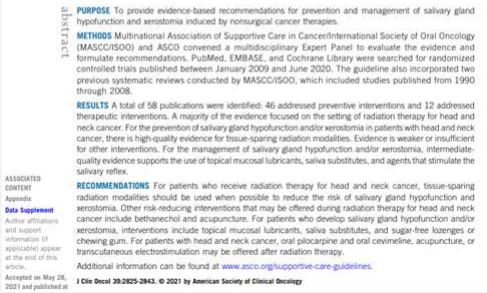
- Joint guideline statements from the International Society of Oral Oncology, the Multinational Association for Supportive Care of Cancer and the American Society of Clinical Oncology
- Guidance addressing oral care, dental management, mucositis, hypersalivation, osteoradionecrosis and other general problems in patients with advanced cancers
- National Comprehensive Cancer Network guidelines for head and neck cancers, with the latest version referred to having been released in May 2016
 |  |
The Head And Neck Cancer Patient Journey456
Diagnosis → H&N MDT Meeting Discussion → Work-up to Cancer Treatment → Surgery → H&N MDT Meeting Rediscussion → Radiotherapy +/- Chemotherapy → Rehabilitation → Chronic side effects and survivorship
Lecturer — Long-Term Care
The patient journey also includes referral, immunotherapy in some cases, and long-term surveillance. Management of chronic side effects and survivorship care continue after rehabilitation.
Signs And Symptoms Suspicious For Head And Neck Cancer7
Duration for longer than 2-3 weeks:
- Persistent sore throat
- Foul breath unexplained by hygiene
- Hoarseness and changes in voice
- Swelling in throat
- Persistent ear ache and constant pain
- Lump, bump or mass in the neck without pain
- White or red patch in the mouth
- Persistent nasal obstruction or congestion
- Blocked nose on one side or bloody discharge from nose
- Difficulty swallowing (dysphagia)
- Trouble breathing
- Unexplained facial palsy, weakness or severe facial pain/numbness
Lecturer — Delayed Diagnosis
Head and neck cancers may produce relatively subtle symptoms, particularly when lesions involve the pharynx or oropharyngeal regions. Diagnosis may therefore occur only once disease is advanced.
- Dentists may be the first clinicians to identify a suspicious lesion, including one of which the patient is unaware.
- Earlier diagnosis was associated with a better course and prognosis.
- Persistent ulcers
Lecturer — Referral Threshold
Suspicious signs or symptoms that remain unresolved for longer than two weeks should be investigated or referred. These features also have differential diagnoses, including infection, benign conditions and other non-head-and-neck-cancer causes, which can contribute to delayed diagnosis.
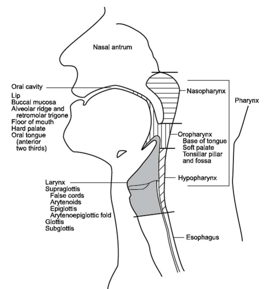
Anatomic Sites And Subsites8
- Nasal antrum
- Oral cavity
- Lip
- Buccal mucosa
- Alveolar ridge and retromolar trigone
- Floor of mouth
- Hard palate
- Oral tongue (anterior two thirds)
- Nasopharynx
- Pharynx
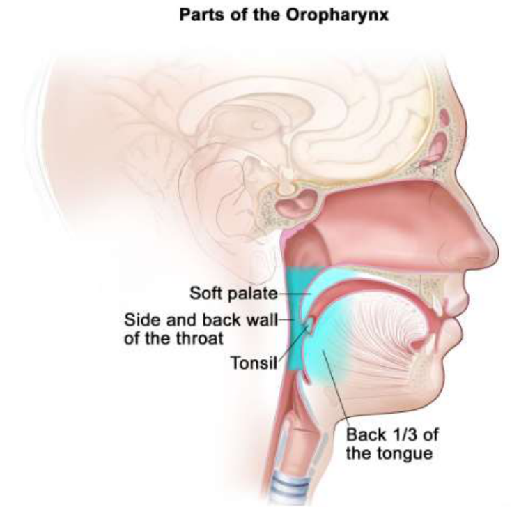
- Oropharynx
- Base of tongue
- Soft palate
- Tonsillar pillar and fossa
- Hypopharynx
- Oropharynx
- Larynx
- Supraglottis
- False cords
- Arytenoids
- Epiglottis
- Arytenoepiglottic fold
- Glottis
- Subglottis
- Supraglottis
- Esophagus
NCCN version 2.2026 – National Comprehensive Cancer Network
Lecturer — Head and Neck Spread
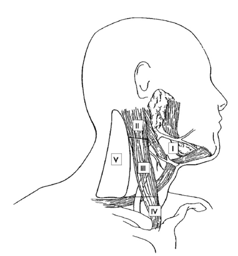
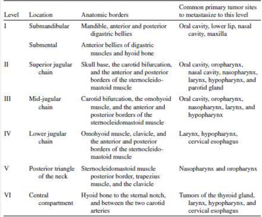
Head and neck squamous cell carcinomas may spread by local invasion, regional lymphatic dissemination or vascular dissemination. They tend to metastasise to regional lymph nodes rather than primarily through the bloodstream; distant metastases usually occur later, with the lungs the most common distant site.
- Metastasis to the liver without lung metastases was described as unusual for head and neck squamous cell carcinoma.
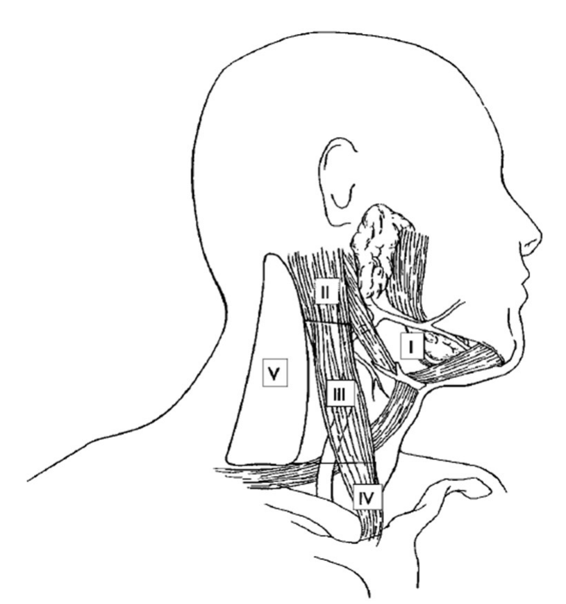
Cervical Lymphatics Level Designation910
NCCN version 2.2026 – National Comprehensive Cancer Network
-
Use 1-2 kg of pressure when palpating
-
Fixation
-
Enlargement
-
Unilateral
-
Tenderness
NCCN version 2.2026 – National Comprehensive Cancer Network
Lecturer — Cervical Node Examination
Concerning cervical lymph node features include hard consistency and enlargement greater than approximately 1 to 1.5 cm. Firm palpation is important because lightly touching or “bottle” palpation may not provide sufficient information; approximately 1 to 2 kg of pressure was described, with the node palpated against the underlying structure.
 |  |
 |
TNM Staging11
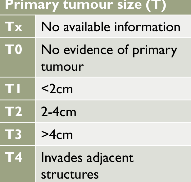
Primary tumour size (T)12
| Stage | Description |
|---|---|
| Tx | No available information |
| T0 | No evidence of primary tumour |
| T1 | <2cm |
| T2 | 2-4cm |
| T3 | >4cm |
| T4 | Invades adjacent structures |
Regional lymph node involvement (N)
| Stage | Description |
|---|---|
| Nx | Nodes cannot be / were not assessed |
| N0 | No regional nodes involved |
| N1 | Ipsilateral single node <3cm diameter |
| N2a | Ipsilateral single node 3-6cm |
| N2b | Ipsilateral multiple nodes <6cm |
| N2c | Bilateral or contralateral nodes <6cm |
| N3 | Any node >6cm |
Involvement by distant metastases (M)
| Stage | Description |
|---|---|
| Mx | Distant metastasis not assessed |
| M0 | No evidence of distant metastasis |
| M1 | Distant metastasis is present |
| Classification | Staging |
|---|---|
| Clinical = cTNM | Stage 0 – carcinoma in situ |
| Pathological = pTNM | Stage I and II – tumours localised to organ of origin |
| Stage III – locally extensive spread, particularly to regional lymph nodes | |
| Stage IV – distant metastasis |
Lecturer — TNM Classification
TNM staging is an anatomical classification system that represents the extent of cancer and functions as a shorthand system. It was initially conceived in the 1940s and has been adopted by the Union for International Cancer Control and the American Joint Committee on Cancer; the American system is used in Australia.
- The exact T and N classifications differ according to the anatomical cancer site.
- Pathological TNM classification after surgery helps guide decisions about adjuvant therapy.
HPV Positive Oropharyngeal Cancer13
- Rising incidence of oropharyngeal cancer due to increasing prevalence of Human papillomavirus (HPV)-induced cancers
- Incidence of HPV-associated tonsil and BOT cancers increasing 5% increase per year since 1990.
- Younger, healthier individuals with little or no tobacco exposure
- Viral aetiological factor: types 16 and 18
- Type 16 most prevalent subtype in 95% of cases
- Preventable with vaccines
- Gardasil 9: HPV types 6, 11, 16, 18, 31, 33, 45, 52, 58
- Highly responsive to treatment and has excellent prognosis
- Immunohistochemistry: P16 biomarker significantly correlated with HPV status
Lecturer — HPV Vaccination
HPV vaccination was discussed as a way of reducing the incidence of HPV-associated cancers. Although vaccination had traditionally been directed towards females in relation to cervical cancer, males were also being included in the national immunisation program at approximately 12 to 13 years of age.
- HPV-associated cancers generally have a better prognosis than HPV-negative cancers associated with smoking and alcohol.
Diagnostic Evaluation
Clinical Assessment14
- Head and neck examination
- Fiber-optic nasopharyngolaryngoscopy
- EUA to assess tumour extent and obtain biopsy
Imaging
- CT scan
- MRI
- PET/CT (if no obvious primary on imaging)
- FNA biopsy of metastatic or equivocal nodes (US or CT guided needle biopsy)
- OPG (mandibultomy, mandibulectomy, radiotherapy)
Lecturer — Diagnostic Evaluation
Diagnostic evaluation identifies the tumour type, anatomical site, clinical stage and extent of disease, and these findings influence treatment planning. A detailed history, physical examination, blood tests and thorough examination for a primary disease site may be included.
- Examination under anaesthesia is particularly important when metastatic carcinoma is present in the neck and the primary site has not been identified.
- An orthopantomogram is important from the dental perspective as part of the pre-radiotherapy dental assessment.
Advantages of CT vs MRI
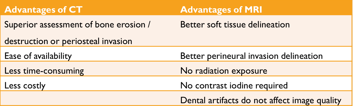
| Advantages of CT | Advantages of MRI |
|---|---|
| Superior assessment of bone erosion / destruction or periosteal invasion | Better soft tissue delineation |
| Ease of availability | Better perineural invasion delineation |
| Less time-consuming | No radiation exposure |
| Less costly | No contrast iodine required |
| Dental artifacts do not affect image quality |
Comprehensive Cancer Care15
-
Cancer treatment
-
General medical care
-
Pain and symptom management
-
Nutritional support (enteral feeding, oral nutrition)
-
Dental care
-
Speech and swallowing therapy
-
Smoking and alcohol cessation
-
Audiology
-
Tracheostomy care
-
Wound management
-
Depression and anxiety management
-
Social work and case management
-
Supportive care (palliative care)
-
Physical therapy (lymphoedema)
-
Health literacy and social circumstances
-
End-of-life care
-
Occupational therapy and lymphoedema management
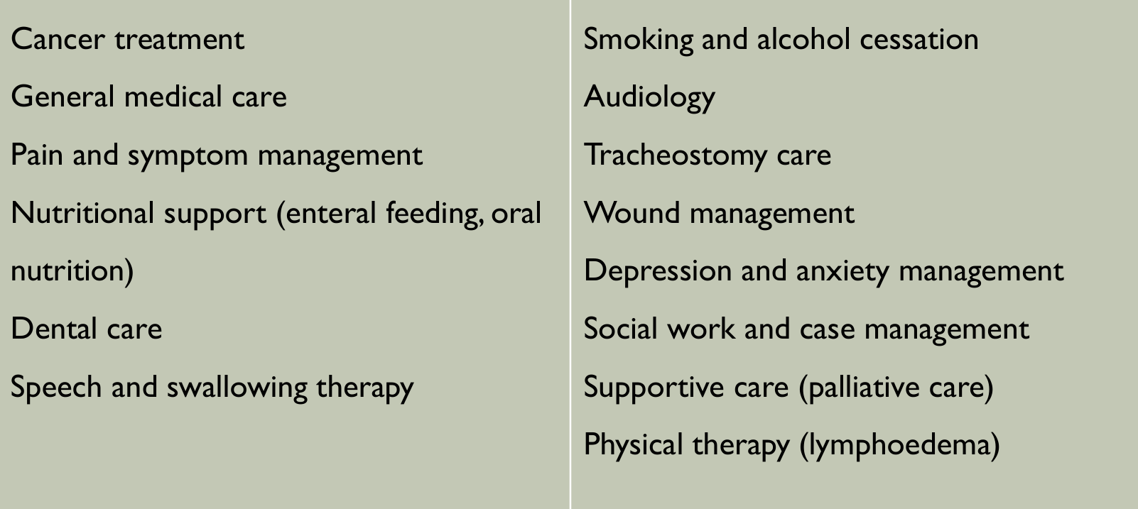
Head And Neck Multidisciplinary Team Members16
- Otolaryngologist (Ear Nose Throat) surgeon
- Plastic and reconstructive surgeon
- Oral and maxillofacial surgeon
- Radiation Oncologist
- Medical Oncologist
- Dental: Special Needs Dentistry, Oral Medicine, Maxillofacial prosthodontist
- Radiologist
- Pathologist
- Anaesthetist
- High risk assessment physician
- Head and neck cancer coordinator
- Clinical nurse specialist
- Speech-language therapist
- Dietitian
- Social worker
- Adjunctive services:
- Pain specialist
- Palliative care specialist
- Neurosurgeon
- Ophthalmologist
- Psychologist/Psychiatrist
- Occupational therapist
- Addiction services
- Audiologist
Lecturer — MDT Responsibilities
Head and neck cancer treatment requires multiple teams because treatment may be disfiguring and may substantially impair normal function.
- The MDT assesses the patient, stages the cancer, develops and coordinates the treatment plan, and decides whether treatment is curative or palliative.
- It reviews progress, monitors for relapse, and liaises with community healthcare professionals after discharge.
- A designated home team generally coordinates the patient’s care and communicates the recommendations to the patient.
Lecturer — MDT Meetings
MDT meetings may include new patients, patients with recurrence, patients requiring review of new investigations, and patients requiring pathology review after surgery.
- The team may review tumour histology and determine TNM staging.
- It agrees on the proposed treatment and develops a surveillance plan.
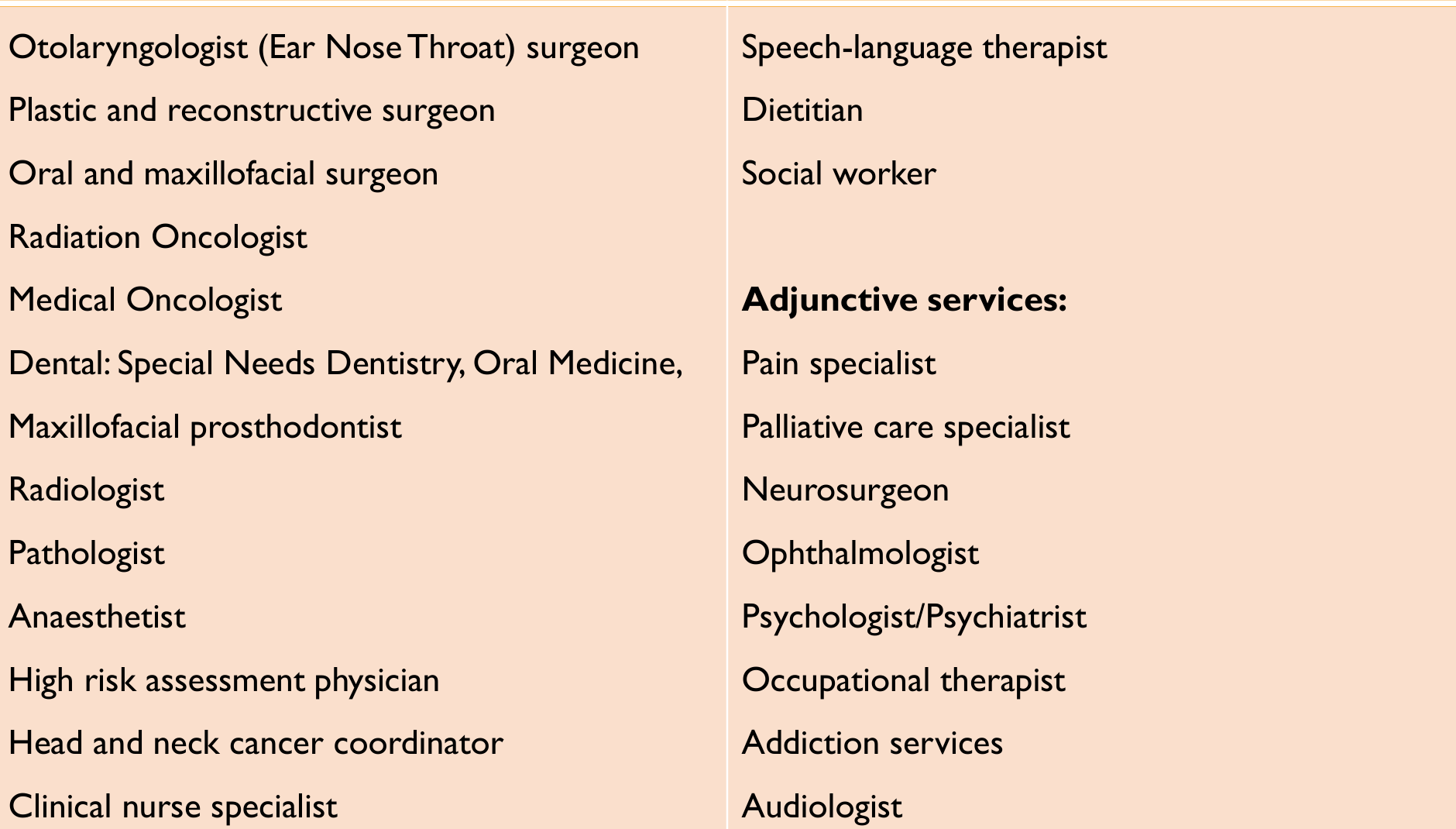
Factors Influencing Recommended Treatment17
| Tumor Factors | Patient Factors | Physician Factors |
|---|---|---|
| Site | Medical condition and comorbidities | Expertise |
| Size | The impact of treatment on the quality of life of the patient | Availability of a comprehensive team: nursing, dental, speech therapy, dietitian, rehabilitation, and social support services |
| Depth of invasion | Performance status | Personal preferences of members of the team |
| Degree of spread | Treatment cost | Preferences of the institution |
| Location and proximity to critical structures | Patient compliance | |
| Previous treatment | Previous treatment | |
| Need for reconstructive surgery | Patient preference |
- Curative Intent
- Palliative Intent
O’Sullivan B, Brierley J, D’Cruz A, et al. UICC Manual of Clinical Oncology. Chicester: John Wiley & Sons, Incorporated, 2015.
Lecturer — Treatment Decision Factors
Treatment is primarily determined by expected oncological outcomes, but functional and cosmetic outcomes are also important.
- Potential treatment side effects and cost-effectiveness also influence the recommendation.
- These factors help determine whether treatment is delivered with curative or palliative intent.
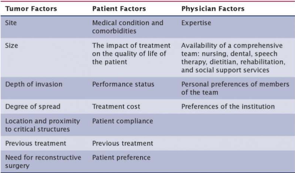
Standard Care Treatments18
Neo-Adjuvant = Shrink tumour first
- Surgery
- Radiotherapy
- Chemotherapy
Treatment modalities:
- Surgery, unimodality
- Neo-adjuvant radiotherapy
- Primary radiotherapy, unimodality
- Surgery + Adjuvant radiotherapy +/- chemotherapy
- Primary chemoradiotherapy
- Palliative radiotherapy / systemic therapy
Lecturer — Treatment Morbidity
Surgery and radiotherapy were described as the proven curative modalities, while chemotherapy is commonly used as an adjunct. Generally, the greater the number of modalities used, the greater the associated morbidity.
Neoadjuvant Immunotherapy
- Standard of care in Australia in past 4 months
- Locally advanced HNSCC (stage III and IV)
- Enhances anti-tumour immunity
- Primes T-cells to target cancer cells via PDL-1 pathway
- 3 weekly injections
- 2 cycles pre-op (surgery after 1 week)
- 15 cycles post-treatment (surgery + RT +/- CT)
- 12% increase in disease-free survival at 36 months
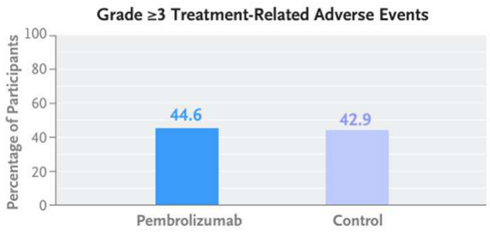
- Similar incidence of adverse events to control group
Source Article19
The NEW ENGLAND JOURNAL of MEDICINE — ORIGINAL ARTICLE: “Neoadjuvant and Adjuvant Pembrolizumab in Locally Advanced Head and Neck Cancer” — R. Uppaluri, R.I. Haddad, Y. Tao, C. Le Tourneau, N.Y. Lee, W. Westra, R. Chernock, M. Tahara, K.J. Harrington, A.L. Klochikhin, I. Braña, G. Vasconcelos Alves, B.G.M. Hughes, M. Oliva, I. Pinto Figueiredo Lima, T. Ueda, T. Rutkowski, U. Schroeder, P.S. Mauz, T. Fuereder, S. Laban, N. Oridate, A. Popovtzer, N. Mach, Y. Korobko, D.A. Costa, A. Hooda-Nehra, C.P. Rodriguez, R.B. Bell, C. Manschot, K. Benjamin, B. Gumuscu, and D. Adkins, for the KEYNOTE-689 Investigators. N Engl J Med 2025;393:37-50. DOI: 10.1056/NEJMoa2415434. Copyright © 2025 Massachusetts Medical Society.
 |  |
Abstract
Background — The benefit of the addition of perioperative pembrolizumab to standard care with surgery and adjuvant therapy for patients with locally advanced head and neck squamous-cell carcinoma (HNSCC) is unclear.
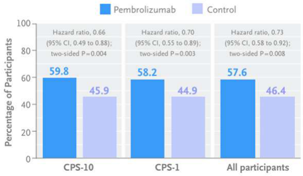
Methods — In this phase 3, open-label trial, participants with locally advanced HNSCC were randomly assigned in a 1:1 ratio to receive 2 cycles of neoadjuvant pembrolizumab and 15 cycles of adjuvant pembrolizumab (both at a dose of 200 mg every 3 weeks) in addition to standard care (pembrolizumab group) or standard care alone (control group). Standard care was surgery and adjuvant radiotherapy with or without concomitant cisplatin. The primary end point was event-free survival, sequentially assessed in participants whose tumors expressed programmed death ligand 1 (PD-L1) with a combined positive score (CPS) of 10 or more (CPS-10 population), participants whose tumors expressed PD-L1 with a CPS of 1 or more (CPS-1 population), and all the participants. A higher CPS indicates a higher proportion of cells that express PD-L1.
Event-Free Survival Results (Figure)
Percentage of participants (axis: 100, 80, 60, 40, 20, 0):
- CPS-10: Pembrolizumab 59.8 vs Control 45.9; hazard ratio 0.66 (95% CI, 0.49 to 0.88); two-sided P=0.004
- CPS-1: Pembrolizumab 58.2 vs Control 44.9; hazard ratio 0.70 (95% CI, 0.55 to 0.89); two-sided P=0.003
- All participants: Pembrolizumab 57.6 vs Control 46.4; hazard ratio 0.73 (95% CI, 0.58 to 0.92); two-sided P=0.008
The article was published on June 18, 2025, at NEJM.org.
Management Of Head And Neck Cancer By Intent And Stage
UICC 2015
Curative Intent20
Non-metastatic HNC (Stage I–IVB) (except for those unable to tolerate standard treatment due to co-morbidity or poor performance status)
- Early stage (I/II): Single modality (surgery/RT)
- Surgery preferred in oral cavity, paranasal sinuses
- Laser surgery is a suitable alternative in early glottic cancers
- Robotic surgeries have an emerging role in early oropharyngeal cancers
- RT preferred in oropharynx, nasopharynx, larynx, and hypopharynx
- Surgery/radiotherapy in glottic cancers
- Advanced stage (III/IVA/IVB): Multimodality
- Oral cavity, paranasal sinuses: surgery with PORT with/without concurrent chemotherapy
- Oropharynx, nasopharynx: CCRT with/without salvage surgery
- Larynx/hypopharynx: CCRT or ICT followed by RT/CCRT if deemed suitable for organ preservation; alternatively surgery with PORT with/without concurrent chemotherapy
- Bioradiotherapy with cetuximab is another treatment option
Lecturer — Choosing Surgery
Surgery alone may be preferred when the cancer is early stage, the lesion is accessible, or surgery is less morbid than radiotherapy.
- Avoiding radiotherapy-related bone damage may be desirable.
- In a young patient, radiotherapy may be preserved for a possible second primary or recurrence.
Lecturer — Choosing Radiotherapy
Primary radiotherapy may be preferred when surgery would impair function or prosthetic rehabilitation, carry high morbidity, or produce a poor cosmetic outcome.
- It may also be preferred when the tumour is particularly radiosensitive or surgical risk is high because of comorbidities.
Palliative Intent
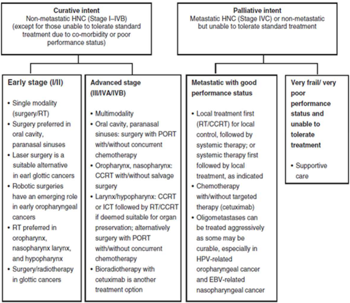
Metastatic HNC (Stage IVC) or non-metastatic but unable to tolerate standard treatment
- Metastatic with good performance status:
- Local treatment first (RT/CCRT) for local control, followed by systemic therapy; or systemic therapy first followed by local treatment, as indicated
- Chemotherapy with/without targeted therapy (cetuximab)
- Oligometastases can be treated aggressively as some may be curable, especially in HPV-related oropharyngeal cancer and EBV-related nasopharyngeal cancer
- Very frail/very poor performance status and unable to tolerate treatment:
- Supportive care
Lecturer — Palliative Treatment
Palliative treatment is used to improve quality of life and reduce symptoms rather than aim for complete tumour eradication.
- Treatment may reduce tumour size and relieve symptoms.
- Options include palliative radiotherapy, systemic chemotherapy, and immunotherapy.
HNC – head and neck cancer; RT – radiotherapy; PORT – postoperative radiotherapy; CCRT – concurrent chemoradiotherapy; ICT – induction chemotherapy; EBV – Epstein-Barr virus; HPV – human papillomavirus
Oral Cavity Management
Slide marked “DON’T HAVE TO KNOW”
NCCN Guidelines Version 2.2026 — Cancer of the Oral Cavity (Including Mucosal Lip) — OR-1
Buccal mucosa, floor of mouth, oral tongue, alveolar ridge, retromolar trigone, hard palate
Workup (OR-1)21
- History and physical (H&P), including a complete head and neck exam; mirror and fiberoptic examination as clinically indicated
- Biopsy
- As clinically indicated:
- Chest CT (with or without contrast)
- CT with contrast and/or MRI with and without contrast of primary and neck
- Consider FDG-PET/CT
- Examination under anesthesia (EUA) with endoscopy
- Preanesthesia studies
- Dental/prosthodontic evaluation, including Panorex or dental CT without contrast
- Nutrition, speech, and swallowing evaluation/therapy
- Smoking cessation counseling
- Fertility/reproductive counseling
- Screening for hepatitis B
- PD-L1 testing by immunohistochemistry (IHC) (combined positive score [CPS])
- Multidisciplinary consultation as clinically indicated
- Genetic testing in the context of early age of onset or family history
 |  |
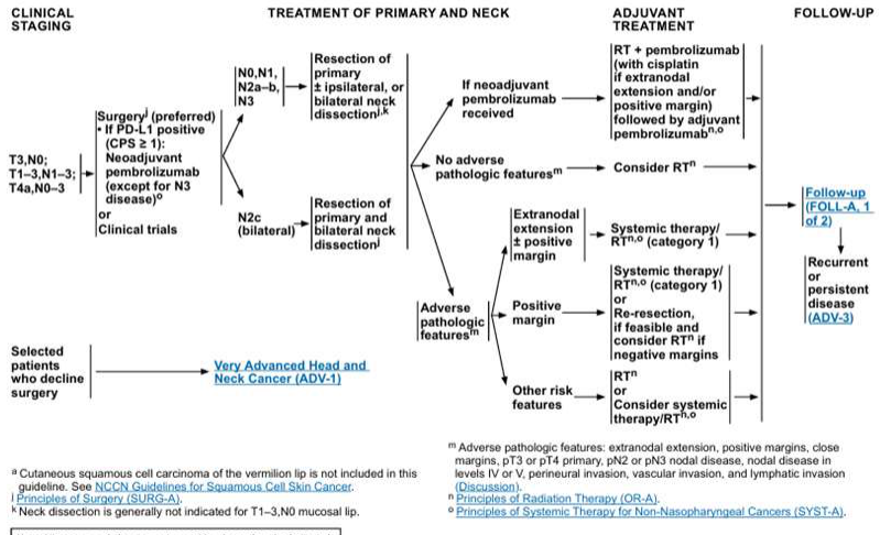 |
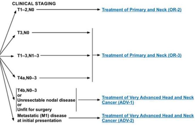
Clinical Staging (OR-1)
- T1–2,N0 → Treatment of Primary and Neck (OR-2)
- T3,N0; T1–3,N1–3; T4a,N0–3 → Treatment of Primary and Neck (OR-3)
- T4b,N0–3 or Unresectable nodal disease → Treatment of Very Advanced Head and Neck Cancer (ADV-1)
- Unfit for surgery; Metastatic (M1) disease at initial presentation → Treatment of Very Advanced Head and Neck Cancer (ADV-2)
Treatment of Primary and Neck (OR-2)
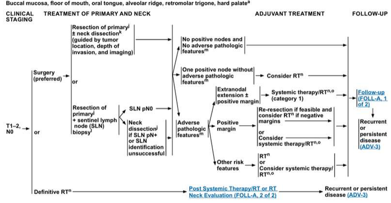
T1–2,N0:
- Surgery (preferred): Resection of primary; neck dissection (guided by tumor location, depth of invasion, and imaging)
- Adjuvant treatment:
- No positive nodes and no adverse pathologic features → Consider RT (category 1)
- One positive node without adverse pathologic features → Systemic therapy/RT
- Adverse pathologic features:
- Positive margin → RT or Consider systemic therapy
- Other risk features → RT or Consider systemic therapy
- Or: Resection of primary + sentinel lymph node (SLN) biopsy
- SLN pN0 → Re-resection if feasible and consider RT if negative margins
- Neck dissection if SLN pN+ or SLN identification unsuccessful
- Or: Definitive RT → Post Systemic Therapy/RT or RT Neck Evaluation (FOLL-A, 2 of 2) → Recurrent or persistent disease (ADV-3)
Follow-up (FOLL-A, 1 of 2) → Recurrent or persistent disease (ADV-3)
Treatment of Primary and Neck (OR-3)
T3,N0; T1–3,N1–3; T4a,N0–3:
- Surgery (preferred). If PD-L1 positive (CPS ≥ 1): Neoadjuvant pembrolizumab (except for N3 disease) or clinical trials
- Neck treatment:
- N0,N1; N2a-b; N3 → Resection of primary (± ipsilateral, or bilateral neck dissection)
- N2c (bilateral) → Resection of primary and bilateral neck dissection
- Adjuvant treatment:
- If neoadjuvant pembrolizumab received → RT + pembrolizumab (with cisplatin if extranodal extension and/or positive margins) followed by adjuvant pembrolizumab
- No adverse pathologic features → Consider RT
- Adverse pathologic features:
- Extranodal extension + positive margin → Systemic therapy/RT (category 1)
- Positive margin → Systemic therapy/RT (category 1) or re-resection, if feasible and consider RT if negative margins
- Other risk features → RT or Consider systemic therapy/RT
- Selected patients who decline surgery → Very Advanced Head and Neck Cancer (ADV-1)
Follow-up (FOLL-A, 1 of 2) → Recurrent or persistent disease (ADV-3)
Footnotes
- Cutaneous squamous cell carcinoma of the vermilion lip is not included in this guideline. See NCCN Guidelines for Squamous Cell Skin Cancer.
- H&P should include documentation and quantification (pack years smoked) of tobacco use history, as well as alcohol use and counseling. All patients who currently smoke should be advised to quit smoking, and those who formerly smoked should be advised to remain abstinent from smoking. For additional cessation support, refer to the Smoking Cessation and Treatment Resources in the NCCN Guidelines for Smoking Cessation.
- Screen for distress. Refer to the NCCN Distress Thermometer and Problem List, which includes social determinants of health. See NCCN Guidelines for Distress Management (DIS-A).
- Image-guided (ultrasound [US] or CT) needle biopsy of cystic neck nodes may offer better diagnostic yield than fine-needle aspiration (FNA) by palpation alone for initial diagnosis in this setting. For unresectable or metastatic disease where there is a plan for systemic therapy, a core biopsy would allow for biomarker testing.
- Principles of Imaging (IMG-A); Principles of Oral/Dental Evaluation and Management (DENT-A); Principles of Nutrition: Management and Supportive Care (NUTR-A); Principles of Systemic Therapy for Non-Nasopharyngeal Cancers (SYST-A).
- Adverse pathologic features: extranodal extension, positive margins, close margins, pT3 or pT4 primary, pN2 or pN3 nodal disease, nodal disease in levels IV or V, perineural invasion, vascular invasion, and lymphatic invasion (Discussion).
Note: All recommendations are category 2A unless otherwise indicated.
Version 2.2026, 09/12/2026 © 2026 National Comprehensive Cancer Network® (NCCN®). All rights reserved. NCCN Guidelines® and this illustration may not be reproduced in any form without the express written permission of NCCN.
Oropharyngeal Management
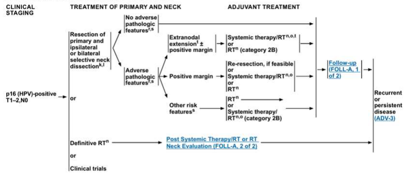
NCCN Guidelines Version 2.2026 — Cancer of the Oropharynx — Base of Tongue/Tonsil/Posterior Pharyngeal Wall/Soft Palate — ORPH-1, ORPH-2, ORHPV-1
Workup (ORPH-1)22
- Tumor human papillomavirus (HPV) testing by p16 IHC required
- H&P, including a complete head and neck exam; mirror and fiberoptic examination as clinically indicated
- Biopsy of primary site or fine-needle aspiration (FNA) of the neck
- CT with contrast and/or MRI with and without contrast of primary and neck
- As clinically indicated:
- EUA with endoscopy
- Preanesthesia studies
- FDG-PET/CT
- Chest CT (with or without contrast)
- Dental evaluation including Panorex
- Nutrition, speech, and swallowing evaluation/therapy, and audiogram
- Smoking cessation counseling
- Fertility/reproductive counseling
- Screening for hepatitis B
- PD-L1 testing by IHC (CPS)
- Multidisciplinary consultation as clinically indicated
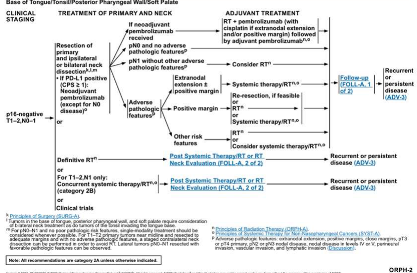 |  |
Clinical Staging (ORPH-1)
p16-negative:
- T1–2,N0–1 → ORPH-2
- T3–4a,N0–1 → ORPH-3
- T1–4a,N2–3 → ORPH-4
- T4b,N0–3, unresectable, or unfit for surgery → Treatment of Very Advanced Head and Neck Cancer (ADV-1)
- Metastatic (M1) disease at initial presentation → Treatment of Very Advanced Head and Neck Cancer (ADV-2)
p16 (HPV)-positive:
- T1–2,N0 → ORHPV-1
- T0–2,N1 (single node ≤3 cm) → ORHPV-2
- T0–2,N1 (single node >3 cm, or 2 or more ipsilateral nodes ≤6 cm), or T1–2,N2 or T3,N0–2 → ORHPV-3
- T0–3,N3 or T4,N0–3 → ORHPV-4
p16-Negative: T1–2,N0–1 (ORPH-2)
- Resection of primary and ipsilateral or bilateral neck dissection
- If neoadjuvant pembrolizumab received:
- pN0 and no adverse pathologic features → RT + pembrolizumab (with cisplatin if extranodal extension and/or positive margin) followed by adjuvant pembrolizumab
- Consider RT
- Adverse pathologic features:
- Extranodal extension ± positive margin → Systemic therapy/RT; or re-resection, if feasible; or systemic therapy/RT
- Positive margin → RT or systemic therapy/RT
- Other risk features → RT or Consider systemic therapy/RT
- If neoadjuvant pembrolizumab received:
- Or: Definitive RT; or for T1–2,N1 only: concurrent systemic therapy/RT (category 2B); or clinical trials → Post Systemic Therapy/RT or RT Neck Evaluation (FOLL-A, 2 of 2)
- Follow-up (FOLL-A, 1 of 2) → Recurrent or persistent disease (ADV-3)
p16 (HPV)-Positive: T1–2,N0 (ORHPV-1)
- Resection of primary and ipsilateral or bilateral selective neck dissection
- No adverse pathologic features → Systemic therapy/RT or RT (category 2B)
- Adverse pathologic features:
- Extranodal extension ± positive margin → Systemic therapy/RT or RT (category 2B); or re-resection, if feasible, or systemic therapy/RT
- Positive margin → RT or systemic therapy/RT
- Other risk features → RT or systemic therapy/RT (category 2B)
- Or: Definitive RT; or clinical trials → Post Systemic Therapy/RT or RT Neck Evaluation (FOLL-A, 2 of 2)
- Follow-up (FOLL-A, 1 of 2) → Recurrent or persistent disease (ADV-3)
Footnotes
- Principles of p16 Testing for HPV-Mediated Oropharyngeal Cancer (ORPH-B); Principles of Surgery (SURG-A); Principles of Imaging (IMG-A); Principles of Radiation Therapy (ORPH-A); Principles of Systemic Therapy for Non-Nasopharyngeal Cancers (SYST-A).
- H&P should include documentation and quantification (pack years smoked) of tobacco use history, as well as alcohol use and counseling. All patients who currently smoke should be advised to quit smoking, and those who formerly smoked should be advised to remain abstinent from smoking. For additional cessation support, refer to the Smoking Cessation and Treatment Resources in the NCCN Guidelines for Smoking Cessation.
- Screen for distress. Refer to the NCCN Distress Thermometer and Problem List, which includes social determinants of health. See NCCN Guidelines for Distress Management (DIS-A).
- Image-guided (US or CT) needle biopsy of cystic neck nodes may offer better diagnostic yield than FNA by palpation alone for initial diagnosis in this setting. For unresectable or metastatic disease where there is a plan for systemic therapy, a core biopsy would allow for biomarker testing.
- Prior to treatment, EUA with biopsy confirmation of the oropharyngeal primary site is recommended for patients presenting with a p16+ cervical lymph node. See Principles of Surgical Management (SURG-A).
- Tumors in the base of tongue, posterior pharyngeal wall, and soft palate require consideration of bilateral neck treatment as do tumors of the tonsil invading the tongue base.
- For pN0-N1 and no poor pathologic risk features, single-modality treatment should be considered whenever possible. For T1-T2 primary tumors near midline and resected to adequate margins and with no adverse pathologic features, a staged contralateral neck dissection can be performed in order to avoid RT. Lateral tumors pN0-N1 resected with favorable pathologic features can be observed.
- The clinical staging definitions are based on the AJCC 8th edition for oropharynx cancer (see ST-4 for p16-, and see ST-7 for p16+). Definitions for nodal staging criteria previously used in clinical trials (AJCC 7th edition) for appropriate staging criteria are included.
- Adverse pathologic features: extranodal extension, positive margins, close margins, pT3 or pT4 primary, pN2 or pN3 nodal disease, nodal disease in levels IV or V, perineural invasion, vascular invasion, and lymphatic invasion (Discussion). In the context of HPV+ disease, close margins are defined as <3 mm; the definition of an adverse pathologic feature in HPV+ disease is an area of active research, including the presence and extent of extranodal extension and the number of involved nodes. The recommendations for patients at high risk with extranodal extension + positive margins are based on randomized studies involving patients for whom the HPV status of their tumors was not specified.
Note: All recommendations are category 2A unless otherwise indicated.
- Students were not expected to memorise the detailed oral cavity treatment pathways; the diagrams demonstrate that defined pathways exist for particular cancer types and stages.
Lecturer — HPV Positive Disease
p16-positive tumours have separate treatment pathways because HPV-associated disease generally has a better prognosis and differs epidemiologically from HPV-negative disease.
Version 2.2026, 09/12/2026 © 2026 National Comprehensive Cancer Network® (NCCN®). All rights reserved. NCCN Guidelines® and this illustration may not be reproduced in any form without the express written permission of NCCN.
Pre-Treatment Work-Up And Support
Many appointments are required before treatment can begin.
Pre-Treatment Appointments23
- Anaesthetic assessment
- Dental assessment
- Nutrition assessment
- Speech and swallow evaluation
- Radiation oncology and medical oncology consults
- Audiogram
- Smoking cessation counselling
- Psychological counselling
- Any further investigations (e.g. incidental findings on imaging)
Contact: Cancer Nurse Coordinator
Lecturer — Treatment Coordination
Although one surgical team may act as the main home team, substantial coordination is required between the various services.
Support Services And Contacts
Cancer Council24
13 11 20 for information and support (Monday to Friday, general office hours)

Cancer Nurse Coordination Service
(08) 6457 0855 (Monday to Friday, 8.30am–4.30pm)
Lecturer — Nurse Coordination
The cancer nurse coordinator service acts as a valuable liaison service throughout the patient journey.
- Acts as a central point of contact.
- Helps patients navigate the patient journey.
- Connects patients with hospital teams.
- Supports communication between patients and healthcare services.
Crisis Care
- (08) 9223 1111
- 1800 199 008 (freecall STD)
Lifeline
13 11 14
Mental Health Emergency Response Line
- Perth Metro: 1300 555 788
- Peel Region: 1800 676 822
- RuralLink WA: 1800 552 002
WA Psycho-Oncology Service
Clinical psychology care for adults affected by cancer.
The service is administered by the Cancer Network WA.
Lecturer — Psycho-Oncology Support
The psycho-oncology service provides targeted psychological support so that patients do not have to rely solely on a general psychology waiting list.
- It is particularly relevant to the depression, anxiety and psychosocial effects associated with cancer.
We are proud to be a smoke-free site. Thank you for not smoking or vaping. Ask a staff member about free nicotine therapy to help your cravings during your hospital stay.
Contact Details
- Cancer Network WA, Queen Elizabeth II Medical Centre, Locked Bag 2012, Nedlands WA 6009
- (08) 6457 0855
- cnwa@health.wa.gov.au
This document can be made available in alternative formats on request.
© North Metropolitan Health Service 2023.
Oral Side Effects Of Head And Neck Cancer Radiotherapy
Short Term25
-
Oral mucositis
-
Altered taste
-
Oral soreness
-
Erythema
-
Ulceration
-
Changes in saliva
-
Difficulty eating
-
Increased mucus
-
Oral discomfort
-
Fungal infection
 |  |
 |  |
Long Term
-
Altered saliva quality and quantity
-
Oral thrush
-
Dysphagia
-
Trismus
-
Periodontal susceptibility
-
Radiation caries
-
*Osteoradionecrosis
-
Xerostomia
-
Dental erosion
-
Reduced healing capacity
-
Muscle fibrosis
-
Difficulties wearing dentures
-
Changes in oral flora
Role Of Dental Practitioner Pre-Radiotherapy26
-
Identify existing oral disease & potential risk of oral disease
-
Remove dental infections and teeth with poor long term prognosis prior to commencing cancer therapy
-
Avoid unscheduled interruption to primary treatment due to dental issues
-
Prepare the patient for expected side effects of cancer therapy
-
Develop a plan for preventive care and follow up to meet the increasing challenges during cancer therapy
-
Establish multidisciplinary collaboration within the cancer team
-
Pay particular attention to teeth receiving high-dose radiation
-
Reduce the risk of osteoradionecrosis
Lecturer — Patient Dental History
Many patients have poorly maintained dentitions, particularly because heavy smoking and alcohol use are common risk factors.
- An Australian study from 2011 found that most head and neck cancer patients had not visited a dentist during the preceding 12 months.
- The mean time since the last dental visit was approximately five and a half years.
Multidisciplinary Work Up27
-
Surgical teams — altered anatomy affecting function and rehabilitation, teeth removed as part of resection, extractions due to impingement on healing flap, extractions to monitor cancer margins, extractions during time of cancer surgery
-
Radiation Oncology — urgency to commence radiotherapy, anticipated radiation fields, risks vs benefits of extractions
-
Medical Oncology — neo-adjuvant therapy affecting timings, patient factors
-
Speech therapy and dietetics — dysphagia, tube feeding, nutritional supplementation
-
The effect of mandibulotomy, segmental mandibulectomy or temporomandibular joint removal on later extraction access
-
Potential effects on healing and the timing of dental treatment in relation to systemic therapy
Lecturer — Dental Treatment Timing
In selected complex patients, dental treatment may be performed during the same general anaesthetic as cancer surgery.
- This may be useful when the patient has significant comorbidities or would otherwise require two separate medical optimisations.
- Other indications include blood thinners, social or behavioural concerns, particularly difficult dental treatment, or access becoming more difficult after surgery.
- This approach is not used for every patient because of the logistical demands and additional time required.
- Nutritional status and the implications of altered anatomy and dental treatment
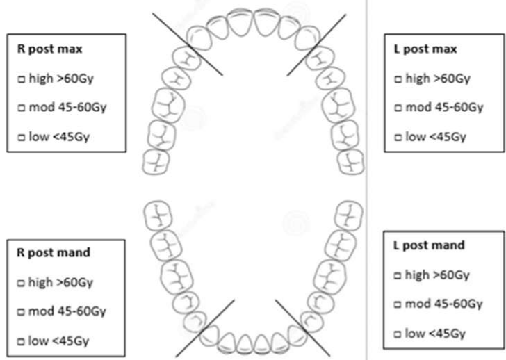
Provisional Radiotherapy Fields28
Anticipated RT dose/#:
- Ant max: □ high >60Gy □ mod 45–60Gy □ low <45Gy
- R post max: □ high >60Gy □ mod 45–60Gy □ low <45Gy
- L post max: □ high >60Gy □ mod 45–60Gy □ low <45Gy
- R post mand: □ high >60Gy □ mod 45–60Gy □ low <45Gy
- L post mand: □ high >60Gy □ mod 45–60Gy □ low <45Gy
- Ant mand: □ high >60Gy □ mod 45–60Gy □ low <45Gy
Lecturer — Treatment Priorities
Dental treatment is prioritised according to the tooth’s relationship to the radiation field, anticipated dose, long-term prognosis, likelihood of future mucosal trauma, and urgency of cancer treatment.
- Different health systems may have different arrangements.
- Some patients, particularly rural patients, may receive dental care in general practice rather than travelling to a major city.
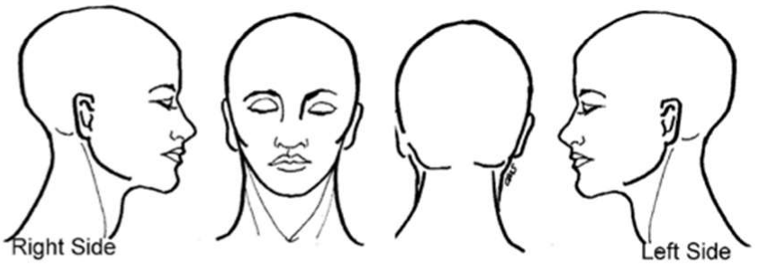
Form fields:
- H&N MDT Date:
- Patient Name:
- UR Number:
- Cancer site / TNM:
- New / Path Review / Known recurrence
- Proposed treatment:
- Rad Onc Consultant:
- Comments:
- Right Side / Left Side
 |  |
Surgery And Adjuvant Therapy Decisions2930
Diagnosis → H&N MDT Meeting Discussion → Work-up to Cancer Treatment → Surgery → H&N MDT Meeting Rediscussion → Radiotherapy +/- Chemotherapy → Rehabilitation → Chronic side effects and survivorship
- After surgery, the pathology is reviewed to determine whether adjuvant treatment is required.
Anatomical Changes Of Relevance To Dentistry31
Diagnosis → H&N MDT Meeting Discussion → Work-up to Cancer Treatment → Surgery → H&N MDT Meeting Rediscussion → Radiotherapy +/- Chemotherapy → Rehabilitation → Chronic side effects and survivorship
Lecturer — Surgical Changes
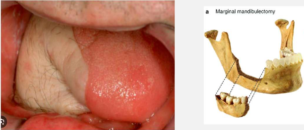
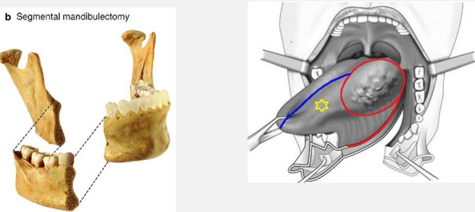
Cancer surgery and reconstruction can significantly alter oral and facial anatomy.
- Loss of teeth during resection
- Mandibulotomy
- Segmental mandibulectomy
- Temporomandibular joint removal
- Partial maxillectomy
- Marginal mandibulectomy
- Tongue resection
- Neck dissection
- Tracheostomy
- Free flap reconstruction
- Hair-bearing free flaps placed into the oral cavity
Hair-bearing flaps may cause tickling, hygiene difficulties, gagging, nausea, and saliva collecting in the hair, which can make the area difficult to clean. An eyebrow plucker may be used to remove hair from an intraoral flap when required. Surgical changes may also make later dental extractions more difficult, especially when mandibular mobility is reduced.
 |  |
Adjuvant Radiotherapy For Oral Cancers
Definitive Factors for Adjuvant Radiotherapy32
- pT3/T4
- pN2/N3
- Close margins <5mm
- Depth of invasion >10mm
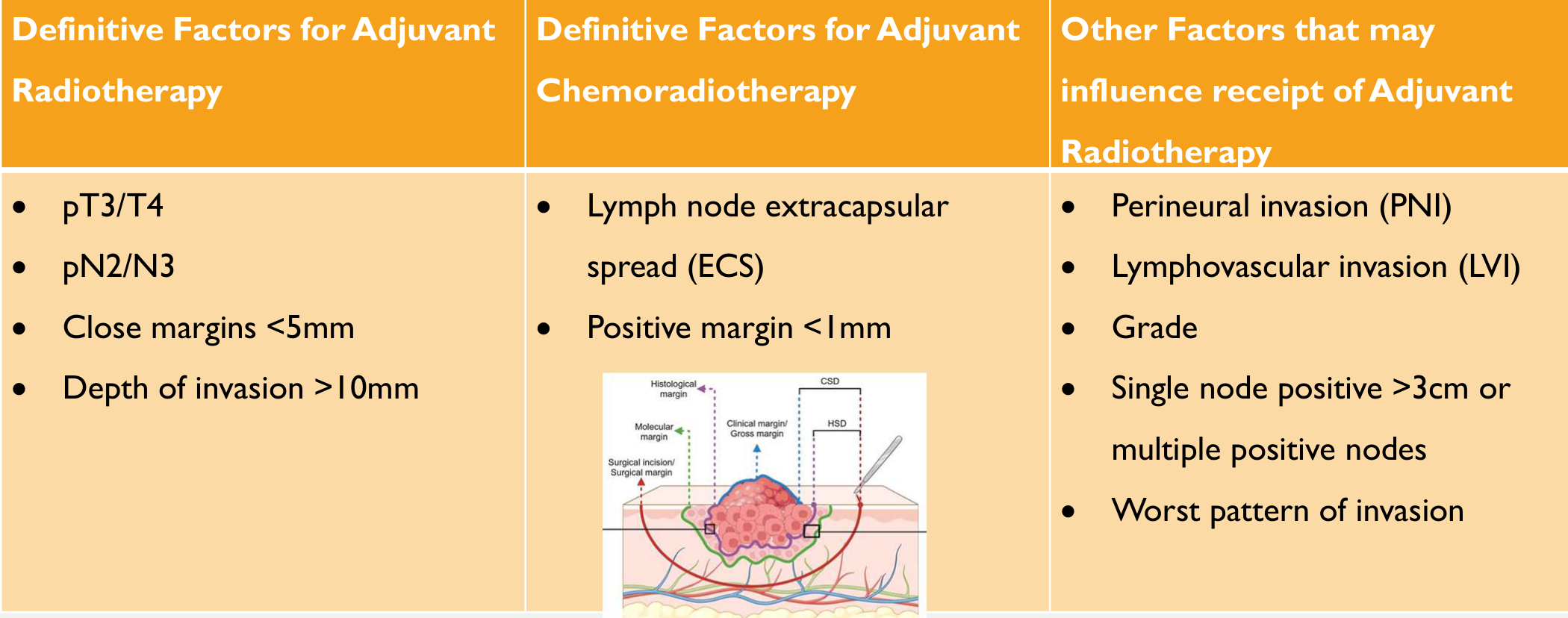 | 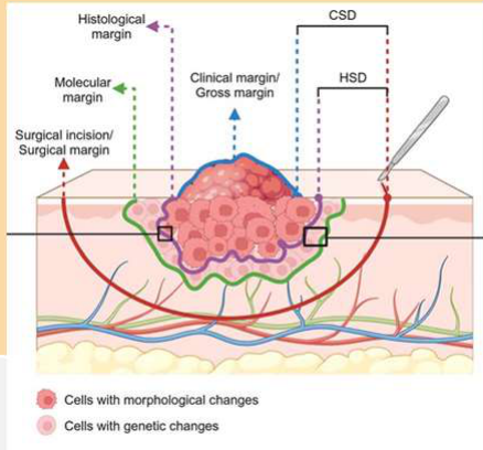 |
Definitive Factors for Adjuvant Chemoradiotherapy
- Lymph node extracapsular spread (ECS)
- Positive margin <1mm
Lecturer — Chemoradiotherapy
Adjuvant chemoradiotherapy is used when adverse pathological features indicate the need for additional systemic treatment alongside radiotherapy. Radio-sensitising chemotherapy may augment the effect of radiotherapy.
Other Factors that may influence receipt of Adjuvant Radiotherapy
- Perineural invasion (PNI)
- Lymphovascular invasion (LVI)
- Grade
- Single node positive >3cm or multiple positive nodes
- Worst pattern of invasion
Lecturer — Treatment Selection
The final decision is influenced by more than the pathological findings.
- Expected oncological benefit
- Patient comorbidities
- Functional outcomes
- Cosmetic outcomes
- Previous treatment
- The patient’s ability to tolerate further treatment
- Overall treatment intent
D’Cruz A, Vaish R, Dhar H. Oral cancers: current status. Oral Oncol. 2018;87:64-9
Chen et al. Surgical margins in head and neck squamous cell carcinoma: A narrative review. Int J Surg. 2024; 110: 3680-3700
Radiotherapy33
Diagnosis → H&N MDT Meeting Discussion → Work-up to Cancer Treatment → Surgery → H&N MDT Meeting Rediscussion → Radiotherapy +/- Chemotherapy → Rehabilitation → Chronic side effects and survivorship
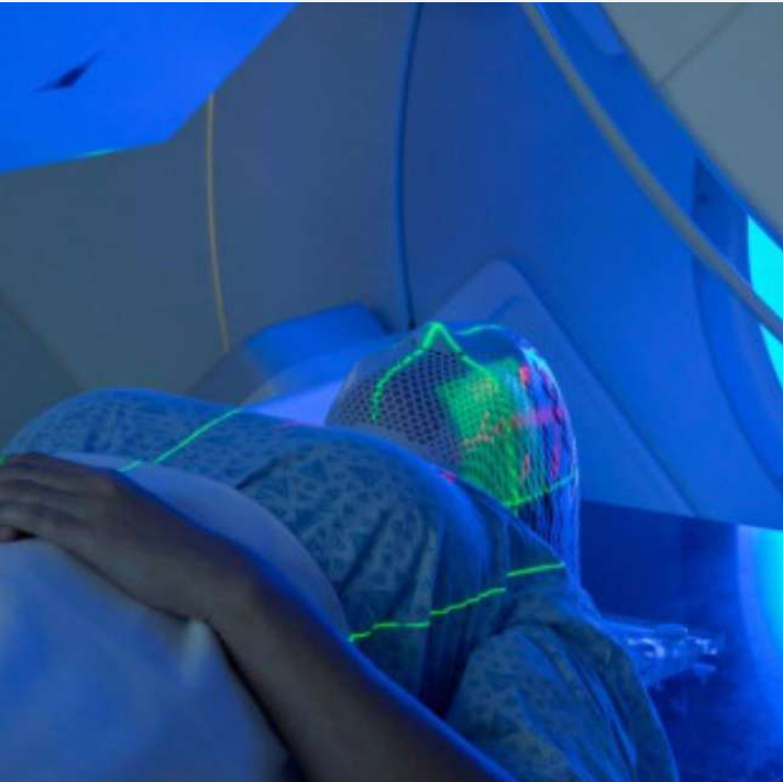
Radiotherapy Fundamentals34
- Used to treat approximately 50-60% of patients with HNC
- Aims: destroy cancer cells, reduce size of tumours
- Unit of measurement: Gray (Gy) = absorption of 1 joule of energy per kilogram of matter
- Delivered in fractions (# = treatment sessions)
- Gold standard = commence within 6 weeks of MDT discussion or surgery
Lecturer — Radiation Mechanism
Radiotherapy uses ionising radiation to induce loss of reproductive potential through DNA damage.
- The relevant damage particularly includes double-stranded DNA breaks.
- The matter absorbing radiation may be water or human tissue.
- For example, 70 Gy in 35 fractions means 70 Gy delivered over 35 treatment sessions
Lecturer — Treatment Coordination
Postoperative radiotherapy should ideally commence as soon as possible, with a goal of approximately six weeks after surgery. Achieving this requires careful planning and coordination.
Radiation Schedules
Curative Radiotherapy (complete eradication)35
- Definitive Radiotherapy: 60-72 Gy in 25-40 fractions over 6-7 weeks
- Adjuvant Radiotherapy:
- Primary and involved nodes: 56-60 Gy in 28-30 fractions (2 Gy) over 6 weeks
- High risk sites (e.g. ECS, residual disease, positive tumour margins): 4-10 Gy dose augmentation (up to 66-70 Gy)
Palliative Radiotherapy (symptom control)
-
30-50 Gy, in 5-20 fractions
-
Its purpose is symptom control and reduction of tumour burden rather than complete eradication
Prophylactic Radiotherapy (prevent growth in high-risk sites)
Challenges Of Treatment Planning Before Radiotherapy36
Most medical procedures have a modest effect on oral health risk factors. Predicting the patient’s future oral health trajectory is often fairly straightforward: we examine their past trajectory.
Lecturer — Radiotherapy Trajectory
Head and neck radiotherapy creates a complex oral health trajectory because the risk of dental disease increases after treatment, but the extent of oral deterioration varies.
- Radiation affects salivary glands and oral tissues.
- The patient’s previous dental trajectory may not predict the post-radiotherapy trajectory.
- Dental treatment timing must be balanced against the urgency of cancer treatment.
But here’s where it gets tricky…
For Head & Neck radiotherapy patients, their risk of dental disease almost always increases after treatment… but the extent can vary from mild to catastrophic. Predicting their future oral health trajectory is more complex because there are many more variables.
- Historical oral health trajectory → Post-treatment oral health trajectory
- Historical oral health trajectory → Post-radiotherapy oral health trajectory
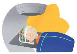
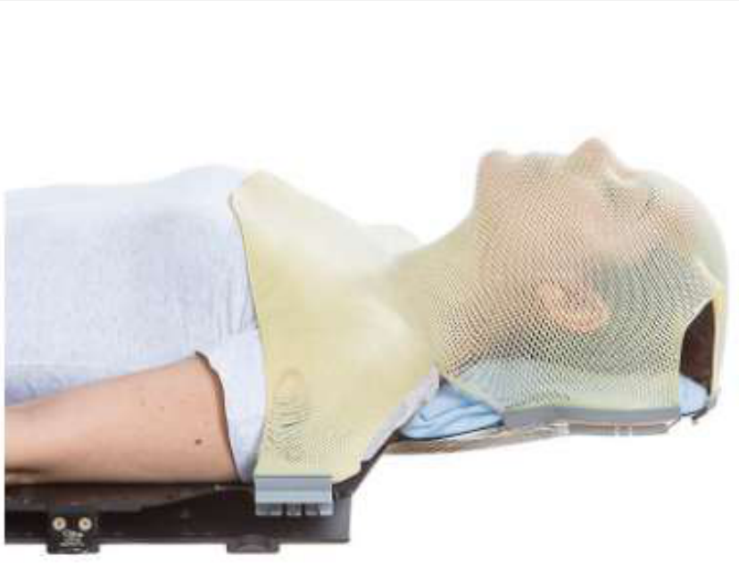
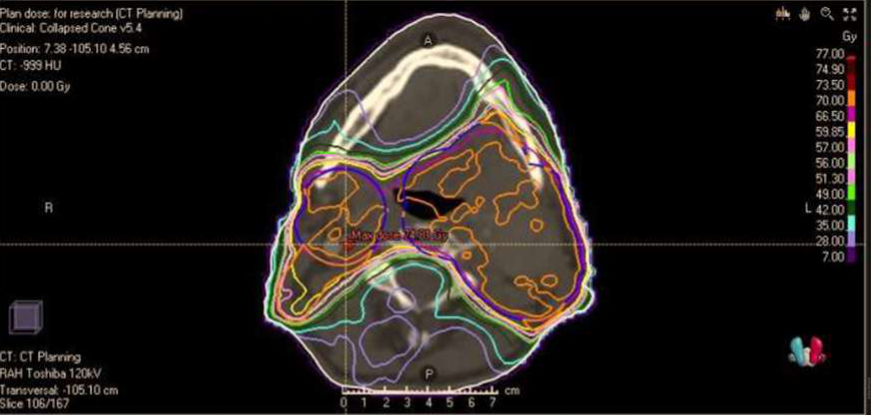
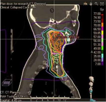
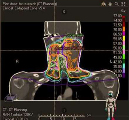
Radiation Field Plans37
 |  |
 |  |
Osteoradionecrosis Prevention Guidelines
Lecturer — Radiation Field Planning
Modern radiation field plans are highly contoured to maximise the dose to the cancer site while minimising the dose to healthy tissues.
- Earlier three-dimensional conformal radiotherapy produced broader fields and exposed more healthy tissue.
- Intensity-modulated radiotherapy allows more precise contouring of the treatment site.
- Dental planning should identify teeth receiving more than 60 Gy, areas receiving approximately 50 Gy or more, and whether the posterior mandible receives high-dose radiation.
- Dental treatment may need to be more aggressive for teeth receiving high doses.
Prevention and Management of Osteoradionecrosis in Patients With Head and Neck Cancer Treated With Radiation Therapy: ISOO-MASCC-ASCO Guideline
Douglas E. Peterson, DMD, PhD, FDS RCSED, FASCO; Shlomo A. Koyfman, MD; Noam Yarom, DMD, MPH; Charlotte Duch Lynggaard, MD, PhD; Nofisat Ismaila, MD, MSc; Lone E. Forner, DDS, PhD; Clifton David Fuller, MD, PhD; Yvonne M. Mowery, MD, PhD; Barbara A. Murphy, MD; Erin Watson, DMD, MHSc; David H. Yang, DDS, FRCD(C); Ivan Alajbeg, DMD, MSc, PhD; Paolo Bossi, MD; Michael Fritz, MD; Neal D. Futran, MD, DMD; Daphna Y. Gelblum, MD; Edward King, JD; Salvatore Ruggiero, DMD, MD, FACS; Derek K. Smith, DDS, PhD, MPH; Alessandro Villa, DDS, MPH, PhD; Jonn S. Wu, BMSc, MD, FRCPC; and Deborah Saunders, DMD
DOI: https://doi.org/10.1200/JCO.23.02750
Purpose38
To provide evidence-based recommendations for prevention and management of osteoradionecrosis (ORN) of the jaw secondary to head and neck radiation therapy in patients with cancer.
Methods
The International Society of Oral Oncology–Multinational Association for Supportive Care in Cancer (ISOO-MASCC) and ASCO convened a multidisciplinary Expert Panel to evaluate the evidence and formulate recommendations. PubMed, EMBASE, and Cochrane Library databases were searched for randomized controlled trials and observational studies, published between January 1, 2009, and December 1, 2023. The guideline also incorporated systematic reviews conducted by ISOO-MASCC, which included studies published from January 1, 1990, through December 31, 2008.
Results
A total of 1,539 publications were initially identified. There were 487 duplicate publications, resulting in 1,052 studies screened by abstract, 104 screened by full text, and 80 included for systematic review evaluation.
Recommendations
Due to limitations of available evidence, the guideline relied on informal consensus for some recommendations. Recommendations that were deemed evidence-based with strong evidence by the Expert Panel were those…
J Clin Oncol 00:1-22. © 2024 by American Society of Clinical Oncology. Accepted January 22, 2024; published May 1, 2024. ISOO-MASCC approval: November 9, 2023; Evidence Based Medicine Committee approval: December 15, 2023.
Pre-Therapy Dental Management39
- Extract teeth with doubtful long-term prognosis (especially mandibular teeth planned for >60 Gy) at least 10-14 days prior to RT
- Balance against the risks of delaying RT (e.g. OAC, co-morbidities that may contribute to delayed healing, MRONJ)
- Recontour/restore sharp teeth, failing restorations, carious cavities with potential to traumatise soft tissues or flap
- Assess dentures = modify ill-fitting dentures if possible
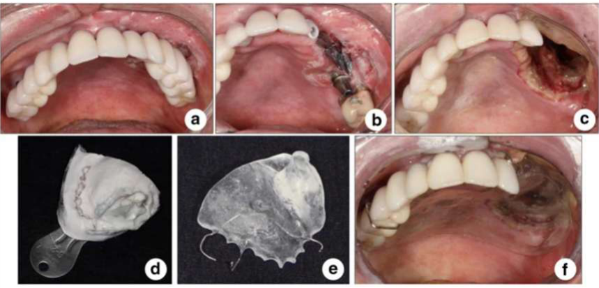
- Joint planning with the surgical team if surgery will alter the oral cavity (e.g. maxillectomy requiring obturator construction, dental extractions as part of resection or cancer margin monitoring)
Key Factors To Determine Treatment Approach
Cancer-Related Factors40
-
Cancer prognosis
-
Urgency to commence radiotherapy
-
Irradiation dose >50 Gy (risk ↑ 11x if >66 Gy)
-
Site:
- Posterior > anterior
- Mandible > maxilla 24:1
-
Tooth extraction timing (<21 days pre-RT)
-
Cancer-related trismus
-
Whether the patient is being treated with curative or palliative intent
Patient-Related Factors
-
Other co-morbidities
-
Physiological tolerance / risks of dental tx
-
Patient prognosis
-
Patient function (e.g. morbidity of surgery, dysphagia, QOL, wishes)
-
Patient motivation and psychology
-
Maintenance (motivation, access, ability)
-
Risks of extraction (OAC, IAN, MRONJ)
-
Periodontal health and oral hygiene
-
Alcohol/tobacco use
-
Nutritional status
-
Dentate > edentate
-
Poor prosthetic adjustment
-
Willingness to attend regular dental appointments Peterson DE, et al. Prevention and Management of Osteoradionecrosis in Patients with Head and Neck Cancer Treated with Radiation Therapy: ISOO-MASCC-ASOC Guideline. J Clin Oncol. 2024;42(16):1975-1996.
Nabil S, Samman N. Risk factors for osteoradionecrosis after head and neck radiation: a systematic review. Oral Surg Oral Med Oral Path Oral Radiol. 2012;113(1):54-69.
Frankart AJ, et al. Osteoradionecrosis: Exposing the evidence not the bone. Int J Radiat Oncol Biol Phys. 2021;109(5):1206-18.
Beadle B, et al. Evaluating the impact of patient, tumor, and treatment characteristics on the development of jaw complications in patients treated for oral cancers: A SEER–Medicare analysis. Head Neck. 2012;35:1599-605.
Raguse J-D, et al. Patient and treatment-related risk factors for osteoradionecrosis of the jaw in patients with head and neck cancer. Oral Surg Oral Med Oral Pathol Oral Radiol. 2016;121(3):215-21.
Pre-RT Extractions41
-
Teeth with doubtful prognosis should be extracted as early as possible in the patient journey before RT.
- Ideally allow 3 weeks healing before RT, minimum 10-14 days
-
Pre-RT extractions still pose a risk of developing ORN
- Irradiation of healing bone
- 2017 Moon et al: pre-RT tooth extractions an independent risk factor for ORN (hazard ratio 4.52)
- 2013 Cochrane review: no evidence from RCTs to support or refute extraction of healthy or diseased teeth prior to ORN
- 1986–2006: ORN pooled incidence after pre-RT extraction 3.0-3.2%, post-RT extraction 3.1-3.5%
- Prophylactic extractions of healthy/restorable molars within the anticipated RT field → no longer considered appropriate
Lecturer — Healing After Extraction
Extraction before radiotherapy does not eliminate the risk of osteoradionecrosis because the healing socket may still receive high-dose radiation.
- Bone turnover remains rapid in the healing socket.
- Oncological treatment often cannot be delayed until complete healing.
- Full resolution of the extraction site may take up to a year.
- Most established guidelines still argue for pre-RT extraction of teeth with poor to hopeless long-term prognoses
- Majority of studies report higher rate of ORN post-RT extractions
- Beumer et al: ORN more often requires radical resection with post-RT extraction (45.4%) than pre-RT (11.7%)
Kumar N, et al. The oral management of oncology patients requiring radiotherapy, chemotherapy and/or bone marrow transplantation. London: Royal College of Surgeons and British Society for Disability & Oral Health; 2018. Available from: https://www.rcseng.ac.uk/dentalfaculties/fds/publications-guidelines/clinical-guidelines/.
McCaul LK, et al. Predicting and managing oral and dental complications of surgical and non-surgical treatment for head and neck cancer - A clinical guideline. United Kingdom: Restorative Dentistry UK, British Association of Head and Neck Oncology Nurses, British Association of Head and Neck Oncologists, Royal College of Speech & Language Therapists, ENT UK, British Dieticians Association, British Association of Oral and Maxillofacial Surgeons; 2016.
Eliyas S, Al-Khayatt A, Porter RWJ, Briggs P. Dental extractions prior to radiotherapy to the jaws for reducing post-radiotherapy dental complications. The Cochrane database of systematic reviews. 2013;2013(2):CD008857.
Moon DH, et al. Incidence of, and risk factors for, mandibular osteoradionecrosis in patients with oral cavity and oropharynx cancers. Oral Oncol. 2017;72:98-103.
Beech NM, Porceddu S, Batstone MD. Radiotherapy-associated dental extractions and osteoradionecrosis. Head Neck. 2017;39(1):128-32.
Chang DT, et al. Do pre-irradiation dental extractions reduce the risk of osteoradionecrosis of the mandible? Head Neck. 2007;29(6):528.
Beumer 3rd J, et al. Osteoradionecrosis: Predisposing factors and outcomes of therapy. Head Neck Surg. 1984;64:819-27.
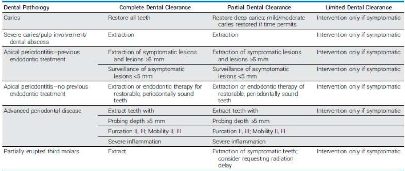
Dental Treatment Protocols Prior To Radiotherapy42
ORN Prevention and Management in Head and Neck Cancer
| Dental Pathology | Complete Dental Clearance | Partial Dental Clearance | Limited Dental Clearance |
|---|---|---|---|
| Caries | Restore all teeth | Restore deep caries; mild/moderate caries restored if time permits | Intervention only if symptomatic |
| Severe caries/pulp involvement/dental abscess | Extraction | Extraction | Intervention only if symptomatic |
| Apical periodontitis—previous endodontic treatment | Extraction of symptomatic lesions and lesions ≥5 mm; surveillance of asymptomatic lesions <5 mm | Extraction of symptomatic lesions and lesions ≥5 mm; surveillance of asymptomatic lesions <5 mm | Intervention only if symptomatic |
| Apical periodontitis—no previous endodontic treatment | Extraction or endodontic therapy for restorable, periodontally sound teeth | Extraction or endodontic therapy of restorable, periodontally sound teeth | Intervention only if symptomatic |
| Advanced periodontal disease | Extract teeth with: probing depth ≥5 mm; furcation II, III; mobility II, III; severe inflammation | Extract teeth with: probing depth ≥5 mm; furcation II, III; mobility II, III; severe inflammation | Intervention only if symptomatic |
| Partially erupted third molars | Extract | Extraction of symptomatic teeth; consider requesting radiation delay | Intervention only if symptomatic |
NOTE. The proper protocol should be selected by the oncologist and dentist according to the patient’s medical status. Limited clearance would be indicated for patients treated with noncurative intent. Partial and complete clearance protocols are based on patients being treated with curative-intent radiotherapy and appropriate access to care. In general, patients with at least 2 weeks of healing available before radiation can be treated with complete clearance and those with less than 2 weeks of time can be treated with partial clearance.
Clearance protocols should be applied to teeth that will receive a therapeutic dose of radiation. For teeth that will receive below a therapeutic dose, minimal clearance protocols can be applied.
Peterson DE, et al. Prevention and Management of Osteoradionecrosis in Patients with Head and Neck Cancer Treated with Radiation Therapy: ISOO-MASCC-ASOC Guideline. J Clin Oncol. 2024;42(16):1975-1996.
Lalla RV, Hodges JS, Treister NS, et al. Tooth-level predictors of tooth loss and exposed bone after radiation therapy for head and neck cancer. J Am Dent Assoc. 2023;154:519-528.e4.
Watson E, Dorna Mojdami Z, Oladega A, et al. Clinical practice guidelines for dental management prior to radiation for head and neck cancer. Oral Oncol. 123:105604, 2021.
Clinical Decision Making For Extractions
Rationale43
- Reducing initiating factors for ORN within an irradiated site
- Curative-intent radiotherapy
Medical, Dental and Social Considerations
- Long-term prognosis of tooth
- Ability to tolerate extractions (pain, cancer-induced trismus)
- Risks of extraction (other medical co-morbidities, OAC, IAN, MRONJ)
- Ability for tooth to be maintained post-surgery & RT (restricted mouth opening, surgical flaps/grafts)
- Compliance with ongoing preventive oral care
- Ongoing alcohol and tobacco use
- Concomitant oral disease (periodontal disease, periapical pathology) = infection risk factor for ORN
- Focus on preventive oral hygiene measures
- Education about life-long risks of ORN
- Tooth location
Entire or Localised Areas of the Mouth
- Anticipated radiation fields >50-60 Gy
- Maxilla vs mandible
- Anterior vs posterior
- Mucosal trauma during period of mucositis
Optimal Timeframes
-
Ideally future-proof for life-long foreseeable future
-
Urgency to commence RT vs time available for healing (min 14 days healing, ideally 2-3 wks)
-
Prioritise care that reduces mucosal trauma or need for future oral surgery impacting on bone (e.g. poor prognosis tooth extraction, poorly fitting dentures)
-
If extractions will alter VDO, perform prior to immobilisation mask fabrication
-
Rapidly progressing tumours: defer extractions, do not delay RT initiation
-
If extractions alter the occlusal vertical dimension after the mask is made, the radiotherapy plan may require remaking
Treatment Approaches
- Curative intent tx: typically aggressive pre-RT, then conservative post-RT
- Long-term medical prognosis, physiological tolerance
- Resulting QOL (# appts, functionality)
Lecturer — Lower Dentures
Retaining lower teeth where possible may avoid a full lower denture in a dry mouth. A full lower denture may be difficult to retain in a dry mouth and may increase the risk of thrush.
Chemotherapy And Immunotherapy
-
Not a curative treatment for HNC alone
-
Contexts:
- Induction (neo-adjuvant) chemotherapy
- Primary chemoradiotherapy
- Adjuvant chemoradiotherapy
- Palliation
-
Typical chemotherapy drugs: cisplatin, carboplatin, docetaxel, 5-fluorouracil and hydroxyurea
-
Typical schedule: 3 doses, 3 weekly during RT (start, middle, end)
-
Chemotherapy may also be used as a radio-sensitising treatment.
-
Cisplatin is administered intravenously in the cancer centre, with the infusion taking approximately four hours.
Chemotherapy44
Immunotherapy45
form of biological therapy that stimulates the body’s immune system to detect and act against cancer cells
-
Examples in H&N cancer context: cetuximab, nivolumab
-
Growing field
-
Advantages:
- Higher specificity against malignant cells thus fewer side-effects
- Ability to target both microscopic disease and widespread metastases
- Increased efficacy against cancer cells that are slowly dividing or quiescent (do not preferentially attack dividing cells c.f. radiation and chemotherapy)
- Potential to provide long-term control (immune system’s ability to develop “memory” cells)
-
Neoadjuvant immunotherapy may be administered before surgery, followed by further cycles afterwards.
Impacts Of Cancer Treatment On The Mouth
Short-Term Effects46
- Mouth lining
- Increased inflammation, damage to cell layering → mucositis
- Tastebuds
- Altered taste sensation; loss of taste sensation
Long-Term Effects
-
Salivary glands
- Decreased saliva volume; change in quality
-
Oral flora
- Changed environment → changed organism profile
-
Jaw joints & facial musculature
- Fibrosis → limited mouth opening, tightness on opening
-
Bone of the jaws
- Decreased blood/oxygen supply → reduced healing capacity
-
Osteoradionecrosis and periodontal susceptibility may occur as long-term effects.
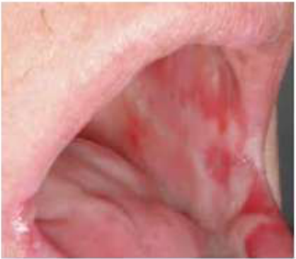
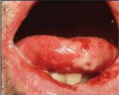
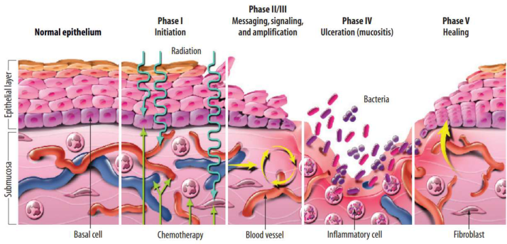
Mucositis47
- Radiotherapy and chemotherapy damages cells → inflammation of the mouth lining (mucosa).
- Initially erythema → progresses to painful ulcers with white/yellow fibrinous slough
- Onset ~2 weeks into treatment
- Effects generally short term:
- Usually resolves at 2 to 6 weeks after completion of therapy.
Phases of mucositis (figure content): Normal epithelium; Phase I Initiation (radiation, basal cell, chemotherapy); Phase II/III Messaging, signaling, and amplification (blood vessel); Phase IV Ulceration – mucositis (bacteria, inflammatory cell); Phase V Healing (fibroblast); epithelial layer; submucosa.
Sonis S. A biological approach to mucositis. J Support Oncol. 2005;2(1):21-36.
 |  |
 |
Complications48
- Painful to speak, eat, swallow
- Infection, sepsis
- Nutritional deficiencies, weight loss
- Dose-limiting factor, necessitating dose reduction or treatment delay
Lecturer — Severe Mucositis
Severe mucositis can make solid food intolerable and may require nutritional support.
- Patients may require nasogastric tube feeding.
- Difficulty maintaining oral hygiene and treatment-related weight loss may also complicate severe mucositis.
Lecturer — Mucositis Management
Management should be tailored to the patient’s presentation and reviewed regularly during radiotherapy.
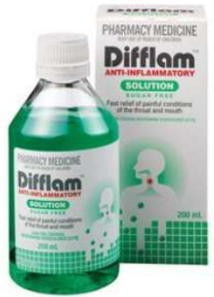
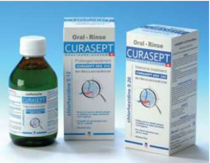
- Chlorhexidine may be used when appropriate, including 0.12% or 0.2% formulations; a 0.2% formulation may be diluted 50:50 with water if too strong while retaining some substantivity.
- A preventive fluoride rinse may be started before radiotherapy and continued until soreness develops, although ethanol-containing products may irritate the mucosa.
- Ice chips may be sucked for approximately 30 minutes before chemotherapy sessions.
Severity / Healing Determined By
-
Radiotherapy dose
-
Radiation field size
-
Fractionation schedules (hyperfractionated)
-
Saliva volume
-
Combination of chemo and radiotherapy
-
Host immune and genomic factors
-
Hepatic and renal impairment
-
Tobacco smoking
-
The patient’s oral condition, nutritional status, infection, degree of ulceration, and ability to tolerate food and fluids also influence severity and healing.
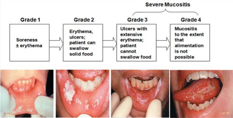
Severe Mucositis — Grading
- Grade 1: Soreness ± erythema
- Grade 2: Erythema, ulcers; patient can swallow solid food
- Grade 3: Ulcers with extensive erythema; patient cannot swallow food
- Grade 4: Mucositis to the extent that alimentation is not possible
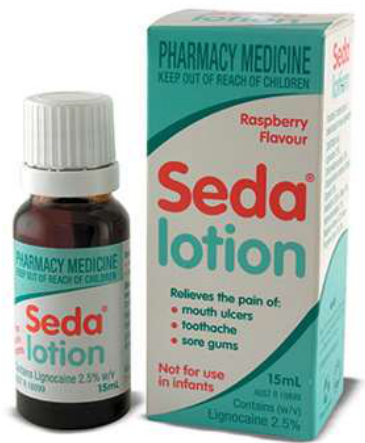
Management Of Mucositis49
 |  |  |
 |  |  |
 |  |  |
 |
Diet Typically Acceptable50
- Liquids
- Purees
- Ice
- Custards
- Non-acidic fruits (banana, mango, melon, peach)
- Soft cheese
- Eggs
Food to Avoid
-
Rough foods (potato crisps, toast)
-
Spicy foods
-
Salt
-
Acidic fruit (grapefruit, lemon, orange)
-
Foods that irritate ulcerated mucosa
Lifestyle Habits to Avoid
- Smoking
- Alcohol
Lecturer — Plaque Removal
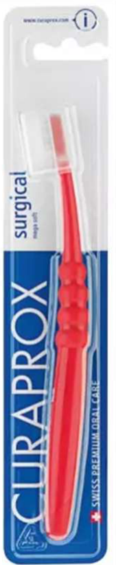
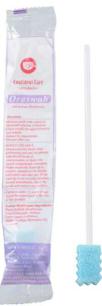
A very soft toothbrush, including a Curaprox surgical toothbrush, may help patients continue removing plaque when conventional bristles are too painful. If brushing is not tolerated, sponge sticks are preferable to doing nothing, although mechanical plaque removal remains preferable.
Lecturer — Topical Pain Relief
Topical pain relief may include 2% viscous lignocaine, known as “pink lady,” held in the mouth and swallowed approximately 30 minutes before meals. It may also be diluted or mixed with Mylanta or Gaviscon to create a thicker coating that helps food pass more comfortably.
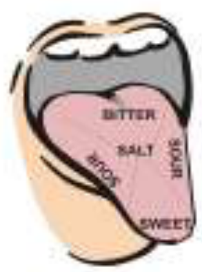
Loss Of Taste51
- Onset at start of cancer treatment
- Related to dry mouth and direct damage to taste buds
- May start to recover within 2 to 4 months
- Taste often returns with an unpleasant interim period of altered taste
- Typically restored by 6 months
- If radiation dose >60 Gy, loss of taste may be permanent
Lecturer — Altered Taste
Altered taste may begin approximately two weeks into radiotherapy.
- Patients may experience a metallic taste.
- Sugar may taste unpleasant, salty foods may taste bland, and enjoyment of food may be reduced.
- Taste commonly begins to recover after treatment, although the timing varies.
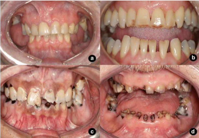
Salivary Changes And Secondary Complications52
Saliva becomes viscous and acidic, with reduced pH and buffering capacity, and reduced quantity.
- Difficulty with speech, eating (mastication) and swallowing (dysphagia)
- Mouth soreness
- Difficulty/discomfort wearing dentures
- Taste alteration/loss
- Reduced food clearance
- Changes in oral flora → fungal, viral, bacterial infections (e.g. oral thrush, salivary gland infections, gingival/periodontal disease progression)
- Radiation caries
- Dental erosion
- Increased risk of tooth wear and gingival recession (hyposalivation important contributor) → tooth sensitivity
- → Quality of life
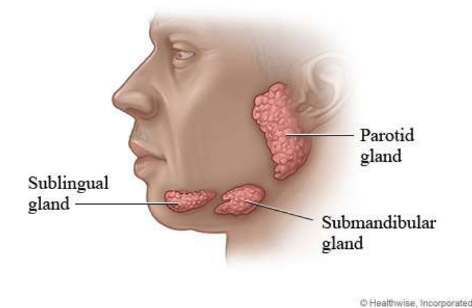
Lecturer — Salivary Dose Effects
Radiation doses of approximately 10 grays may temporarily alter salivary flow and composition, while permanent damage may occur at approximately 30 grays.
 |  |
Managing Dry Mouth Symptoms53
Lecturer — Dry Mouth Management
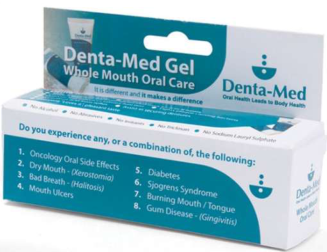
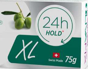
Dry-mouth management may include frequent sips of water, saliva substitutes, oral moisturising products, fluoride-containing products, and regular preventive dental care.
- Sugar-free chewing gum may help where functional salivary tissue remains, but is less effective when salivary tissue has been significantly damaged.
- Saliva substitutes mentioned included Biotene, Oral7, and products incorporating some enzymes found in natural saliva.
- Lips may be kept moist with plant- or animal-based products such as lanolin or beeswax; petroleum-based products such as Vaseline act mainly as a barrier rather than moisturising the lips.
 |  |  |
 |  |  |
 |
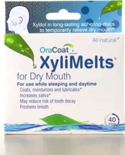
XyliMelts For Dry Mouth
XyliMelts — OraCoat, for dry mouth. 40 discs.
Xylitol in long-lasting adhering discs to temporarily relieve dry mouth*. All-natural.
- For use while sleeping and daytime
- Coats, moisturizes and lubricates*
- Increases saliva*
- May reduce risk of tooth decay
- Freshens breath
Instructions For Use54
Place the tan adhesive side of disc on the outside of a molar and/or adjoining gums, upper or lower, white side touching cheek. Use your tongue to push the disc to a comfortable spot. Once there, do not touch it with your tongue for 10 seconds, as this movement will disrupt the adhesion.
- Before bedtime: Use two (2) discs, placing one on each side of the mouth.
- Everyday use: For dry mouth, use more as needed throughout the day.
Lecturer — XyliMelts Duration
XyliMelts may be particularly useful overnight when dry mouth is worse and causes repeated waking.
- Adhesion strengthens over several minutes.
- A disc may last approximately four to six hours at night and one to three hours during the day, depending on dry-mouth severity.
- The discs may be used while eating or drinking and should not interfere with speaking or appearance.
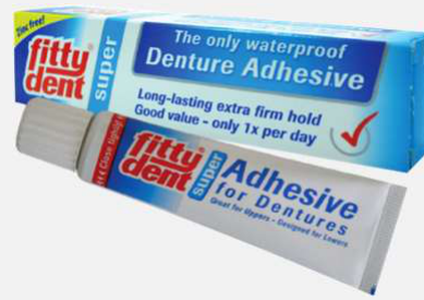
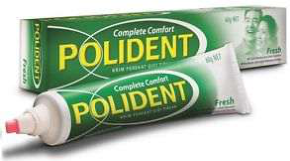
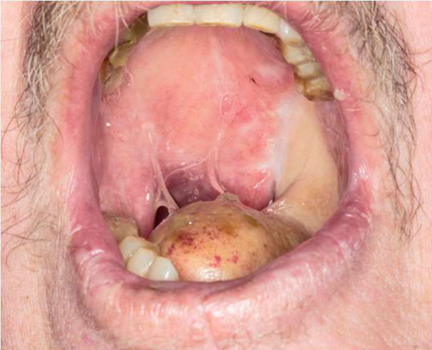
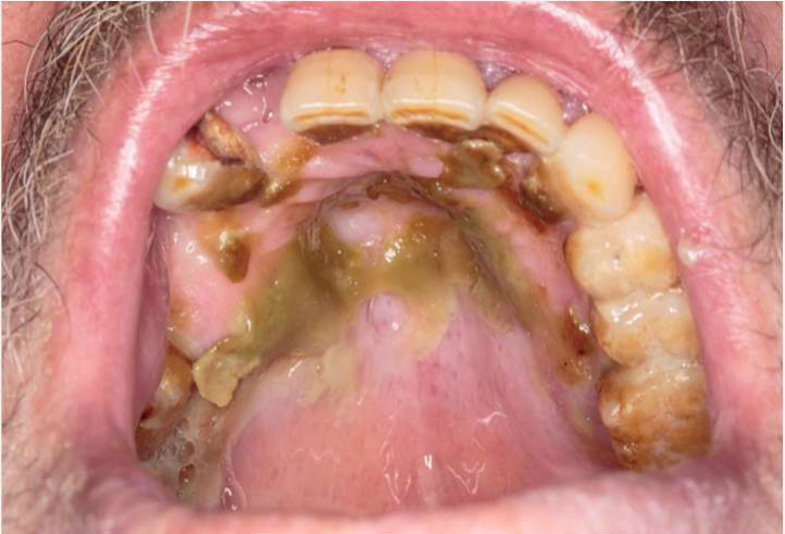
Difficulties With Dentures555657
- Susceptibility to oral thrush
- Denture hygiene
- Well-fitting denture
- Ill-fitting dentures
- Post-surgical change
- Weight loss
- Changes in sensation
- Dry mouth
Lecturer — Denture Retention
Mucositis, oral ulcers, reduced denture retention, and altered oral structures may contribute to denture problems.
- Denture adhesives may be painful on mucositis or ulcerated mucosa.
- Oral moisturising gel may sometimes improve retention by providing a more suitable surface for suction and capillary action.
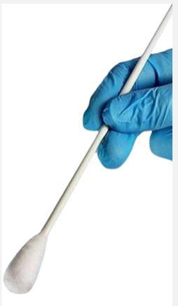
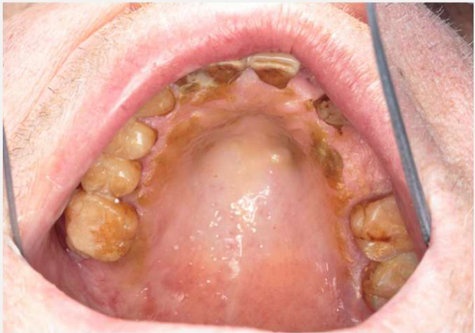



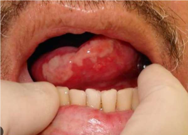
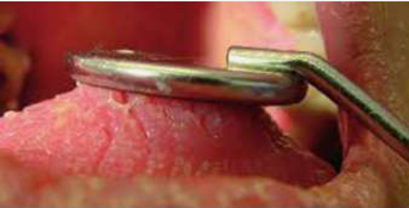
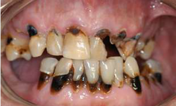
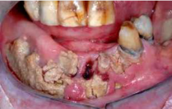
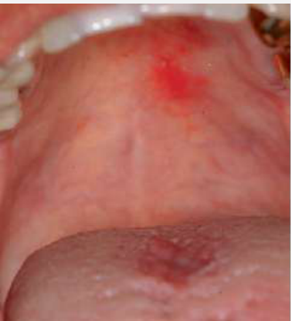
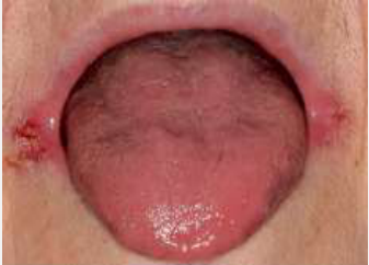
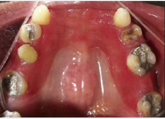
Fungal Infections (Oral Thrush)58
- Candida albicans: harmless organism that inhabits the mouths of up to 50% of the population.
- ‘Opportunistic’ fungal infection
- Symptoms are variable, and not always specific. They may include:
- White spots on the palate
- Central red patch on tongue (can be “kissing” a red spot on the palate)
- Thickly coated tongue
- Red patch delineated by a denture-bearing area
- Cracked corners of the mouth
- Burning sensation in the mouth
Images: Contemporary Oral Medicine 2018 (Farah, Balasubramaniam, McCullough)
- More acidic saliva, reduced salivary flow, moist altered oral environments, denture use, and changes in oral flora increase susceptibility to oral thrush.
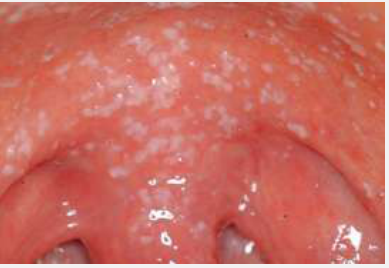 | 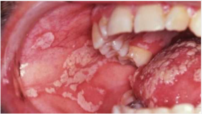 |  |
 |  |
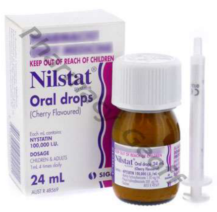
Management Of Fungal Infections59
Lecturer — Thrush Management
Management may include antifungal medication, sodium bicarbonate rinses, good denture hygiene, avoiding smoking, natural yoghurt particularly containing probiotics, and maintaining oral hygiene.
 |  |
 |
Weight Maintenance And Dietary Advice60
During Chemo-Radiotherapy61
- Dietician advice
- High calorie foods and drinks
- Nutritional supplements (contain sugar)
- Moisten foods, thickened liquids
- PEG feeding
- Soda water (effervescent)
Lecturer — Nutrition Priority
During treatment, maintaining nutrition and body weight is an immediate priority even when some high-energy foods contain sugar and may increase the risk of dental decay.
- Dietitians may recommend custard, doughnuts, ice cream, jelly, and other foods that help maintain weight.
- Dental and dietetic advice must be coordinated so oral-health recommendations do not conflict with nutritional requirements.
 |  |
 |
After Chemo-Radiotherapy
- Weight stabilisation may still be a priority
- Taste alterations (John lost sweet and salty sensations)
- Taste aversions
- Risk assessment for dental caries
How would this impact on the dietary advice you would routinely provide patients with for the prevention of dental caries?
-
Liaise with dietician: ensure our advice does not conflict with theirs!
-
Focus on prevention:
- Sip water or bicarbonate rinses after meals
- Fluoride / CPP-ACP
- Oral hygiene
- Use of straws
-
Weight loss may result from pain, nausea, altered taste, reduced appetite, reflux, difficulty eating, reduced enjoyment of food, or mucositis.
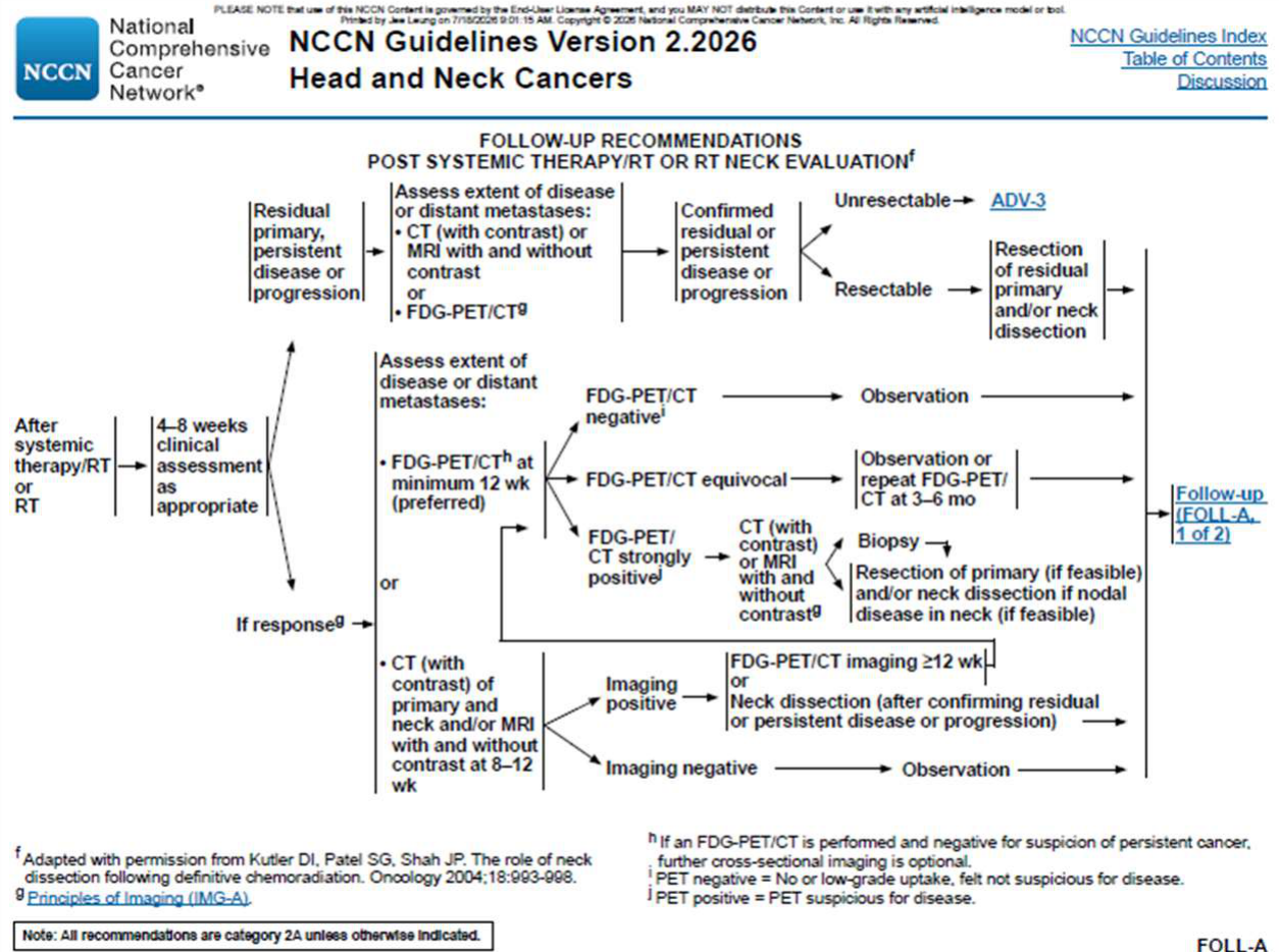
Post-Treatment Follow-Up62
-
4–8 week clinical assessment
-
3 month post-treatment FDG-PET/CT
-
(3–4 month post-surgical CT and/or MRI)
-
Dental review around the three-month post-radiotherapy period, ongoing cancer surveillance, assessment of oral complications, and review of rehabilitation needs.
Rehabilitation And Post-Radiotherapy Care6364
Diagnosis → H&N MDT Meeting Discussion → Work-up to Cancer Treatment → Surgery → H&N MDT Meeting Rediscussion → Radiotherapy +/- Chemotherapy → Rehabilitation → Chronic side effects and survivorship
Diagnosis → H&N MDT Meeting Discussion → Work-up to Cancer Treatment → Surgery → H&N MDT Meeting Rediscussion → Radiotherapy +/- Chemotherapy → Rehabilitation → Chronic side effects and survivorship
Ongoing Dental Issues Post-Radiotherapy65
-
Salivary changes and their long-term impacts
-
Muscle fibrosis and limited mouth opening (trismus)
-
Difficulties with dentures
- Trismus, surgery, hyposalivation
-
Osteoradionecrosis
- Spontaneous, trauma-induced (extractions, dentures)
-
Risk of recurrence
- Oral surveillance
- Lymph node assessment
- Smoking cessation and counselling regarding alcohol intake
-
Altered oral anatomy
Role Of Dental Practitioner Post-Radiotherapy66
-
Management of hyposalivation
-
Prevention and minimisation of trismus
-
Prevention and management of dental caries, periodontal disease, ORN and oral candidiasis
-
Performing dental work deferred during RT
-
Consultation with the treating radiation oncologist if extractions or implants are to be considered
-
Continued monitoring for local recurrence, metastatic lesions in the head and neck region, and second cancers
-
Maintain oral hygiene
-
Monitor teeth exposed to high-dose radiation
-
Manage denture problems
-
Support rehabilitation
-
Coordinate with specialist services
-
Provide regular surveillance after the patient returns to community care
-
Patients may eventually be discharged back to community dental services, so general dentists need to understand the treatment history and chronic effects
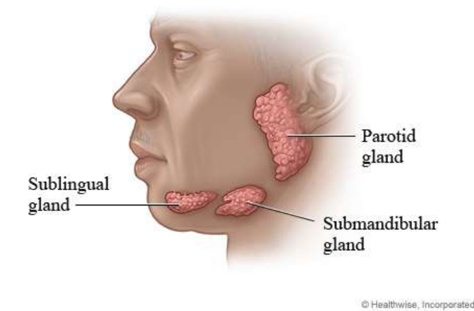
Salivary Recovery And Testing67
Viscous, acidic, reduced buffering capacity, reduced quantity
- Difficulty with speech, eating (mastication) and swallowing (dysphagia)
- Mouth soreness
- Difficulty/discomfort wearing dentures
- Taste alteration/loss
- Reduced food clearance
- Changes in oral flora → fungal, viral, bacterial infections (e.g. oral thrush, salivary gland infections, gingival/periodontal disease progression)
- Radiation caries
- Dental erosion
- Increased risk of tooth wear and gingival recession (hyposalivation important contributor) → tooth sensitivity
- → QUALITY OF LIFE
 | |
Salivary Recovery Outlook68
- Usually yes, to some degree
- Timing varies between individuals
- Gains usually start within 4 weeks to 6 months
- Can be last up to 2 years post-radiotherapy, but is often permanent
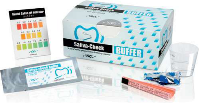
Saliva Testing
Saliva Testing Products69
-
Dental Saliva pH Indicator
-
Saliva-Check BUFFER
-
Biotene
-
Oral7
-
Other saliva substitutes and moisturising products
The Challacombe Scale of Clinical Oral Dryness
King’s College London
The Challacombe Scale was developed from research conducted at King’s College London Dental Institute under the supervision of Professor Stephen Challacombe. The purpose of this scale is to be able to visually identify and quantify whether your patient has xerostomia (dry mouth) and if so, how it changes over time and the most appropriate therapy options. This scale is applicable whatever your profession.
The Challacombe Scale works as an additive score of 1 to 10: 1 being the least and 10 being the most severe. Each feature scores 1 and symptoms will not necessarily progress in the order shown, but summed scores indicate likely patient needs. Score changes over time can be used to monitor symptom progression or regression.
| Score | Feature | Interpretation |
|---|---|---|
| 1 | Mirror sticks to buccal mucosa | Score 1–3 = mild dryness. May not need treatment or management. Sugar-free chewing gum for 15 mins, twice daily and attention to hydration is needed. Many drugs will cause mild dryness. Routine checkup monitoring required. |
| 2 | Mirror sticks to tongue | |
| 3 | Saliva frothy | |
| 4 | No saliva pooling in floor of mouth | Score 4–6 = moderate dryness. Sugar-free chewing gum or simple sialagogues may be required. Needs to be investigated further if reasons for dryness are not clear. Saliva substitutes and topical fluoride may be helpful. Monitor at regular intervals especially for early decay and symptom change. |
| 5 | Tongue shows generalised shortened papillae (mild depapillation) | |
| 6 | Altered gingival architecture (i.e. smooth) | |
| 7 | Glassy appearance of oral mucosa, especially palate | Score 7–10 = severe dryness. Saliva substitutes and topical fluoride usually needed. Cause of hyposalivation needs to be ascertained and Sjögren’s Syndrome excluded. Refer for investigation and diagnosis. Patients then need to be monitored for changing symptoms and signs, with possible further specialist input if worsening. |
| 8 | Tongue lobulated / fissured | |
| 9 | Cervical caries (more than two teeth) | |
| 10 | Debris on palate or sticking to teeth |
Prevention Of Dental Decay70
-
Colgate NeutraFluor® 5000 Plus Freshmint — 5000ppm fluoride professional toothpaste, contains 1.23% w/w neutral sodium fluoride, for individuals at high risk of caries (pharmacist only medicine; keep out of reach of children)
-
GC Tooth Mousse (original, Strawberry, and Plus Mint — CPP-ACP)
-
ReCalDent (リカルデント, dental-practice use, CPP-ACP 2倍配合)
-
Clinpro 5000 — 1.1% sodium fluoride anti-cavity toothpaste
-
McKenzie’s Bi-Carb Soda (baking soda)
-
Xylitol
-
Adequate fluoride
-
Regular dental review
-
Mechanical plaque removal
-
Avoidance of unnecessary dietary sugar once nutritional priorities permit
-
Maintaining calcium and phosphate availability in saliva or through topical products
-
Use of modified toothbrushes when mouth opening or anatomy is restricted
- End-tufted toothbrushes for posterior teeth
- Curved toothbrushes that can contact buccal, lingual and occlusal surfaces
- Other adaptations when the patient cannot open the mouth normally
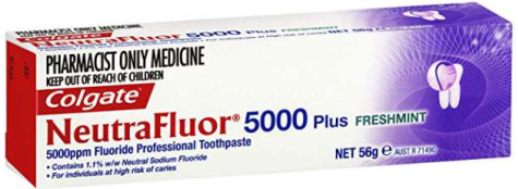 |  | 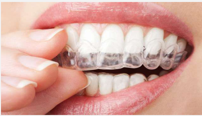 |
 | 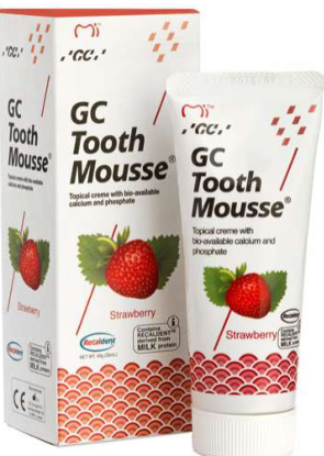 |  |
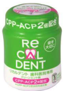 | 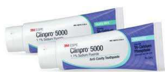 |  |
Trismus7172
- Muscle and joint capsule stiffness (trismus)
- Reduced jaw muscle movement
- Exercises can help to maintain and improve mouth opening and movement
Anatomy shown: temporal mandibular joint, maxilla, masseter, mandible.
- Trismus may also result from radiotherapy, fibrosis, cancer itself, surgical changes, or changes involving the jaw joint and facial muscles
- Trismus can make oral hygiene, dental examinations, extractions, posterior access and prosthetic treatment more difficult oral hygiene maintenance.*
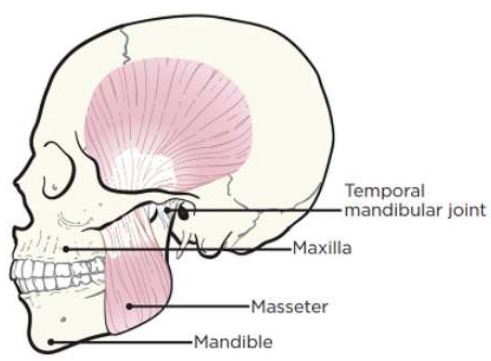 | 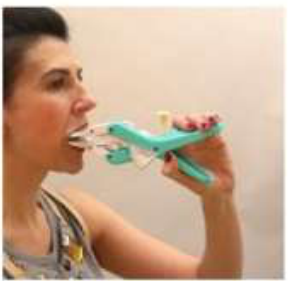 | 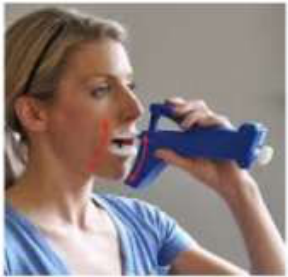 |
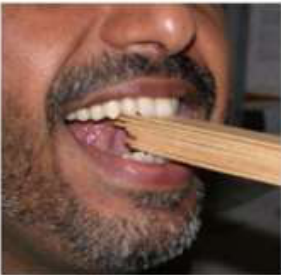 | 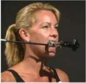 | 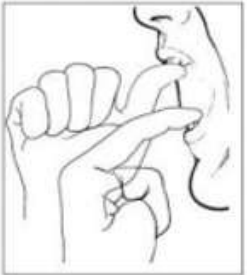 |
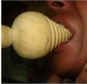 | 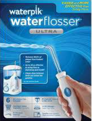 | 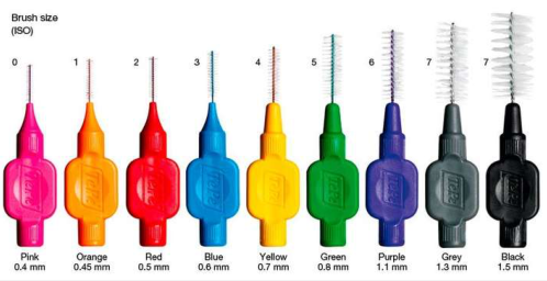 |
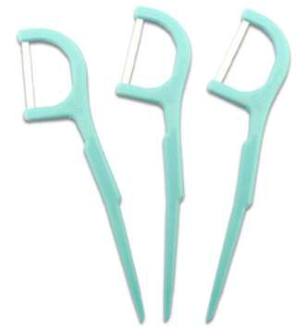 |  |  |
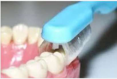 |
Osteoradionecrosis Of The Jaw73
- “Radiation-induced bone death”
- Changes in the jaw bone’s healing ability following dental extractions and trauma
- Occasionally seen as a spontaneous condition
- Typically within first 2 years post-RT
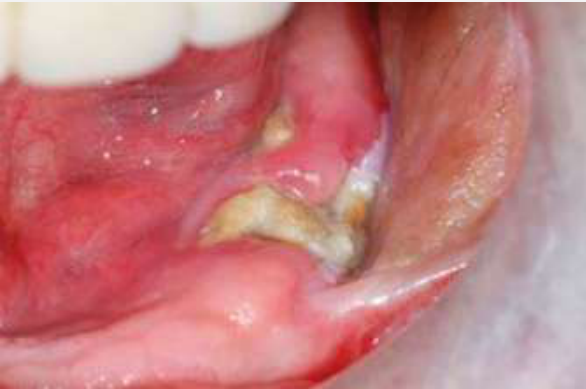 | 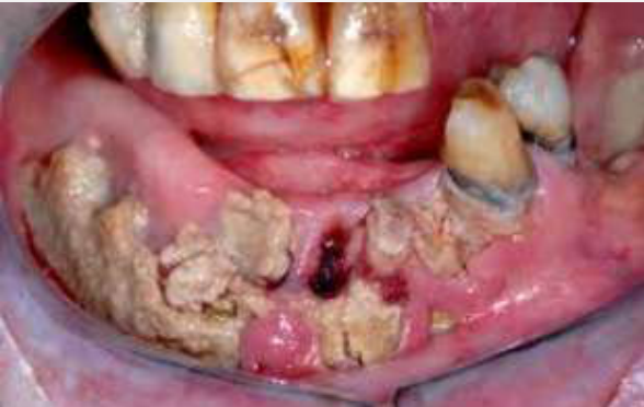 |
Definitions Of Osteoradionecrosis
Traditional ORN74
- “Exposed bone through overlying skin or mucosa that presents as a non-healing wound for three months or more, without residual or recurrent tumour, in those who have received radiation” (Marx 1983)
- However, this is not strictly correct since:
- ORN can be shown radiographically without any breach of the oral mucosa or cervicofacial skin, by virtue of its characteristic appearance
- Pathological fractures due to ORN have been described without evidence of bone exposure (He et al 2015, Owosho et al 2015, Lyons et al 2014, Store & Boysen 2000, Van Merkesteyn et al 1994)
Radiographic ORN
- “Radiographic evidence of bone necrosis (i.e. bone sequestrum or pathologic bone fracture) within the radiation field, where tumour recurrence has been excluded, and mucosa is still intact” (Owosho et al 2015, Støre & Boysen, 2000, Van Merkesteyn et al 1994)
Clinical Features Of ORN75
| Signs | Symptoms |
|---|---|
| Non-healing ulcer or necrosis of oral mucosa | Pain |
| Exposed necrotic bone | Paraesthesia / dysaesthesia / anaesthesia — when IAN irritated or severed by the process |
| Oral malodour | Trismus |
| In advanced stages: ulceration of overlying skin; chronic drainage or orocutaneous fistula; pathologic fractures | Difficulties chewing / speaking / swallowing |
| Complications: repeated episodes of superimposed bacterial infection and osteomyelitis |
Lecturer — ORN Risk
Osteoradionecrosis is a major lifelong concern after high-dose radiotherapy.
- The risk is particularly increased above approximately 50 grays and at doses exceeding approximately 66 grays.
- It is especially relevant in the posterior mandible and remains greater in the mandible than the maxilla.
- Risk remains even when teeth are extracted before radiotherapy and the mucosa appears to have healed.
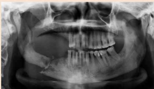 | 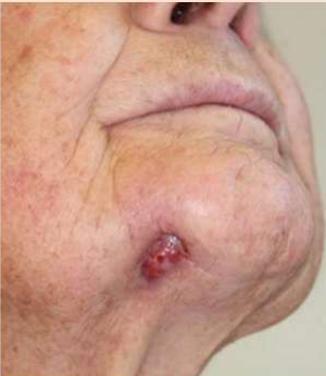 |
Radiographic Changes Following Head And Neck Radiotherapy7677
- Widened Periodontal Ligament Space (WPLS): subclinical, increased width of PDL space >0.5mm along entire length of tooth root without a specific epicentre and adjacent bone destruction
- Bone sclerosis (BS): region of increased bone density resulting from an increased number of bone trabeculae
- Bone resorption (BR): region of decreased bone density resulting from decreased number of bone trabeculae
- Periodontal disease-like bone loss (PDBL): vertical bone loss in the alveolar process as measured from the level of the CEJ of the adjacent tooth
Radiographic features shown as panels A and B.
Chan K. Jaw Bone Changes on Panoramic Imaging after Head and Neck Radiotherapy. In: Pharoah M, editor.: ProQuest Dissertations Publishing; 2012.
Chan KC, Perschbacher SE, Lam EWN, Hope AJ, McNiven A, Atenafu EG, et al. Mandibular changes on panoramic imaging after head and neck radiotherapy. Oral Surg Oral Med Oral Pathol Oral Radiol. 2016;121(6):666-72.
- After radiotherapy in particular, it is important to have regular dental monitoring
- This may be more often than normal
- Allows dental team to work with you to prevent and manage problems before they become severe
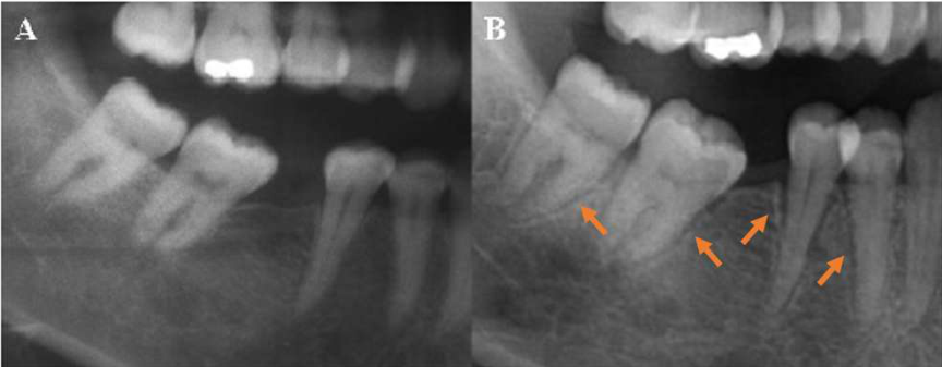 | 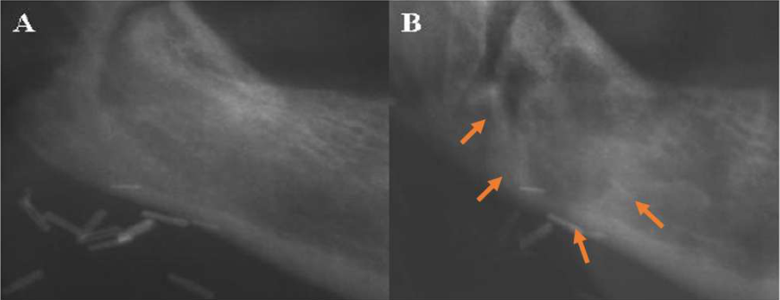 |
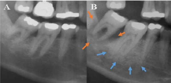 |  |
Post-Radiotherapy Extractions78
Radiation tissue injury vs time (Marx and Johnson 1987)
Marx RE, Johnson RP. Studies in the radiobiology of osteoradionecrosis and their clinical significance. Oral Surg Oral Med Oral Pathol. 1987;64(4):379-90.
Kanatas A, Rogers S, Martin M. A practical guide for patients undergoing exodontia following radiotherapy to the oral cavity. Dent Update. 2002;29(10):498
Nabil S, Samman N. Incidence and prevention of osteoradionecrosis after dental extraction in irradiated patients: a systematic review. Int J Oral Maxillofac Surg. 2011;40(3):229-43
Lecturer — Extraction Decisions
Decisions about post-radiotherapy extractions should consider several factors.
- The radiation dose at the tooth site
- The location of the tooth
- The condition of the surrounding bone
- Patient comorbidities and healing ability
- The urgency of treatment
- Whether conservative management is possible
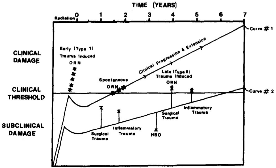
3D-CRT Versus IMRT Radiotherapy79
| 3DCRT | vs. | IMRT |
|---|---|---|
| Target conformity | Highly conformal | |
| Uniform intensity beams | Intensity-modulated beams | |
| Higher dose to nearby OARs | Better sparing of OARs | |
| Shorter | Longer | |
| Simple to moderate | Complex | |
| Dose gradient less sharp | Sharp dose fall-off | |
| Basic | Advanced | |
| Suitable for simple-shaped | Best for irregular tumors |
Lecturer — Radiotherapy Planning
Earlier three-dimensional conformal radiotherapy produced broader radiation fields and exposed more healthy tissue. Intensity-modulated radiotherapy allows more highly contoured fields and aims to minimise radiation to healthy tissues.
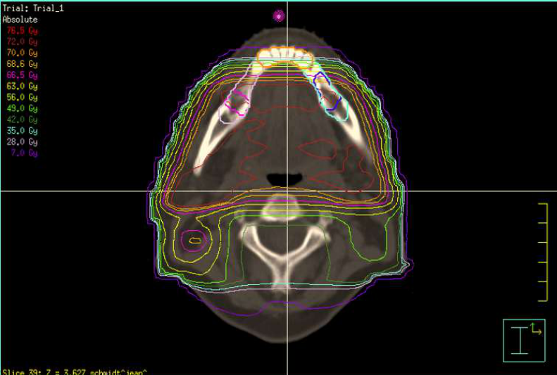 | 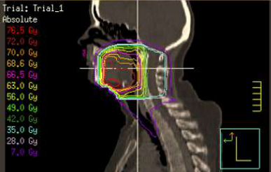 | 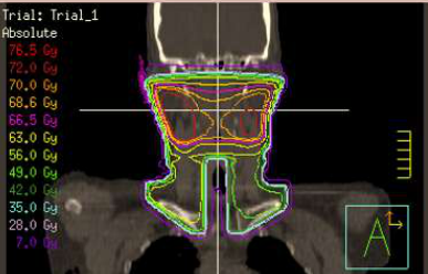 |
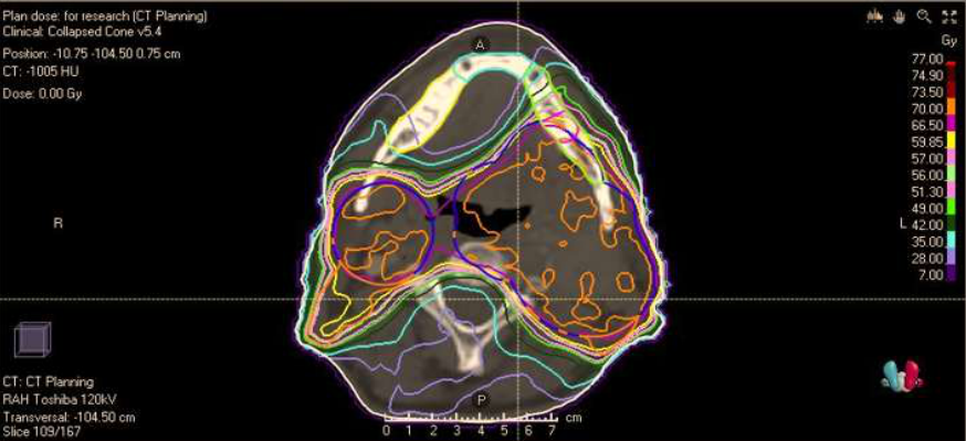 | 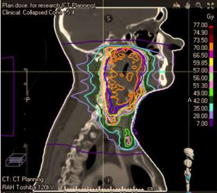 | 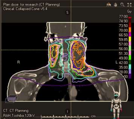 |
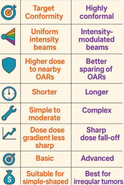 |
Hyperbaric Oxygen Therapy80
- Based on Marx’s “3H” theory of ORN: Hypocellularity, Hypovascularity and Hypoxia
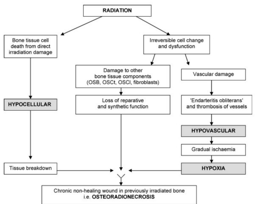 | 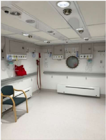 |
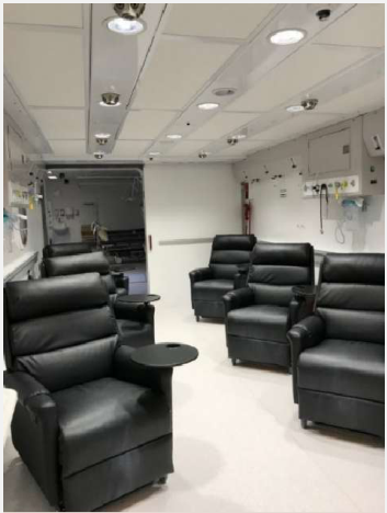 | 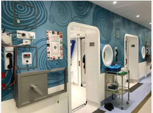 |
Pentoclo81
- Based on Delanian and Lefaix’s radiation-induced fibroatrophic theory of ORN
- Impaired healing results from endothelial injury, release of cytokines (inflammation) and abnormal fibroblast activity
- End result is hypocellular bone and reduced bone matrix formation, compensated by fibrosis → bone and tissue necrosis
| Pentoxifylline | α-Tocopherol (Vitamin E) | Clodronate |
|---|---|---|
| Vasodilator and antifibrotic | Antioxidant | Non-nitrogenous bisphosphonate |
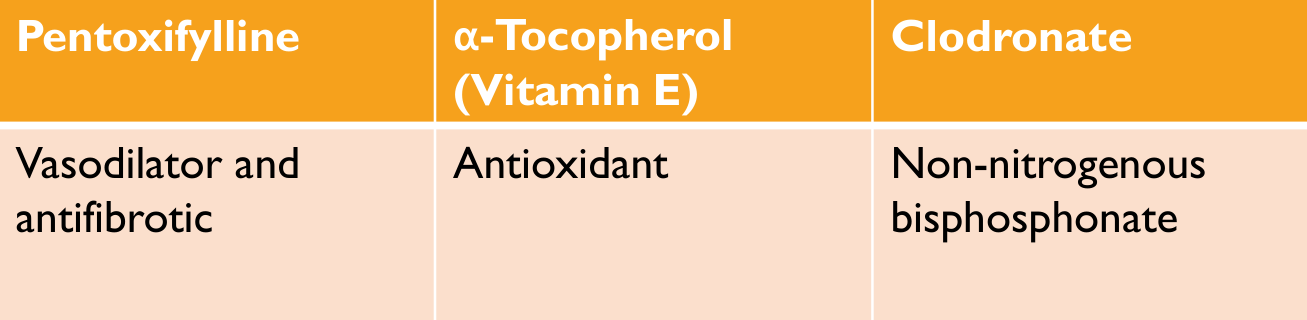 | 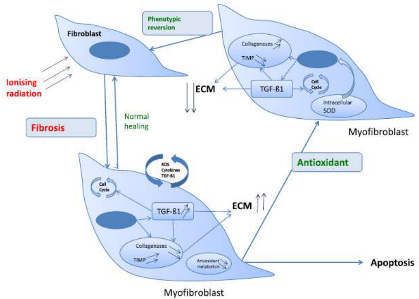 |
 |
Conservative ORN Management82
- Strict oral hygiene
- Irrigation (saline)
- Chlorhexidine rinses or gel
- Cessation of tobacco and alcohol consumption
- Analgesics
- Antibiotic therapy
- Minimal surgical debridement
Cancer Surveillance83
| Follow-up intervals | Regular follow-ups to assess | Increased risk of recurrence |
|---|---|---|
| Year 1: every 1-2 months | Symptoms | Buccal cancers |
| Year 2: every 2-6 months | Possible tumour recurrence | Ongoing smoking |
| Years 3-5: every 4-8 months | Health behaviours | Immunosuppressants |
| >5 years: every 12 months | Nutrition | Larger cancers |
| Dental health | Long history of SCCs (“hot mouth”, “field cancerisation”) | |
| Speech and swallowing function |
Lecturer — Surveillance Imaging
Surveillance includes imaging as well as clinical review. FDG-PET or CT may be performed at approximately three months after treatment, alongside ongoing assessment by the cancer team, dental monitoring and communication with community healthcare providers.
Long-Term Community Support84
| General medical practitioner | Social worker |
| Clinical nurse specialist (including tracheotomy care and wound management) | Psychologist |
| Speech and swallowing therapist | Psychiatrist |
| Dietician (enteral feeding and oral nutrition) | Counsellor |
| Dentist/Dental hygienist | Pain specialist |
| Physiotherapist | Smoking and alcohol cessation services |
| Occupational therapist (lymphedema management) | Audiology |
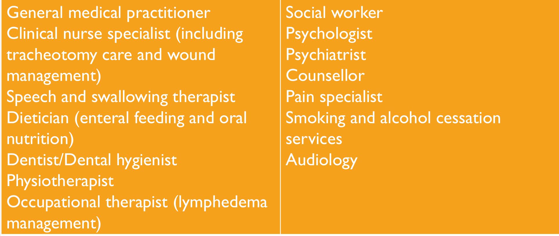
Psychosocial Considerations85
- Depression during and post treatment
- Social support/isolation
- Affect on family (children, spouses)
- Quality of life impacts
- Social adaptation
- Occupation
- Survivorship
- Treatment guiding your life — what next?
- Fear of recurrence
Lecturer — Psychosocial Effects
Head and neck cancer can have major psychosocial effects because treatment may be disfiguring and functionally disabling.
- Patients may have difficulty eating and speaking.
- Depression and anxiety may occur.
- Challenges may be greater for patients with low health literacy or vulnerable backgrounds.
- The MDT may involve psychology, psycho-oncology, social work, supportive care and palliative care.
Prognosis8687
- 5-year survival
- Overall survival for oral cancer globally = 50%
| Stage I | Stage II | Stage III | Stage IV |
|---|---|---|---|
| 85% | 66% | 41% | 9% |
Lecturer — Prognostic Factors
Prognosis is influenced by tumour and patient factors as well as treatment response.
- Tumour site and stage
- Regional lymph node involvement and distant metastases
- HPV or p16 status
- Pathological findings and treatment response
- Patient comorbidities, functional status and ability to complete treatment
HPV-associated or p16-positive oropharyngeal cancers generally have a better prognosis than HPV-negative cancers associated with smoking and alcohol.
- Steady increase in HNC survivors
- Advances in treatment
- Decreased smoking rates
- Improved prognosis of HPV-associated oropharyngeal cancers (80% long term survival)

Thank You And Questions88
THANK YOU!
QUESTIONS?

Clinical Cases
Case: Post-recurrence mandibular non-union and free flap complications
Question
Scenario: A patient with a history of left tongue squamous cell carcinoma (SCC) who developed a recurrence and subsequently underwent further surgery.
What’s shown: A clinical image showing the result of the surgical intervention, highlighting the site of the previous surgery in relation to the adjacent canine tooth, as well as the presence of a free flap in the oral cavity.
Consider: What surgical complications occurred following the recurrence, what dental issues arise as a result of the surgical site, and what secondary complications are associated with the use of a free flap in the mouth?
Answer
Observations:
- The image shows the site of a previous surgical resection, clearly visible next to the canine tooth.
- The patient developed a non-union despite the use of plating.
- The surgical team performed an ankylosis/ankyloectomy for the recurrence.
- There are teeth on either side of the surgical site.
- The free flap transferred into the mouth contains hair follicles.
Reasoning:
- The non-union developed despite plating, necessitating further surgical management (ankylosis) for the recurrence.
- The teeth adjacent to the surgical cages/site are prone to developing periodontal issues, trauma, and occlusal problems as a result of the altered anatomy and surgical access.
- Because the free flap is taken from a hairy area (like an arm or leg), the hair follicles transfer into the mouth. This causes a tickling sensation, difficulty with saliva management, poor oral hygiene, gagging, and nausea.
Outcome:
- The patient requires management of the dental issues adjacent to the surgical site.
- The hair growth from the free flap requires management, which may include radiotherapy to the hair follicles or the use of an eyebrow plucker to remove the hair.
Takeaway:
- Extensive mandibular surgery for cancer recurrence can lead to non-union and adjacent tooth complications, while free flap reconstruction introduces secondary issues like intraoral hair growth that require specific management strategies.
Case: Dental workup for a patient with oropharyngeal squamous cell carcinoma
Question
Scenario: A gentleman presents for dental evaluation prior to head and neck radiotherapy for a left oropharyngeal T4N2Mx p16-positive squamous cell carcinoma, which will involve bilateral neck irradiation down to level 4.
What’s shown: The patient’s dental status, including carious teeth, teeth with significant periodontal pockets, teeth with large lost fillings, and lower anterior teeth (32, 31, 41, 42) that are grade 1 mobile.
Consider: How to manage the lower anterior teeth that are only grade 1 mobile, considering the planned radiation fields and the risk of osteoradionecrosis.
Answer
Observations:
- The patient has multiple teeth with varying prognoses, including carious teeth, teeth with 6-7 mm periodontal pockets, and teeth with large lost fillings.
- The lower anterior teeth (32, 31, 41, 42) are grade 1 mobile.
- The anticipated radiation field includes these lower anterior teeth, with a planned dose of about 70 Gy.
Reasoning: The risk of osteoradionecrosis significantly increases at radiation doses above 50-60 Gy. Although grade 1 mobile lower anterior teeth would typically be retained in a patient not planned for radiotherapy, the high radiation dose (70 Gy) to this specific area changes the risk-benefit analysis.
Outcome: The decision was made to extract the lower anterior teeth (along with the other poor-prognosis teeth) prior to radiotherapy.
Takeaway: When planning dental extractions prior to head and neck radiotherapy, the anticipated radiation dose to specific teeth must be considered; teeth in fields receiving >60 Gy may need to be extracted even if they are only mildly mobile and would otherwise be retained.
Case: 19-year-old with tongue cancer and clear aligner orthodontics
Question
Scenario: A 19-year-old female university student is diagnosed with a left lateral tongue squamous cell carcinoma (T3N0M0). Her treatment plan includes neoadjuvant chemotherapy, left hemiglossectomy, left selective neck dissection, tracheostomy, NG tube placement, left radial forearm free flap reconstruction, and post-operative radiotherapy. She is currently 90 days away from completing a one-year course of clear aligner orthodontic therapy, with clear attachments placed on teeth 1 to 5 in each quadrant.
What’s shown: The clinical dilemma of managing active clear aligner orthodontic appliances in a patient about to undergo extensive oral surgery and radiotherapy.
Consider: How to manage her orthodontic appliances and retention in the context of her upcoming major surgery and anticipated post-treatment oral soreness.
Answer
Observations:
- The patient has clear aligner attachments on teeth 1 to 5 in each quadrant.
- She is 90 days from completing her one-year orthodontic course.
- She requires extensive head and neck surgery and post-operative radiotherapy.
Reasoning:
- The orthodontic attachments and aligners pose a risk of trauma to the healing surgical sites in the mouth.
- Therefore, the appliances must be removed partway through the orthodontic treatment.
- A removable retainer (similar to an Invisalign-style retainer, avoiding rigid acrylic and wires) is fabricated. This allows the patient the option of not wearing it if her mouth becomes too sore post-surgery or during acute radiotherapy side effects, while her mouth adapts to the new surgical and functional changes.
Outcome:
- The clear aligner attachments were removed.
- A flexible, removable retainer was fabricated to maintain orthodontic results while accommodating post-surgical soreness and radiotherapy side effects.
Takeaway:
- In patients undergoing major head and neck surgery and radiotherapy, orthodontic appliances that risk trauma to surgical sites should be removed, and flexible, removable retainers should be provided to allow for non-compliance during periods of severe oral soreness.
Case: Crusted mucus on the palate after extensive head and neck surgery and chemoradiotherapy
Question
Scenario: A patient who underwent extensive surgery (including partial maxillectomy, bilateral neck dissection, and tracheostomy) followed by post-operative chemoradiotherapy.
What’s shown: Intraoral photographs showing profound, dried mucus crusted on the palate, and the underlying tissue after removal.
Consider: What is the nature of the palatal presentation, and how was it managed clinically?
Answer
Observations:
- Profound, dried mucus sticking to the palate.
- A crusted layer on the top of the palate.
- Underlying tissue revealed after the thick mucus was removed.
Reasoning: The mucus becomes so thick and profound due to the dry mouth and altered anatomy that it dries up and sticks to the palate, forming a crust.
Outcome: The lecturer used tweezers to peel off the crusted mucus from the palate.
Takeaway: Severe mucus buildup and crusting can occur in the altered anatomy of the palate post-surgery and chemoradiotherapy, requiring careful mechanical removal.
Case: Thick mucus and tongue debris three weeks into radiotherapy
Question
Scenario: A patient three weeks into post-operative radiotherapy following surgery.
What’s shown: Intraoral photographs showing thick, rosy, mucousy saliva and debris building up on the tongue.
Consider: What are the clinical findings regarding the saliva and oral cavity in this patient, and what are the contributing factors?
Answer
Observations:
- Saliva has become thick, rosy, mucousy, and clammy, rather than the normal watery consistency.
- Debris is building up on the tongue.
Reasoning: The lack of normal natural flow (saliva) in the dry mouth environment leads to the accumulation of debris on the tongue and the thickening of saliva.
Outcome: Management includes using jumbo swabs dipped in bicarbonate or chlorhexidine to clean the palate, and bicarbonate mouth rinses (half a teaspoon in a glass of warm water) to help break down and soften the mucus.
Takeaway: Radiotherapy can cause profound changes in saliva consistency and oral clearance, requiring specific mechanical and chemical interventions to manage mucus and debris buildup.
Case: Full dental clearance and prosthodontic rehabilitation for a head and neck cancer patient
Question
Scenario: A lady diagnosed in June 2024 with a head and neck cancer, planned for partial mandibulectomy, marginal alveolar bulectomy, bilateral neck dissection, free flap, and post-operative chemoradiotherapy.
What’s shown: The significant alterations to the patient’s anatomy following surgery, making denture fabrication challenging.
Consider: What was the dental treatment plan in conjunction with her surgery, and how was her prosthetic rehabilitation managed?
Answer
Observations:
- Very poor dentition.
- Significant alterations to the anatomy following extensive surgery.
Reasoning: Due to the poor dentition, a full dental clearance was planned at the same time as the ENT surgery. The significant anatomical changes made conventional denture fabrication tricky.
Outcome: The patient was referred to prosthodontic colleagues, who undertook the process of making her a conventional full upper denture.
Takeaway: Patients undergoing extensive head and neck surgery with poor dentition may require full dental clearance at the time of surgery, and complex anatomical changes necessitate referral to prosthodontic specialists for denture fabrication.
Case: Maxillofacial prosthesis for facial rehabilitation
Question
Scenario: A case presented by a colleague demonstrating the rehabilitation of entire facial structures (eyes, ears, noses) by a maxillofacial prosthetist.
What’s shown: A maxillofacial prosthesis featuring natural hairs.
Consider: What specific detail is highlighted regarding the construction of this prosthesis?
Answer
Observations:
- A silicone prosthesis for facial structures.
- Natural hairs individually threaded into the silicon.
Reasoning: The rehabilitation of facial structures requires highly detailed, artistic work to match natural features, such as individually threading hairs into the silicone base.
Outcome: The prosthesis was successfully crafted to rehabilitate the patient’s facial structures.
Takeaway: Maxillofacial prosthetics involves highly specialized, artistic techniques to rehabilitate facial structures, such as individually threading natural hairs into silicone.
Footnotes
-
Original PDF page 1: W1 HNC Patient Journey, p.1 ↩
-
Original PDF page 2: W1 HNC Patient Journey, p.2 ↩
-
Original PDF page 3: W1 HNC Patient Journey, p.3 ↩
-
Original PDF page 4: W1 HNC Patient Journey, p.4 ↩
-
Original PDF page 13: W1 HNC Patient Journey, p.13 ↩
-
Original PDF page 22: W1 HNC Patient Journey, p.22 ↩
-
Original PDF page 5: W1 HNC Patient Journey, p.5 ↩
-
Original PDF page 6: W1 HNC Patient Journey, p.6 ↩
-
Original PDF page 7: W1 HNC Patient Journey, p.7 ↩
-
Original PDF page 8: W1 HNC Patient Journey, p.8 ↩
-
Original PDF page 10: W1 HNC Patient Journey, p.10 ↩
-
Original PDF page 9: W1 HNC Patient Journey, p.9 ↩
-
Original PDF page 11: W1 HNC Patient Journey, p.11 ↩
-
Original PDF page 12: W1 HNC Patient Journey, p.12 ↩
-
Original PDF page 14: W1 HNC Patient Journey, p.14 ↩
-
Original PDF page 15: W1 HNC Patient Journey, p.15 ↩
-
Original PDF page 16: W1 HNC Patient Journey, p.16 ↩
-
Original PDF page 17: W1 HNC Patient Journey, p.17 ↩
-
Original PDF page 18: W1 HNC Patient Journey, p.18 ↩
-
Original PDF page 19: W1 HNC Patient Journey, p.19 ↩
-
Original PDF page 20: W1 HNC Patient Journey, p.20 ↩
-
Original PDF page 21: W1 HNC Patient Journey, p.21 ↩
-
Original PDF page 23: W1 HNC Patient Journey, p.23 ↩
-
Original PDF page 24: W1 HNC Patient Journey, p.24 ↩
-
Original PDF page 25: W1 HNC Patient Journey, p.25 ↩
-
Original PDF page 26: W1 HNC Patient Journey, p.26 ↩
-
Original PDF page 27: W1 HNC Patient Journey, p.27 ↩
-
Original PDF page 28: W1 HNC Patient Journey, p.28 ↩
-
Original PDF page 29: W1 HNC Patient Journey, p.29 ↩
-
Original PDF page 31: W1 HNC Patient Journey, p.31 ↩
-
Original PDF page 30: W1 HNC Patient Journey, p.30 ↩
-
Original PDF page 32: W1 HNC Patient Journey, p.32 ↩
-
Original PDF page 33: W1 HNC Patient Journey, p.33 ↩
-
Original PDF page 34: W1 HNC Patient Journey, p.34 ↩
-
Original PDF page 35: W1 HNC Patient Journey, p.35 ↩
-
Original PDF page 36: W1 HNC Patient Journey, p.36 ↩
-
Original PDF page 37: W1 HNC Patient Journey, p.37 ↩
-
Original PDF page 38: W1 HNC Patient Journey, p.38 ↩
-
Original PDF page 39: W1 HNC Patient Journey, p.39 ↩
-
Original PDF page 40: W1 HNC Patient Journey, p.40 ↩
-
Original PDF page 41: W1 HNC Patient Journey, p.41 ↩
-
Original PDF page 42: W1 HNC Patient Journey, p.42 ↩
-
Original PDF page 43: W1 HNC Patient Journey, p.43 ↩
-
Original PDF page 44: W1 HNC Patient Journey, p.44 ↩
-
Original PDF page 45: W1 HNC Patient Journey, p.45 ↩
-
Original PDF page 46: W1 HNC Patient Journey, p.46 ↩
-
Original PDF page 47: W1 HNC Patient Journey, p.47 ↩
-
Original PDF page 48: W1 HNC Patient Journey, p.48 ↩
-
Original PDF page 49: W1 HNC Patient Journey, p.49 ↩
-
Original PDF page 50: W1 HNC Patient Journey, p.50 ↩
-
Original PDF page 51: W1 HNC Patient Journey, p.51 ↩
-
Original PDF page 52: W1 HNC Patient Journey, p.52 ↩
-
Original PDF page 53: W1 HNC Patient Journey, p.53 ↩
-
Original PDF page 54: W1 HNC Patient Journey, p.54 ↩
-
Original PDF page 55: W1 HNC Patient Journey, p.55 ↩
-
Original PDF page 56: W1 HNC Patient Journey, p.56 ↩
-
Original PDF page 70: W1 HNC Patient Journey, p.70 ↩
-
Original PDF page 57: W1 HNC Patient Journey, p.57 ↩
-
Original PDF page 58: W1 HNC Patient Journey, p.58 ↩
-
Original PDF page 60: W1 HNC Patient Journey, p.60 ↩
-
Original PDF page 59: W1 HNC Patient Journey, p.59 ↩
-
Original PDF page 61: W1 HNC Patient Journey, p.61 ↩
-
Original PDF page 62: W1 HNC Patient Journey, p.62 ↩
-
Original PDF page 63: W1 HNC Patient Journey, p.63 ↩
-
Original PDF page 64: W1 HNC Patient Journey, p.64 ↩
-
Original PDF page 65: W1 HNC Patient Journey, p.65 ↩
-
Original PDF page 66: W1 HNC Patient Journey, p.66 ↩
-
Original PDF page 67: W1 HNC Patient Journey, p.67 ↩
-
Original PDF page 68: W1 HNC Patient Journey, p.68 ↩
-
Original PDF page 69: W1 HNC Patient Journey, p.69 ↩
-
Original PDF page 71: W1 HNC Patient Journey, p.71 ↩
-
Original PDF page 72: W1 HNC Patient Journey, p.72 ↩
-
Original PDF page 73: W1 HNC Patient Journey, p.73 ↩
-
Original PDF page 74: W1 HNC Patient Journey, p.74 ↩
-
Original PDF page 75: W1 HNC Patient Journey, p.75 ↩
-
Original PDF page 76: W1 HNC Patient Journey, p.76 ↩
-
Original PDF page 77: W1 HNC Patient Journey, p.77 ↩
-
Original PDF page 78: W1 HNC Patient Journey, p.78 ↩
-
Original PDF page 79: W1 HNC Patient Journey, p.79 ↩
-
Original PDF page 80: W1 HNC Patient Journey, p.80 ↩
-
Original PDF page 81: W1 HNC Patient Journey, p.81 ↩
-
Original PDF page 82: W1 HNC Patient Journey, p.82 ↩
-
Original PDF page 83: W1 HNC Patient Journey, p.83 ↩
-
Original PDF page 84: W1 HNC Patient Journey, p.84 ↩
-
Original PDF page 85: W1 HNC Patient Journey, p.85 ↩
-
Original PDF page 86: W1 HNC Patient Journey, p.86 ↩
-
Original PDF page 87: W1 HNC Patient Journey, p.87 ↩
-
Original PDF page 88: W1 HNC Patient Journey, p.88 ↩