Cracked Teeth and Vertical Root Fractures: A New Look at a Growing Problem1
ENDODONTICS: Colleagues for Excellence
2022 Edition
Published for the Dental Professional Community by the American Association of Endodontists — aae.org/colleagues
 |  |
Introduction
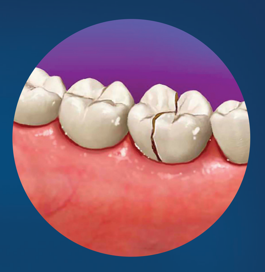
The incidences of cracked teeth have increased significantly in the United States over the last decade! Even before the pandemic, the AAE 2015 Special Committee on the Methodology of Cracked Tooth Studies conducted a survey and found 49% of 941 endodontists had seen an increase of cracked teeth and vertical root fractures compared with the previous decades. (1) Poor occlusion, bruxism/parafunctional habits, and wedging events combined with repetitive forces are all potential etiologies for tooth cracks. As these early cracks develop, bacteria and biofilms can form, eventually progressing towards the pulp and resulting in pulpal necrosis. (2) Cracks when left untreated, can propagate into fractures resulting in catastrophic failures and tooth loss, such as in the incidence of a split tooth. (3)
Early in the COVID-19 pandemic, a Sept. 11, 2020, article in USA Today reported that endodontists were seeing twice as many cracked teeth as they did in the prior year. (4) The March 21, 2021, ADA Huddle reported that an ADA Health policy survey had found a rise in stress-related tooth damage linked to pandemic stress. (5) More recently, the JOE published an article comparing the incidence of cracked teeth in a private endodontic practice over three years (2019, 2020, and 2021), showing a significant increase in the incidence of cracked teeth in the 40-60 age group (2020) and males 40-60 and over-60 age groups in 2021. (6) Regardless of direct or indirect causes, the diagnosis of cracked teeth was already highly prevalent before the pandemic and has subsequently and substantially increased since that time. We are at a point where we must include cracked teeth and vertical root fractures in almost every differential diagnosis of tooth pain.
Let us look at the new definitions of cracked teeth and vertical root fractures.
The New Vocabulary for Cracked Teeth and Vertical Root Fractures2
The American Association of Endodontists formed a Special Committee on Cracked Tooth Initiatives in May 2014. (7) The charge of this committee was to identify consistent terminology for the AAE Glossary of Endodontic Terms. The definitions found in the 2012 Glossary of Endodontic terms, those used in the 2008 “Cracking the Cracked Tooth Code,” and by the International Association of Dental Traumatology were inconsistent with each other. At issue was the overlap in terminologies and the ambiguous and subjective nature of the definitions. Also, omissions of specific terms and elaborations within the definitions were confusing. Moreover, the clinical appropriateness associated with some of these terminologies was questionable. (8)
The following were the new definitions that were approved by the 2015-2016 Board of Directors of the AAE:
- Abfraction: A V-shaped loss of hard tooth structure caused by biomechanical loading forces such as from flexure and/or chemical fatigue degradation of enamel and/or dentin at some location distant from the actual point of loading.
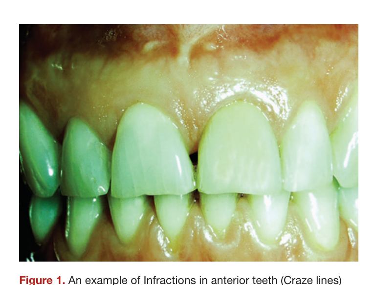
- Infraction (Craze Line): A crack of the enamel without loss of tooth structure (Figure 1)
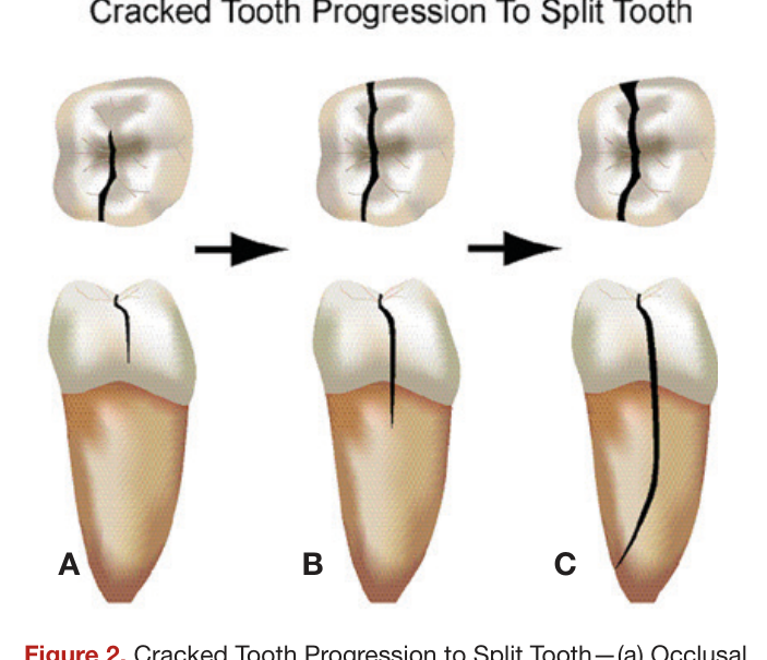
- Cracked tooth: A thin surface disruption of enamel and dentin, and possibly cementum, of unknown depth or extension. (Figure 2)
- Crown infraction: was removed from the glossary
- Fracture: A split or break in bone, cartilage or tooth structure. When involving tooth structure, it is a disruption of the enamel and/or dentin and/or cementum that is assumed to be of greater depth than a crack, of unknown depth or extension, visible or not visible clinically or radiographically, and with separated or unseparated segments.
- Root Fractures: a fracture that exists or extends into the root, to include dentin, cementum, and possibly pulpal space, which may progress to or from the enamel.
- Longitudinal Fracture: a root fracture extending in the axial plane within the tooth.
- Vertical Root Fracture: a fracture in the root whereby the fractured segments are incompletely separated; it may occur buccal-lingually or mesial-distally; it may cause an isolated periodontal defect(s) or sinus tract; it may be radiographically evident.
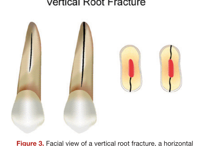
- Split Root: a continuation of a crack or vertical root fracture whereby the fractured segments are completely separated longitudinally; it may occur buccal-lingually or mesial-distally; it may cause an isolated periodontal defect(s) or sinus tract; it may be radiographically evident. (Figure 3)
These terms are found in the 2020 Glossary of Terms, 10th Edition. (8)
Other authors and researchers have suggested other conceptual ways to define cracked teeth and vertical root fractures. These included terms such as “crown originating fracture” or “root originating fracture,” emphasizing the direction the fracture may be progressing. (9) These terms and definitions are not presently found in the AAE Glossary of Terms. Notably, none of the fractures
resulting from trauma are listed in this report, as the etiologies are usually the result of acute force rather than repetitive forces that seem to be associated with cracked teeth and vertical root fractures. So, “what are the diagnostic features we look for in a cracked tooth and vertical root fractures?”
Diagnosing Cracked Teeth and Vertical Root Fractures3
Diagnosing cracked teeth and vertical root fractures begins with the Dental History and an objective clinical exam. What is the patient’s chief complaint? Does the pain occur when chewing (either biting down or upon release)? Are fillings fracturing or dislodging? Are there brief episodes of cold sensitivity? Is there tooth mobility? The following steps are involved when performing the objective exam. Current recommendations dictate the use of a surgical operating microscope to visualize the tooth because the naked eye often misses the subtle details of cracks or fractures. Greater visual acuity is also achieved when transilluminating the tooth surface from multiple directions. Minute flexure at the site of the crack can sometimes be seen under magnification, and staining using a dye can accentuate the visualization of cracks.
In addition, the clinical tests that are recommended when trying to diagnose a cracked or fractured tooth are as follows:
- Soft tissue exam for localized swelling or sinus tract(s)
- Percussion
- Palpation
- Biting tests (each cusp should be tested)
- Vitality tests (Electric pulp testing and cold testing)
- Periodontal probing (flexible plastic probes)
- Restoration removal (if necessary for complete visualization)
Figure 1. An example of Infractions in anterior teeth (Craze lines)
Figure 2. Cracked Tooth Progression to Split Tooth—(a) Occlusal and distal/proximal views of a cracked tooth affecting the distal marginal ridge of the mandibular right molar; the crack has not yet extended onto the root. (b) Growth/propagation of the crack to include both mesial and distal marginal ridges and extending onto the distal root surface; a restoration is usually not present and the crack is more centered as viewed from the proximal. (c) Further propagation results in a split tooth in which a separable segment is detected; mesial and distal marginal ridges are involved and the fracture extends deeply in the root.
Figure 3. Facial view of a vertical root fracture, a horizontal cross-section of a VRF affecting only the lingual root surface, and a horizontal cross section of a VRF affecting both the buccal and the lingual root surfaces; root canal-filling material is shown in the canal space.
- Transillumination (to look for disruptions in light transmission which are common in cracked teeth)
- Dye Staining (either before or after restoration removal)
- Surgical Assessment
- A radiographic exam which includes both periapical films (PA) and Cone Beam Computerized Tomography (CBCT) should be performed routinely.
 |  |
 |
CBCT and New Imaging Approaches4
With the increased use of Cone Beam Computed Tomography (CBCT) in dental offices over the past decade, newer indirect methods for identifying possible cracks or fractures are available. Several authors have suggested that isolated areas of bone loss adjacent to the root surface or those associated with narrow periodontal probings may indicate the presence of a crack or fracture in the absence of generalized periodontal disease. (10, 11) Loss of cortical bone as seen on a CBCT, is also thought to be more closely associated with vertical root fractures. (11)
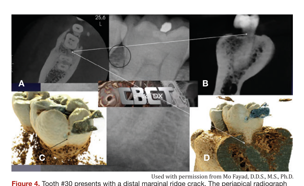
Proprietary imaging software that comes with most CBCT machines generally does not allow for artifact reduction. “Beam hardening” can obscure the accurate interpretation of endodontically treated or restored teeth. (2a) Artificial Intelligence (AI) networks are presently being developed that allow for the actual visualization and rendering of fractures. (12) Currently, an alternative radiographic imaging program is available that utilizes filters to reduce the “blooming artifact” and “metal artifact.” Other proprietary aspects of the program allow for rendering and visualization of external cracks, as seen in Figure 4. (13)
Findings Highly Suggestive of Cracked or Fractured Teeth
The AAE 2016 Special Committee on The Methodology Of Cracked Tooth Studies found the following findings were highly suggestive of cracked or fractured teeth. (1)
- Triggered biting pain
- Swelling
- One or more sinus tracts
- Percussion sensitive and mobility
- A root crack associated with a deep probing is visible with or without magnification
- Probings greater than 6 mm
- Conventional radiographs that show
- Visible fracture or separation of root or
- “J” shaped radiolucency
- Small FoV CBCT imaging with visible fracture or separation of root
- Bone loss associated with an isolated periodontal defect
Small Field of View (FoV) CBCT may be beneficial in identifying isolated bone loss, but flap reflection with visualization of the fracture using a Surgical Operating Microscope is still the “gold standard.”
These steps should allow the dental professional to formulate both a pulpal and periapical diagnosis, which will help to determine the recommended clinical treatment or a suspected cracked or fractured tooth. So, what do you tell the patient once there is a diagnosis? What are the recognized “Best Practices” today for cracked teeth and vertical root fractures?
Treatment and Prognosis
crowned had an 80% chance of never needing root canal treatment. (14, 15) Patients should be informed that all cracked teeth require the placement of a crown as part of the treatment. A recent article has confirmed that early placement of a crown on a cracked tooth can improve the prognosis of the tooth by 98% over an 11-year period. (16) If symptoms persist and either irreversible pulpitis or pulpal necrosis develops, root canal treatment can be performed. Patients with reversible pulpitis that elect to have the tooth crowned should be told that if root canal treatment is needed, it can be done through the existing crown and restored after treatment without crown replacement. However, what do you tell your patient regarding “success” or “survival” statistics?
The Iowa Staging Index: Communicating Success vs. Survival5
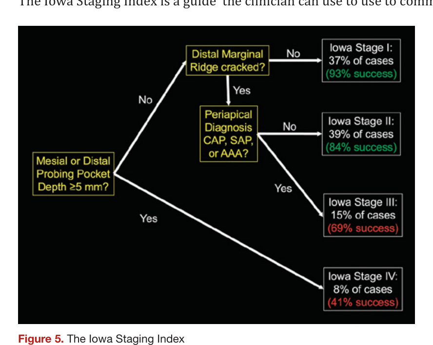
The Iowa Staging Index is a guide the clinician can use to communicate to patient possible outcomes for “success.” (17) For purposes of discussion, “Success” is defined as the “resolution of previous radiographic pathosis and the absence of signs or symptoms.” This terminology is often associated with “clinician-centered” outcomes. This definition is stricter than “Survival,” which has become more frequently used as a “patient-centered” term.
“Survival” means the “tooth is present, asymptomatic and functional.” “Survival” can include teeth that are asymptomatic but still have signs of apical pathosis, either radiographically or clinically. Older studies called these teeth “questionable.” To be clear, “Survival” statistics are always better than “Success” statistics. Let us look closer at the Iowa Staging Index. (See Figure 5.)
Figure 5 shows the decision “tree” for factors that have influenced success in past cases after root canal treatment and when the teeth were crowned. Three factors were identified as significantly affecting the outcomes following root canal treatment. The first factor is the depth of the periodontal probing associated with the crack. In the first three stages, all have periodontal probings less than 5 mm, with the second factor being the location of the crack. Distal marginal ridge cracks were associated with decreased success rates. The third factor was the initial diagnosis of periapical pathosis. Older diagnostic terminology was used because the study period covered 25 years and the new accepted diagnostic terms came out in the last ten years of the study. Using the most recent terms, periapical diagnoses of acute apical abscess, symptomatic apical periodontitis, asymptomatic apical periodontitis, and chronic apical abscess were all associated with decreased prognoses.
Success Rates by Iowa Stage
Let us look at some other features of the Iowa Staging Index. Iowa Stage 1 was if the tooth had less than a 5 mm probing, no apical pathosis, and only a mesial marginal ridge crack. The “success” rate was 93%, and “survival” has been reported as high as 97%. (18) For Iowa Stage 2, the tooth had less than a 5 mm probing, a distal marginal ridge crack, but no apical pathosis. The success rate for Stage 2 was 84%. At Iowa Stage 3 the tooth had less than a 5 mm probing, a distal marginal ridge crack, and a periapical diagnosis indicating apical pathosis. The success rate for Stage 3 was 69%. At Iowa Stage 4, if the tooth had greater than a 5 mm probing, any marginal ridge crack, the success rate was 41%. (17)
Two different meta-analyses and one large clinical retrospective study have come up with similar findings with respect to the Iowa Staging Index. All three papers concluded periodontal probings greater than or equal to 5 mm associated with a crack decrease the survival. Timely crown placement improves survival. (16, 19-21) Root canal treatment of cracked teeth is more successful than previously thought and should be considered (84% survival at 60 months). (16-21)
Clinical Treatment Improvements When Treating Cracked Teeth
The protocol for treating cracked teeth requiring endodontic therapy has changed over the past fifteen years. There is still a continued emphasis on reducing repetitive forces on the tooth and improving survivability. Reduction of occlusion is highly recommended as part of the initial treatment because the repetitive stress from bruxing or grinding is thought to lead to the propagation of a crack leading to fracture.
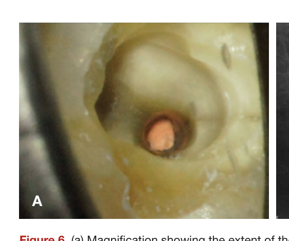
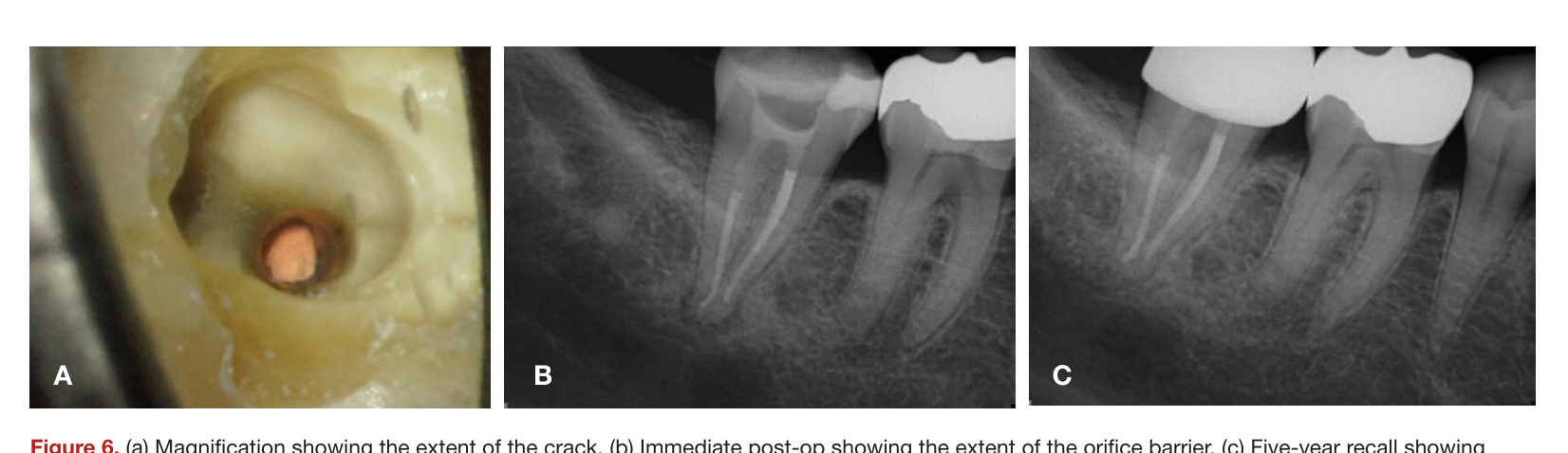
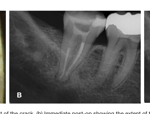
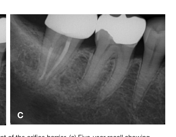
Repetitive occlusal forces were the only explanation for root fractures found in non-endodontically treated teeth! (22, 23) Davis and Shariff (18) have recommended that after obturation, orifice barriers be placed to the deepest extent
of the crack, which may help strengthen the root and seal the canal from further bacterial penetration. The tooth is taken out of occlusion, and the patient is encouraged to chew on the other side until a crown has been placed. Additionally, the crown should be placed as soon as possible after root canal treatment. The authors’ survival rate using this protocol and strict recall every six months has resulted in a 97% survival rate at four years of follow-up. (Figure 6)
Several studies have shown the benefit of timely crown placement on teeth with root canal treatment. (24) Two meta-analyses of cracked teeth and two large population retrospective studies have all re-emphasized the importance of a crown following treatment. (16, 17, 18, 22) Once again, when deciding the treatment options for the patient, a crown must be an integral part of the treatment plan.
Treatment Options for Vertical Root Fractures6
Teeth that have developed vertical root fractures (or root originating fractures) have guarded prognoses. Single rooted teeth with a vertical root fracture will generally need to be extracted. Frequently, these teeth have been endodontically treated and subsequently develop periapical pathosis over time. Endodontically treated teeth with posts or excessive root structure removed are at the greatest risk for fractures, but even non-endodontically teeth can develop root fractures, as previously mentioned. (22,23)
 |  |  |
 |  |  |
 |
Natural Teeth vs. Implants
As dental implants have become more prone to failure, the question “What is in the patient’s best interest?” becomes tantamount. In an editorial by the then editor of the Journal of Dental Research in 2016, Giannoble and Lang stated:
“Teeth even compromised because of periodontal disease or endodontic problems may have a longevity that surpasses by far that of the average implant.” (Carnevale et al. 1998; Hardt et al. 2002; Lang and Zitzmann 2012; Salvi et al. 2014; Klinge et al. 2015) (26-30)
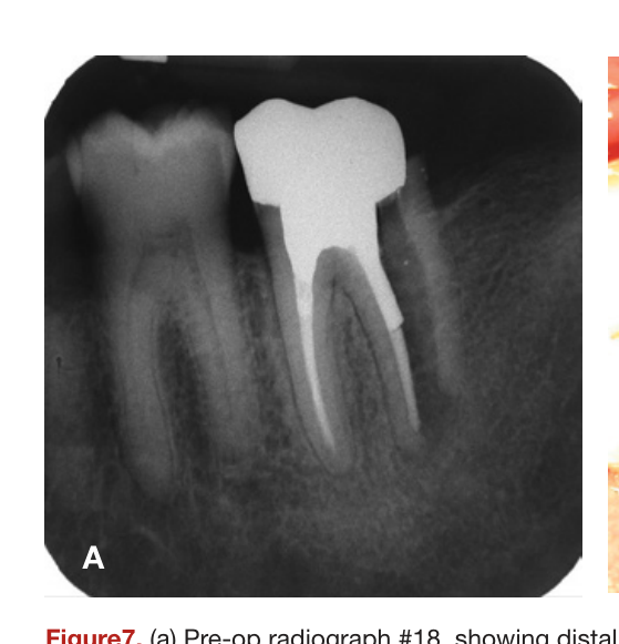
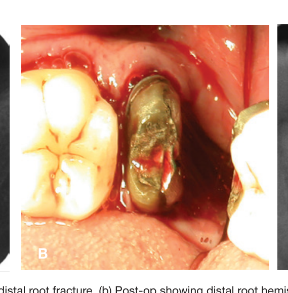
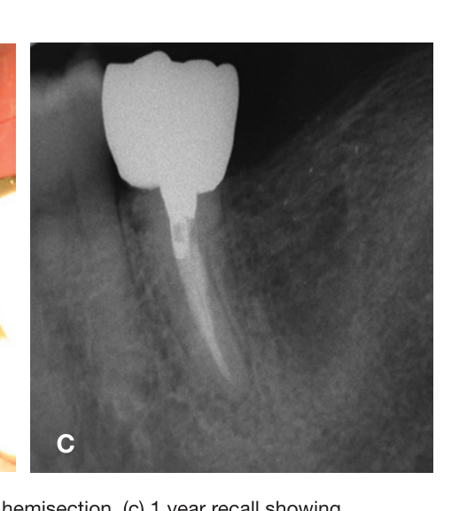
Historically, multi-rooted teeth have been saved with either a root resection, root amputation, or hemisection procedure rather than extraction and replacement with an implant. Root amputations have been shown to have a 92% survival over 12 years (31) and hemisections 79 to 91% survival over seven to 30 years. (32, 33) (Figure 7). The editors’ argument that these older treatment options were more conservative and have demonstrated outcomes that can exceed implants was a plea for dentists to slow down with extractions of cracked teeth and teeth with vertical root fractures and consider some “tried and true” procedures with better outcomes. Quite simply, natural dentition preserves bone and should be preserved.
Vahdati (34), compared patients with both a single tooth implant and those with a tooth with non-surgical root canal treatment. Patients experienced four times as many appointments when agreeing to a single tooth implant versus patients having non-surgical root canal treatment and a crown with over twice the expense. Both success rates were the same, but more extended periods occurred before final restoration, and more complications were found with the single tooth implants.
So, for multi-rooted teeth with a single root with a vertical root fracture, “hopeless” is no longer the only word to be used when addressing the patient. These other documented options with outcome studies dating back decades show long-term success when correctly done. These procedures may require referral to a specialist.
Conclusions7
Cracked Teeth
Over the past decade, several factors have become more evident in preserving cracked teeth. The earlier cracked teeth can be identified, the higher the success rate for the long-term preservation of the tooth. Not all cracked teeth require root canal treatment, but all cracked teeth require a crown! Occlusion should be evaluated during the initial exam, and adjustments to reduce occlusal forces should be made. If root canal treatment is required, the use of orifice barriers placed below the level of the crack as seen in a surgical operating microscope should be placed. A crown should be placed as soon as possible, though a well-fitted temporary crown may be left in place until symptoms subside for cases with reversible pulpitis. Close recall appointments and monitoring of the occlusion are important.
Vertical Root Fractures
The diagnosis of a vertical root fracture has improved with the use of CBCT but is still considered catastrophic for the root. Single rooted teeth will need to be extracted, as no successful treatment options have been developed for these teeth. For multi-rooted teeth, clinical studies show that the removal of the fractured root and restoration of the remaining root(s) are more successful than many of the implants placed. Removal of the fractured root on multi-rooted teeth with the treatment of the remaining tooth should be an option offered to patients. This conclusion may require referral to a specialist for the optimal result. Close monitoring and occlusal adjustment as needed should be recommended.
References
- Friedman S, A.A., Bruder GA, Krell KV et al Guidelines for the methodology of cracked tooth studies. 2016.
- Ricucci, D., et al., The cracked tooth: histopathologic and histobacteriologic aspects. J Endod, 2015. 41(3): p. 343-52.
- Rivera, R.M.a.W., R.E., Cracking the cracked tooth code: detection and treatment of various longitudinal tooth fractures. Colleagues for Excellence. Chicago, American Association of Endodontists, 2008.
- Shannon, J., Cracked teeth, gross gums: Dentists see surge of problems, and the pandemic is likely to blame. USA Today, 2020. Published 9-11-2020: p. 3.
- Huddle, A., HPI: Prevalence of stress-related teeth damage risding, M. Huddle, Editor. 2021, ADA: ADA.org.
- Nosrat, A., et al., Endodontic Specialists’ Practice During the Coronavirus Disease 2019 Pandemic: 1 Year after the Initial Outbreak. J Endod, 2022. 48(6): p. 699-706.
- Louis H. Berman, C., Scott L. Doyle, Gary G. Goodell, Keith V. Krell, Mark A. Odom, D., Special Committee on Cracked Tooth Initiative. 2015, American Association of Endodontists.
- Scott B. McClanahan, D.D.S., M.S., ChairSpecial Committee to Revise the Glossary (2020), et al., American Association of Endodontists (2020) Glossary of endodontic terms Tenth Edition. 2020: p. 48.
- Bakland, L.K. and A. Tamse, Categorization of Dental Fractures, in Vertical Root Fractures in Dentistry, A. Tamse, I. Tsesis, and E. Rosen, Editors. 2015, Springer International Publishing: Cham. p. 7-28.
- Zhang, L., et al., In Vivo Detection of Subtle Vertical Root Fracture in Endodontically Treated Teeth by Cone-beam Computed Tomography. J Endod, 2019. 45(7): p. 856-862.
- Alaugaily, I. and A.A. Azim, CBCT Patterns of Bone Loss and Clinical Predictors for the Diagnosis of Cracked Teeth and Teeth with Vertical Root Fracture. J Endod, 2022. 48(9): p. 1100-1106.
- Shah, H., et al., Automatic quantification framework to detect cracks in teeth. Proc SPIE Int Soc Opt Eng, 2018. 10578.
References
- Bueno, M.R., B.C. Azevedo, and C. Estrela, A Critical Review of the Differential Diagnosis of Root Fracture Line in CBCT scans. Braz Dent J, 2021. 32(5): p. 114-128.
- Krell, K.V. and E.M. Rivera, A six year evaluation of cracked teeth diagnosed with reversible pulpitis: treatment and prognosis. J Endod, 2007. 33.
- Abbott, P. and N. Leow, Predictable management of cracked teeth with reversible pulpitis. Aust Dent J, 2009. 54(4): p. 306-15.
- de Toubes, K.M.S., et al., The Correlation of Crack Lines and Definitive Restorations with the Survival and Success Rates of Cracked Teeth: A Long-term Retrospective Clinical Study. J Endod, 2022. 48(2): p. 190-199.
- Krell, K.V. and D.J. Caplan, 12-month Success of Cracked Teeth Treated with Orthograde Root Canal Treatment. J Endod, 2018. 44(4): p. 543-548.
- Davis, M.C. and S.S. Shariff, Success and Survival of Endodontically Treated Cracked Teeth with Radicular Extensions: A 2- to 4-year Prospective Cohort. J Endod, 2019. 45(7): p. 848-855.
- Olivieri, J.G., et al., Outcome and Survival of Endodontically Treated Cracked Posterior Permanent Teeth: A Systematic Review and Meta-analysis. J Endod, 2020. 46(4): p. 455-463.
- Leong, D.J.X., et al., Outcomes of endodontically treated cracked teeth: a systematic review and meta-analysis. Clin Oral Investig, 2020. 24(1): p. 465-473.
- Chen, Y.T., et al., Factors Related to the Outcomes of Cracked Teeth after Endodontic Treatment. J Endod, 2021. 47(2): p. 215-220.
- Chan, C.P., et al., Vertical root fracture in nonendodontically treated teeth—a clinical report of 64 cases in Chinese patients. J Endod, 1998. 24(10): p. 678-81.
- Yang, S.F., E.M. Rivera, and R.E. Walton, Vertical root fracture in nonendodontically treated teeth. J Endod, 1995. 21(6): p. 337-9.
- Aquilino, S.A. and D.J. Caplan, Relationship between crown placement and the survival of endodontically treated teeth. J Prosthet Dent, 2002. 87(3): p. 256-63.
- Giannobile, W.V. and N.P. Lang, Are Dental Implants a Panacea or Should We Better Strive to Save Teeth? J Dent Res, 2016. 95(1): p. 5-6.
- Carnevale, G., R. Pontoriero, and G. di Febo, Long-term effects of root-resective therapy in furcation-involved molars. A 10-year longitudinal study. J Clin Periodontol, 1998. 25(3): p. 209-14.
- Hardt, C.R., et al., Outcome of implant therapy in relation to experienced loss of periodontal bone support: a retrospective 5-year study. Clin Oral Implants Res, 2002. 13(5): p. 488-94.
- Lang, N.P., T. Berglundh, and P. Working Group 4 of Seventh European Workshop on, Periimplant diseases: where are we now?—Consensus of the Seventh European Workshop on Periodontology. J Clin Periodontol, 2011. 38 Suppl 11: p. 178-81.
- Salvi, G.E., et al., Risk factors associated with the longevity of multi-rooted teeth. Long-term outcomes after active and supportive periodontal therapy. J Clin Periodontol, 2014. 41(7): p. 701-7.
- Klinge, B., et al., The patient undergoing implant therapy. Summary and consensus statements. The 4th EAO Consensus Conference 2015. Clin Oral Implants Res, 2015. 26 Suppl 11: p. 64-7.
- Basten, C.H., W.F. Ammons, Jr., and R. Persson, Long-term evaluation of root-resected molars: a retrospective study. Int J Periodontics Restorative Dent, 1996. 16(3): p. 206-19.
- Svardstrom, G. and J.L. Wennstrom, Periodontal treatment decisions for molars: an analysis of influencing factors and long-term outcome. J Periodontol, 2000. 71(4): p. 579-85.
- Derks, H., et al., Retention of molars after root-resective therapy: a retrospective evaluation of up to 30 years. Clin Oral Investig, 2018. 22(3): p. 1327-1335.
- Vahdati, S.A., et al., A Retrospective Comparison of Outcome in Patients Who Received Both Nonsurgical Root Canal Treatment and Single-tooth Implants. J Endod, 2019. 45(2): p. 99-103.
Acknowledgments8
The AAE wishes to thank Keith V. Krell, D.D.S., M.S., M.A., for authoring this issue of the newsletter, as well as the following article reviewers: Drs. Mark B. Desrosiers, Craig Dunlap, Craig S. Hirschberg and Stefan I. Zweig. Special thanks as well to Drs. Matthew C. Davis, Gary Matt and Mohamed I. Fayad for their contributions.
This issue of the Colleagues newsletter is available online at aae.org/colleagues.
 |  |  |
 |  |  |
Contact Information
American Association of Endodontists
180 N. Stetson Ave. Suite 1500 Chicago, IL 60601 Phone: 800-872-3636 (U.S., Canada, Mexico) or 312-266-7255 Fax: 866-451-9020 (U.S., Canada, Mexico) or 312-266-9867 Email: info@aae.org
- facebook.com/endodontists
- @savingyourteeth
- youtube.com/rootcanalspecialists
- @savingyourteeth
- www.aae.org
Clinical Resources
The AAE offers a variety of clinical resources, including publications, guidelines, position statements and treatment planning resources reflecting the latest research, techniques and technologies in endodontics. Whether an endodontist, general dentist or dental student, these tools are valuable references to support the highest standards of patient care. Visit aae.org to view our collection.
Disclaimer
Information in this newsletter is designed to aid dentists. Practitioners must use their best professional judgment, taking into account the needs of each individual patient when making diagnosis/treatment plans. The AAE neither expressly nor implicitly warrants against any negative results associated with the application of this information. If you would like more information, consult your endodontic colleague or contact the AAE.
Feedback
Are there topics you would like to cover in the future? We want to hear from you! Send your comments and questions to the American Association of Endodontists at the address below, and visit the Colleagues online archive at aae.org/colleagues for back issues of the newsletter.
© 2022 American Association of Endodontists (AAE), All Rights Reserved
Footnotes
-
Original PDF page 1: AAE cracked teeth, p.1 ↩
-
Original PDF page 2: AAE cracked teeth, p.2 ↩
-
Original PDF page 3: AAE cracked teeth, p.3 ↩
-
Original PDF page 4: AAE cracked teeth, p.4 ↩
-
Original PDF page 5: AAE cracked teeth, p.5 ↩
-
Original PDF page 6: AAE cracked teeth, p.6 ↩
-
Original PDF page 7: AAE cracked teeth, p.7 ↩
-
Original PDF page 8: AAE cracked teeth, p.8 ↩