 |  |  |
 |  |  |
Etiology and Pathophysiology
- Derived from the dental lamina or it’s remants (cell rests of Serres )
- Originate form the enamel organ (tooth primordium) of a tooth befre hard tissues develop
Contributory factors :
Prevalence
- Constitutes 5 -10 % of all jaw cysts
Histological Features
- regular startified squamous epithelium
- Thin epithelial layer (5-8 cells thick)
- Palisaded basal layer
- basal cells have a hyperchromatic blue nuclues
- Slightly columnar appearance
- Corrugated surface which can be para or orthokeratinized
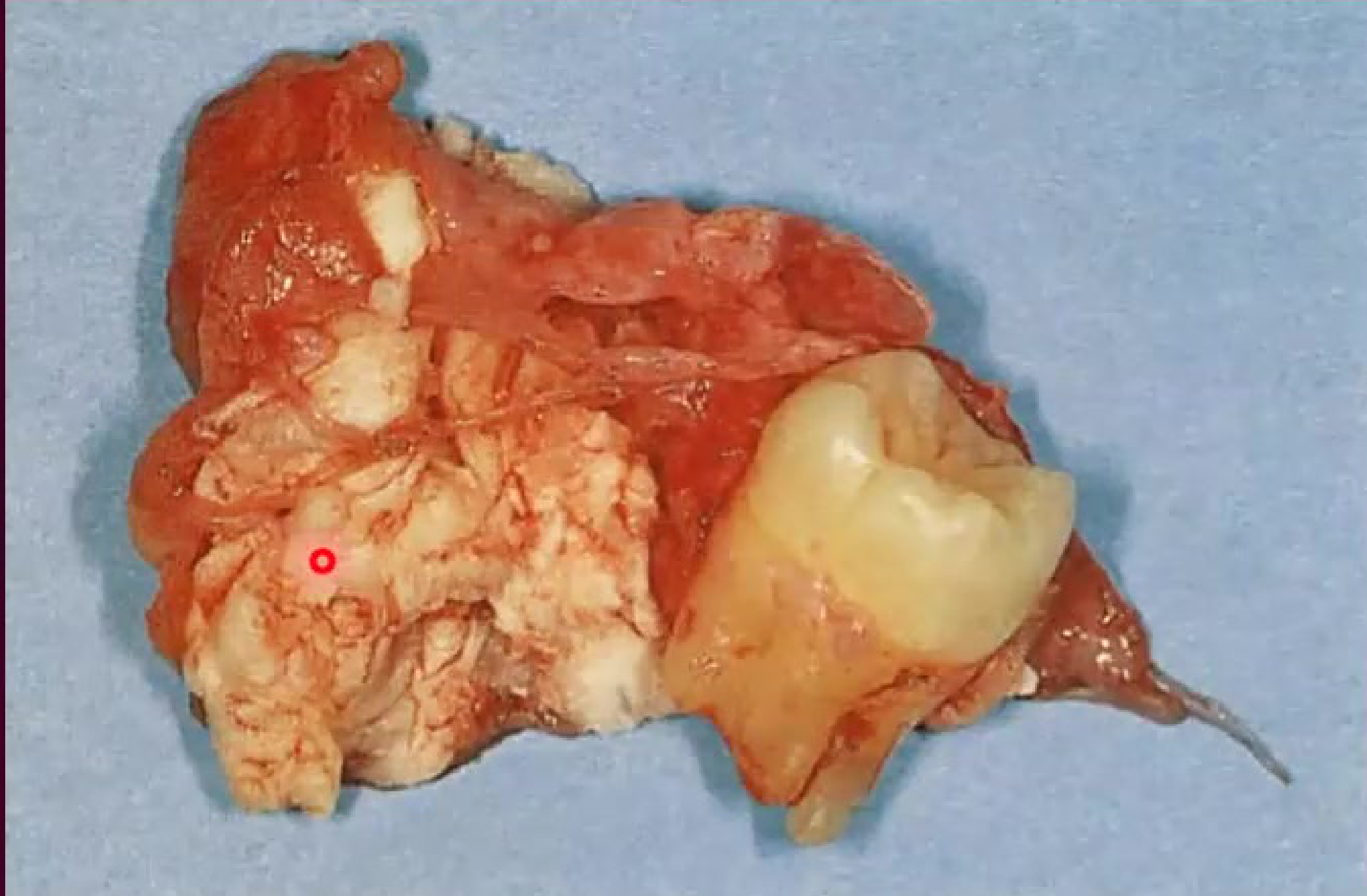
- Thin friable fibrous capsule
- Satellite cysts in the cyst wall
Clinical Features
- often asymptomattic
- swelling , discharge, pain , pathological fracutre, tooth displacement and rarely: buccal expansion
- High recurrence rate
- 10% of cases of keratocysts have multiple cysts
Location
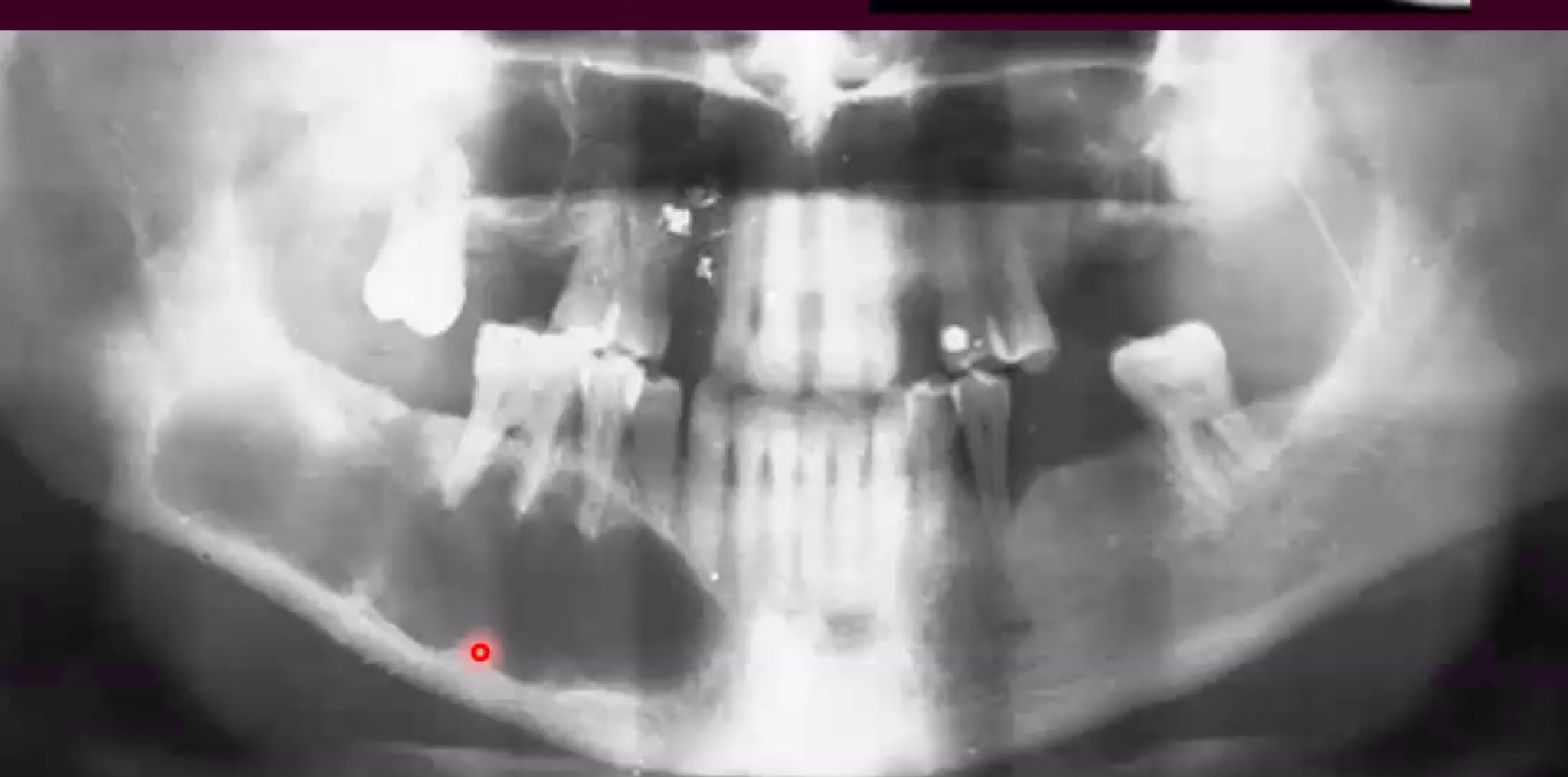
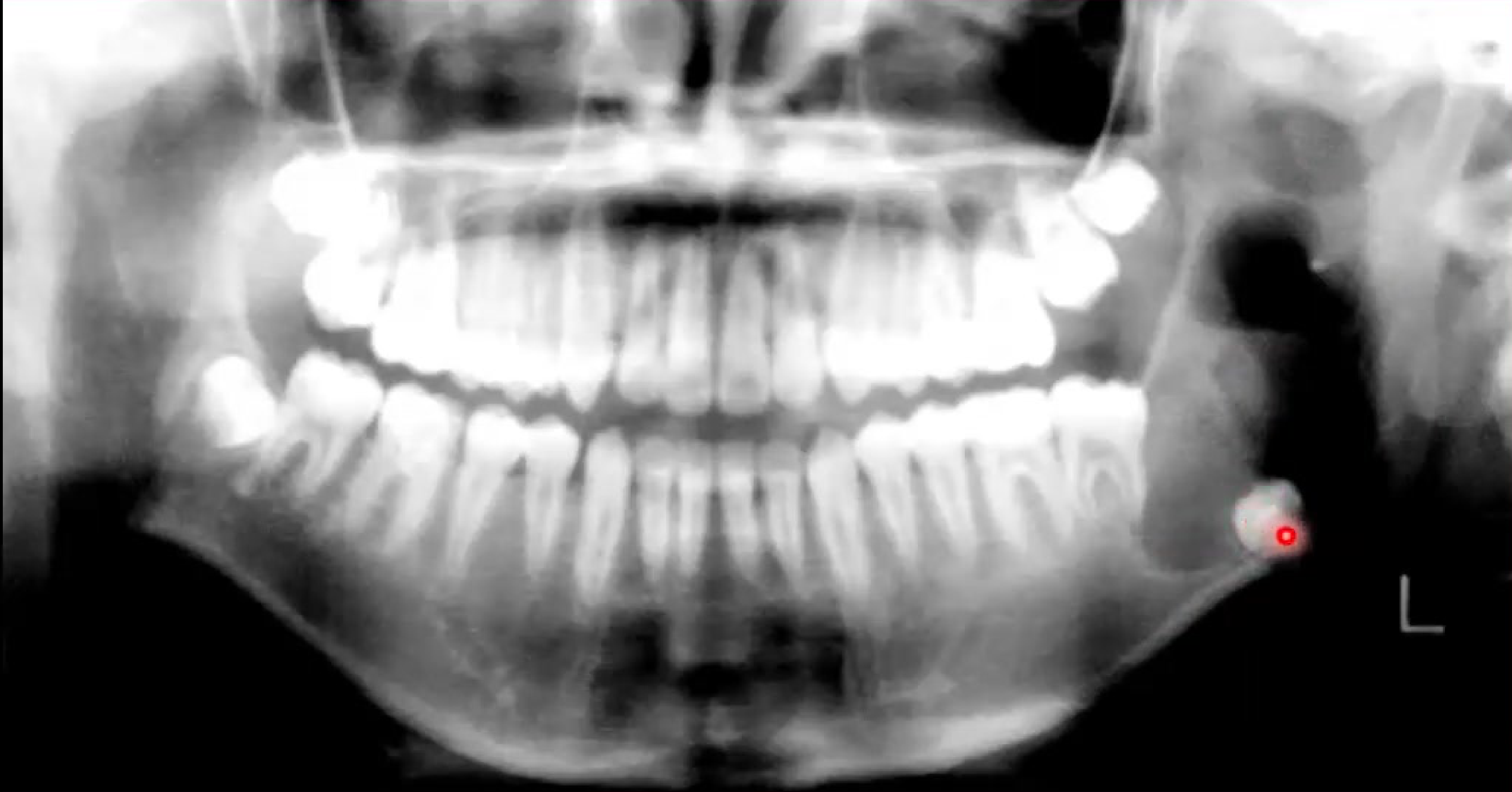
- Most frequenty (80%) found in the posterior mandible
Appearance
Radiographic:
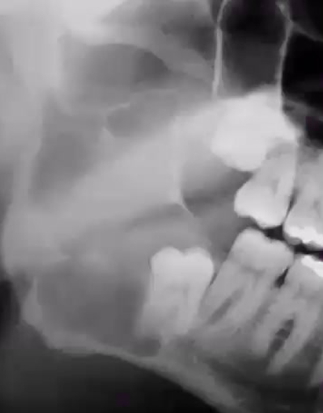
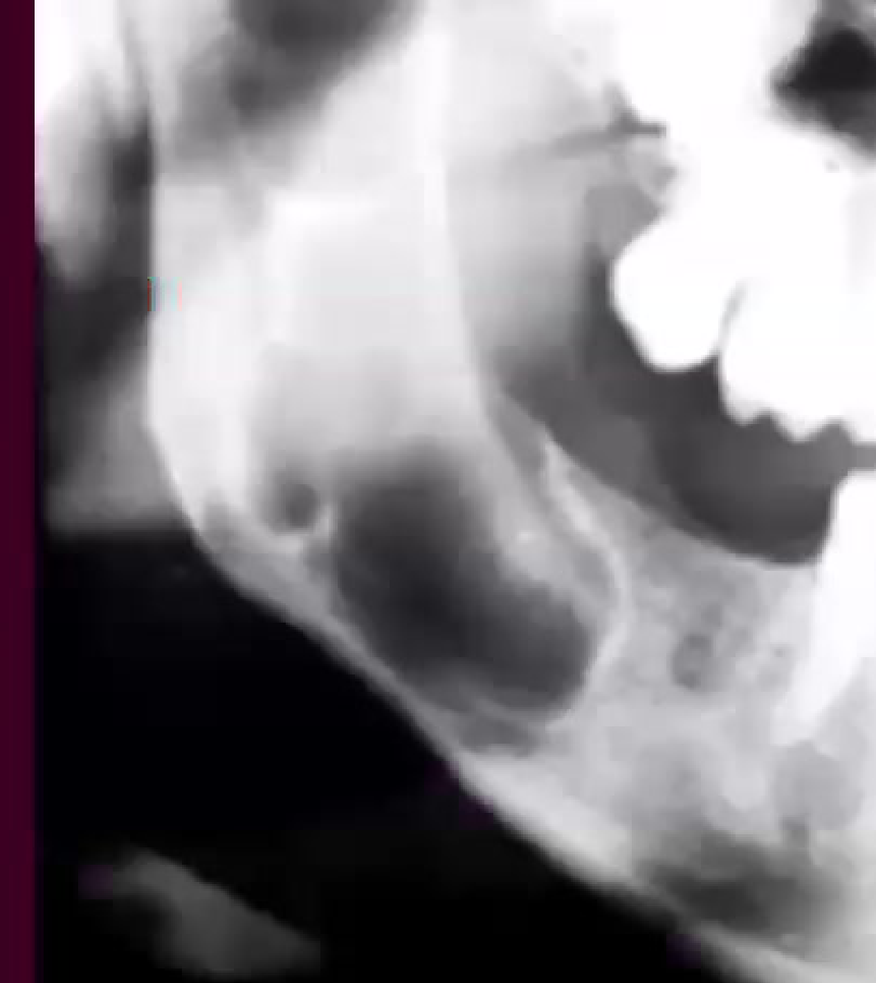
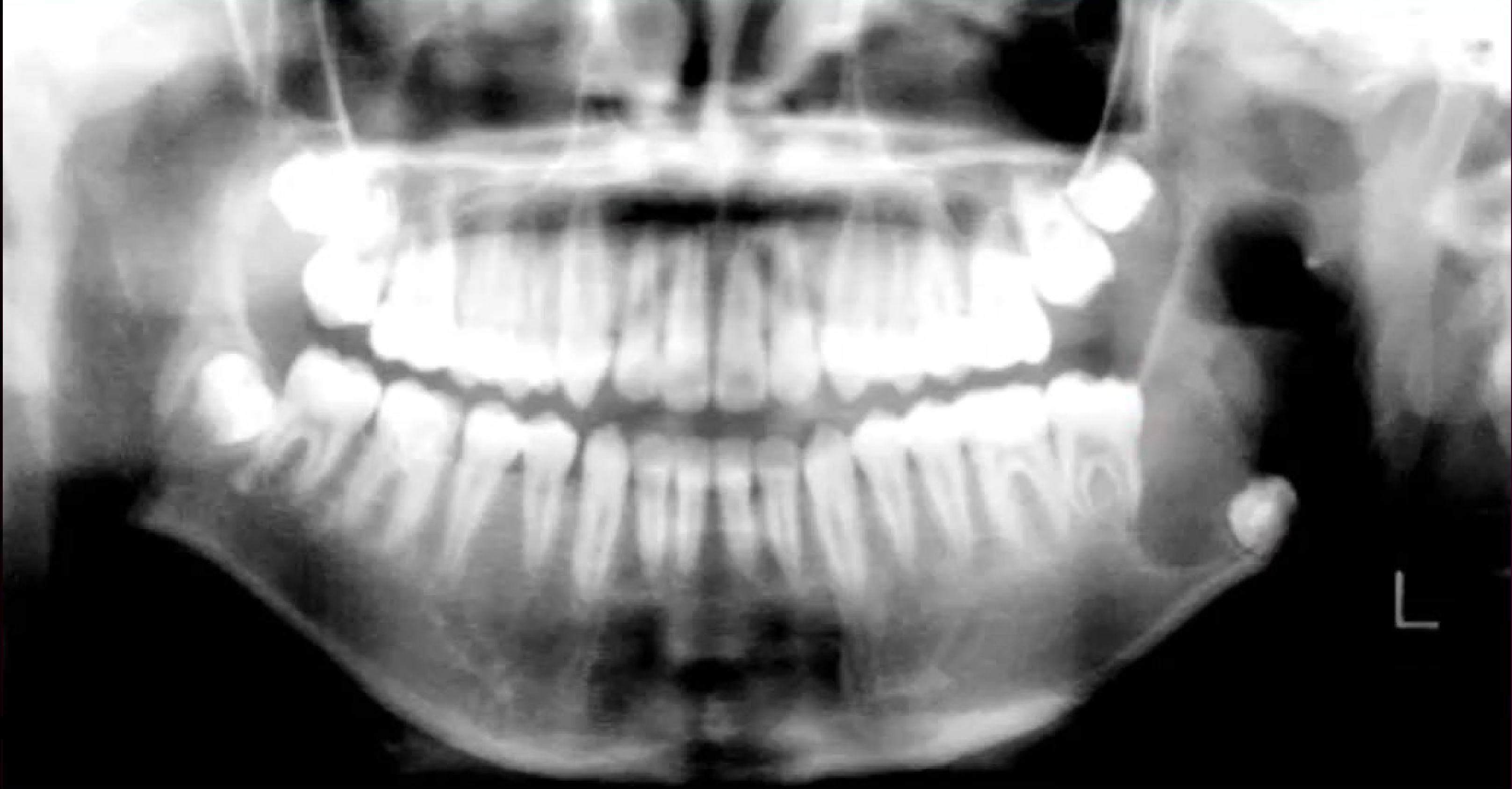
- well demarcated radiolucency
- Pseudolocular or multilocular, often with Scalloped periphery
- Root or tooth displacement
Differential Diagnoses
- orthokeratinising odontogenic cyst
- Ameloblastic carcinoma
- Dentigerous cyst
Relevant Clinical Investigations
- OPG
- Biopsy
- Aspiration cytology (to rule out unicystic amelobalstoma)
- PTCH1 gene mutation to rule out Gorlin-Goltz syndrome
- MRI (if soft tissue involvement is suspected)
Patient Management
- marsupialization (decompression) folled by enucleation
- Enucleation either alone or with the removal of the associated tooth
- Can use adjunctive techqniques like Carnoy’s solution, cryotherapy