Oral Pathology1
Table Of Contents
Clinical Categories and Disease Classifications2
- Developmental Conditions
- Mucosal Lesions—Reactive
- Mucosal Lesions—Infections
- Mucosal Lesions—Immunologic Diseases
- Mucosal Lesions—Premalignant
- Mucosal Lesions—Malignant
- CT Tumors—Benign
Connective Tissue Tumors
Connective tissue tumors are located within the submucosal layer and typically manifest as “lumps or bumps.” These tumors generally fall into two categories:
- Reactive: Reacting to trauma or another stimulus.
- Neoplastic: Referring to new, abnormal growth.
These lesions are further divided by tissue type, including fibrous, neural, vascular, muscular, and fat tissue.
- CT Tumors—Malignant
- Salivary Gland Diseases—Reactive
- Salivary Gland Diseases—Benign
- Salivary Gland Diseases—Malignant
- Lymphoid Neoplasms
- Odontogenic Cysts
- Odontogenic Tumors
- Bone Lesions—Fibro-Osseous
- Bone Lesions—Giant Cell
- Bone Lesions—Inflammatory
- Bone Lesions—Malignant
- Hereditary Conditions
Reactive Mucosal Lesions
Clinical Concepts3
- Also known as traumatic fibroma, irritation fibroma, and hyperplastic scar.
- Caused by chronic trauma or irritation.
- Characterized as fibrous hyperplasia of the oral mucosa.
- It is a very benign and common connective tissue tumor, often found on the buccal mucosa.
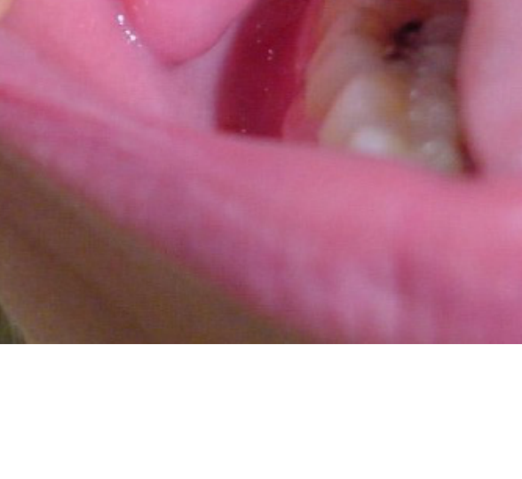
Fibroma
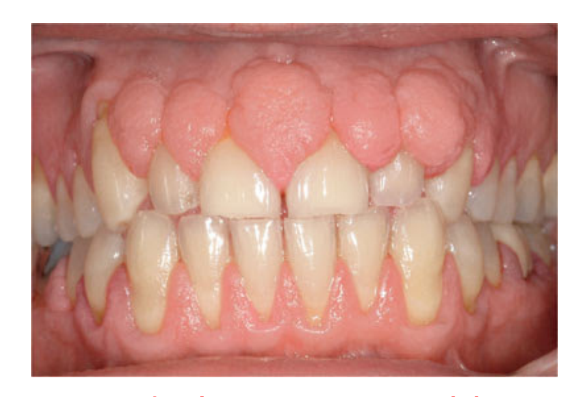
Gingival Hyperplasia
Drug-Induced Etiology4
- Calcium channel blockers
- Dilantin (anticonvulsant)
- Cyclosporine (immunosuppressant)
Treatment and Clinical Notes
- Treatment: Gingivectomy and discontinue drug if possible.
- CDC is concerned about gingival hyperplasia.
CDC Mnemonic
A helpful mnemonic for drug-induced gingival hyperplasia is CDC:
- C - Calcium Channel Blockers (e.g., verapamil, nifedipine, amlodipine)
- D - Dilantin (phenytoin)
- C - Cyclosporine
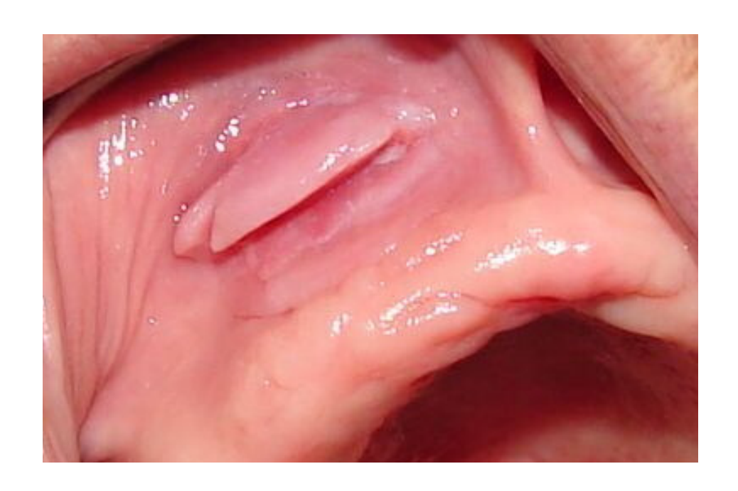
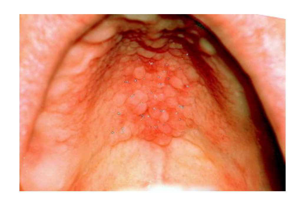
Denture Induced Fibrous Hyperplasia
Clinical Variants5
- Epulis fissuratum: Located in the vestibule.
- Papillary hyperplasia: Located on the palate
- Associated Condition: Angular Cheilitis is often grouped with these as part of a “triad” of denture-associated lesions. It occurs at the corners of the mouth and is linked to fungal infections (candidiasis) when the corners of the mouth fold in due to poor denture fit.
 |  |
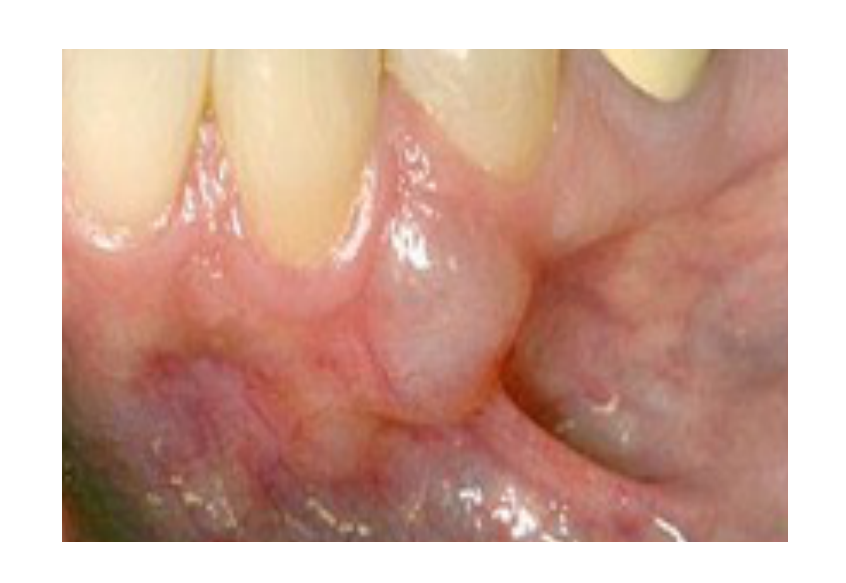
Traumatic Neuroma
Pathophysiology and Location6
- Entangled submucosal mass of neural tissue and scar.
- Caused by injury to a nerve.
- Most common at the mental foramen.
- The mental foramen is located near the mandibular canine and premolar area.
Associated Syndromes
- Multiple Endocrine Neoplasia (MEN 2B): Characterized by multiple neuromas, medullary thyroid cancer, and pheochromocytoma of the adrenal gland.
- A mnemonic to remember the association is that “MEN” is short for “mental,” as in the mental foramen.
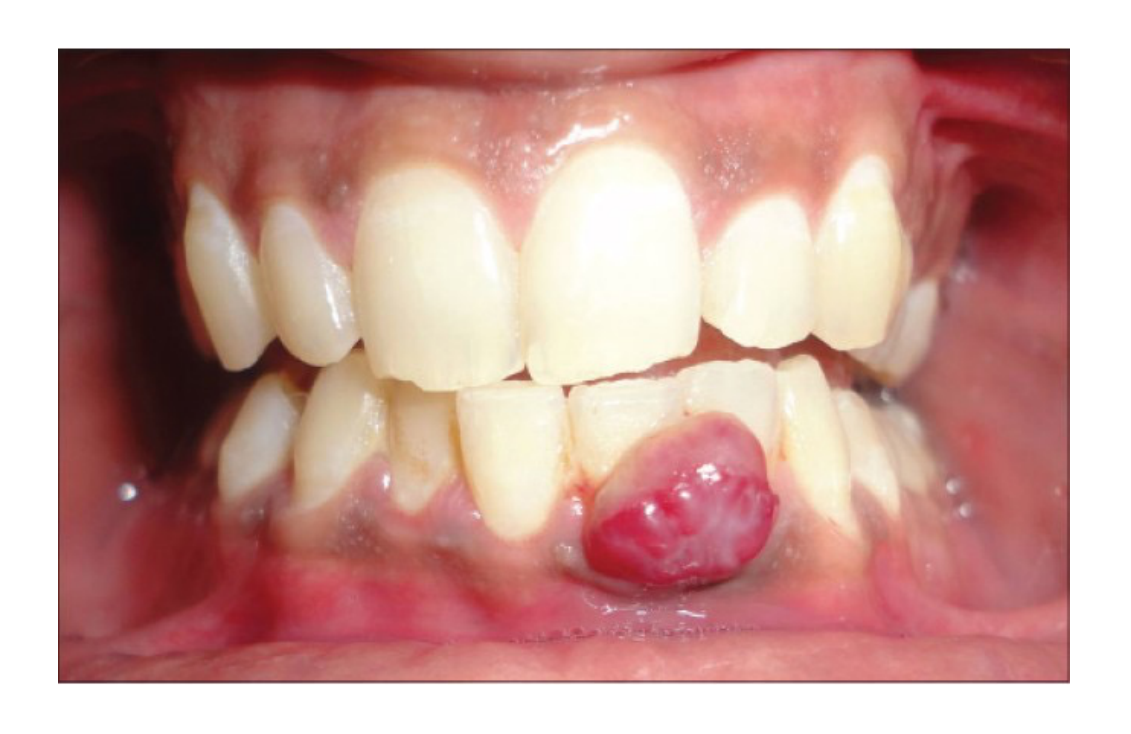
Pyogenic Granuloma
Clinical Features7
- Hyperplasia of capillaries (appears red).
- Presentation: Usually a bright, vividly red lesion.
- Caused by chronic trauma or irritation.
- Common in the gingiva.
Benign Connective Tissue Tumors
Clinical Characteristics8
- Neoplasm of fibroblasts.
- Easy to eradicate and rarely recurs.
- Treatment: Surgical excision.
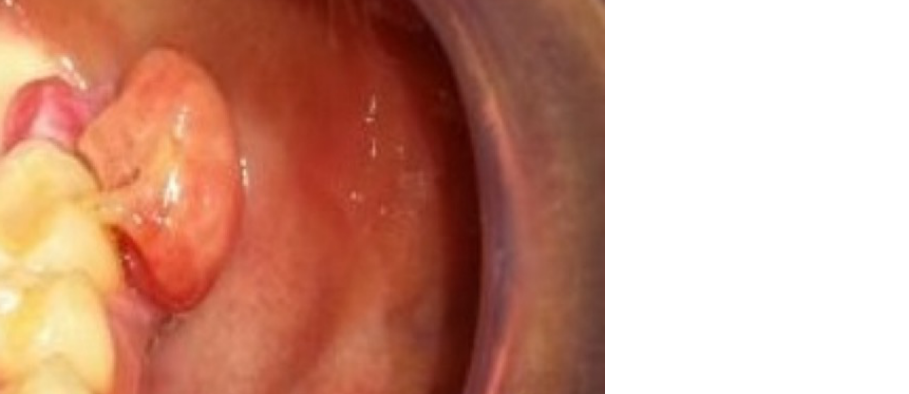
Nodular Fasciitis
- Pathophysiology: A neoplasm of fibroblasts (cells that secrete collagen).
- Clinical Characteristics: Easy to eradicate and rarely recurs after surgical excision.
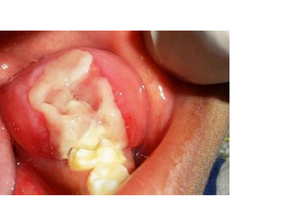
Fibromatosis
Clinical Characteristics9
- Neoplasm of fibroblasts.
- Difficult to eradicate and often recurs.
- Pathophysiology: Also a neoplasm of fibroblasts.
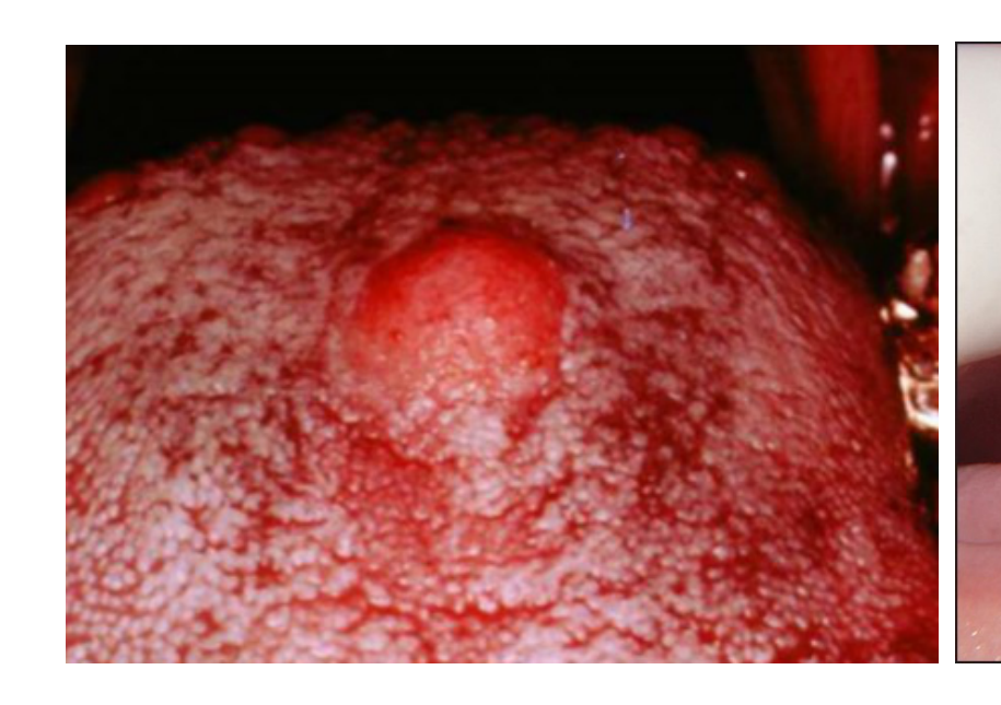
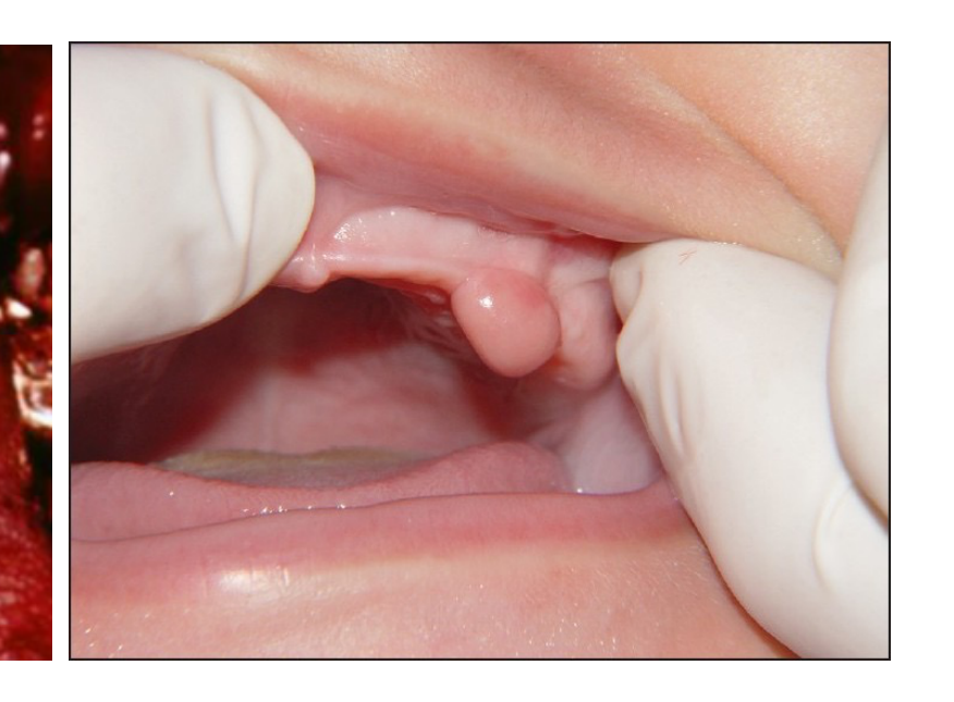
Granular Cell Tumor
Pathophysiology10
- Neoplasm of Schwann cells.
- Named for tumor cells containing granular cytoplasm.
- Cell Type: A neoplasm of Schwann cells (responsible for myelin).
- Pseudoepitheliomatous hyperplasia (PEH) in this tumor mimics Squamous Cell Carcinoma (SCC).
- PEH features a proliferative epithelium that mimics the appearance of squamous cell carcinoma under a microscope.
 |  |
Clinical Presentation
- Most common on the dorsal tongue.
- Congenital epulis of newborn: A variant found on the gingiva (does not exhibit PEH).
Schwannoma
- Terminology: Also known as a neurolemoma.
- Pathophysiology: A neoplasm of Schwann cells.
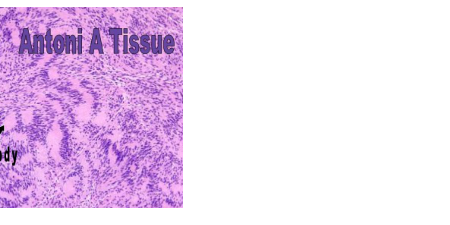
Histologic Features11
- Neoplasm of Schwann cells.
- Characterized by acellular Verocay bodies in Antoni A tissue (resembling a “line of scrimmage”).
- These form a pattern described as a “line of scrimmage,” appearing as two distinct lines in the histology.
Neurofibroma
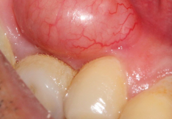
Pathophysiology12
- Neoplasm of Schwann cells and fibroblasts.
Neurofibromatosis Type I (Von Recklinghausen’s Disease)
- Characterized by multiple neurofibromas.
- Skin manifestations: Multiple skin freckles (café au lait spots) and axillary freckles (Crowe’s sign).
- Ocular manifestations: Iris freckles (Lisch spots).
- Complications: Neurofibromas can transform into neurofibrosarcomas in this condition.
Muscle And Fat Tumors
Leiomyoma
- Neoplasm of smooth muscle cells.
Rhabdomyoma
- Neoplasm of skeletal muscle cells.
Leiomyoma13
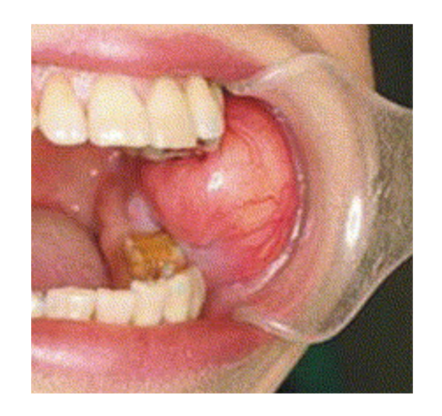
Lipoma
- Neoplasm of fat cells.
- Most common on the buccal mucosa.
- Typically presents with a yellowish nature due to its fatty composition.
Footnotes
-
Original PDF page 1: Oral Pathology - 07 - Connective Tissue Benign Tumors, p.1 ↩
-
Original PDF page 2: Oral Pathology - 07 - Connective Tissue Benign Tumors, p.2 ↩
-
Original PDF page 3: Oral Pathology - 07 - Connective Tissue Benign Tumors, p.3 ↩
-
Original PDF page 4: Oral Pathology - 07 - Connective Tissue Benign Tumors, p.4 ↩
-
Original PDF page 5: Oral Pathology - 07 - Connective Tissue Benign Tumors, p.5 ↩
-
Original PDF page 6: Oral Pathology - 07 - Connective Tissue Benign Tumors, p.6 ↩
-
Original PDF page 7: Oral Pathology - 07 - Connective Tissue Benign Tumors, p.7 ↩
-
Original PDF page 8: Oral Pathology - 07 - Connective Tissue Benign Tumors, p.8 ↩
-
Original PDF page 9: Oral Pathology - 07 - Connective Tissue Benign Tumors, p.9 ↩
-
Original PDF page 10: Oral Pathology - 07 - Connective Tissue Benign Tumors, p.10 ↩
-
Original PDF page 11: Oral Pathology - 07 - Connective Tissue Benign Tumors, p.11 ↩
-
Original PDF page 12: Oral Pathology - 07 - Connective Tissue Benign Tumors, p.12 ↩
-
Original PDF page 13: Oral Pathology - 07 - Connective Tissue Benign Tumors, p.13 ↩