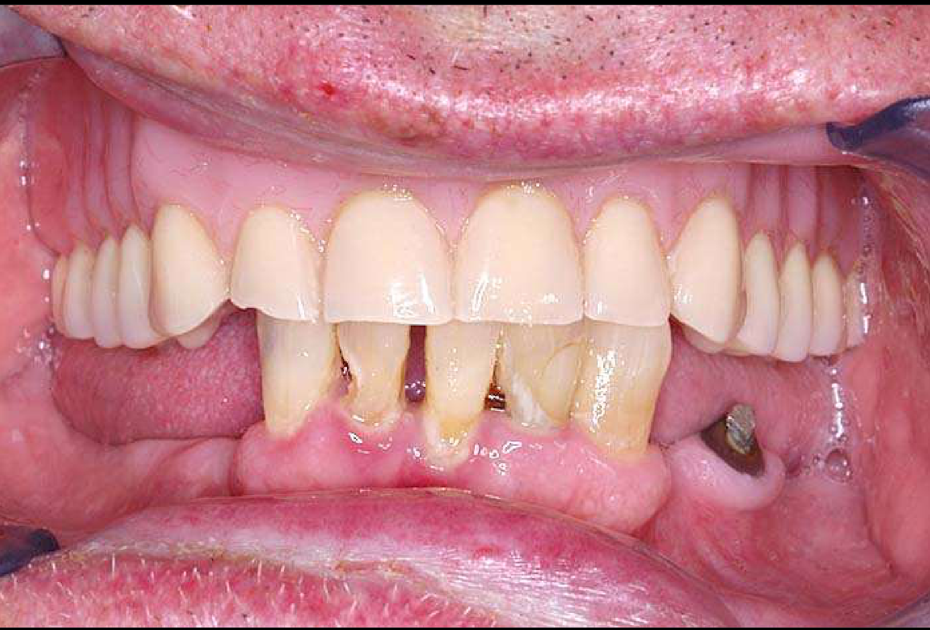
Clinical Case Study: Kennedy Class IV Restorative Treatment Intraoral and Extraoral Observations1
 |  |
Clinical Presentation and Visual Analysis
Patient History and Case Description
A 68-year-old male presented for a routine appointment. His primary complaint was an inability to chew effectively, and he requested information regarding restorative treatment options. The patient noted that his last dental visit was “a long time ago.”
Medical and Social History
- Medical Conditions: Hypertension, hypercholesterolaemia, ischaemic heart disease, osteoarthritis, and skin cancers.
- Social History:
- Non-smoker for 30 years.
- Alcohol consumption: Average of 1–2 drinks per week.
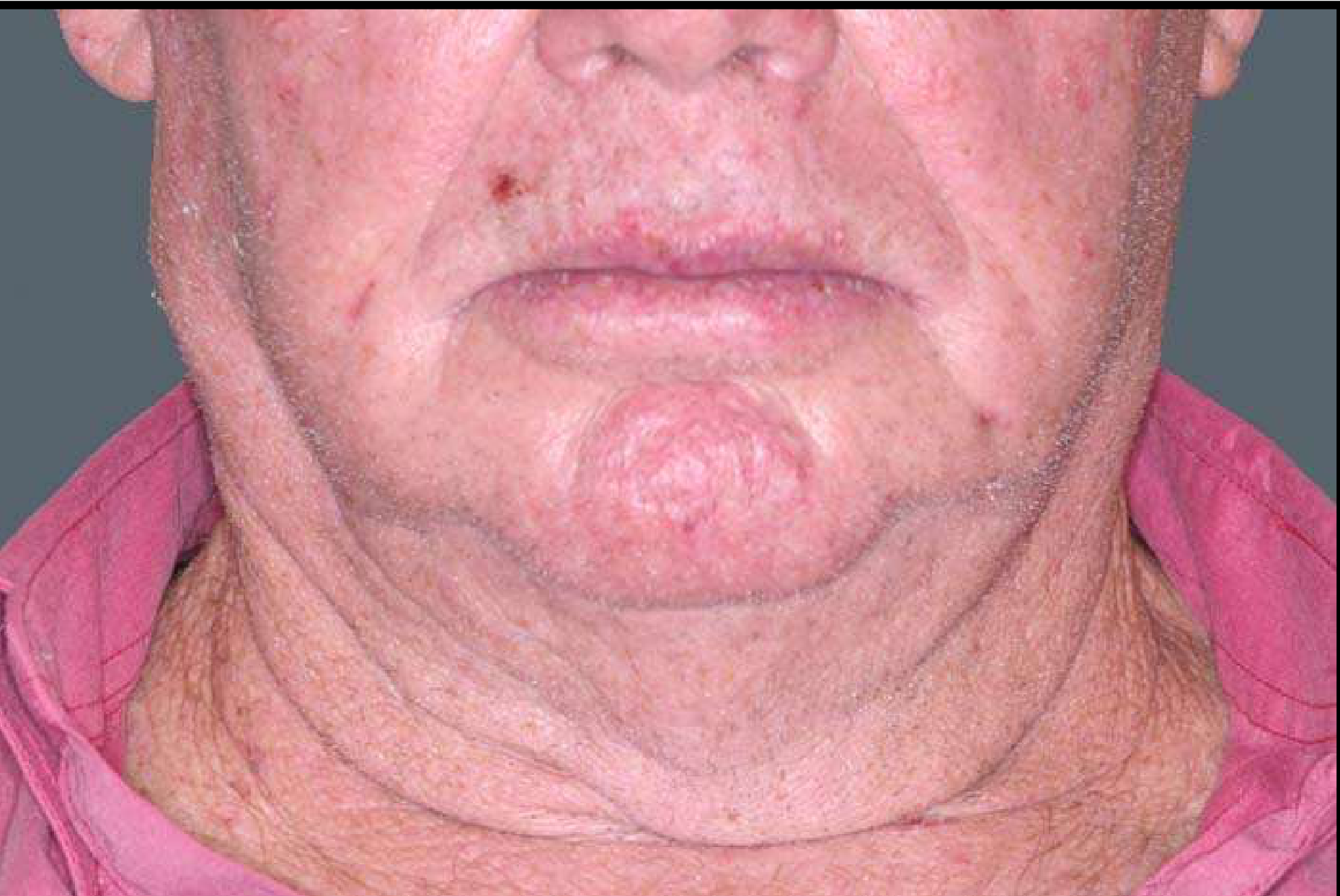
Identify the pathosis and describe the clinical features.
Extra orally
- Asymmetric lesion
- palpate the area
- feels soft, doesn’tfeel different from other tissue
- Feels hard:
- need to know if its mobile or fixed!
- need to know if its painful
- palpate the area
What is the differential diagnosis?
Lump/Asymmetry Extra orally
- Lipoma (if soft lump)
- Lymphoadenopathy
- looking for source of infection
- lymph nodes up to 1 cm shouldn’t be worried about , afterwards its likely to be reactive
- Metastatic deposit in the lymph node
Neck Lumps
Never ignore patients with neck lumps, particularly if they are hard
- you need to always follow up
What are the relevant clinical investigations?
Extra orla lump
- Fine needle aspiration/ biopsy
- CT of the head and the neck
- If it is a metastatic deposit we need to know where its from
- Results show that its a metastatic deposit of a squamous cell carcinoma but CT doesn’t show anything?
- this is an example of an unknown primary !
Percentage of head and neck cancers with unknown primary cancer
- between 1-4% of head and neck cancers have an unknown primary
- Immune system dealt with the primary, or it lost blood supply
- in this patients case he might have had a skin cancer removed but the lesion may have spread
What is the diagnosis?
How would you manage this patient?
Footnotes
-
Original PDF page 1: 10. Epithelial Pathosis II, p.1 ↩