Facial Form Analysis
Course Exercise1
- Course Code: DENT5310
- Instructor: Dr. Daniel Fernandes
Introduction to Facial Form Analysis23
Facial Form Analysis is the use of simple diagnostic tools to assess the facial form of a patient.
Analysis of facial proportions and dental-facial relationships is an essential step in the diagnosis of malocclusion.
Dentofacial Proportions
Keep the following principles in mind when completing a facial analysis:
General Assessment Principles4
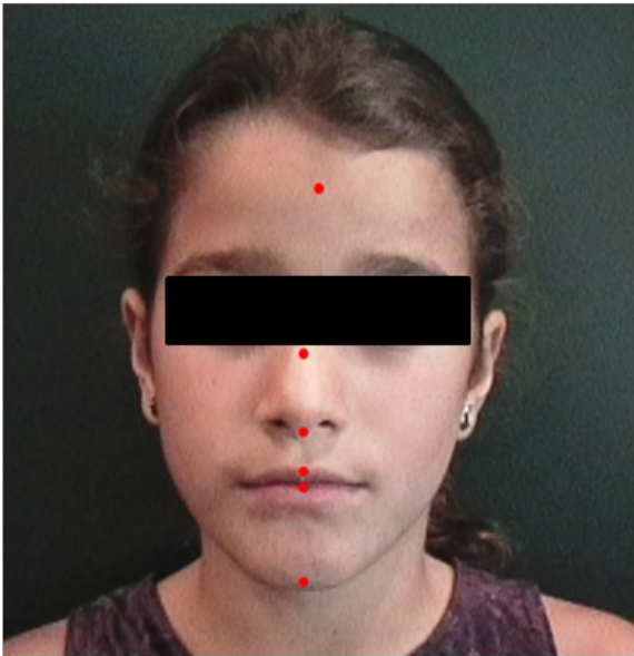
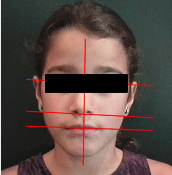
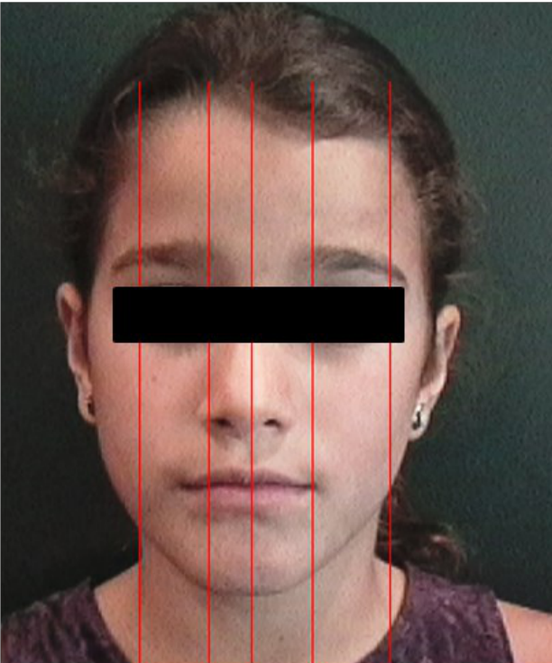
- Line Orientation: Check the patient’s vertical symmetry with a vertical facial centerline.
- Horizontal Positioning: Guide the patient to a natural position (relaxed face, closed eyes).
- Facial Midline: Draw the midline through the center of the patient’s ears and nose.
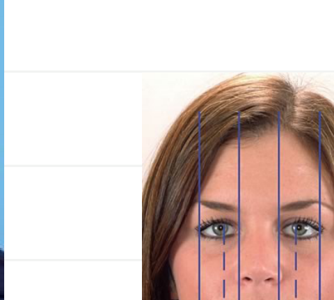
- Horizontal Proportions: Measure the horizontal width of the face in five equal vertical sections.
- Human Growth: The craniofacial complex grows and develops differently over time.
 |  |  |
 |  |
Vertical and Transverse Proportions
Vertical Proportions
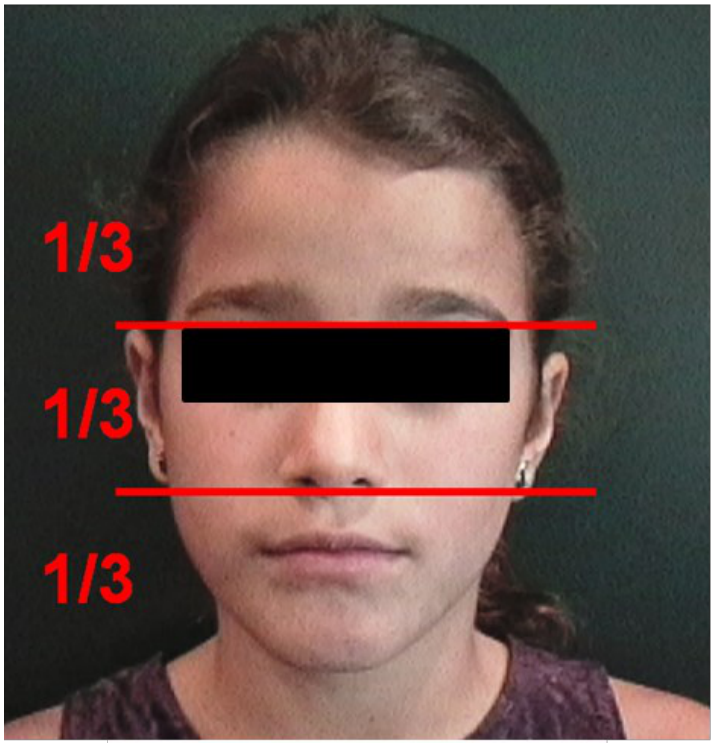
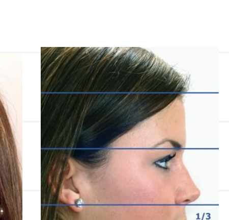
Measure the patient’s face in three horizontal thirds (upper, middle, and lower) by drawing imaginary lines.
Anatomic Considerations for Measurements
- The upper and lower thirds of the face should be approximately equal.
- The eyes should be positioned 1/2 down the upper third of the face.
- The lower third of the upper third of the face (including upper and lower eyelids and cheeks) should be a little lower than the eyes.
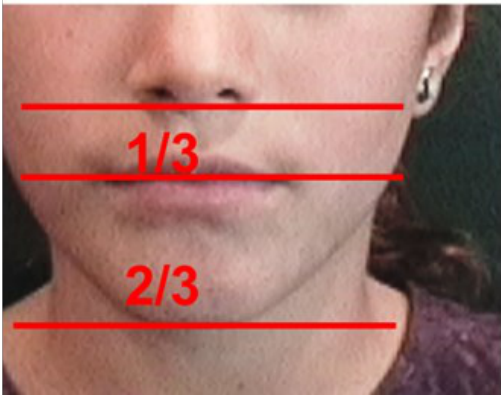
- The mouth height is approximately 1/2 of the middle third of the face.
- The mouth usually appears to be about 3mm wide when the lips are closed and centered.
- The angles of the lateral commissures of the mouth (corners of lips) should be slightly wider than 90 degrees.
- The point of the chin should appear 3mm at the lowest point.
- The point of the chin should not be more than 1mm below the level of the lower lip when the patient is in a natural resting position.
- The lower third of the face should be evenly distributed between the lower part of the nose, the mouth, and the chin.
Key Anatomic Landmarks
- The upper and lower facial thirds
- The point of the chin
- The position of the eyes
- The position of the mouth
- The angle of the mouth
- Vertical symmetry with a vertical facial centerline
Keep these principles in mind when completing a facial analysis:
Harmonious Proportions5
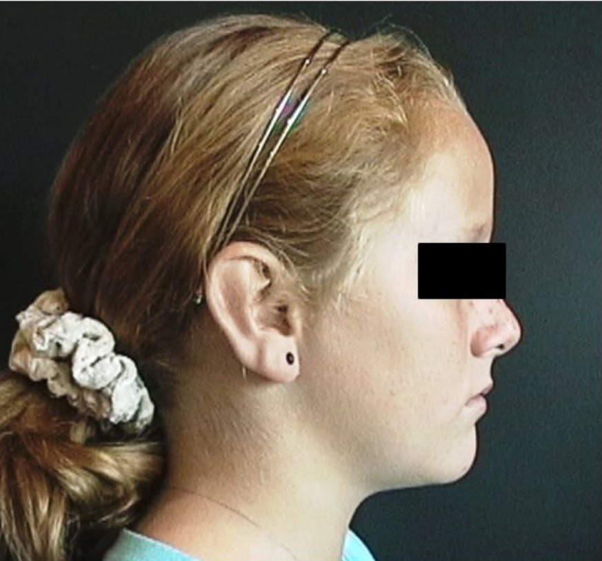
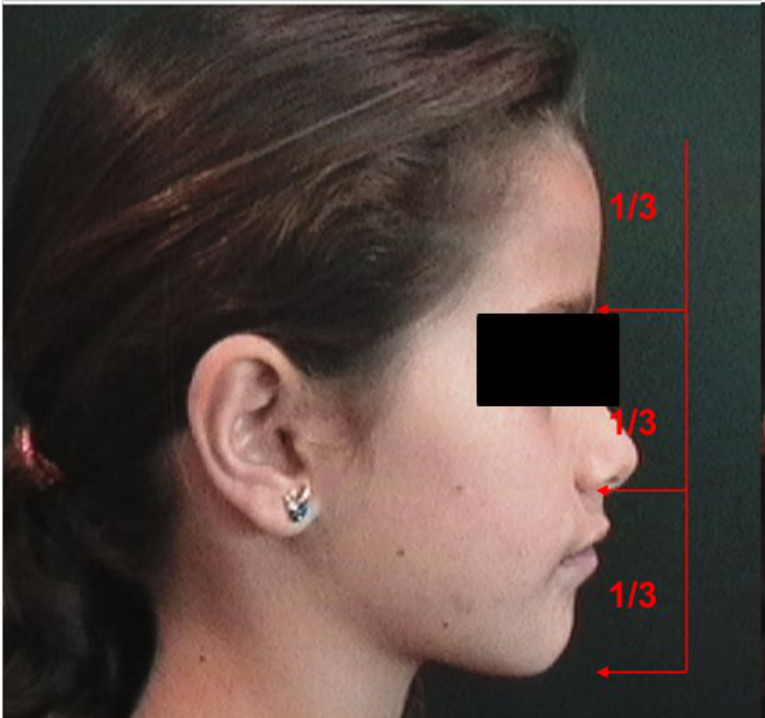
The face appears most pleasing and proportional when divided into three approximately equal vertical thirds:
- Upper Third: Forehead to Glabella (root of nose).
- Middle Third: Glabella to Subnasale (bottom of nose).
- Lower Third: Subnasale to Menton (bottom of chin).
 |  |
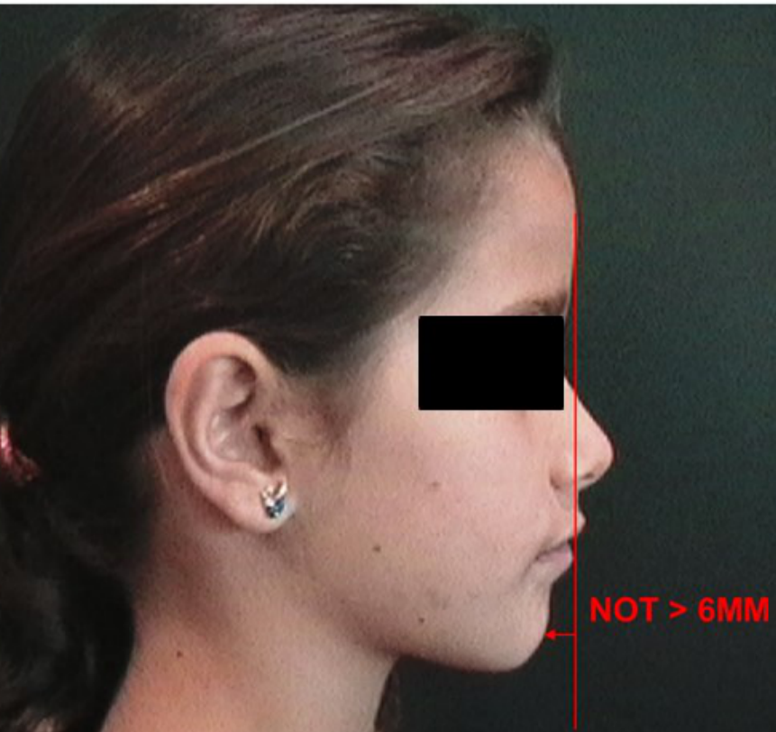 |
Ricketts’ E Line (Lp Line)
The distance from the soft tissue pterion (pterygomandibular raphe) to the tip of the nose should be not greater than 6mm.
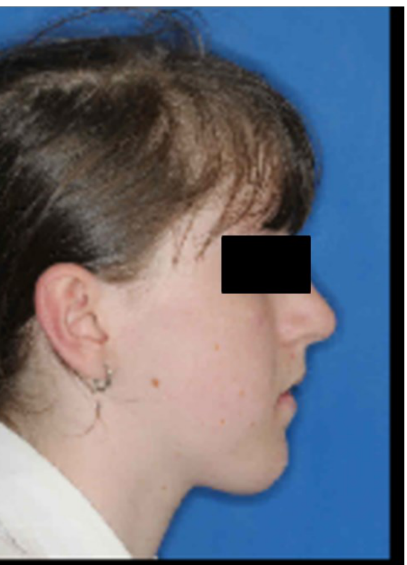
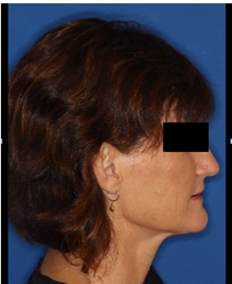
Facial Height Classifications6
- Long
- Short
 |  |
Clinical Observations of Facial Form
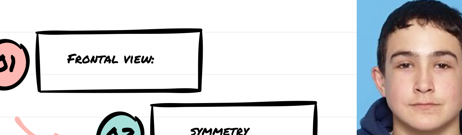
Frontal View Components7
- Frontal View
- Symmetry
- Vertical and Transverse Facial Proportions
- Tooth Display
 |  |  |
 |  |  |
 |
Clinical Assessment Criteria8
- Facial and Dental Midlines
- Transverse Facial Proportions
- Vertical Facial Proportions
 | | |
 |  |
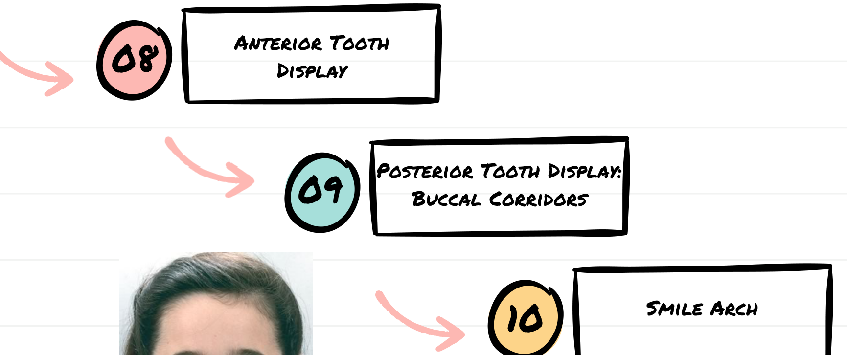
Tooth Display and Smile Aesthetics
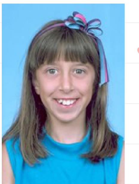
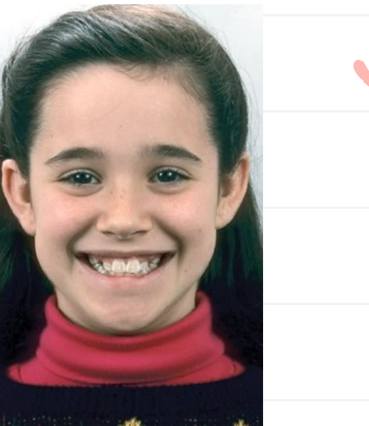
Smile Aesthetics Components9
- Anterior Tooth Display
- Posterior Tooth Display: Buccal Corridors
- Smile Arch
 |  |
 |  |
Patient Aesthetic Judgment10
1. Facial Symmetry
- Within normal limits or asymmetric
- If present, location of asymmetry
2. Incisor Tooth Display
- OK, too little, or too much (excess gingiva)
3. Buccal Corridor Width
- OK, too little, or too much
4. Smile Arc
- OK, flat, or excessive
Summary
Summarize the facial form of your patient based on the above criteria.
Facial Profile Analysis111213
As part of a Facial Form Analysis, a facial profile analysis is considered a simplified version of Cephalometric Analysis.
Facial Profile analysis is performed to identify the underlying skeletal relationships. This is an important diagnostic tool for identification of patients with a severe dentofacial disproportion.
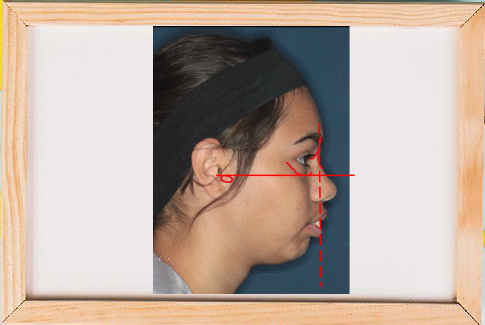
Evaluation of the facial profile using horizontal and vertical reference lines to assess skeletal and soft tissue relationships.
Goals of Profile Analysis14
- Establishing Anterior-Posterior (AP) Positioning: Determining whether the jaws are proportionately positioned in the AP plane of space.
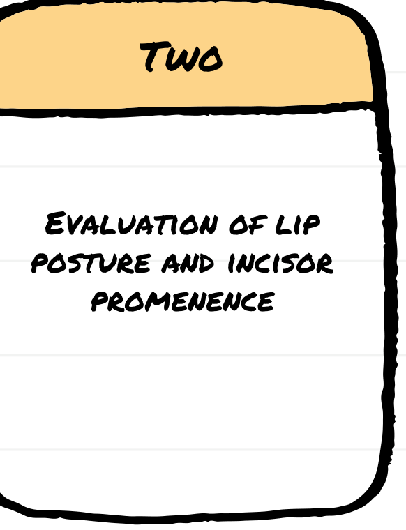
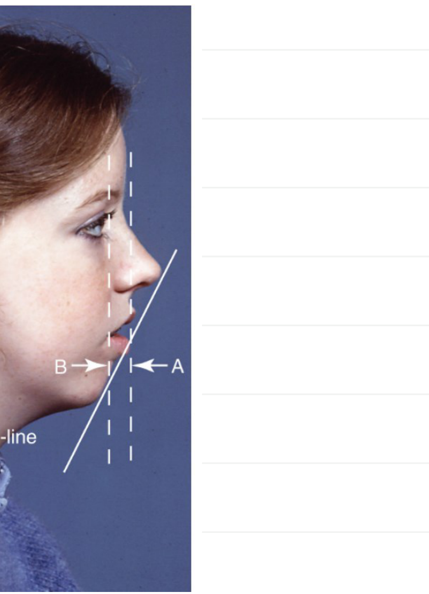
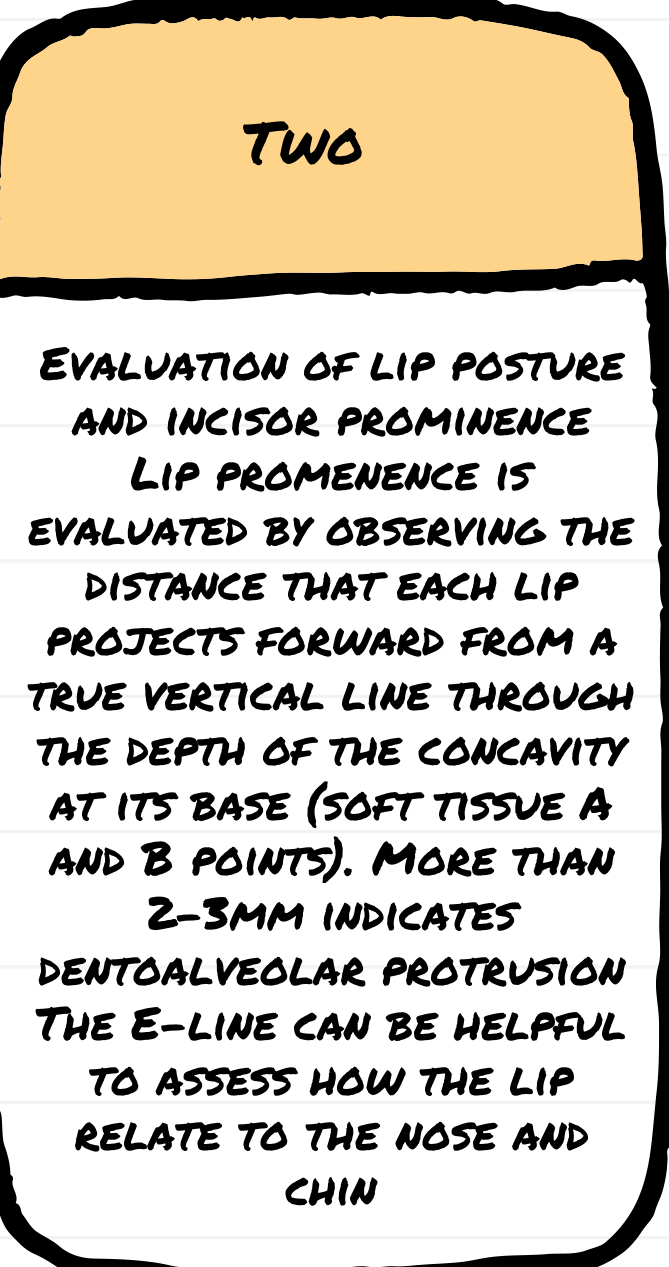
- Lip Posture and Incisor Prominence: Evaluation of the soft tissue profile and dental support.
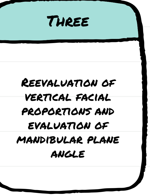
- Vertical Proportions and Mandibular Plane: Reevaluation of vertical facial proportions and evaluation of the mandibular plane angle.
 |  |
 |
Anterior-Posterior Jaw Relationships15
In natural head position, note the relationship between two lines: one dropped from the bridge of the nose to the base of the upper lip, and a second one extending from that point downward to the chin.
 |  |
Profile Classifications16
- A: Convex
- B: Straight
- C: Concave
Visual assessment of the relationship between the maxilla and mandible in the anterior-posterior plane to determine skeletal classification.
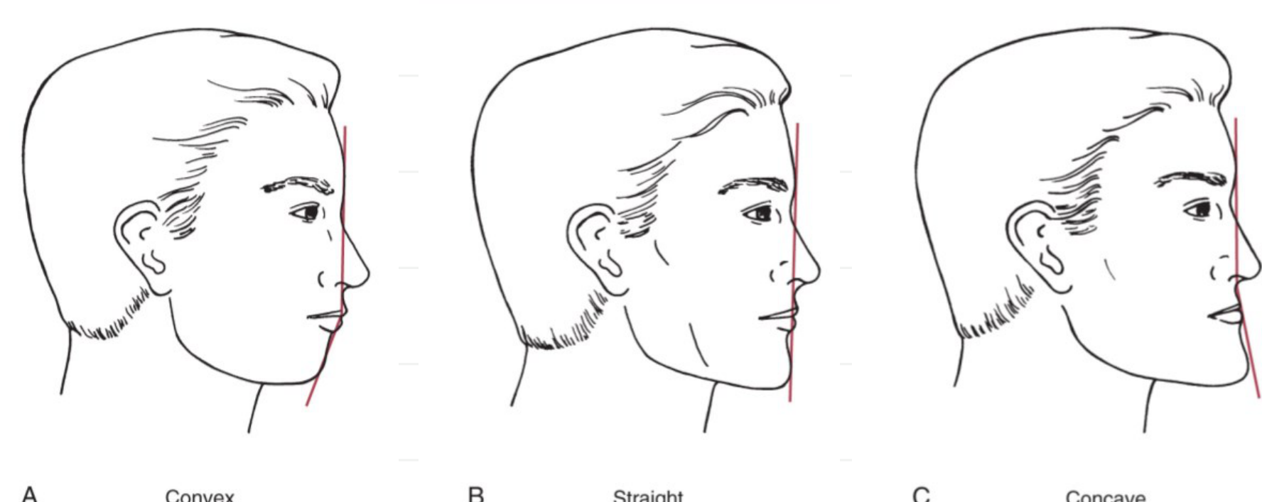
Mandibular Plane and Vertical Proportions
Mandibular Plane Angle Assessment17
- Reevaluation of vertical facial proportions.
- Evaluation of the mandibular plane in relation to the true horizontal plane.
- The mandibular plane angle is measured between the true horizontal plane and the mandibular plane to determine if the angle is steep, moderate, or low.
Profile Landmarks and Points
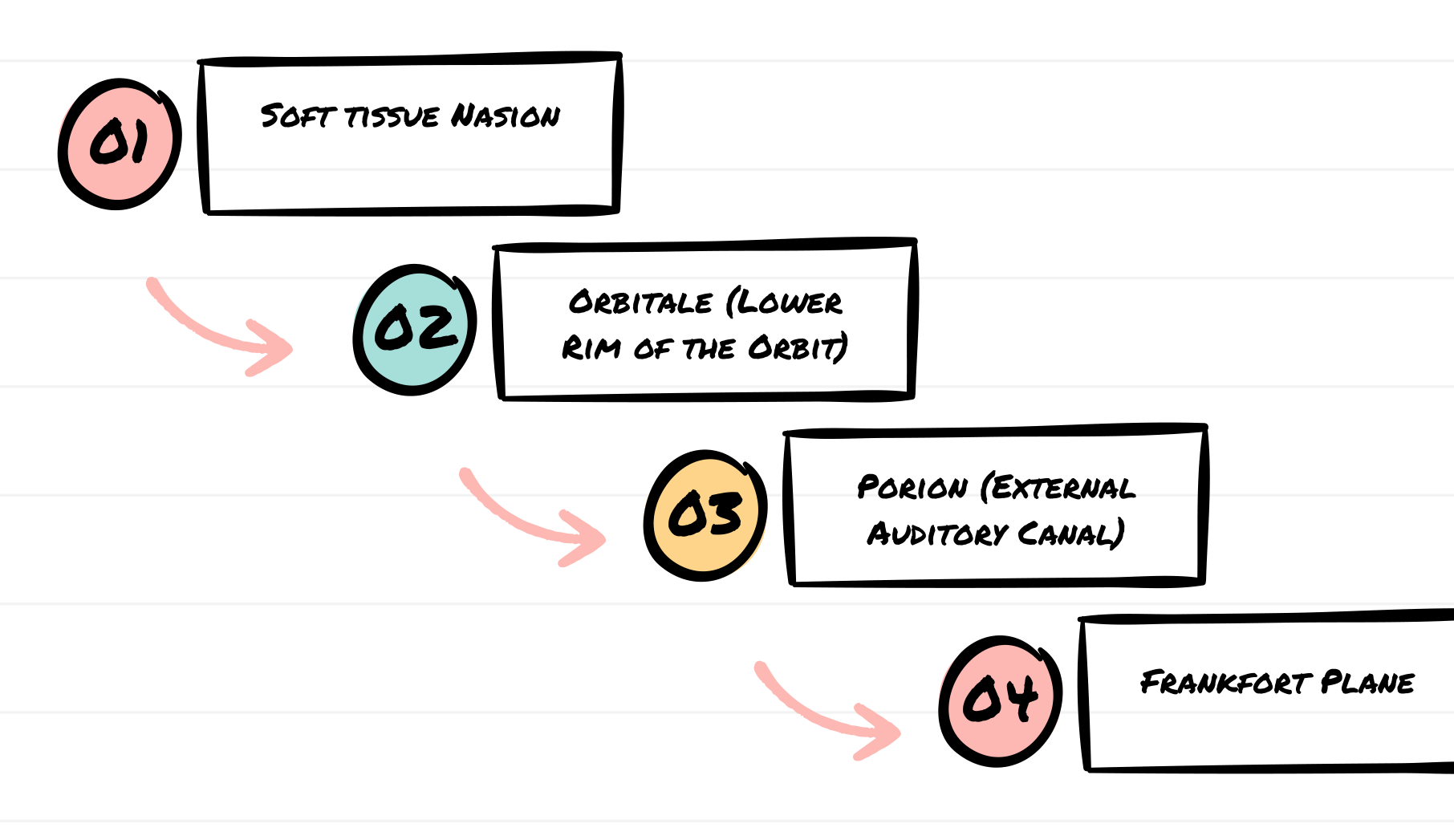
Profile Landmarks18
- Soft Tissue Nasion
- Orbitale: Lower rim of the orbit
- Porion: External auditory canal
- Frankfort Plane
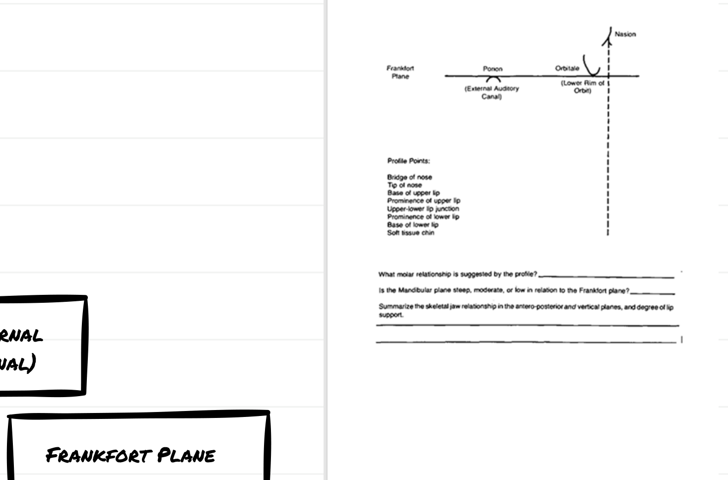 |  |
Profile Points for Analysis
- Bridge of nose
- Tip of nose
- Base of upper lip
- Prominence of upper lip
- Upper-lower lip junction
- Prominence of lower lip
- Base of lower lip
- Soft tissue chin
Clinical Diagnostic Questions
- What molar relationship is suggested by the profile?
- Is the Mandibular plane steep, moderate, or low in relation to the Frankfort plane?
- Summarize the skeletal jaw relationship in the antero-posterior and vertical planes, and degree of lip support.
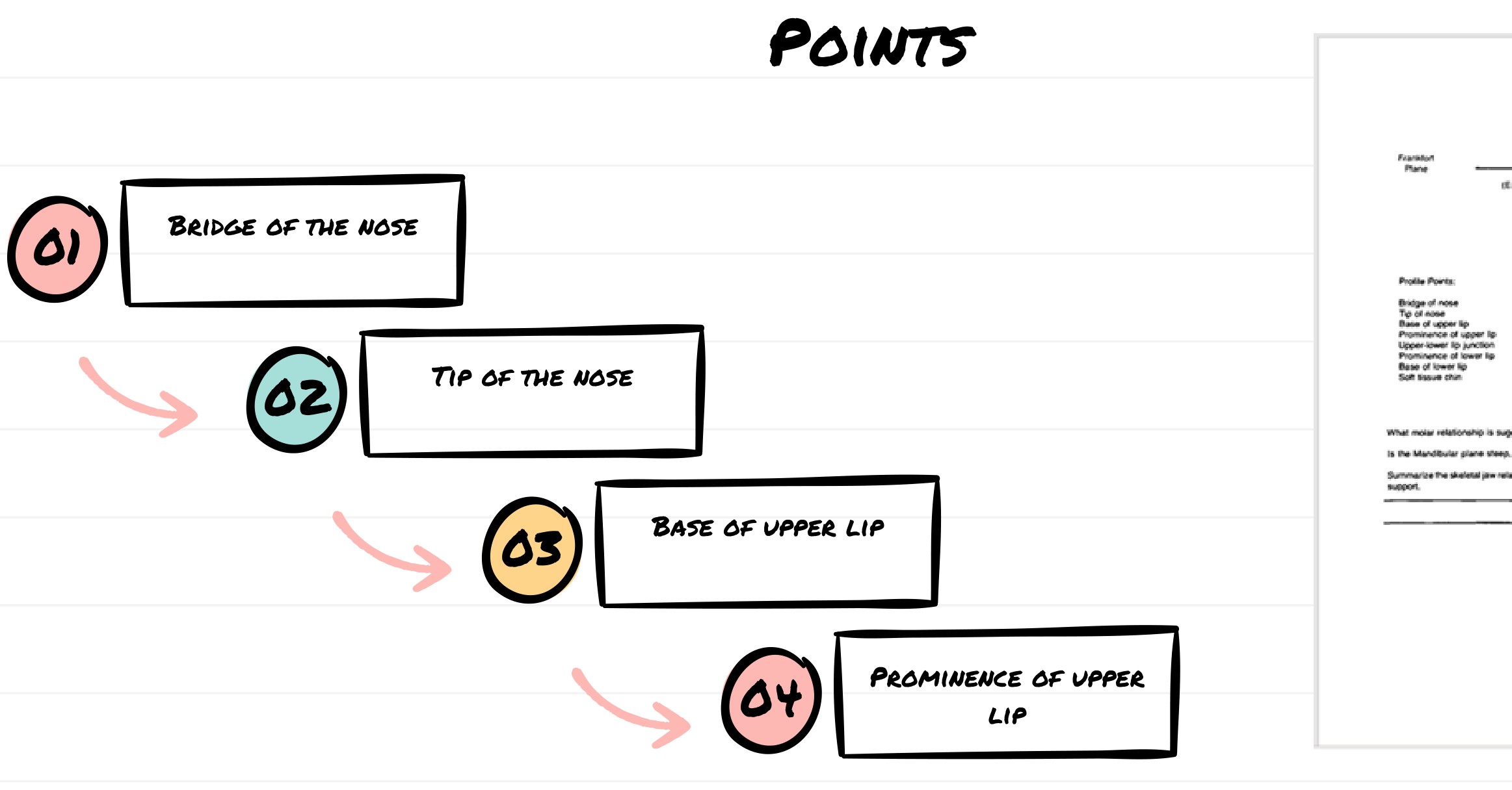
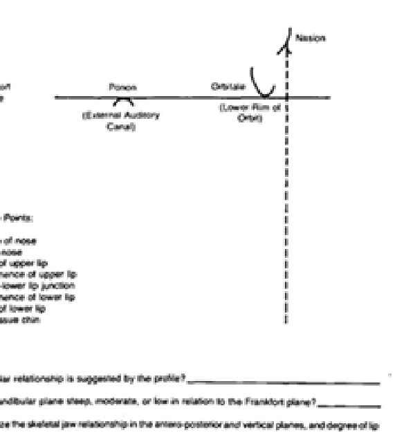
Primary Profile Points19
- Bridge of the nose
- Tip of the nose
- Base of upper lip
- Prominence of upper lip
 |  |
Secondary Profile Points20
- Upper-lower lip junction
- Prominence of lower lip
- Base of lower lip
- Soft tissue chin
 | 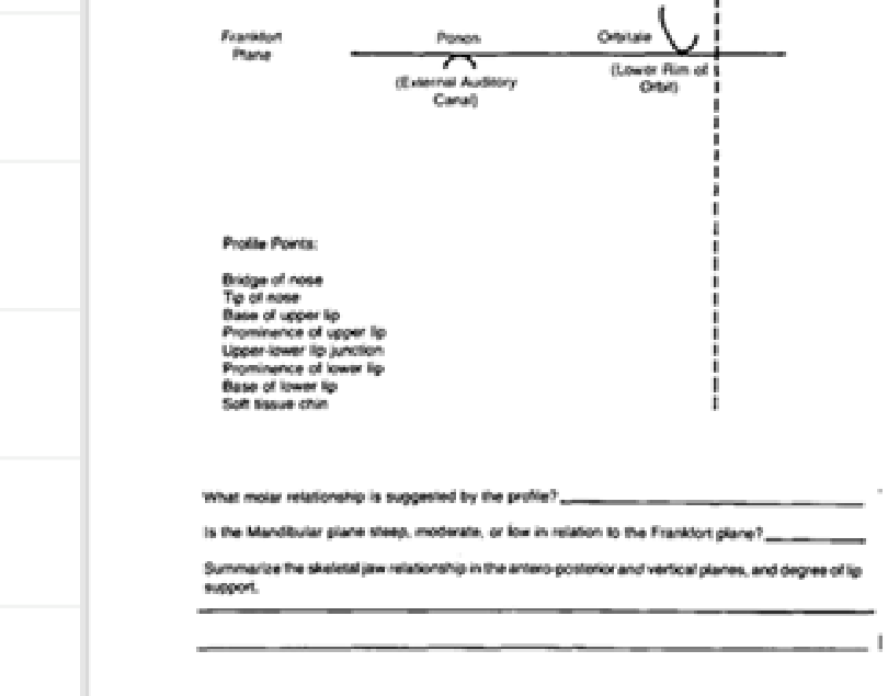 |
Reference Planes and Landmarks
- Frankfort Plane
- Porion: External auditory canal
- Orbitale: Lower rim of orbit
- Nasion
Clinical Assessment
- Determine suggested molar relationship.
- Evaluate mandibular plane angle (steep, moderate, or low) relative to the Frankfort plane.
- Summarize skeletal jaw relationships (AP and vertical) and lip support.
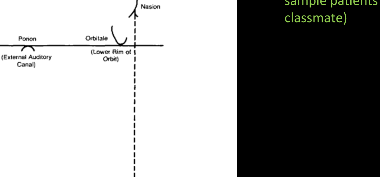
Frankfort Plane Components21
- Porion: External auditory canal
- Orbitale: Lower rim of orbit
- Nasion
Profile Points List
- Bridge of nose
- Tip of nose
- Base of upper lip
- Prominence of upper lip
- Upper-lower lip junction
- Prominence of lower lip
- Base of lower lip
- Soft tissue chin
Summary Evaluation
- Suggested molar relationship
- Mandibular plane angle assessment
- Summary of skeletal jaw relationship and lip support
Note: This analysis will be completed for sample patients (selected classmate).
References
Prediction of Dental and Skeletal Relationships from Facial Profiles in Preschool Children22
Authors: Henry W. Fields, William F. Vann
Source: Pediatric Dentistry 1979
This paper reviews the rationale for assessing the facial profile of the patient with a developing dentofacial complex. The purpose of the study was to assess one method of profile analysis utilizing 4-year-old children.
-
Methodology:
- Sixteen orthodontists and 16 pedodontists were asked to classify existing skeletal or dental relationships.
- Assessments were based solely on soft tissue profile tracings or lateral facial photographs.
-
Key Findings:
- For this age group, neither the orthodontists nor the pedodontists could accurately predict the existing skeletal or dental pattern based solely on the soft tissue profile tracing or the lateral facial photograph.
- No significant differences were found between the predictions of the orthodontists and the pedodontists.
-
Conclusion:
- The investigation notes that even with highly similar dental relationships, significant variability in skeletal and soft tissue relationships may occur in preschool children.
Clinical Exercises and Assessment
Predicted Occlusion Assessment23
Based on the provided facial profile, what type of malocclusion or occlusion do you expect to see?
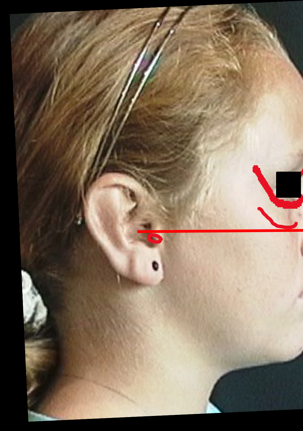 | 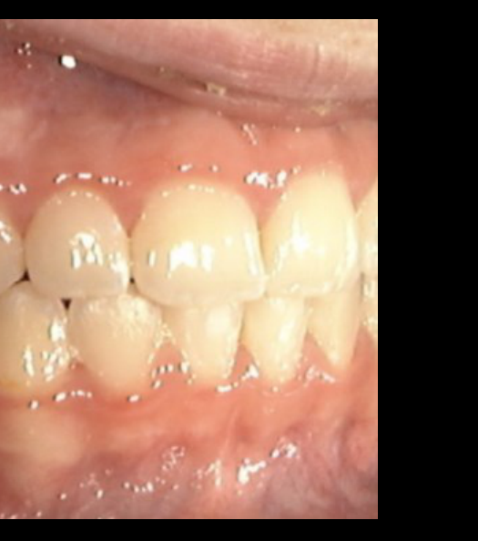 |
Malocclusion Identification Exercises242526
- Class I occlusion or malocclusion
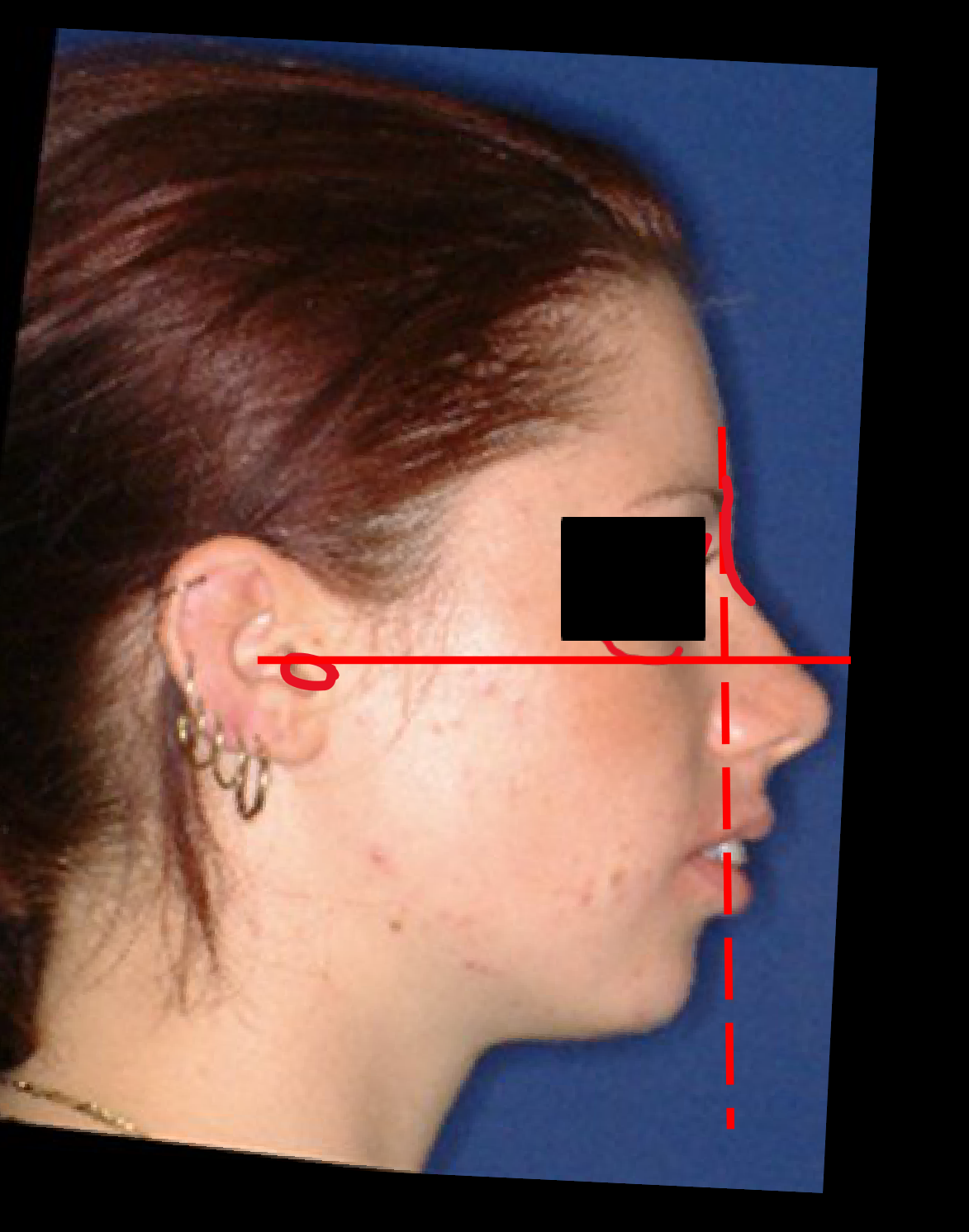
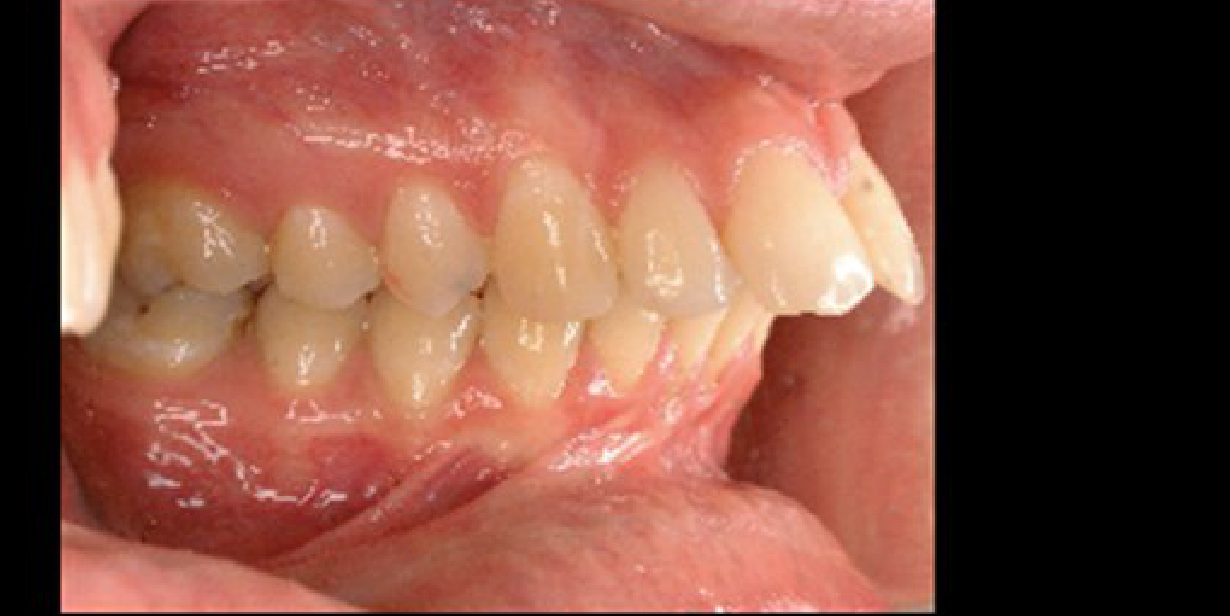
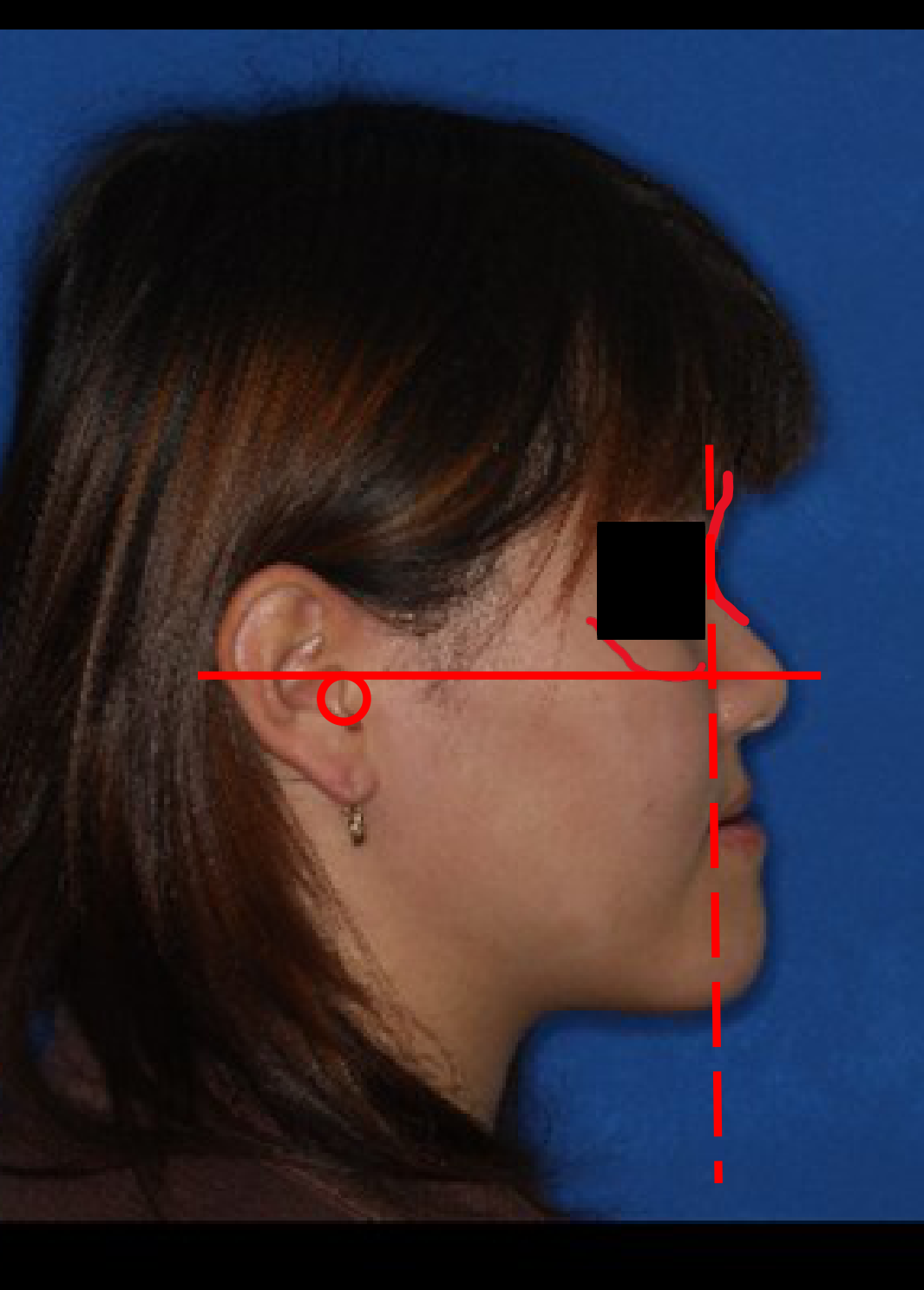
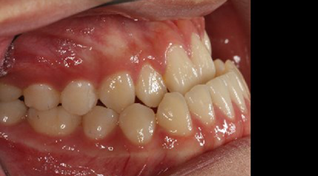
Predicted Occlusion Assessment27
What type of malocclusion do you expect to see?
- Class II malocclusion
What type of malocclusion do you expect to see?
- Class II malocclusion
What type of malocclusion do you expect to see?
- Class III malocclusion
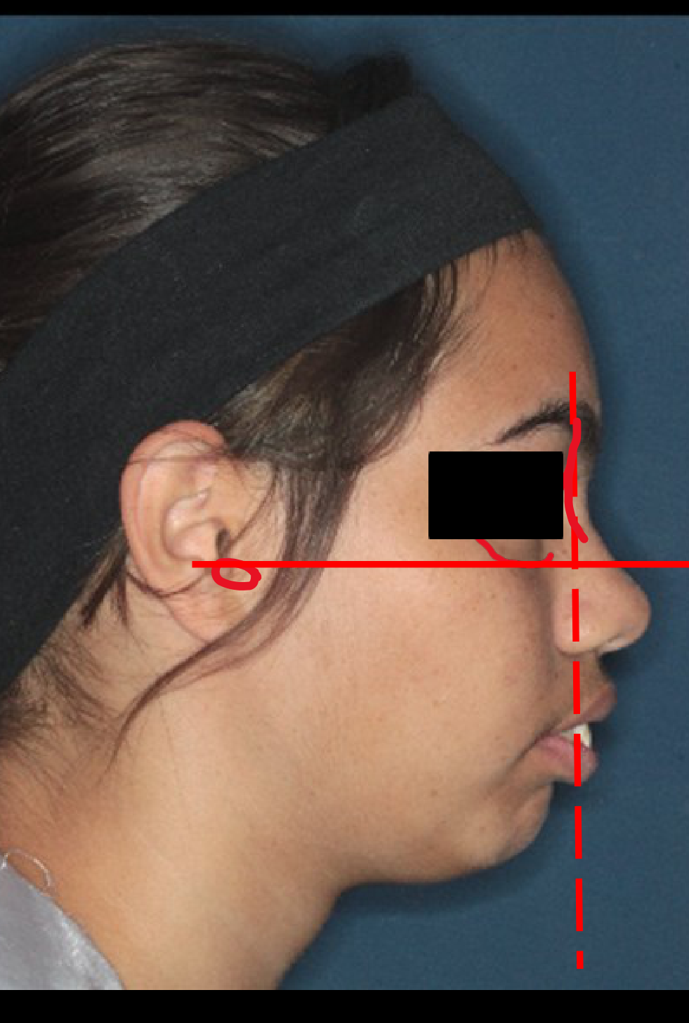 | 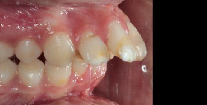 |
Skeletal and Soft Tissue Compensations
Diagnostic Considerations28
Because this is a Soft Tissue Analysis attempting to diagnose an underlying skeletal problem, would you expect the skeletal measurements from a radiograph to be more or less significant?
Clinical Observations
- Dental and Soft Tissue Compensations
Facial Profile Analysis Criteria29
For your patient(s), provide a judgment on the following:
- Antero-posterior Skeletal Jaw Relationship
- Class I (Normal)
- Class II
- Class III
- Vertical Skeletal Jaw Relationship
- Normal
- Long Face
- Short Face
- Tooth Support for Lip and Lip Prominence
- Excessive
- Normal
- Inadequate
Patient Assessment Forms
Clinical Exercise Instructions30
Two patients are required to complete the following assessments:
- Facial Form Analysis
- Facial Profile Analysis
 |  |
Facial Form Analysis Record31
For your patient(s), provide a judgment on the following:
- Facial Symmetry
- Within normal limits or asymmetric
- If present, specify location of asymmetry: __________________________
- Incisor Tooth Display
- OK, too little, or too much (excess gingiva)
- Notes: __________________________
- Buccal Corridor Width
- OK, too little, or too much
- Notes: __________________________
- Smile Arc
- OK, flat, or excessive
- Notes: __________________________
Summary Summarize the facial form of your patient: __________________________
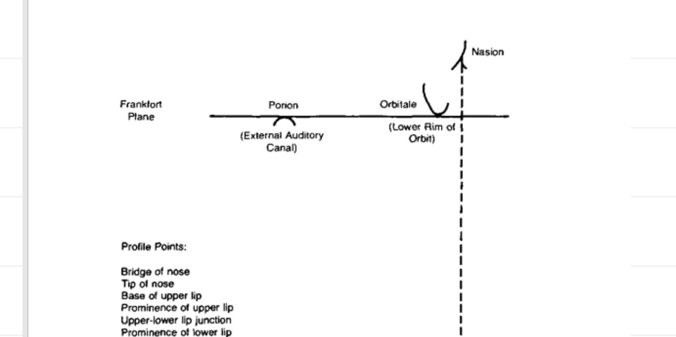
Profile Points and Skeletal Relationship32
Profile Points for Assessment:
- Bridge of nose
- Tip of nose
- Base of upper lip
- Prominence of upper lip
- Upper-lower lip junction
- Prominence of lower lip
- Base of lower lip
- Soft tissue chin
Clinical Findings:
- What molar relationship is suggested by the profile? __________________________
- Is the Mandibular plane steep, moderate, or low in relation to the Frankfort plane? __________________________
Summary of Findings: Summarize the skeletal jaw relationship in the antero-posterior and vertical planes, and the degree of lip support.
Facial Profile Analysis Record33
For your patient(s), provide a judgment on the following:
- Antero-posterior Skeletal Jaw Relationship
- Class I (Normal)
- Class II
- Class III
- Vertical Skeletal Jaw Relationship
- Normal
- Long Face
- Short Face
- Tooth Support and Lip Prominence
- Excessive
- Normal
- Inadequate
Submission Requirements
Assignment Submission Guidelines34
Format Requirements:
- Submit as a Word file or PDF.
- Alternatively, use A4 paper with attached de-identified patient photographs.
Submission Method:
- Use the University of Western Australia’s Learning Management System (Blackboard) via Turnitin to forward the assignment.
Course Details:
- Unit Code: DENT5310
- Assignment Name: Facial Form Analysis and Profile Analysis Practical Exercise
Footnotes
-
Original PDF page 1: L2 FacialFormAnalysis2026, p.1 ↩
-
Original PDF page 2: L2 FacialFormAnalysis2026, p.2 ↩
-
Original PDF page 3: L2 FacialFormAnalysis2026, p.3 ↩
-
Original PDF page 4: L2 FacialFormAnalysis2026, p.4 ↩
-
Original PDF page 5: L2 FacialFormAnalysis2026, p.5 ↩
-
Original PDF page 6: L2 FacialFormAnalysis2026, p.6 ↩
-
Original PDF page 7: L2 FacialFormAnalysis2026, p.7 ↩
-
Original PDF page 8: L2 FacialFormAnalysis2026, p.8 ↩
-
Original PDF page 9: L2 FacialFormAnalysis2026, p.9 ↩
-
Original PDF page 10: L2 FacialFormAnalysis2026, p.10 ↩
-
Original PDF page 11: L2 FacialFormAnalysis2026, p.11 ↩
-
Original PDF page 12: L2 FacialFormAnalysis2026, p.12 ↩
-
Original PDF page 13: L2 FacialFormAnalysis2026, p.13 ↩
-
Original PDF page 14: L2 FacialFormAnalysis2026, p.14 ↩
-
Original PDF page 16: L2 FacialFormAnalysis2026, p.16 ↩
-
Original PDF page 15: L2 FacialFormAnalysis2026, p.15 ↩
-
Original PDF page 17: L2 FacialFormAnalysis2026, p.17 ↩
-
Original PDF page 18: L2 FacialFormAnalysis2026, p.18 ↩
-
Original PDF page 19: L2 FacialFormAnalysis2026, p.19 ↩
-
Original PDF page 20: L2 FacialFormAnalysis2026, p.20 ↩
-
Original PDF page 21: L2 FacialFormAnalysis2026, p.21 ↩
-
Original PDF page 22: L2 FacialFormAnalysis2026, p.22 ↩
-
Original PDF page 23: L2 FacialFormAnalysis2026, p.23 ↩
-
Original PDF page 25: L2 FacialFormAnalysis2026, p.25 ↩
-
Original PDF page 26: L2 FacialFormAnalysis2026, p.26 ↩
-
Original PDF page 27: L2 FacialFormAnalysis2026, p.27 ↩
-
Original PDF page 24: L2 FacialFormAnalysis2026, p.24 ↩
-
Original PDF page 28: L2 FacialFormAnalysis2026, p.28 ↩
-
Original PDF page 29: L2 FacialFormAnalysis2026, p.29 ↩
-
Original PDF page 30: L2 FacialFormAnalysis2026, p.30 ↩
-
Original PDF page 31: L2 FacialFormAnalysis2026, p.31 ↩
-
Original PDF page 32: L2 FacialFormAnalysis2026, p.32 ↩
-
Original PDF page 33: L2 FacialFormAnalysis2026, p.33 ↩
-
Original PDF page 34: L2 FacialFormAnalysis2026, p.34 ↩