Oral Pathology1

Table Of Contents
Clinical Categories and Disease Classifications2
- Developmental Conditions
- Mucosal Lesions—Reactive
- Reactive Lesions: These refer to physical and chemical injuries to the oral mucosa.
- Mucosal Lesions—Infections
- Mucosal Lesions—Immunologic Diseases
- Mucosal Lesions—Premalignant
- Mucosal Lesions—Malignant
- CT Tumors—Benign
- CT Tumors—Malignant
- Salivary Gland Diseases—Reactive
- Salivary Gland Diseases—Benign
- Salivary Gland Diseases—Malignant
- Lymphoid Neoplasms
- Odontogenic Cysts
- Odontogenic Tumors
- Bone Lesions—Fibro-Osseous
- Bone Lesions—Giant Cell
- Bone Lesions—Inflammatory
- Bone Lesions—Malignant
- Hereditary Conditions
Reactive Mucosal Lesions
Focal Hyperkeratosis And Ulcerations
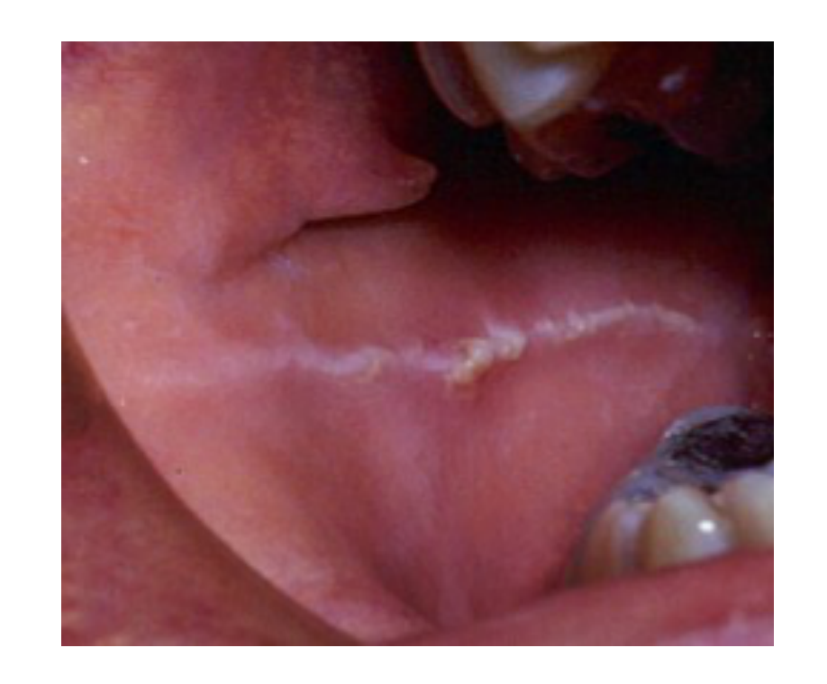
Linea Alba3
- White line appearing in the buccal mucosa.
- Literally translates to "white line."
- Appears at the plane of occlusion where the upper and lower teeth meet.
- A type of focal hyperkeratosis resulting from chronic friction on the mucosa.
- Similar to a callus, often caused by cheek biting.
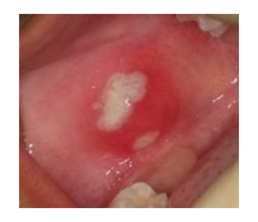
Traumatic Ulcer4
- Very common clinical finding.
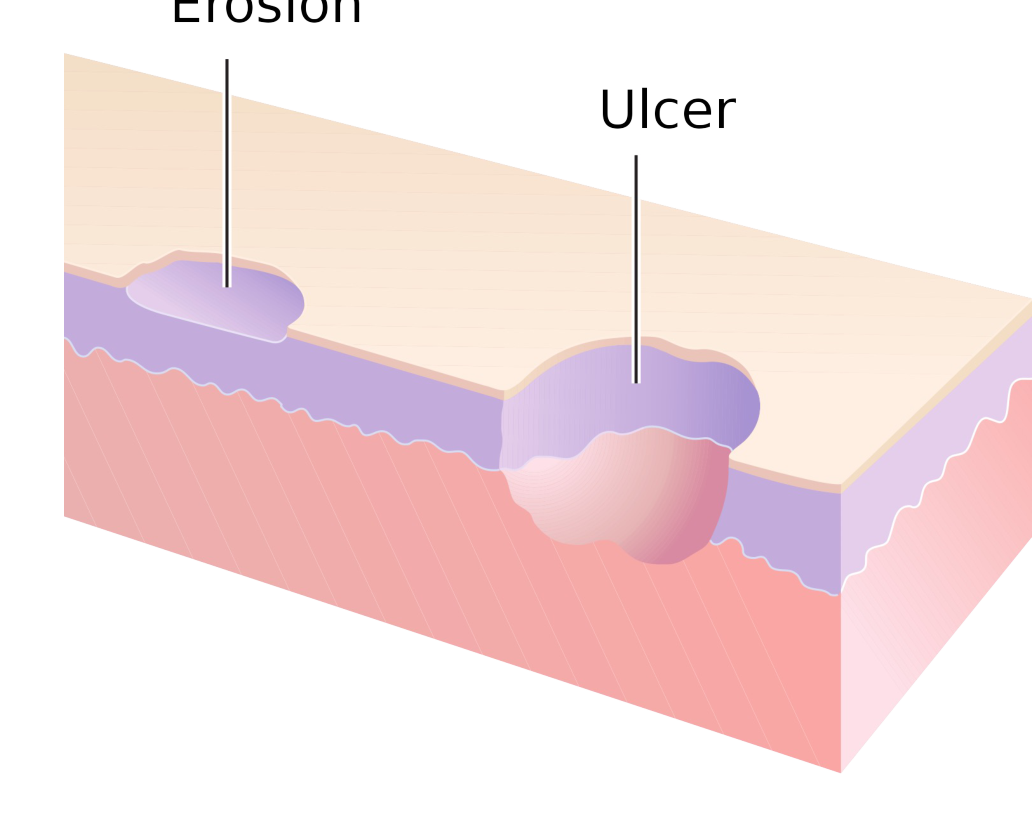
- Erosion: An incomplete break in the epithelium.
- Limited to the mucosal layer.
- Ulcer: A complete break through the epithelium.
- Extends into the submucosal layer.
- Typically more painful and noticeable due to its depth.
 |  |
Chemical And Thermal Injuries
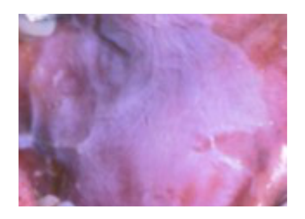
Chemical Burn Etiology5
Common causative agents include:
- Aspirin
- Hydrogen peroxide
- Silver nitrate
- Phenol
- Often presents as a whitish, sloughing appearance of the tissue.
- Aspirin is a common cause cited in examinations.
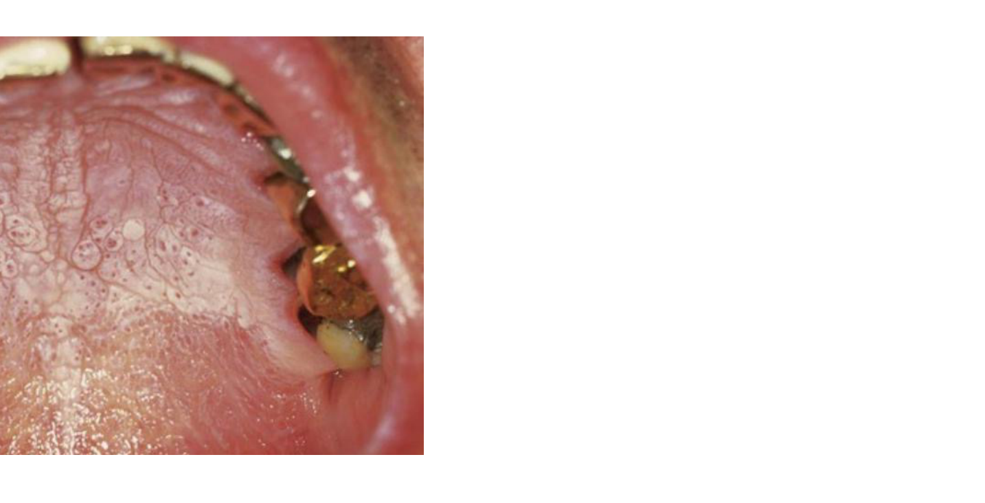
Nicotine Stomatitis6
- Characterized by inflamed salivary duct openings.
- Presents as red dots on the hard palate.
- Seen in chronic heavy smokers.
- Only considered premalignant if related to “reverse smoking” (holding the lighted end of the cigarette inside the mouth).
Pigmented Lesions
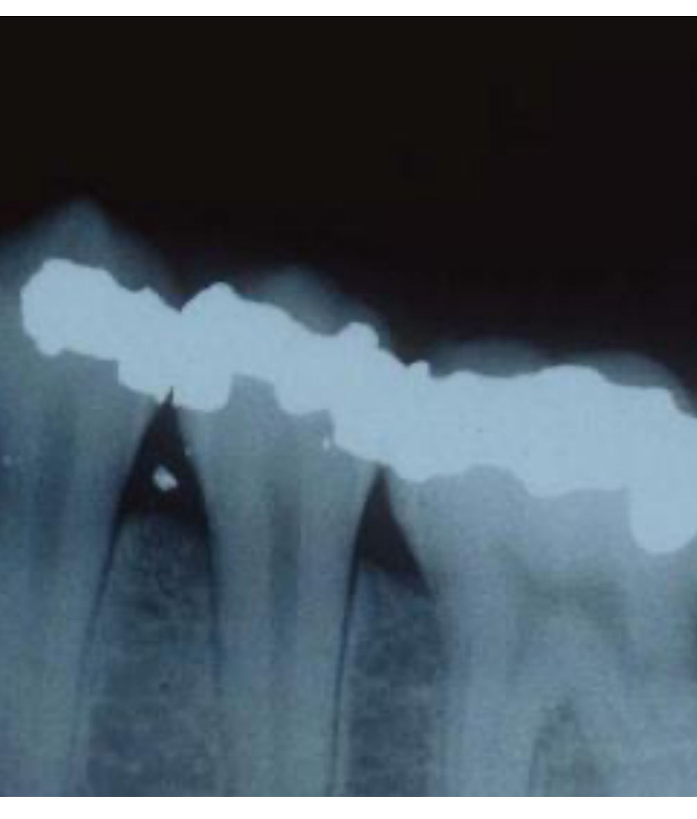
Amalgam Tattoo7
- Caused by the traumatic implantation of amalgam particles into the oral mucosa.
- Appears as tiny radiopaque particles on radiographic images (X-rays).
- Often occurs during the condensation of amalgam if particles leak or are not rinsed out.
- Confirmed as floating opaque particles in the soft tissue or gum area.
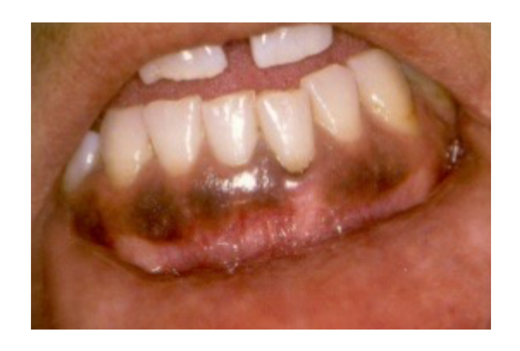
Smoking-Associated Melanosis8
- Pathophysiology: Chemicals in tobacco stimulate melanocytes to produce excess pigment.
- Presentation: Brown, diffuse, and irregular macules.
- Location: Typically observed in the anterior gingiva.
- Also associated with the use of smokeless tobacco.
- Treatment: Reversible if the patient discontinues smoking.
Melanotic Macule9
- A benign hyperpigmentation of the mucous membrane.
- Essentially functions as a freckle of the mucosa.
- Peutz-Jeghers Syndrome: A condition characterized by the presence of these freckles in combination with intestinal polyps.
Papillary And Surface Alterations
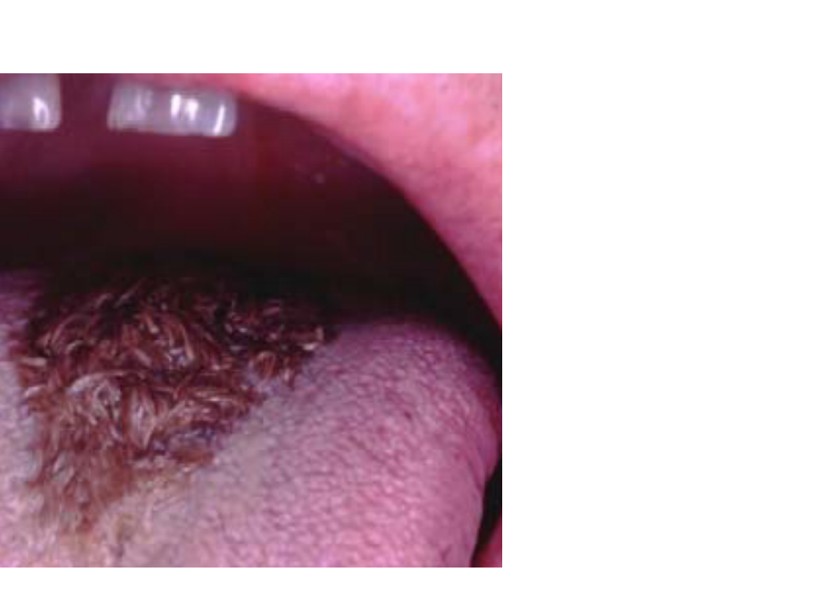
Hairy Tongue10
- Characterized by the elongation of the filiform papillae.
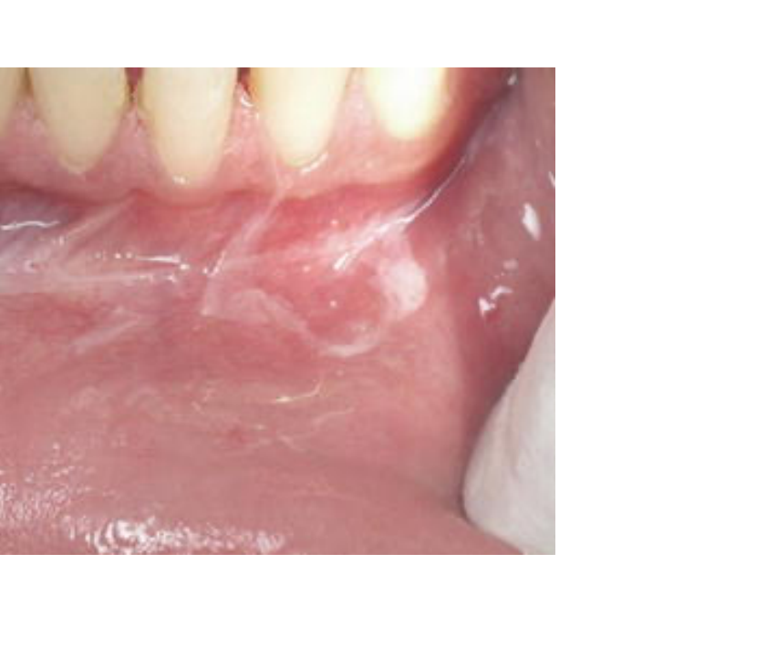
Dentifrice-Associated Sloughing11
- Related to the surfactant ingredient sodium lauryl sulfate found in many toothpastes.
- Management involves suggesting alternative products such as Tom’s of Maine or Rembrandt toothpaste.
- Presents as a white sloughing appearance of the mucosa.
Submucosal Hemorrhage
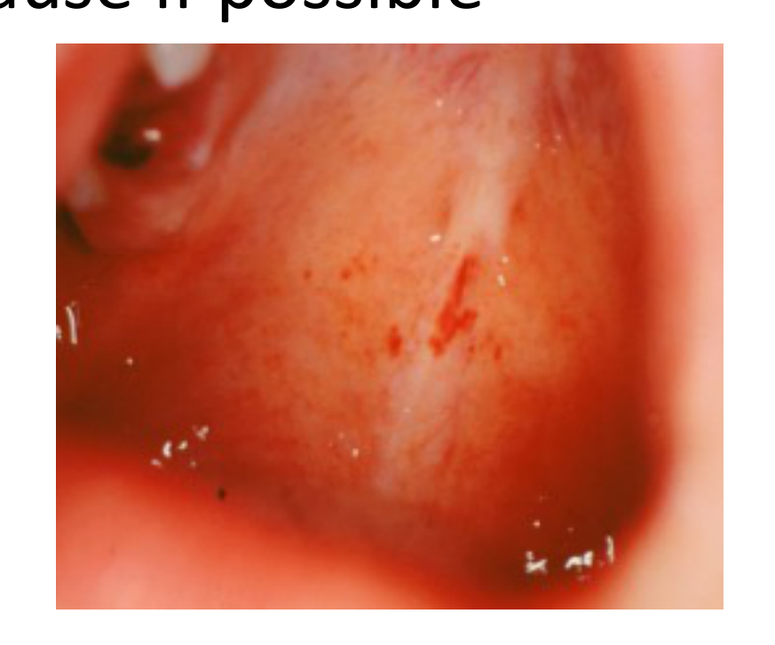
Clinical Characteristics of Submucosal Hemorrhage12
- These are extravascular lesions that do not blanch upon pressure.
- In contrast, vascular lesions (such as hemangiomas and telangiectasias) do blanch during a diascopy test.
Pathophysiology of Blanching
Vascular lesions blanch because the blood is contained within vessels. Extravascular lesions (hemorrhages) do not blanch because the blood has leaked out of the vessels into the tissues.
Classifications
- Petechiae: 1mm hemorrhages; often caused by the Valsalva maneuver, violent coughing, or fellatio.
- Purpura: Hemorrhagic spots slightly larger than petechiae.
- Ecchymosis: A bruise measuring 1cm or larger
- Typically measures between 1 cm to 2 cm in diameter.
- Hematoma: A mass of blood collected within the tissue, typically caused by trauma to the oral mucosa, such as from an anesthetic needle
- Example: Often caused by an anesthetic needle during a PSA (Posterior Superior Alveolar) block.
Treatment
- No specific treatment is required; eliminate the underlying cause if possible.
Audio Appendix
Additional Audio Content
The following sections from the lecture audio did not correspond to any heading in the main document.
Peutz-Jeghers (PJ) Syndrome
- Components: A combination of melanotic macules (freckles) on the face, lips, and inside the mouth, and intestinal polyps.
- Mnemonic: Think of someone home sick in their “PJs” with freckles and intestinal distress.
Footnotes
-
Original PDF page 1: Oral Pathology - 02 - Mucosal Reactive Lesions, p.1 ↩
-
Original PDF page 2: Oral Pathology - 02 - Mucosal Reactive Lesions, p.2 ↩
-
Original PDF page 3: Oral Pathology - 02 - Mucosal Reactive Lesions, p.3 ↩
-
Original PDF page 4: Oral Pathology - 02 - Mucosal Reactive Lesions, p.4 ↩
-
Original PDF page 5: Oral Pathology - 02 - Mucosal Reactive Lesions, p.5 ↩
-
Original PDF page 6: Oral Pathology - 02 - Mucosal Reactive Lesions, p.6 ↩
-
Original PDF page 7: Oral Pathology - 02 - Mucosal Reactive Lesions, p.7 ↩
-
Original PDF page 8: Oral Pathology - 02 - Mucosal Reactive Lesions, p.8 ↩
-
Original PDF page 9: Oral Pathology - 02 - Mucosal Reactive Lesions, p.9 ↩
-
Original PDF page 10: Oral Pathology - 02 - Mucosal Reactive Lesions, p.10 ↩
-
Original PDF page 11: Oral Pathology - 02 - Mucosal Reactive Lesions, p.11 ↩
-
Original PDF page 12: Oral Pathology - 02 - Mucosal Reactive Lesions, p.12 ↩