[[2. Physical and Chemical Injuries of the Oral Cavity#physical-and-chemical-injuries-of-the-oral-cavity-| Physical and Chemical Injuries of the Oral Cavity]]
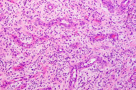
[More Examples at Libre Pathology](Granulation tissue - Libre Pathology)
Granulation tissue is a hallmark of wound healing, recognized by its distinctive histological features. It consists of proliferating capillaries, fibroblasts, inflammatory cells, and a loose extracellular matrix, together creating a supportive environment for tissue repair.12345
Histological Examples of Granulation Tissue
- Proliferating Capillaries: Abundant, thin-walled blood vessels are a defining feature; these newly formed capillaries may be seen as solid cords or with lumens, supplying nutrients and oxygen for healing.125
- Fibroblast Proliferation: Numerous plump fibroblasts actively produce extracellular matrix, initially rich in type III collagen, which is later replaced by type I collagen as the wound matures.451
- Mixed Inflammatory Cells: Lymphocytes, plasma cells, neutrophils, and macrophages are scattered within the stroma, clearing debris and orchestrating tissue repair.354
- Loose Edematous Stroma: The matrix appears pink or pale on H&E stain, reflecting edema from leaky new vessels and active synthesis of ground substances.53
- Granular Organization: The tissue may display a meshwork pattern with collagen fibers running parallel to the surface and vessels perpendicular, aiding the formation of a stable wound environment.6
What to Look for in Microscopy
- Look for numerous, randomly arranged capillaries lined by plump endothelial cells.125
- Identify clusters of fibroblasts, often with oval nuclei and pale cytoplasm, situated between capillaries.
- Note the presence of mixed inflammatory cells, which are less densely packed than in classic inflammatory infiltrates.5
- The stroma should appear loose, edematous, and slightly granular, differentiating granulation tissue from mature fibrous scar.34
- There is typically no abnormal architecture—collagen fibers align parallel to the epidermal or mucosal surface; blood vessels usually grow perpendicular.6
Granulation tissue is most commonly seen in wound beds, healing ulcers, post-surgical sites, and sometimes in pyogenic granulomas and pulp polyps. Its identification aids in staging wound healing and guiding clinical management.Granulation tissue is easily identified histologically by its characteristic combination of proliferating capillaries, fibroblasts, and loosely arranged inflammatory cells within a soft extracellular matrix.4135
Key Histological Features
- Numerous, thin-walled capillaries formed by angiogenesis; these proliferating blood vessels are a hallmark microscopic feature and give the tissue its granular appearance.245
- Plump, active fibroblasts that produce collagen and extracellular matrix; initially the collagen is type III, later replaced by type I as healing continues.145
- Mixed inflammatory cells, mainly macrophages, lymphocytes, and plasma cells (plus neutrophils and eosinophils at times), scattered throughout but not densely clustered as in pure inflammatory infiltrate.35
- Loose, edematous stroma: the tissue appears pink and loose due to the leaky new capillaries and developing ground substance from fibroblast activity.53
- Organization: Collagen fibers run parallel to the surface, while new vessels grow perpendicular, creating a supporting meshwork for repair.6
What to Look For in Microscopy
- Check for abundant new blood vessels with plump endothelial cells.15
- Scan for numerous fibroblasts with oval nuclei and pale cytoplasm between capillaries.21
- Observe a loose matrix, granular texture, and widespread mild inflammation.34
- The combination of all these features distinguishes granulation tissue from both mature scar and pure inflammatory infiltrate.5
Common sites to see granulation tissue include wound beds, healing ulcers, pyogenic granulomas, and dental pulp polyps. Recognizing these features under the microscope is crucial for monitoring healing and guiding clinical decision-making.45 789
Footnotes
-
https://www.ncbi.nlm.nih.gov/books/NBK554402/ ↩ ↩2 ↩3 ↩4 ↩5 ↩6 ↩7 ↩8
-
https://librepathology.org/wiki/Granulation_tissue ↩ ↩2 ↩3 ↩4 ↩5
-
https://ilovepathology.com/granulation-tissue/ ↩ ↩2 ↩3 ↩4 ↩5 ↩6 ↩7 ↩8
-
https://en.wikipedia.org/wiki/Granulation_tissue ↩ ↩2 ↩3 ↩4 ↩5 ↩6 ↩7 ↩8 ↩9
-
https://medicine.osu.edu/-/media/files/medicine/departments/otolaryngology/atlas-of-head-and-neck-pathology/g/granulationtissue2.pdf?rev=51795c2257614f4a83ccb7a3627de158\&hash=F27478FF823D33778D9CD86F0713F677 ↩ ↩2 ↩3 ↩4 ↩5 ↩6 ↩7 ↩8 ↩9 ↩10 ↩11 ↩12 ↩13 ↩14 ↩15
-
https://lms.vet.unimelb.edu.au/pathology_prac/250214/pracs/wh/intro/granulation.html ↩ ↩2 ↩3
-
https://www.sciencedirect.com/topics/medicine-and-dentistry/granulation-tissue ↩
-
https://www.sciencedirect.com/topics/immunology-and-microbiology/granulation-tissue ↩