 ([source](Oral lichenoid lesions: distinguishing the benign from the deadly | Modern Pathology))
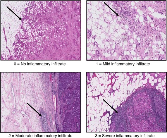
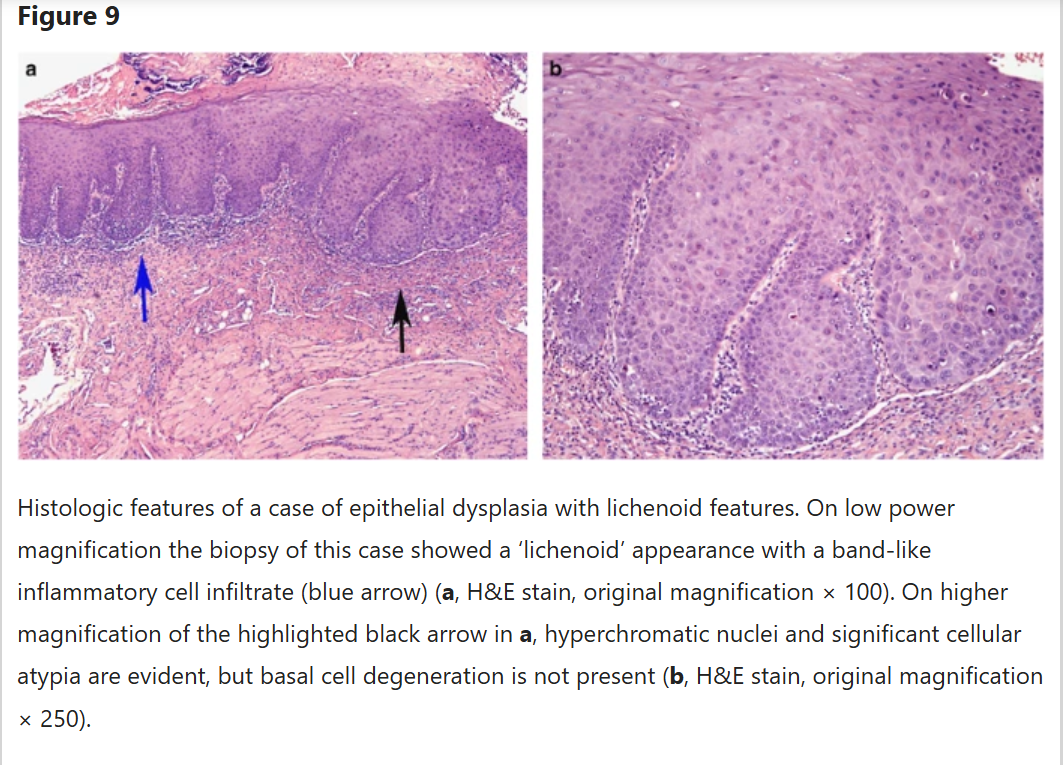
Histological inflammatory infiltrate refers to clusters or diffuse accumulations of immune cells—such as neutrophils, lymphocytes, plasma cells, eosinophils, and macrophages—within tissue, an indicator of tissue inflammation. Recognizing and differentiating these from granulation tissue is essential in pathology.123
What to Look For: Identification Features
- Inflammatory Infiltrate: Appears as increased numbers of leukocytes within connective or other tissues, visible as dense blue or purple clusters under hematoxylin and eosin (H&E) stain.21
- Acute: Dominated by neutrophils, often near sites of tissue injury or infection.43
- Chronic: Comprised mainly of lymphocytes, plasma cells, and macrophages, often less tightly clustered and dispersed through stroma or lamina propria.562
- Pattern: Can be perivascular, interstitial, or diffuse, depending on the nature and chronicity of inflammation.73
- Cell Identification: Leukocytes are characterized by large, dark nuclei (lymphocytes), granular cytoplasm (eosinophils), or segmented nuclei (neutrophils); less cytoplasm compared to stromal cells.65
Differentiation: Inflammatory Infiltrate vs. Granulation Tissue
- Granulation Tissue:
- Consists of new small blood vessels (angiogenesis), proliferating fibroblasts, and a looser extracellular matrix.89
- Contains fewer, but often mixed, inflammatory cells compared to typical inflammatory infiltrate.
- Characteristic features: prominent capillaries lined by plump endothelial cells, loose arrangement of proliferating fibroblasts, and variable amounts of mononuclear inflammatory cells.9
- Inflammatory Infiltrate:
Practical Histological Clues
- Granulation tissue has a classic “granular” appearance owing to angiogenesis and fibroplasia, while inflammatory infiltrate focuses on leukocyte accumulation.89
- Granulation tissue represents a reparative (healing) process, whereas infiltrate signals active inflammation, often extending into and between tissue structures—not just in healing zones.398
- On H&E, granulation tissue looks pink and loose due to matrix and vessel proliferation, with scattered dark nuclei; inflammatory infiltrate appears as denser blue/black cell clusters.9
Correct identification relies on evaluating cell types and tissue architecture together, sometimes requiring special stains or clinical correlation.Histological inflammatory infiltrate refers to the accumulation of immune cells—such as neutrophils, lymphocytes, plasma cells, and macrophages—in tissue, distinguishing inflamed from normal or healing tissue.173529
What to Look for in Histological Samples
- Inflammatory infiltrate typically appears as dense collections or scattered groups of leukocytes within tissue, making the area stain darker (dark blue/purple on H&E).431
- Cell types give important clues:
- In some organs (like skin or colon), look for these cells within or around blood vessels, in the connective tissue, or beneath surface epithelium.734
How to Differentiate from Granulation Tissue
| Inflammatory Infiltrate | Granulation Tissue |
|---|---|
| Dense clusters/sheets of leukocytes (acute: more neutrophils; chronic: lymphocytes, plasma cells, macrophages)12. | Proliferation of small capillaries (angiogenesis), loose arrangement of fibroblasts, some scattered inflammatory cells9. |
| Minimal or absent new blood vessels and fibroblasts. | Numerous new capillaries lined by plump endothelial cells; fibroblasts producing new extracellular matrix9. |
| Often replaces or disrupts normal architecture, especially near a site of ongoing damage/infection. | Loose, edematous, pink-appearing stroma, mixed with cells but less densely cellular overall9. |
Look for a combination of cell density, cell type, new vascular channels, and stromal features to confidently distinguish between active inflammatory infiltrate and restorative granulation tissue. If you see abundant new delicate blood vessels and fibroblasts mixed with less dense inflammation, it’s likely granulation tissue; if you see focal, dense sheets of neutrophils or mononuclear cells, it’s likely an inflammatory infiltrate.12859 101112131415161718
Footnotes
-
https://www.sciencedirect.com/topics/medicine-and-dentistry/inflammatory-infiltrate ↩ ↩2 ↩3 ↩4 ↩5 ↩6 ↩7 ↩8
-
https://www.6patterns.org/cellular-infiltrates ↩ ↩2 ↩3 ↩4 ↩5 ↩6 ↩7
-
https://academic.oup.com/ecco-jcc/article/18/2/175/7247579 ↩ ↩2 ↩3 ↩4 ↩5 ↩6
-
https://journals.plos.org/plosone/article?id=10.1371%2Fjournal.pone.0072494 ↩ ↩2
-
https://pmc.ncbi.nlm.nih.gov/articles/PMC5885876/ ↩ ↩2 ↩3 ↩4
-
https://www.ncbi.nlm.nih.gov/books/NBK554402/ ↩ ↩2 ↩3 ↩4 ↩5 ↩6 ↩7 ↩8 ↩9 ↩10
-
https://www.sciencedirect.com/topics/pharmacology-toxicology-and-pharmaceutical-science/inflammatory-infiltrate ↩
-
https://www.sciencedirect.com/science/article/pii/S0022202X15332917 ↩
-
https://www.sciencedirect.com/science/article/pii/S1578219016302025 ↩
-
https://teachmesurgery.com/skills/wounds/chronic-inflammation/ ↩
-
https://dermnetnz.org/cme/dermatopathology/inflammatory-skin-diseases ↩