[Palisading of basal cells in OKC:](Pathology Outlines - Odontogenic keratocyst)
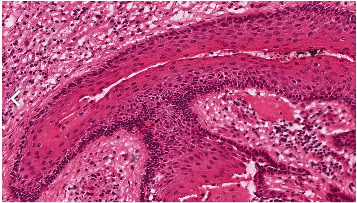
palisaded basal cells images and histological significance
“Palisaded basal cells” refers to a histological arrangement where the basal cells, especially in tumors like basal cell carcinoma (BCC), are aligned in a columnar or fence-like pattern along the periphery of tumor nests or lobules. This peripheral palisading is a key diagnostic feature in BCC.
Images
- Histology images of basal cell carcinoma show nests of basaloid cells with prominent peripheral palisading where nuclei line up neatly at the edge of tumor islands.
- Within these images, the palisaded basal cells create a “picket fence” or “rail fence” appearance around the darker, crowded central nuclei of the tumor nests.
- The tumor nests are often separated from the surrounding stroma by a cleft-like space, an artifact due to mucin or stroma shrinkage during tissue processing.
Histological Significance
- Palisading basal cells indicate a characteristic growth pattern of basal cell carcinoma, arising from basal keratinocytes of the epidermis or follicular epithelium.
- This organization reflects the tumor’s relatively slow growth and local invasiveness with a well-defined stromal interface.
- Peripheral palisading helps differentiate BCC from other basaloid tumors and non-malignant conditions.
- The clefting artifact and palisading combined are hallmarks that aid in confirming the diagnosis histologically.
Summary Table
| Feature | Description | Significance |
|---|---|---|
| Palisaded basal cells | Basal cells aligned in a fence-like pattern at tumor periphery | Characteristic for basal cell carcinoma, aids in diagnosis |
| Cleft formation | Separation artifact between tumor nests and stroma | Helps differentiate BCC, related to mucin degradation or shrinkage during processing |
| Basaloid cells | Small, darkly staining cells with hyperchromatic nuclei | Indicative of neoplastic basal keratinocyte origin |
Representative Histology Images
- Tumor nests with peripheral palisading and clefting in BCC can be seen in histology micrographs stained with hematoxylin and eosin (H&E).
- These images highlight the typical nodular, basaloid clusters with palisaded peripheral nuclei and stromal clefts.
If a histological image is helpful, one typical micrograph shows dense basaloid nests with the peripheral cells neatly palisaded, surrounded by lighter stained connective tissue stroma with clear clefts in between.123

Micrograph of a basal-cell carcinoma stained with H&E.

High-magnification micrograph of basal-cell carcinoma.
In summary, palisaded basal cells in histology primarily suggest basal cell carcinoma and serve as an important diagnostic microscopic feature of tumor growth and organization.123 4567
Footnotes
-
https://dermnetnz.org/topics/basal-cell-carcinoma-pathology ↩ ↩2
-
https://meridian.allenpress.com/aplm/article/141/11/1490/65518/Histologic-Mimics-of-Basal-Cell-Carcinoma ↩ ↩2
-
https://www.sciencedirect.com/science/article/pii/S2374289521000592 ↩
-
https://dermnetnz.org/imagedetail/24276-basal-cell-carcinoma-histopathology ↩