Radiologic Interpretation Panoramic Radiograph Part 11
Radiologic Interpretation - Panoramic Radiograph Part 1.
Dr Dayea Oh OMF Radiologist
Learning Objectives2
- Revise Panoramic radiographic anatomy and artefacts
- Review indications for panoramic imaging / OPG
- Understand basic interpretation skills
- Radiographic features of dento-alveolar findings
- Use appropriate radiology terminology
- Follow systematic approach
Radiographic Anatomy and Interpretation Principles
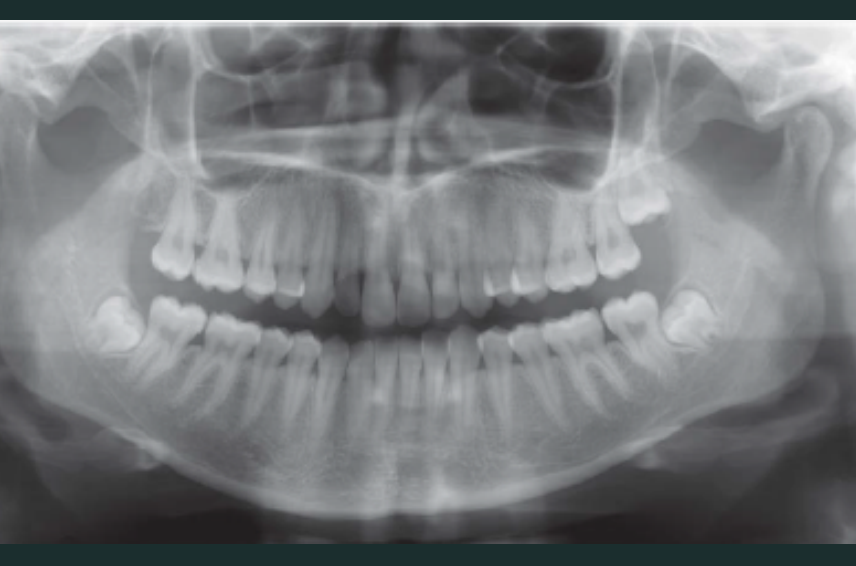
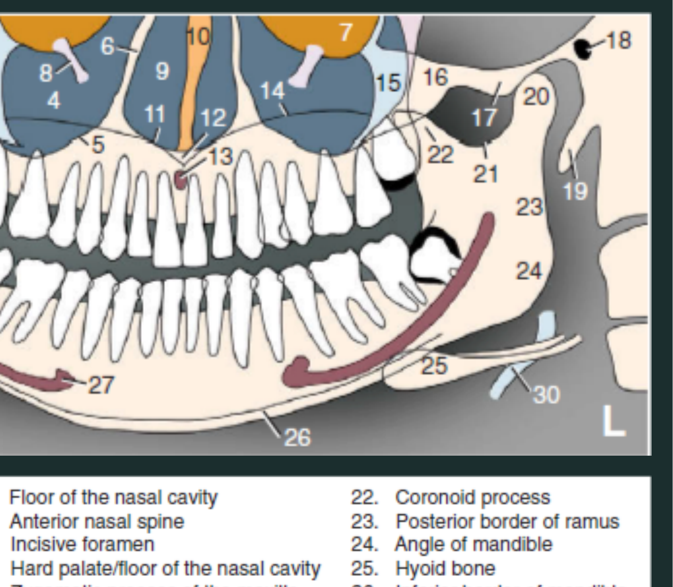
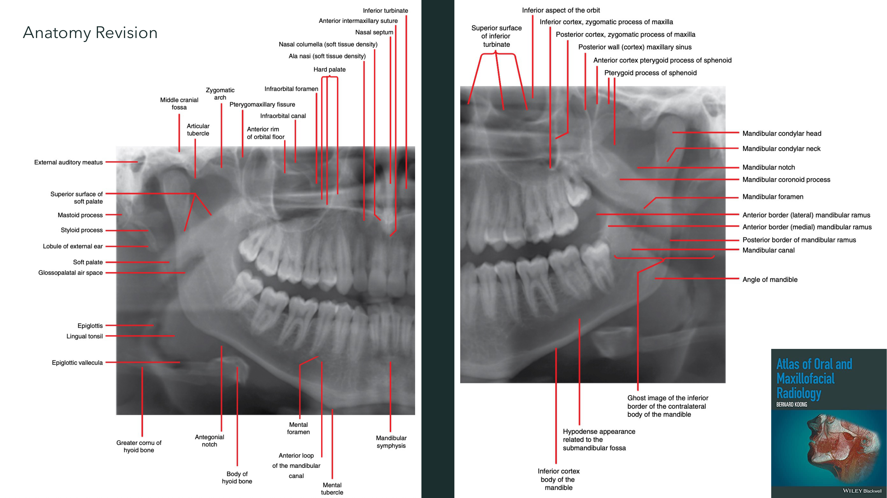
Anatomical Landmarks of the Maxilla34
Note the double image of palate, hyoid & spine
- Pterygomaxillary fissure
- Posterior border of maxilla
- Maxillary tuberosity
- Maxillary sinus
- Floor of the maxillary sinus
- Medial border of maxillary sinus/lateral border of the nasal cavity
- Floor of the orbit
- Infraorbital canal
- Nasal cavity
- Nasal septum
- Floor of the nasal cavity
- Anterior nasal spine
- Incisive foramen
- Hard palate/floor of the nasal cavity
- Zygomatic process of the maxilla
- Zygomatic arch
- Articular eminence
- External auditory meatus
- Styloid process
- Mandibular condyle
- Sigmoid notch
- Coronoid process
- Posterior border of ramus
- Angle of mandible
- Hyoid bone
- Inferior border of mandible
- Mental foramen
- Mandibular canal
- Cervical vertebrae
- Epiglottis
Atlas of Oral and Maxillofacial Radiology BERNARD KOZUNG WILEY Blackwell
 |  |
 |
General Indications for Panoramic Radiography5
General dental screening? OPGs are NOT accurate for caries assessment.
- Heavily compromised dentition - gross caries, multiple periapical inflammatory lesions
- Periodontal screening (periodontal bone loss)
- Impacted third molars
- Mixed dentition status
- Orthodontics
- Crowding, impacted canines, etc.
- Dental anomalies
- Jaw (intrabony) Pathology
- TM Joints (gross morphological change)
- Trauma (esp. mandibular fracture)
Medicolegal Responsibilities and Reporting6
- Radiation dosages with CBVT and panoramic film must be explained
- There is an increased duty to explain dosages and risks of radiographs on dentist’s own premises
- Dentists who record panoramic radiographs need to take responsibility for all non-dental diagnosis or have them assessed by or referred to a DMF radiologist or radiologist
- Dentists who record small volume CBVT need to assess whether a referral to a DMF radiologist/radiologist is appropriate
- Dentists who record large volume CBVT need to refer all data sets to DMF radiologists/radiologists for review, as they have the highest medicolegal risk
- There needs to be thorough discussion of dosage of CBVT for paediatric patients
- If the dentist has a CBVT on-site, it is not advisable to expose paediatric patients to CBVT
- Dentist “self-reports” must be held to a similar standard as a specialist report
Radiology Report Statistics7
- All dental images MUST have radiology reports
- ~ 40% of dental imaging is self-reported by dentists
- 1.5% unreported
- ~40% reported by medical radiologists
- ~20% are reported by DMF radiologists
Ref: Selim, DG, C. Sexton, and P. Monsour. “Dentomaxillofacial Radiology in Australia and Dentist Satisfaction with Radiology Reports.” Australian Dental Journal 63, no. 4 (2018): 402-13.
Referral Pathways for Radiology Reports8
- Request scan & report to an imaging clinic (Medicare Rebate)
- Patient details
- Referrer details
- Select imaging modality (e.g., OPG / Occlusograph / CT / …)
- Write reasons for referral (i.e., clinical indications / history)
- Report only (Private)
- Teleradiology (online referral with enclosed image)
Key Points of Interpretation9
- Review the initial indication for OPG
- Identify OPG artefacts
- Must know normal radiographic anatomy of maxillofacial region
- So, abnormal changes to normal anatomy can be identified
- Study radiographic features of various jaw pathologies
- Systematic approach
- Have a routine
Panoramic Radiograph Artefacts10
- Foreign materials (e.g., earrings, necklace, denture left in situ, etc.)
- Ghost images
- Incorrect patient positioning
- Chin down
- Chin up
- Head tilting
- Head rotation
- Movement
- Horizontal (following the beam or opposite to the beam direction)
- Vertical
- Swallowing
- Soft tissue position
- Tongue to the palate
- Lips closed
- Focal trough position
- Head in front of the focal trough
- Head behind the focal trough
- Neck extension
- Collision with shoulder
Systematic Approach to Interpretation11
- Any Technical or Positional Error?
- Address Clinical Concerns
- Dentoalveolar
- Count teeth & identify ectopic and/or impacted teeth
- Dental Anomalies (Position, Shape,…)
- Periodontal bone loss
- Dental pathology (non-caries tooth loss, caries, fracture, periapical pathologies, etc.)
- Maxilla and Mandible
- Abnormal (radio)lucencies
- Abnormal (radio)opacities
- Altered trabecular pattern
- Fractures
- Jaw Asymmetry
- Maxillary sinuses (esp. mucosal changes)
- TM Joints (esp. morphological change of condyles)
- Soft Tissues (swelling, soft tissue calcifications e.g., tonsiloliths, salivary gland stones, etc.)
- Other sites: Spine, orbits, etc. (structural changes)
Clinical Case Studies12
OPG CASES
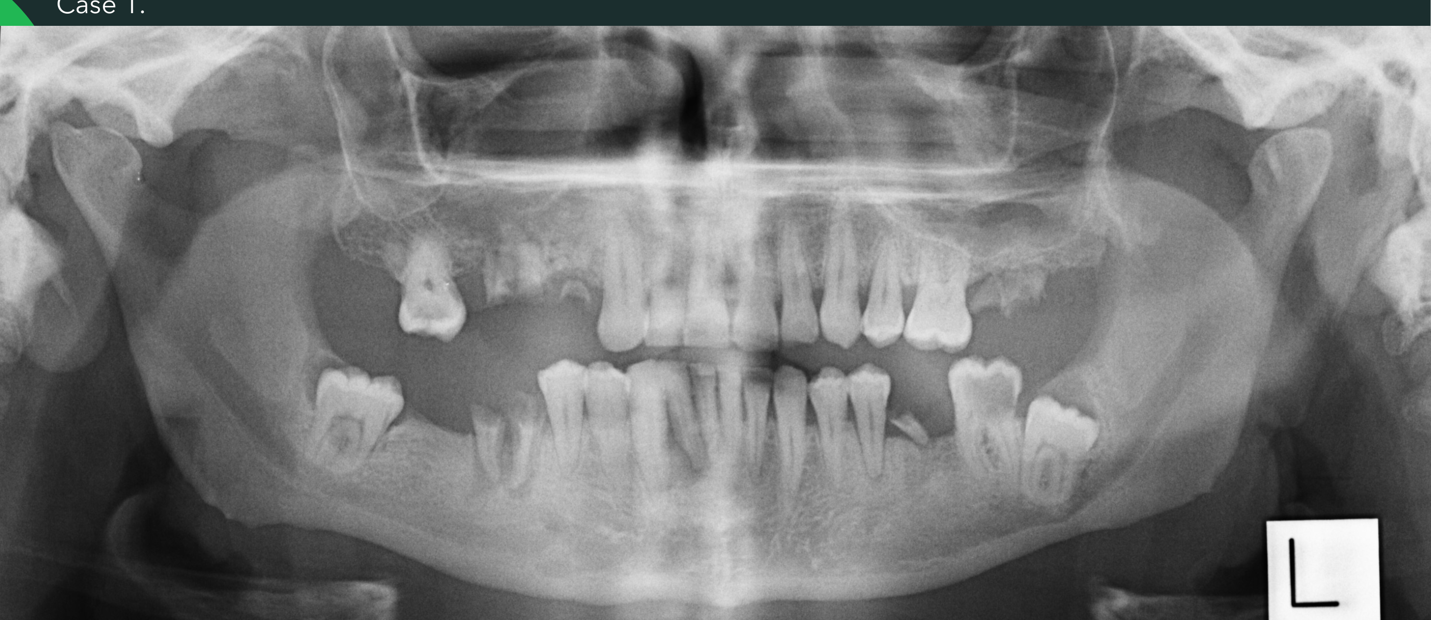
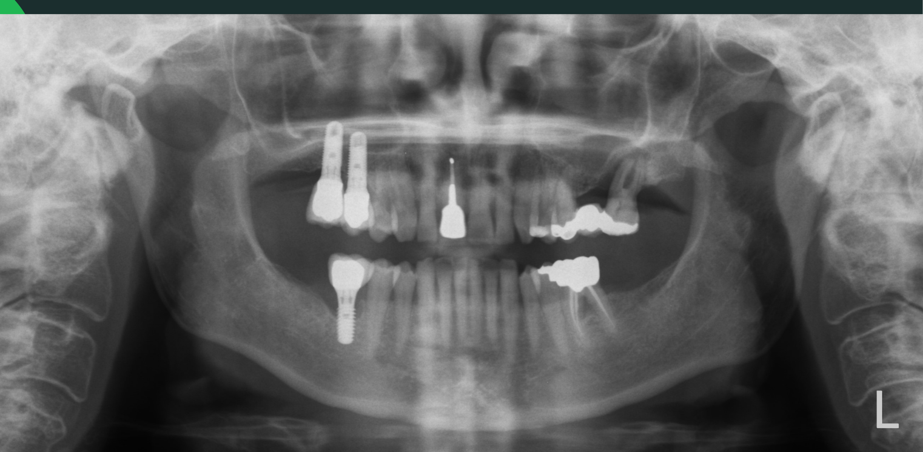
Case 1 Heavily Compromised Dentition131415
Case 1.
Reason for Scan: Heavily compromised dentition
- Count teeth including root remnants
- Identify impacted teeth – give brief description
- Periodontal bone loss
- Existing restoration
- Caries
- Existing endodontic therapy
- Inflammatory lesions
Relationship with adjacent vital structures eg. Maxillary Sinuses and Mandibular Canals
 |  |
OTHER FINDINGS - Systematic Approach16
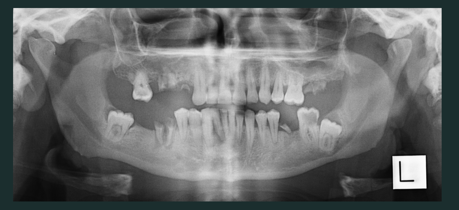
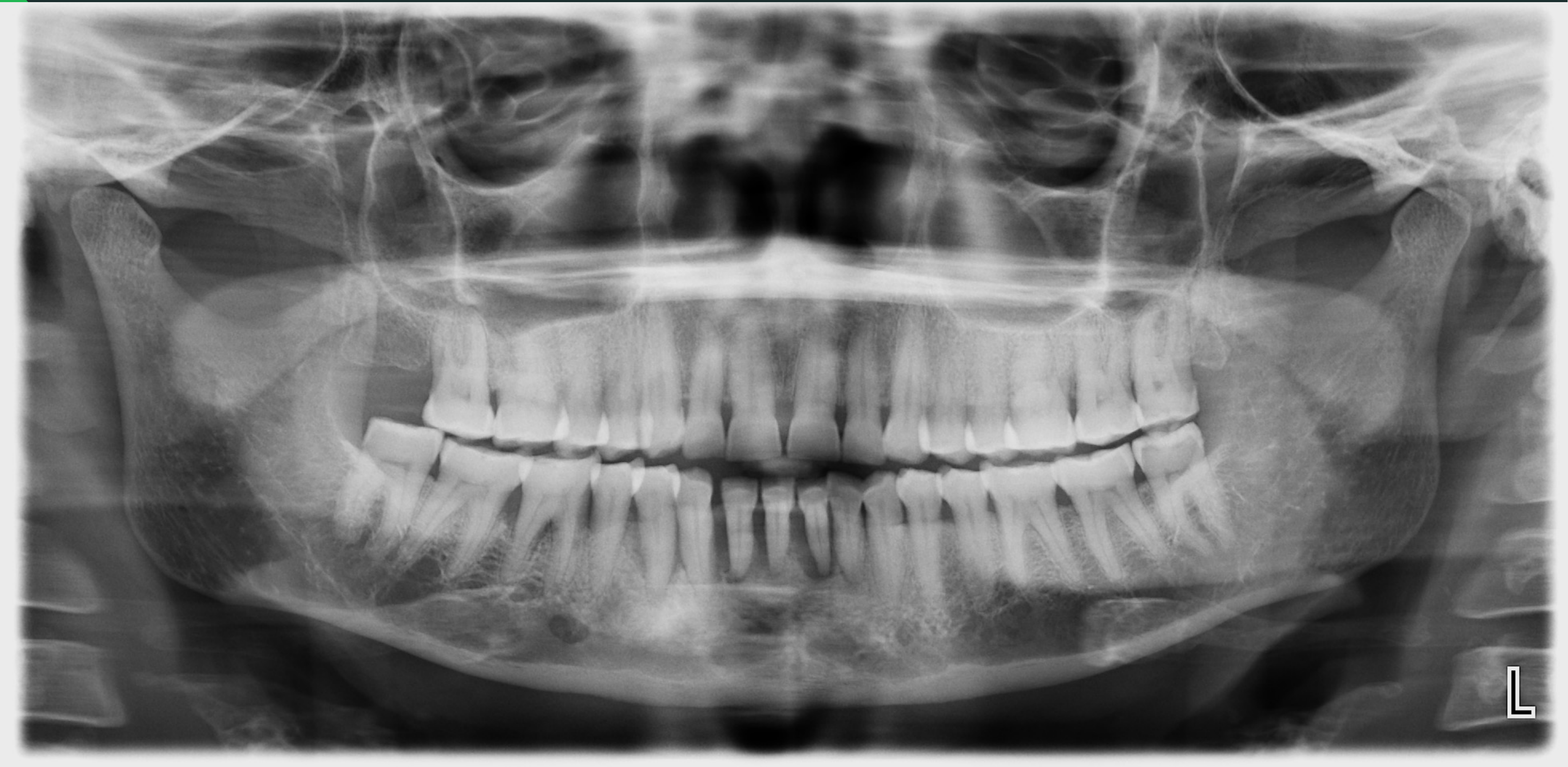
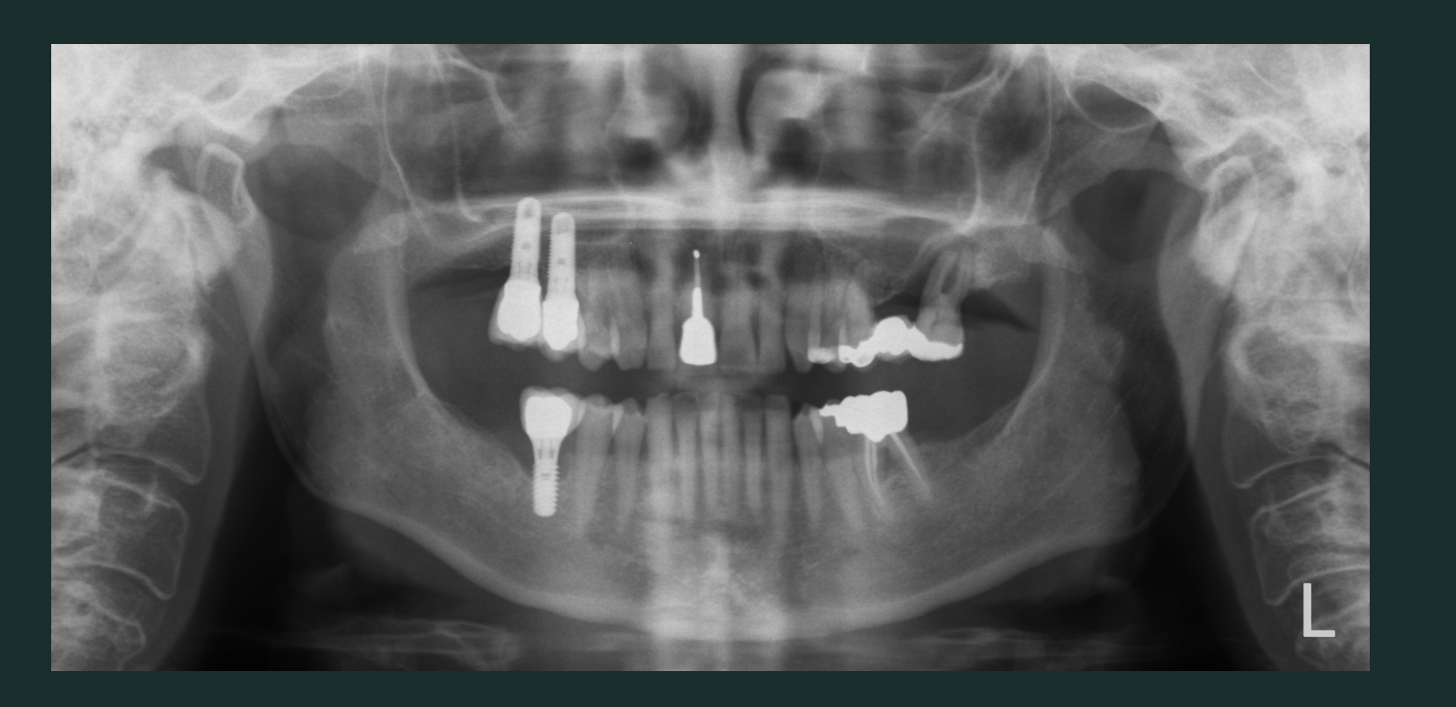
Case 2 Periodontal and Peri-Implant Bone Loss171819
Case 2 Main Focus: Periodontal Bone Loss
- Count teeth
- Count implants
- Peri-implant bone loss
- Periodontal bone loss
- Existing restoration
- Caries
- Existing endodontic therapy
- Inflammatory lesions
- Relationship with adjacent vital structures eg. Maxillary Sinuses and Mandibular Canals
 |  |
OTHER FINDINGS - Systematic Approach20
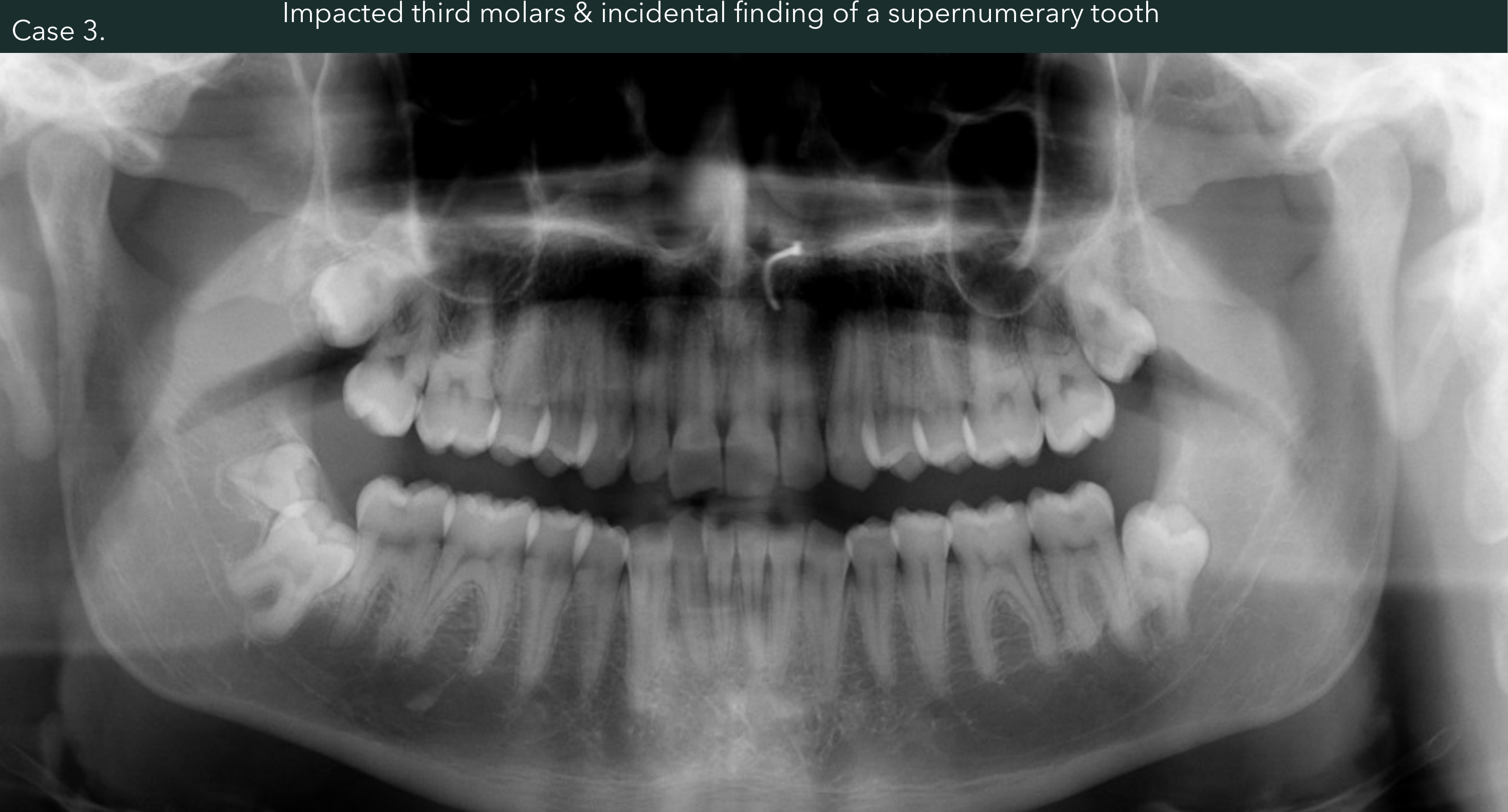
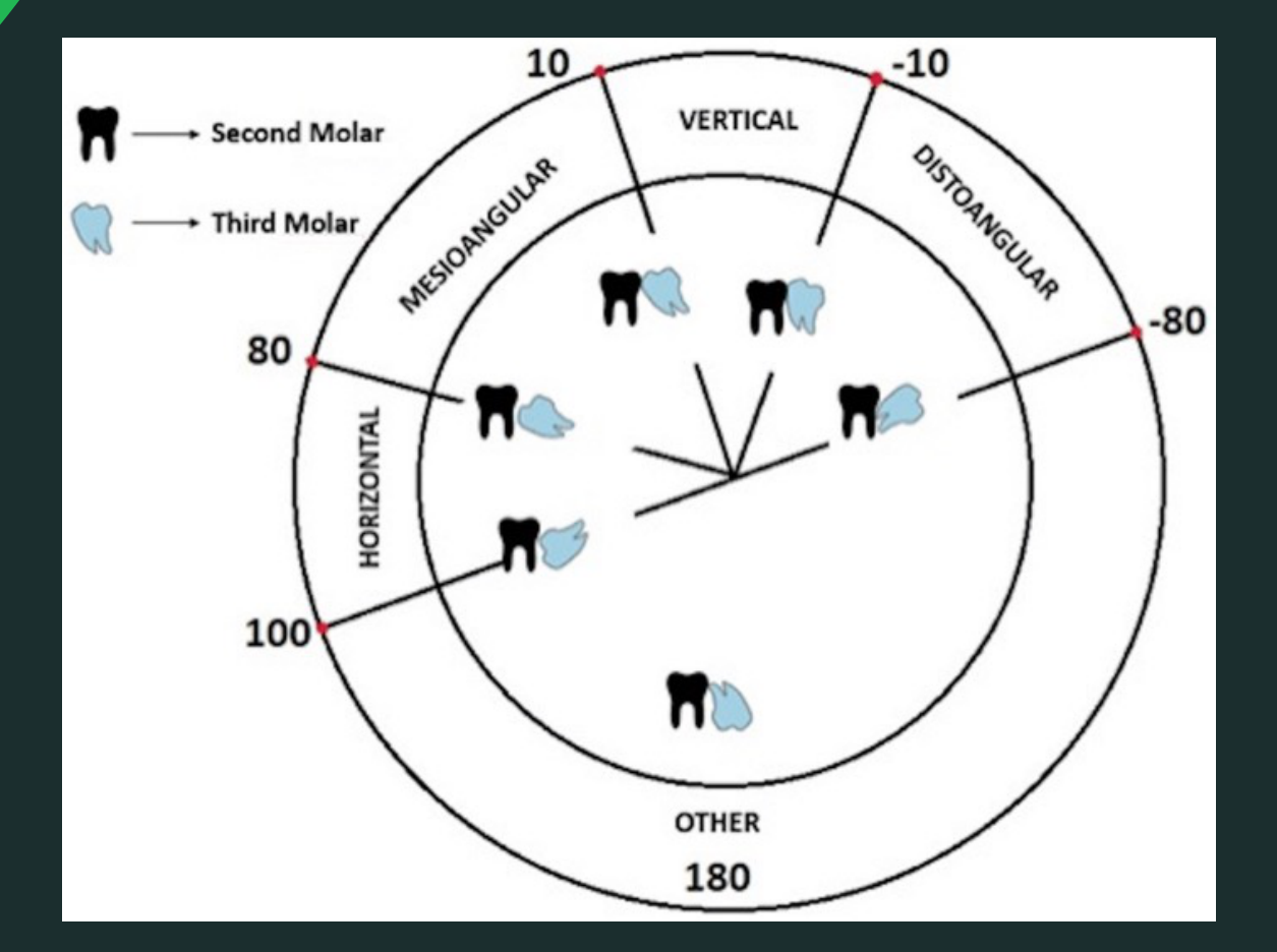
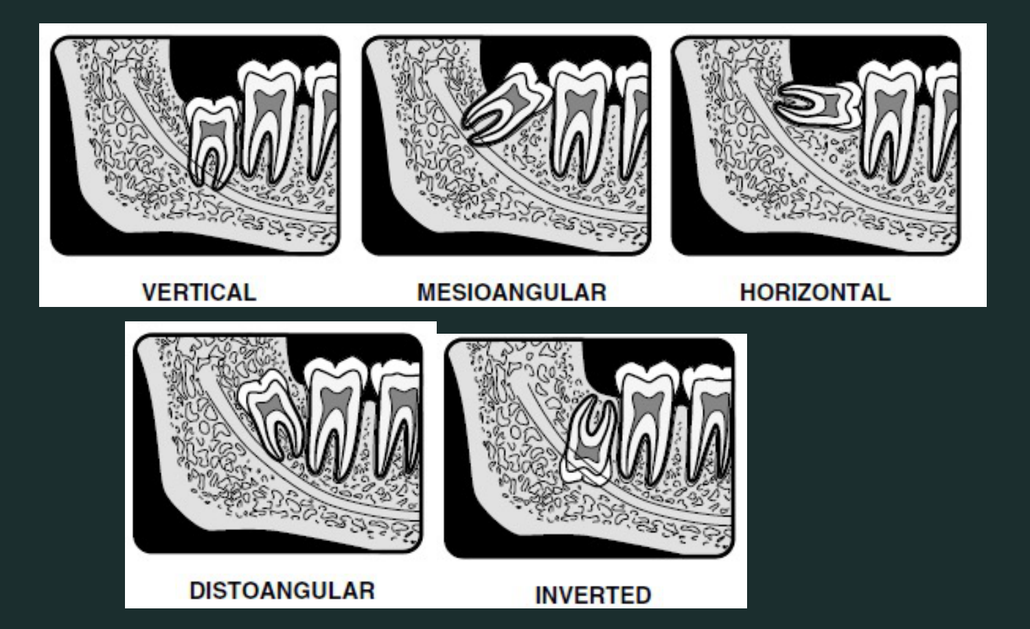
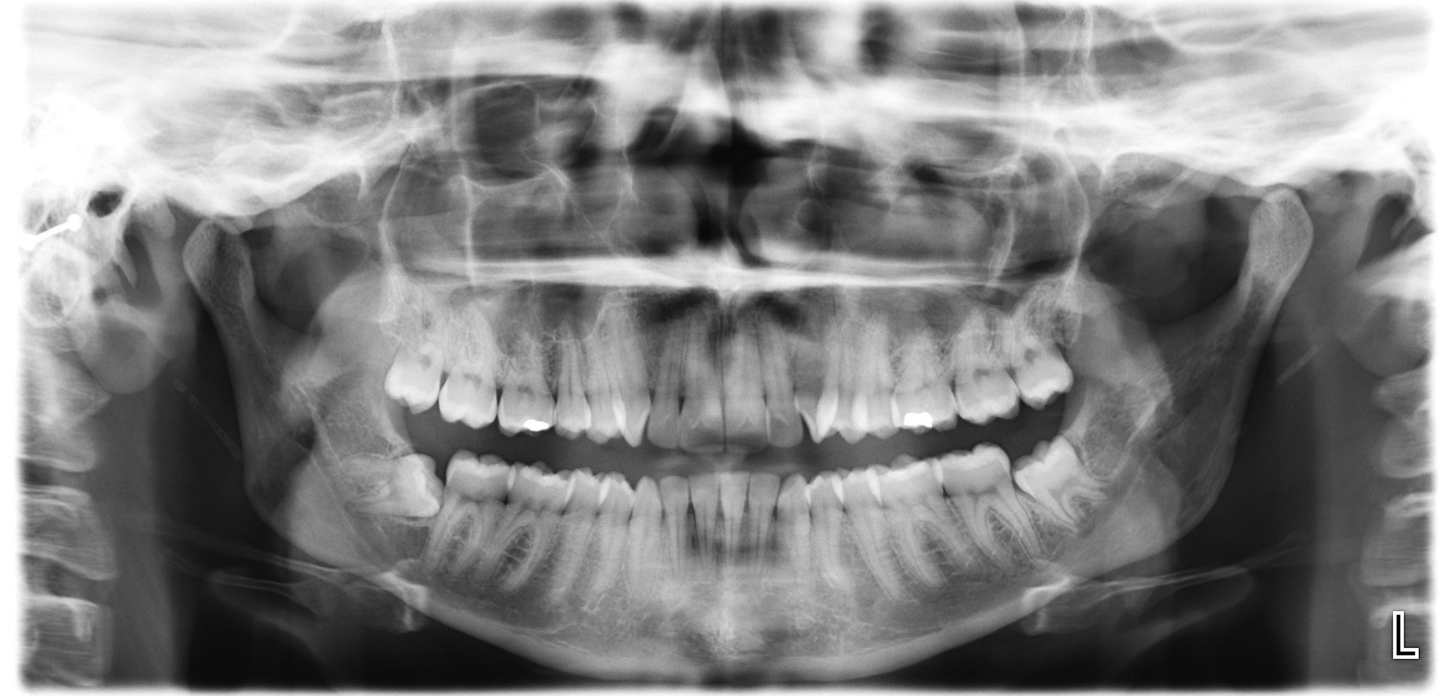
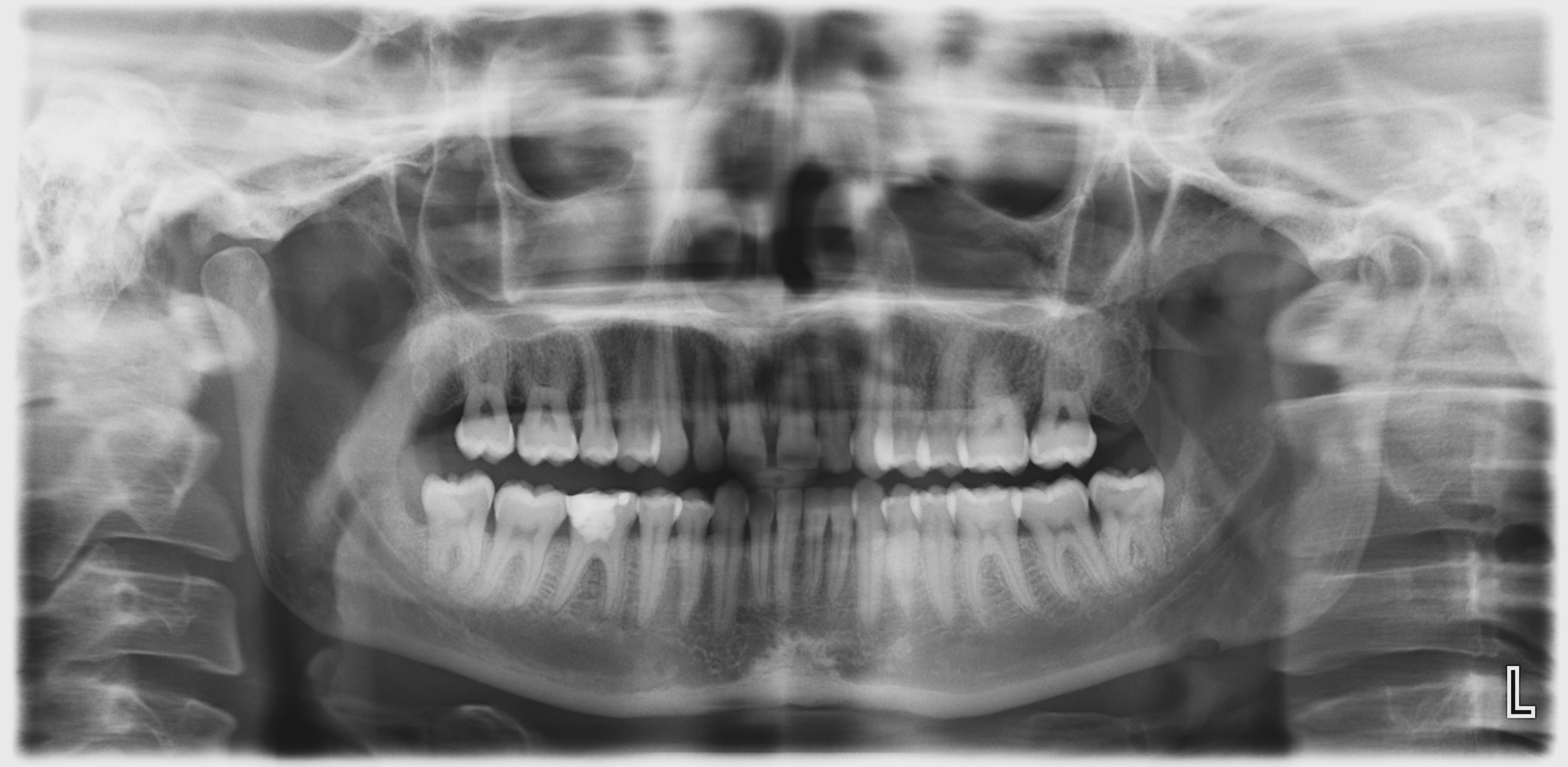
Case 3 Impacted Third Molars and Supernumerary Teeth21222324
Third molar impaction – Winter’s Classification
Case 3 Main Focus: Impacted Third Molars
- Count teeth including supernumerary
- Describe the supernumerary tooth
- Describe the impacted teeth
- Periodontal bone loss
- Existing restoration
- Caries
- Existing endodontic therapy
- Inflammatory lesions
- Relationship with adjacent vital structures eg. Maxillary Sinuses and Mandibular Canals
 |  |
 |  |
OTHER FINDINGS - Systematic Approach25
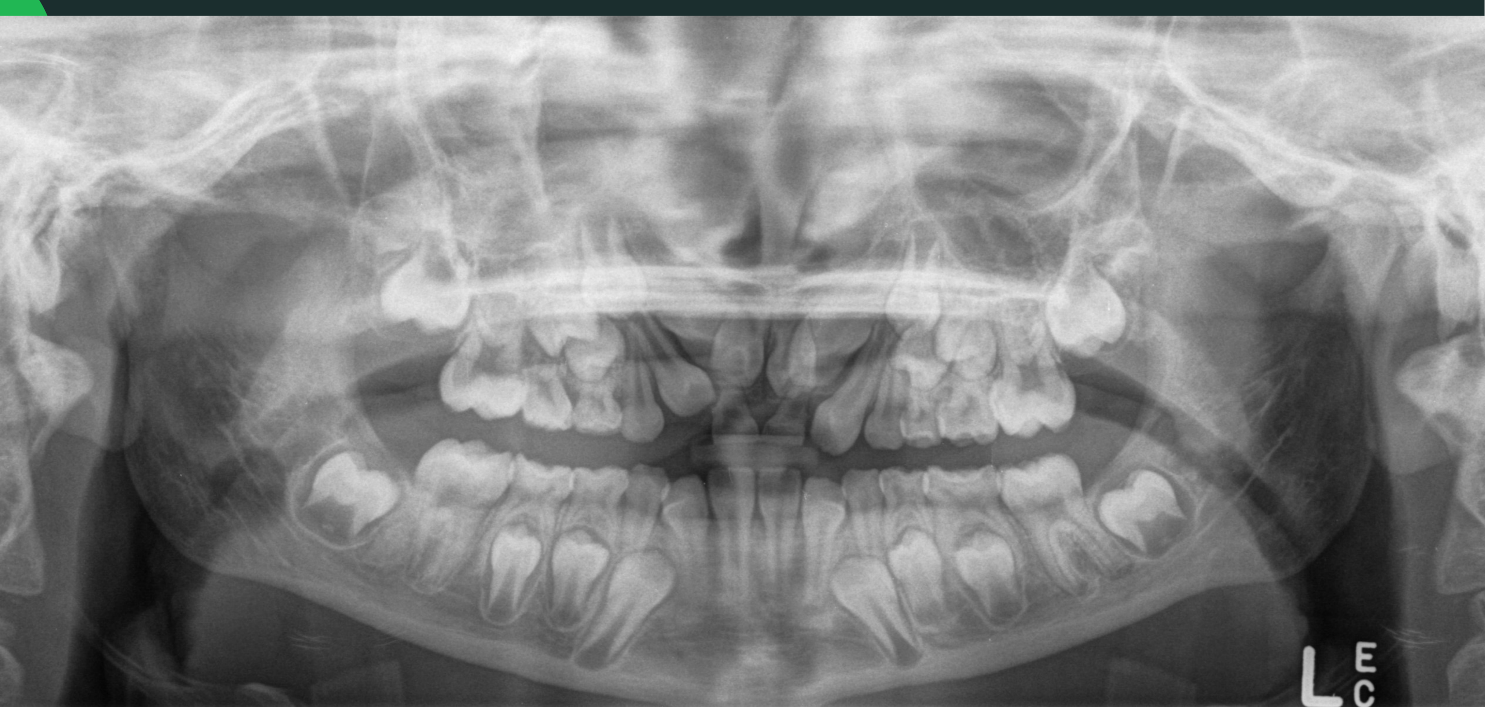
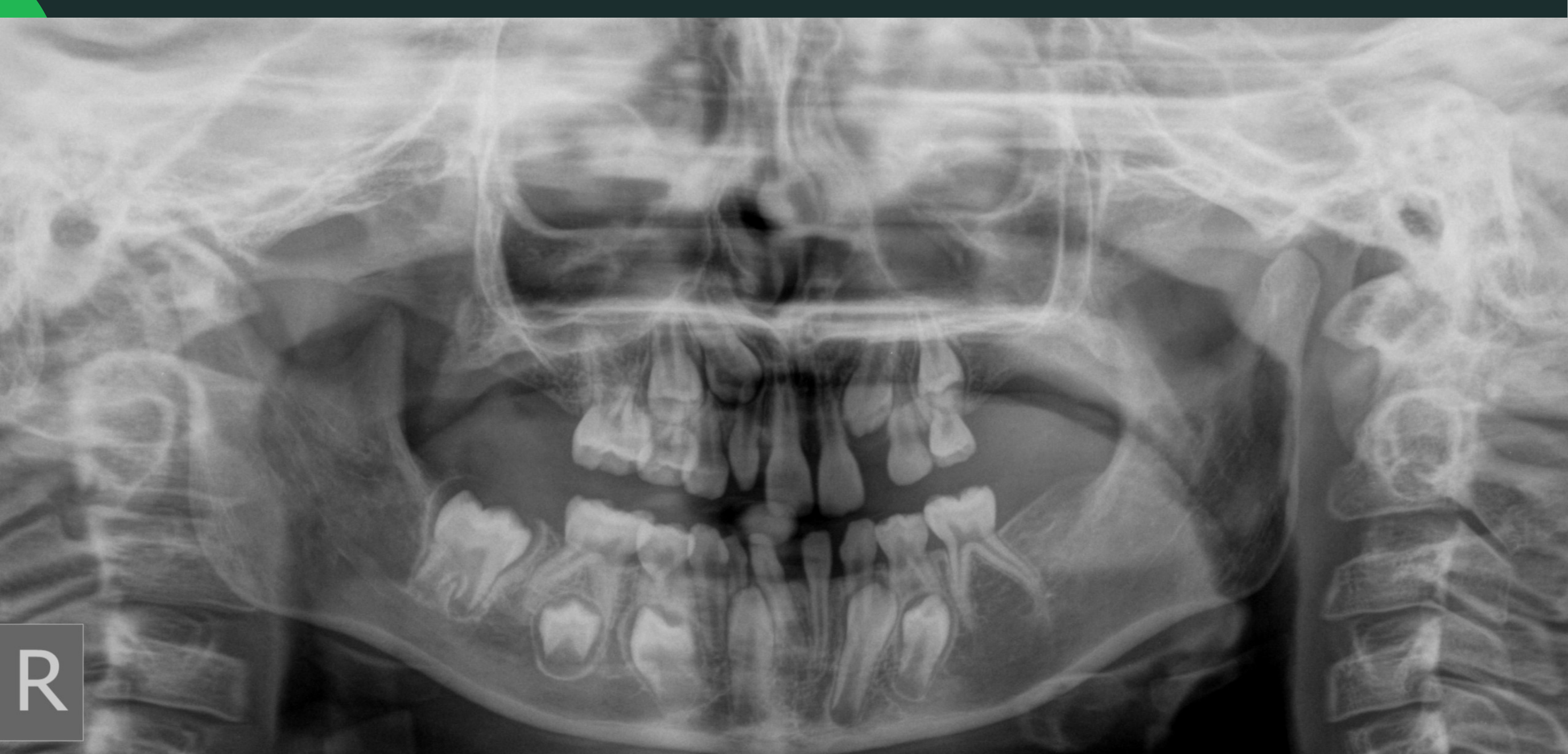
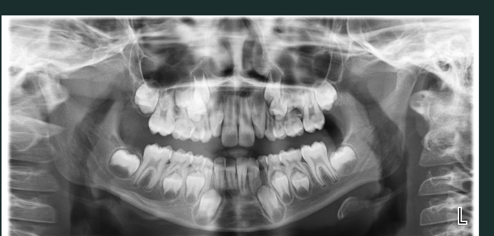
Case 4 Mixed Dentition and Development262728
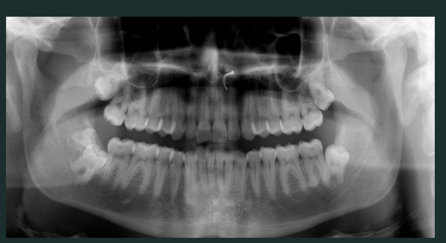
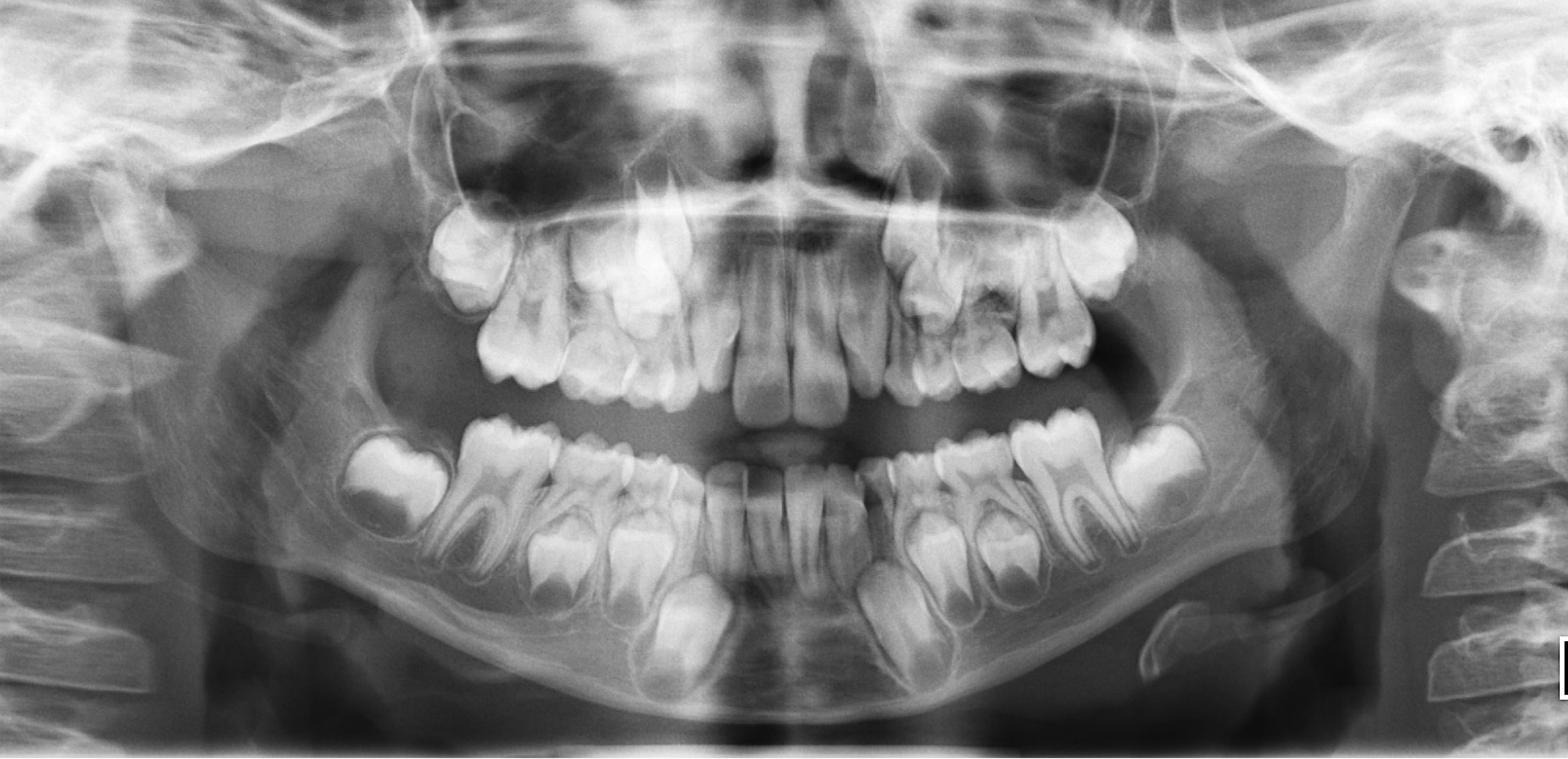
Case 4.
Case 4 Main Focus: Mixed Dentition
- Count teeth
- Check permanent teeth development and position
- Is the dentition appropriate for age?
- Crowding and rotated teeth
- Check Dental Anomalies
- Periodontal bone loss
- Existing restoration
- Caries
- Inflammatory lesions
- Relationship with adjacent vital structures eg. Maxillary Sinuses and Mandibular Canals
 |  |
 |  |
OTHER FINDINGS - Systematic Approach29
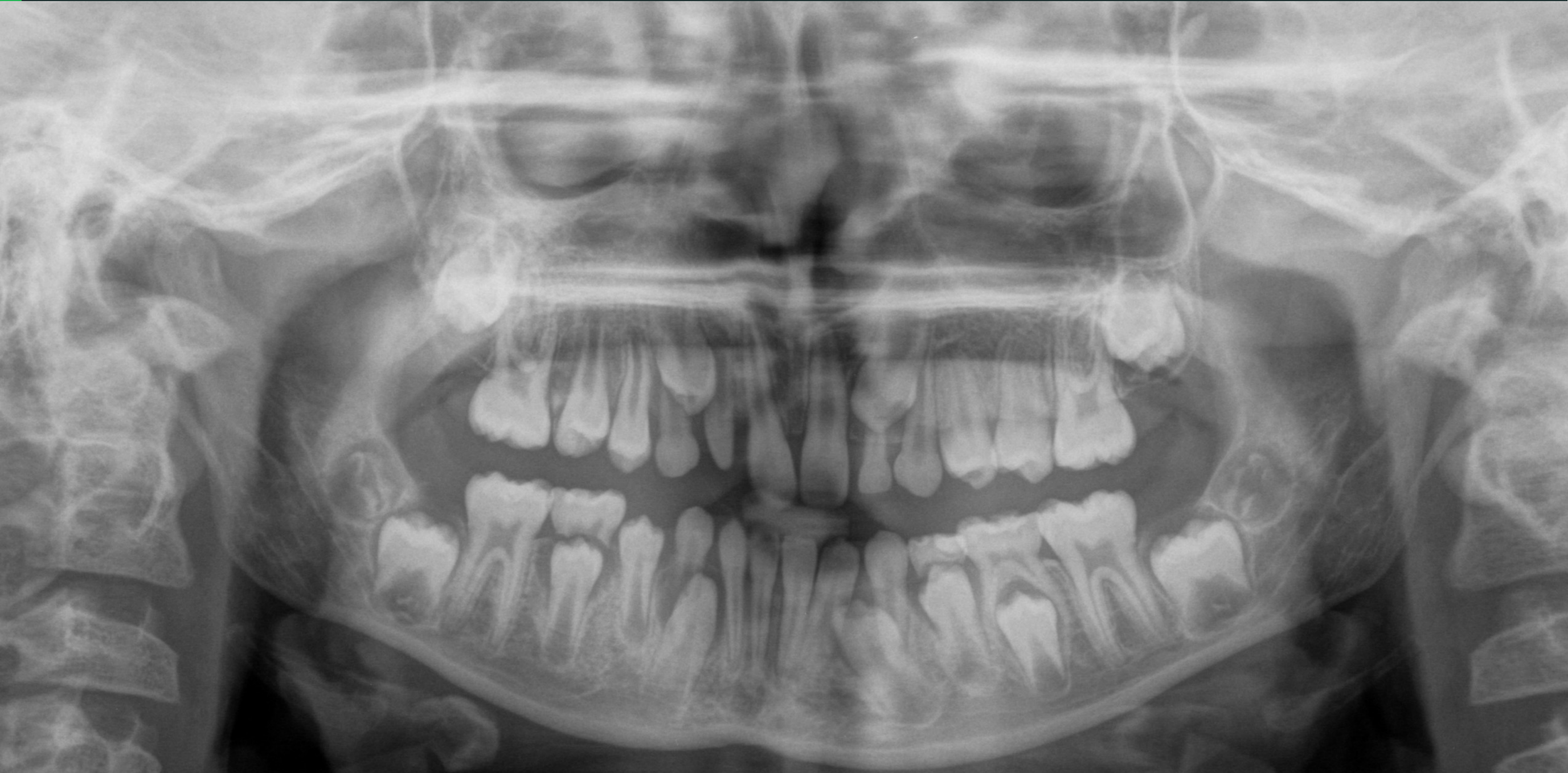
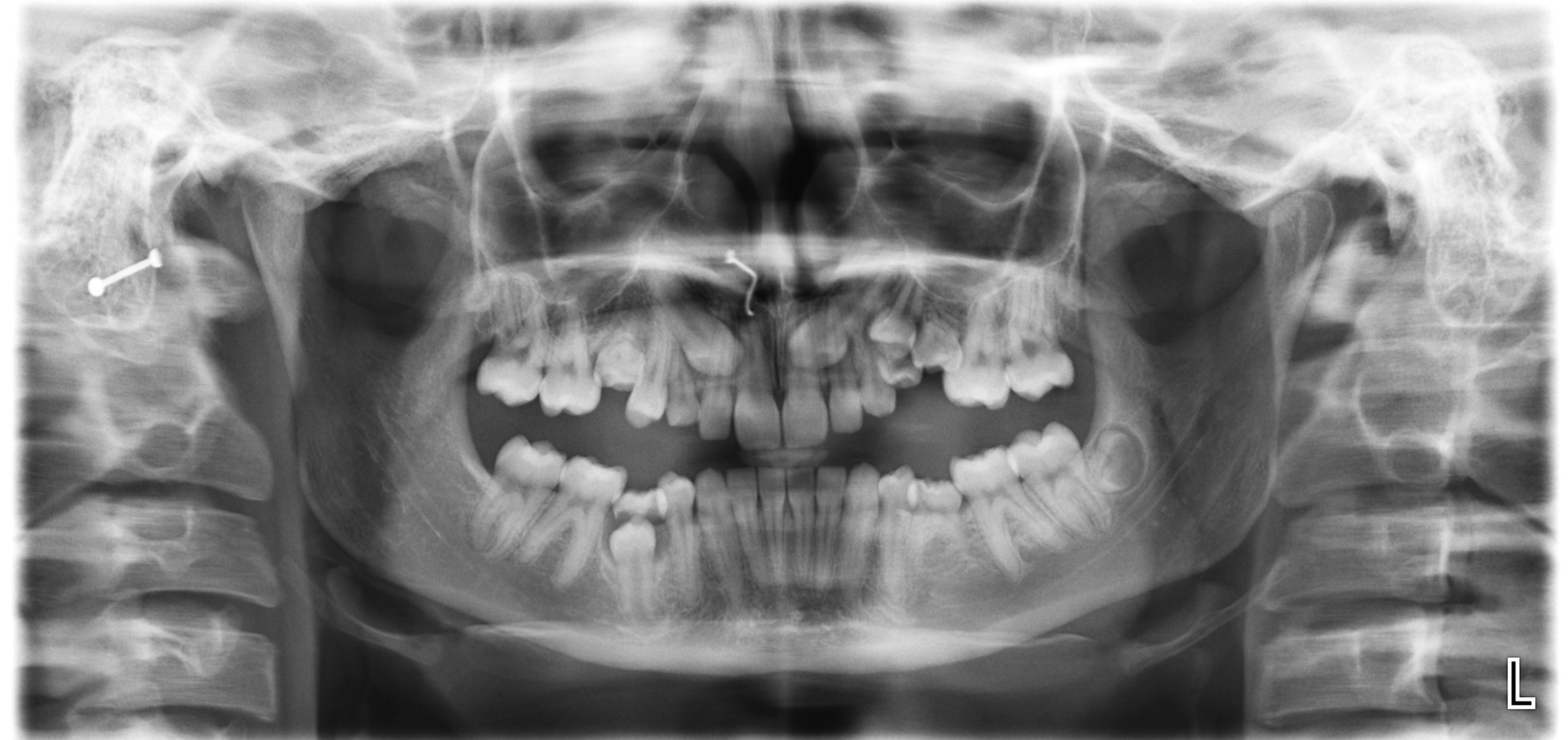
Case 5 Submerged Deciduous Molars
 |  |
Footnotes
-
Original PDF page 1: L12 PR1, p.1 ↩
-
Original PDF page 2: L12 PR1, p.2 ↩
-
Original PDF page 3: L12 PR1, p.3 ↩
-
Original PDF page 4: L12 PR1, p.4 ↩
-
Original PDF page 5: L12 PR1, p.5 ↩
-
Original PDF page 6: L12 PR1, p.6 ↩
-
Original PDF page 7: L12 PR1, p.7 ↩
-
Original PDF page 8: L12 PR1, p.8 ↩
-
Original PDF page 9: L12 PR1, p.9 ↩
-
Original PDF page 10: L12 PR1, p.10 ↩
-
Original PDF page 11: L12 PR1, p.11 ↩
-
Original PDF page 12: L12 PR1, p.12 ↩
-
Original PDF page 13: L12 PR1, p.13 ↩
-
Original PDF page 15: L12 PR1, p.15 ↩
-
Original PDF page 16: L12 PR1, p.16 ↩
-
Original PDF page 14: L12 PR1, p.14 ↩
-
Original PDF page 18: L12 PR1, p.18 ↩
-
Original PDF page 19: L12 PR1, p.19 ↩
-
Original PDF page 20: L12 PR1, p.20 ↩
-
Original PDF page 17: L12 PR1, p.17 ↩
-
Original PDF page 21: L12 PR1, p.21 ↩
-
Original PDF page 23: L12 PR1, p.23 ↩
-
Original PDF page 24: L12 PR1, p.24 ↩
-
Original PDF page 25: L12 PR1, p.25 ↩
-
Original PDF page 22: L12 PR1, p.22 ↩
-
Original PDF page 26: L12 PR1, p.26 ↩
-
Original PDF page 28: L12 PR1, p.28 ↩
-
Original PDF page 29: L12 PR1, p.29 ↩
-
Original PDF page 27: L12 PR1, p.27 ↩