Panoramic Radiograph Analysis
How to use this note
- Start with the Quick Annotation Checklist, then read the OPG in that order and fill in the Report Template at the end.
- Sister note for intraoral films: Periapical Radiograph Analysis.
- White & Pharoah (W&P) is the primary source. Lecture and case images fill gaps. Where sources disagree, both are stated.
Quick Annotation Checklist
Systematic OPG read (same order every time)
- Before you look: reason for the scan, and deal with the clinical concern first. Check that the image carries patient name, DOB, R/L marker, exam date and dentist. Note the stage of dentition (deciduous / mixed / permanent)123
- Image quality and positioning: any significant OPG error? Smile line, anterior tooth width, L vs R molar and ramus width, tongue air band, spine shadow, blur, ghosts of jewellery or apron243
- Teeth: count them in FDI, including root remnants and supernumeraries. Missing teeth, implants (peri-implant crestal bone), impacted or ectopic teeth (location, angulation, crown/root morphology, relation to neighbours), developing teeth, anomalies253
- Restorations and endo: existing restorations and faults (overhangs, deficient margins), caries including recurrent, NCTL, existing endodontic therapy2
- Periodontal bone levels: horizontal vs angular/vertical loss, furcations, perio-endo defects, calculus2
- Periapices: periapical lucencies, and any related root resorption, antral floor remodelling, reactive sclerosis or antral mucosal thickening2
- Jaws, bilaterally: abnormal lucencies or opacities, trabecular pattern, fractures, asymmetry. Trace the cortical outline around the whole mandible and maxilla, and compare L vs R, using the unaffected side as the internal comparator231
- IAN canal and third molars: canal course and width; third molar orientation, roots and relation to the canal, sinus floor and tuberosity32
- Maxillary sinuses and nasal cavity: symmetry, size, opacity, floor and walls intact23
- TMJ and condyles: symmetry, deformity, remodelling, sclerosis, pitting, beaking/lipping, loose bodies2
- Hyoid, airway, soft tissue: airspace, cervical spine, soft-tissue calcifications (submandibular, tonsillar, carotid bulb regions), pterygomaxillary fissure, orbits21
- Impression and next step: summary; is further imaging needed, and which modality (CBCT is the usual step up for IAN or sinus proximity)21
- The Oral Surgery workshop gives the same read as a fixed seven-region order after the indication and the artefact screen: dentoalveolar → maxilla/mandible → maxillary sinuses → TMJ condyles → soft tissues (calcifications) → other visible structures16
Technique and Positioning Errors
Focal trough and image formation
- The OPG is a body-section (tomographic) image. The x-ray source and receptor rotate around the head, and the receptor moves at the same rate that the beam sweeps through the arch on the receptor side3
- Focal trough (image layer): a curved 3D zone of sharpness. Structures at its centre are clearest. Objects outside it are blurred, magnified or reduced, and can be distorted beyond recognition3
- Structures on the tube side are blurred and magnified into ghosts. Only structures near the receptor are usefully imaged3
- Posteriorly the OPG is a sagittal (lateral) view; anterior to the premolars it is a coronal view3

Positioning error table
| Error | Appearance | Cause | Fix |
|---|---|---|---|
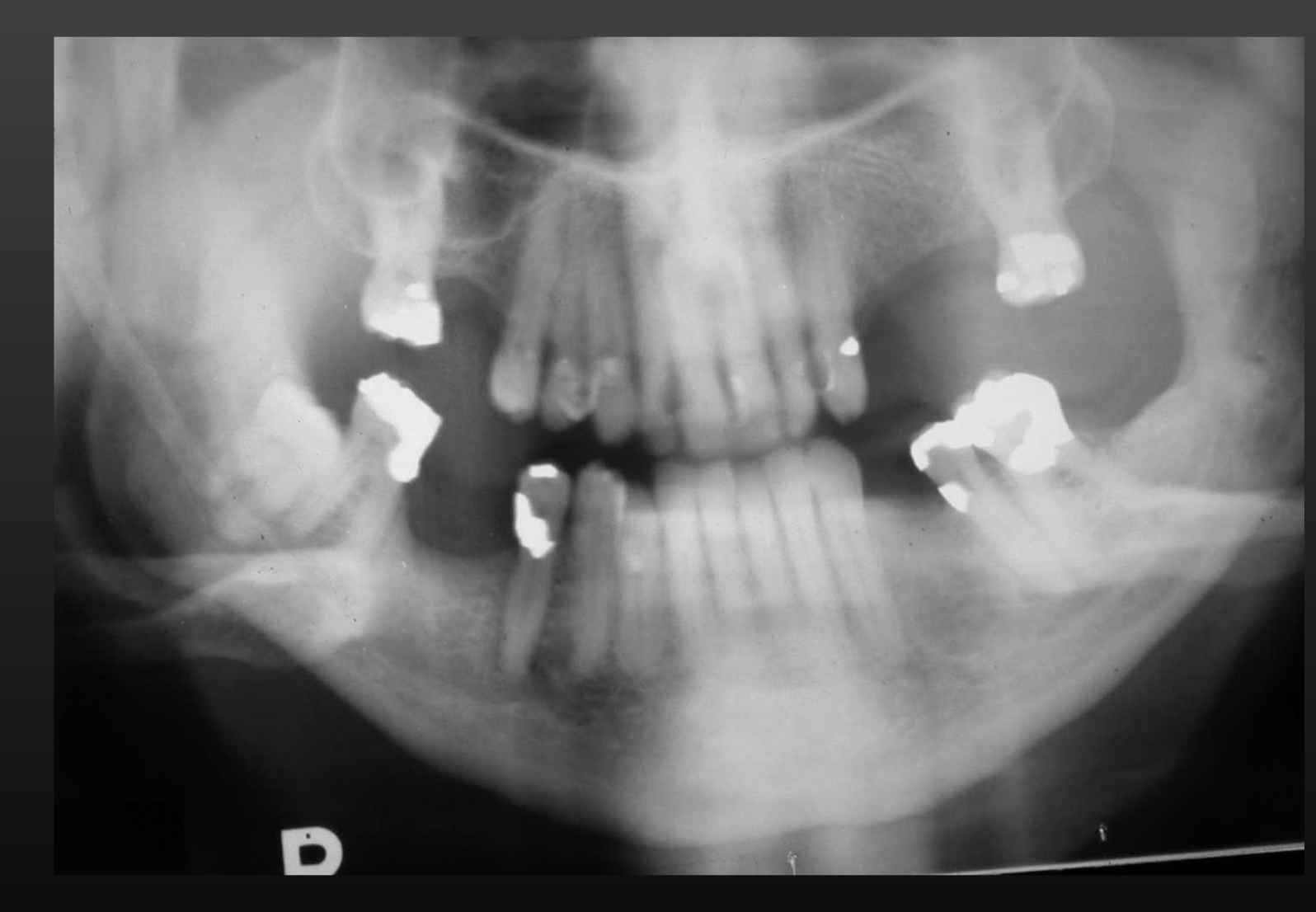
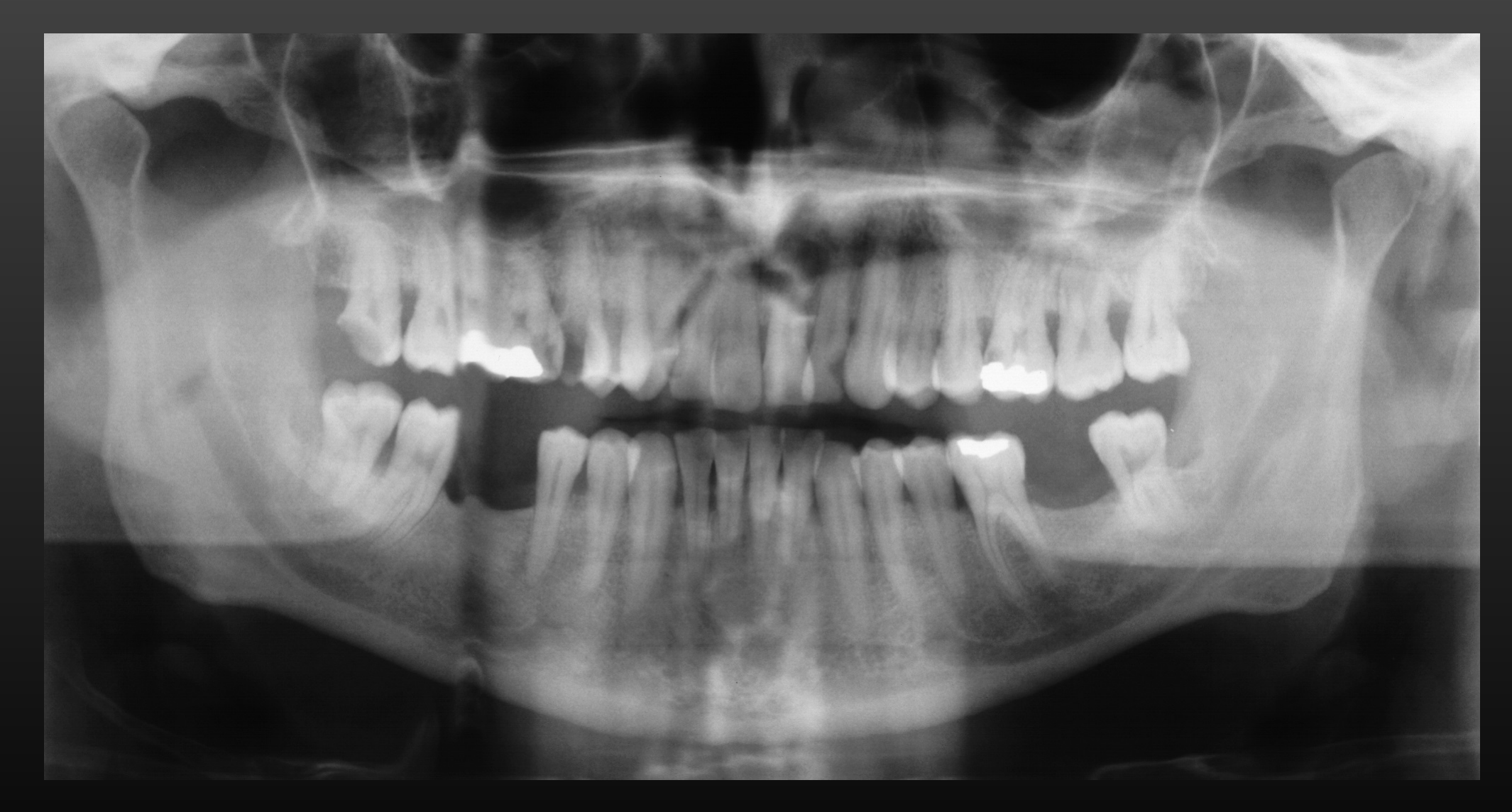
| Chin up | Occlusal plane flat or inverted (reverse smile / frown); hard palate shadow over maxillary roots; condyles may not show; maxillary incisors blurred and magnified34 | Chin and occlusal plane tipped upward; Frankfort plane not parallel to the floor34 | Tragus-to-outer-canthus line parallel to the floor; occlusal plane slightly lower anteriorly3 |
| Chin down | Exaggerated smile line; teeth severely overlapped; symphysis may be cut off; condyles projected off the top edge; mandibular incisors blurred with short roots; hyoid higher34 | Chin tipped too low3 | As above: Frankfort plane parallel to the floor4 |
| Too far forward (anterior to trough) | Anterior teeth narrow (thin teeth); spine may be visible34 | Incisors in front of the bite-block notch3 | Incisal edges in the bite-block notch; focal trough light at the tip of the maxillary canine34 |
| Too far back (posterior to trough) | Anterior teeth wide (fat teeth); ramus may not be fully shown34 | Incisors behind the notch3 | Incisal edges in the bite-block notch3 |
| Head rotated | Molars and ramus wider on one side, narrower on the other; severe premolar overlap on the wide side. The side the head is turned to is magnified34 | Midsagittal plane not in the machine’s rotational midline3 | Midsagittal plane perpendicular to the floor, light centred on the nose4 |
| Head tilted | Face asymmetric; one side sits lower; condyles asymmetric in height41 | Midsagittal plane tilted4 | Midline perpendicular to the Frankfort plane4 |
| Tongue not on palate | Radiolucent band under the hard palate over the maxillary apices (palatoglossal / glossopalatal air space)34 | Air left between tongue dorsum and palate3 | Swallow, then hold the tongue flat against the palate (and don’t swallow during the scan)34 |
| Slumped spine / neck | Large midline radiopacity (cervical spine ghost) over the symphysis; may need a retake34 | Back not erect, neck flexed forward3 | Sit or stand tall, neck extended; gentle upward pressure at the mastoids; seat the patient if needed34 |
| Lips not closed | Dark radiolucent shadow around the anterior teeth4 | Lips apart4 | Close lips around the bite block4 |
| Movement | Horizontal movement gives deletion and doubling; step in the image; swallowing lifts the soft palate and hyoid4 | Patient moved during the ~10–20 s exposure4 | Explain and demonstrate the machine first; children look forward, not at the tube head34 |
| Lead apron | Radiopaque, cone-shaped artefact in the centre of the image45 | Apron or thyroid collar in the beam4 | Use an apron without a thyroid collar for OPG4 |
| Shoulder collision | Cone-cut appearance1 | Receptor or tube head hits the shoulder4 | — |
| Metal left in situ | Real image plus blurred ghost on the opposite side (see Ghost and Double Images)3 | Earrings, necklaces, hairpins, appliances, dentures34 | Remove all metal from the head and neck first3 |
- Quick rotation check: compare the mesiodistal width of the mandibular first molars on each side. The smaller side was too close to the receptor3
- Poor midline positioning is a common error, according to W&P3. The Oral Surgery workshop calls tongue-to-palate failure the most frequent1
- Don’t read a rotation asymmetry as a real facial asymmetry3
 |  |
 |  |
 |  |
 |  |
Magnification and distortion limits
- Don’t make linear or angular measurements. Distortion depends on beam angle, source–object distance, the path of the rotation centre, the object’s position in the trough and patient anatomy, so no preset magnification factor applies3
- Horizontal magnification depends on position in the trough. Lingual displacement (toward the source) makes structures wider; buccal displacement makes them narrower. The effect is much stronger anteriorly than posteriorly3
- A lesion can look like it is enlarging or healing on follow-up OPGs purely from positioning3
- Vertical magnification depends on source–object distance and is relatively constant on some units3
- The beam tilts slightly upward (caudocranial). Structures nearer the source project higher, so vertical relationships are unreliable. A lingual IAN canal projects closer to the molar apex than a buccal one at the same true height3
- In the posterior regions the buccal external oblique ridge and the lingual mylohyoid ridge both look sharp, even though they are millimetres apart3

Why an OPG does not replace BWs and PAs
- Lower resolution: small caries, fine marginal periodontium and early periapical disease are not shown as well as on PAs3
- OPG under 10 lp/mm vs about 20 lp/mm intraoral4
- Premolar proximal surfaces usually overlap, which interferes with caries diagnosis3
- The cervical spine can hide lesions in the incisor region; objects outside the trough may be distorted or missing3
- A full-mouth series adds little to general care when an OPG is taken as well. An OPG plus bitewings (± selected PAs) gives similar information to a full-mouth series3
- Best for broad coverage: trauma, third molars, large lesions, eruption and development (mixed dentition), and patients who cannot tolerate intraoral films3
- Dose: roughly four F-speed bitewings (skin exposure, film-screen) per W&P3; about two to three bitewings per L84
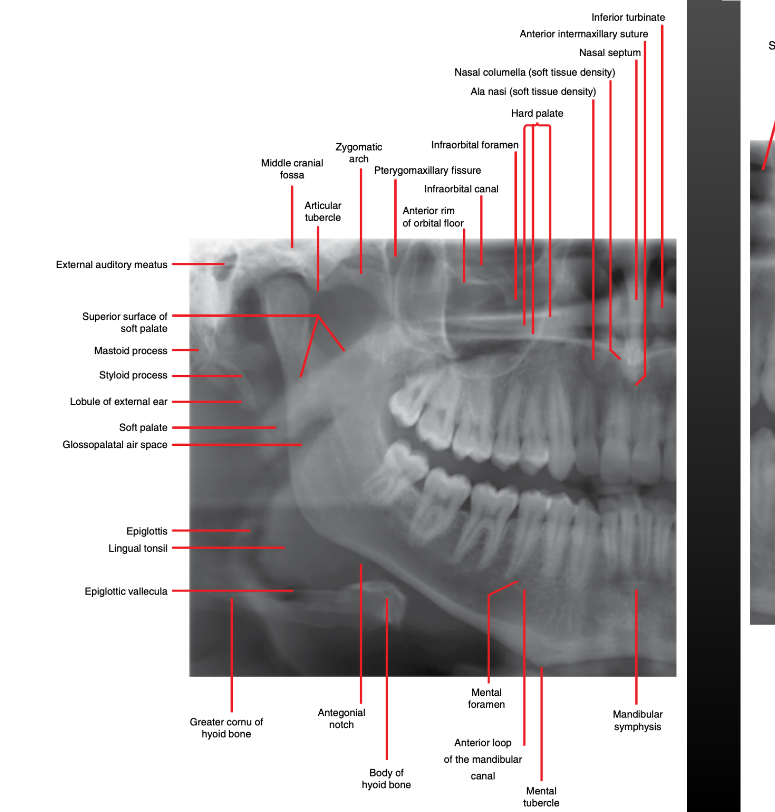
Normal Panoramic Anatomy
- Read it as if facing the patient: the patient’s right is on the viewer’s left3
- The “midface” includes the temporal, zygoma, frontal, maxilla, sphenoid, ethmoid, vomer, nasal, conchae and palate, so don’t call the whole region “the maxilla”3

Midface and Maxilla
| Landmark | Appearance on OPG | Tip |
|---|---|---|
| Hard palate | Horizontal radiopacity over the maxillary sinus posteriorly, often two lines; the upper, diffuse line is the opposite side. W&P conflicts on the lower line: receptor side (Fig 9.9 caption) vs tube side (text)3 | Chin up drops it over the maxillary roots3 |
| Nasal cavity and septum | Lateral border and anterior rim show as a radiopaque line anteriorly; septum in the midline; anterior nasal spine and incisive foramen may be seen3 | Incisive foramen: ovoid lucency between the central incisor roots7 |
| Turbinates (conchae) | Large homogeneous soft-tissue densities; coronal anteriorly, sagittal posteriorly; may overlie the sinuses and nasopharynx3 | Normal soft-tissue shadow3 |
| Maxillary sinus | Check posterior, anterior, floor and roof (= orbital floor, seen anteriorly) for complete cortex and symmetry. Posterior part is more opaque from zygoma superimposition3 | Asymmetry and septa are common variants. Look for retention cysts and mucosal thickening3 |
| Zygomatic process of maxilla / arch | Process arises over the maxillary first and second molars (innominate line); arch runs back to the articular tubercle and glenoid fossa34 | The zygomaticotemporal suture can mimic a fracture. Sinus pneumatisation of the process gives a corticated lucency over molar roots3 |
| Articular eminence | Radiopaque slope in front of the glenoid fossa7 | Mastoid pneumatisation can make it look multilocular (a normal variant)3 |
| Glenoid fossa | Concavity of the temporal bone above the condyle7 | Same “soap-bubbly” pneumatisation variant3 |
| Pterygomaxillary fissure | Inverted teardrop behind the maxilla; pterygoid plates form its posterior border3 | Check both sides. Loss of its anterior border (the posterior maxilla) suggests sinus mucocele or carcinoma3 |

 | 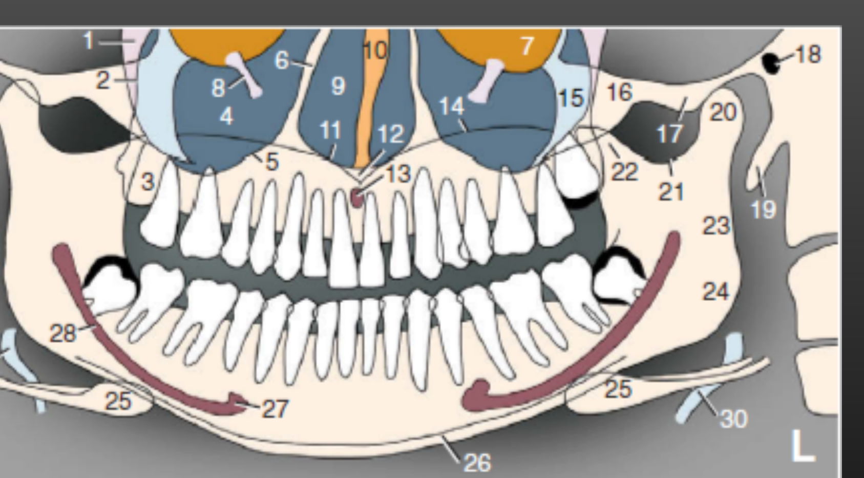 |
Legend for both numbered images 1
1 pterygomaxillary fissure · 2 posterior border of maxilla · 3 maxillary tuberosity · 4 maxillary sinus · 5 floor of maxillary sinus · 6 medial border of maxillary sinus / lateral border of nasal cavity · 7 floor of orbit · 8 infraorbital canal · 9 nasal cavity · 10 nasal septum · 11 floor of nasal cavity · 12 anterior nasal spine · 13 incisive foramen · 14 hard palate / floor of nasal cavity · 15 zygomatic process of maxilla · 16 zygomatic arch · 17 articular eminence · 18 external auditory meatus · 19 styloid process · 20 mandibular condyle · 21 sigmoid notch · 22 coronoid process · 23 posterior border of ramus · 24 angle of mandible · 25 hyoid bone · 26 inferior border of mandible · 27 mental foramen · 28 mandibular canal · 29 cervical vertebrae · 30 epiglottis

Mandible
| Landmark | Appearance on OPG | Tip |
|---|---|---|
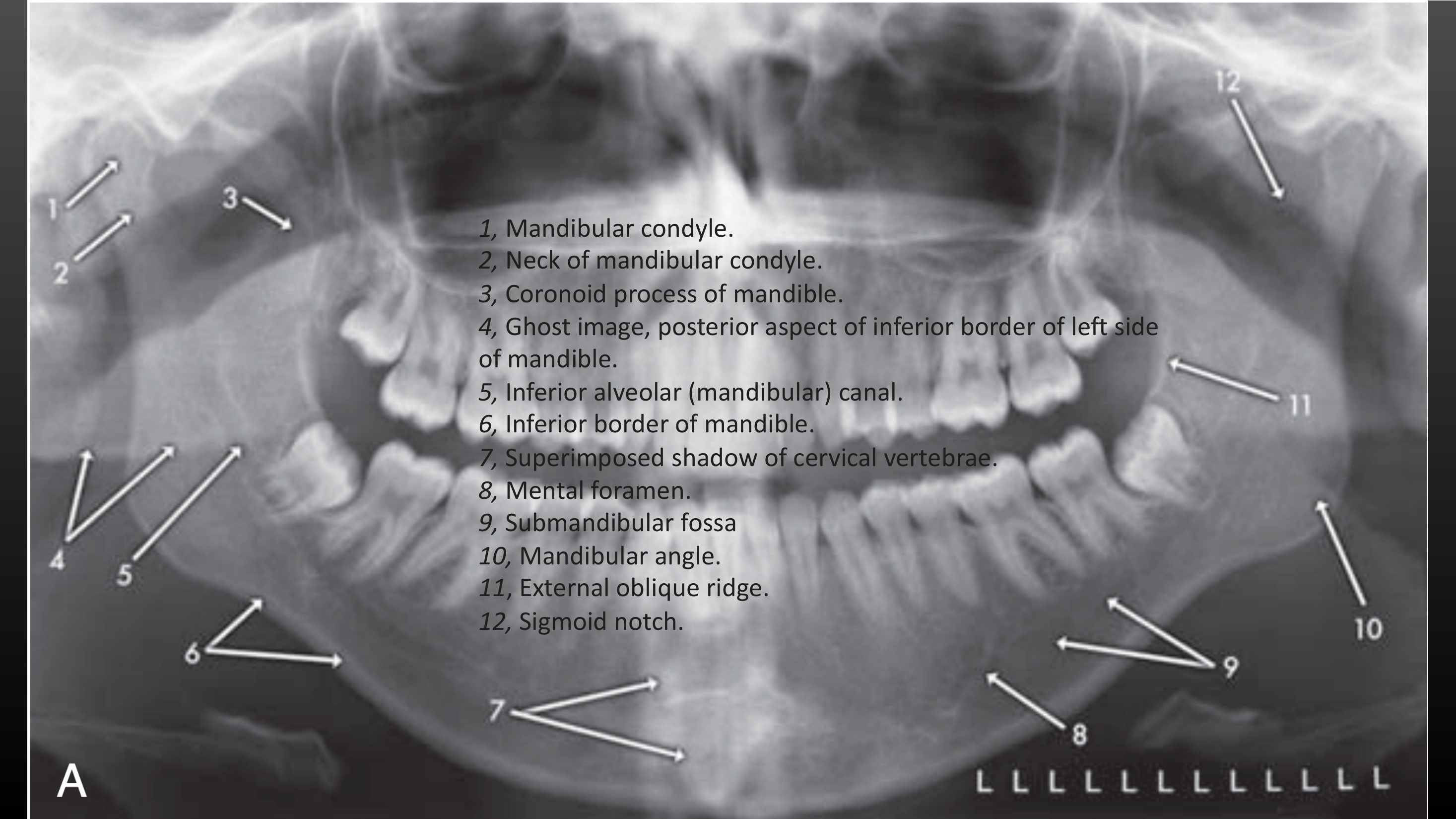
| Condyle | Sits slightly anteroinferior because the patient protrudes onto the bite block3 | Gross bony change only: no disc or soft tissue; CBCT/MDCT for bone, MRI for disc3. The articular cortex is not seen until about age 187 |
| Coronoid process | Anterior process of the ramus: no. 22 on the numbered images below, next to the sigmoid notch (21)4 | Part of the fixed mandible survey: condyle → coronoid → ramus → body/angle → anterior sextant3 |
| IAN (mandibular) canal | Radiolucent band with uniform width or gentle taper, from the mandibular foramen to the mental foramen3 | If only one wall shows, it is usually the inferior one. Less distinct at the first molar and premolars. Expansion suggests neurovascular pathology3 |
| Mandibular foramen | Canal entry in the ramus3 | Slight widening where the canal enters the body is normal3 |
| Mental foramen | Oval lucency where the canal rises, typically near the second premolar apex37 | Anterior loop may run several mm in front of it (implant planning)3 |
| Inferior border | Smooth, continuous cortex, ≥3 mm thick in adults, symmetric3 | Step = fracture. Thinning = expansile lesion or systemic disease3 |
| Submandibular fossa | Relative lucency below the mylohyoid ridge at the molar–premolar roots3 | Normal variant, not a lesion3 |
| Hyoid | Projected below or onto the inferior border; casts double and ghost images3 | Rises when the chin is down or the patient swallows4 |
| Styloid process | Spike from the temporal bone behind the ramus; 5–50 mm long7 | Stylohyoid ligament calcification is reported in up to 30%7 |
| Cervical spine | Midline and lateral opacity from double and ghost images3 | More opaque midline is normal; worse with slumping or pronounced lordosis3 |
 |  |
Soft Tissue and Air Spaces
| Landmark | Appearance on OPG | Tip |
|---|---|---|
| Tongue | Soft-tissue arch under the hard palate, roughly from one mandibular angle to the other3 | If it is off the palate, an air band shows over the maxillary apices3 |
| Soft palate | Soft-tissue band running back from the hard palate over each ramus3 | Lifts into the nasopharynx on swallowing4 |
| Pharyngeal airway | Radiolucent bands: nasopharynx (behind turbinates, above the hard palate), velopharynx (behind the soft palate), oropharynx (below the uvula)3 | Oropharyngeal and nasopharyngeal shadows cross the ramus. Note narrowing of the posterior pharyngeal wall34 |
| Ear lobe | Soft-tissue density over the ramus region3 | Also check for earrings and their ghosts3 |
| Epiglottis / thyroid cartilage | Epiglottis lies behind the tongue dorsum; thyroid cartilage low in the neck3 | The superior thyroid cartilage can be mistaken for a vascular calcification3 |
| Other | Lip line (mid-image), nose, nasolabial folds, posterior pharyngeal wall3 | OPGs can show soft-tissue swellings and tonsilloliths4 |
 |  |
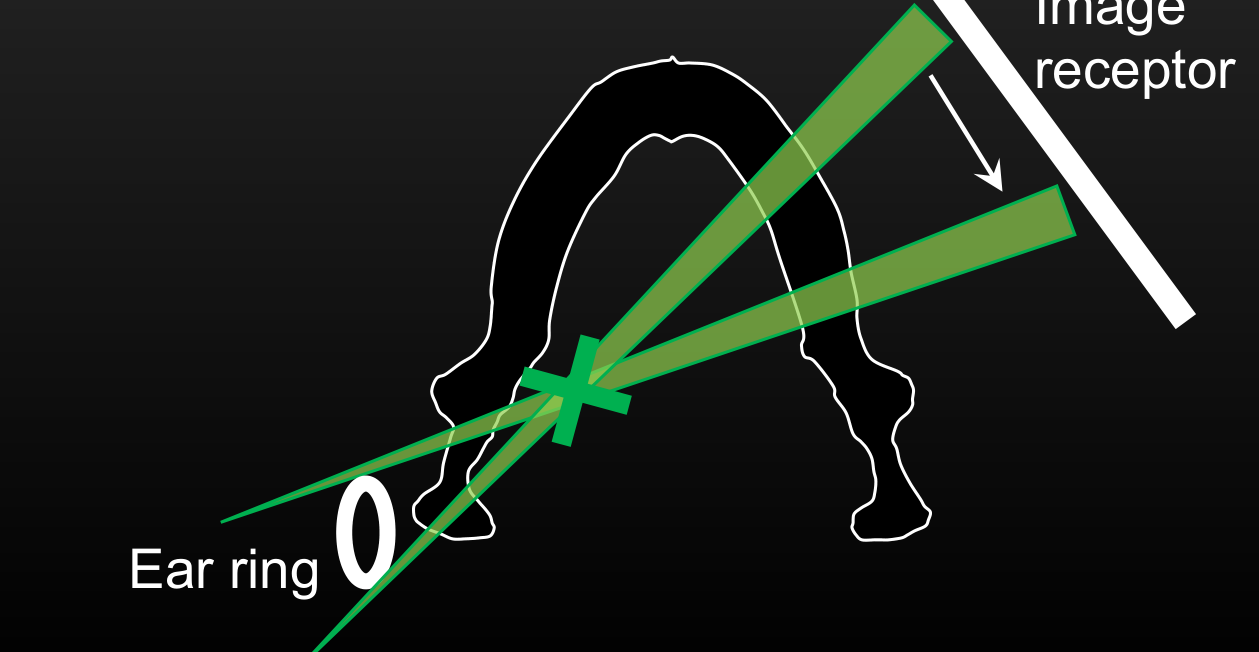
Ghost and Double Images
| Real | Double | Ghost | |
|---|---|---|---|
| Where the object is | Between the rotation centre and the receptor3 | Behind the rotation centre, crossed by the beam twice3 | Between the x-ray source and the rotation centre3 |
| Appearance | Sharp if in the trough, blurred if outside it3 | Appears on both sides; mirror images in the same position on each side34 | Opposite side, higher, larger (magnified) and blurred34 |
| Examples | Teeth, jaws on the receptor side3 | Hyoid, epiglottis, cervical spine3; L8 adds the palate4 | Opposite ramus and mandible, hyoid, spine, palate, earrings, necklaces, hairpins3 |
- Some zones produce both double and ghost images (where the two zones overlap)3
- Ghosts of metal can hide anatomy, hide pathology, or mimic pathology3
- Ghosts look like the real object but sit higher and on the other side. Remove radiodense objects before exposure4
 |  |
Common mimics
| Mimic | What it looks like | How to tell |
|---|---|---|
| Hyoid | Opacity below or onto the inferior border of the mandible, plus double and ghost images3 | Double image: mirror images in the same position on both sides34 |
| Earring / jewellery ghost | Blurred, magnified opacity on the opposite side, higher up, often over the ramus34 | Ghost is higher, larger and blurred; the sharp real image is on the opposite side34 |
| Airway lucencies | Palatoglossal band over the maxillary apices; pharyngeal air over the ramus3 | Palatoglossal band is a tongue-position error (see table above); pharyngeal shadows cross the ramus34 |
| Spine ghost | Midline opacity over the symphysis and incisors3 | Worse with slumping; can hide incisor-region lesions3 |
| Palate | Two horizontal lines over the sinus or maxillary roots3 | The upper, diffuse line is the opposite side. Which side the lower line is differs within W&P (see Midface table)3 |
| Opposite mandible ghost | Ghost of the opposite mandible and inferior border34 | Higher and blurrier than the real border34 |
| Thyroid cartilage | Opacity low in the neck3 | Can be mistaken for vascular calcification3 |
 |  |
Dental Findings on OPG
What the OPG is good at
Tooth count and development
| Check | What to look for | Source |
|---|---|---|
| Count and chart | Every erupted and developing tooth in FDI. Absent teeth: developmental or extracted? | 39 |
| Dental age | Presence/absence and developmental stage of the permanent teeth vs chronological age | 38 |
| Mixed dentition | Primary teeth, erupting successors, tooth germs. Development complicates interpretation | 83 |
| Ectopic canines | OPG is the usual first image when the canine is not palpable by 9–10 years | 8 |
| Patient positioning | Anterior teeth too wide or narrow → patient malpositioned in the focal trough. One side wider → sagittal plane rotated | 3 |

Number and position anomalies
| Finding | Key facts | Source |
|---|---|---|
| Hypodontia | 3–10% of people (excluding third molars). Most often missing: third molars, then mandibular second premolars, maxillary laterals and mandibular central incisors | 9 |
| Delayed vs missing | Some teeth (especially mandibular second premolars) can erupt years late, so late development is not the same as absence. Call a tooth missing only when it isn’t seen clinically or on imaging and there’s no history of extraction | 9 |
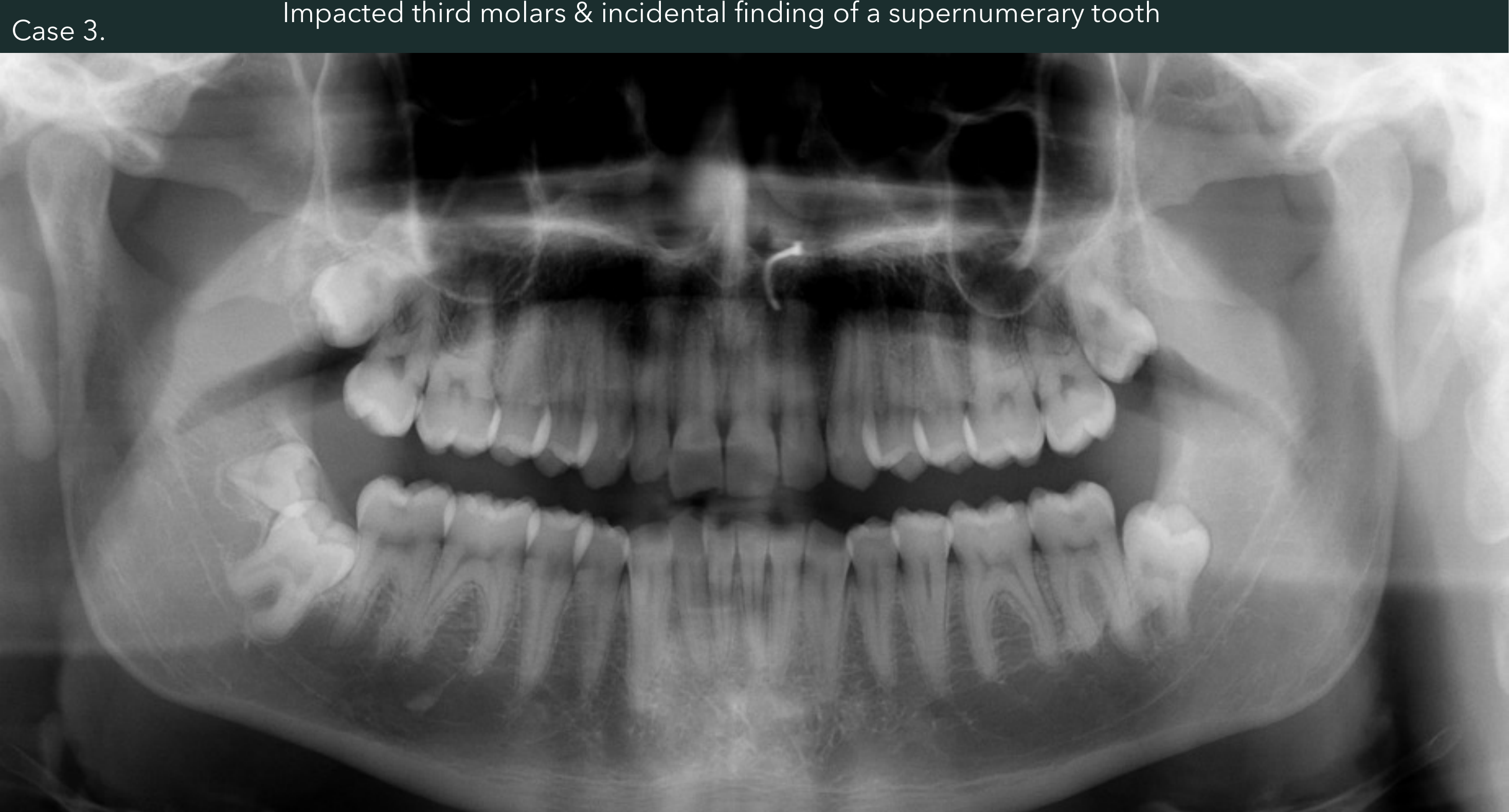
| Supernumerary teeth | 1–4% of people. Single ones are most common as a mesiodens (anterior maxilla) or a distodens (maxillary molar region). Multiple ones are most common in the mandibular premolar region, often lingual | 9 |
| Supernumerary on OPG | In the anterior maxilla a mesiodens can be hidden by the cervical spine shadow, or distorted if it lies outside the focal trough. Use CBCT to find the exact position and count | 98 |
| Transposition | Adjacent teeth swapped. The most common pair is the canine and first premolar | 9 |
| Impaction | Most often impacted: third molars, then maxillary canines, then mandibular premolars | 10 |

Caries, restorations, endo and implants
| Finding | What the OPG shows | Limits | Source |
|---|---|---|---|
| Caries | Gross caries only | Premolar contacts overlap, so interproximal caries is hard to read. Low sensitivity, especially anteriorly. Use BW for caries | 38 |
| Restorations | Record existing crowns and fixed work | Lower resolution than intraoral films, so check detail on a BW or PA | 3 |
| Endo | Record root fillings. Gross periapical disease may show | Subtle periapical change needs a PA | 3 |
| Implants: planning | First-line imaging for an implant patient. Gives an overview of sinus floors and the IA canals | Distortion is unpredictable and there are ghost images. No buccolingual information. CBCT is the modality of choice before surgery | 11 |
| Implants: follow-up | Use a PA if symptom-free. An OPG may be indicated after extensive implant work | CBCT is poor for peri-implant bone because of metal artefact. OPG and PA are better | 1112 |
Third Molars and the IAN Canal
What the OPG can't show
What to record for every impacted third molar
- Orientation of the molar; number and shape of roots3
- Relation to the mandibular canal, floor and back wall of the maxillary sinus, the tuberosity and neighbouring teeth3
- Any changes in the bone around the crown or roots (follicle, cyst)3
- The canal usually runs buccal to the third molar, but it is still close to the roots10
- Worked example: 48 pushed down to the inferior border (the case note attributes this to a ruptured dentigerous cyst) and a mesioangular 3814
Seven OPG signs of IAN proximity
| # | Sign | Note | Source |
|---|---|---|---|
| 1 | Darkening of the roots | Root is less radiopaque where it crosses the canal, meaning the canal has notched the root or vice versa | 131 |
| 2 | Interruption of the white line | The canal’s corticated border is lost where it crosses the root | 131 |
| 3 | Diversion of the canal | 131 | |
| 4 | Dark and bifid apex | 131 | |
| 5 | Deflection of the roots | 131 | |
| 6 | Narrowing of the canal | 131 | |
| 7 | Narrowing of the roots | 131 | |
| + | Juxta-apical area | Rare but highly prognostic. Dark area just distal to the root | 131 |
- Most significant: interruption of the white line and darkening of the root1
- Consent figures: temporary paraesthesia 0.5–5%, permanent <1%131
 |  |
Classification
Mandibular: Winter (angulation vs the second molar’s long axis)1310
| Class | Notes |
|---|---|
| Mesioangular | Most common: about 42% per the OS LOs1, about 43% per OMFS10. Least difficult, especially when only partly impacted10 |
| Vertical | Second most common. Distal side often covered by ramus bone10 |
| Horizontal | Uncommon. Harder than mesioangular10 |
| Distoangular | Uncommon. Hardest of the four main types10 |
| Transverse (buccolingual) | Rare. Buccal or lingual position needs a cross-sectional occlusal view or CBCT10 |
| Inverted | Rare13 |
Mandibular: Pell & Gregory1310
| Depth (highest point of the third molar) | Ramus space (from the distal of the second molar to the anterior ramus) |
|---|---|
| Level A: at or above the occlusal plane | Class 1: space > mesiodistal width of the third molar crown |
| Level B: below the occlusal plane, above the second molar’s cervical line | Class 2: space < crown width |
| Level C: below the second molar’s cervical line | Class 3: tooth embedded in the ramus |
- Write Winter and Pell & Gregory together, e.g. mesioangular / Class 1 / Level A = easiest; distoangular / Class 3 / Level C = hardest10
 |  |
Maxillary: Archer (angulation 1–7 plus depth a–e)131
| Angulation | Depth (occlusal surface of the third molar relative to the second molar) |
|---|---|
| 1 Mesioangular · 2 Distoangular · 3 Vertical · 4 Horizontal · 5 Buccoangular · 6 Linguoangular · 7 Inverted | a level with the occlusal surface · b mid-crown · c cervical line · d along the root · e above the root |
- A7E is much harder than A1A13
- Maxillary third molar angulation: vertical 63%, distoangular 25%, mesioangular 12%10
- Sinus risk on OPG: no overlap with the sinus suggests it is likely safe. Darkening of the roots means a close sinus relationship. A break in the sinus floor cortex means high risk of oroantral communication13
- Assessing the upper third molar on PA or OPG is hard because of projection13
When to step up to CBCT
- Roots very close to or overlapping the IA canal on the OPG103
- Any of the proximity signs above → consider CBCT for consent and planning. CBCT is the default step-up from the OPG in oral surgery1
- Third molar assessment is a listed CBCT indication15
- Justify the dose: use CBCT only when a PA or OPG can’t answer the question16
- The upper third molar–sinus relationship is shown best on CBCT, but CBCT isn’t needed in every case13
Periodontal Overview
Role of the OPG in perio
| Read on the OPG | Criteria | Source |
|---|---|---|
| Normal crest | 0.5–2.0 mm apical to the CEJ. Measure bone loss from this level, not from the CEJ | 17 |
| Horizontal bone loss | Crest height is lost but the crest stays parallel to the line joining adjacent CEJs. Buccal and lingual plates and interdental bone lost together | 17 |
| Vertical (angular) defect | Loss localised to one or both root surfaces of one tooth. Seen in Stage III–IV | 17 |
| Pseudo-angular crest | When adjacent CEJs are at different heights, the crest looks angled but is still parallel to the CEJ line. Not a vertical defect | 17 |
| Localised vs generalised | Localised: <30% of teeth affected. Generalised: ≥30% | 12 |
| Report | Severity, pattern (horizontal/vertical), distribution (localised/generalised) | 12 |
- Staging: express radiographic bone loss as a % of root length at the worst tooth: <15% (Stage I), 15–33% (Stage II), middle third and beyond (Stage III–IV)17. Full criteria and grading: Periodontitis Staging and Grading
OPG vs BW, PA and CBCT
| OPG | BW / PA | CBCT | |
|---|---|---|---|
| Strength | Quick, comfortable. One-image overview of both arches17. Lower dose than full-mouth PAs12 | BW: beam nearly at right angles to the teeth, so the CEJ-to-crest distance is most accurate17 | 3D view of craters, furcations, buccal/lingual plate loss and complex vertical defects17 |
| Weakness | Superimposition, distortion, lower resolution, especially anteriorly. Positioning errors17. Uneven, unpredictable magnification12 | 2D: craters and furcations are poorly shown12 | Higher dose. Not recommended for follow-up12 |

TMJ, Condyles and Maxillary Sinus
Condyles and TMJ on the OPG
| OPG can show | OPG can’t show | Source |
|---|---|---|
| Gross change: asymmetry, large erosions, large osteophytes, tumours, fractures. Left–right comparison. Dental causes of TMJ pain | Condylar position or function, because the mouth is slightly open and protruded. Mild erosions. The articular eminence is hidden by the skull base and zygomatic arch | 18 |
- Screening only: for some patients it’s the only image needed before conservative treatment. Add more imaging when detail is needed. The “TMJ programmes” on panoramic machines are still too thick and oblique18
- Condylar hyperplasia: enlarged condyle, which may include the ramus and body on that side18
- Condylar hypoplasia: small condyle, short ramus, deep antegonial notch18
- Coronoid hyperplasia: tips ≥1 cm above the lower rim of the zygomatic arch. Best seen on OPG and CT18
- Bifid condyle: usually an incidental OPG finding18
- Progressive condylar resorption: flattened, elongated front of the condyle with a backward tilt (“toadstool”). Mostly females in their teens18
- Air cell in the articular eminence = normal variant18
- Condylar bony prominence on an OPG: differentials are osteochondroma vs osteophyte from degenerative joint disease → MSCT5
 |  |
Maxillary sinus on the OPG
Imaging pathway
The PA shows the most detail at the apices and sinus floor. The OPG covers more of the sinus (floor, back wall and front-inner wall). Ghost images and overlap make it hard to compare the left and right floors. If the findings are positive, refer to an OMF radiologist19.
| Finding | Appearance | Next step | Source |
|---|---|---|---|
| Mucosal thickening (mucositis) | Normal mucosa (<1 mm) is invisible. Thickened mucosa shows as a smooth, non-corticated radiopaque band following the sinus wall. If centred over a diseased tooth, it’s odontogenic | Often an incidental finding needing no further investigation. Odontogenic thickening resolves days to weeks after the tooth is treated | 19 |
| Retention pseudocyst | Smooth dome, non-corticated, homogeneous. Usually on the sinus floor. Adjacent lamina dura and PDL intact | No treatment; it resolves by itself. Not linked to dental disease | 19 |
| Odontogenic cyst vs pseudocyst | Only the odontogenic cyst has a corticated edge. It pushes the sinus floor up | Workup of the cyst | 19 |
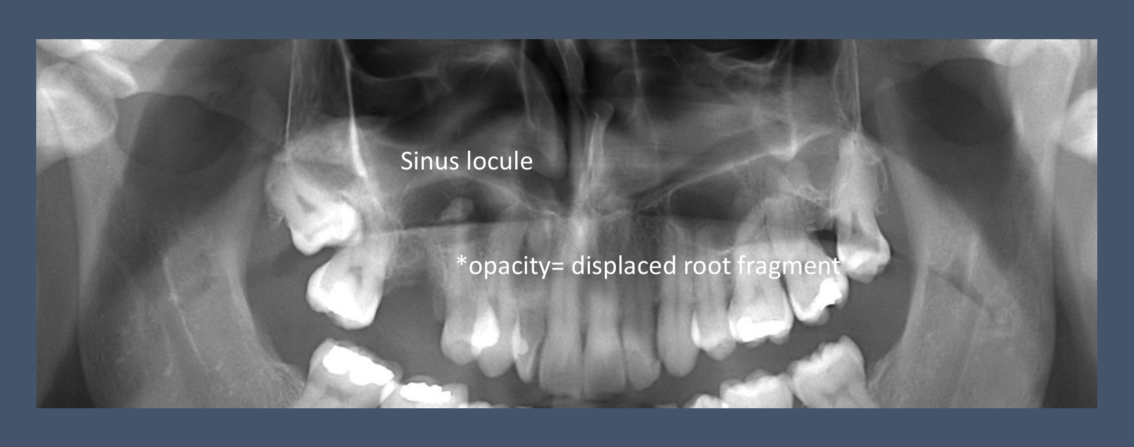
| Root in sinus | Radiopaque fragment, usually near the floor (gravity). If lamina dura and PDL are visible, it’s still in the alveolus. A canal or pulp chamber distinguishes a root from an antrolith | CBCT to check the sinus wall and locate the fragment | 192 |
| Opacification | Diffuse mucosal thickening or complete opacification. An air–fluid level means secretions. Compare with the other side | Any hint of bone destruction → MDCT or MRI. Suspect cancer in an older patient with first-time chronic sinusitis | 19 |
| Mimic | A sinus loculus draping over the roots can look like a lesion | Recognise it as normal anatomy | 19 |
 |  |
Trauma
One OPG is never enough to rule out a fracture
A fracture is a plane, not a line. If the beam doesn’t pass along that plane, a single 2D image can miss it20. Add a second view at right angles, e.g. an open-mouth Towne, or CBCT. This matters most for condylar neck greenstick fractures, whose displacement is in the coronal plane1820. OMFS recommends two or more of: OPG, open-mouth Towne, PA mandible, lateral oblique21.
Fracture signs on the OPG
- Sharp radiolucent line(s) within the bone outline. A line that runs beyond the mandible is probably a superimposed structure20
- Change in shape: asymmetry, a step in the occlusal plane
- Break in a cortical border, including a “step defect” where fragments are displaced
- Increased radiopacity where fragments overlap. On the condylar neck this is the “overlapping fragment sign”18
- Undisplaced fractures through both buccal and lingual plates can look like two lines meeting at the edges20
Sites and patterns
| Point | Detail | Source |
|---|---|---|
| Sites | Dentoalveolar, symphysis/parasymphysis, body, angle, ramus, coronoid, condyle | 2022 |
| Frequency | W&P: symphysis, body and angle are most frequent, then condyle/neck and ramus. DI L4 (Radiopaedia): condyle 30%, angle 25%, body 25%, parasymphysis 15%, ramus 3%, coronoid 2% | 2022 |
| Indirect fractures | A blow to one side can fracture the opposite condylar neck. A blow to the anterior mandible often fractures one or both condylar necks, so check both condyles | 20 |
| Condylar fractures | Neck fractures are more common than head fractures. The head is pulled anteromedially (and inferiorly) by lateral pterygoid. About half have a body fracture too | 2018 |
| Favourable vs unfavourable | Depends on whether masseter or medial pterygoid pulls the fragments together or apart along the fracture line | 20 |
 |  |
What the OPG misses
- Undisplaced condylar head fractures: often impossible to see20
- Greenstick condylar neck fractures: the change is in the coronal plane18
- Anterior dentoalveolar injuries: resolution too low, so use intraoral films (two PAs at different horizontal angles, plus the opposing arch)20
- Mimics: the air gap between tongue and soft palate across the angle20; the soft palate over the ramus20; the zygomaticotemporal suture looking like a break in the lower border of the zygomatic arch20

Escalation
| Scenario | Modality | Source |
|---|---|---|
| Suspected mandibular fracture, conscious patient | OPG first, plus occlusal (body/alveolus) or Towne (condyle) | 20 |
| Limited dentoalveolar trauma | Small-FOV CBCT is the modality of choice (AAOMR-AAE) when 2D is inconclusive | 2022 |
| Condylar fracture | CT/CBCT shows the 3D relation of the condylar head to the fossa | 2018 |
| Multiple or complex mandibular fractures | CBCT or, preferably, MDCT | 20 |
| Midface / multiple facial bones | CT (MDCT preferred over CBCT for soft tissue). CT is the gold standard | 202221 |
| TMJ capsule or disc injury | MRI | 20 |
- CBCT suits dentoalveolar and jaw fractures but has a small field of view, poorer soft tissue and more noise than CT22
Jaw Lesions: Description and Differentials
Describing a lesion
Describe first, name second
Work through the five W&P steps in order and write each one down before you commit to a diagnosis. The analysis only narrows the lesion to a disease category (inflammation, cyst, benign neoplasm, malignancy, bone dysplasia, vascular, metabolic, trauma), and then you decide how to proceed23
| Step | What to record | Key terms and what they imply |
|---|---|---|
| 1. Location | Epicentre (anatomic position); localized or generalized; unilateral or bilateral; single or multifocal23 | Odontogenic lesions sit in the alveolar process, superior to the IAC2425. Epicentre coronal to an unerupted crown means the lesion is pericoronal, e.g. dentigerous cyst24 |
| 2. Periphery and shape | Well or poorly defined; border type; shape23 | Punched out: sharp, no bone reaction, e.g. myeloma. Corticated: thin uniform RO line, seen in cysts and benign tumours. Sclerotic: wider, non-uniform reactive bone, e.g. PCOD. Soft tissue capsule: RL rim around an RO centre, e.g. odontoma, cementoblastoma. Blending: gradual trabecular transition, e.g. sclerosing osteitis, fibrous dysplasia. Invasive: few or no trabeculae at a wide, enlarging RL margin, i.e. malignancy23 |
| Shape23 | Circular / hydraulic (balloon-like) suggests a cyst. Scalloped (contiguous arcs) is seen in cysts such as OKC, simple bone cysts and some benign tumours. Irregular23 | |
| 3. Internal structure | Totally RL, totally RO, or mixed (describe the pattern)23 | Septa, abnormal trabecular pattern, dystrophic calcification, amorphous bone, tooth structure23 |
| 4. Effects on surrounding structures | Teeth, lamina dura, PDL; IAC and mental foramen; antrum; surrounding bone density and trabeculae; outer cortex and periosteal reaction23 | Tooth displacement and resorption point to a slow, benign process. Irregular PDL widening with lamina dura loss points to malignancy. IAC displaced superiorly points to fibrous dysplasia. Onion-skin periosteal new bone is mostly inflammatory; spiculated / sunray bone suggests prostate metastasis or osteosarcoma23 |
| 5. Interpretation | Normal vs abnormal → developmental vs acquired → disease category → how to proceed (more imaging, biopsy, watchful waiting, treatment)23 | Weigh the features: an invasive, destructive border outweighs benign-looking tooth effects23 |
- OPG checklist wording (L15): for any abnormal lucency or opacity, record location → border (well or ill-defined) and shape → internal architecture → effects on adjacent structures → surgical-sieve differentials5
Differential by category
| Category | Key OPG features | Examples |
|---|---|---|
| Inflammatory | RL centred on the apex, with loss of the apical lamina dura and a widened apical PDL. Periphery usually poorly defined, with a sclerotic zone that widens with chronicity26. OPG pattern: lucent → mixed lucent–sclerotic → sclerotic; may efface the sinus floor2 | Rarefying / sclerosing osteitis, Radicular Cyst (radiographically “rarefying osteitis” until biopsied)24; Osteomyelitis: posterior mandible, sequestra are the hallmark26 |
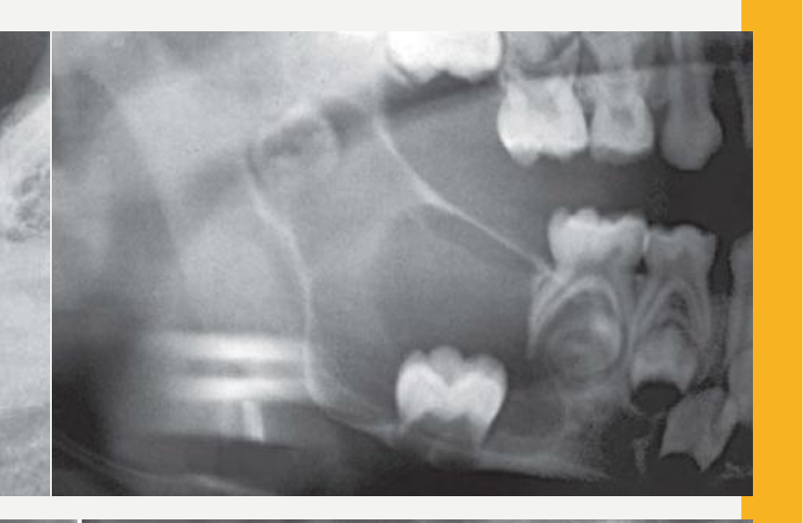
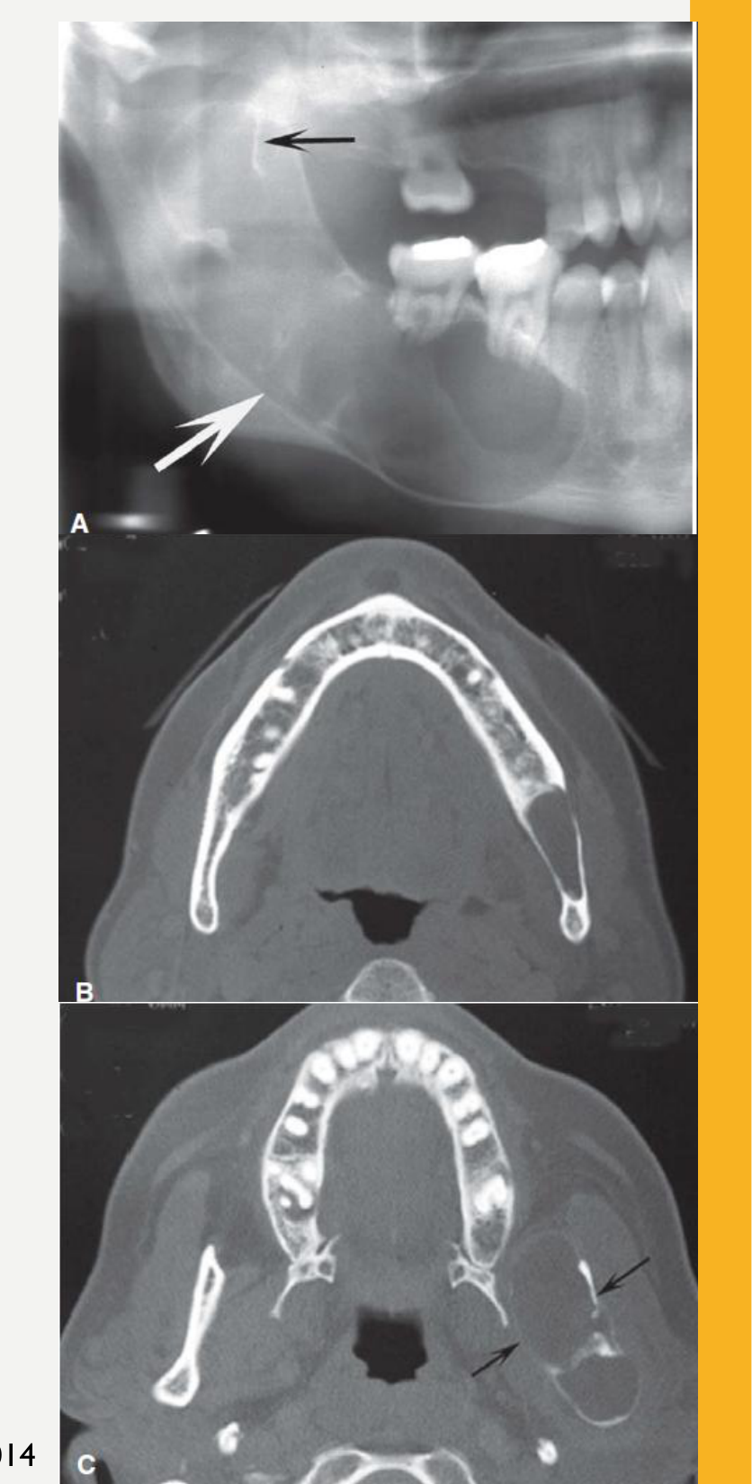
| Cysts | Well defined, thin uniform cortex, round/oval hydraulic shape, usually totally RL. Slow expansion with a smooth curved surface; they displace the IAC inferiorly and the antral floor upward; directional root resorption24 | Dentigerous Cyst: epicentre coronal to the crown, attaches at the CEJ. Odontogenic Keratocyst: posterior mandible/ramus, scalloped, tunnels with minimal expansion. Residual Cyst: in the former periapical region of a missing tooth. Nasopalatine Duct Cyst: midline, round or heart-shaped24 |
| Pseudocysts | Traumatic Bone Cyst (simple bone cyst): delicate cortex, scallops between roots, spares the lamina dura and follicle cortices, teeth vital. Stafne Defect: below the IAC, anterior to the antegonial notch, thick cortex, no effect on teeth24 | Stafne: well-defined lucency with a thick corticated border on OPG2 |
| Benign tumours | Well defined; cortex more obvious the slower the growth. Concentric expansion; teeth displaced or resorbed25 | Ameloblastoma: 80% in the molar–ramus region; coarse curved septa (soap bubble / honeycomb); extensive root resorption; greater expansion than OKC25. Cementoblastoma: RO mass fused to the root with a RL rim (see below)25 |
| Fibro-osseous / dysplasia | Periapical Cemento-Osseous Dysplasia: periapical, above the IAC; matures RL → mixed (cotton-wool) → RO with a RL rim; sclerotic border2723. Florid Cemento-Osseous Dysplasia: usually bilateral, both jaws27 (lecture: multifocal, ≥2 quadrants2). Fibrous Dysplasia: maxilla > mandible, blending border, ground-glass/granular, fusiform expansion, may displace the IAC superiorly27 | Early PCOD can’t be told from rarefying osteitis on imaging alone, so test vitality2627 |
| Malignant | Destructive: irregular shape, poorly defined invasive border, no cortication; cortex, sinus floor and IAC destroyed; PDL widened irregularly (Garrington sign); “floating teeth”; usually no periosteal reaction, but sometimes sunburst / hair-on-end28 | SCC by direct extension; metastases (breast/prostate may be sclerotic); multiple myeloma: punched-out, non-corticated, multiple, posterior mandible28. Refer urgently2 |
| Systemic / treatment-related | Osteoradionecrosis: history of radiotherapy, poorly defined resorption with sclerosis, uniform PDL widening without an epicentre; diagnosis relies on sequestra plus history26. Medication-Related Osteonecrosis of the Jaw: often no specific imaging finding; otherwise looks like chronic osteomyelitis (sclerosis + sequestra), PDL widening, thick lamina dura26 | Myeloma may cause generalized osteopenia; hyperparathyroidism is the metabolic look-alike28 |
- More radiolucent look-alikes: Radiolucent Lesion differentiation
 |  |
 |  |

Malignancy on the OPG: DI L6 additions
| Point | Detail |
|---|---|
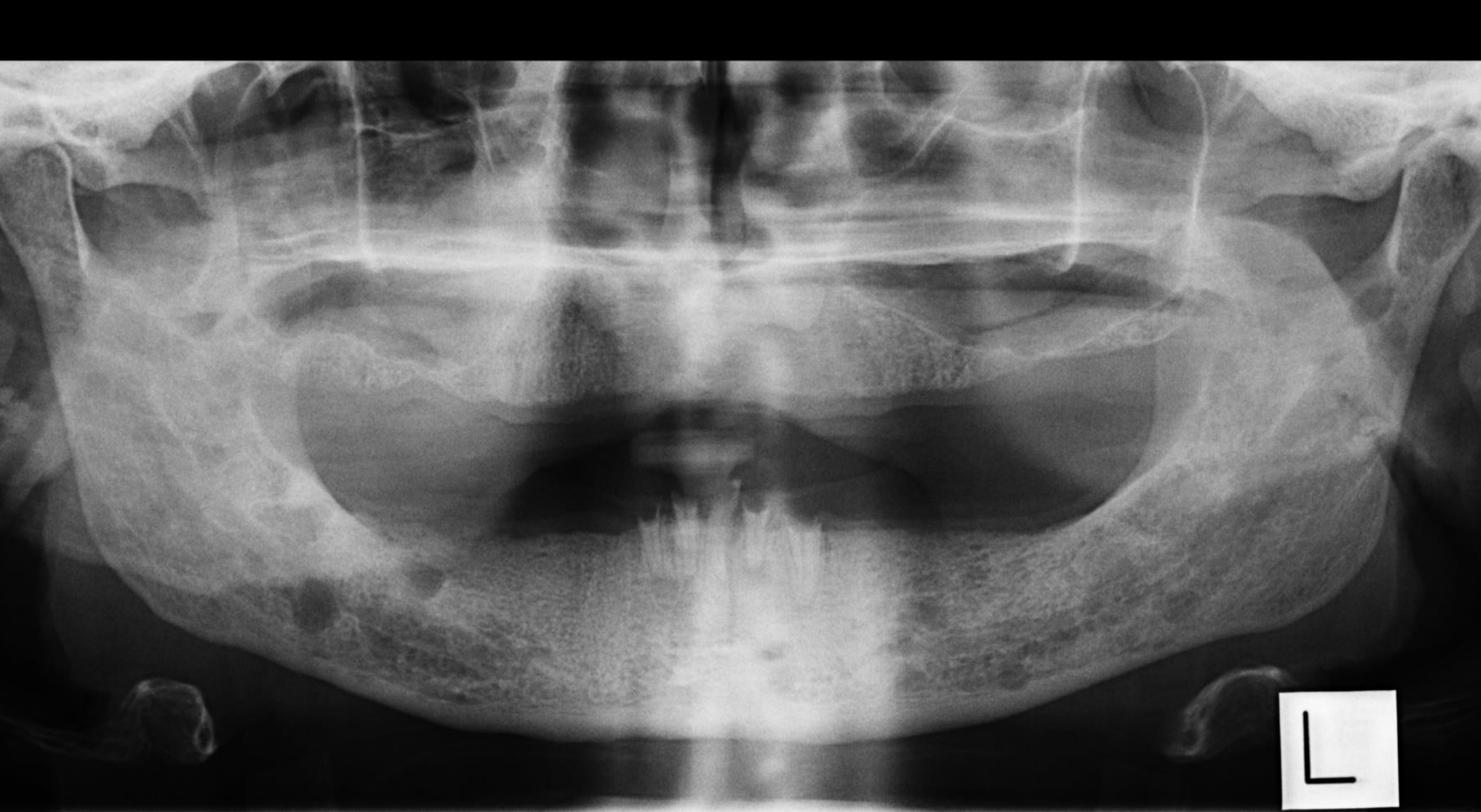
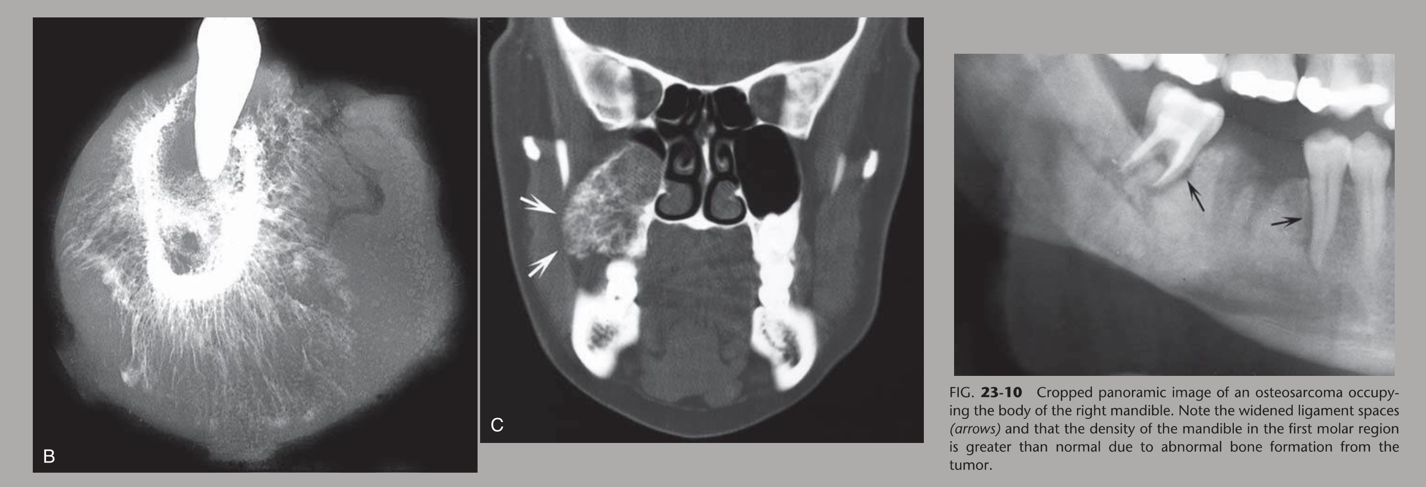
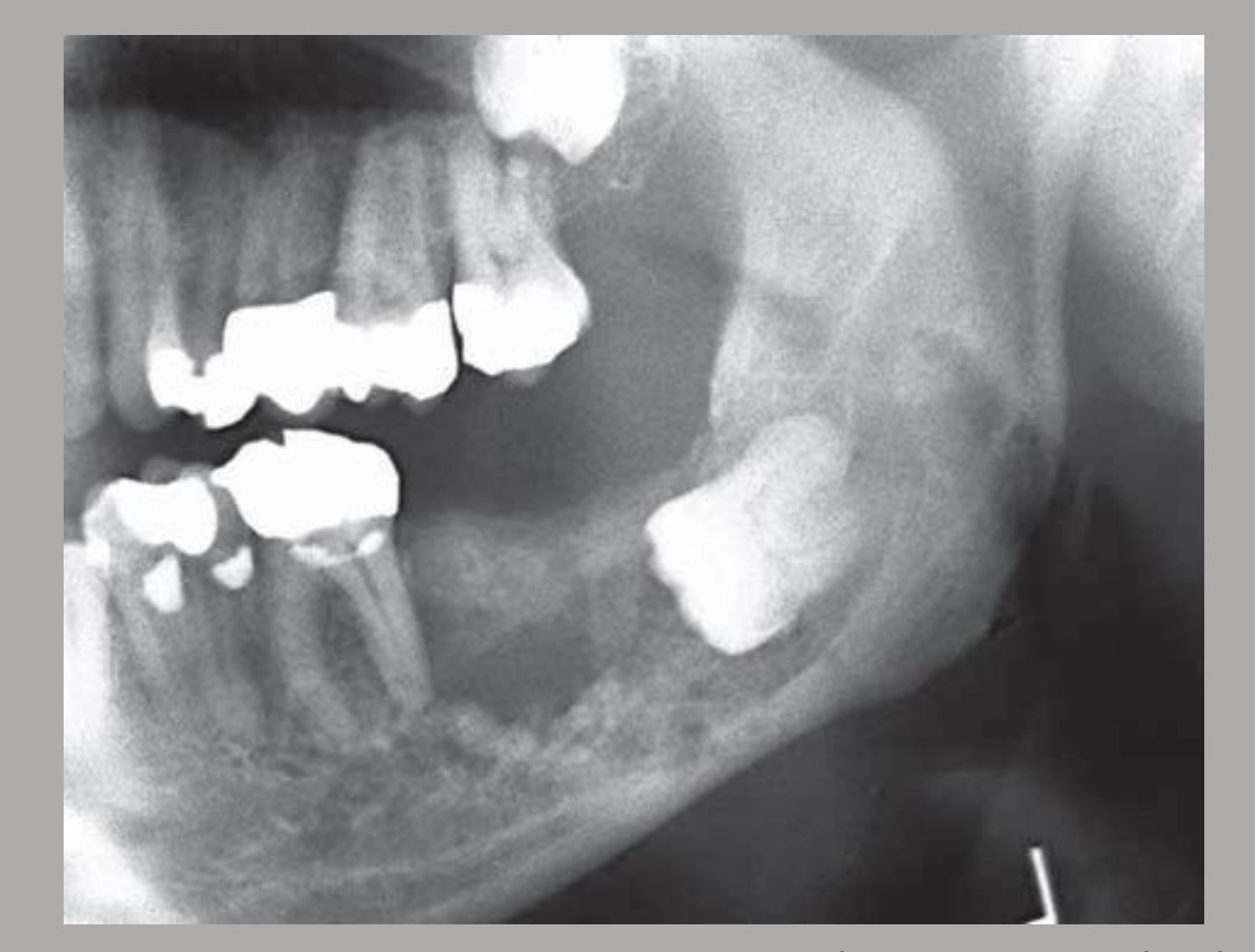
| Carcinoma vs sarcoma | Carcinomas are mainly destructive and lytic. Sarcomas can also form new bone, so they may be mixed density. Osteosarcoma may look denser than normal, while SCC is lucent. Both can destroy cortex, widen the PDL irregularly and erase the lamina dura and mandibular canal29 |
| SCC on OPG | Ill-defined “moth-eaten” lucency; destroyed cortices, lamina dura and antral floor; irregular PDL widening (“floating” teeth if extensive); irregular widening or loss of the mandibular canal29 |
| Carcinoma in a cyst | A carcinoma arising in a dentigerous cyst loses the cyst cortex and invades the adjacent bone29 |
| Central mucoepidermoid carcinoma | Uni- or multilocular, expansile, often well corticated, so it mimics ameloblastoma29 |
| Osteosarcoma | 75% occur before age 20. Cotton-ball opacities, sunburst periosteal reaction, enlarged neurovascular canals; can be subtle on an OPG29 |
| Lymphoma | Irregular PDL widening and loss of the lamina dura, cortex and sinus floor. Can look identical to SCC29 |
| Leukaemia (children) | Lucency is subtle because child bone is less dense. Look for loss of the lamina dura, loss of follicle cortices and displaced developing teeth29 |
| Myeloma | The most common primary malignant bone cancer in adults: multiple punched-out lytic lesions29 |
| Metastasis | Primaries are usually below the clavicle (breast, kidney, lung, prostate, thyroid, stomach). The mandibular body is the most common site. Paraesthesia is a key sign. Often multiple and ill-defined. Sclerotic from breast or prostate, lytic from renal or thyroid primaries. Multiple sclerotic foci in the condyles, rami or C-spine are suspicious (bone islands in the condyles are rare)29 |
| Next step | Aggressive features → urgent OMFS referral. MSCT with contrast, MRI (marrow, soft tissue, perineural spread, nodes) and PET/CT for staging29 |
 |  |
 | 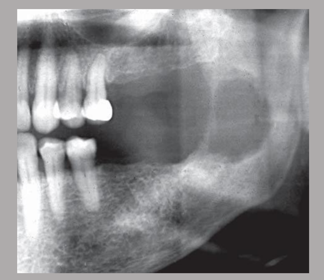 |
Cysts on the OPG
- Location: odontogenic cysts sit in the tooth-bearing jaw. In the maxilla they generally lie below the antral floor. They are rare in the condyle and coronoid30
- Shape follows surroundings: a cyst suspended in the sinus stays spherical; one against thick cortex develops a flattened edge30
- Secondary infection: the thin cortex becomes a thicker sclerotic border, or becomes less apparent30
- Long-standing cysts: sparse, dot-like dystrophic calcification; septa show as thin RO lines30
- 2D limits: expansion and cortical thinning are hard to see on PA, BW and OPG. CBCT or MSCT shows them30
| Cyst | OPG points from DI L3.1 (not in the table above) |
|---|---|
| Radicular Cyst | Maxilla > mandible; 40–50% in the anterior maxilla. Lecture threshold: a periapical RL <10 mm is called apical periodontitis. Curved root resorption. Can enter the sinus with the floor intact but elevated30 |
| Residual Cyst | Round, corticated RL at an extraction site. After treatment, healing bone may radiate like wheel spokes, and a central lucency (fibrous scar) is normal30 |
| Dentigerous Cyst | About 75% involve an unerupted lower 8. Frequency: lower 8s > upper 3s > upper 8s > lower 5s; about 5% involve supernumeraries. More likely a cyst if the follicular cortex is >5 mm from the crown. Pushes the involved tooth apically30 |
| Odontogenic Keratocyst | About 80% in the mandible (posterior body and ramus), epicentre above the IAC. In the posterior maxilla, think NBCCS. Septa are incomplete residual bone, not true loculi. Balloons only in the upper ramus and coronoid. MSCT for extent and cortical perforation30 |
| Inflammatory collateral cyst | Paradental: distobuccal to a lower 8. Mandibular buccal bifurcation cyst: buccal to a lower 6 or 7 in children, often bilateral. Subtle RL superimposed over the roots; molar tipped with root tips in the lingual cortex; tooth vital, lamina dura intact30 |
| Nasopalatine Duct Cyst | Incisive canal >6 mm is considered a cyst. The heart shape shows best on 2D imaging. Incisors vital, lamina dura intact30 |
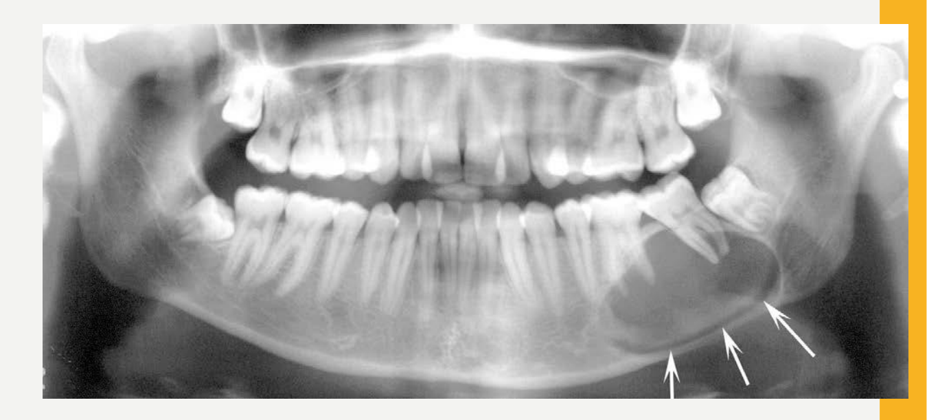 | 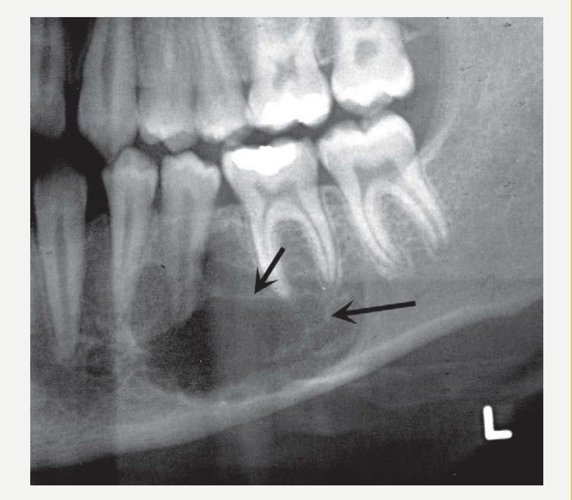 |
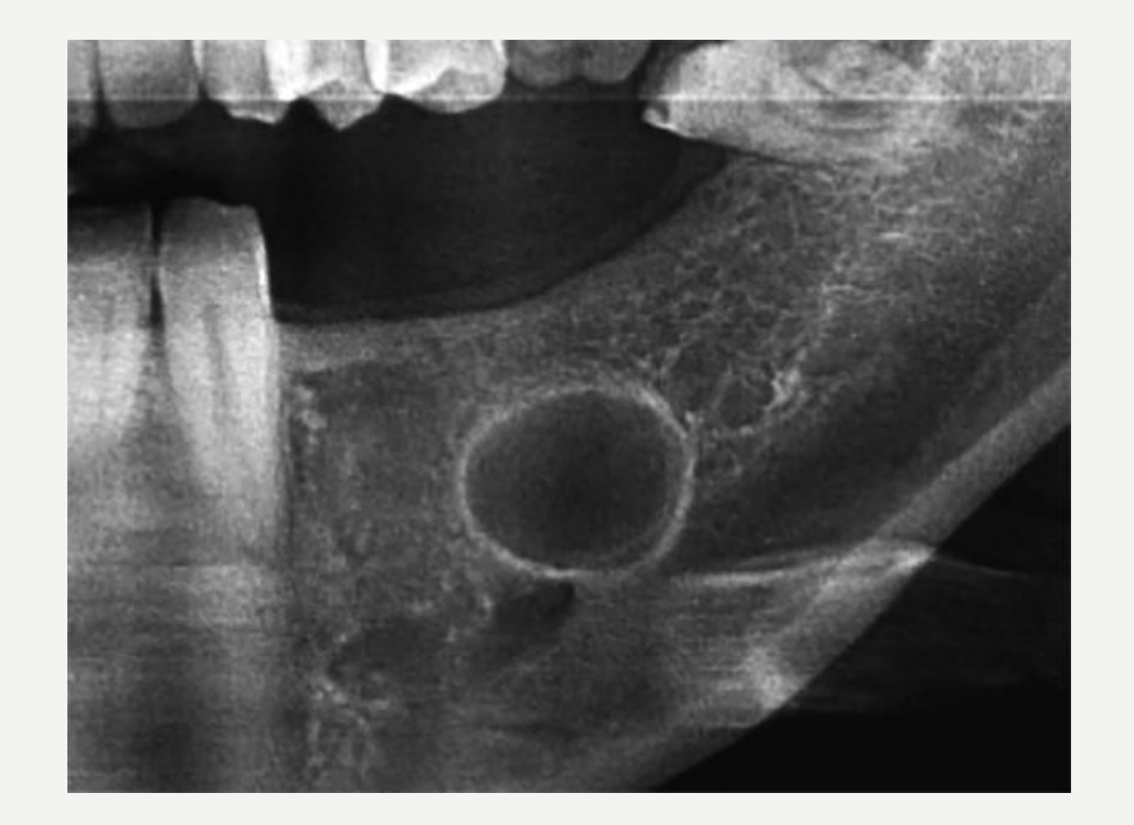 |  |
 |
Radiopacities and Signs of Past Infection
Three different questions
- Which opacity is this? Use group (a).
- Is this the trace of an infection that is over? Use group (b).
- Is this infection still active or chronic? Use group (c). Sequestra and periosteal new bone belong here, not under “healed”.
(a) Periapical opacities to differentiate
| Feature | Dense bone island (idiopathic osteosclerosis, enostosis) | Sclerosing osteitis (“condensing osteitis”) | Hypercementosis | Cementoblastoma |
|---|---|---|---|---|
| Nature | Hamartomatous growth of cortical bone into cancellous bone25. Lectures: central osteoma “aka dense bone islands”31; “also considered hamartomas”2 | Inflammatory bone deposition around a root. W&P discourages the term “condensing”26 | Bulbous roots; can be a root response to periapical inflammation26. Also seen with Paget disease27 | Benign neoplasm of cementoblasts: a bulbous mass of cementum on the root2532 |
| Cause visible? | No. Asymptomatic; presence doesn’t correlate with teeth25 | Yes. Usually an obvious cause, such as a large restoration or caries25 | May accompany inflammation26 | — |
| Location / epicentre | Mandible > maxilla, premolar–molar region252 | Just apical to the root apex25 | Root | Mandible (78%), premolar/first molar (90%)25; apical third of the root32 |
| Periphery | Well defined and abuts normal bone; sometimes blends. Narrow zone of transition2526 | Poorly defined blending border; wide transition2326 | Root outline enlarged | Well defined and corticated25 |
| Internal | Ground glass to uniformly RO25 | Thick, more numerous trabeculae, becoming dense enough to obscure them26 | — | Mostly RO, amorphous or wheel-spoke; hides the root outline2532 |
| PDL / RL rim | Uniform PDL, lamina dura continuous; no RL periphery2625 | Widened apical PDL, often with a small RL focus at the apex; no RL rim2625 | PDL present but thinner than a cementoblastoma rim25 | RL band around the mass25; lecture: RL rim continuous with the PDL32 |
| Root and jaw | May cause limited, self-limiting root resorption (usually mandibular first molar) with a thin PDL kept25 | May cause root resorption or hypercementosis26 | No root resorption, no expansion25 | Root resorption (about ⅔ of cases32); expansion, sometimes cortical perforation without periosteal reaction25 |
| Pulp | Vital25 | Non-vital tooth (pulp necrosis is the usual cause)26 | n/a | W&P: vital25; lecture: vital in about 80%32 |
| Action | None; if ≥5 DBIs, consider adenomatous polyposis coli25 (Gardner syndrome31). Monitor2 | — | — | Endo plus root amputation, or extraction25 |
- DBI on OPG: bone islands can make neighbouring normal bone look falsely lucent. If the lamina dura and PDL are intact, the “lesion” is likely an artefact33
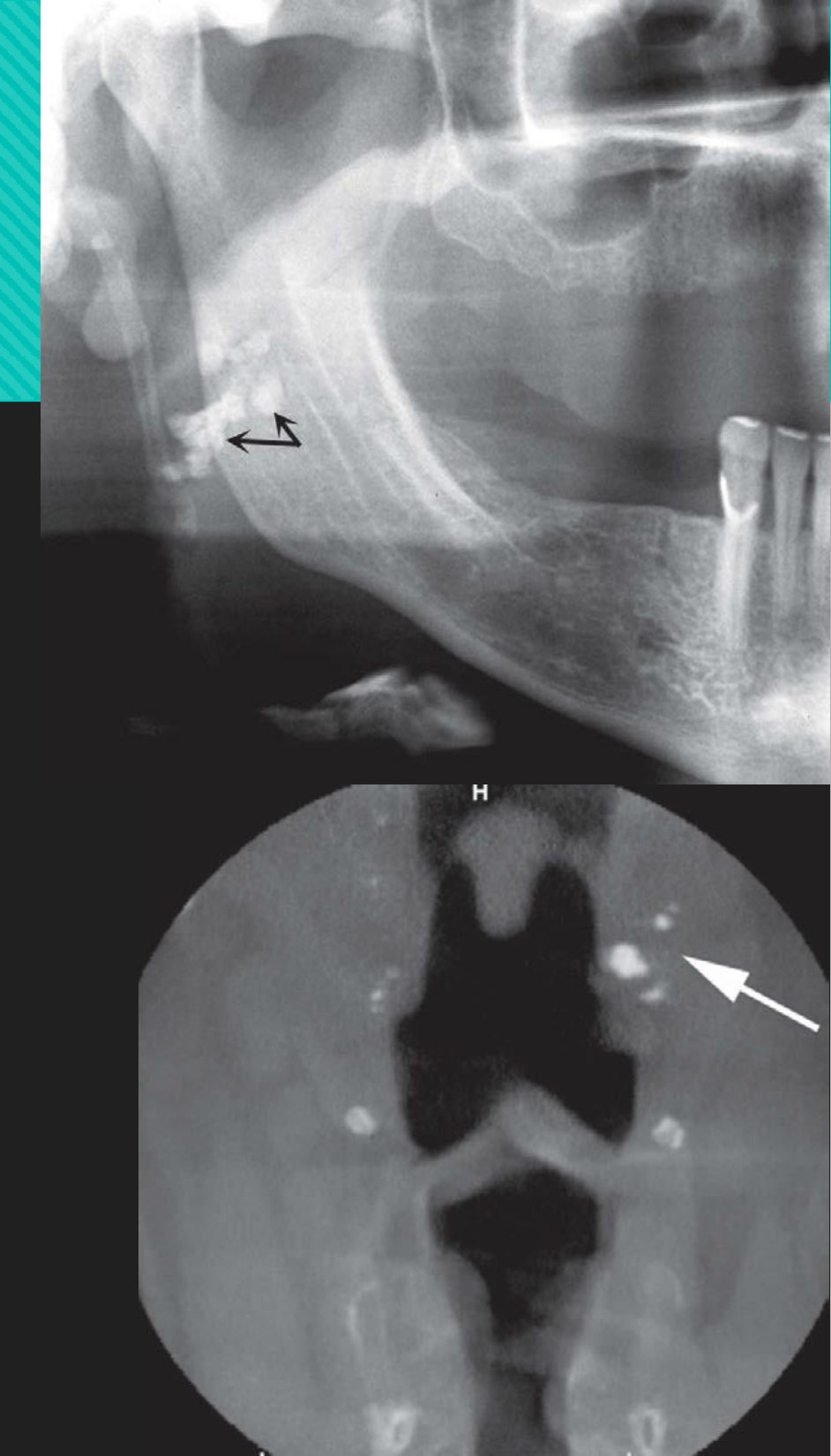
- DBI vs tonsillolith: may be hard to separate on an OPG34
- DBI vs sclerotic metastasis: periosteal change is not expected with a bone island29
 |  |

(b) Signs of past infection
| Sign | Density | What it means | How to confirm |
|---|---|---|---|
| Fibrous scar after successful orthograde RCT or apical surgery | Radiolucent centre (dense connective tissue, healing by secondary intention). “Doughnut” pattern: rolled border with granular bone or radiating trabeculae at the edge; concentric or spoke-like rings26 | Healed or healing. Not active disease, but it mimics rarefying osteitis26 | Treatment history plus clinical signs and symptoms26 |
| Calcified lymph node, isolated, level I | RO, cauliflower-like | Usually post-inflammatory. More extensive nodal calcification means active or previously treated disease (e.g. TB, sarcoidosis)34 | History; see Soft-Tissue Calcifications |
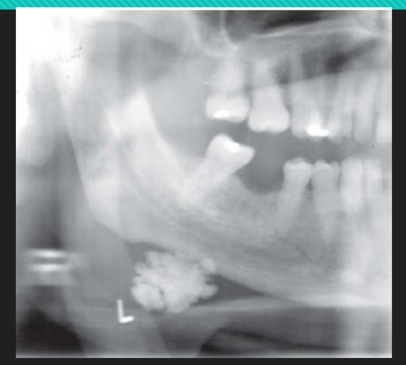
| Tonsillolith | RO cluster | Lecture heading: “post-inflammatory tonsillar calcifications”34 | See Soft-Tissue Calcifications |
| Sialolith | RO | May relate to a previous inflammatory condition or ongoing chronic sialadenitis34 | Symptoms; US / MSCT34 |
Residual sclerosis: not supported as a "healed" sign
None of DI L1/L2 or W&P 22/25 says that sclerosis persists after an extraction or RCT. W&P says only that apical PDL widening “may persist” in a lesion that looks entirely sclerotic26. Don’t call residual sclerosis “healed” from these sources.

(c) Active or chronic infection (not healed)
| Sign | Appearance | Classification in the sources |
|---|---|---|
| Sequestrum | RO island of bone inside a region of resorption (RL). Ranges from a dot (young patients) to large islands; CT is better26 | Necrotic bone (dying or dead) separated from healthy bone. Hallmark of osteomyelitis26; “key feature”33. Stays a focus of infection that antibiotics penetrate poorly33. Also the key finding in ORN and MRONJ26 |
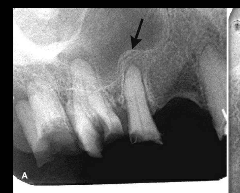
| Periosteal new bone / onion skin | Paired RL (fibrous periosteum) and RO (new bone) lines parallel to the cortex; one layer per exacerbation. At the sinus floor it is a “halo”26 | Ongoing, cyclical inflammation26. Adults: thin single layer or solid; children: thick, multilayered onion skin33. W&P: proliferative periostitis is more common in children26 |
| Chronic sclerosis with filling-in | Layers fill in with sclerotic bone; the original cortex may disappear; the jaw enlarges26 | Chronic osteomyelitis (chronic = >4 weeks, non-suppurative, sclerosing)33. Mixed lucent and opaque appearance33 |
| Fistula | Well-defined break in the cortex or periosteal new bone26 | Chronic osteomyelitis26 |
- Onion skin isn’t always infection: LCH, leukaemia, lymphoma and Ewing sarcoma can also produce it. Destruction of adjacent structures or of the periosteal bone itself should raise suspicion of malignancy26
- Imaging: rarefaction and sclerosis show on an OPG; occlusal views show periosteal new bone; CBCT or MDCT is the modality of choice for sequestra and periosteal bone26
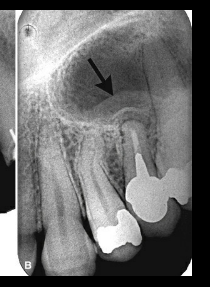
- Sinus-floor periostitis: periapical inflammation from root remnants or infected teeth lifts and scleroses the adjacent antral floor, a reactive periosteal response33. W&P calls this the “halo”26
 |  |
 |  |
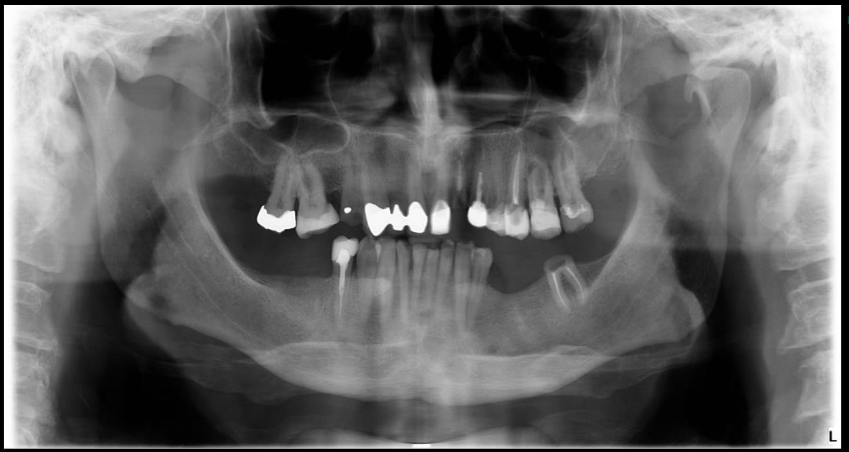
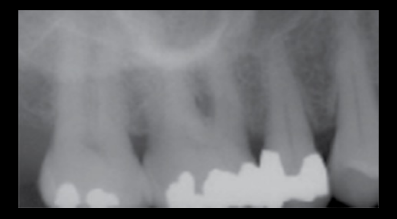
- Where the OPG fits: good for surveying multiple lesions and heavily compromised dentitions. Use CBCT when PA or OPG findings are inconclusive33
- Restorations hide apices: on the OPG, superimposition over heavily restored maxillary molars showed no clear apical lucency. CBCT of the 16 then showed an apical lucency, sinus-floor perforation, reactive mucosal thickening and root resorption33
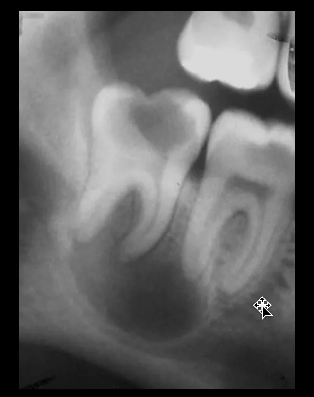
- Canal displacement: a periapical lucency that displaces or compresses the mandibular canal suggests a radicular cyst rather than a granuloma or abscess33
 |  |
 |
Soft-Tissue Calcifications
General rules
| Calcification | Where on the OPG | Shape / internal | How to confirm or act |
|---|---|---|---|
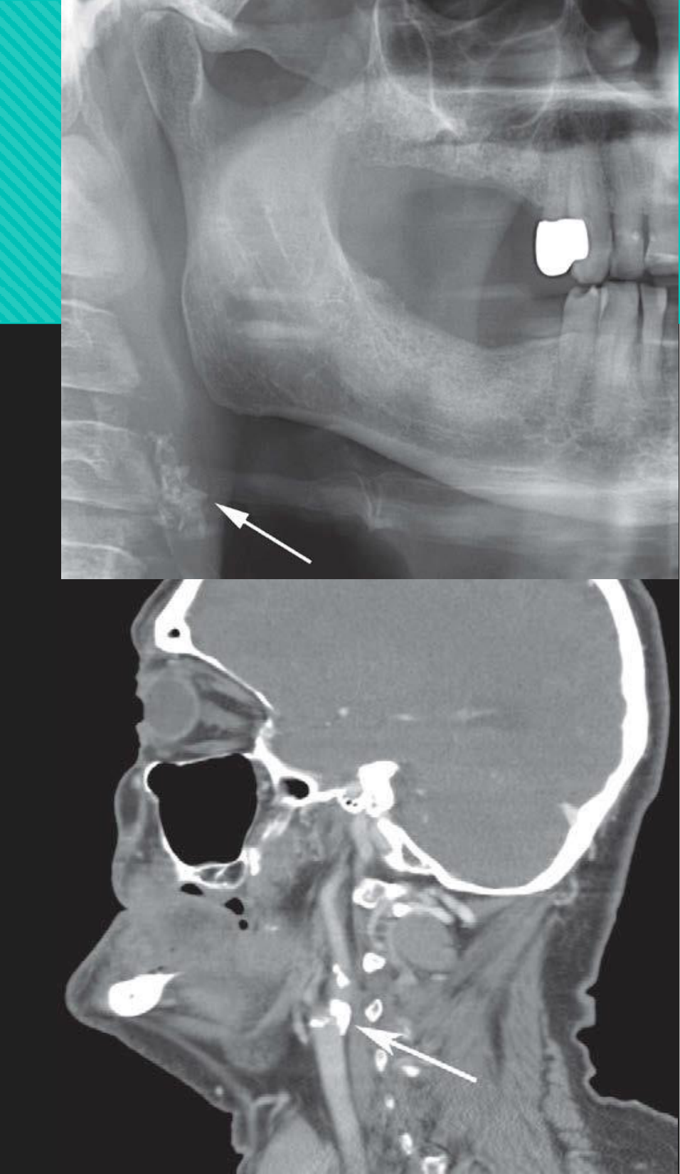
| Calcified atheromatous plaque (carotid) | Neck soft tissue, above or below the greater cornu of the hyoid, next to C3–C4 or the space between them (carotid bifurcation)3534 | Multiple, irregular, vertical linear distribution; heterogeneous with RL voids35; may be C-shaped or circular34 | Don’t confuse with triticeous or thyroid cartilage, which are uniform in size and shape35. Refer to the GP / medical review, especially in younger patients, those with risk factors, or with large plaques34. About 1 in 7 have significant stenosis34 |
| Tonsillolith | Over the mid-ramus, where the dorsum of the tongue crosses it; often just below the IAC. On CT: medial to the ramus at the lateral pharyngeal wall3534 | Cluster of small, ill-defined RO bodies; density about that of cortical bone35 | Main differential is a DBI in the ramus. Confirm with a PA skull/mandible or reverse Towne view, or CBCT/MDCT35 |
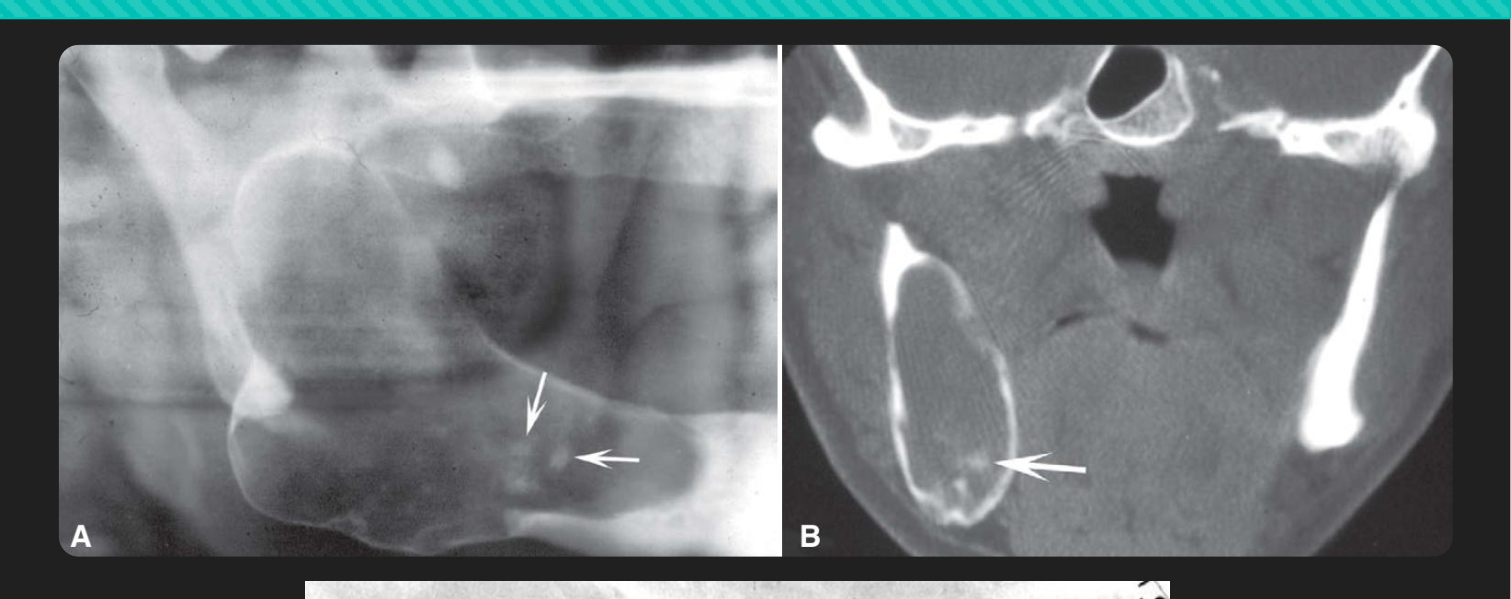
| Calcified lymph node | Submandibular, at or below the inferior border near the angle; or between the ramus and the C-spine; can form a chain (cervical)3534 | Well defined, irregular, cauliflower-like; variable density; sometimes eggshell or laminated35 | Sialolith is smooth, a node is irregular. Salivary symptoms or sialography separate them35. Treat the underlying cause34 |
| Sialolith | Submandibular gland 83–94%, parotid 4–10%34. In the submandibular hilum it projects near the inferior cortex just anterior to the angle35 | Ductal: cylindrical, smooth; hilar: larger, ovoid/irregular; homogeneous or laminated34 | Some are RL: <20% of submandibular and 40% of parotid stones. US or MSCT preferred; CBCT not indicated34. See Sialolithiasis |
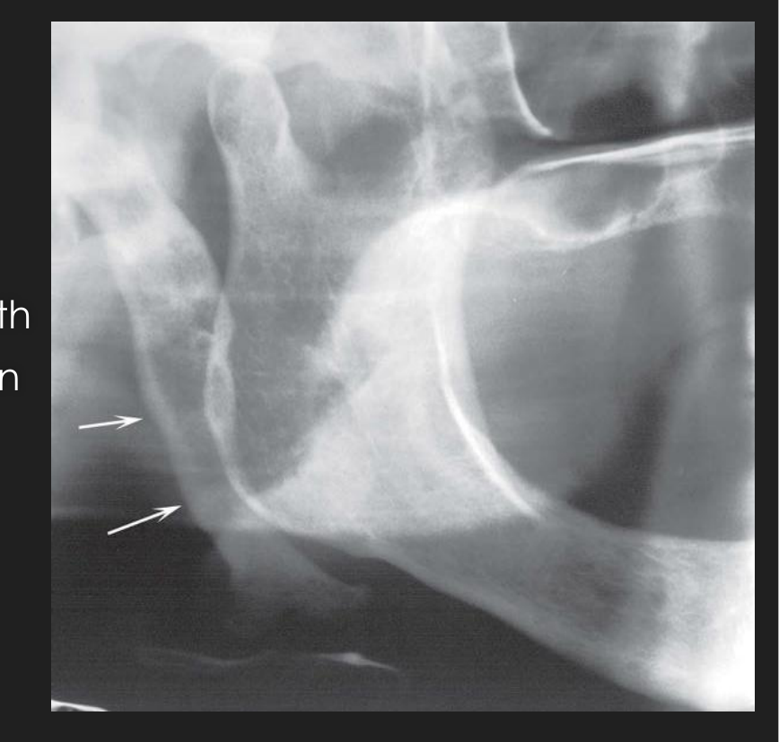
| Ossified stylohyoid ligament | Linear, from the mastoid region, crossing the posteroinferior ramus towards the hyoid3534 | Long tapering process; normal styloid is 0.5–2.5 cm; may have RL pseudoarticulations; a cortex appears as it thickens35 | Usually incidental and asymptomatic. If symptomatic (Eagle), palpate the tonsillar fossa to reproduce the symptoms35 |

 |  |
 |  |
| Point | DI L2 detail |
|---|---|
| Calcified nodes: sites | Most often submandibular and superficial or deep cervical nodes (levels Ib and II). Preauricular and submental nodes are less common. A single node or a linear chain (“LN chaining”)34 |
| Node vs phlebolith | Phleboliths are usually smaller and multiple, with a targetoid look (lucent centre)34 |
| Tonsillolith vs parotid calcification | Parotid parenchymal calcifications are usually smaller and round, and project over or behind the posterior ramus34 |
| Triticeous cartilage (carotid mimic) | Below the greater cornu of the hyoid, next to the superior border of C4. Smooth “grain of wheat”, 7–9 × 2–4 mm, homogeneous. A normal ageing change: no action34 |
| Mönckeberg arteriosclerosis | On the OPG, the facial artery is involved more often than the carotid. Parallel thin RO lines (“tram-track”, “pipe-stem”), often bilateral. When the carotid is outlined, it is almost always atheroma34 |
| Dystrophic calcification in a cyst | A long-standing, chronically infected residual cyst can contain internal opacities34 |
| Styloid length | Lecture: up to 3 cm is normal; about 18% of people have ossification longer than 3 cm34. W&P (table above): styloid 0.5–2.5 cm; calcification in up to 30%357 |
 |  |
 |  |
 |
Report Template and Worked Examples
Report template
Fill-in version of the L13 / L15 OPG report checklist25:
OPG REPORT
1.  Technical errors:            ______ (positioning, e.g. chin up/down, ghosts, artefacts)
2.  Dentition stage:             deciduous / mixed / permanent
3.  Missing teeth:               ______   Supernumeraries: ______
    Implants (crestal bone level): ______   Root remnants: ______
4.  Impacted / ectopic teeth:    tooth __ | location | angulation | crown & root morphology |
                                 relation to adjacent teeth / sinus / IAC
5.  Dental anomalies:            ______
6.  Periodontal bone loss:       horizontal / angular (vertical) / furcation / perio-endo
7.  Calculus:                    ______ (where)
8.  Restorations:                ______   Faulty (overhang, deficient margin): ______
9.  Caries (incl. recurrent):    ______
10. Non-carious tooth loss:      ______ (e.g. attrition)
11. Endodontic therapy:          ______
12. Periapical lucencies:        ______ + root resorption / antral floor remodelling /
                                 reactive sclerosis / reactive antral mucosal thickening
13. Other lucencies/opacities:   location | border (well/ill-defined) & shape |
                                 internal architecture | effects on adjacent structures |
                                 surgical-sieve differentials
14. Extragnathic:                maxillary sinuses (symmetry, size, opacity, floor/walls) |
                                 condyles (symmetry, remodelling, sclerosis, beaking, loose bodies) |
                                 airspace | C-spine | pterygomaxillary fissure | orbits |
                                 soft-tissue calcifications (submandibular, tonsillar, carotid bulb)
15. Summary:                     ______   Further imaging? modality: ______
Worked example 1
Case: adult, comprehensive-care work-up. The student labelled normal anatomy on the OPG with numbered arrows, then reported the findings as a region-by-region table36.
| Checklist item | Finding |
|---|---|
| Dentition | Permanent. One premolar missing in each quadrant; third molars present in all four quadrants36 |
| Restorations / endo | Heavily restored dentition. Root-filled 12, 11 and 36: radiopaque canal fillings are visible in the upper incisors and the lower left first molar36 |
| Periodontium | Generalized mild horizontal bone loss, maxilla and mandible; no furcation involvement36 |
| Bone | Normal density and trabeculation in both jaws. Two RO spots on the right mandibular body, superimposed over 45, reported as amalgam tattoos36 |
| Mandibular cortices | Continuous and even: rami, coronoids, condyles, inferior borders36 |
| Maxilla / sinuses | NAD for cortices, zygoma, nasal cavity and palate. RO shadows in the sinuses are superimposed inferior nasal conchae and zygomatic buttresses, not sinus disease36 |
| Periphery | NAD. Styloid processes, pharynx and mandibular foramina were recorded as “not identified”36 |
What this example teaches
 |  |
Worked example 2
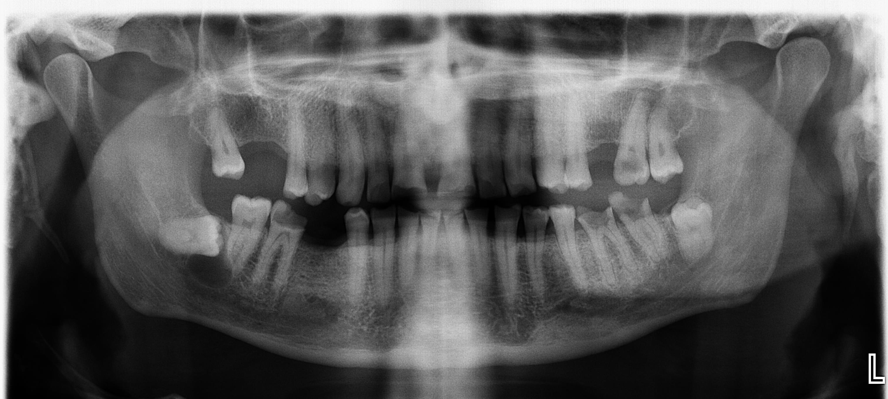
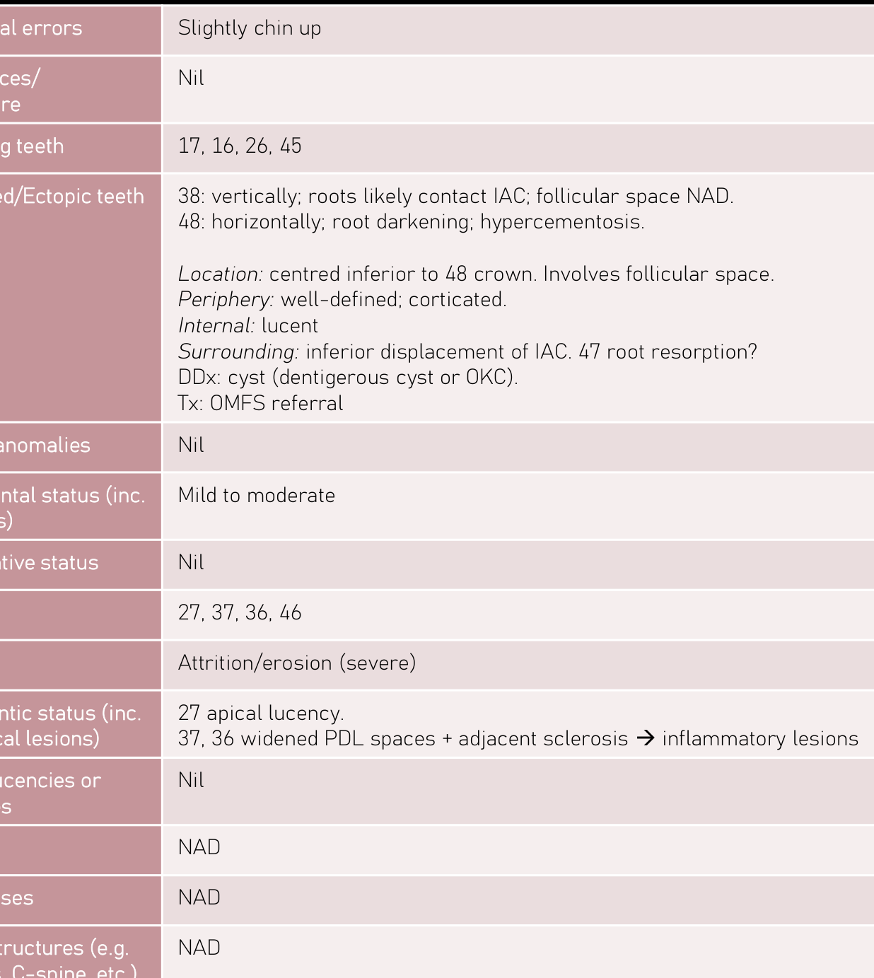
Case: adult, L15 OPG workshop. Pericoronal lucency on an impacted lower third molar5.
| Checklist item | Finding |
|---|---|
| Technical | Slightly chin up5 |
| Missing teeth | 17, 16, 26, 455 |
| Impacted | 38 vertical; roots likely contact the IAC; follicular space NAD. 48 horizontal, with root darkening and hypercementosis5 |
| Lesion at 48 | Location: centred inferior to the 48 crown, involving the follicular space. Periphery: well defined, corticated. Internal: lucent. Surrounding: IAC displaced inferiorly; possible 47 root resorption5 |
| Differential | Cyst: Dentigerous Cyst or Odontogenic Keratocyst. Refer to OMFS5 |
| Perio / caries / NCTL | Mild to moderate bone loss. Caries 27, 37, 36, 46. Severe attrition/erosion5 |
| Endo / periapical | 27 apical lucency. 37 and 36 have widened PDL with adjacent sclerosis, read as inflammatory lesions (sclerosing osteitis pattern)5 |
| TMJ / sinuses / other | NAD5 |
Dentigerous vs OKC on this film
- A dentigerous cyst attaches at the CEJ and has a propensity to displace and resorb teeth24
- An OKC is favoured if the cyst meets the tooth apical or coronal to the CEJ, or if the bone has barely expanded. The two may be indistinguishable in a pericoronal position24
- Both displace the IAC inferiorly, so this sign doesn’t separate them. CBCT (or MDCT if the cortex is perforated) is the next step for a suspected OKC24
 |  |
Related Notes
- MOC Radiography
- Periapical Radiograph Analysis
- Radiolucent Lesion differentiation
- Periodontitis Staging and Grading
- Cysts: Radicular Cyst · Residual Cyst · Dentigerous Cyst · Odontogenic Keratocyst · Nasopalatine Duct Cyst
- Tumours: Ameloblastoma · Cementoblastoma
- Bone: Periapical Cemento-Osseous Dysplasia · Florid Cemento-Osseous Dysplasia · Fibrous Dysplasia · Osteomyelitis · Stafne Defect · Traumatic Bone Cyst · Medication-Related Osteonecrosis of the Jaw · Osteoradionecrosis
- Salivary: Sialolithiasis
Sources
Footnotes
-
Lecture and Workshop LOs ↩ ↩2 ↩3 ↩4 ↩5 ↩6 ↩7 ↩8 ↩9 ↩10 ↩11 ↩12 ↩13 ↩14 ↩15 ↩16 ↩17 ↩18 ↩19 ↩20 ↩21
-
L13 PR2 ↩ ↩2 ↩3 ↩4 ↩5 ↩6 ↩7 ↩8 ↩9 ↩10 ↩11 ↩12 ↩13 ↩14 ↩15 ↩16 ↩17 ↩18 ↩19 ↩20 ↩21
-
09 9 Panoramic Imaging ↩ ↩2 ↩3 ↩4 ↩5 ↩6 ↩7 ↩8 ↩9 ↩10 ↩11 ↩12 ↩13 ↩14 ↩15 ↩16 ↩17 ↩18 ↩19 ↩20 ↩21 ↩22 ↩23 ↩24 ↩25 ↩26 ↩27 ↩28 ↩29 ↩30 ↩31 ↩32 ↩33 ↩34 ↩35 ↩36 ↩37 ↩38 ↩39 ↩40 ↩41 ↩42 ↩43 ↩44 ↩45 ↩46 ↩47 ↩48 ↩49 ↩50 ↩51 ↩52 ↩53 ↩54 ↩55 ↩56 ↩57 ↩58 ↩59 ↩60 ↩61 ↩62 ↩63 ↩64 ↩65 ↩66 ↩67 ↩68 ↩69 ↩70 ↩71 ↩72 ↩73 ↩74 ↩75 ↩76 ↩77 ↩78 ↩79 ↩80 ↩81 ↩82 ↩83 ↩84 ↩85 ↩86 ↩87 ↩88 ↩89 ↩90 ↩91 ↩92 ↩93 ↩94 ↩95 ↩96 ↩97 ↩98 ↩99 ↩100 ↩101 ↩102 ↩103 ↩104 ↩105 ↩106 ↩107 ↩108 ↩109 ↩110 ↩111 ↩112 ↩113 ↩114 ↩115 ↩116 ↩117 ↩118 ↩119 ↩120 ↩121 ↩122 ↩123 ↩124 ↩125 ↩126 ↩127 ↩128 ↩129
-
L8 Panoramic Radiography ↩ ↩2 ↩3 ↩4 ↩5 ↩6 ↩7 ↩8 ↩9 ↩10 ↩11 ↩12 ↩13 ↩14 ↩15 ↩16 ↩17 ↩18 ↩19 ↩20 ↩21 ↩22 ↩23 ↩24 ↩25 ↩26 ↩27 ↩28 ↩29 ↩30 ↩31 ↩32 ↩33 ↩34 ↩35 ↩36 ↩37 ↩38 ↩39 ↩40 ↩41 ↩42 ↩43 ↩44 ↩45 ↩46 ↩47
-
11 10 Principles of Management of Impacted Teeth ↩ ↩2 ↩3 ↩4 ↩5 ↩6 ↩7 ↩8 ↩9 ↩10 ↩11 ↩12 ↩13 ↩14
-
L3 Wisdom Teeth ↩ ↩2 ↩3 ↩4 ↩5 ↩6 ↩7 ↩8 ↩9 ↩10 ↩11 ↩12 ↩13 ↩14 ↩15 ↩16 ↩17 ↩18
-
30 30 Temporomandibular Joint Abnormalities ↩ ↩2 ↩3 ↩4 ↩5 ↩6 ↩7 ↩8 ↩9 ↩10 ↩11 ↩12 ↩13
-
27 27 Trauma ↩ ↩2 ↩3 ↩4 ↩5 ↩6 ↩7 ↩8 ↩9 ↩10 ↩11 ↩12 ↩13 ↩14 ↩15 ↩16 ↩17 ↩18 ↩19 ↩20
-
18 18 Principles of Radiographic Interpretation ↩ ↩2 ↩3 ↩4 ↩5 ↩6 ↩7 ↩8 ↩9 ↩10 ↩11 ↩12 ↩13 ↩14
-
24 24 Benign Tumors and Neoplasms ↩ ↩2 ↩3 ↩4 ↩5 ↩6 ↩7 ↩8 ↩9 ↩10 ↩11 ↩12 ↩13 ↩14 ↩15 ↩16 ↩17 ↩18 ↩19 ↩20 ↩21 ↩22 ↩23 ↩24 ↩25 ↩26
-
22 22 Inflammatory Conditions of the Jaws ↩ ↩2 ↩3 ↩4 ↩5 ↩6 ↩7 ↩8 ↩9 ↩10 ↩11 ↩12 ↩13 ↩14 ↩15 ↩16 ↩17 ↩18 ↩19 ↩20 ↩21 ↩22 ↩23 ↩24 ↩25 ↩26 ↩27 ↩28 ↩29 ↩30 ↩31
-
25 25 Diseases Affecting the Structure of Bone ↩ ↩2 ↩3 ↩4 ↩5
-
L6 - Imaging for Jaw Malignancies ↩ ↩2 ↩3 ↩4 ↩5 ↩6 ↩7 ↩8 ↩9 ↩10 ↩11
-
L1 - Imaging for Infection and Inflammatory Lesions of the Jaws ↩ ↩2 ↩3 ↩4 ↩5 ↩6 ↩7 ↩8 ↩9 ↩10
-
L2 - Opacities in Dental Imaging ↩ ↩2 ↩3 ↩4 ↩5 ↩6 ↩7 ↩8 ↩9 ↩10 ↩11 ↩12 ↩13 ↩14 ↩15 ↩16 ↩17 ↩18 ↩19 ↩20 ↩21 ↩22 ↩23
-
31 31 Soft Tissue Calcifications and Ossifications ↩ ↩2 ↩3 ↩4 ↩5 ↩6 ↩7 ↩8 ↩9 ↩10 ↩11 ↩12 ↩13 ↩14 ↩15 ↩16