Periapical Radiograph Analysis
How to use this note
Work top to bottom when annotating a PA: run the checklist, rule out technique faults, confirm normal anatomy, then assess teeth, periodontium and periapex. Check the mimics before calling anything pathology, and write it up with the report template. Built from White & Pharoah (8th ed.), the DENT4213 OMFR lectures, DMD3S2 Diagnostic Imaging, and Cohen’s Pathways of the Pulp.
Quick Annotation Checklist
Systematic PA read (work top to bottom, every time)
- Before you look: reason for the X-ray, previous X-rays to compare, patient ID, good viewing conditions1
- View, region, teeth: PA vs BW vs occlusal; maxilla or mandible, anterior or posterior; orient by the dot or marker; count the teeth and name them in FDI (e.g. 36, 37)123
- Image quality and technique: full roots plus ≥2 mm periapical bone? Contacts open? Elongation or foreshortening? Cone cut, blur, too light or dark, receptor artefacts?435
- Teeth: crowns (enamel, dentine, pulp chamber); caries (depth: enamel / EDJ / dentine / near pulp; approximal, occlusal, buccal/lingual, root, next to a restoration); pulp size, stones, canal morphology; root number and shape1
- Restorations and endo: material by radiodensity; margins (overhangs, open or deficient margins), voids; RCT length, density, extrusion; posts, crowns, ortho appliances1
- Periodontium: crest level relative to CEJ; is the crest corticated? Horizontal vs vertical/angular loss, furcation involvement, calculus, widened PDL, lamina dura continuity; enamel pearl16
- Periapex: is the PDL/lamina dura continuous around every apex? Radiolucency (rarefying osteitis) or sclerosis (sclerosing osteitis) centred on the apex? Rule out superimposed mental or incisive foramen (tube shift)789
- Other bone, anatomy, soft tissue: trabecular pattern, sinus floor, canals and foramina, unerupted teeth, calcifications, foreign bodies61
- Any lesion, described: location/epicentre → periphery and shape → internal structure → effects on neighbours (teeth, lamina dura, PDL, canal, sinus, cortex)6
- Impression and next step: normal vs abnormal → developmental vs acquired → category; differentials in order of likelihood; clinical tests or further views62
Image Quality and Technique
Paralleling vs bisecting angle
| Paralleling (right-angle / long-cone) | Bisecting angle | |
|---|---|---|
| Principle | Receptor parallel to tooth long axis; central ray perpendicular to both4 | Receptor against the lingual/palatal tooth surface; central ray perpendicular to the imaginary bisector of the tooth–receptor angle (Cieszynski’s rule of isometry)4 |
| Holder | Always needs a holder, ideally with an aiming ring104 | Bisecting instrument with aiming ring is best; a finger-held receptor bends and causes cone cuts4 |
| Geometry | Receptor sits away from teeth → more OID. A long cone (long source–object distance) offsets the magnification and unsharpness84 | Tooth length correct in theory, but shape is distorted; each root of a multirooted tooth needs a different angle4 |
| Bone levels | Crest–CEJ relationship accurate10 | Alveolar crest projects coronal to where it really is (bone looks higher than it is)84 |
| Use | Preferred for all PAs; endo working length; crestal bone around implants810 | When anatomy stops parallel placement (shallow palate, floor of mouth, tori) or with large rigid sensors4 |
| Downsides | Harder receptor placement, less comfortable10 | Distortion likely; zygoma often over the palatal root apex of maxillary molars49 |
 |  |
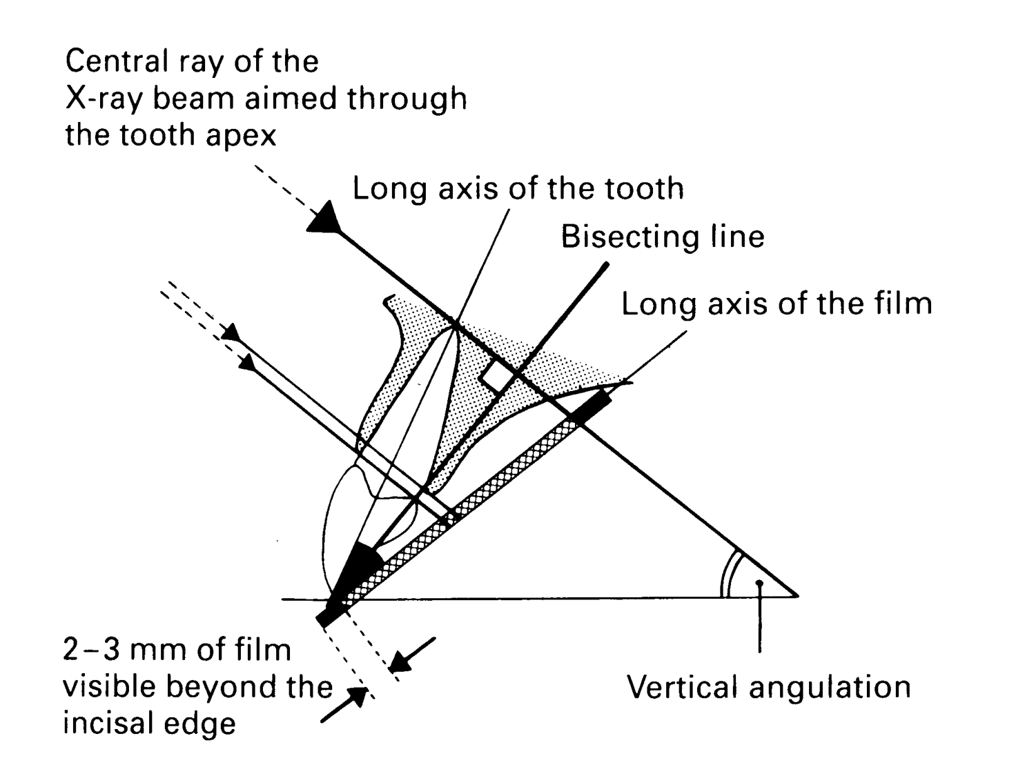
- Shape distortion rules8
- Foreshortening: central ray perpendicular to the receptor but the tooth is not parallel to it → image shorter than the tooth
- Elongation: central ray perpendicular to the tooth but not to the receptor → image longer than the tooth
- In maxillary molars the palatal root can look too long relative to the buccal roots, and the crest–CEJ relationship is distorted8
- Vertical angle diagnosis (bisecting): foreshortening = vertical angle too high; elongation = vertical angle too low4
- Magnification: the beam diverges from a point source, so even paralleling magnifies about 10% (range 6.4–16.2%). Reduce it with a long cone and rectangular collimation9
Endo "modified paralleling" (Abbott, DENT4212)
- Add about +15° vertical angulation to a paralleling set-up. This deliberately foreshortens the image by about 10% to cancel magnification and gives more apical detail911
- A decreased vertical angle only elongates the image and has no diagnostic value9
- This is the endo lecturer’s protocol. W&P describes standard paralleling with the beam perpendicular to the receptor4
 |  |
Is this PA acceptable?
- Full root length plus ≥2 mm of periapical bone visible4
- Receptor extends 2–3 mm beyond the incisal edge or occlusal plane (vertical receptor for anteriors, horizontal for posteriors)10
- Any abnormality shown entirely, with a rim of normal bone around it. If a PA can’t do this, add an occlusal, panoramic or CBCT4
- Minimal distortion: tooth length and shape look right, no elongation or foreshortening4
- Contacts open: the horizontal beam direction controls crown overlap (BW standard: overlap < ⅓ of enamel thickness)4
- Density and contrast good enough to separate enamel, dentine and pulp43
- No cone cut, blur, receptor bending or processing/plate artefact35
- You don’t need a retake if the missing information is already on another view4
Error table
| Error | Appearance | Cause | Fix |
|---|---|---|---|
| Elongation | Teeth too long; apices may run off the image | Vertical angle too shallow; beam not perpendicular to the receptor; receptor tilted (e.g. cotton roll on the wrong side of the bite block)489 | Increase the vertical angle; paralleling holder with aiming ring; cotton roll between the target tooth and the bite block9 |
| Foreshortening | Teeth short and stubby | Vertical angle too steep; tooth not parallel to receptor (e.g. shallow palate)4 | Reduce the vertical angle; place the receptor deeper, toward the midline4 |
| Horizontal overlap | Contacts closed; crowns superimposed | Central ray not directed through the contacts in the horizontal plane4 | Aim through the contacts, with the cone edge parallel to the buccal surfaces4 |
| Cone cut | Sharp-edged clear (white) unexposed area | Tube head misaligned with the receptor; posterior holder assembled for the wrong quadrant3129 | Align the PID with the aiming ring; check the holder assembly49 |
| Receptor bending | Stretched, distorted anatomy; dark lines/creases (film); streaks or permanent damage (PSP) | Finger-held receptor, receptor bent against the palate or tori435 | Holder with a rigid backing9 |
| Reversed film | Too light, with a herringbone/embossed lead-foil pattern; left and right reversed if you orient by the dot | Packet placed back to front3 | Convex side of the raised dot (marked tube side of the packet) toward the tube3 |
| Motion | Blurred outlines | Patient or receptor moved3 | Stabilise patient and receptor (holder)3 |
| Overexposure | Too dark | Excessive mA, kVp or time; source–film distance too short. Film only: overdevelopment, light leak312 | Correct the exposure chart; processing QA13 |
| Underexposure | Too light (film); noisy/grainy (digital) | Too little mA, kVp or time; distance too long. Film only: underdevelopment, depleted developer35 | Correct the settings; replenish chemicals3 |
| Low contrast / fog | Grey enamel and grey pulps; dark with no detail | Underdevelopment, underexposure, excessive kVp; safelight/light leaks, old film3 | Darkroom and film-stock QA313 |
| Film processing marks | Dark spots/lines (developer, fingerprints, static, rollers); light spots (fixer splash); yellow-brown stain (poor washing); straight-edged partial image (not fully immersed)312 | Handling and processing faults3 | Clean handling; correct tank levels and wash times3 |
| PSP: light exposure | Faded image; uneven density where plates overlapped | Plate exposed to ambient light before scanning5 | Keep exposed plates shielded until scanned5 |
| PSP: damage | Thin light streak (scratch) that can mimic a root filling; circular or blotchy artefacts | Scratches, bending, harsh disinfectant, gas under the coating513 | Inspect plates monthly and retire damaged ones13 |
| PSP: double image | Two superimposed images | Previous image not fully erased5 | Erase fully; retake5 |
| Scanner/software | Straight radiopaque line (dust on the scanner lens); halos at restoration edges that look like recurrent caries (edge-enhancement filter) | Scanner maintenance; filter overuse5 | Clean the scanner; check the unfiltered image5 |
| Other | Double exposure; jewellery or appliances in the field10 | Operator error10 | Remove metal objects before exposure10 |
 |  |  |
 |  |  |
 |  |
Artefact or pathology?
Don't diagnose a technique error
- Does it respect anatomy? An artefact often crosses anatomical boundaries. In Fig 4.22A the “root filling” streak runs straight through the crown and out into air5
- Does it repeat? Check a second view taken at a different angle. Plain images at slightly different angles, and views at right angles, add real information6
- Superimposition: the mental or incisive foramen over an apex mimics a periapical radiolucency. It moves off the apex on a tube-shifted view; a true lesion stays centred on the apex89
- Angulation: poor angulation or underexposure can fake a periodontal pocket. A bent or wrongly oriented receptor can fake a root defect. Retake with a holder before you diagnose9
- Bone height: bisecting and off-angle views project the crest coronally or distort crest–CEJ distances, so assess bone loss on paralleling views84
- Processing/software: edge enhancement can create “recurrent caries” at restoration margins. Check the unfiltered image5
- Size: lesions are always larger histologically and surgically than they look on the radiograph9
Tube shift / SLOB (buccal object rule)
- Take two PAs of the same area, the second with the tube shifted horizontally or vertically. Compare the object with a fixed reference, e.g. an apex (Clark’s rule)810
- SLOB: Same Lingual, Opposite Buccal8
- Object moves in the same direction as the tube → lingual/palatal
- Object moves in the opposite direction → buccal
- Doesn’t move → same bucco-lingual depth as the reference
- Uses: impacted or supernumerary teeth, foreign bodies, separating superimposed canals/roots, moving foramina off apices89
- Endo applications11
- Lower incisors: always shift (about 50–60% have two canals)
- Lower molars: mesial shift → the mesiolingual canal projects more mesially
- Upper molars: distal shift first (to separate MB2), mesial shift to show the DB root
- Don’t mistake the buccal and lingual PDL outlines for a missed canal
- Clue to beam direction without a record: the zygomatic process (buccal to the molars) moves mesially as the beam comes from more distal, and projects occlusally as vertical angulation increases8
- Alternative: right-angle technique: PA plus a cross-sectional (true) occlusal view. Easiest in the mandible810
 |  |

Normal Radiographic Anatomy
Rule of thumb
Normal structures often look like disease on a PA. Before you call a periapical lesion, check whether the lamina dura is still intact around the apex, and whether the shadow moves when you change beam angle (a normal structure shifts position against the apex).14
Tooth and Supporting Structures
| Landmark | Radiodensity | How to recognise / location | Pitfall (mimic) |
|---|---|---|---|
| Enamel | RO (most) | Uniform opaque cap on the crown. About 96% mineral, the densest tissue in the body14 | — |
| Dentine | RO (less than enamel) | Makes up most of the tooth. About 75% mineral, so similar density to bone. Looks homogeneous. The DEJ is a distinct interface14 | — |
| Pulp chamber / root canal | RL | Runs from the crown to the apex. The canal is often not visible in the apical ~1–2 mm1415 | Two canals in one root can superimpose (e.g. MB root of maxillary molar)14 |
| Cementum | Not visible | Too thin, and too close to dentine in density to show1416 | Visible only with hypercementosis16 |
| PDL space | RL (thin dark line) | Lies between the root and the lamina dura. Thinnest at mid-root, wider at the crest and apex14 | Can look double where the root surface has two convexities (e.g. mesial of mandibular molar roots). Can look widened where the IAC overlaps the apex14 |
| Lamina dura | RO (thin white line) | Cortical socket wall, continuous with the alveolar crest. Looks dense because the beam passes through it tangentially (the eggshell effect)14 | Can be absent or blurred on a normal root when the beam is oblique. An intact lamina dura at the apex strongly suggests a vital pulp14 |
| Alveolar crest | RO (cortical line) | Normally 0.5–2 mm apical to the CEJ. A pointed peak between incisors, flat between posterior teeth. Meets the lamina dura at a sharp angle14 | A rounded crest–lamina dura junction suggests periodontal disease. Missing cortex between incisors may mean early disease14 |
| Trabecular (cancellous) bone | RO trabeculae / RL marrow spaces | Anterior maxilla: fine and granular. Anterior mandible: coarser, horizontal plates. Posterior mandible: large marrow spaces, sparse below the molar apices14 | Sparse trabeculae can mimic a lesion. Compare with the other side or older films14 |
| Cervical burnout | RL (ill-defined) | Mesial or distal root surface between the enamel edge and the alveolar crest. Caused by less tissue for x-rays to pass through there, plus contrast with the enamel and bone on either side1415 | Root caries. Burnout shows intact proximal edges, and you should expect it on almost every tooth14 |
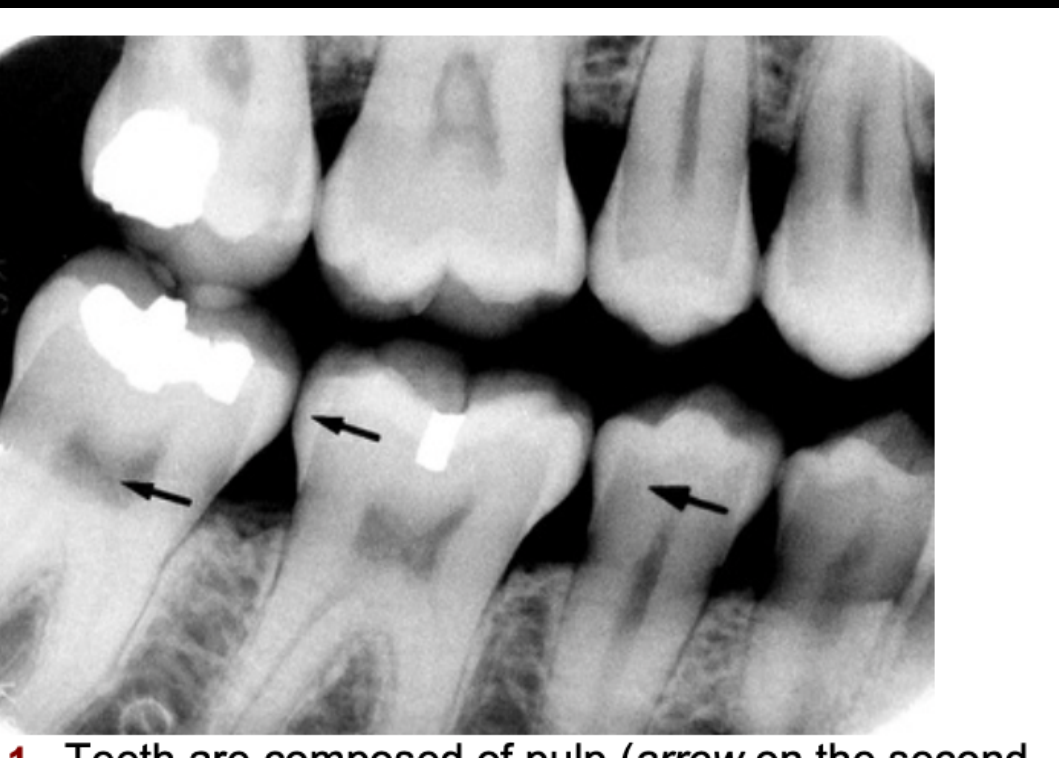
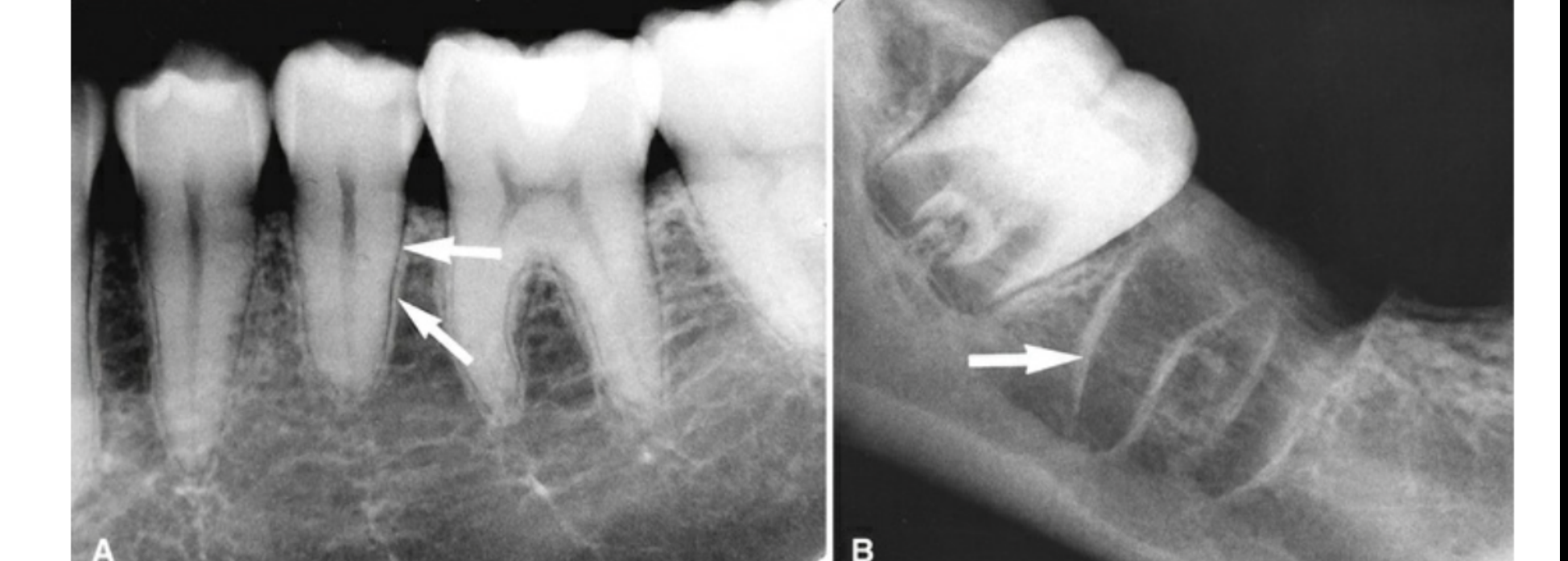
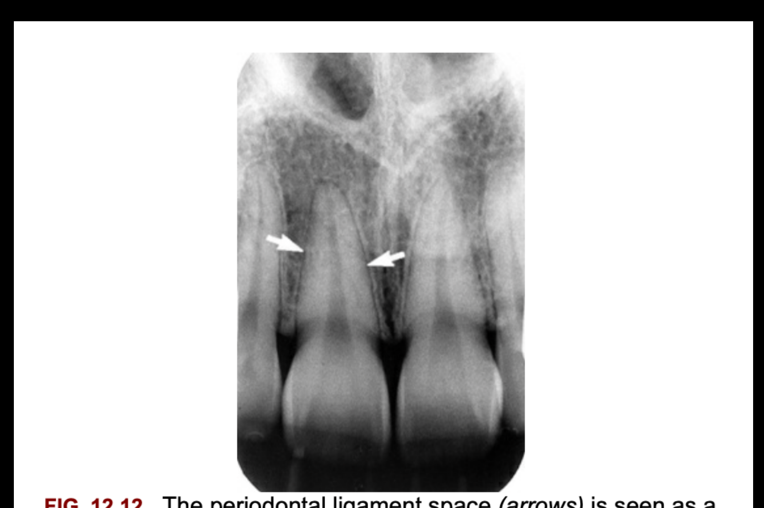
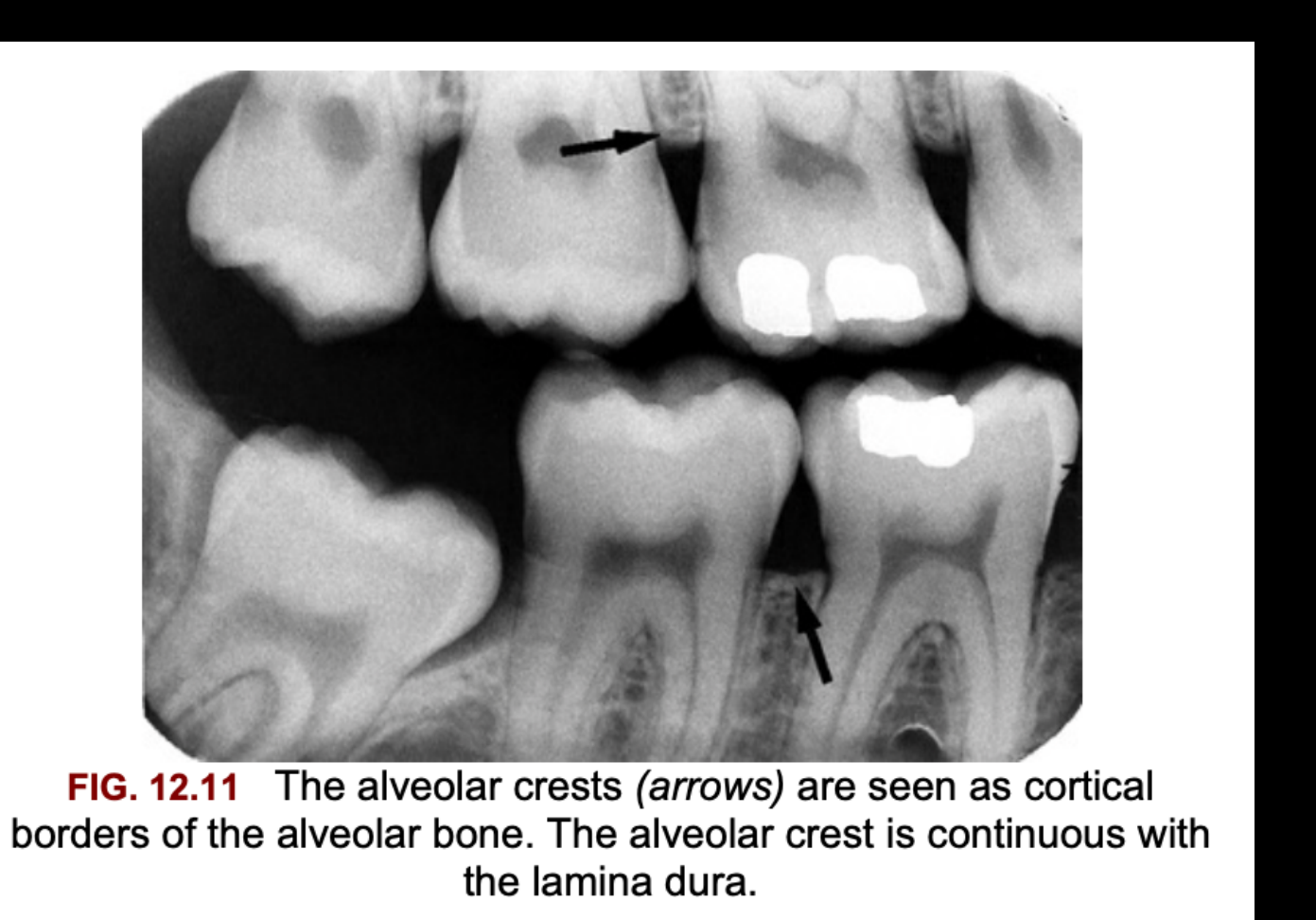
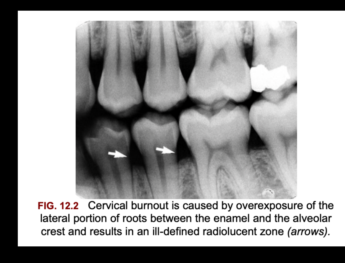
Maxillary Anterior (Incisor/Canine) PA
| Landmark | Radiodensity | How to recognise / location | Pitfall (mimic) |
|---|---|---|---|
| Intermaxillary (median) suture | RL line | Thin midline line between the central incisors, running from the alveolar crest up to the ANS. Has cortical borders on both sides1415 | Can end in a V-shaped widening at the crest. This is normal, not periodontal bone loss15 |
| Anterior nasal spine (ANS) | RO | Midline V-shaped projection about 1.5–2 cm above the alveolar crest, at the base of the nasal septum14 | — |
| Nasal fossa (aperture) and floor | Fossa RL / floor RO line | The floor is an opaque line running out to each side from the ANS. The RL nasal cavity sits above it. Can project high on a canine PA1415 | In posterior views, the nasal floor can look like a sinus septum or a sinus roof14 |
| Nasal septum | RO (wide, ill-defined) | Rises in the midline from the ANS. Made of cartilage and vomer, with mucosa on each side. Often slightly off the midline1415 | — |
| Inferior concha | Soft-tissue RO | Projects from the lateral nasal wall into the nasal fossa14 | — |
| Incisive foramen / nasopalatine canal | RL (ovoid, often diffuse borders) | Between the central incisor roots, in the middle to apical third. The canal walls can show as two vertical RO lines running up to the nasal floor14 | Can overlap one incisor apex and mimic a periapical lesion (check for intact lamina dura). Suspect a nasopalatine duct cyst if it is wider than 1 cm or enlarging over time14 |
| Superior foramina of the incisive canal (foramina of Stensen) | RL (paired, round) | In the nasal floor, just lateral to the septum and behind the ANS. Mostly seen with a steep vertical angle (occlusal view)1415 | — |
| Lateral (incisive) fossa | RL (diffuse) | A depression in the buccal bone near the lateral incisor apex1415 | Periapical lesion. An intact lamina dura and no symptoms mean it is normal14 |
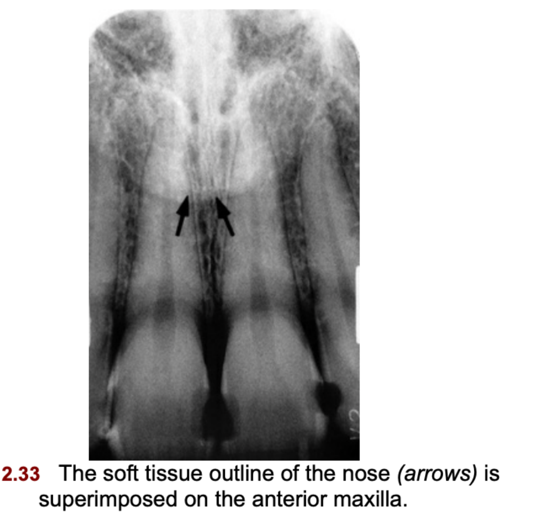
| Soft tissue of the nose | Soft-tissue RO, sharp edge | Uniform faint opacity over the central and lateral incisor roots. The nares can show as RL areas at a steep angle1415 | — |
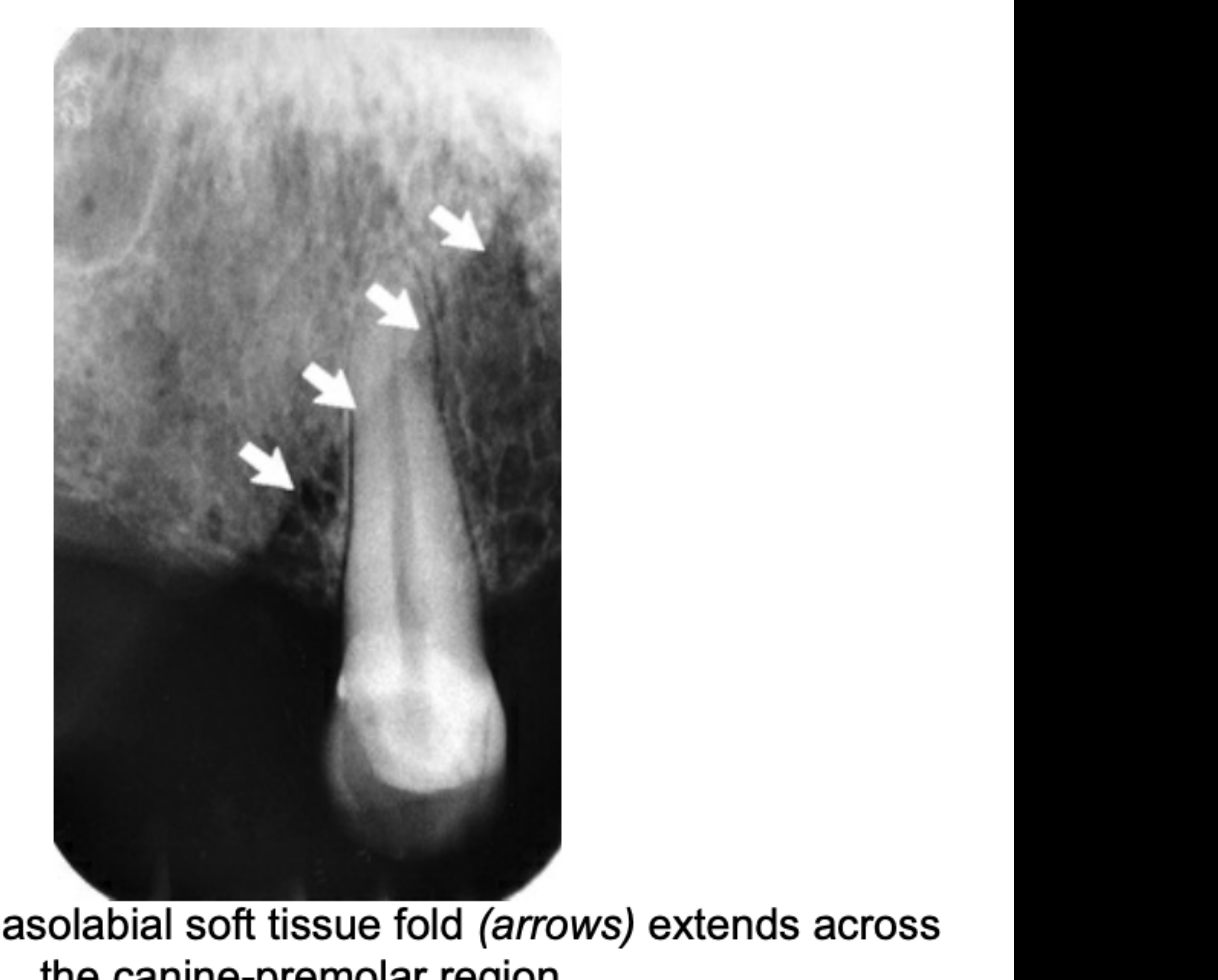
| Nasolabial fold | Faint RO veil, sharp oblique line | Runs across the canine–premolar region. The opaque zone lies behind (posterior to) the line. More obvious with age1415 | Useful for telling which side an edentulous film comes from14 |
| Inverted Y | RO lines | On a canine PA, the floor of the nasal fossa and the anterior wall of the maxillary sinus cross, making an inverted Y1415 | — |
| Canine eminence | — | Not described in L5 or W&P ch.12 | — |
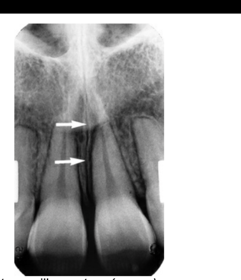
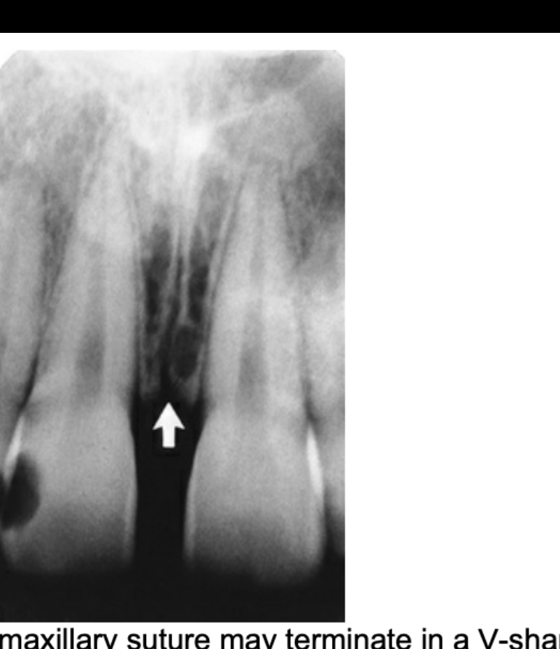
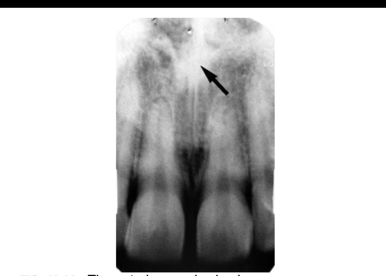
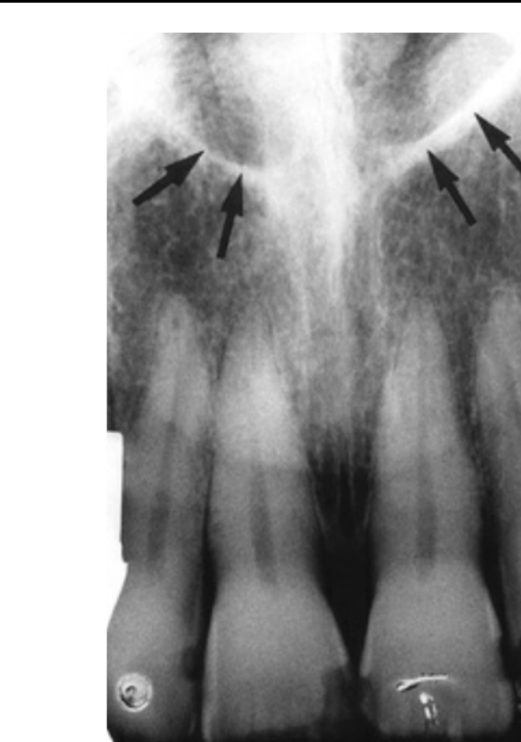
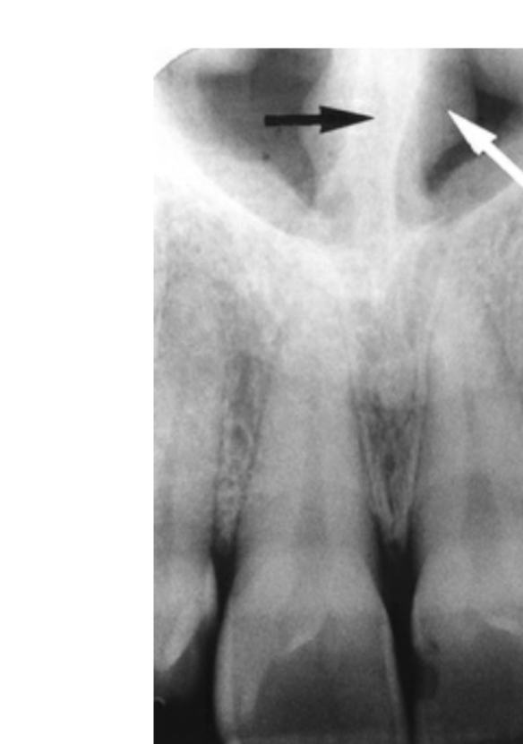

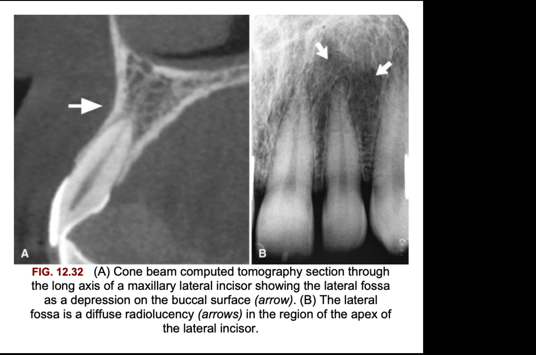

Maxillary Posterior (Premolar/Molar) PA
| Landmark | Radiodensity | How to recognise / location | Pitfall (mimic) |
|---|---|---|---|
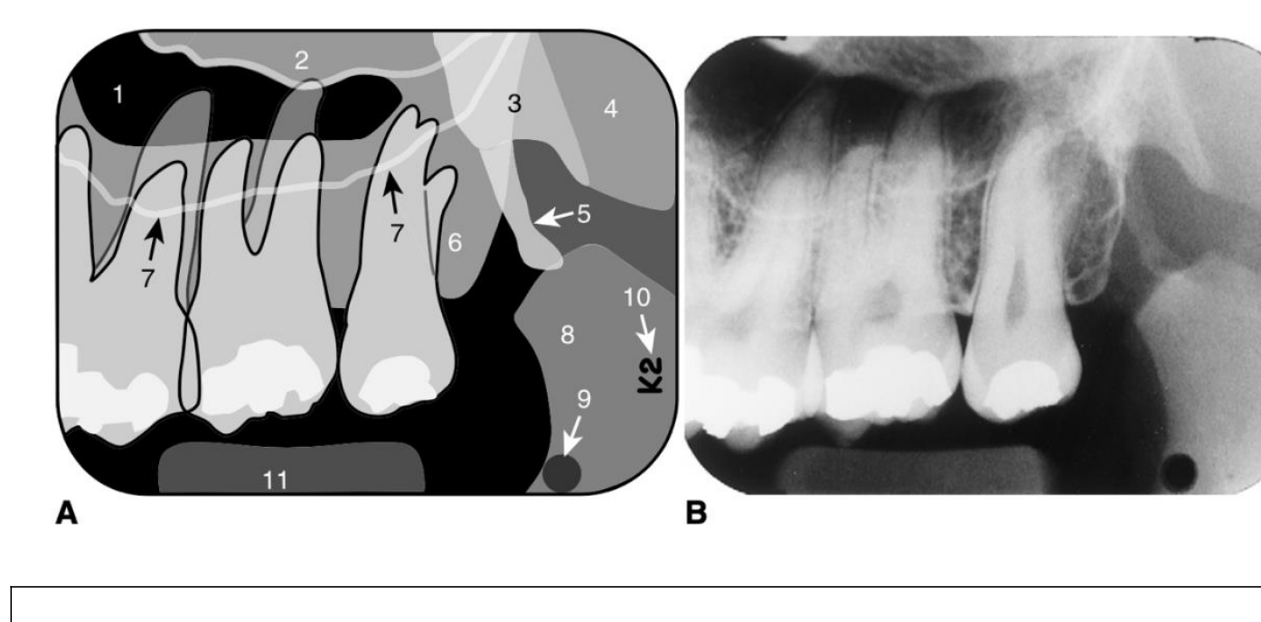
| Maxillary sinus (air space) | RL | Spans from distal of the canine back to the tuberosity. Reaches adult size at 15–18 yr14 | Roots can appear to project into the sinus. This is an illusion from superimposition14 |
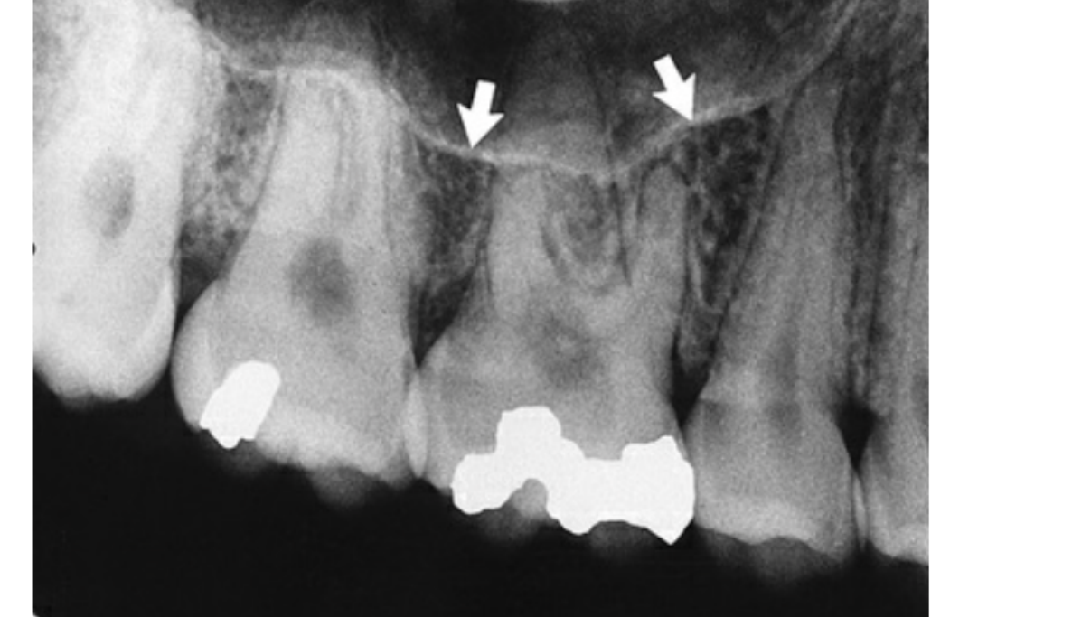
| Sinus floor | RO (thin line) | Thin cortical line near the premolar and molar apices. Tiny gaps in it are usually overlapping marrow spaces1415 | The lamina dura can rarely be absent on a molar root that sits in the sinus14 |
| Alveolar recess | RL | The sinus floor dips toward the ridge, especially after tooth loss1417 | Not a lesion. A prominent recess at an edentulous site is normal17 |
| Sinus septa | RO (thin, vertical lines) | Folds of cortical bone projecting a few mm from the floor or wall. Seldom divide the sinus fully14 | Can mimic periapical disease. They also complicate the search for a root displaced into the sinus (use CBCT)14 |
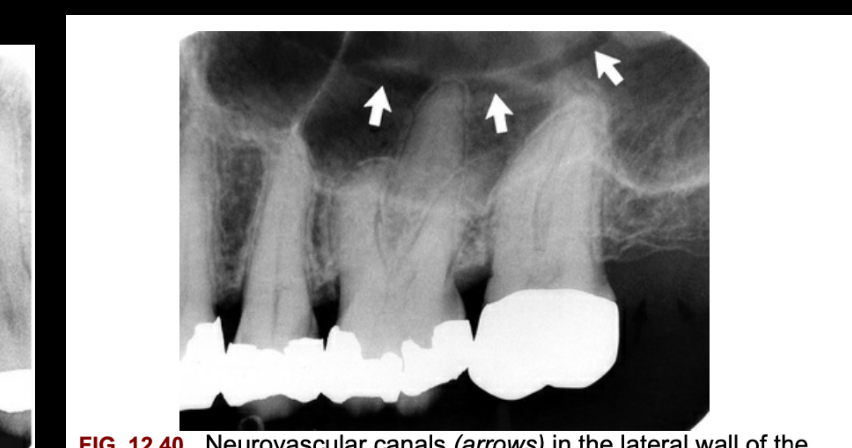
| Neurovascular canals | RL lines with uniform width | Grooves in the lateral sinus wall for the PSA vessels and nerves. Usually curved and convex toward the ridge1415 | Normal finding. Recognise them by their uniform width and curved course14 |
| Bony nodule on the sinus floor | RO | Small projection that shows trabeculation and blends into the floor14 | Root tip. A root is homogeneous and has a canal14 |
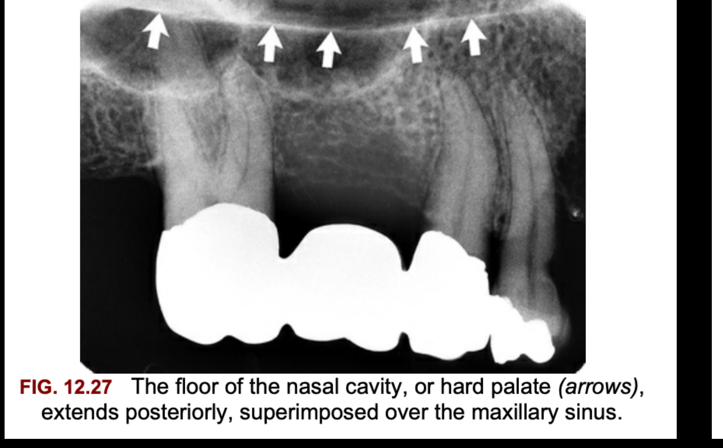
| Floor of the nasal cavity / hard palate | RO line (thick) | Roughly horizontal, high up on the film, superimposed over the sinus. Often thicker than the sinus walls1415 | Can look like a sinus septum or a sinus roof14 |
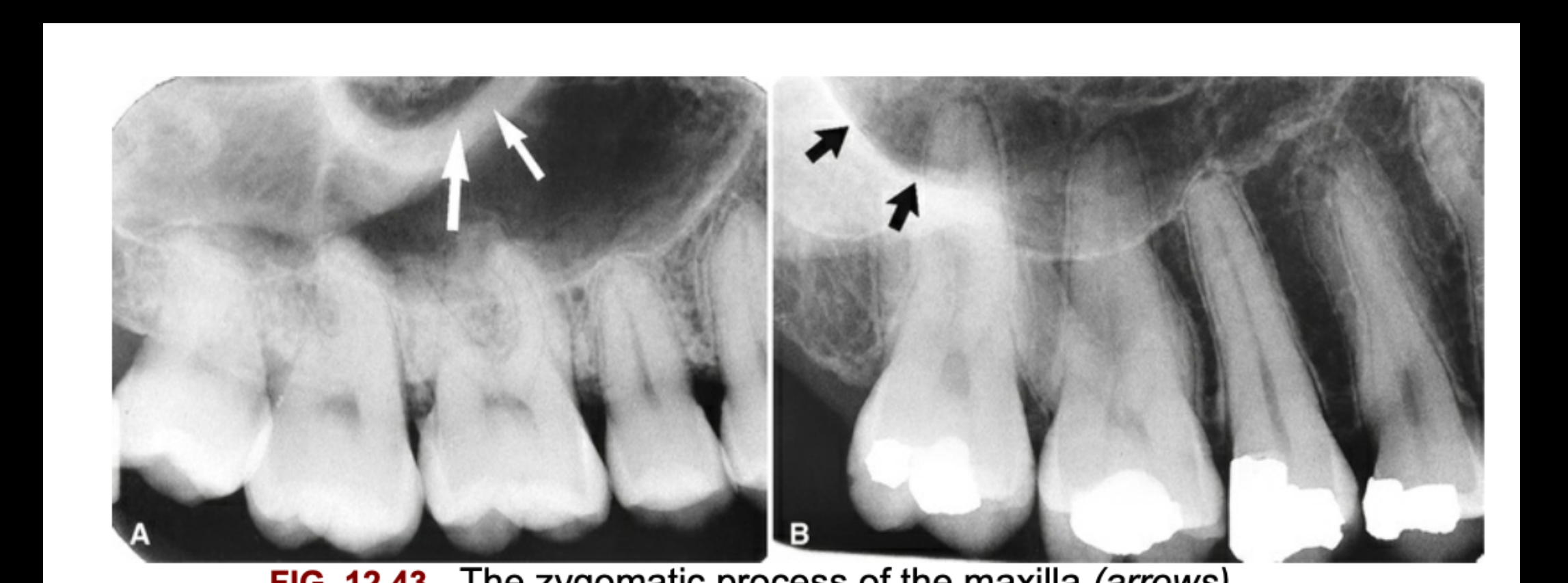
| Zygomatic process of maxilla (zygomatic buttress) | RO | U-shaped line, open end up, with its rounded end over the M1/M2 apices. Can be small and thick-walled, or large and thin-walled when the sinus extends into it1416 | Can hide the molar apices14 |
| Zygoma (inferior border) | RO (uniform band) | Runs back from the zygomatic process, over the molar apices14 | Hides periapical detail. Change the beam angle14 |
| Maxillary tuberosity | RO (alveolar bone) | Rounded distal end of the alveolar process behind the last molar1614 | — |
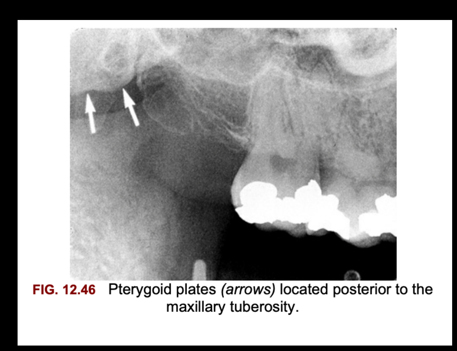
| Pterygoid plates (medial/lateral) | RO (homogeneous) | Behind the tuberosity. Often not captured on the film at all1415 | — |
| Hamular process | RO | Projects down from the medial pterygoid plate, behind the tuberosity. May show trabeculae1416 | — |
| Coronoid process of mandible | RO (triangular) | Apex points up and forward, over the M3 region (sometimes as far forward as M2). Shows up because the mouth is open1416 | Can be taken for a root fragment. Retake with the mouth barely open and see whether it moves14 |





Mandibular Anterior PA
| Landmark | Radiodensity | How to recognise / location | Pitfall (mimic) |
|---|---|---|---|
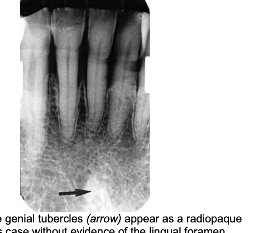
| Genial tubercles | RO (3–4 mm mass) | Midline, lingual, below the incisor roots and just above the inferior border. Attachment for genioglossus (superior) and geniohyoid (inferior). Sometimes not visible1416 | — |
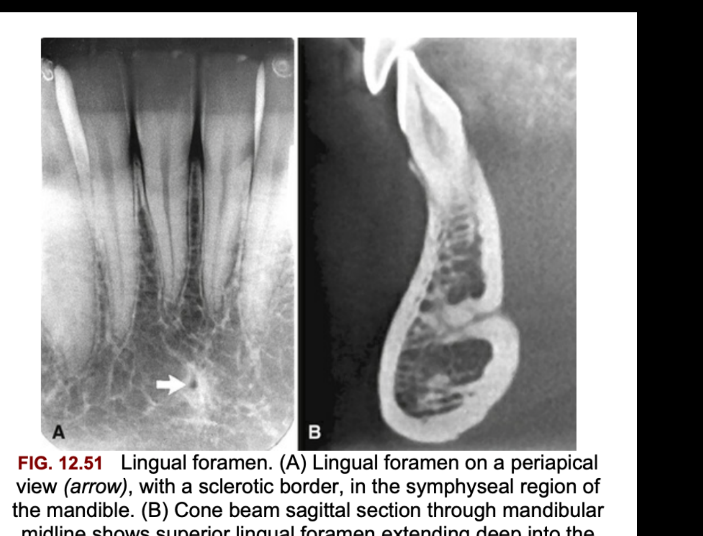
| Lingual foramen | RL dot, often with a sclerotic RO rim | Midline, inside or just above the genial tubercles. Present in 96–100% of people1415 | Periapical lesion. Matters when planning implants because of bleeding risk14 |
| Mental ridge | RO (paired lines) | Lines sweep up and forward from the premolar region to the midline, below or over the incisor roots. Most obvious with bisecting-angle technique14 | — |
| Mental fossa | RL (ill-defined) | Labial depression above the mental ridge, over the incisor roots. The bone is thin here1415 | Periapical disease of the incisors14 |
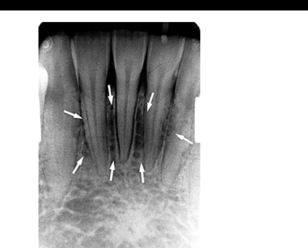
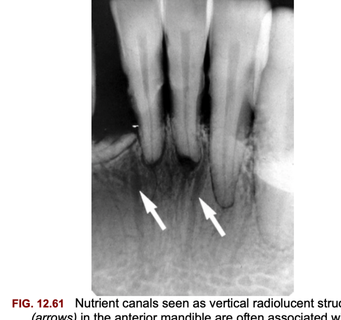
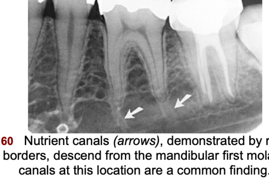
| Nutrient canals | RL lines with uniform width, sometimes RO borders | Vertical lines in the interdental septa between the incisors. More common with periodontal disease, age and hypertension1415 | End-on canal = small round RL that looks like a lesion14 |
| Inferior border (cortex) | RO (dense, broad) | Band at the bottom of the film1416 | — |
| Mandibular symphysis (infant) | RL midline line | Fuses by the end of the 1st year14 | If still seen in an older patient: think fracture or cleft14 |

Mandibular Posterior PA
| Landmark | Radiodensity | How to recognise / location | Pitfall (mimic) |
|---|---|---|---|
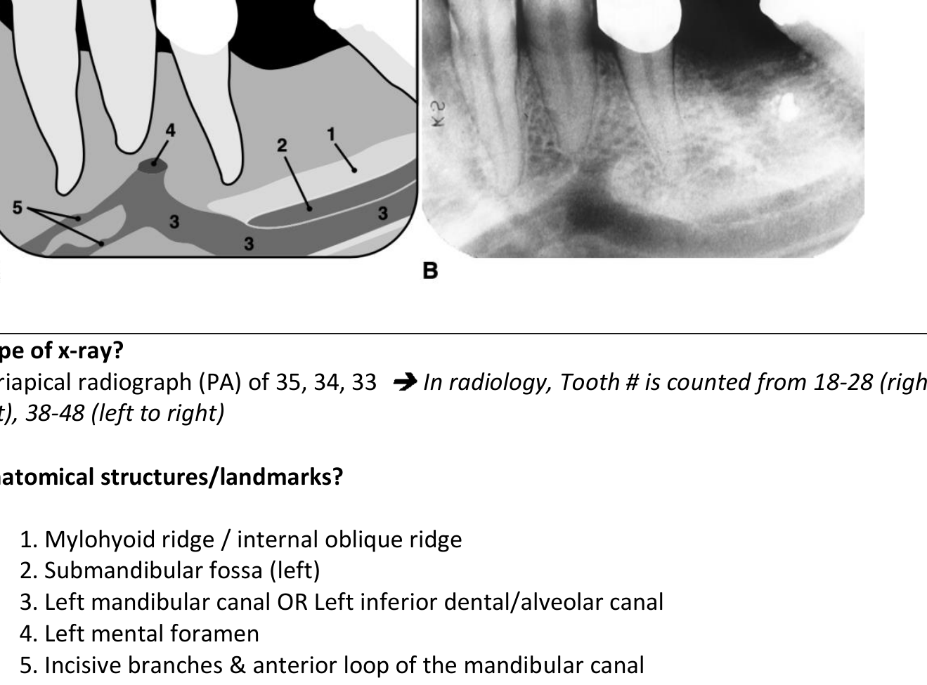
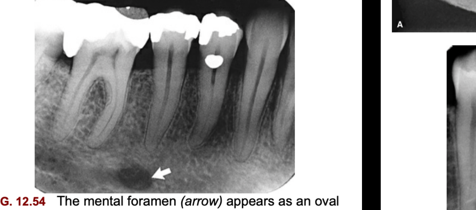
| Mental foramen | RL (oval, round or slit-like; often no cortical rim) | Halfway between the crest and the inferior border, usually at the PM2 apex. Can project anywhere from mesial of M1 to mesial of PM1. Visible on only about half of PAs1416 | Periapical lesion on a premolar. Look for an intact lamina dura and the IAC running into it, and check whether it moves on a second angled film1415 |
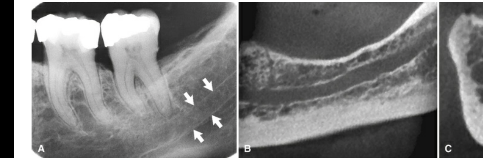
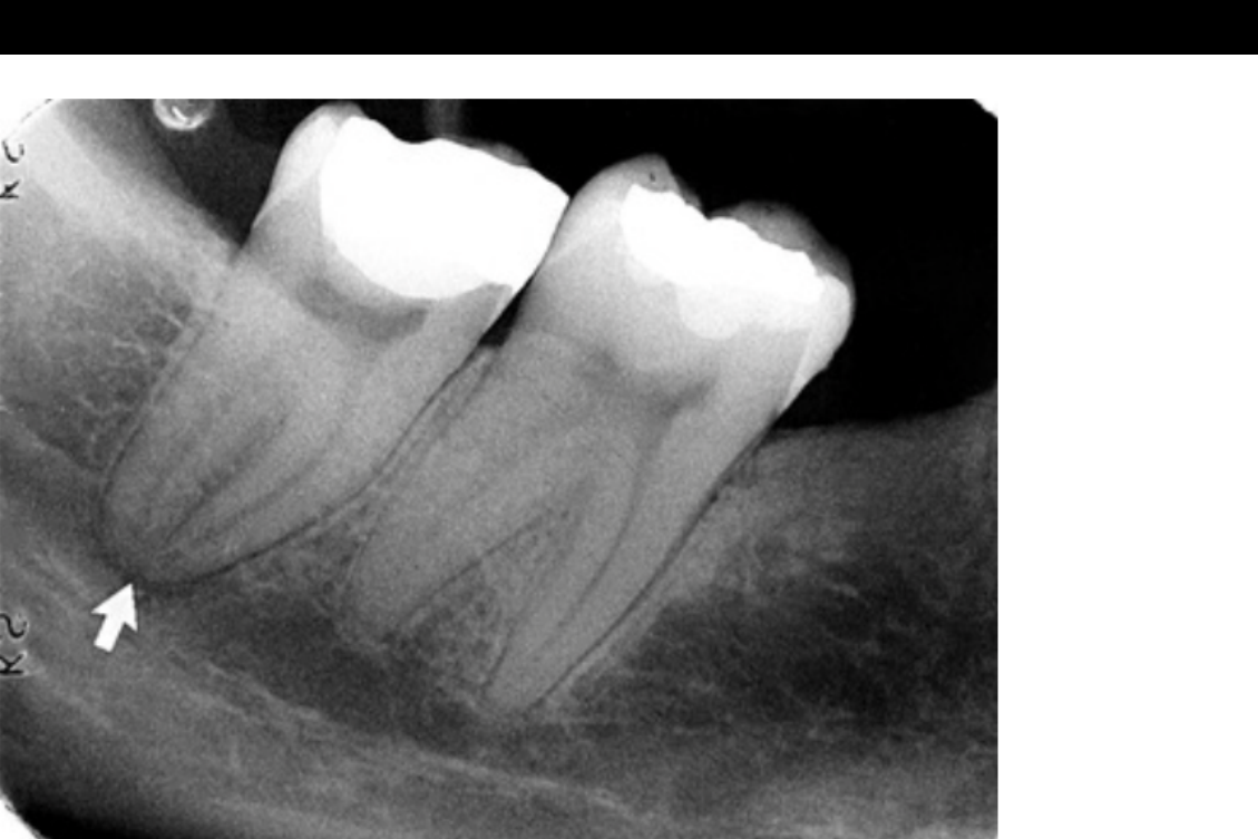
| Mandibular (inferior alveolar) canal | RL band between thin RO superior and inferior borders | Runs from the mandibular foramen to the mental foramen, just below the molar apices, usually touching M31415 | Where it overlaps a molar apex, the PDL can look widened. An intact lamina dura rules out disease. Changing the vertical angle rarely separates the two1415 |
| Anterior loop / incisive branch | RL | The canal continues forward past the mental foramen, then loops back to exit through it1416 | Relevant when placing implants14 |
| Nutrient canals | RL lines with uniform width, sometimes RO borders | Run vertically from the IAC to an apex, e.g. under M11415 | Can look like a lesion when seen end-on14 |
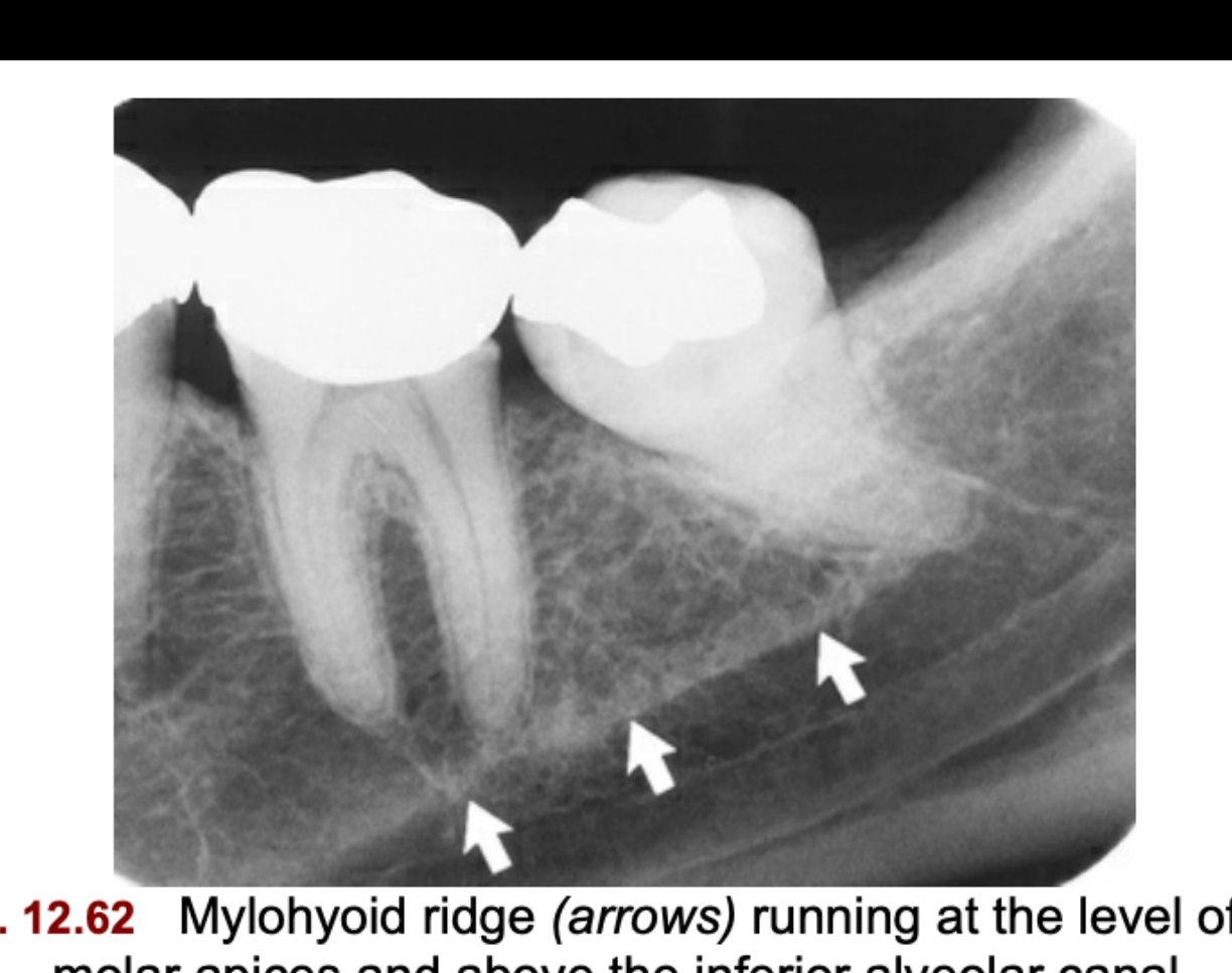
| Mylohyoid (internal oblique) ridge | RO (diffuse, variable width) | Runs diagonally down and forward from M3 to the premolars, at about apex level and above the IAC. Denser with excessive negative angulation1415 | Can overlap the molar roots14 |
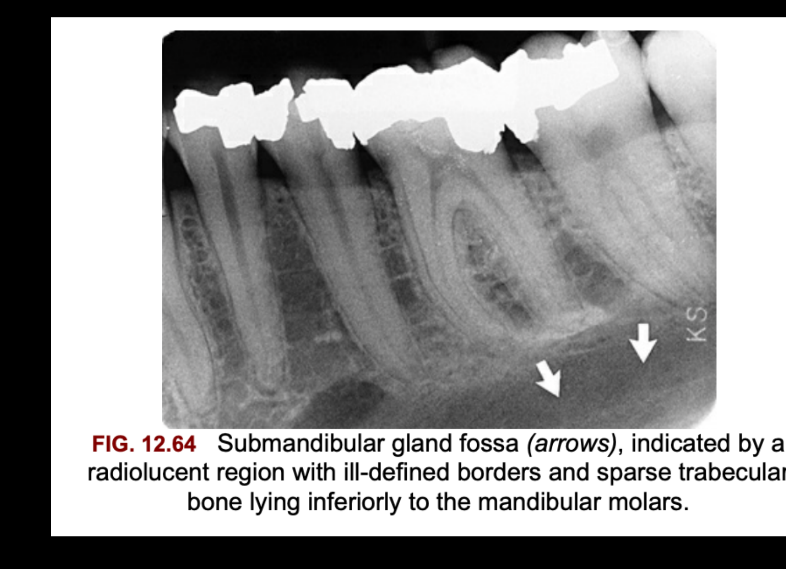
| Submandibular (gland) fossa | RL, sparse trabeculae | Below the mylohyoid ridge in the molar region, bounded below by the inferior border. Poorly defined front and back1415 | Bony lesion / cyst14 |
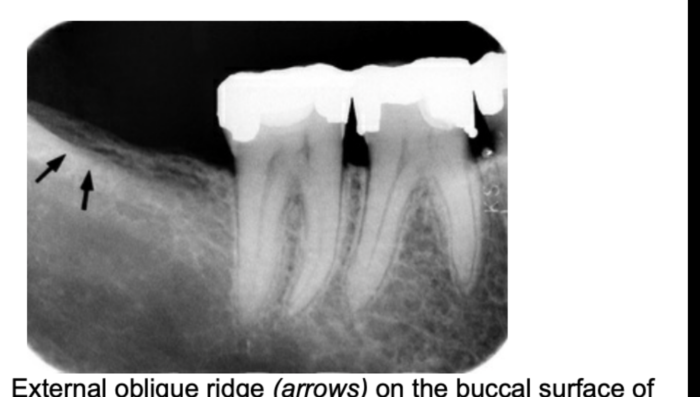
| External oblique ridge | RO line | Continues from the anterior ramus onto the buccal bone near the crest at M3, and fades below M1. Projects above and roughly parallel to the mylohyoid ridge1415 | — |
| Inferior border | RO (dense, broad band) | Seen at the bottom of the film when the PA is placed low1415 | — |

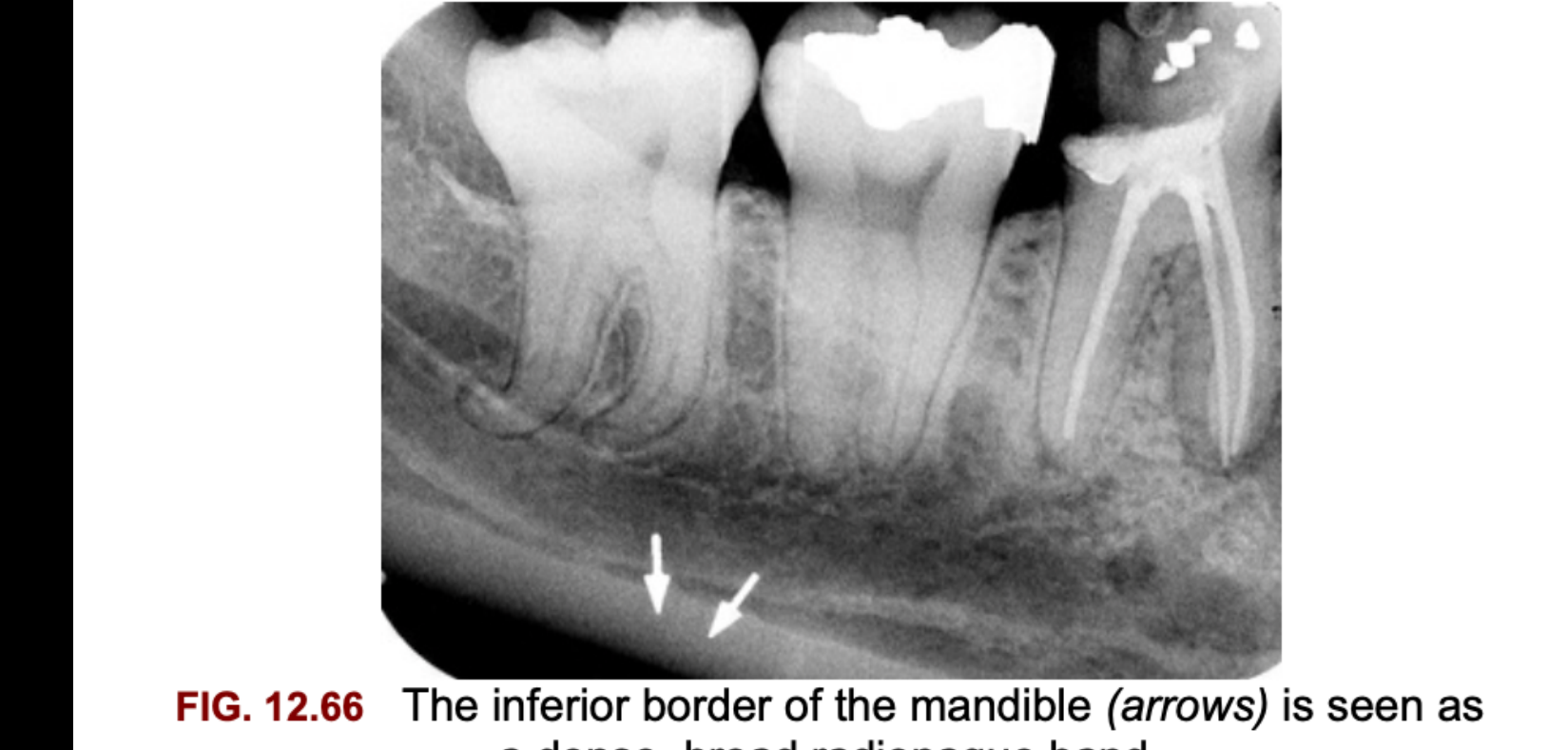
Tooth-Level Findings
Where this fits
This is step 2 of the lecture’s systematic approach: identify restorative, endodontic and orthodontic work, and check the margins for overhangs, insufficient margins and voids. Step 1 covers teeth and surrounding structures, and step 3 covers abnormal radiolucencies and radiopacities.18
Caries
Principle: demineralised tooth lets more photons through, so caries shows as a radiolucency. About 35% of the enamel must be demineralised before a proximal lesion is visible, so the true (histological) depth is usually greater than it looks.19
| Site | Where to look | Radiographic appearance |
|---|---|---|
| Interproximal | Between the contact point and the free gingival margin. With bone loss, look lower (at or near the CEJ). A surface touching a lesion or restoration is higher risk | Enamel lesion: a triangle with its base at the surface and its apex toward the DEJ (it can also look like a band, rectangle or notch). At the DEJ it spreads, then forms a second, wider triangle in dentine pointing at the pulp19 |
| Occlusal | Under pits and fissures | Enamel-only lesions are nearly impossible to see (too much sound enamel overlaps them). A dentine lesion is semicircular or round with its base at the DEJ, centred on the pit. Clinical exam is more accurate for small lesions19 |
| Buccal / lingual | Cervical smooth surfaces, pits | Round or oval, well defined, surrounded by intact tooth. Buccal vs lingual can’t be told apart on one image, so take a second image at a different horizontal angle (tube shift)19 |
| Root | Exposed root, only with recession or bone loss | Cementum is only 20-50 µm thick near the CEJ, so these lesions progress fast. Main differential: cervical burnout19 |
| Recurrent / secondary | At the tooth-restoration margin | New radiolucency at the junction. Poor vertical angulation lets the restoration hide it. Residual caries is different: it was left behind on purpose (e.g. indirect pulp cap)19 |
ICCMS radiographic depth staging (proximal)19
| Score | Radiolucency reaches | Cavitation likelihood / management |
|---|---|---|
| 0 | Sound | — |
| RA1 | Outer ½ enamel | Non-operative (OHI, fluoride), then monitor |
| RA2 | Inner ½ enamel ± DEJ | Non-operative, then monitor |
| RA3 | Outer ⅓ dentine | ~32% cavitated; usually non-operative |
| RB4 | Middle ⅓ dentine | ~72% cavitated (middle ⅓ or deeper); operative |
| RC5 | Inner ⅓ dentine | Operative |
| RC6 | Into the pulp | Operative. Also take a PA to check the apex |
- A single image can’t tell active from arrested. You need a second image at a later date, ideally with the same geometry.19

Mimics
| Mimic | Why it happens | How to tell it apart |
|---|---|---|
| Cervical burnout (the commonest false positive) | Fewer photons are absorbed where the beam passes tangentially through the convex root just below the CEJ, near crest height. It also appears where multirooted teeth don’t overlap | Its shape follows tooth anatomy, and the surface is intact. Caries has an irregular border and a break in the surface. If there’s no bone loss, a cervical lucency is very likely burnout19 |
| Mach band | A visual (retinal) effect: a dark band in dentine next to the bright enamel at the DEJ, most obvious occlusally | Mask the enamel. If the band goes away, it was Mach band. If it stays, the DEJ is involved. Image-only with no clinical sign: monitor19 |
| Radiolucent restorative materials, liners and bases | Older composites and some bases are lucent | Well-defined, regular (cavity-prep) outline, often with a radiopaque liner at the edge. Liners and bases shouldn’t reach the cavosurface margin, so a lucency at the margin is suspicious. Check the history19 |


PA vs bitewing for caries
- The bitewing is the preferred image for interproximal caries. Its beam is nearly perpendicular and the contacts are open. On a PA, steep vertical angulation can let the crown or restoration overlap and hide proximal and recurrent lesions.19
- Overlapping contacts (wrong horizontal angle) are the commonest technical cause of false negatives.19
- A restoration can hide buccal or lingual recurrent lesions whatever the beam angle.18

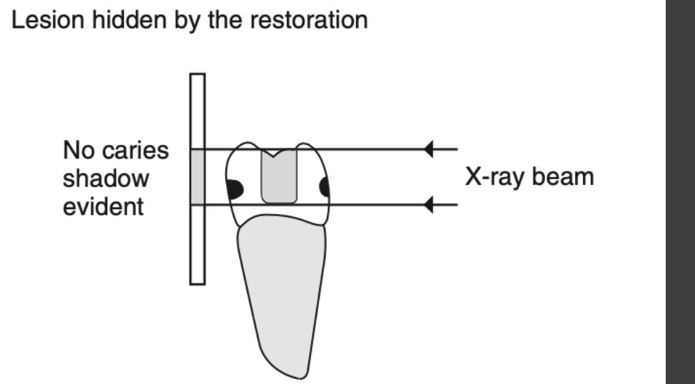
Restorations and Materials
Radiodensity depends mostly on atomic number, then on thickness and density.14
| Material | Radiodensity / appearance |
|---|---|
| Amalgam | Completely radiopaque14 |
| Gold (cast crown, inlay, foil) | As opaque as amalgam14 |
| Stainless steel crowns and pins | Highly radiopaque14 |
| Composite | Partially radiopaque, and it varies. Older composites can be radiolucent and mimic caries1419 |
| Porcelain / PFM | Porcelain is partially radiopaque and looks relatively lucent over the metal coping, which is fully opaque14 |
| GIC | Restorative GICs are formulated to be radiopaque (e.g. Fuji Bulk)20 |
| Base / liner (e.g. calcium hydroxide) | Usually made radiopaque, but less opaque than amalgam, so that recurrent caries can be seen. Some bases are lucent1419 |
| Gutta-percha | Radiopaque, but less than amalgam14 |
| Silver points (old RCTs) | Highly radiopaque14 |
| Orthodontic appliances | Radiopaque, less than stainless steel crowns14 |
| Implants | Radiopaque fixture. On a baseline PA, check for bone against the threads and a re-formed crest. Failure signs: a uniform lucent line around the fixture (no osseointegration) or crestal bone loss spreading apically (peri-implantitis)21 |
What to assess on every restoration182221
- Overhangs and poorly contoured or overextended margins. They trap plaque and cause localised bone loss.
- Open or insufficient margins, i.e. gaps between restoration and tooth. Check for recurrent caries at the margin.
- Voids inside the restoration.
- Open contacts and uneven marginal ridges, which let food pack in.
- Implants: aim the beam perpendicular to the fixture. A few degrees off blurs the threads and can hide a gap between abutment and fixture.

Endodontic Assessment
| Check | Acceptable | Problem signs |
|---|---|---|
| Obturation length | GP ends 0.5-2 mm short of the radiographic apex23 | Short (under-filled) or extruded past the apex |
| Density | Uniform density along the whole canal, with no voids23 | Voids, lucent gaps, poorly condensed fill |
| Material | GP is radiopaque but less dense than amalgam. Very opaque silver points mean an older RCT14 | — |
| Post length | Leave 4-5 mm of apical GP for the seal. Post length ≥ crown length (about ⅔ of tooth length, or ½ the clinical root)24 | Too short; no apical GP left |
| Post width | ≤ ⅓ of root width24 | Wide post with thin dentine walls |
| Perforation | Fill stays within the root outline | A 2D PA can make a root filling look acceptable when it has actually left the root. Suspect this if a lesion doesn’t match the canal. CBCT is the tool for this25 |
| Periapical status | Compare with the pre-op or baseline image | Record the PDL and lamina dura at the apex (see periapical section) |

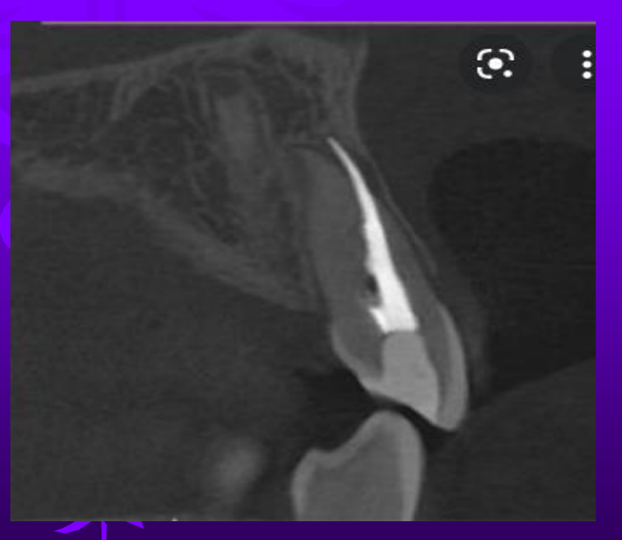
Pulp and Root Changes
| Finding | Appearance | Notes |
|---|---|---|
| Secondary / tertiary dentine | Pulp chamber and canals get smaller. From ageing it’s generalised, with the normal shape kept (pulp horns go first). From a local stimulus (caries, attrition, restoration) it’s localised and changes the chamber’s shape26 | Looks the same as primary dentine on imaging |
| Pulp stones | Discrete radiopacities in the chamber or canal. Single or multiple, round or oval, or shaped to fit the chamber. Most common in molars26 | No treatment. Matters for endo access. Only large stones show up |
| Pulpal sclerosis | Diffuse, ill-defined fine radiopacities through the chamber and canals26 | Linked to age. Hard to tell apart from small stones, and it doesn’t matter clinically |
| Hypercementosis | Bulbous, usually smooth enlargement of the apical root. Cementum is slightly less opaque than dentine. The PDL space and lamina dura go around the extra cementum26 | Differentials: dense bone island, periapical osseous dysplasia, cementoblastoma. The PDL wrapping round it is the key sign. Paget disease gives an exuberant, irregular form |
Internal vs external resorption26
| Feature | Internal | External |
|---|---|---|
| Origin | Pulp wall (odontoclasts inside the canal) | Outer root surface; needs a blood supply from soft tissue |
| Shape | Round or oval, sharp, smooth or slightly scalloped ballooning of the canal | Apical: smooth blunting. Lateral: irregular, often more on one side |
| Canal outline | Lesion is continuous with the canal; the canal wall is lost inside it | The normal canal can be traced through the lucency |
| Tube shift | Lucency stays fixed to the canal | Lucency moves relative to the canal |
| Internal density | Homogeneously lucent, no trabeculae | Bone can grow into the defect (inostosis) |
| Lamina dura | — | Usually keeps up with the shortened root (intact). It’s lost if the cause is periapical inflammation |
| Differential | Buccal or lingual caries (more diffuse borders; separates from the canal on tube shift) | Caries, internal resorption |
- Replacement (ankylosis-related) resorption: no PDL space visible, with root structure being replaced by bone. Clinically: a metallic percussion note, no mobility, and infra-position in growing patients. Expected after delayed replantation of avulsed teeth. It can happen alongside inflammatory resorption, which shows root or bone resorption anywhere around the root.27


Dental Anomalies Seen on PAs
| Anomaly | PA appearance |
|---|---|
| Supernumerary / mesiodens | Normal, smaller or conical tooth; can be inverted; may block eruption of neighbouring teeth. CBCT for number and position26 |
| Dens invaginatus | Inverted teardrop lucency with a radiopaque (enamel) border, most often a maxillary lateral incisor. Severe forms: open apex and periapical lesion (pulp necrosis)26 |
| Taurodontism | Elongated pulp chamber, apically placed furcation, short roots; normal crown size26 |
| Dilaceration | Sharp bend in the root. Bent buccally or lingually it looks like a bull’s-eye (radiopaque ring, central canal, PDL halo)26 |
| Concrescence | Roots joined by cementum; often can’t be confirmed on one image. No PDL between the roots supports it. Take extra angles or CBCT before extraction26 |
| Fusion vs gemination | Both: a large crown and unusual pulp. Fusion has one tooth fewer; gemination keeps a normal count with a single, enlarged, partly split pulp chamber26 |
| Enamel pearl | Smooth, round opacity of enamel density, 1-3 mm, at or just below the CEJ in molar furcations. Can look like calculus; a site for pockets26 |
| Amelogenesis imperfecta | Hypoplastic: very thin enamel, no contacts (“picket fence”). Hypomineralised: enamel radiodensity reduced26 |
| Dentinogenesis imperfecta | Bulbous crowns, cervical constriction, short thin roots, obliterated pulps26 |
| Dentin dysplasia | Type I: short or absent roots, obliterated pulps, periapical lucencies on caries-free teeth. Type II: thistle-tube / flame-shaped pulp chambers, pulp stones, normal roots26 |


Periodontium
Normal
| Structure | Normal appearance |
|---|---|
| Alveolar crest height | 0.5-2.0 mm apical to the CEJ.1422 The perio lecture also gives an average CEJ-to-crest distance of 1.5-2 mm.25 Measure bone loss from this normal level, not from the CEJ22 |
| Crest shape | Posterior: flat, parallel to the line joining neighbouring CEJs. Anterior: a pointed, corticated tip22 |
| Crest cortex | A thin radiopaque cortex means no periodontitis. No cortex doesn’t prove disease (it can be missing in health)22 |
| Crest-lamina dura junction | A sharp angle next to the root (posterior)1422 |
| Lamina dura | Thin radiopaque line continuous with the crest. It may not be continuous in health25 |
| PDL space | Thin, even lucency around the whole root. Normally wider at the crest and apex, thinner mid-root; wider cervically in premolars and erupting teeth. Still normal if the crest-lamina dura angle stays sharp1422 |

Bone Loss
| Feature | What you see |
|---|---|
| Early changes | Crest cortex fades; the crest-lamina dura angle gets “rounded off”; anterior crests blunt. X-rays lag behind real bone loss (stage I may not show)22 |
| Horizontal | Crest height is reduced, but the crest stays parallel to the line between CEJs. Buccal plate, lingual plate and interdental bone are all lost22 |
| Vertical / angular (infrabony) | V-shaped defect down one root, oblique to the CEJ line. It starts as PDL widening at the crest. One-, two- or three-walled types can’t be reliably told apart in 2D. A GP point can trace the defect22 |
| Interdental crater | Band or trough of lower density at the crest, between buccal and lingual walls that are still there; more common posteriorly2225 |
| Buccal / lingual plate loss | Root looks more lucent near the crest (semicircular); easy to miss if the interproximal bone is intact22 |
| Furcation | Early: PDL widening at the tip of the interradicular crest. Advanced: an obvious furcation lucency. Maxillary molars: less clear because the palatal root overlaps; look for an inverted “J” or a triangle pointing at the furcation. In the mandible, the external oblique ridge or converging roots can hide third molar furcations2225 |
| Trabecular reaction | Acute: more lucent (rarefaction). Chronic: sclerosis. Usually a mix. Perio lesions next to the sinus can cause mucositis22 |
| Pseudo-defects | CEJs at different heights make the crest look angled; supraerupted teeth or passive eruption from attrition put the CEJ far from the crest without periodontitis22 |
How much bone is lost (% of root length, using the PA)2225
| Stage (2017) | Radiographic bone loss |
|---|---|
| I | Coronal ⅓, < 15% |
| II | Coronal ⅓, 15-33% |
| III / IV | Reaches the middle ⅓ and beyond (IV = III complexity + severe ridge defect / < 20 teeth) |
- The perio e-form uses thirds: mild < 33%, moderate 33-66%, severe > 66%. Also record the pattern (horizontal or vertical) and the distribution (localised < 30% of sites, generalised > 30%).25
- For full staging and grading, see Periodontitis Staging and Grading.
- One image shows past destruction, not current activity. A re-formed crestal cortex is a good sign the disease has stabilised.2225


Local Factors
| Factor | Appearance |
|---|---|
| Calculus | Small angular radiopaque spurs sticking out between teeth, or radiopaque bands across the root (circumferential). Most common on mandibular incisors22 |
| Overhangs / poor contours | Excess radiopaque restoration past the margin, with localised bone loss22 |
| Open contacts, uneven marginal ridges, tipped teeth | Food trap sites where localised disease can start22 |
| Enamel pearls / cervical enamel projections | Enamel-density opacities in the furcation; plaque-retentive2226 |

PDL Widening: Causes
| Cause | Clues |
|---|---|
| Occlusal trauma | Widened PDL, thickened lamina dura, more and bigger trabeculae; hypercementosis or root fracture possible. Diagnosis is clinical only2225 |
| Mobility (trauma or reduced support) | Single root: hourglass-shaped socket. Multirooted: widening at the apices and furcation. Lamina dura broad and hazy22 |
| Inflammation | Early vertical defect: widening at the crest. Early furcation involvement: widening at the interradicular crest22 |
| Malignancy | Irregular widening along the whole root length with a ragged edge and lamina dura destroyed22 |
| Superimposition (false widening) | Inferior alveolar canal over the apex. An intact lamina dura rules out disease14 |
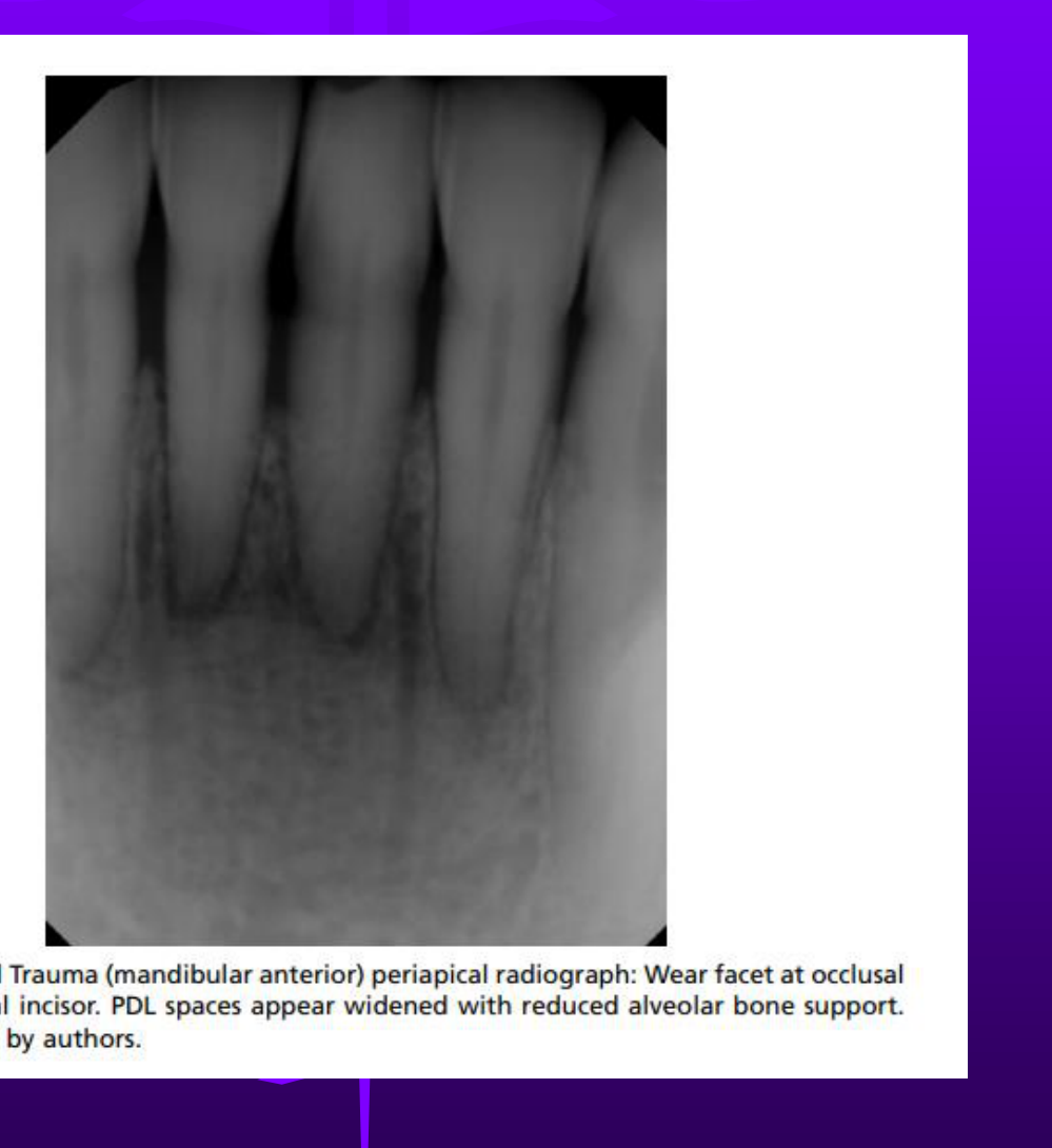
Endo-Perio Lesion Clue
- A deep angular defect running from the crest to the apex and joining a periapical rarefying osteitis. It’s usually of even width or widens slightly at the crest (funnel-shaped).2225
- It can be on one surface, several, or all the way round the root. Treatment needs both endo and perio therapy. See Endo-Periodontal Lesions.22

When it isn't periodontitis
- Periodontal bone loss starts at the crest. Defects are wider coronally and narrower at the apical leading edge.22
- Be suspicious of these:22
- one or a few loose teeth when the rest of the mouth is healthy
- ill-defined destruction with no sclerotic reaction
- irregular PDL widening with the lamina dura destroyed (malignancy, e.g. SCC)
- destruction centred mid-root with a spared crest (Langerhans cell histiocytosis, which gives an “ice-cream scoop” or “floating teeth” look)
Limits of 2D for the periodontium
- Buccal and lingual plates overlap the defect, so the number of walls, craters and plate loss are under-read. Dehiscences and fenestrations can exist in health and still not show.2225
- X-rays underestimate bone loss and show no pocket depth or soft-tissue attachment, so interpret them alongside probing.2225
- The PA shows the whole root, so use it for the % of root length. The bitewing measures the CEJ-crest distance more accurately: on maxillary PAs a steep beam can project the buccal crest near the palatal CEJ, so bone looks higher than it is.22
- Changing beam angle or exposure changes how the crest looks. Sequential images can look like bone has “grown back”, or an over-exposed image can look like the crest is resorbed.2225
- Use CBCT only for selected surgical cases (complex vertical defects, furcations, plate loss). It isn’t for routine use or monitoring.2225
Periapical Region and Lesion Description
Describing Any Lesion
Run the same five steps on every abnormality, then interpret.628
| Step | Ask | Vocabulary / what it implies |
|---|---|---|
| 1. Location | Epicentre? Localized or generalized? Unilateral or bilateral? Single or multiple? | Periapical / pericoronal / furcation. Mandible: above IAC → likely odontogenic; below IAC → unlikely odontogenic; within IAC → neural/vascular. Maxilla: below vs within the antrum (inside the antrum → not odontogenic).628 Multifocal short list: COD, OKC, metastases, myeloma, leukaemia.6 |
| 2. Periphery | Can you trace the border with a pencil? How wide is the zone of transition? | Well-defined: punched-out (no bone reaction, e.g. myeloma), corticated (thin, uniform RO line: cysts, benign tumours), sclerotic (wider, uneven reactive bone, e.g. PCOD), soft-tissue capsule (RL rim inside cortex: odontoma, cementoblastoma). Ill-defined: blending (gradual change in the trabeculae: sclerosing osteitis, fibrous dysplasia), invasive/permeative (lucency eats trabeculae, finger-like extensions: malignancy).6 |
| 3. Shape | Round? Scalloped? Irregular? | Circular/hydraulic (balloon-like) → cyst. Scalloped (contiguous arcs between roots) → OKC, simple bone cyst, some benign tumours. Unilocular / multilocular / pseudo-multilocular.628 |
| 4. Internal structure | Totally RL, totally RO, or mixed? | Mixed: describe the pattern (abnormal trabeculae, ground glass, septa, dystrophic calcification, amorphous bone, tooth structure, wheel-spoke, cotton-wool). Is the “calcification” really inside the lesion or just buccal/lingual to it?628 |
| 5. Effects on surroundings | What has it done to teeth, canal, cortex, antrum? | Teeth: displacement (slow, benign), resorption (chronic/benign, sometimes inflammation). PDL/lamina dura: uniform widening with intact LD = orthodontic; irregular widening with LD destroyed = malignancy. IAC: displaced, widened with cortex kept (benign neural/vascular), irregular widening (malignancy). Bone: sclerosis. Cortex: expansion with cortex kept (slow) vs lost (fast). Periosteal reaction: onion-skin (inflammation), spiculated/sunray (malignancy). Antrum floor: lifted, effaced, remodelled.628 |
- Density scale (most RL → most RO): air/fat/gas → fluid → soft tissue → marrow → trabecular bone → cortical bone and dentine → enamel → metal. A thick enough layer of cortical bone can look as dense as enamel.6
- Terms: radiolucent (RL, dark), radiopaque (RO, light), mixed; corticated = thin uniform RO border; sclerotic = thicker, uneven reactive bone with a wide zone of transition.6
- Interpretation order: normal variant or abnormal → developmental or acquired → disease category → next step (image more, refer, monitor). Normal variants need no treatment, so ruling them out comes first.6
- Surgical sieve (OMFR L13): artefactual · anatomical · anomaly · inflammatory/infection · cysts/cyst-like · hamartoma · benign tumour (odontogenic / non-odontogenic) · idiopathic · fibro-osseous · malignancy · trauma · vascular anomalies · metabolic/systemic · other (giant cell lesion, syndromes).28
- Weighting: aggressive signs outrank reassuring ones. An invasive border plus bone destruction points to malignancy even when teeth are displaced or resorbed.6
Signs that need urgent referral


Periapical Differential Diagnosis
| Condition | Vitality | Location | Periphery | Internal | Effect on tooth / PDL / LD | Key discriminator |
|---|---|---|---|---|---|---|
| Widened apical PDL / early apical periodontitis | Non-vital, or irreversible pulpitis (an inflamed pulp can widen the PDL before necrosis)29 | Apical PDL | Focal widening only | RL band | Apical LD loses definition; PDL focally wider at the apex | Radiograph may look normal early on. Bone change becomes visible only after roughly 12–15% (up to two-thirds) mineral loss7 (Cohen: lesion ≥1–7 mm)29 |
| Rarefying osteitis (chronic apical periodontitis / granuloma) | Non-vital | Epicentre at or just apical to the apex; moves apically as it grows | Usually poorly defined; transition narrow to broad; occasionally corticated | RL, lacks internal bone | LD lost, PDL widening merges into the lesion; ± external root resorption; ± surrounding sclerosis | Centred on the apical foramen. Obvious cause (caries, deep restoration, trauma)730 |
| Periapical (radicular) cyst | Always non-vital31 | Epicentre at the apex; anterior maxilla in 40–50%31 | Well-defined, corticated; sclerotic or lost if infected | RL; ± dystrophic calcification if long-standing | Displaces and resorbs roots (curved outline); expands cortex; displaces IAC inferiorly; lifts the antral floor with the floor kept intact | Hydraulic expansion with displacement of the canal or antral floor. Granulomas and abscesses do not usually do this.3031 L3.1 calls it a cyst above 10 mm,31 but W&P and Cohen say imaging cannot separate cyst from granuloma, whatever the size or cortication.3229 |
| Apical abscess | Non-vital, symptomatic (pain, swelling, tender to percussion, tooth may feel elevated)7 | Apex | Often no bony change yet | Normal, or slight apical RL | ± slight PDL widening | A clinical diagnosis. The acute stage may show little or no bone change.729 Radiographically it is part of the “rarefying osteitis” umbrella32 |
| Sclerosing (condensing) osteitis | Non-vital, or pulpitis29 | Apical to a carious or heavily restored tooth; mandibular premolar/molar; children and young adults29 | Ill-defined, blending, wide zone of transition | RO; thickened trabeculae; small RL focus at the apex | Apical PDL stays widened | A persistent widened PDL, epicentre just apical, and a visible cause separate it from a dense bone island733 |
| PCOD, early | Vital | Periapical; anterior mandible; multifocal; women, non-Caucasian, age 30s–50s3429 | Well-defined | RL | LD lost34 (PDL often preserved per Cohen and DI L1)2930 | Imaging cannot distinguish it from rarefying osteitis, so vitality testing decides734 |
| PCOD, mixed | Vital | As above | Well-defined; may become sclerotic | RO foci in the centre of the RL (cotton-wool, swirling, ground glass) | Root resorption uncommon | Inflammation puts its radiopacity at the periphery; PCOD puts it centrally7 |
| PCOD, mature | Vital | As above | Well-defined with sclerotic border; RL rim that may be thin | Dense amorphous RO | ± hypercementosis (especially florid COD) | RL rim around a dense mass; rarely larger than 1 cm; do not RCT (unnecessary treatment, infection risk)3429 |
| Cementoblastoma | Vital (about 80%)35; often painful33 | Mandibular first molar / premolar root (78% mandible)33 | Well-defined, corticated | Mostly RO, amorphous or wheel-spoke; uniform RL rim continuous with the PDL | Fused to the root; root outline obscured; resorption in about two-thirds; may expand the cortex | Attached to the root, spherical, with a uniform RL rim. PCOD has a more irregular, undulating outline3335 |
| Hypercementosis | Vital (unless there is an inflammatory cause) | Apical third of the root | Smooth or bulbous root outline | Cementum slightly less RO than dentine | PDL and LD wrap around the enlarged root; no resorption | The enveloping PDL/LD separates it from DBI and PCOD. Causes: idiopathic, supra-eruption, inflammation, Paget disease26 |
| Idiopathic osteosclerosis / dense bone island | Vital | Mandibular premolar–molar; size up to 2 cm2933 | Well-defined; abuts normal bone, sometimes blends | Uniform RO to ground glass | PDL uniform; LD continuous; may cause limited self-limiting resorption of a molar root, with a thin PDL kept | No RL rim, no PDL widening, narrow zone of transition. Five or more DBIs → consider adenomatous polyposis coli73330 |
| Residual cyst | N/A (tooth extracted) | Edentulous site of a former apex; mandible > maxilla; above IAC32 | Well-defined, corticated (unless infected) | RL; ± dystrophic calcification | Expands cortex; displaces IAC or antral floor; can displace or resorb adjacent teeth | Lesion in an extraction site; history and previous films confirm it3231 |
| Osteomyelitis | Source tooth non-vital | Posterior mandible; maxilla rare | Ill-defined in the acute phase; wide transition when chronic | RL → mixed → sclerotic; sequestra | Cortical destruction; onion-skin periosteal new bone | Sequestrum is the hallmark. Acute phase may look normal. CT is the gold standard730 |
| Fibrous (surgical) healing defect / scar | N/A (RCT or apicectomy done) | Treated apex | ”Rolled” border; granular bone or radiating trabeculae at the edge | Central RL (dense connective tissue), “doughnut” pattern | Tooth is root-filled; asymptomatic | Treatment history plus no symptoms. Healing by secondary intention731 |
Per-condition notes
- Early apical periodontitis / rarefying osteitis
- Earliest sign: the apical lamina dura loses definition and the apical PDL widens focally.7
- A lesion may also sit laterally (accessory canal, perforation, root fracture). A lucency centred on the furcation of an infected molar suggests a vertical root fracture or crack.730
- Sclerosis builds up at the periphery with chronicity. At the antral floor this gives periostitis (“halo”) and mucositis.730
- If a PA is equivocal in a symptomatic tooth, order limited-FOV CBCT. It shows small apical lucencies and cortical perforation that PAs miss.3029


- Radicular cyst
- Most common jaw cyst (60% of odontogenic cysts). Peak in the 4th–5th decades.31
- Treatment is the same for cyst and granuloma: RCT or extraction.29
- “True” cysts (fully epithelium-lined, no communication with the canal) may not heal after RCT. “Pocket” cysts, which open into the canal, are more likely to heal.29

- Sclerosing osteitis vs dense bone island


- Periapical cemento-osseous dysplasia (PCOD)
- Lesions mature in three stages, mineralizing from the centre outwards: RL → mixed → RO with a RL rim.34
- External root resorption is more typical of inflammation than of PCOD.7
- Mature foci are poorly vascular and prone to secondary infection or osteomyelitis. Avoid biopsy and extractions; encourage good oral hygiene.3429
- Multi-quadrant disease = florid COD. Take an OPG to see the full extent.3429


- Cementoblastoma

-
Hypercementosis: no treatment is needed. Its main significance is a difficult extraction.26
-
Residual cyst
- Arises from epithelium left behind after an inflammatory or dentigerous cyst.32
- Differentials: OKC (expands less); Stafne defect (lower in the mandible, below the tooth-bearing area, thick cortex).32
- See Residual Cyst and Stafne Defect.

- Osteomyelitis

- Fibrous healing defect: a central lucency can persist after successful RCT or apical surgery. It is fibrous scar tissue and counts as a normal outcome.731

Test vitality before calling a periapical radiolucency endodontic
- Early PCOD cannot be separated from rarefying osteitis on imaging. The diagnosis rests on pulp testing.734
- A nasopalatine duct cyst projected over a central incisor apex can mimic a lesion of pulpal origin. Test the incisors.2932
- DI L1 case: a large mixed-density periapical lucency on a tooth that responded to cold and EPT turned out to be ameloblastoma.30
- Pulp tests measure sensibility, not health, and give false positives and false negatives.29 Partial necrosis in a multi-rooted tooth gives mixed responses.29 Pulp testing means nothing on a root-filled tooth.29
- An intact lamina dura around the apex strongly suggests a vital pulp, but LD can be invisible on a normal tooth.14
- A vital tooth with a lucency that does not settle after RCT, or that shows root resorption, mobility, expansion or perforation → biopsy or refer.29
- Related notes: Endodontic Diagnoses (Chapter 8), Radiolucent Lesion differentiation.

Outcome and healing on follow-up
- Most periapical lesions heal within 1 year, but healing can continue for up to 4 years. “Uncertain healing” still present at 4 years counts as failure.36
- Success means a normal PDL space on the radiograph, with no swelling or sinus tract.36
- Incomplete healing with an isolated scar is a recognized outcome category, the same entity as the fibrous scar above.367
- Persistent or recurrent disease after RCT can come from an inadequate RCT, a perforation, unusual canal anatomy, a missed accessory canal, or a root fracture. Limited-FOV CBCT helps find the cause.7
- PCOD is not bacterial, so do not expect bone change at recall even when a necrotic tooth has been treated.29
Trauma
Read every trauma PA for three things
- Tooth: crown, crown-root or root fracture? Level, and pulp involved?
- PDL / lamina dura: widened (extrusion, lateral luxation, concussion), lost (intrusion, later ankylosis), or empty socket (avulsion)?
- Bone: alveolar fracture line? Fragment moving with the tooth?
- Keep the first film as a baseline for follow-up comparison.37
| Injury | PA signs | Pitfall / what to add |
|---|---|---|
| Crown fracture (uncomplicated / complicated) | Loss of enamel-dentin; check distance to pulp | 1 occlusal + 2 PAs (mesial and distal) to exclude luxation and root fracture; film lip/cheek lacerations for fragments38 |
| Crown-root fracture | Oblique line, labial-cervical to below palatal gingiva | Hard to see on 2D; take several horizontal and vertical angulations; consider CBCT for extent3738 |
| Horizontal / oblique root fracture | 1 sharp line, or 2 lines converging at mesial and distal root surfaces; may show only focal PDL widening | Often invisible straight after injury; oblique cervical extension is missed on PA; CBCT if inconclusive3739 |
| Vertical root fracture | Line through canal, fragment separation (greater at apex), focal PDL widening | Buccolingual and nondisplaced VRFs often not seen; CBCT specificity drops with GP and posts (streak artefact)3739 |
| Concussion | Normal, or slight apical PDL widening | 2 PAs (mesial and distal) to exclude displacement3738 |
| Subluxation | No abnormality expected | 2 PAs; CBCT depending on severity38 |
| Extrusion | Widened apical PDL; tooth displaced coronally | 1 occlusal + 2 PAs; CBCT confirms socket integrity3738 |
| Lateral luxation | PDL widening, greater on the impact side; alveolar (facial cortex) fracture common | Labial-palatal displacement poorly shown on PA; occlusal view and CBCT3738 |
| Intrusion | Partial or total loss of PDL space; CEJ sits more apical than neighbours | Minimal intrusion hard to show; total intrusion can mimic avulsion; add occlusal ± lateral view3738 |
| Avulsion | Empty socket with lamina dura still visible (persists for months) | A tooth in the lip can project over bone; take a soft-tissue film; aspiration/ingestion needs a chest/abdominal film37 |
| Alveolar process fracture | Radiolucent line anywhere from crest to periapex; several teeth move as a block | Single-cortex fractures are easily missed; 3 PA angulations + occlusal ± OPG; CBCT for palatal or both-plate fractures3738 |

Root Fractures
Horizontal / oblique37
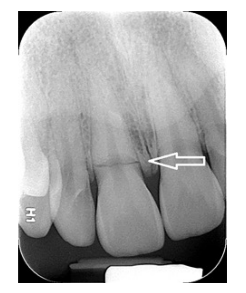
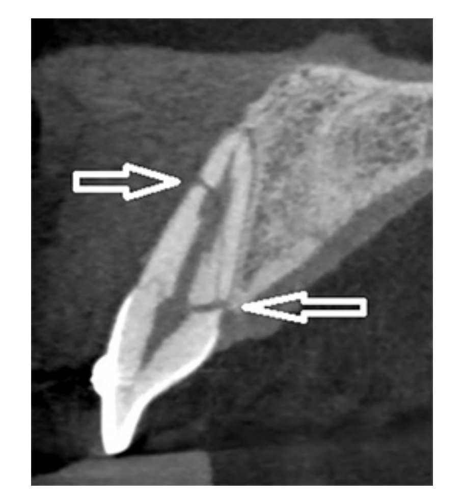
- All root fractures involve the pulp. Most common in maxillary central incisors; most occur in the apical and middle thirds, and the plane is often diagonal.
- Why several angulations: how the fracture looks depends on the beam-to-fracture-plane angle and on fragment separation.
- Beam parallel to the plane → one sharp radiolucent line confined to the root.
- Beam oblique → a poorly defined line, or two lines converging at the mesial and distal surfaces (can look like two fractures).
- Subtle or invisible early on: later inflammation and resorption widen the gap and make it visible. Sometimes the only sign is focal PDL widening at the fracture site.
- Longer-standing fractures: the fracture gap widens (resorption of the fractured surfaces); pulp chamber/canal calcification may follow.
- Level decides prognosis: cervical third = poor (usually extraction unless the root can be orthodontically extruded); middle/apical third = reposition and splint, favourable; closer to the apex = better.
- If the pulp necroses, bone loss appears at the fracture line rather than at the apex.
- Mimics: alveolar fracture lines, neurovascular canals, lip, ala of nose and nasolabial fold superimposed on the root. A true root fracture line does not shift relative to the tooth between angulations.
- Cohen’s: 68.2% of intra-alveolar fractures in one CBCT study were oblique and reached the cervical third, which contradicts earlier PA-only studies.39
Healing patterns on follow-up3937
-
Healing types: hard (calcified) tissue fusion, PDL interposition, or PDL interposition with bone.39
-
Non-healing (coronal pulp necrosis): bone loss at the fracture line.37
-
Healing type and fracture location have the biggest effect on tooth loss; immature roots heal better.39
-
W&P Fig 27.4 shows the range: recent fracture → healed with slight displacement → healed with resorption and separation of the fragments.37


Vertical root fracture (VRF)
- Runs along the long axis. Most often in endodontically treated premolars and molars, especially with metal posts.37
- PA signs: fracture plane through the canal/root filling, with fragments separating (often more at the apex); focal PDL widening next to the fracture may be the only indirect sign.37
- Often not visible on PA: VRFs usually run buccolingually, so they show only when the beam is parallel to the fracture plane. PA sensitivity is about 37%.39 Nondisplaced and mesiodistal VRFs are often undetectable.37
- CBCT: small-FOV CBCT is the modality of choice when the clinical exam and 2D images are inconclusive (AAOMR/AAE).37 However, GP and posts cause streak and “cupping” artefacts that mimic fracture lines and lower CBCT specificity.3739 A vertical bone-loss pattern on CBCT helps the diagnosis.39
- Prognosis poor: extract single-rooted teeth; hemisection is an option for multirooted teeth.37
- Clinical crack work-up: see Diagnosis of Cracked Teeth.


A negative PA does not rule out a root fracture
Crown and Crown-Root Fractures
- Crown: assess how far the fracture is from the pulp chamber. The first film is the baseline for later pulp changes.37
- Crown-root: typical plane runs obliquely from the labial gingival third of the crown to below the palatal/lingual gingival attachment.37
- Hard to see: take PA + occlusal at several horizontal and vertical angulations.37 Imaging: 1 occlusal + 2 PAs (mesial and distal); consider CBCT for extent and direction.38
- Missing fragments: film the lip, cheek or tongue (receptor next to the laceration); consider a chest/abdominal film if a fragment is unaccounted for.37
- Tender to percussion in a crown fracture suggests an added luxation or root fracture.38
Luxation Injuries and Alveolar Fractures
| Injury | Displacement | PA signs |
|---|---|---|
| Concussion | None | Usually normal; may show slight apical PDL widening37 |
| Subluxation | None (mobile) | No abnormality expected38 |
| Extrusion | Coronal (out of socket) | Widened apical PDL, in proportion to the force; tooth elongated37 |
| Lateral luxation | Labial/palatal, often with slight extrusion | PDL widening greater on the impact side; often facial cortex fracture3738 |
| Intrusion | Apical (into bone) | Partial/total loss of apical PDL space; crown depressed; CEJ more apical than neighbours3738 |
| Avulsion | Out of socket | Empty socket with lamina dura still visible37 |
- Usually 2 or more teeth are involved, mainly maxillary incisors.37
- Intrusion and lateral luxation can crush or comminute the alveolus.37
- Intrusion: minimal intrusion is hard to show. A deeply intruded tooth can look avulsed. Use multiple projections including occlusal to show the direction of displacement and relation to the cortex. For total intrusion, consider a lateral view for nasal penetration.3738
- Avulsion: the missing tooth may lie in the lip and project over the alveolus, so take a soft-tissue film. Dense new bone in a healing socket can mimic a retained root.37
- Alveolar process fracture37
- Usually anterior/premolar; several teeth; plane mostly horizontal; can be apical to the teeth or through the socket.
- PA/occlusal: radiolucent line anywhere from crest to periapex. Both-plate fractures are usually visible; single-plate, nondisplaced fractures are easily missed.
- The closer the line is to the crest, the more likely a root fracture coexists. A line that shifts relative to the tooth between angulations is bone, not root.
- A line touching the root apices carries a high risk of later internal/external resorption.
- Posterior maxilla: sinus floor involvement → mucosal thickening or air-fluid level.


Post-Trauma Sequelae on Follow-up
| Sequela | PA appearance | Source |
|---|---|---|
| Pulp canal obliteration | Pulp chamber, then canal, progressively narrowed or obliterated (the pulp survived and dentine deposition sped up) | 37 |
| Pulp necrosis | Periapical rarefying osteitis; dentine deposition stops, so the canal stays wider than in contralateral teeth | 37 |
| Internal (inflammatory) resorption | Focal ballooning of the canal; the lesion stays with the canal on parallax shift | 3739 |
| External inflammatory resorption | Concave, patchy root-surface excavations and adjacent bone loss; irregular borders, loss of lamina dura, often apical | 39 |
| Replacement resorption / ankylosis | PDL space disappears; direct union of root with bone; tooth immobile with a distinctive percussion note | 39 |
| Arrested root development (immature tooth) | Root “frozen in time”: canal uniformly wide from chamber to apex; possible osteodentin cap over the open apex | 37 |
- The Yu and Abbott table in L4 groups follow-up responses. Favourable: pulp canal calcification, revascularisation, fibrous healing. Unfavourable: necrosis ± infection, internal/external resorption (surface, inflammatory, replacement, invasive), ankylosis, cessation of root development.38
- Osteodentin cap vs internal resorption: the cap has a uniformly widened canal; internal resorption has a focally widened canal.37
- PA misses small resorptions. In one study PAs detected 68.8% of internal resorptions against 100% on CBCT.39
- Sensibility can stay falsely negative for up to 3 months after a root fracture, so read follow-up PAs alongside vitality testing.37
- IADT: yearly clinical + PA review for up to 5 years after some injuries.39


Imaging Protocol and When to Use CBCT
IADT PA set for maxillary incisor trauma38
- 1 parallel PA centred on the midline (both maxillary centrals).
- 1 parallel PA centred on the right lateral incisor (showing canine and central).
- 1 parallel PA centred on the left lateral incisor (showing canine and central).
- 1 maxillary occlusal.
- ≥1 parallel PA of the mandibular incisors (more if lower teeth are injured).
- W&P: at least 2 PAs at different horizontal angulations for root fractures, and always image the opposing arch. If mouth opening is limited, use cross-sectional occlusals (size 2 receptor in children).37
- OPG is fine for locating injuries and mandibular fractures, but its resolution is too low for anterior dentoalveolar detail.37
- Root fracture level: an occlusal shows apical/middle-third fractures; 2 PAs at varied horizontal angles are needed for cervical-third fractures.38
| Use PA first | Add small-FOV CBCT when |
|---|---|
| Initial assessment of all injuries; follow-up reviews | Symptomatic patient but PAs show no fracture38 |
| Concussion, subluxation, uncomplicated crown fracture | Suspected root, crown-root or alveolar fracture that 2D does not resolve (AAE recommends CBCT for these)3839 |
| Lateral luxation or labial-palatal displacement; oblique or vertical fracture suspected38 | |
| Late stage: unclear healing, necrosis, root resorption, ankylosis38 | |
| PA placement not possible or would need retakes; foreign bodies in lip, cheek or tongue39 |
- CBCT accuracy for horizontal root fractures, from studies cited in L4: CBCT sensitivity 98.8%, specificity 95.6%, against PA 97.2% and 78.5% (in vivo). In vitro: CBCT 0.97/0.93 against PA 0.83/0.885.38
- Use small FOV, voxel ideally <0.1 mm. Voxels above 0.3 mm may not improve assessment.3839
- Apply ALARA strictly in children: CBCT should answer a specific question that PA or OPG cannot.39
- Read CBCT with MPR planes aligned to the tooth’s long axis. The corrected coronal view resembles a PA.38
PA vs CBCT in one line
Anatomy That Mimics Pathology
Rule of thumb: ==intact lamina dura + normal PDL around the apex + no symptoms + vital tooth = anatomy, not disease.== A second film at a different angle shifts a superimposed structure relative to the apex.1430
| Mimic | Mimics what | How to tell |
|---|---|---|
| Mental foramen | Periapical RL on a mandibular premolar (usually the second premolar) | LD intact around the apex. The IAC can be traced into the lucency. A second film at another angle shows the LD and shifts the foramen relative to the apex. Beware: burnout can hide a thin LD over the foramen.14 |
| Incisive foramen / canal | Periapical RL on a central incisor, or apparent PDL widening | LD intact around the incisor and no symptoms. Size and shape vary widely with beam angle.14 DI L1 case: the incisive canal projected as distal PDL widening, and only CBCT showed the real small apical lesion.30 |
| Large incisive foramen vs nasopalatine duct cyst | Cyst vs normal | Suspect a cyst if the foramen is wider than 1 cm (W&P) or the canal is wider than 6 mm (DI L3.1), if it enlarges on successive films, or if there is expansion or incisor roots diverge.143231 A cyst is well-defined and corticated, ± heart-shaped (anterior nasal spine superimposed), with uniform PDL around the vital incisors and palatal to them on horizontal tube shift. Occlusal view or CBCT confirms.3231 Rarefying osteitis of an incisor instead shows lost LD with a widened PDL.32 See Nasopalatine Duct Cyst. |
| Lateral (incisive) fossa | RL at the maxillary lateral incisor apex | Diffuse, ill-defined lucency. LD intact and no symptoms.14 |
| Maxillary sinus recess over roots | Roots “inside” the sinus; apical RL on maxillary premolars/molars | Roots projecting into the sinus is a projection illusion. The thin bone over the root shows as the LD fused with the sinus floor. The sinus extends further after tooth loss. Use CBCT if the relationship matters.14 |
| Inferior alveolar canal over a molar apex | Missing LD or a widened apical PDL | The overlying LD is overexposed. Test vitality. Changing the vertical angle rarely separates canal and apex, because the canal sits just below the apices.14 |
| Nutrient canals | Vertical RL lines, or a small round RL simulating a lesion | Uniform width, often with corticated (hyperostotic) walls. Run from the IAC to an apex or interdentally in the mandibular incisor region. A canal seen end-on looks like a round “hole”.14 |
| Submandibular fossa | Large RL below the mandibular molar apices | Sparse trabeculae. Sharply bounded above by the mylohyoid ridge and below by the inferior border; poorly defined front and back. LD and PDL of the teeth stay intact.1430 |
| Mental fossa | RL at the mandibular incisor apices | Labial depression with ill-defined borders; looks like the submandibular fossa.14 |
| Sparse trabeculae (anterior maxilla, anterior mandible) | “Lesion” in an asymptomatic patient | Compare with the contralateral side and with previous films. Only a change makes it suspicious.14 |
| Cervical burnout | Root-surface caries | Ill-defined RL band between enamel and alveolar crest caused by lower x-ray absorption. Close inspection shows intact proximal surface edges. Expect it on almost all teeth.14 |
| Mach band | Caries spreading along the DEJ | RL line at the occlusal DEJ caused by the contrast difference between enamel and dentine.19 |
| Nasolabial fold | Diffuse opacity or “sclerosis” over premolar roots | Sharp oblique line with a veil of slight radiopacity posterior to it (thick cheek tissue). More obvious with age.14 |
| Nose soft tissue | RO over the maxillary incisor roots | Uniform, slightly opaque, sharp border. The nares may appear as RLs at steep vertical angles.14 |
| Tori / hyperostoses | Periapical RO: sclerosing osteitis or dense bone island | Dense, well-defined, convex or lobulated RO superimposed over premolar and molar roots, often bilateral. Bony-hard on palpation, so confirm clinically. Palatal tori rarely show on PAs.3329 See Tori and Exostoses. |
| Anterior Stafne defect (developmental, not anatomy) | Well-defined periapical RL near the lower anterior apices | Near but not at the apex; teeth respond to pulp testing; no restorative history. CBCT shows a lingual cortical concavity that does not involve the roots.29 See Stafne Defect. |


Report Template
Six-point intraoral write-up (OMFR L7)
This is a <type of radiograph>, showing the <tooth numbers>. <Identify the normal anatomy>. There is:
- Periodontal status:
- Restorations (bonus: material):
- Caries:
- Resorption:
- Periapical lesions (not applicable for BW):
- Other findings:
Template verified against the L7 PDF slide2. In the Findings, describe only; put your interpretation and differentials last, in order of likelihood6
Add before and after the six points
Worked example
Filled template (my read of W&P Fig 18.14)
This is a periapical radiograph (mandibular posterior), showing a two-rooted mandibular molar (presumed first molar, 36 or 46; you can’t tell the side without the orientation dot), the premolar mesial to it, and part of the next molar distally. Normal anatomy: enamel, dentine, pulp chamber and root canals of the molar; alveolar crest; normal trabecular pattern near the crest (long arrow). Quality: roots and periapical bone fully covered with plenty of bone below the apices; crowns sit right at the top edge of the image (little clearance beyond the occlusal plane); no obvious elongation or foreshortening. There is:
- Periodontal status: interdental crests between the premolar and the molar sit close to the CEJ. No obvious horizontal or angular bone loss.
- Restorations: none seen on the molar. The partially imaged distal tooth can’t be assessed.
- Caries: large radiolucency in the molar crown, reaching the pulp chamber region. Consistent with deep caries and probable pulpal involvement.
- Resorption: no internal or external root resorption evident.
- Periapical lesions: diffuse increased radiopacity (dense trabeculae) around and below the molar apices, extending mesially under the premolar. The periphery is poorly defined and blends into normal bone (short arrow); there is no discrete corticated border. The apical PDL is hard to assess against the dense bone.
- Other findings: none.
Impression: periapical sclerosing osteitis related to the carious molar (inflammatory, bone-forming response)67 Differentials: dense bone island, which keeps a uniform PDL width; periapical cemento-osseous dysplasia, where the tooth is vital. These are much less likely because a deep carious lesion is present7 Next step: pulp sensibility testing and periapical tests on the molar, clinical caries assessment, then endodontic diagnosis7
Related Notes
- Periapical conditions: Radicular Cyst, Residual Cyst, Periapical Cemento-Osseous Dysplasia, Cementoblastoma, Osteomyelitis, Nasopalatine Duct Cyst
- Differentials: Radiolucent Lesion differentiation, Tori and Exostoses, Traumatic Bone Cyst, Stafne Defect
- Endodontics: Endodontic Diagnoses (Chapter 8), Diagnosis of Cracked Teeth
- Periodontics: Periodontitis Staging and Grading, Endo-Periodontal Lesions
- Hub: MOC Radiography
Sources
Footnotes
-
05 5 Film Imaging ↩ ↩2 ↩3 ↩4 ↩5 ↩6 ↩7 ↩8 ↩9 ↩10 ↩11 ↩12 ↩13 ↩14 ↩15 ↩16 ↩17 ↩18
-
07 7 Intraoral Projections ↩ ↩2 ↩3 ↩4 ↩5 ↩6 ↩7 ↩8 ↩9 ↩10 ↩11 ↩12 ↩13 ↩14 ↩15 ↩16 ↩17 ↩18 ↩19 ↩20 ↩21 ↩22 ↩23 ↩24 ↩25 ↩26 ↩27
-
04 4 Digital Imaging ↩ ↩2 ↩3 ↩4 ↩5 ↩6 ↩7 ↩8 ↩9 ↩10 ↩11 ↩12 ↩13
-
18 18 Principles of Radiographic Interpretation ↩ ↩2 ↩3 ↩4 ↩5 ↩6 ↩7 ↩8 ↩9 ↩10 ↩11 ↩12 ↩13 ↩14 ↩15 ↩16 ↩17 ↩18 ↩19 ↩20
-
22 22 Inflammatory Conditions of the Jaws ↩ ↩2 ↩3 ↩4 ↩5 ↩6 ↩7 ↩8 ↩9 ↩10 ↩11 ↩12 ↩13 ↩14 ↩15 ↩16 ↩17 ↩18 ↩19 ↩20 ↩21 ↩22 ↩23
-
06 6 Projection Geometry ↩ ↩2 ↩3 ↩4 ↩5 ↩6 ↩7 ↩8 ↩9 ↩10 ↩11 ↩12 ↩13 ↩14
-
L16 Endo Diagnostic radio_formatted_text ↩ ↩2 ↩3 ↩4 ↩5 ↩6 ↩7 ↩8 ↩9 ↩10 ↩11 ↩12 ↩13 ↩14
-
12 12 Radiographic Anatomy ↩ ↩2 ↩3 ↩4 ↩5 ↩6 ↩7 ↩8 ↩9 ↩10 ↩11 ↩12 ↩13 ↩14 ↩15 ↩16 ↩17 ↩18 ↩19 ↩20 ↩21 ↩22 ↩23 ↩24 ↩25 ↩26 ↩27 ↩28 ↩29 ↩30 ↩31 ↩32 ↩33 ↩34 ↩35 ↩36 ↩37 ↩38 ↩39 ↩40 ↩41 ↩42 ↩43 ↩44 ↩45 ↩46 ↩47 ↩48 ↩49 ↩50 ↩51 ↩52 ↩53 ↩54 ↩55 ↩56 ↩57 ↩58 ↩59 ↩60 ↩61 ↩62 ↩63 ↩64 ↩65 ↩66 ↩67 ↩68 ↩69 ↩70 ↩71 ↩72 ↩73 ↩74 ↩75 ↩76 ↩77 ↩78 ↩79 ↩80 ↩81 ↩82 ↩83 ↩84 ↩85 ↩86 ↩87 ↩88 ↩89 ↩90 ↩91 ↩92 ↩93 ↩94 ↩95 ↩96 ↩97 ↩98 ↩99 ↩100 ↩101 ↩102 ↩103 ↩104 ↩105 ↩106 ↩107 ↩108
-
L5 Intraoral Radiographic Anatomy ↩ ↩2 ↩3 ↩4 ↩5 ↩6 ↩7 ↩8 ↩9 ↩10 ↩11 ↩12 ↩13 ↩14 ↩15 ↩16 ↩17 ↩18 ↩19 ↩20 ↩21 ↩22 ↩23 ↩24 ↩25 ↩26
-
19 19 Dental Caries ↩ ↩2 ↩3 ↩4 ↩5 ↩6 ↩7 ↩8 ↩9 ↩10 ↩11 ↩12 ↩13 ↩14 ↩15 ↩16
-
20 20 Periodontal Diseases ↩ ↩2 ↩3 ↩4 ↩5 ↩6 ↩7 ↩8 ↩9 ↩10 ↩11 ↩12 ↩13 ↩14 ↩15 ↩16 ↩17 ↩18 ↩19 ↩20 ↩21 ↩22 ↩23 ↩24 ↩25 ↩26 ↩27 ↩28 ↩29 ↩30 ↩31 ↩32 ↩33 ↩34
-
L2 Radiography in Periodontology ↩ ↩2 ↩3 ↩4 ↩5 ↩6 ↩7 ↩8 ↩9 ↩10 ↩11 ↩12 ↩13 ↩14
-
21 21 Dental Anomalies ↩ ↩2 ↩3 ↩4 ↩5 ↩6 ↩7 ↩8 ↩9 ↩10 ↩11 ↩12 ↩13 ↩14 ↩15 ↩16 ↩17 ↩18
-
International Association of Dental Traumatology guidelines for the management of traumatic dental injuries 2. Avulsion of permanent teeth ↩
-
03 3 Lesions that mimic endodontic pathosis ↩ ↩2 ↩3 ↩4 ↩5 ↩6 ↩7 ↩8 ↩9 ↩10 ↩11 ↩12 ↩13 ↩14 ↩15 ↩16 ↩17 ↩18 ↩19 ↩20 ↩21 ↩22 ↩23 ↩24
-
L1 - Imaging for Infection and Inflammatory Lesions of the Jaws ↩ ↩2 ↩3 ↩4 ↩5 ↩6 ↩7 ↩8 ↩9 ↩10 ↩11 ↩12 ↩13 ↩14 ↩15
-
24 24 Benign Tumors and Neoplasms ↩ ↩2 ↩3 ↩4 ↩5 ↩6 ↩7 ↩8 ↩9 ↩10
-
25 25 Diseases Affecting the Structure of Bone ↩ ↩2 ↩3 ↩4 ↩5 ↩6 ↩7 ↩8
-
27 27 Trauma ↩ ↩2 ↩3 ↩4 ↩5 ↩6 ↩7 ↩8 ↩9 ↩10 ↩11 ↩12 ↩13 ↩14 ↩15 ↩16 ↩17 ↩18 ↩19 ↩20 ↩21 ↩22 ↩23 ↩24 ↩25 ↩26 ↩27 ↩28 ↩29 ↩30 ↩31 ↩32 ↩33 ↩34 ↩35 ↩36 ↩37 ↩38 ↩39 ↩40 ↩41 ↩42 ↩43
-
L4 - Imaging for Trauma ↩ ↩2 ↩3 ↩4 ↩5 ↩6 ↩7 ↩8 ↩9 ↩10 ↩11 ↩12 ↩13 ↩14 ↩15 ↩16 ↩17 ↩18 ↩19 ↩20 ↩21 ↩22 ↩23 ↩24 ↩25 ↩26
-
02 2 Radiographic interpretation ↩ ↩2 ↩3 ↩4 ↩5 ↩6 ↩7 ↩8 ↩9 ↩10 ↩11 ↩12 ↩13 ↩14 ↩15 ↩16 ↩17 ↩18 ↩19 ↩20