Intraoral Radiograph Interpretation1
Dr Dayea Oh OMF Radiologist
Table Of Contents2
- Normal vs Abnormal findings
- Systematic approach
- Assessment of:
- Caries
- Periodontal disease
- Periapical lesions
Systematic Approach To Interpretation
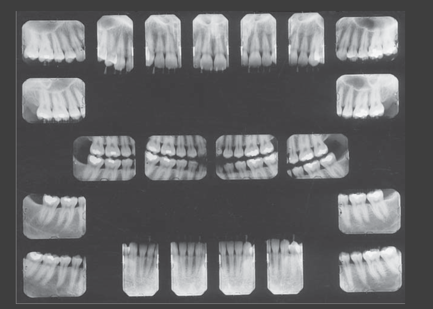
Radiographic Checklist And Viewing Conditions
Checklist3
- Reason for x-ray
- Previous x-rays
- Optimal Viewing Conditions
- Patient ID
- Orientation →
- Identify the type of x-ray
- Image Quality
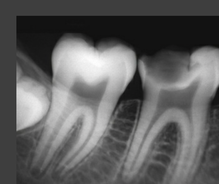
Differentiating Normal And Abnormal Findings
Normal vs Abnormal4
- Normal
- Anatomical
- Review tooth morphology
- Bilateral & symmetrical (extra-oral imaging)
- Anatomical
- Abnormal
- Pathological
- Artefactual
- Superimpositions
Description!!!
Stepwise Review Of Teeth And Restorations
Systematic Approach5
- Step 1. Teeth & Surrounding Structures
- Count teeth
- Tooth morphology
- Crown – enamel, dentine, pulp chamber, etc.
- Roots – PDL space, lamina dura, # of roots, root morphology
- Periodontal bone
- Other
- Step 2. Identify existing restorative therapy, endodontic therapy & orthodontic therapy
- Assess restorative margins – presence of overhangs? Insufficient margins?
- Presence of voids?
- Step 3. Identify & describe abnormal radiolucencies & radiopacities
- Location
- Size & Extent
- Morphology & Borders
- Effects on surrounding structures
- Internal architecture
- Step 4. Differential diagnosis
Caries Assessment
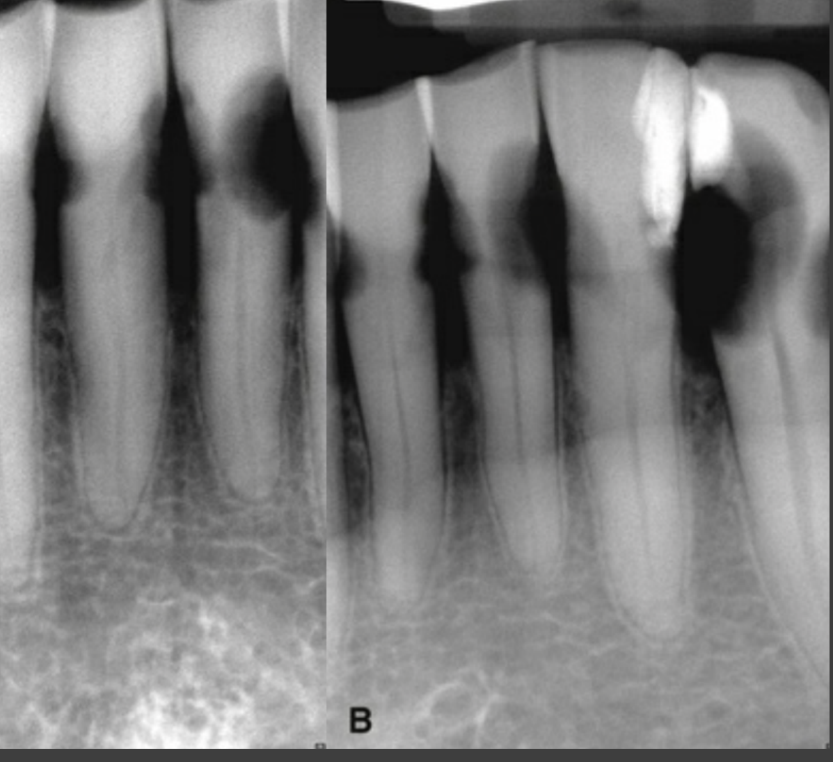
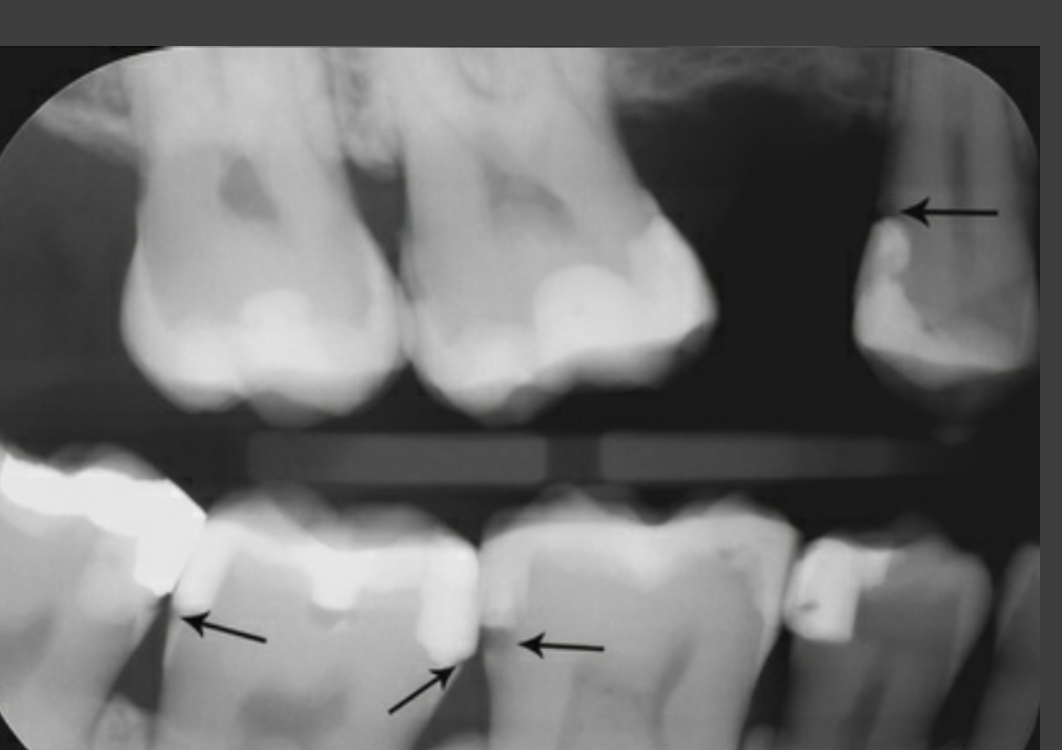
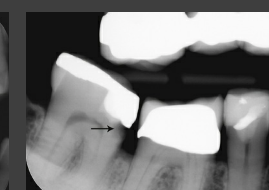
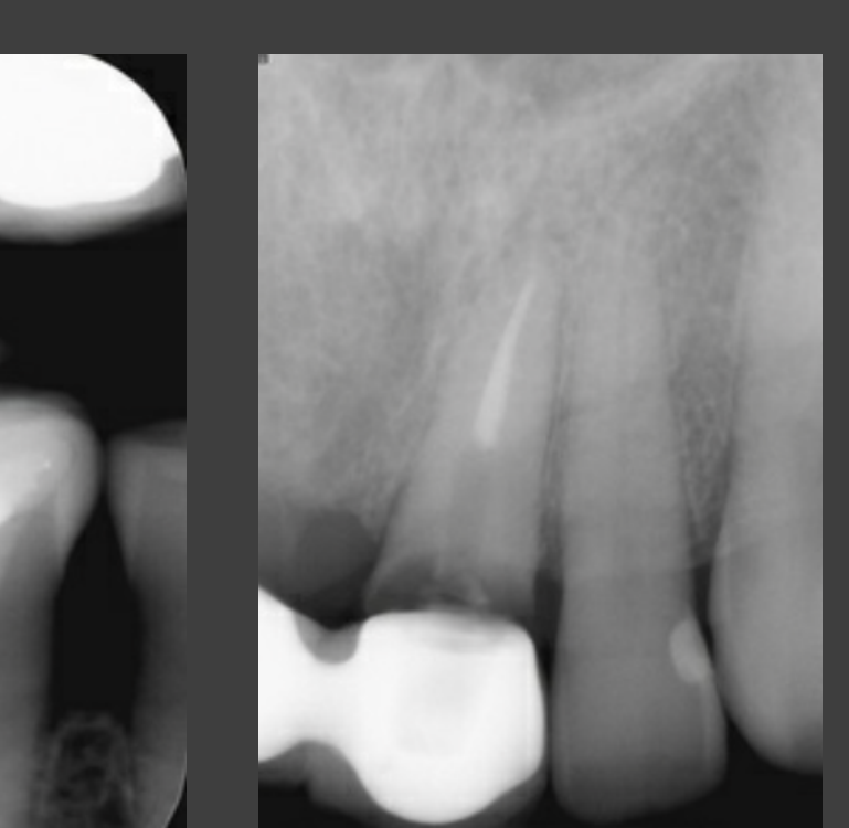
Classification Of Caries Lesions6
- triangular
- Dentinal caries is larger
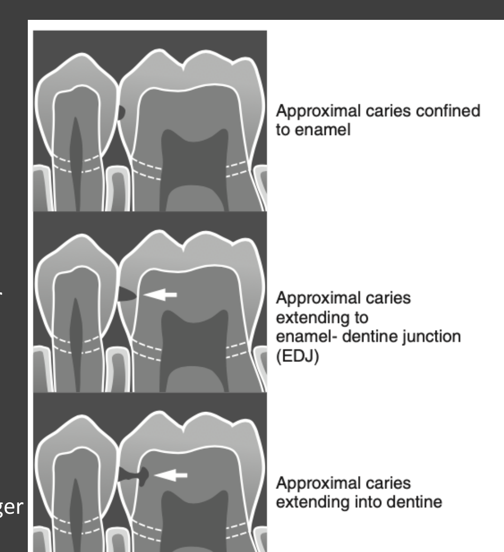
- Approximal caries confined to enamel:
- Approximal caries extending to enamel-dentine junction (EDJ):
- Approximal caries extending into dentine:
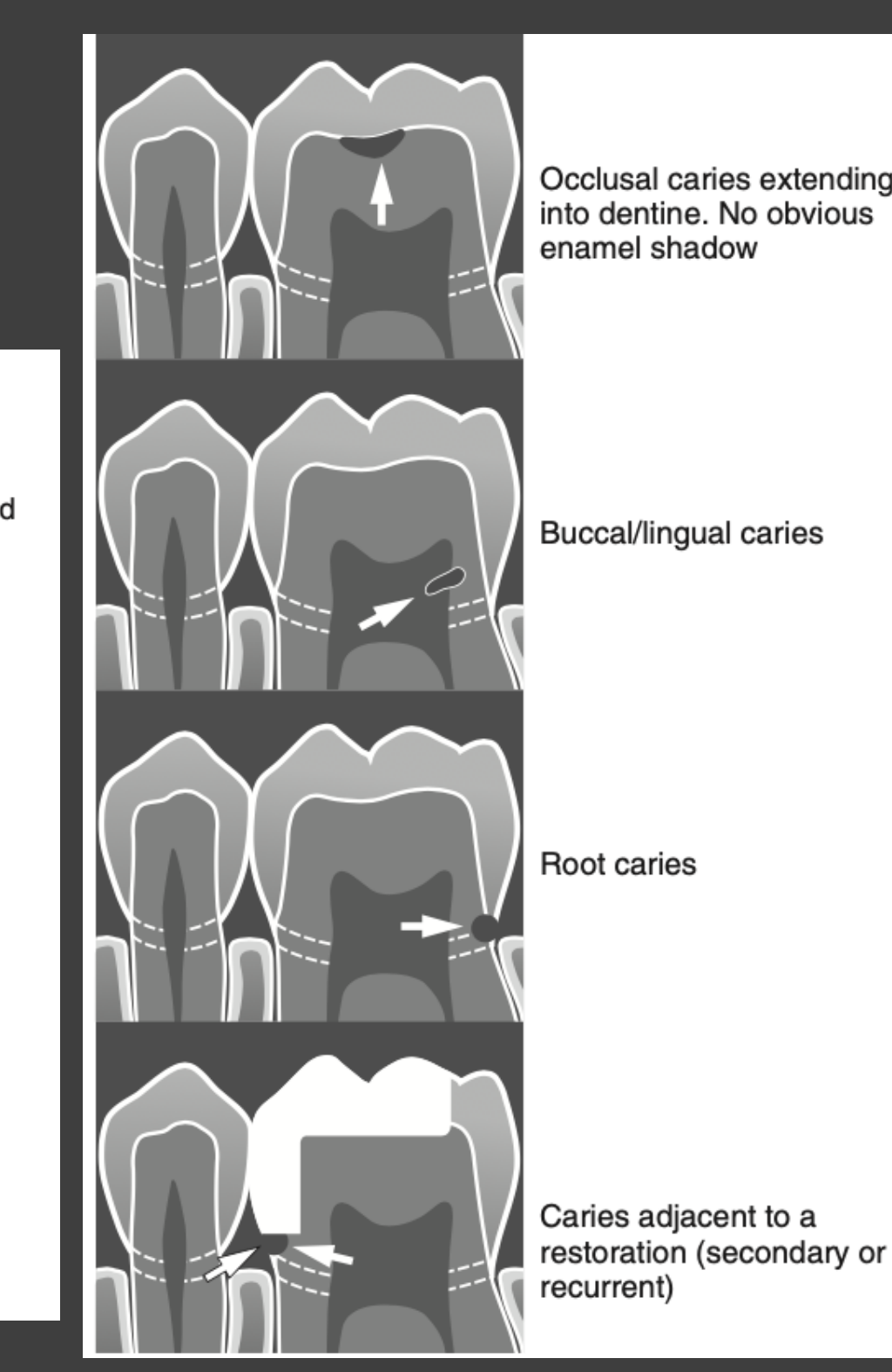
- Occlusal caries extending into dentine. No obvious enamel shadow:
- Buccal/lingual caries:
- Root caries:
- Caries adjacent to a restoration (secondary or recurrent):
 |  |
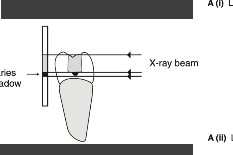
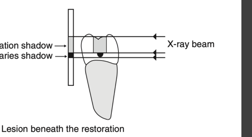
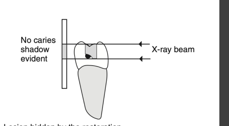
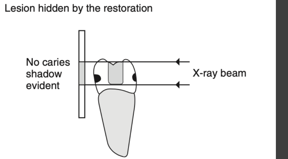
Recurrent Caries And Radiographic Limitations7
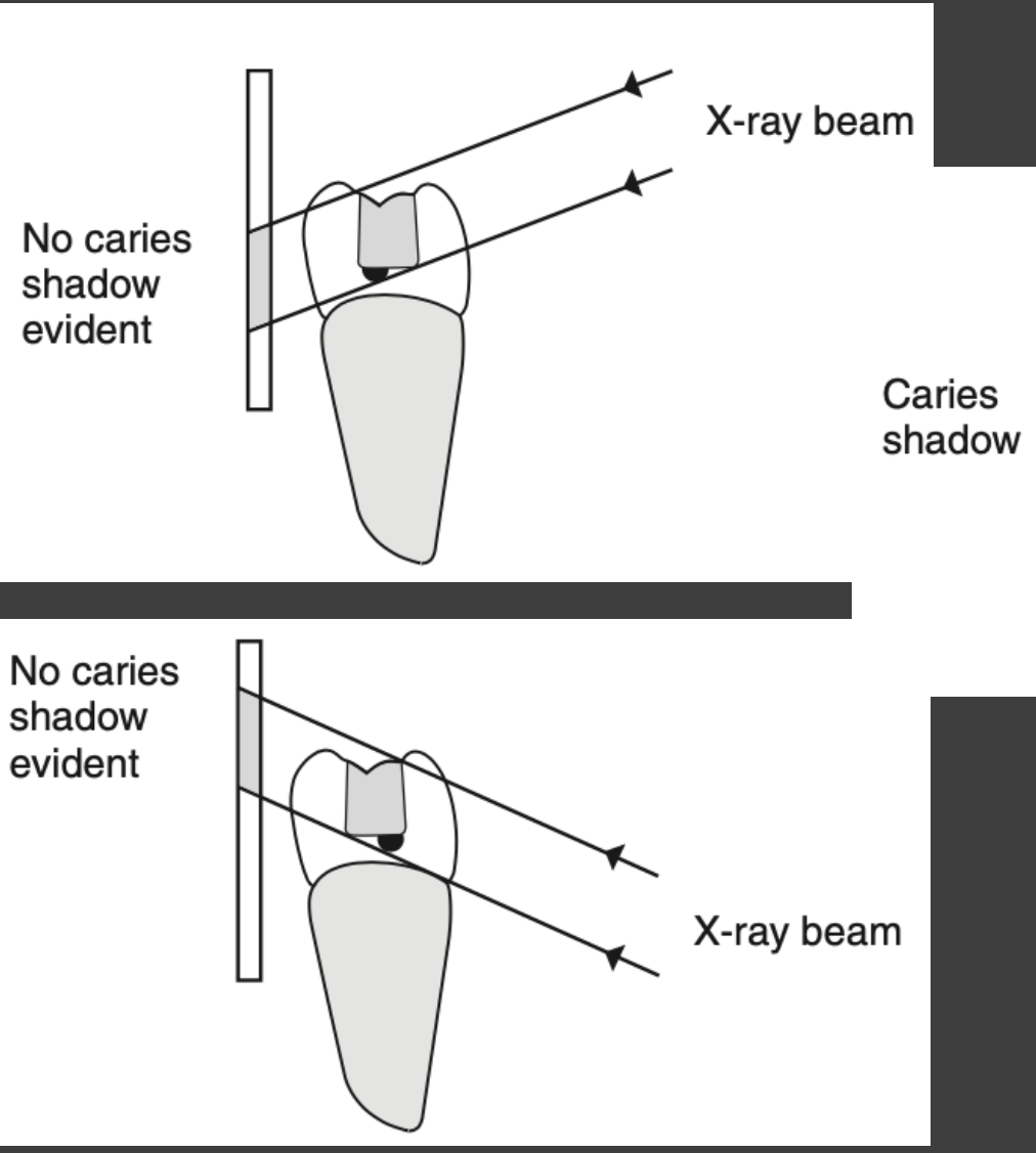
- A (i) Lesion beneath the restoration
- Left side (Top): No caries shadow evident due to X-ray beam angle.
- Center: Caries shadow evident.
- Right side: Restoration shadow and Caries shadow identified; X-ray beam parallel.
- A (ii) Lesion hidden by the restoration
- Left side (Bottom): No caries shadow evident due to X-ray beam angle.
- Right side: No caries shadow evident as lesion is obscured by the restoration.
- B Buccal and lingual lesions hidden by the restoration
- No caries shadow evident.
 |  |  |
 |  |
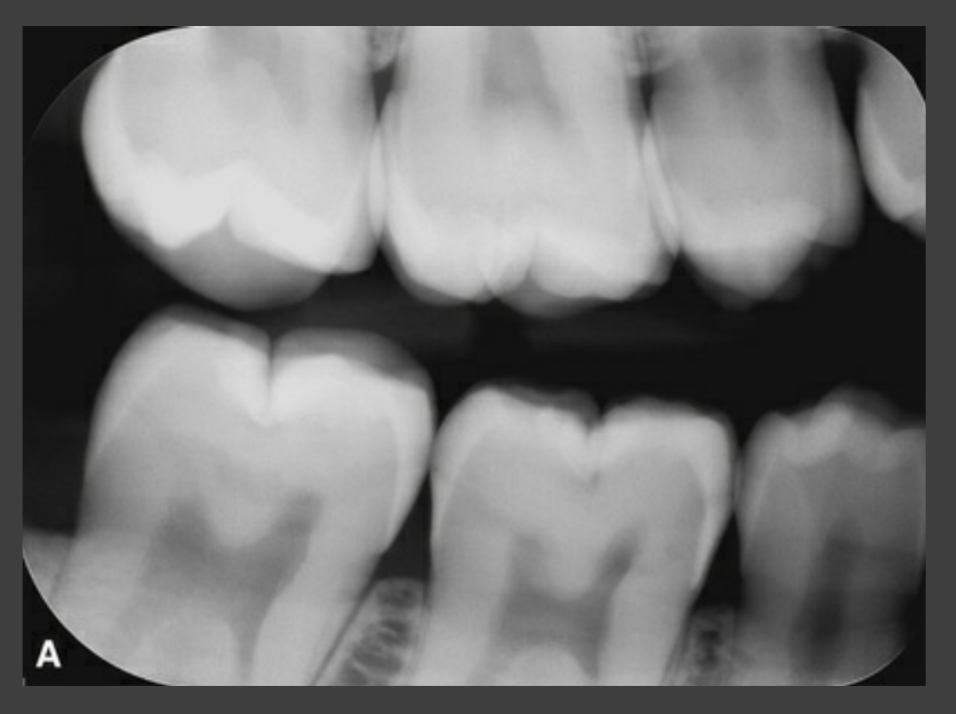
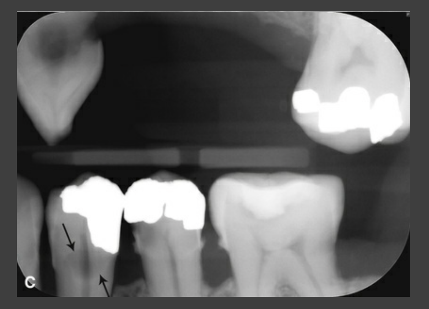
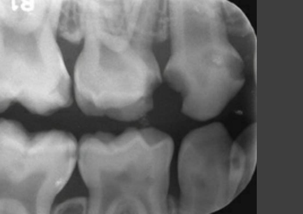
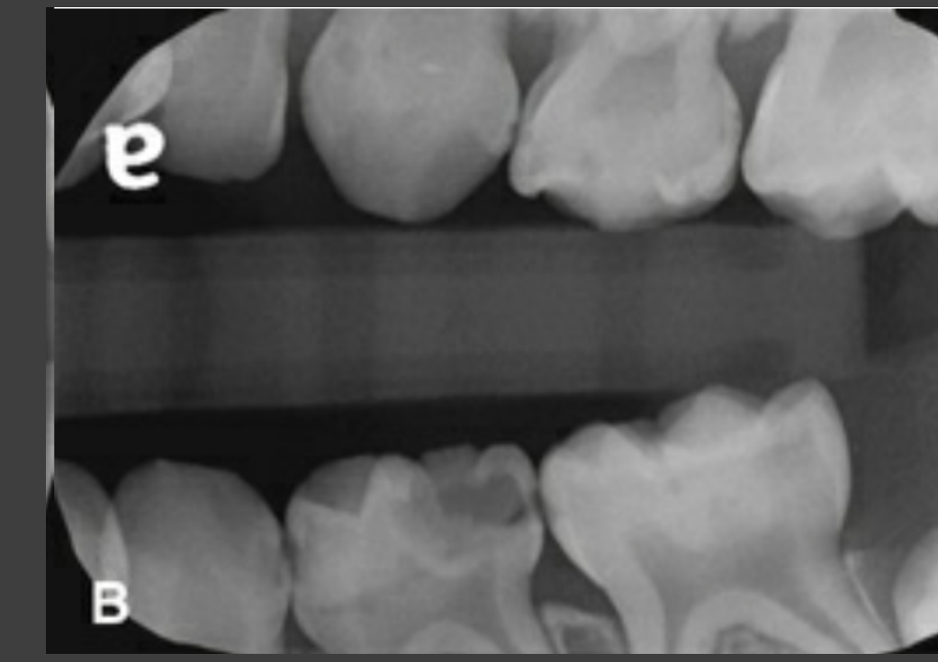
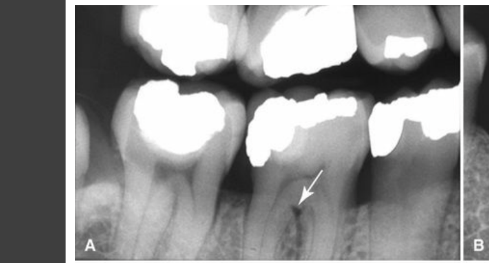
Clinical Radiographic Examples Of Caries891011
A B
 |  |  |
 |  |  |
 |  |
Periodontal Disease Assessment
Bone Loss Patterns And Calculus
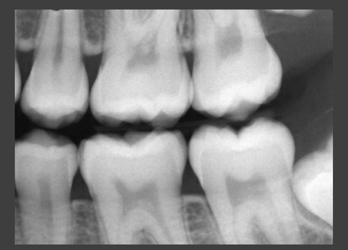
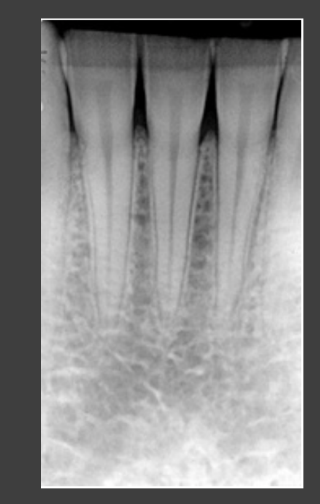
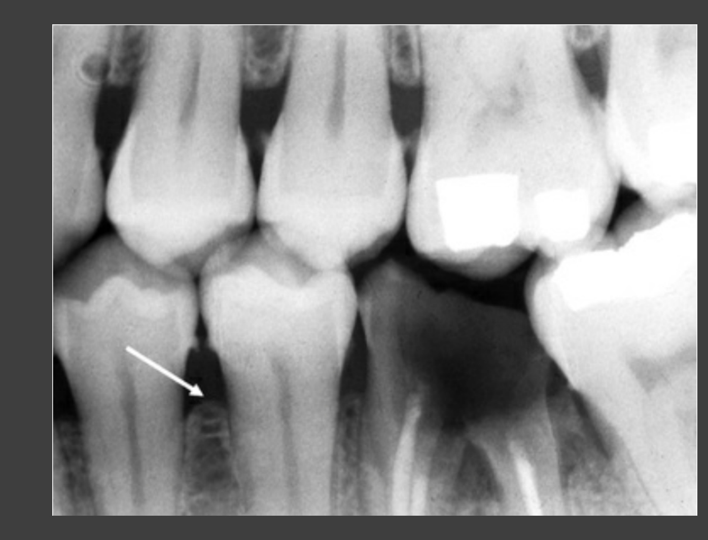
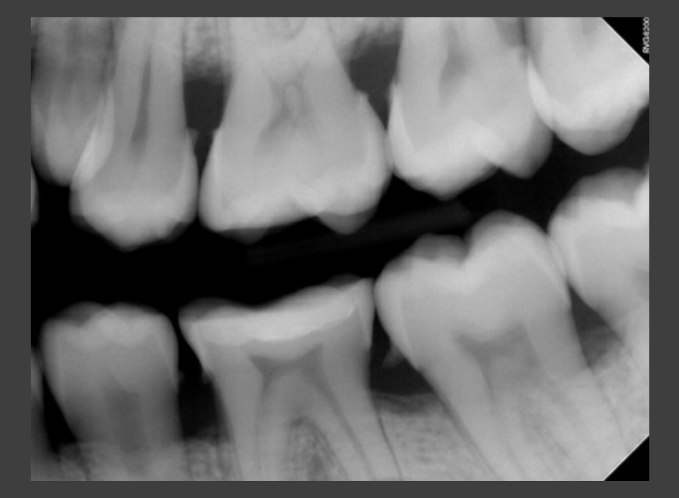
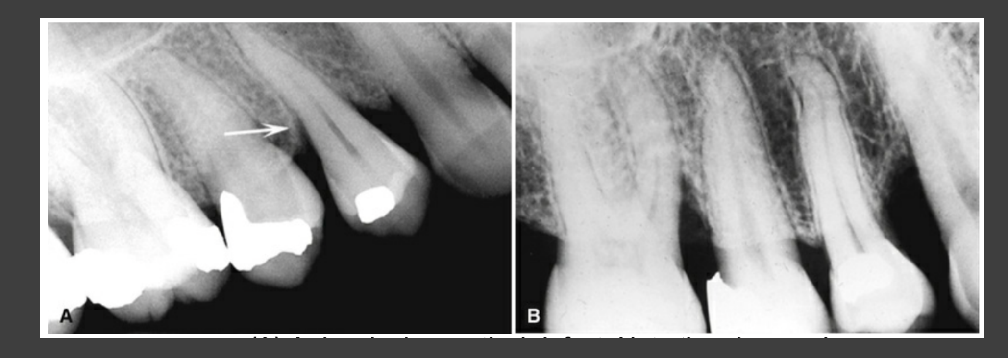
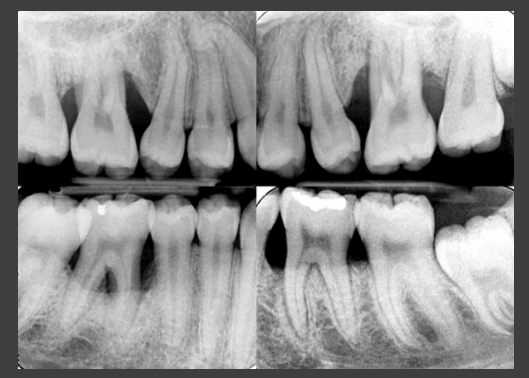
Periodontal Disease12
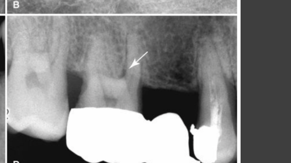
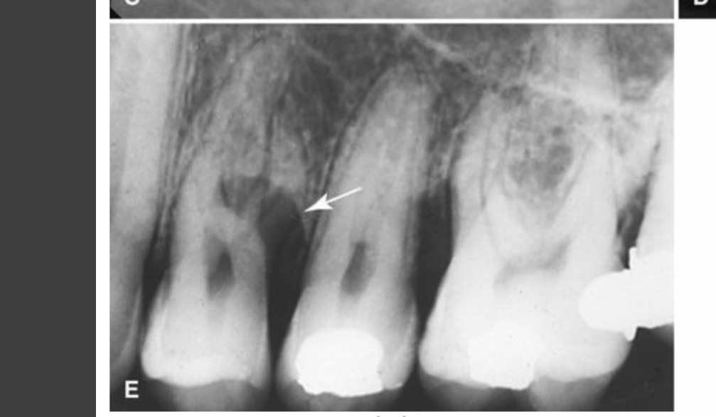
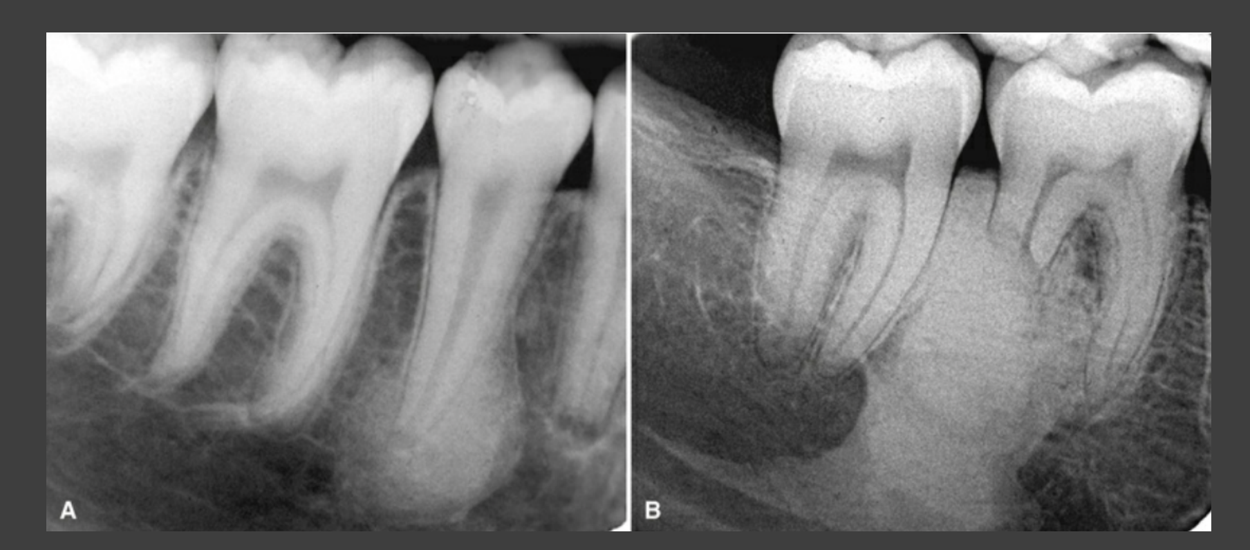
- Periodontal bone loss
- Horizontal
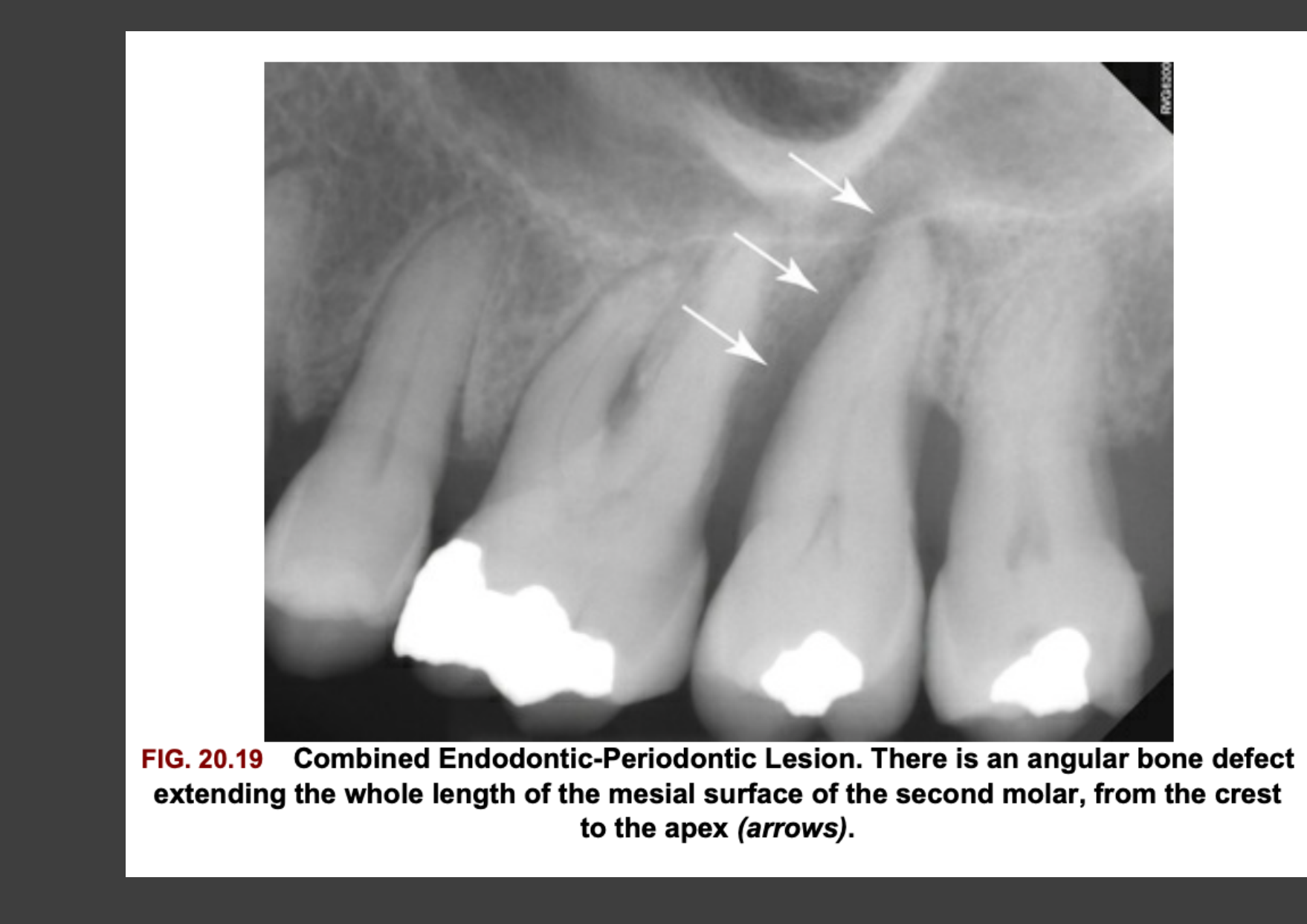
- Vertical / angular bone defect
- Furcation involvement
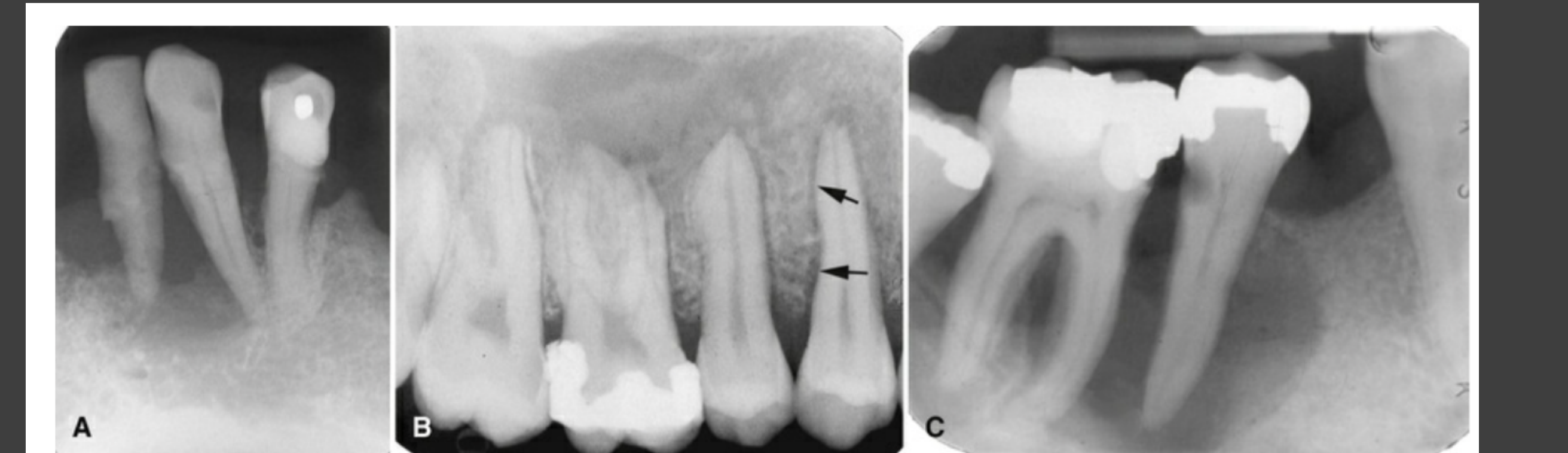
- Alveolar crest → cortical?
- Calculus
- Periodontal ligament space widening
- Anatomic considerations
- Enamel pearl
Clinical Radiographic Examples Of Periodontal Disease1314151617
 |  |  |
 |  |  |
Cortical Bone And Alveolar Crest Evaluation181920
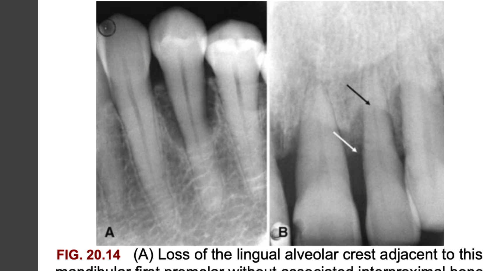
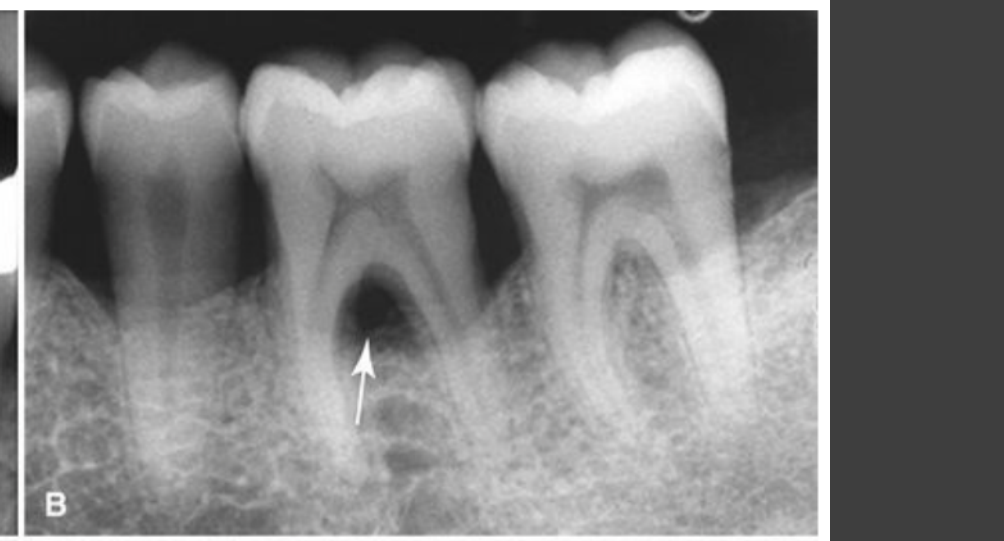
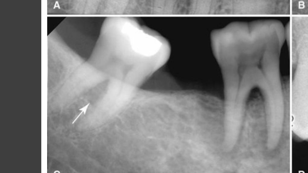
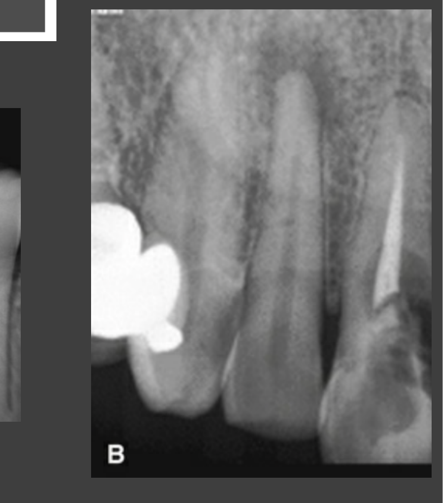
FIG. 20.14 (A) Loss of the lingual alveolar crest adjacent to this mandibular first premolar without associated interproximal bone loss. (B) Loss of the buccal cortical bone adjacent to the maxillary central and lateral incisors. The black arrow indicates the level of the buccal alveolar crest, which demonstrates more profound loss relative to the lingual alveolar crest (white arrow).
 |  |  |
 |  |  |
 |
Periapical Lesions Assessment
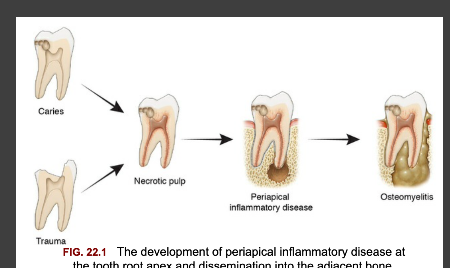
Pathogenesis Of Periapical Inflammatory Disease21
Periapical Inflammatory Lesions
- Abscess
- Granuloma
- Cyst
FIG. 22.1 The development of periapical inflammatory disease at the tooth root apex and dissemination into the adjacent bone.
- Caries → Necrotic pulp
- Trauma → Necrotic pulp
- Necrotic pulp → Periapical inflammatory disease → Osteomyelitis
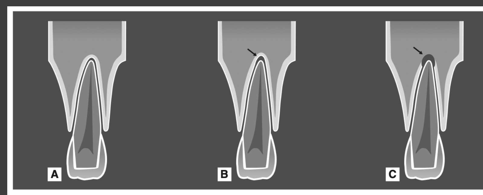
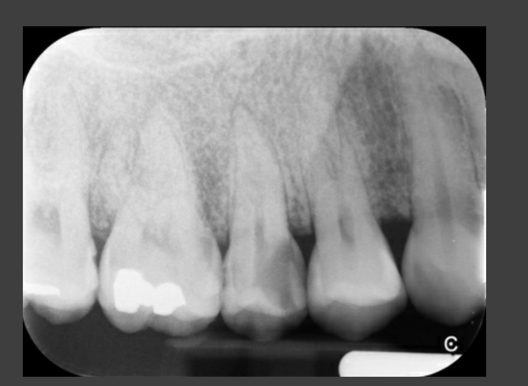
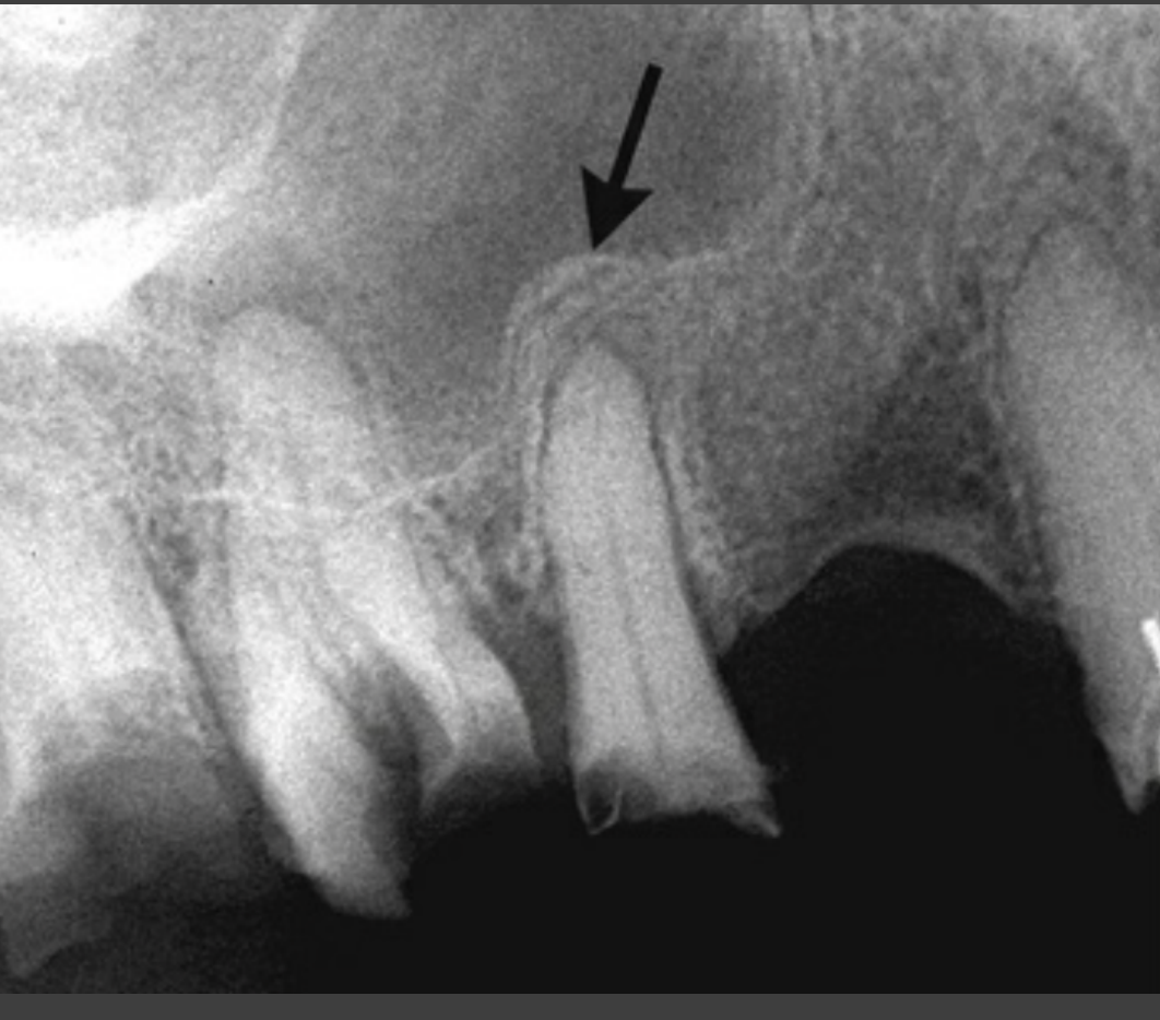
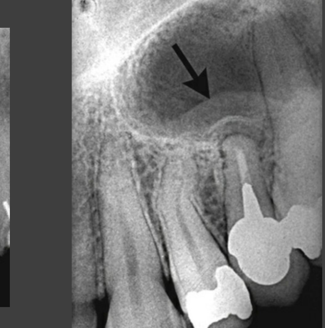
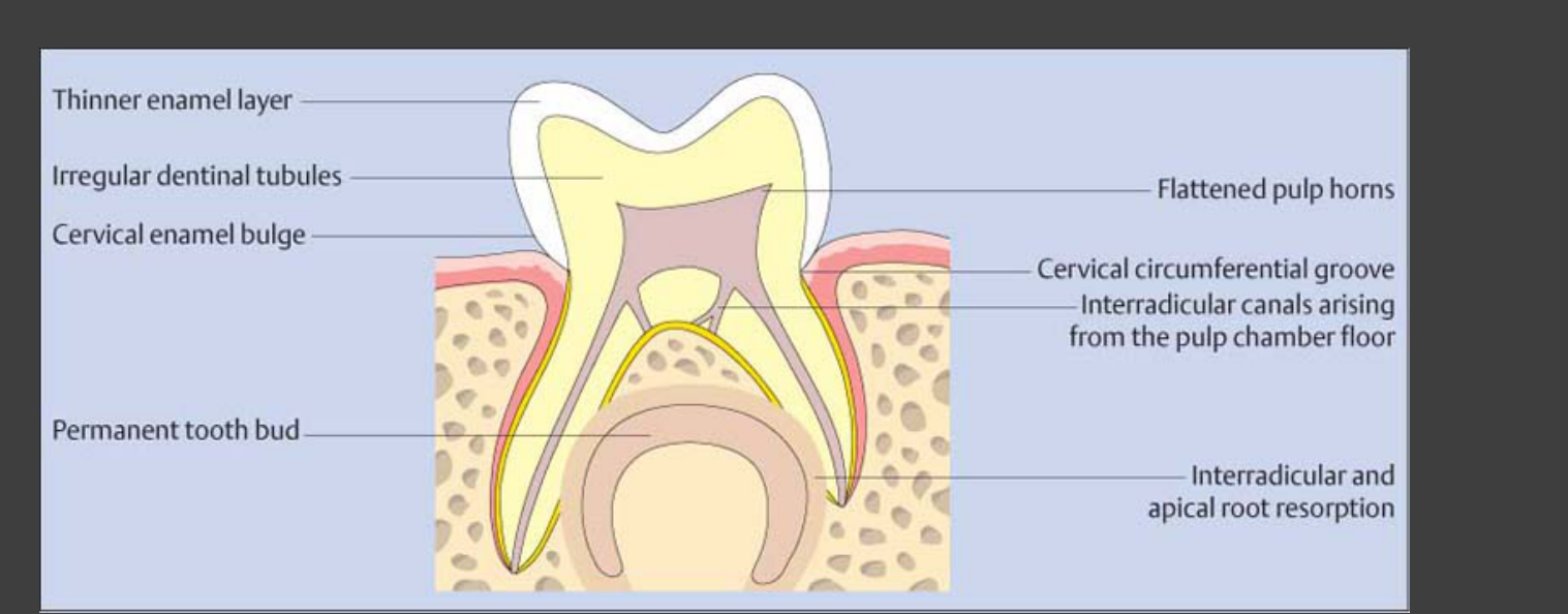
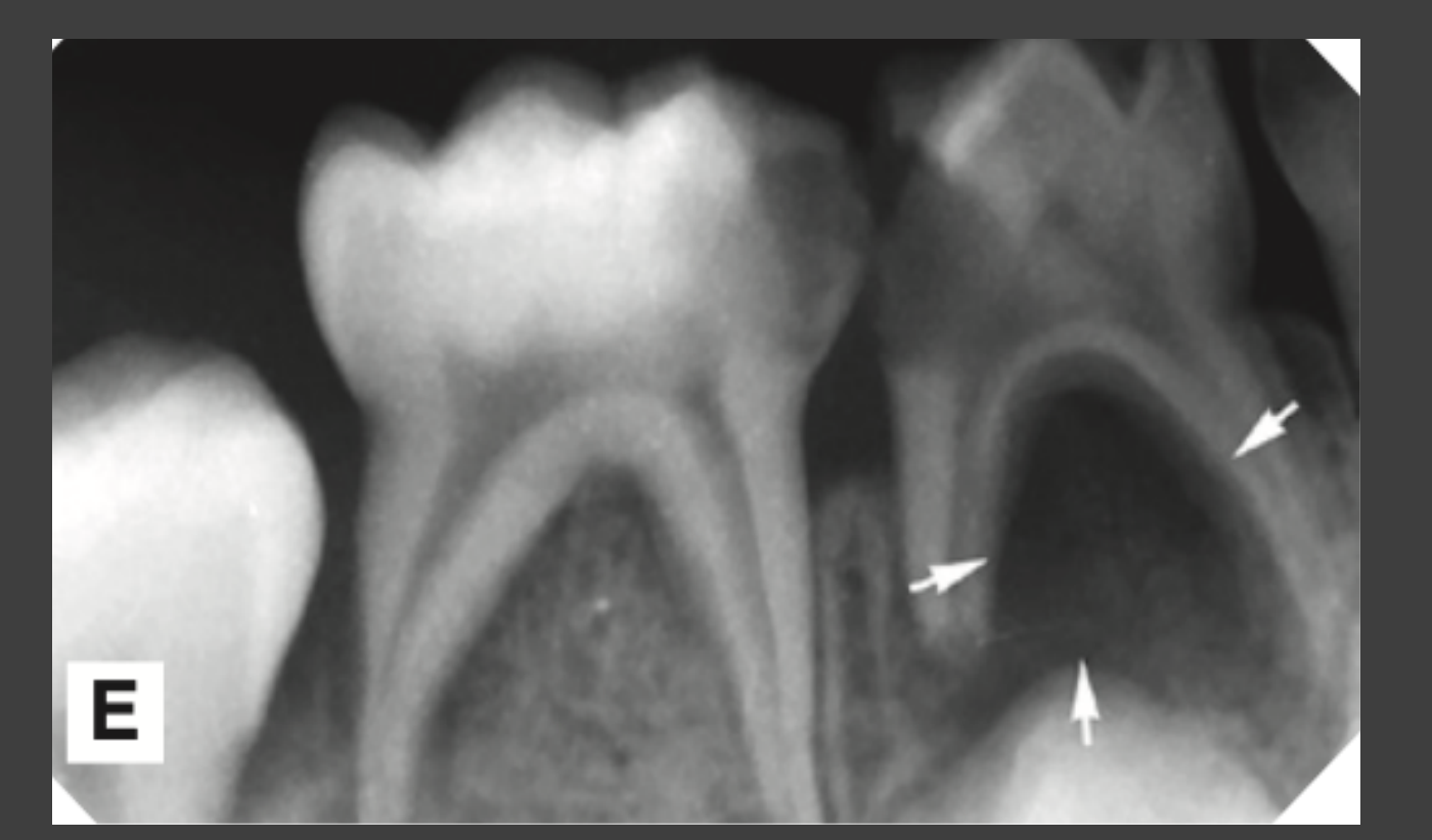
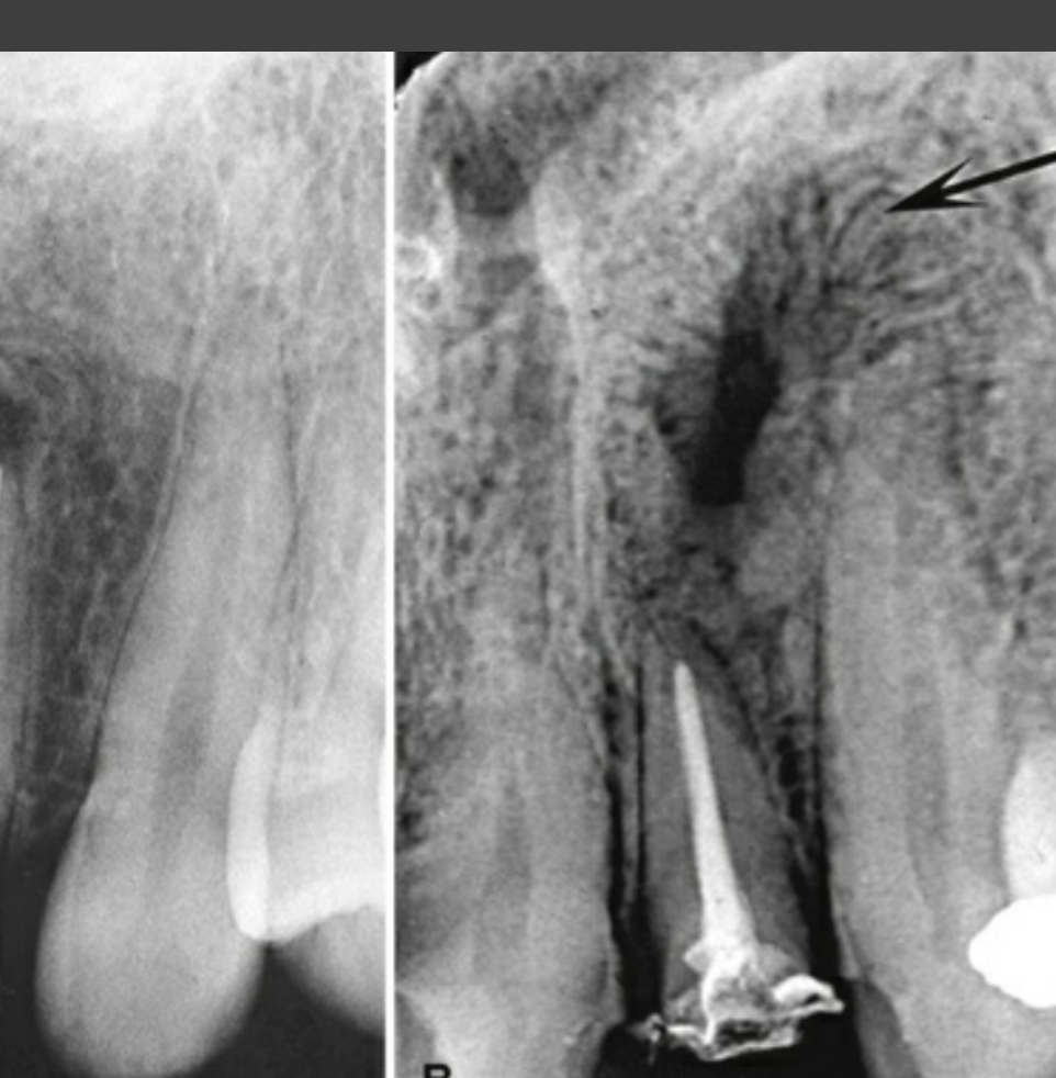
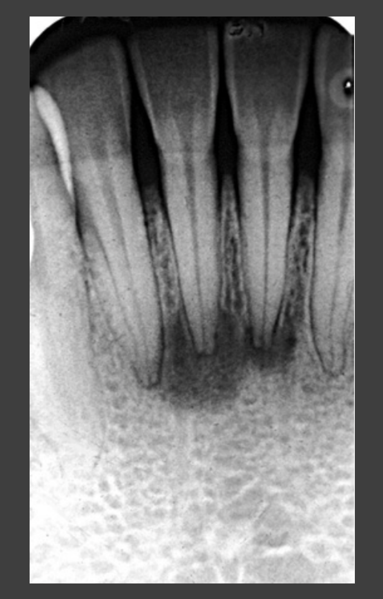
Clinical Radiographic Examples Of Periapical Lesions22232425262728
 |  |  |
 |  |  |
 |  |  |
 |  |  |
Clinical History And Treatment Outcomes29
before after A B Importance of clinical history

Conclusion30
End
Footnotes
-
Original PDF page 1: L6 Intraoral Radiograph Interpretation, p.1 ↩
-
Original PDF page 2: L6 Intraoral Radiograph Interpretation, p.2 ↩
-
Original PDF page 3: L6 Intraoral Radiograph Interpretation, p.3 ↩
-
Original PDF page 4: L6 Intraoral Radiograph Interpretation, p.4 ↩
-
Original PDF page 5: L6 Intraoral Radiograph Interpretation, p.5 ↩
-
Original PDF page 6: L6 Intraoral Radiograph Interpretation, p.6 ↩
-
Original PDF page 7: L6 Intraoral Radiograph Interpretation, p.7 ↩
-
Original PDF page 8: L6 Intraoral Radiograph Interpretation, p.8 ↩
-
Original PDF page 9: L6 Intraoral Radiograph Interpretation, p.9 ↩
-
Original PDF page 10: L6 Intraoral Radiograph Interpretation, p.10 ↩
-
Original PDF page 11: L6 Intraoral Radiograph Interpretation, p.11 ↩
-
Original PDF page 12: L6 Intraoral Radiograph Interpretation, p.12 ↩
-
Original PDF page 13: L6 Intraoral Radiograph Interpretation, p.13 ↩
-
Original PDF page 14: L6 Intraoral Radiograph Interpretation, p.14 ↩
-
Original PDF page 15: L6 Intraoral Radiograph Interpretation, p.15 ↩
-
Original PDF page 16: L6 Intraoral Radiograph Interpretation, p.16 ↩
-
Original PDF page 17: L6 Intraoral Radiograph Interpretation, p.17 ↩
-
Original PDF page 18: L6 Intraoral Radiograph Interpretation, p.18 ↩
-
Original PDF page 19: L6 Intraoral Radiograph Interpretation, p.19 ↩
-
Original PDF page 20: L6 Intraoral Radiograph Interpretation, p.20 ↩
-
Original PDF page 21: L6 Intraoral Radiograph Interpretation, p.21 ↩
-
Original PDF page 22: L6 Intraoral Radiograph Interpretation, p.22 ↩
-
Original PDF page 23: L6 Intraoral Radiograph Interpretation, p.23 ↩
-
Original PDF page 24: L6 Intraoral Radiograph Interpretation, p.24 ↩
-
Original PDF page 25: L6 Intraoral Radiograph Interpretation, p.25 ↩
-
Original PDF page 26: L6 Intraoral Radiograph Interpretation, p.26 ↩
-
Original PDF page 27: L6 Intraoral Radiograph Interpretation, p.27 ↩
-
Original PDF page 28: L6 Intraoral Radiograph Interpretation, p.28 ↩
-
Original PDF page 29: L6 Intraoral Radiograph Interpretation, p.29 ↩
-
Original PDF page 30: L6 Intraoral Radiograph Interpretation, p.30 ↩