Basic Osteology Of The Skull And Radiographic Anatomy
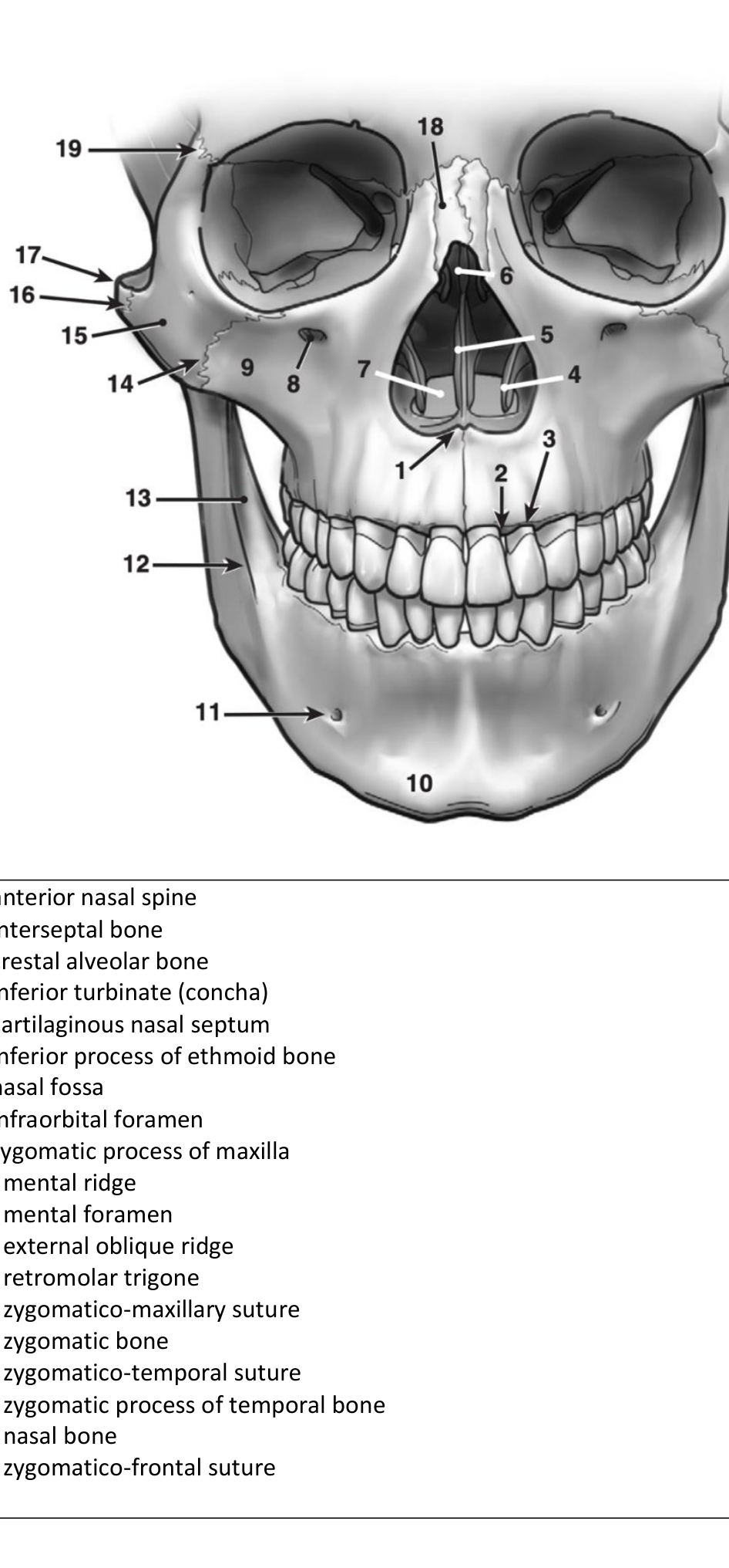
Labelled diagram of skull anatomy illustrating basic osteology, with numbered identifiers for structures such as nasal bones, zygomatic processes, and dental alveoli.
Basic Osteology of the Skull1
Anatomical Diagrams And Osteology
Skull And Facial Bones
- anterior nasal spine
- interseptal bone
- crestal alveolar bone
- inferior turbinate (concha)
- cartilaginous nasal septum
- inferior process of ethmoid bone
- nasal fossa
- infraorbital foramen
- zygomatic process of maxilla
- mental ridge
- mental foramen
- external oblique ridge
- retromolar trigone
- zygomatico-maxillary suture
- zygomatic bone
- zygomatico-temporal suture
- zygomatic process of temporal bone
- nasal bone
- zygomatico-frontal suture
Vascular Anatomy Of The Head2
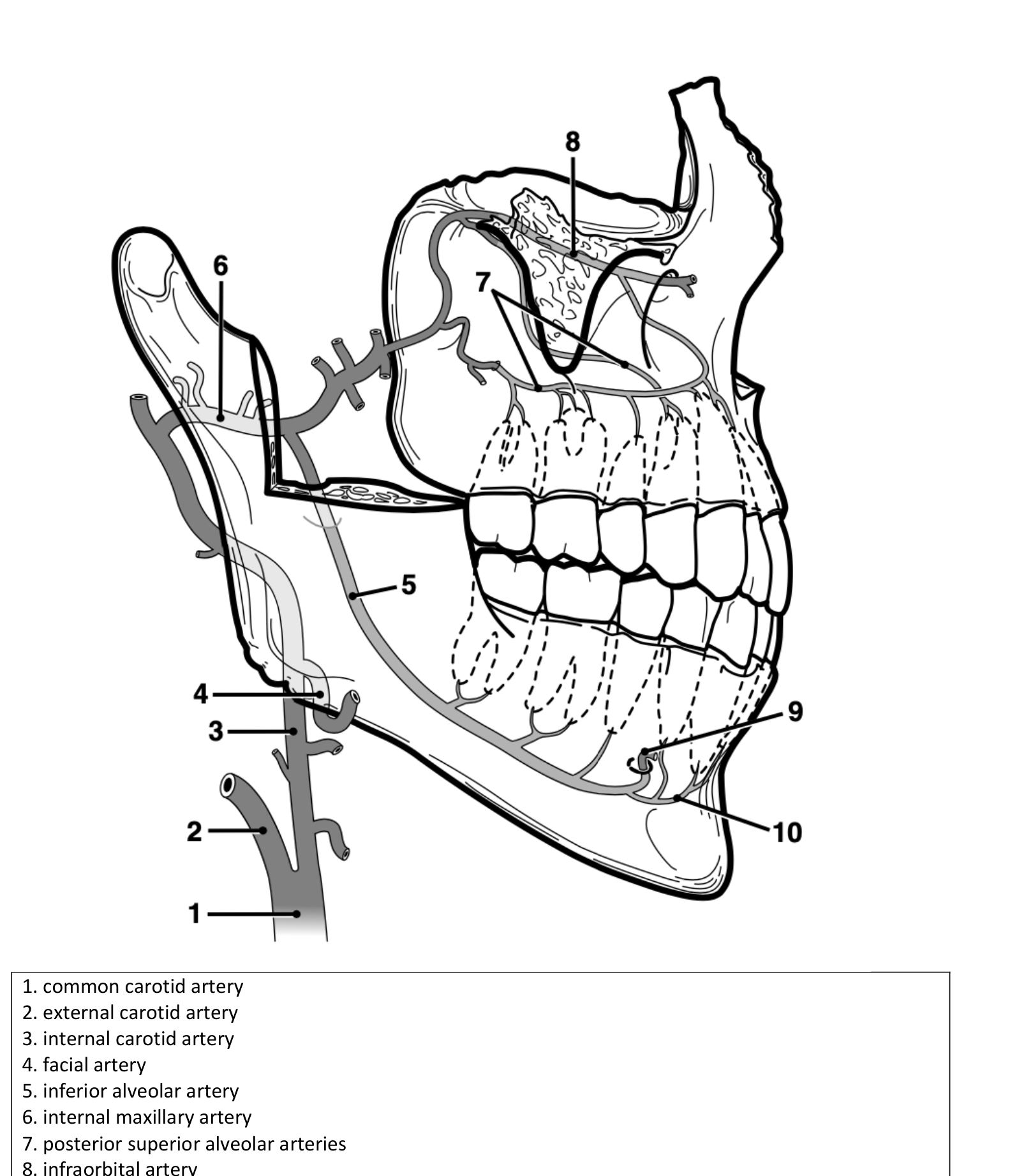
- common carotid artery
- external carotid artery
- internal carotid artery
- facial artery
- inferior alveolar artery
- internal maxillary artery
- posterior superior alveolar arteries
- infraorbital artery
- labial artery
- incisive artery
 |  |
Maxillary And Palatal Structures34
 |  |
 |
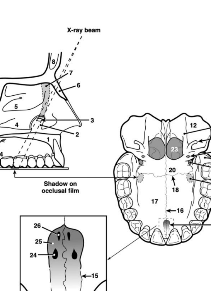
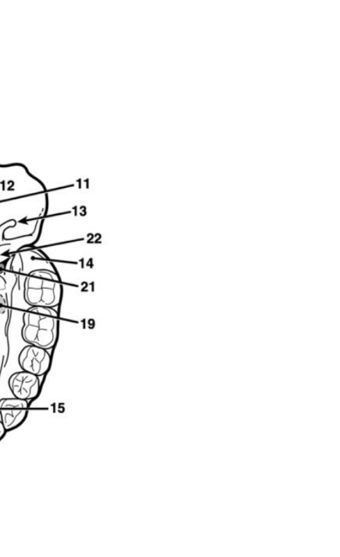
Maxilla5
-
hard palate
-
infundibulum
-
ostium of nasolacrimal canal
-
inferior turbinate
-
middle turbinate
-
nasal bone
-
nasolacrimal canal
-
frontal sinus
-
sphenoid sinus
-
pituitary fossa
-
medial pterygoid plate
-
lateral pterygoid plate
-
hamular process of medial pterygoid plate (hamulus)
-
maxillary tuberosity
-
incisive foramen
-
median maxillary suture (midpalatal suture)
-
palatal process of maxilla
-
palato-maxillary suture (transverse)
-
shadow of nasolacrimal duct
-
palatine bone
-
greater palatine foramen
-
lesser palatine foramen
-
posterior nares
-
foramen of Stenson
-
incisive canal
-
foramen of Scarpa
-
lateral fossa
-
maxilla
-
vertical process of maxilla
-
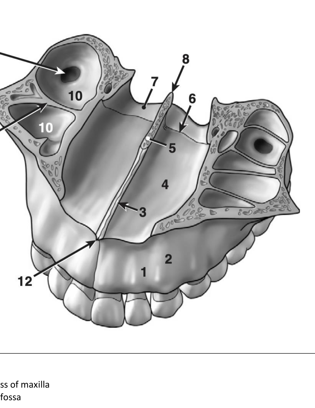
floor of nasal fossa
-
vomer bone
-
palato-maxillary suture
-
palatal bone
-
posterior nasal spine
-
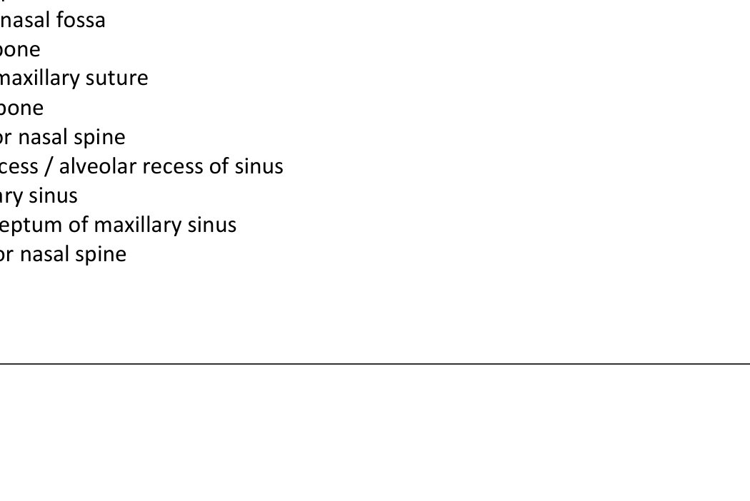
sinus recess / alveolar recess of sinus
-
maxillary sinus
-
bony septum of maxillary sinus
-
anterior nasal spine
 |  |
 |  |
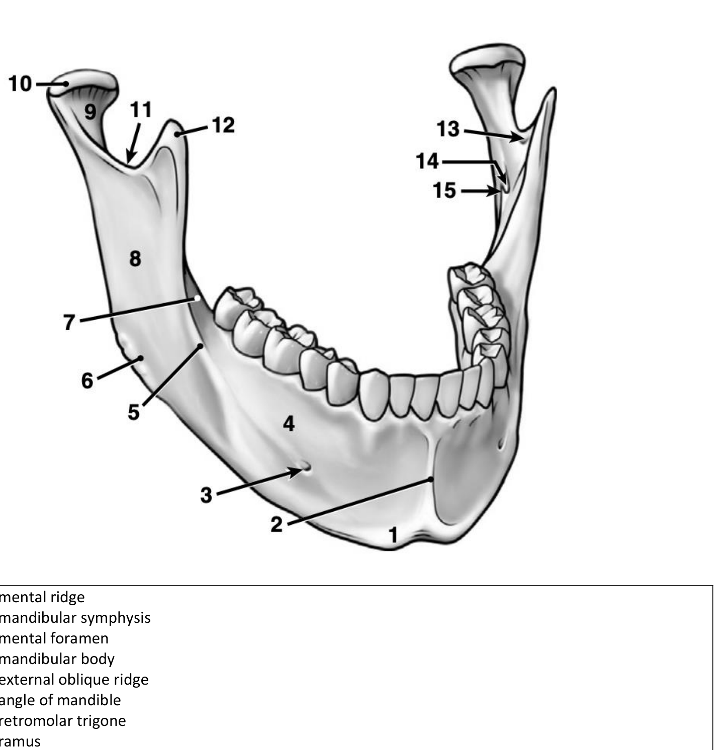
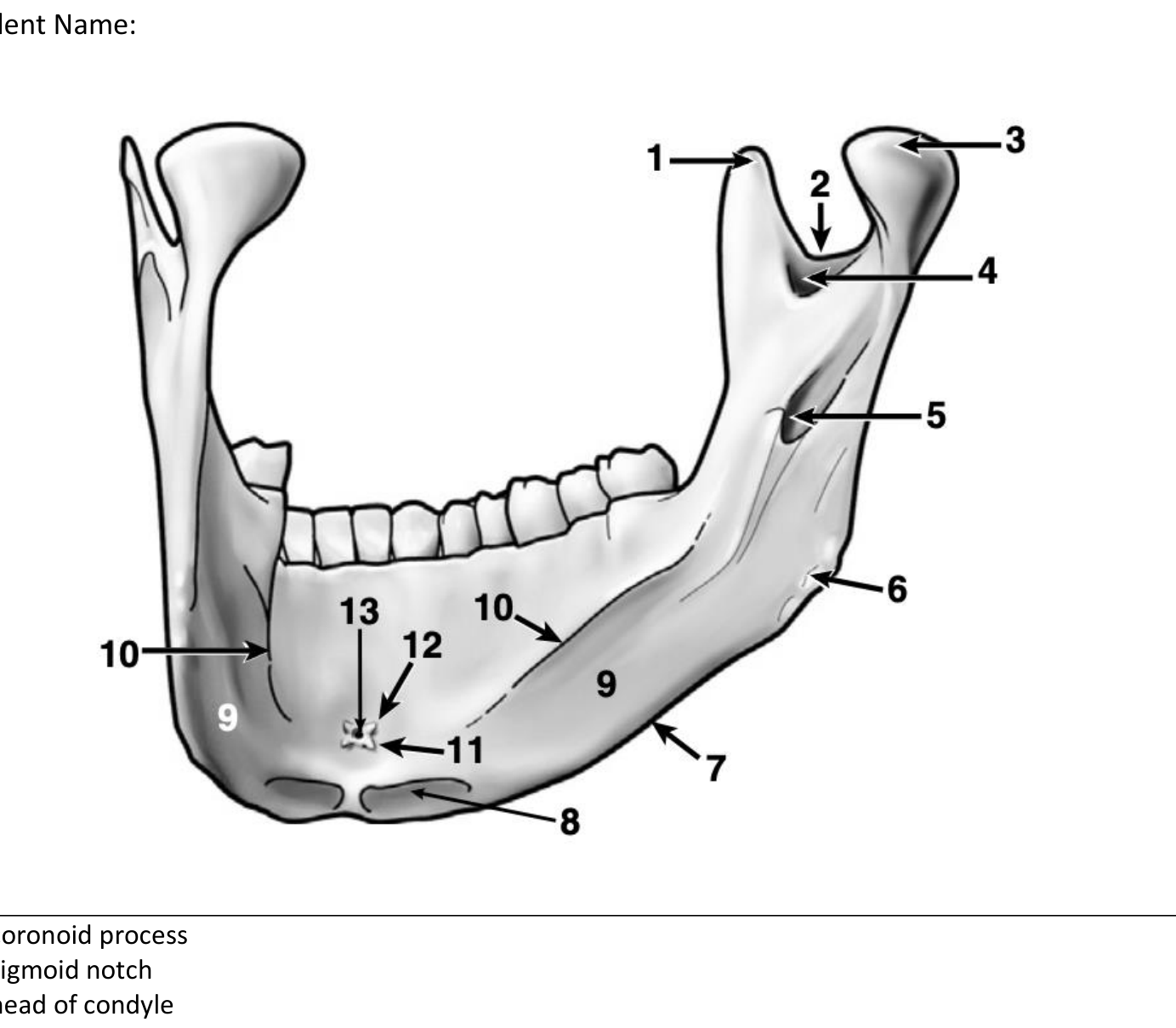
Mandibular Osteology6
- coronoid process
- sigmoid notch
- head of condyle
- medial sigmoid depression
- mandibular foramen
- angle of mandible
- inferior cortex of mandible
- digastric fossa
- submandibular (salivary gland) fossa
- mylohyoid (internal oblique) ridge
- inferior genial tubercle of geniohyoid muscle
- superior genial tubercle of genioglossus muscle
- accessory lingual foramen for lingual vessels (lingual foramen)
 |  |
Radiographic Anatomy Exercises
Introduction To Radiographic Identification7
Exercises
- Identify the type of x-ray
- Identify anatomical structures / landmarks
Example
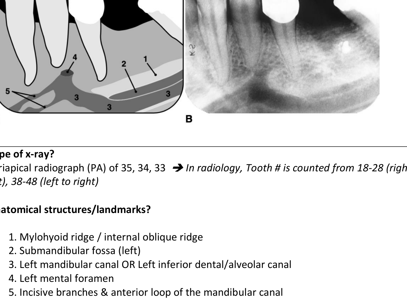
Q. Type of x-ray? A: Periapical radiograph (PA) of 35, 34, 33 → In radiology, Tooth # is counted from 18-28 (right to left), 38-48 (left to right)
Q. Anatomical structures/landmarks? A:
-
- Mylohyoid ridge / internal oblique ridge
-
- Submandibular fossa (left)
-
- Left mandibular canal OR Left inferior dental/alveolar canal
-
- Left mental foramen
-
- Incisive branches & anterior loop of the mandibular canal
Mandibular And Dental Radiography8
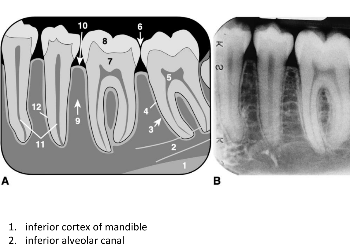
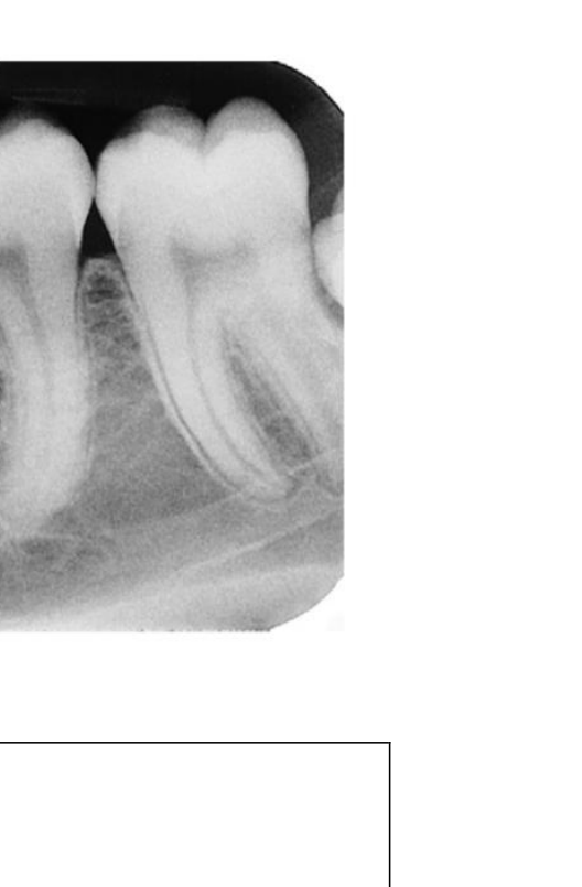
- inferior cortex of mandible
- inferior alveolar canal
- lamina dura (radiopaque thin white line)
- periodontal membrane space (radiolucent thin black line)
- coronal pulp space (pulp chamber)
- interproximal contact point
- dentin
- enamel
- interseptal alveolar bone (interradicular alveolar bone)
- crestal plate (thin white radiopaque line)
- root canal space (containing the dental pulp)
- cementum (frequently not distinguishable from the subadjacent dentin unless hypercementosis is present)
 |  |
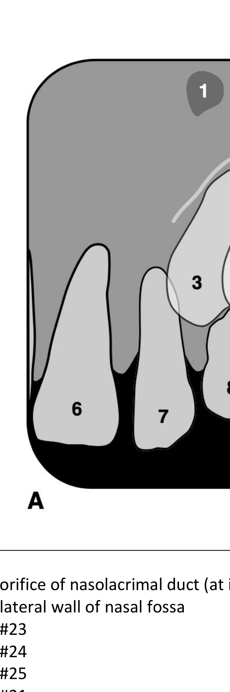
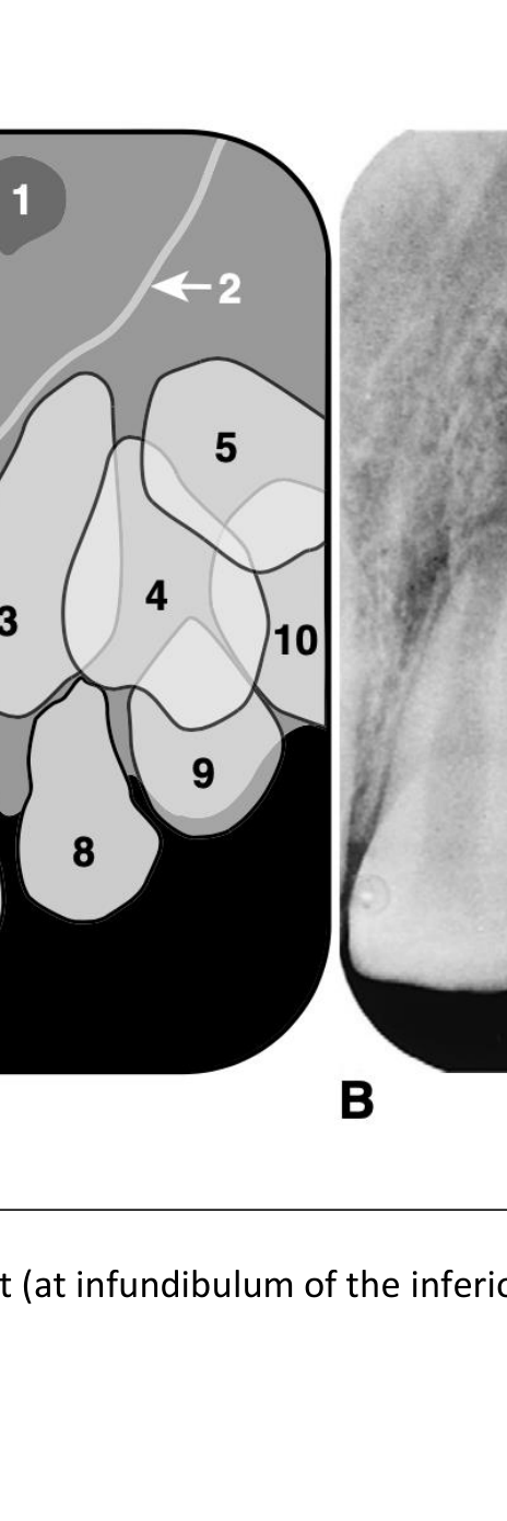
Anterior Maxillary Radiography910
-
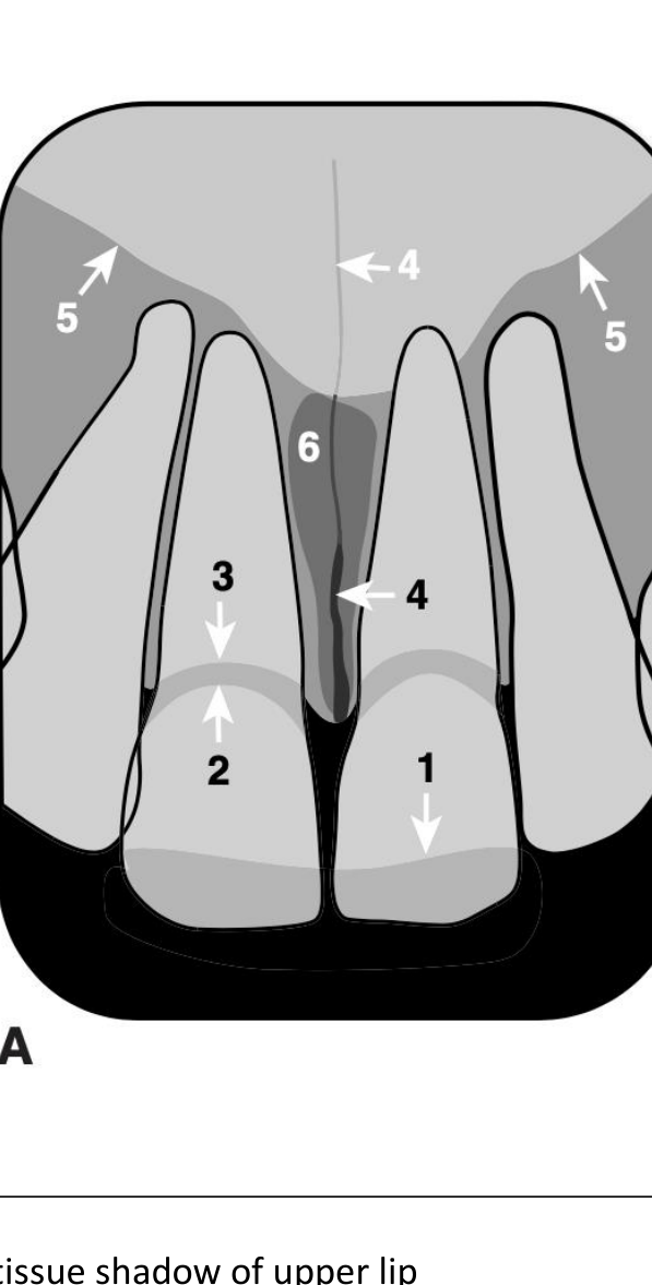
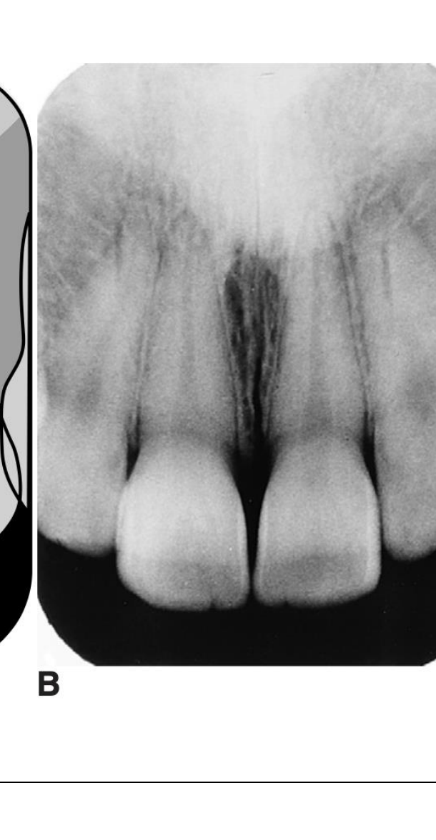
soft tissue shadow of upper lip
-
cervical margin (line) of enamel
-
alveolar bone margin (dark band between 2 and 3 is the root)
-
median maxillary suture
-
soft tissue outline of nose
-
incisive foramen
-
orifice of nasolacrimal duct (at infundibulum of the inferior turbinate not seen)
-
lateral wall of nasal fossa
-
#23
-
#24
-
#25
-
#21
-
#22
-
#63
-
#64
-
#65
 |  |
 |  |
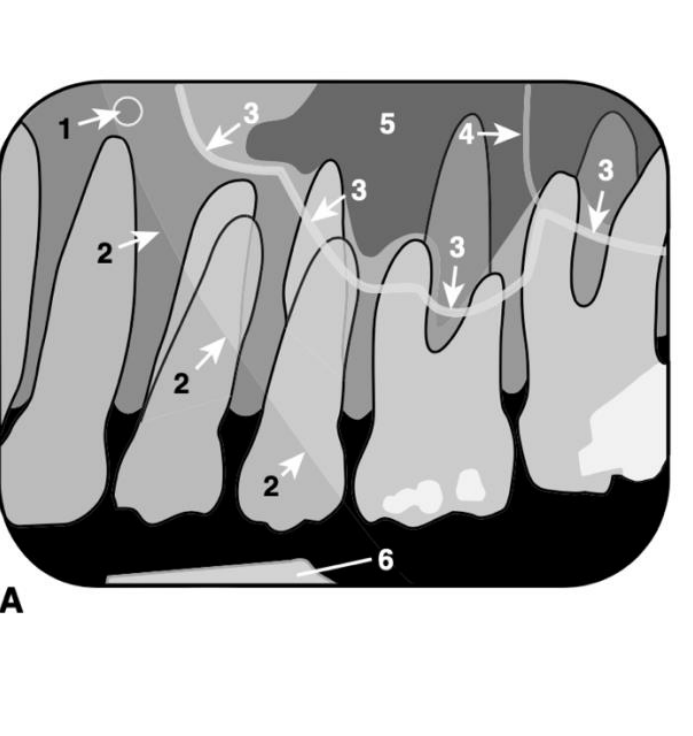
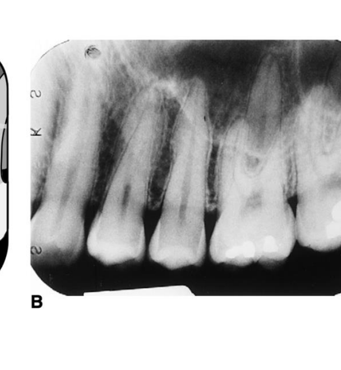
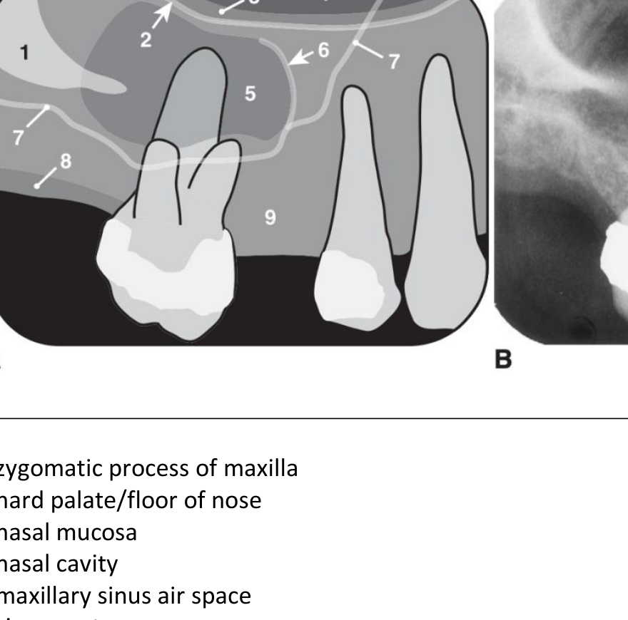
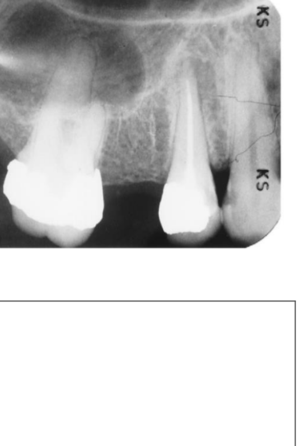
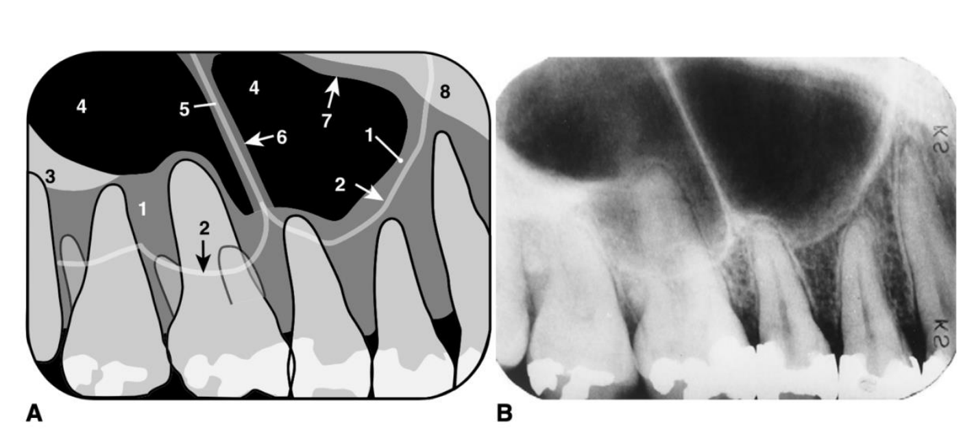
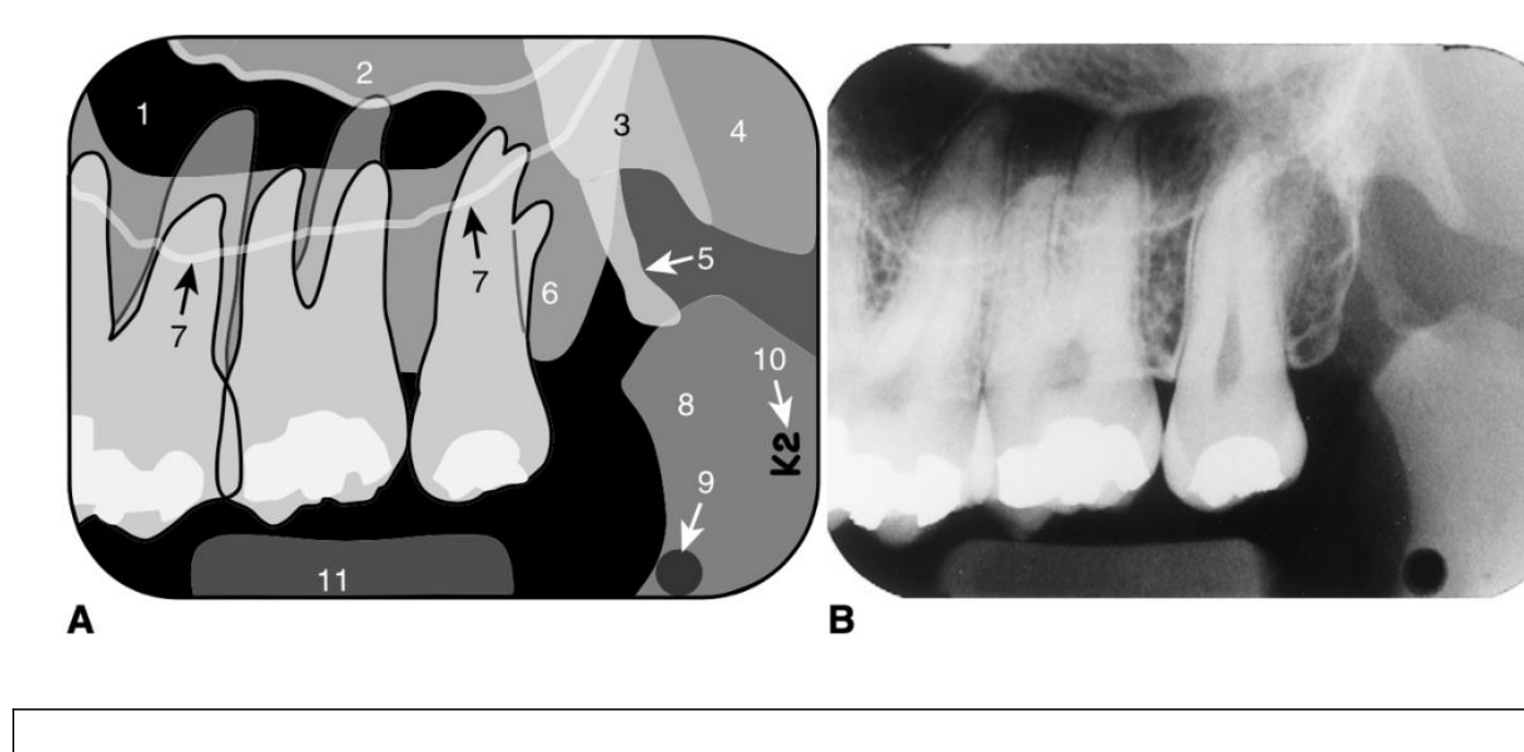
Posterior Maxillary And Sinus Radiography11121314
-
embossed dot on film
-
soft tissue outline of nasolabial fold
-
floor of maxillary sinus
-
bony septum in maxillary sinus
-
air space of maxillary sinus
-
metal part in bite-block
-
zygomatic process of maxilla
-
hard palate/floor of nose
-
nasal mucosa
-
nasal cavity
-
maxillary sinus air space
-
sinus septum
-
floor of maxillary sinus
-
mucosa of alveolar ridge
-
alveolar bone of maxilla
-
sinus floor mucosa
-
floor of maxillary sinus
-
zygomatic process of maxilla
-
air space of maxillary sinus
-
bony septum in maxillary sinus
-
sinus mucosa on both sides of septum
-
mucosal lining
-
soft tissue superimposition
-
maxillary sinus air space
-
zygomatic arch
-
medial pterygoid plate
-
lateral pterygoid plate
-
hamular process of medial pterygoid plate
-
maxillary tuberosity
-
floor of maxillary sinus
-
coronoid process of mandible
-
embossed dot in film
-
“ks” marking indicating “D” speed film
-
bite-block
 |  |  |
 |  |  |
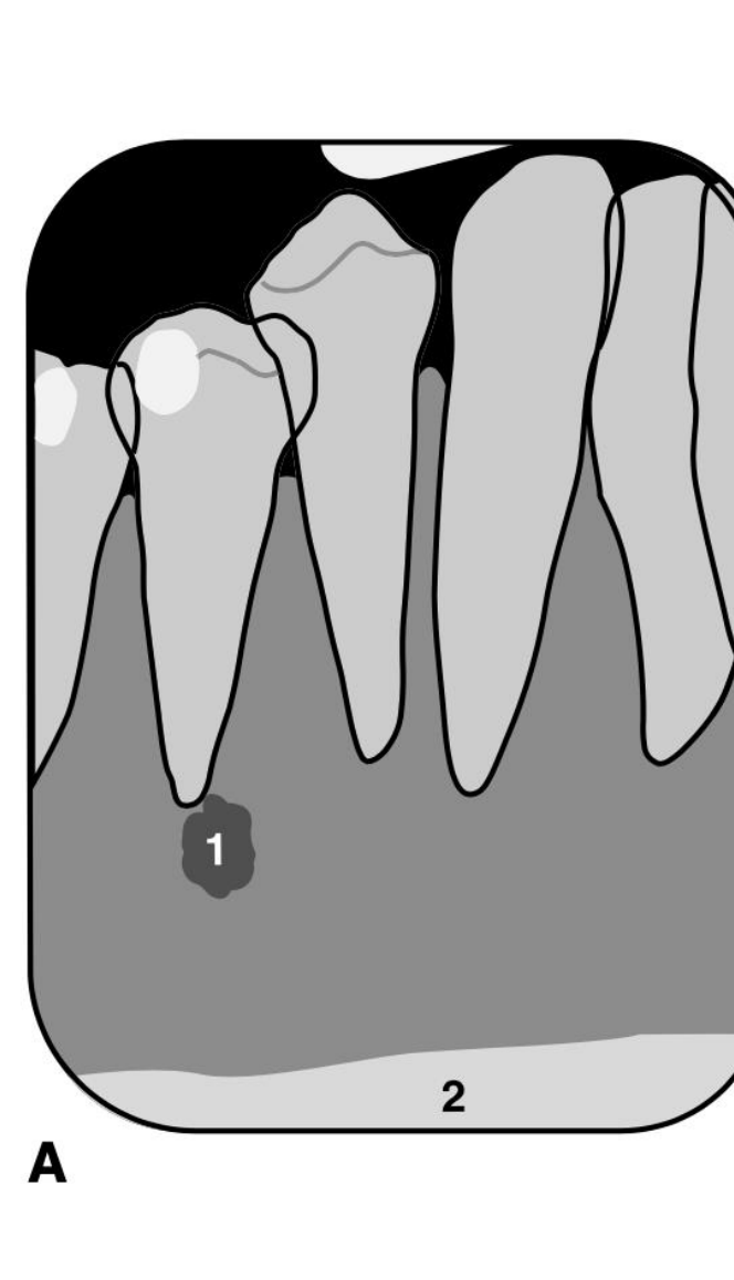
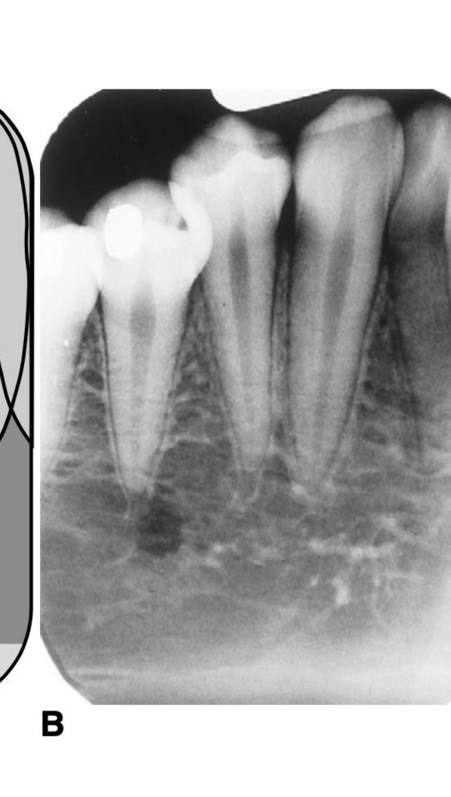
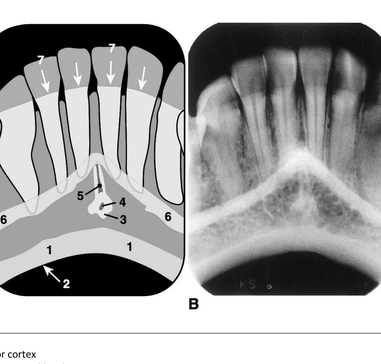
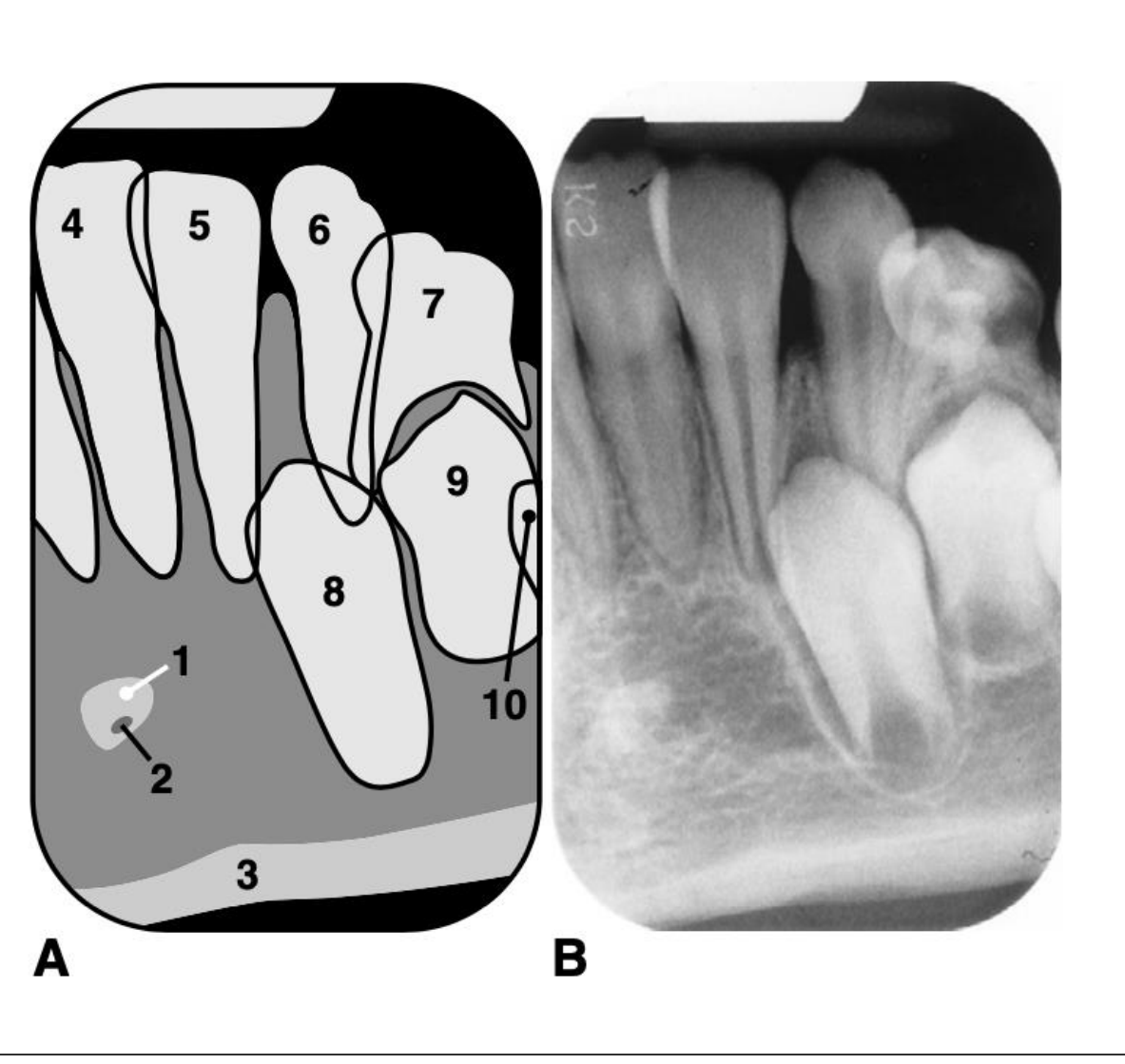
Anterior Mandibular Radiography151617
-
mental foramen (does not usually have a corticated margin)
-
inferior cortex of mandible
-
inferior cortex
-
inferior cortical border
-
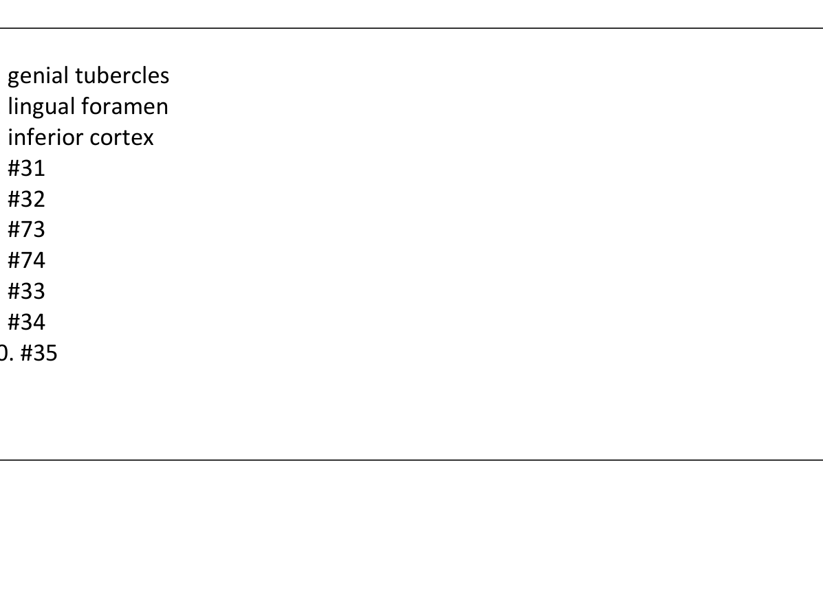
genial tubercles
-
lingual foramen
-
lingual canal
-
mental ridge
-
soft tissue shadow of lower lip
-
genial tubercles
-
lingual foramen
-
inferior cortex
-
#31
-
#32
-
#73
-
#74
-
#33
-
#34
-
#35
 |  |  |
 |  |  |
 |
Footnotes
-
Original PDF page 1: Lecture 4 Excercise answers, p.1 ↩
-
Original PDF page 2: Lecture 4 Excercise answers, p.2 ↩
-
Original PDF page 4: Lecture 4 Excercise answers, p.4 ↩
-
Original PDF page 5: Lecture 4 Excercise answers, p.5 ↩
-
Original PDF page 3: Lecture 4 Excercise answers, p.3 ↩
-
Original PDF page 6: Lecture 4 Excercise answers, p.6 ↩
-
Original PDF page 7: Lecture 4 Excercise answers, p.7 ↩
-
Original PDF page 8: Lecture 4 Excercise answers, p.8 ↩
-
Original PDF page 9: Lecture 4 Excercise answers, p.9 ↩
-
Original PDF page 10: Lecture 4 Excercise answers, p.10 ↩
-
Original PDF page 11: Lecture 4 Excercise answers, p.11 ↩
-
Original PDF page 12: Lecture 4 Excercise answers, p.12 ↩
-
Original PDF page 13: Lecture 4 Excercise answers, p.13 ↩
-
Original PDF page 14: Lecture 4 Excercise answers, p.14 ↩
-
Original PDF page 15: Lecture 4 Excercise answers, p.15 ↩
-
Original PDF page 16: Lecture 4 Excercise answers, p.16 ↩
-
Original PDF page 17: Lecture 4 Excercise answers, p.17 ↩