Seale et al 1981 - Dog Bleaching Experiment
Contextual takeaway
In the dog model using 35% hydrogen peroxide for 30 minutes weekly over four weeks, early histologic resorption was reported to have stopped with healing evident by 60 days; this does not estimate human incidence or risk.
Methods and reported outcomes
- Model: dog teeth; hydrogen peroxide was applied to the enamel surface.
- Regimen: 35% hydrogen peroxide for 30 minutes once per week for four weeks.
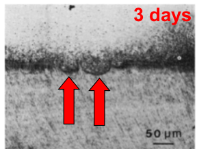
- At three days, L2 reports internal surface resorption and active dentinoclasts.
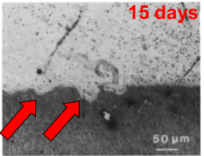
- At 15 days, the lecturer note says resorption was more advanced.
- At 60 days, L2 reports that resorption had stopped and healing was evident.
- The excerpt gives no sample count, comparator or control details, or full article citation. The Fig. 6 caption mentions heat, but L2 does not give a heat protocol.
The citation in L2 is reproduced as written: Seale et al - J Dent Res 1981.1 No title, author initials, volume, issue, or page range is supplied in the lecture excerpt.
Figures and captions from L2
These are the original L2 image embeds and captions associated with the study:
Exact evidence extracts from L2
The following text is from L2, External Whitening. The citation and study details are repeated within that L2 section; the later occurrence adds the 60-day outcome.1
Seale et al - J Dent Res 1981
- 35% hydrogen peroxide applied to enamel surface of dog’s teeth
- Applied for 30 mins, once per wk for 4 wks
- After only 3 days:
- Internal surface resorption noted
- Active dentinoclasts seen
- After 60 days:
- The resorption had stopped
- Healing was evident
After 15 days, the resorption was more advanced. After the whitening treatment ended, teeth left for up to 60 days showed cessation of resorption and evidence of healing.
Recording-derived cases (L2 audio)
These two cases were first woven into L11 by mistake: L11 had been linked to the L2 recording. They come from the L2 audio and now live in L2’s Clinical Cases; the Seale 1981 dog-peroxide study appears on the L2 slides. Neither case names Seale or gives a full citation, so they are shown as lecture appearances and do not extend the study findings.
Dog-histology case
From L2, Clinical Cases.2
Scenario: A 1981 histological study on dog teeth investigating the effects of hydrogen peroxide applied to the enamel surface to induce internal surface resorption.
What’s shown: Histological images of the teeth at different time points (3 days, 15 days, and up to 60 days) after the bleaching procedure.
Consider: What are the histological findings at each time point, and what do they indicate about the nature of the resorption?
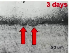
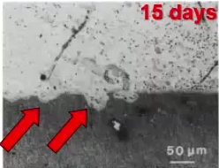
Observations:
- At 3 days, internal surface resorption is occurring with active dentinoclasts (transcribed as “dentin class”) present.
- At 15 days, the resorption is more advanced.
- At 60 days, the resorption has stopped and healing is evident.
Reasoning: The progression and subsequent cessation of resorption with evident healing indicate that internal surface resorption is likely a transient condition that can self-limit if the irritant is removed.
Outcome: The study demonstrates that the resorption is transient and heals over time when the irritant is no longer present.
Takeaway: Internal surface resorption can be a transient, self-limiting condition when the irritant is removed, though it is rarely diagnosed clinically or radiographically.
Sealed/non-infected-group vignette
From L2, Clinical Cases. The case does not name Seale.3
Scenario: A clinical photo from a study showing an anterior tooth, illustrating the healing phase of a resorptive process in a sealed, non-infected group.
What’s shown: A clinical image of an anterior tooth.
Consider: What is visible in the image and what does it indicate about the resorptive process in this group?
Observations:
- Hard tissue formation is visible in the image.
Reasoning: The presence of hard tissue forming indicates that the resorptive process was transient and that healing is occurring, which contrasts with the unsealed, infected group that experienced extensive and prolonged colonization of resorbing cells.
Outcome: The image demonstrates the transient nature of the resorption and the subsequent healing and hard tissue formation when bacteria are excluded.
Takeaway: When the irritant (such as bacteria) is absent, the resorptive process can be transient, leading to hard tissue formation and healing, whereas the presence of bacteria leads to internal inflammatory resorption.
Footnotes
-
Original PDF page 10: L2 Resorption 1 - Introduction, General Internal Resorption, p.10. ↩ ↩2
-
Recording-derived case from the L2 audio (formerly misplaced in L11); now in L2 Resorption 1 - Introduction, General Internal Resorption. No Seale citation in the case block. ↩
-
Recording-derived case from the L2 audio (formerly misplaced in L11); now in L2 Resorption 1 - Introduction, General Internal Resorption. No named publication. ↩